Study of the interaction of an α-helical transmembrane peptide with phosphatidylcholine bilayer...
13
Study of the interaction of an α-helical transmembrane peptide with phosphatidylcholine bilayer membranes by means of densimetry and ultrasound velocimetry Peter Rybar a , Roland Krivanek a , Tomas Samuely a , Ruthven N.A.H. Lewis b , Ronald N. McElhaney b , Tibor Hianik a, ⁎ a Department of Nuclear Physics and Biophysics, Faculty of Mathematics, Physics and Computer Sciences, Comenius University, 842 48 Bratislava, Slovak Republic b Department of Biochemistry, University of Alberta, Edmonton, Alberta, Canada T6G 2H7 Received 1 November 2006; received in revised form 8 February 2007; accepted 9 March 2007 Available online 19 March 2007 Abstract We applied precise densimetry and ultrasound velocimetry methods to study the interaction of a synthetic α-helical transmembrane peptide, acetyl-K 2 -L 24 -K 2 -amide (L 24 ), with model bilayer lipid membranes. The large unilamellar vesicles (LUVs) utilized were composed of a homologous series of n-saturated diacylphosphatidylcholines (PCs). PCs whose hydrocarbon chains contained from 13 to 16 carbon atoms, thus producing phospholipid bilayers of different thicknesses and gel to liquid-crystalline phase transition temperatures. This allowed us to analyze how the difference between the hydrophobic length of the peptide and the hydrophobic thickness of the lipid bilayer influences the thermodynamical and mechanical properties of the membranes. We showed that the incorporation of L 24 decreases the temperature and cooperativity of the main phase transition of all LUVs studied. The presence of L 24 in the bilayer also caused an increase of the specific volume and of the volume compressibility in the gel state bilayers. In the liquid crystalline state, the peptide decreases the specific volume at relatively higher peptide concentration (mole ratio L 24 :PC = 1:50). The overall volume compressibility of the peptide-containing lipid bilayers in the liquid- crystalline state was in general higher in comparison with pure membranes. There was, however, a tendency for the volume compressibility of these lipid bilayers to decrease with higher peptide content in comparison with bilayers of lower peptide concentration. For one lipid composition, we also compared the thermodynamical and mechanical properties of LUVs and large multilamellar vesicles (MLVs) with and without L 24 . As expected, a higher cooperativity of the changes of the thermodynamical and mechanical parameters took place for MLVs in comparison with LUVs. These results are in agreement with previously reported DSC and 2 H NMR spectroscopy study of the interaction of the L 24 and structurally related peptides with phosphatidylcholine bilayers. An apparent discrepancy between 2 H NMR spectroscopy and compressibility data in the liquid crystalline state may be connected with the complex and anisotropic nature of macroscopic mechanical properties of the membranes. The observed changes in membrane mechanical properties induced by the presence of L 24 suggest that around each peptide a distorted region exists that involves at least 2 layers of lipid molecules. © 2007 Elsevier B.V. All rights reserved. Keywords: α-helical transmembrane peptide; Phosphatidylcholine bilayers; Lipid–peptide interaction; Densimetry; Ultrasound velocimetry; Volume compressibility 1. Introduction Lipid–protein interactions are of fundamental importance for understanding both the structural integrity and the functions of biological membranes [1,2]. In particular, the chemical com- position and physical properties of bilayer lipid membranes (BLMs) can markedly influence the activity, thermal stability, and the location and disposition of a large number of integral Biochimica et Biophysica Acta 1768 (2007) 1466 – 1478 www.elsevier.com/locate/bbamem Abbreviations: L 24 , acetyl-K 2 -L 24 -K 2 -amide; P 24 , acetyl-K 2 -G-L 24 -K 2 -A- amide; LUV, large unilamellar vesicle; MLV, large multilamellar vesicle; FTIR, Fourier transform infrared; PC, phosphatidylcholine; DMPC, dimyristoylpho- sphatidylcholine; DPPC, dipalmitoylphosphatidylcholine; DSC, differential scanning calorimetry; T m , gel to liquid-crystalline phase transition temperature ⁎ Corresponding author. Fax: +421 7 65425882. E-mail address: [email protected] (T. Hianik). 0005-2736/$ - see front matter © 2007 Elsevier B.V. All rights reserved. doi:10.1016/j.bbamem.2007.03.005
-
Upload
independent -
Category
Documents
-
view
0 -
download
0
Transcript of Study of the interaction of an α-helical transmembrane peptide with phosphatidylcholine bilayer...

1768 (2007) 1466–1478www.elsevier.com/locate/bbamem
Biochimica et Biophysica Acta
Study of the interaction of an α-helical transmembrane peptide withphosphatidylcholine bilayer membranes by means of densimetry
and ultrasound velocimetry
Peter Rybar a, Roland Krivanek a, Tomas Samuely a, Ruthven N.A.H. Lewis b,Ronald N. McElhaney b, Tibor Hianik a,⁎
a Department of Nuclear Physics and Biophysics, Faculty of Mathematics, Physics and Computer Sciences,Comenius University, 842 48 Bratislava, Slovak Republic
b Department of Biochemistry, University of Alberta, Edmonton, Alberta, Canada T6G 2H7
Received 1 November 2006; received in revised form 8 February 2007; accepted 9 March 2007Available online 19 March 2007
Abstract
We applied precise densimetry and ultrasound velocimetry methods to study the interaction of a synthetic α-helical transmembrane peptide,acetyl-K2-L24-K2-amide (L24), with model bilayer lipid membranes. The large unilamellar vesicles (LUVs) utilized were composed of ahomologous series of n-saturated diacylphosphatidylcholines (PCs). PCs whose hydrocarbon chains contained from 13 to 16 carbon atoms, thusproducing phospholipid bilayers of different thicknesses and gel to liquid-crystalline phase transition temperatures. This allowed us to analyzehow the difference between the hydrophobic length of the peptide and the hydrophobic thickness of the lipid bilayer influences thethermodynamical and mechanical properties of the membranes. We showed that the incorporation of L24 decreases the temperature andcooperativity of the main phase transition of all LUVs studied. The presence of L24 in the bilayer also caused an increase of the specific volumeand of the volume compressibility in the gel state bilayers. In the liquid crystalline state, the peptide decreases the specific volume at relativelyhigher peptide concentration (mole ratio L24:PC=1:50). The overall volume compressibility of the peptide-containing lipid bilayers in the liquid-crystalline state was in general higher in comparison with pure membranes. There was, however, a tendency for the volume compressibility ofthese lipid bilayers to decrease with higher peptide content in comparison with bilayers of lower peptide concentration. For one lipid composition,we also compared the thermodynamical and mechanical properties of LUVs and large multilamellar vesicles (MLVs) with and without L24. Asexpected, a higher cooperativity of the changes of the thermodynamical and mechanical parameters took place for MLVs in comparison withLUVs. These results are in agreement with previously reported DSC and 2H NMR spectroscopy study of the interaction of the L24 and structurallyrelated peptides with phosphatidylcholine bilayers. An apparent discrepancy between 2H NMR spectroscopy and compressibility data in the liquidcrystalline state may be connected with the complex and anisotropic nature of macroscopic mechanical properties of the membranes. The observedchanges in membrane mechanical properties induced by the presence of L24 suggest that around each peptide a distorted region exists that involvesat least 2 layers of lipid molecules.© 2007 Elsevier B.V. All rights reserved.
Keywords: α-helical transmembrane peptide; Phosphatidylcholine bilayers; Lipid–peptide interaction; Densimetry; Ultrasound velocimetry; Volume compressibility
Abbreviations: L24, acetyl-K2-L24-K2-amide; P24, acetyl-K2-G-L24-K2-A-amide; LUV, large unilamellar vesicle; MLV, large multilamellar vesicle; FTIR,Fourier transform infrared; PC, phosphatidylcholine; DMPC, dimyristoylpho-sphatidylcholine; DPPC, dipalmitoylphosphatidylcholine; DSC, differentialscanning calorimetry; Tm, gel to liquid-crystalline phase transition temperature⁎ Corresponding author. Fax: +421 7 65425882.E-mail address: [email protected] (T. Hianik).
0005-2736/$ - see front matter © 2007 Elsevier B.V. All rights reserved.doi:10.1016/j.bbamem.2007.03.005
1. Introduction
Lipid–protein interactions are of fundamental importance forunderstanding both the structural integrity and the functions ofbiological membranes [1,2]. In particular, the chemical com-position and physical properties of bilayer lipid membranes(BLMs) can markedly influence the activity, thermal stability,and the location and disposition of a large number of integral
1467P. Rybar et al. / Biochimica et Biophysica Acta 1768 (2007) 1466–1478
membrane proteins in both model and biological membranes[2]. For these reasons, many studies of the interactions ofmembrane proteins with their host BLM have been carried out,in both biological and reconstituted model systems, employinga wide range of different physical techniques [3–6]. Toovercome the problem of the complicated structure of integralproteins and the problems with their isolation and purification, anumber of workers have designed and synthesized peptidemodels of specific regions of natural membrane proteins andhave studied their interactions with model lipid membranes ofdefined composition (see [7,8]). In particular, the study of themechanisms of the interactions of peptides with BLM has alsovery important practical significance for understanding of themechanism of interaction of e.g. neuropeptides [9] orantimicrobial peptides [10] with membranes.
The synthetic peptide acetyl-K2-G-L24-K2-A-amide (P24)and its structural analogs, e.g. acetyl-K2-L24-K2-amide (L24),have been successfully utilized as a model of the hydrophobictransmembrane α-helical segments of integral proteins [8,11].These peptides contain a long sequence of hydrophobic leucineresidues capped at both the N- and C-termini with twopositively charged lysine residues. The central polyleucineregion of these peptides was designed to form a maximallystable α-helix which will partition strongly into the hydro-phobic environment of the lipid bilayer core, while the dilysinecaps were designed to anchor the ends of these peptides to thepolar surface of the BLM and to inhibit the lateral aggregationof these peptides. In fact, circular dichroism (CD) [11] and FTIR[12] spectroscopic studies of P24 have shown that it adopts avery stable α-helical conformation both in solution and in lipidbilayers, and X-ray diffraction [13], fluorescence quenching[14] and FTIR [12] and deuterium nuclear magnetic resonance(2H-NMR) [15] spectroscopic studies have confirmed that P24and its analogs assume a transbilayer orientation with the N- andC-termini exposed to the aqueous environment and the hydro-phobic polyleucine core embedded in hydrocarbon core of theBLM when reconstituted with various phosphatidylcholines(PCs) [16]. 2H-NMR [17] and electron spin resonance (ESR)[18] spectroscopic studies have shown that the rotational dif-fusion of P24 about its long axis perpendicular to the membraneplane is rapid in the liquid-crystalline state of the bilayer.
Detailed DSC, FTIR, NMR and EPR studies of interaction ofP24 or L24 with BLM [19] have revealed that the results obtainedfrom different physical techniques generally agree well with oneanother. However, certain discrepancies have been found incomparison of the results obtained by spectroscopic techniques,i.e. FTIR and 2H-NMR. While the 2H-NMR techniqueindicated that incorporation of P24 peptide into the dipalmi-toylphosphatidylcholine (DPPC) bilayers resulted in a decreaseof the ordering of the membrane in gel state and increase in theliquid crystalline state, FTIR experiments suggest that peptideinduced a decrease of the ordering of the lipid bilayer in bothstructural states of the membrane [19]. This discrepancy hasbeen explained by different peculiarities of these two methods.While the order parameters in 2H-NMR spectroscopy areprimarily sensitive to trans/gauche isomerisation, the molecularinterpretation of the changes in membrane ordering based on
changes in frequency of the methylene stretching modes in IRspectroscopy are likely attributed to the sensitivity of the bandposition phenomena other than trans/gauche isomerisation,such as the interchain coupling and the contribution of peptidein the methylene and methyl stretching region. Interchaincoupling is significant enough even in fluid bilayers [19].Therefore, using exclusively FTIR it is difficult to decide whatprocess is dominant in fluid state — interchain coupling ortrans/gauche isomerisation.
In contrast with spectroscopic methods that provide informa-tion about microscopic changes of the lipid bilayer in closeproximity of the protein, macroscopic methods, such are mem-brane compressibility measurements, are sensitive to changes oflarge membrane regions. The sensitivity and utility of measure-ments of volume compressibility has been proved in severalstudies of the interaction of integral proteins with lipid bilayers,e.g. bacteriorhodopsin [4] or peptides like ACTH24 [9] or gra-micidin S [10]. In the case of bacteriorhodopsin, it has beenshown that one molecule of the peptide is able to change thestructural state of the lipid bilayer of whole large unilamellarvesicle (LUV). However, this method has not been applied to thestudy of interaction of synthetic α-helical transmembranepeptides like L24 with lipid bilayers. In this work, we thereforeapplied the method of precise measurement of density andultrasound velocity to study the physical properties of LUVscomposed of a homologous series of n-saturated diacylphospha-tidylcholines (PC) containing the model peptide L24. A combi-nation of these two methods allowed us to determine the changesof compressibility of lipid bilayers induced by the peptide inboth the gel and liquid-crystalline state of the lipid bilayer.
2. Materials and methods
2.1. Chemicals and preparation of vesicles
The n-saturated diacyl-PCs of various number of carbon atoms (13, 14, 15,and 16) were purchased from Avanti Polar Lipids (USA) and used withoutfurther purification. We will use following abbreviation for these phospholipids:13:0 PC; 14:0 PC; 15:0 PC and 16:0 PC, respectively. The peptide L24 has beensynthesized as previously described [20]. The lipid was dissolved in chloroform/methanol (3:1, vol/vol) and mixed with appropriate amounts of peptide stocksolution. These solutions were dried under N2 and the lipid/peptide films wereexposed to high vacuum for 3 h to remove traces of organic solvent. Themultilamellar vesicles (MLVs) containing L24 were prepared by vortexing in anexcess of deionised water at a temperature above the main phase transitiontemperature of phospholipids. The L24-containing large unilamellar vesicles(LUVs) were prepared by the extrusion method of MacDonald et al. [21] using aLiposoFast-Basic Extruder (Avestin Inc, Canada) and polycarbonate filters withpores of 100 nm in diameter. It has been shown that the average diameter ofLUVs prepared under these conditions is∼ 100 nm [21]. Prior to measurements,the aqueous vesicle suspension was degassed using a water vacuum pump. Thelipid concentration was 2 mg/ml.
2.2. Ultrasound velocimetry
The measurement of ultrasound velocity allows us to evaluate the elasticproperties of aqueous media and suspensions such as vesicles [22–24],lipoproteins [25] or cell surface proteins [26] based on a simple relationship:
bS ¼ 1qd u2
ð1Þ
1468 P. Rybar et al. / Biochimica et Biophysica Acta 1768 (2007) 1466–1478
Whereby βS, ρ, and u are the adiabatic compressibility, the density, and thesound velocity of the suspension, respectively. Thus, by measuring the changesof sound velocity and density, one can determine the changes of adiabaticcompressibility [27].
Ultrasound velocity was measured using a fixed-path differentialvelocimeter consisting of two almost identical acoustic cavity resonators ofthe Sarvazyan-type [28] operated at frequencies, ν, around 7.2 MHz. Theresonance frequencies of the cells were measured using a computer controllednetwork analyzer (USAT, USA). The sample volume was 0.7 ml. The resonatorcells were equipped with magnetic stirrers to ensure homogeneously dispersedsamples during the measurements. One resonator contained the vesicle solutionwhereas the other one was filled with the reference liquid (double distilledwater). When starting a series of measurements, the resonance frequencies ofthe resonators were always first compared with one another by filling both cellswith the reference liquid. As the intensity of the sonic signal was smallthroughout (the pressure amplitude in the ultrasonic wave was less than103 Pa), any effects of the sound wave on the structural properties of thevesicles were thus avoided. In general, ultrasonic velocimetry allows thedetermination of the sound velocity or rather its concentration increments, [u],as defined by the equation [27,28]:
u½ � ¼ u� u0u0c
ð2Þ
whereby c is the solute concentration (lipid) in mg/ml and the subscript “0”refers to the solvent (distilled water). The value [u] can be directly determinedfrom the changes of resonance frequencies f and f0 of the resonators (f is theresonance frequency of the sample and f0 that of the reference — distilledwater):
u½ � ¼ u� u0u0c
¼ f � f0f0c
1þ gð Þ ð3Þ
(the coefficient γ≪1 [28] and can be neglected in the calculations).
2.3. Density measurements
A high precision densimetric system (DMA 60 with two DMA 602 Msample chambers, Anton Paar KG, Graz, Austria), operating according to thevibrating tube principle [29], has been used to determine the density, ρ, of thevesicle solution. Apparent specific partial volumes φV have been calculatedfrom the density data using the equation
uV ¼ 1� q� q0c
h id
1q0
¼ 1q0
� q½ � ð4Þ
whereby the subscript 0 again refers to the solvent and [ρ]= (ρ−ρ0) / (ρ0c)denotes the concentration increment of density.
The determination of the specific volume, in addition to the sound-velocityconcentration increment, allows the estimation of the specific adiabaticcompressibility, φK/β0, of the vesicles, which is based on the followingequation:
uK
b0¼ �2 u½ � � 1
q0þ 2uV; ð5Þ
whereby β0 is the coefficient of adiabatic compressibility and ρ0 is the density ofthe buffer [27]. The value of φK/β0 indicates changes of the volumecompressibility of the vesicles relative to the buffer.
2.4. Experimental errors
The uncertainty in the concentration of the phospholipid suspensions wassmaller then 5%. The temperature of the cells was controlled to be within0.02 °C with a Lauda RK 8 CS ultra-thermostat (Lauda, Germany). Theaccuracy of the determination of the sound velocity increment, [u], and thespecific partial volume, φv, was better then 10−3 ml/g. The accuracy of thedetermination of the density was better then 10−3 g/ml. Each series ofmeasurements was performed at least 3 times.
3. Results
Ultrasound velocimetry and densimetry are sensitive tools tostudy the mechanical and thermodynamical properties of modeland biological membranes (see [27,30] and references citedherein). These methods, if used simultaneously to study theproperties of the phase transition of a lipid bilayer, allow one todetermine the phase transition temperature, the degree ofcooperativity of phase transition, and the volume compressi-bility of the lipid bilayer. These methods are therefore veryuseful in the study of protein–lipid interactions, since thedetermination of mechanical values is crucial for an evaluationof the size of the distorted lipid bilayer structure around proteins[4]. In our previous work, we discussed in detail the basicphysical values obtained by density and ultrasound velocimetrystudies and also compared these values with those obtained byDSC [10]. We pointed out that ultrasound velocimetry anddensimetry allow the characterization of physical properties ofvesicles in more detail than, for example, traditional methodslike DSC, by also providing information about the compressi-bility of the membrane. Additional advantages of the ultrasoundvelocimetry method, in comparison with so-called microscopicmethods like ESR, NMR or fluorescence spectroscopy, is thatthe compressibility of the membrane derived from thistechnique is a macroscopic value that reflects the physicalproperties of the membrane as a whole. This means that themechanical properties are suitable to monitor the changes ofphysical properties of larger regions of the membrane(approximately 10% of the membrane) and are less sensitiveto the local perturbations [4].
In the first series of experiments, we studied the ultrasoundvelocity and density of vesicles composed of 14:0 PC(dimyristoylphosphatidylcholine (DMPC)), without and withL24, as a function of temperature and calculated the specificvolume and specific adiabatic compressibility of the lipidbilayer. We also compared the properties of unilamellar (LUV)and multilamellar (MLV) vesicles with and without L24. Themeasurement of the density is crucial for the determination ofthe specific volume of the membrane and thus for evaluation ofthe specific adiabatic compressibility (see Eq. (5)). In addition,as demonstrated earlier (see [30]), from the plot of the specificvolume versus temperature, we can estimate the degree ofcooperativity of the membrane phase transition and the thermalexpansivity at constant pressure, αm, of the lipid bilayer, asaffected by the presence of the peptide (αm=(δV/δT)p, note thatthere is no 1/V factor in this definition [30]).
The results of determinations of the concentration incrementof ultrasound velocity, [u], specific volume, φV, and thecoefficient of specific adiabatic compressibility, φK/β0, ofLUVs composed of DMPC without and with different moleratio of L24 are presented in Fig. 1 as a plot of these valuesversus temperature. A characteristic property of the plot of [u]value vs. temperature is its minimum at the phase transitiontemperature (Tm) (Fig. 1a, curve 1). At temperatures below theTm of DMPC (i.e. T<24 °C), the value of [u] slowly decreaseswith increasing temperature, whereas an increase of [u] ischaracteristic for higher temperature regions above the Tm. This
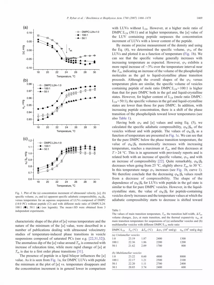
Table 1The values of main transition temperature, Tm, the transition half-width, ΔTm,volume changes, ΔφV at main transition, and the thermal expansivity αm, atmain transition temperature for suspensions of (a) unilamellar vesicles and (b)multilamellar vesicles with different DMPC/L24 mole ratio
DMPC/L24 Tm (°C) ΔTm (°C) ΔφV (105 xml/g) αm (105 xml/g deg)
(a) Unilamellar vesicles1:0 23.19 1.87 2400 1300100:1 22.36 1.86 2200 120050:1 21.62 2.09 1700 800
(b) Multilamellar vesicles1:0 23.22 0.60 4800 8000100:1 22.17 1.21 2500 210050:1 22.20 1.11 2700 240030:1 20.85 3.74 2400 600
Fig. 1. Plot of the (a) concentration increment of ultrasound velocity, [u]; (b)specific volume, φV and (c) apparent specific adiabatic compressibility, φK/β0,versus temperature for an aqueous suspension of LUVs composed of DMPC(14:0 PC) without peptide (○) and with different mole ratio of DMPC/L24:100:1 (●); 50:1 (▲) (see legends). The mean±SD were obtained from 3independent experiments.
1469P. Rybar et al. / Biochimica et Biophysica Acta 1768 (2007) 1466–1478
characteristic shape of the plot of [u] versus temperature and thenature of the minimum of the [u] value, were described in anumber of publications dealing with ultrasound velocimetrystudies of temperature-induced phase transitions in vesiclesuspensions composed of saturated PCs (see e.g. [22,31,32]).The anomalous dip of the [u] value around Tm is connected withincrease of relaxation time, while more rapid change of [u] atTm is due to a first order phase transitions [31].
The presence of peptide in a lipid bilayer influences the [u]value. As it is seen from Fig. 1a, for DMPC LUVs with peptidethe minimum at the plot of [u] vs. temperature disappears andthe concentration increment is in general lower in comparison
with LUVs without L24. However, at a higher mole ratio ofDMPC:L24 (50:1) and at higher temperatures, the [u] value ofthe LUV containing peptide surpasses the concentrationincrement of LUVs with a lower content of the peptide.
By means of precise measurement of the density and usingthe Eq. (4), we determined the specific volume, φV, of theLUVs and plotted it as a function of temperature (Fig. 1b). Wecan see that the specific volume generally increases withincreasing temperature as expected. However, φV exhibits amore rapid increase of ∼3% over the temperature interval nearthe Tm, indicating an increase of the volume of the phospholipidmolecules as the gel to liquid-crystalline phase transitionproceeds. Although the overall shapes of the φV versustemperature plots are similar, the specific volume of vesiclescontaining peptide of mole ratio DMPC:L24=100:1 is higherthan that for pure DMPC both in the gel and liquid-crystallinestates. However, for higher content of L24 (mole ratio DMPC:L24=50:1), the specific volumes in the gel and liquid-crystallinestates are lower than those for pure DMPC. In addition, withincreasing peptide concentration, there is a shift of the phasetransition of the phospholipids toward lower temperatures (seealso Table 1).
Having both φV and [u] values and using Eq. (5), wecalculated the specific adiabatic compressibility, φK/β0, of thevesicles without and with peptide. The values of φK/β0 as afunction of temperature are presented in Fig. 1c. We can see thatfor the pure DMPC below the phase transition temperature, thevalue of φK/β0 monotonically increases with increasingtemperature, reaches a maximum at Tm, and then decreases atT>24 °C. This is in agreement with previously reports and isrelated both with an increase of specific volume, φV, and withan increase of compressibility [22]. Quite remarkably, φK/β0decreases when going from 25 °C, slightly above Tm, to 30 °C.In this temperature range φV increases (see Fig. 1b, curve 1).We therefore conclude that the decreasing φK/β0 values resultfrom a decrease of compressibility. The shape of thedependence of φK/β0 for LUVs with peptide in the gel state issimilar to that for pure DMPC vesicles. However, in the liquid-crystalline state, the value of φK/β0 for peptide-containingvesicles slowly increases and the temperature values at which theadiabatic compressibility starts to decrease is shifted toward
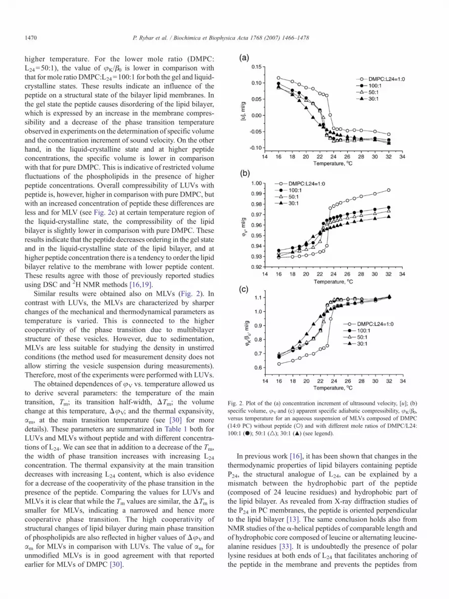
Fig. 2. Plot of the (a) concentration increment of ultrasound velocity, [u]; (b)specific volume, φV and (c) apparent specific adiabatic compressibility, φK/β0,versus temperature for an aqueous suspension of MLVs composed of DMPC(14:0 PC) without peptide (○) and with different mole ratios of DMPC/L24:100:1 (●); 50:1 (△); 30:1 (▲) (see legend).
1470 P. Rybar et al. / Biochimica et Biophysica Acta 1768 (2007) 1466–1478
higher temperature. For the lower mole ratio (DMPC:L24=50:1), the value of φK/β0 is lower in comparison withthat for mole ratio DMPC:L24=100:1 for both the gel and liquid-crystalline states. These results indicate an influence of thepeptide on a structural state of the bilayer lipid membranes. Inthe gel state the peptide causes disordering of the lipid bilayer,which is expressed by an increase in the membrane compres-sibility and a decrease of the phase transition temperatureobserved in experiments on the determination of specific volumeand the concentration increment of sound velocity. On the otherhand, in the liquid-crystalline state and at higher peptideconcentrations, the specific volume is lower in comparisonwith that for pure DMPC. This is indicative of restricted volumefluctuations of the phospholipids in the presence of higherpeptide concentrations. Overall compressibility of LUVs withpeptide is, however, higher in comparison with pure DMPC, butwith an increased concentration of peptide these differences areless and for MLV (see Fig. 2c) at certain temperature region ofthe liquid-crystalline state, the compressibility of the lipidbilayer is slightly lower in comparison with pure DMPC. Theseresults indicate that the peptide decreases ordering in the gel stateand in the liquid-crystalline state of the lipid bilayer, and athigher peptide concentration there is a tendency to order the lipidbilayer relative to the membrane with lower peptide content.These results agree with those of previously reported studiesusing DSC and 2H NMR methods [16,19].
Similar results were obtained also on MLVs (Fig. 2). Incontrast with LUVs, the MLVs are characterized by sharperchanges of the mechanical and thermodynamical parameters astemperature is varied. This is connected to the highercooperativity of the phase transition due to multibilayerstructure of these vesicles. However, due to sedimentation,MLVs are less suitable for studying the density in unstirredconditions (the method used for measurement density does notallow stirring the vesicle suspension during measurements).Therefore, most of the experiments were performed with LUVs.
The obtained dependences of φV vs. temperature allowed usto derive several parameters: the temperature of the maintransition, Tm; its transition half-width, ΔTm; the volumechange at this temperature, ΔφV; and the thermal expansivity,αm, at the main transition temperature (see [30] for moredetails). These parameters are summarized in Table 1 both forLUVs and MLVs without peptide and with different concentra-tions of L24. We can see that in addition to a decrease of the Tm,the width of phase transition increases with increasing L24
concentration. The thermal expansivity at the main transitiondecreases with increasing L24 content, which is also evidencefor a decrease of the cooperativity of the phase transition in thepresence of the peptide. Comparing the values for LUVs andMLVs it is clear that while the Tm values are similar, the ΔTm issmaller for MLVs, indicating a narrowed and hence morecooperative phase transition. The high cooperativity ofstructural changes of lipid bilayer during main phase transitionof phospholipids are also reflected in higher values of ΔφVandαm for MLVs in comparison with LUVs. The value of αm forunmodified MLVs is in good agreement with that reportedearlier for MLVs of DMPC [30].
In previous work [16], it has been shown that changes in thethermodynamic properties of lipid bilayers containing peptideP24, the structural analogue of L24, can be explained by amismatch between the hydrophobic part of the peptide(composed of 24 leucine residues) and hydrophobic part ofthe lipid bilayer. As revealed from X-ray diffraction studies ofthe P24 in PC membranes, the peptide is oriented perpendicularto the lipid bilayer [13]. The same conclusion holds also fromNMR studies of the α-helical peptides of comparable length andof hydrophobic core composed of leucine or alternating leucine-alanine residues [33]. It is undoubtedly the presence of polarlysine residues at both ends of L24 that facilitates anchoring ofthe peptide in the membrane and prevents the peptides from

Table 2Temperature of the main phase transition and hydrophobic thickness of thebilayer formed by selected saturated phosphatidylcholines
PC Tm (°C) a Hydrophobic thickness (nm) a
Gel phase Liquid-crystalline phase Mean b
13:0 12.8 3.15 2.10 2.6314:0 23.7 3.42 2.28 2.8515:0 34.0 3.68 2.45 3.0716:0 41.5 3.94 2.63 3.29
a Adapted from Ref. [16,43].b The mean value of the hydrophobic thickness of the gel and liquid-
crystalline phases.
1471P. Rybar et al. / Biochimica et Biophysica Acta 1768 (2007) 1466–1478
aggregation due to repulsive electrostatic forces. However, thelysine residues do not affect the conformation of the polar headgroups of phospholipids as revealed from FTIR [34] and NMRstudies [33]. Certain influence on phospholipid head groups hasbeen observed only for too short (less than 12 carbons) or toolong (more than 20 carbons) PCs [33]. Moreover, the FTIRstudies of the C_O stretching band of the phospholipidsrevealed that it has not been affected by P24 or phase state of thephospholipid. It has been suggested that P24 probably does notinteract significantly with the polar/apolar region of PC bilayers[34]. Therefore, the study of the influence of the P24 or L24
peptides on physical properties of lipid bilayers was focused onthe analysis of hydrophobic interactions. Based on molecularmodeling studies, it has been estimated, that the hydrophobic
Fig. 3. Plot of the concentration increment of ultrasound velocity [u] versus temperatuchain lengths (a) 13:0 PC; (b) 14:0 PC; (c) 15:0 PC; (b) 16:0 PC. Open symbols in(lipid:peptide=100:1).
length of the α-helix composed of 24 leucine residues isapproximately 3.1 nm [16,19] (note that this is lower incomparison with routinely calculated end-to-end distance of thisα-helical peptide assuming 0.15 nm projection on the z-axis peramino acid residue [11]). Therefore, by using PCs with differentlengths of the hydrocarbon chains, it is possible to analyze theinfluence of L24 on thermodynamical and mechanical propertiesof lipid bilayer depending on the hydrophobic thickness of themembrane. In analogy with a previous study focused on theapplication of DSC and FTIR methods [16], we appliedultrasound velocimetry and densimetry to study the physicalproperties of LUVs composed of 13:0 PC, 14:0 PC, 15:0 PC or16:0 PC without and with L24. These saturated phospholipidsdiffer in the length of their hydrophobic chains, which results indifferent hydrophobic thickness of lipid bilayer, both in the geland in liquid-crystalline states, as well as in different tempe-ratures of the main transition. The temperatures of the maintransition, as well as hydrophobic thickness of these phospho-lipids, are shown in Table 2. As can be seen from this table, thetemperature of the main phase transition from gel to liquid-crystalline state of the bilayer increases with increases in thenumber of carbon atoms in the lipid hydrocarbon chain. This isa well-known phenomenon, which is due to increase of the vander Waals and hydrophobic attractive forces between the chainswith increases of their length. We can also see that for all PCslisted in Table 2, the hydrophobic length of the peptide is lowerthan that for hydrophobic thickness of the bilayer in the gel
re for an aqueous suspension of LUVs composed of PCs of various hydrocarbondicate peptide-free LUVs and filled symbols indicate peptide-containing LUVs

1472 P. Rybar et al. / Biochimica et Biophysica Acta 1768 (2007) 1466–1478
state, but longer then hydrophobic thickness of bilayer in itsliquid-crystalline state. Considering this mismatch between thehydrophobic length of L24 and the hydrophobic thickness of thelipid bilayer, it is interesting to analyze how this mismatch willaffect the physical properties of the lipid bilayers.
The dependences of [u] value vs. temperature for suspensionsof LUVs without and with L24 at mole ratio PC:L24=100:1 forall PC studied are presented in Fig. 3. We can see that for thepure PCs, in all cases the plot of [u] vs. temperature has thetypical shape with a minimum at the temperature of phasetransition of the respective phospholipid. It is also evident thatthe shift of the minimum of [u] toward lower temperature existsfor LUVs containing L24 except 15:0 PC, for which also thedifferences between [u] value for LUVswithout and with L24 areminimal in comparison with other PCs. This peculiarityprobably reflects the fact that the mean hydrophobic thicknessof 15:0 PC almost matches the hydrophobic length of L24. Thisresult agrees well with that obtained earlier by DSC method onthe structurally related peptide P24 with an identical hydro-phobic length to L24 [16]. The plots of specific volume, φV vs.temperature for PCs studied are presented in Fig. 4. It is seen thatfor 13:0 PC, the differences of specific volume for LUVswithout and with peptide in the gel state are not significant,while in a liquid crystalline state there is a tendency to decreaseφV for LUVs containing peptide in comparison with pure LUVs.For LUVs composed of 14:0 PC and 16:0 PC, the φV values forLUVs containing peptide are higher than that for LUVs without
Fig. 4. Plot of the specific volume, φV, versus temperature for an aqueous suspension14:0 PC; (c) 15:0 PC; (b) 16:0 PC. Open symbols indicate peptide-free LUVs and f
peptide. Finally, for LUVs composed of 15:0 PCs, thedifferences between values of φV for LUVs with and withoutpeptide are not significant in either the gel or liquid-crystallinestates. On the other hand, as it is seen from Figs. 1 and 2 forLUVs and MLVs composed of 14:0 PC, at higher concentrationof the peptide there is clear tendency in decrease of φV forvesicles with peptide in comparison with pure 14:0 PC in theliquid-crystalline state of the bilayer. Using both [u] and φV
values, we constructed a plot of specific adiabatic compressi-bility for all the PCs studied. These plots are presented in Fig. 5.In general, the adiabatic compressibility of LUVs containedpeptide in a mole ratio PC:L24=100:1 is higher for pure LUVsboth in gel and liquid crystalline states except for 15:0 PC, forwhich the differences are small in the gel state and negligible in aliquid-crystalline state. Thus, on the base of these results, we canconclude that L24 increases the adiabatic compressibility of bothLUVs and MLVs in the gel state of lipid bilayers for all PCsstudied. The influence of L24 on the adiabatic compressibility ofvesicles in the liquid-crystalline state is not so clearly expressed,but there is certainly a tendency to decrease the adiabaticcompressibility for higher peptide concentration, as it is revealedin experiments with LUVs composed of 14:0 PCs.
On the basis of the results presented in Fig. 4, we constructeda plot of Tm, ΔTm, ΔφVas well asΔαm as a function of numberof carbon atoms in the hydrocarbon chain of the various PCsstudied. These dependencies are presented in Fig. 6. It is seenfrom this figure that for all PCs, except of 15:0 PC, the main
of LUVs composed of PC of various hydrocarbon chain lengths: (a) 13:0 PC; (b)illed symbols indicate peptide-containing LUVs (lipid:peptide=100:1).

Fig. 5. Plot of the apparent specific adiabatic compressibility, φK/β0, versus temperature for an aqueous suspension of LUVs composed of PCs of various hydrocarbonchain lengths: (a) 13:0 PC; (b) 14:0 PC; (c) 15:0 PC; (b) 16:0 PC. Open symbols indicate peptide-free LUVs and filled symbols indicate peptide-containing LUVs(lipid:peptide=100:1).
1473P. Rybar et al. / Biochimica et Biophysica Acta 1768 (2007) 1466–1478
phase transition temperature is slightly lower for LUVs withpeptide in comparison with those without peptide (Fig. 6a). For15:0 PC the Tm was practically the same for pure LUVs and forthat contained L24. The half-width of the phase transition ΔTmalmost linearly decreases with increasing the number of carbonatoms for unmodified LUVs (Fig. 6b), which indicates anincrease in the cooperativity of the transition with increases inthe length of hydrocarbon chains. However, for LUVs contain-ing peptide this plot has a tendency to saturate with increasingnumber of carbon atoms. This means that with increasing thelength of hydrocarbon chains, the influence of the peptide on thetransition cooperativity is more expressed. The decrease of thecooperativity of phase transition of LUVs with peptide is alsoevident from lower values of ΔφVand Δαm in comparison withthat for pure LUVs (Fig. 6c, d).
4. Discussion
The major objective of this work was an experimental studyof the physical properties of phospholipid vesicles containingthe model α-helical transmembrane peptide L24. We wereinterested in how the mismatch between the hydrophobicthickness of bilayer and the peptide will affect the thermo-dynamical and mechanical properties of the vesicles in the geland liquid crystalline states.
The thermodynamical properties of the vesicles withincorporated peptide derived mostly from the study of the
specific volume were similar to that reported earlier on the basisof the application of precise differential scanning calorimetry(DSC) to study of the influence of the structurally related peptideP24 on the properties of bilayers of various hydrophobicthickness [16]. Namely, in both studies the decrease of overallmain phase transition temperature and increase of width of phasetransition were observed. The decrease of the phase transition, aswell as decrease of the cooperativity of the gel to liquid-crystalline phase transition, have been attributed to indirecteffect of the presence of the peptide in a lipid bilayer, that act ascertain impurities (see also [35] for discussion of thisphenomenon). In fact the so-called mattress model of protein–lipid interaction proposed byMouritsen and Bloom [36] predictsthe dependence of the shift of phase transition temperature,ΔTm,upward or downward of the main transition of pure phospholipiddepending on the degree of mismatch between the hydrophobiclength of peptide (dp) and the hydrophobic thickness of lipidbilayer in a gel (dg) and in a fluid (df) state, as well as on the moleratio between peptide and phospholipid, xp, (see also [37]):
DTmf½2ðdf � dpÞ=ðdf � dgÞ � 1�xp ð6Þ
Thus, according to Eq. (6) and using the hydrophobic length ofL24 and membrane hydrophobic thickness in the gel and liquidcrystalline states (see Table 2), we could expect the shift ofΔTmupward relatively to the main transition for 13:0 and 14:0 PCs,almost no effect for 15:0 PC and shift downward for 16:0 PC.

Fig. 6. Plot of the temperature of (a) the main transition, Tm, (b) the transition half-width, ΔTm, (c) the changes of specific volume, ΔφV and (d) the thermalexpansivity, αm as a function of chain length of the phospholipids (N is the number of carbon atoms) for LUV 1- without peptide (open symbols) and at PC/L24 moleratio 100:1 (full symbols), see the legends.
1474 P. Rybar et al. / Biochimica et Biophysica Acta 1768 (2007) 1466–1478
Therefore, when hydrophobic length of the peptide is equal tothe mean hydrophobic thickness of the bilayer, no shift of the Tmis observed. As a matter of fact, using precise DSC method, itwas possible to analyze the influence of peptide P24 on the maintransition for PCs of various lengths of saturated hydrophobicchains. It has been shown that the behavior of the peptide-containing bilayers does not approximate that of ideal “two-dimensional solution” and that the peptide induces a two-component DSC endotherm in the membrane. The sharpcomponent with transition temperature Tf has been attributedto a bulk PC (peptide-poor domains), while the broad componentwith transition temperature Tb corresponded to peptide-asso-ciated phospholipids (peptide-rich domains). The plotΔTm=Tb−Tf vs. mean hydrophobic thickness of lipid bilayer was in goodagreement with the mattress model, i.e.ΔTm was shifted upwardfor PCwithC<15 (C is number of carbon atoms in hydrophobicchain of PC) and downward for C>15. ΔTm=0 for C=15 (see[16] for more details). As a matter of fact, this has beenconfirmed also in our experiments. Certainly the non-zero shiftof Tm was observed for all PCs except 15:0, for which ΔTm waspractically 0. For bilayers composed of 15:0 there were minimaldifferences between the values of [u], φVand φK/β0 without andwith L24 (see Figs. 3–5). However, in the case of specific volumecertain anomaly was observed for other phospholipids. Forexample the differences between the values of φV for vesicleswithout and with the peptide were also rather small for C13:0
like that for C15:0, but these differences were rather high forvesicles composed of C14:0 and C16:0 (see Fig. 4). However, itis not clear what the reason for this behavior is.
The concentration increment of sound velocity [u], togetherwith the specific volume of phospholipids, φV, allowed us todetermine the specific adiabatic compressibility of the lipidbilayer, φK/β0. This value can be considered as a mechanicalcharacteristic of lipid bilayers. This is a macroscopic parameterthat can be used for estimation of the influence of the peptide onthe surrounding lipid bilayer. The plot of the changes ofφK/β0 asa function of the L24/DMPC mole ratio for a gel (T=16 °C) andliquid-crystalline state (T=32 °C) of lipid bilayer is presented inFig. 7a. This plot was constructed using the results presented inFig. 2c. We can see that in the gel state, a remarkable increase ofthe φK/β0 takes place and saturation occurs already at a moleratio of 0.02, while in a liquid-crystalline state only minorchanges ofφK/β0 were observed. Thus, the L24 peptide in the gelstate of the membrane is able to change the structural propertiesof the lipid bilayer that involve around 50 lipid molecules. Thisnumber is two times larger then estimation based onDSC studies[16] and corresponds to approximately 2 “solvation layers” ofphospholipids around each peptide. This higher value may beconnected with the peculiarities of ultrasound velocity methods,which reflects the changes of the physical properties of themembrane as a whole. On the other hand, the value of φK/β0 forMLVat higher temperatures (T>30 °C), when the bilayer is in a

Fig. 7. (a) Plot of the changes of apparent specific adiabatic compressibility(ΔφK/β0=φK/β0− (φK/β0)0, where (φK/β0)0 is adiabatic compressibility ofvesicles without peptide and φK/β0 those at certain peptide/DMPC mole ratio),as a function of the mole ratio of L24/DMPC in MLV at T=16 and 32 °C,respectively. See legend at the figure. (b) Plot of the apparent adiabaticcompressibility as a function of the mass fraction of the peptide (x) in a gel stateof lipid bilayer (T=16 °C) obtained experimentally (●) and using the Eq. (8)(○). The dashed line is a sigmoidal fit of experimentally obtained data (seelegend).
1475P. Rybar et al. / Biochimica et Biophysica Acta 1768 (2007) 1466–1478
liquid-crystalline state, practically does not depend on thecontent of the peptide. This suggests that in the liquid-crystallinestate, which is the natural condition of the cell membranes, thepeptide is not able to change the mechanical properties of themembrane in a longer distance than those corresponding tomaximum one layer of lipids surrounding the peptide. Asrevealed from Fig. 7a, the maximal changes of ΔφK/β0=0.07.This is much lower than the changes of φK/β0 due to the phasetransition of the bilayer from gel (G) to liquid-crystalline (LC)state: ΔφK/β0= (φK/β0)LC− (φK/β0)G=0.47 ml/g.
The question arises about the nature of the changes of themechanical properties of the lipid bilayer induced by the L24. Atleast two types of processes could be responsible for thisbehavior. (1) Incorporation of the peptide disturbs themembrane and this causes the changes of the mechanicalproperties, or incorporation of the peptide affects the mechan-ical properties of the membrane due to contribution from itsown compressibility, or both the peptide compressibility and itsdisturbing effect are responsible for the changes of themembrane compressibility. (2) Changes of compressibility are
due to changes of vesicle hydration caused by incorporation ofthe peptides. Let us discuss these possibilities.
The apparent molar adiabatic compressibility of the system,ΦKS (ΦKS=β0M(φK/β0)S, where M is molecular weight of thesolute) involves the intrinsic compressibility of the lipid bilayer,KL, the peptide KP and the compressibility effect of hydration,ΔKh:
UKS ¼ mLKL þ mpKp þ nhðKSh � KS0Þ ð7Þwhere mL and mP are the mole fractions of lipids and proteins,respectively (mL+mP=1), nh is the number of water moleculesin hydration shell, KSh and KS0 are the partial molar adiabaticcompressibilities of the water in the hydration shell and of thesolvent (bulk water), respectively [38]. Eq. (7) describes theapparent specific adiabatic compressibility of the vesicles andtheir components relative to the distilled water that was used asa solvent. Because the solvent in the vesicle interior is identicalwith that outside the vesicles, its effect on the hydration of thevesicles is symmetrical.
Let us first discuss the possible contribution of the peptideL24 to the compressibility of the vesicles. The substantial partof the peptide is located in the hydrophobic part of the mem-brane. Two positively charged lysines at each peptide end,however, contact the surrounding water. We can thereforeexpect a contribution to the peptide compressibility from both,the compressibility of its hydrophobic part and the hydratedshell of the surrounding lysines. Inspection of Fig. 2c showedthat the adiabatic compressibilities of the vesicles are differentin a gel and in a liquid-crystalline state. While in a gel state thecompressibility depends on the concentration of peptide, in aliquid/crystalline state and at a temperature of 32 °C, thecompressibility of MLV does not depend on the content of thepeptide. However, in the case of LUVs the compressibility ofthe vesicles depends on the peptide content also at highertemperatures (30 °C). On the other hand, this dependence isnon-monotonous, i.e. the compressibility of LUV with higherpeptide content is less in comparison with lower one (see Fig.1c). In this respect the behavior of MLV and LUV is similar atthe temperatures below 30 °C. As it has been shown by FTIRmethod, the peptide of similar structure to L24, i.e. P24, is ratherstable and its structure practically does not change withtemperature in the temperature range studied in this work.Also, the phase transition from the gel to liquid-crystalline statepractically does not affect the peptide structure. We cantherefore assume that also the compressibility of the peptidewill not change substantially with temperature in the range of16–32 °C and following the phase transition. In order to discussthe effect of the compressibility of the peptide into the overallcompressibility of vesicles, it is desirable to estimate the valueof apparent adiabatic compressibility of the peptide, (φK/β0)P.For this purpose we should present the apparent adiabaticcompressibility as follows
uK=b0 ¼ xðuK=b0ÞP þ ð1� xÞðuK=b0ÞL ð8Þ
where x is the mass fraction of the peptide in a peptide–lipidmixture (x=xP / (xP+xL), xP is the mass of the peptide and xL

1476 P. Rybar et al. / Biochimica et Biophysica Acta 1768 (2007) 1466–1478
that of the lipid. In order to apply this equation to theexperimentally obtained data, we recalculated the data pre-sented in Fig. 2 in respect of the mass concentration, c,involving both lipid and peptide. The plot of the value φK/β0 asa function of mass fraction of peptide and for the gel state of thebilayer is shown in Fig. 7b. This plot can be well fitted by thesigmoidal function represented by the dashed line. According tothis fit, at x=1 we obtain for (φK/β0)P=0.74. This is a ratherhigh value in comparison with that for a lipid and withpreviously reported values for α-helical polypeptides andglobular proteins in water [39]. Certainly, the apparent adiabaticcompressibility of the α-helix of polylysine in water is due to ahigh degree of hydration negative (−0.73 ml/g). The adiabaticcompressibility of the globular protein human serum albumin ispositive due to more compact structure that is less exposed to awater and reaches the value of 0.13 ml/g (see also [40,41]). Weshould note that so far the adiabatic compressibility of peptidein a membrane has not been determined. Considering the factthat most part of the peptide is in the hydrophobic interior of themembrane and only the polar part is exposed to a water and thushydrated, we can expect that the overall adiabatic compressi-bility of the peptide should be positive in analogy with globularproteins. However, taking into account the structural stability ofthe peptide with temperature, we also can expect that this shouldbe a rigid structure, at least not softer than the core of theglobular proteins. Therefore, the value obtained above, i.e. (φK/β0)P= 0.74, is likely an overestimation.We assume that (φK/β0)Pshould be similar or lower than that of globular proteins, i.e.lower in comparison with the compressibility of lipid bilayer.Unfortunately, measurements of the adiabatic compressibility ofliposomes with higher peptide content is technically compli-cated. However, let us use the value (φK/β0)P obtained by fittingand construct the theoretical dependence φK/β0 as a function ofthe mass fraction of peptide, x, using the Eq. (8) and knownvalue of φK/β0, for pure lipid bilayer (φK/β0)L=0.63 ml/g. Thisplot is presented in Fig. 7b. We can see that the values calculatedaccording to Eq. (8) are much smaller in comparison with thatobtained experimentally. Obviously, for smaller values of theadiabatic compressibility of the peptide (φK/β0)L, the differ-ences between experimentally and theoretically obtaineddependences should be higher. Therefore, we assume that theincrease of apparent adiabatic compressibility of the vesicleswith increased concentration of peptide is caused by thedisturbing effect of the peptide on the lipid bilayer and not bythe compressibility of the peptide itself.
Let us estimate the effect of hydration. The compressi-bilities of hydration shell and the bulk waters are practicallythe same at the 32 °C. Therefore, also hydration effects athigher temperature should not contribute to the overallcompressibility of the vesicles. The effect of hydration shouldtherefore be taken into account at lower temperatures, wherethe compressibilities of hydrated shell and that of bulk waterare different and dependent on the polarity of dissolvedmolecules. At the highest peptide concentration, the hydratedshell of the polar part of the peptide is approximately 10% ofthe hydrated shell of the vesicles. According to Ref. [38], thecoefficient of adiabatic compressibility of water in the hydrated
shell of charged groups is 15–20% lower in comparison withthe adiabatic compressibility of bulk water. According toKharakoz [42] the compressibility of intermediate polar groupat 20 °C is approximately 10-times higher in comparison withcharged group. Thus, if the hydration effect is dominant, weshould expect a decrease in the of compressibility of vesicleswith increases in the peptide concentration. However, asrevealed in Fig. 7a, this is not the case. Let us estimate thecontribution of hydration to the overall compressibility ofvesicles in a gel state. For this purpose we should modify Eq.(7). Assuming that the peptide compressibility does notcontribute significantly to the overall compressibility of thevesicles, we get:
UKS ¼ KL þ ð1� sÞKhL þ sKhp ð9Þ
where s is the relative area of vesicles occupied by peptide and1-s those occupied by lipids. Khp and KhL are the apparentadiabatic compressibilities of hydrated shell of the chargedgroups of the peptide and the polar groups of phospholipids,respectively. From Ref. [38,42] it is possible to estimate: KhP=−30× 10− 9 ml mol− 1 Pa− 1, that corresponds to averageapparent adiabatic compressibility of charged groups andKhL=−2×10− 9 ml mol− 1 Pa− 1 as for intermediate polargroups at 20 °C. Note that both KhP and KhL are negative, i.e.the compressibility of the hydrated shell is lower than that ofbulk water at 20 °C. The apparent adiabatic compressibility ofthe vesicles at maximal peptide content (molar ratio L24/DMPC= 1:30) can be estimated from the results presented inFig. 2c as ΦKS =Mβ0(φK/β0) = 678 g mol− 1 × 45×10− 11
Pa−1×0.684 ml g−1=2.09×10⁻⁷ ml mol−1 Pa−1 (M=678 g mol−1
is molecular weight of DMPC, β0=45×10− 11 Pa− 1 is adiabatic
compressibility of bulk water at 20 °C, and (φK/β0)=0.84ml g− 1 is specific apparent adiabatic compressibility of vesicleswith peptide at 20 °C). Taking into account that s=0.1, we obtainKL=2.13×10
− 7 ml mol− 1 Pa− 1. Thus, the contribution of thecompressibility of hydration shell into the overall compressibilityof vesicles represents only 2%. We can therefore conclude thatthe main contribution to the changes of adiabatic compressibilityof the vesicles is due to the disturbing effect of the peptide onsurrounding hydrophobic chains of the phospholipids.
Fig. 7a shows also rather interesting phenomena consistingin non-monotonous changes of the adiabatic compressibility athigher peptide concentration. This is especially well expressedfor the gel state of the bilayer. We assume that this may beconnected with the influence of distorted lipid regions aroundpeptide on each other. Similar phenomena were observed earlieron the planar bilayer lipid membranes (BLM) containingbacteriorhodopsin. The elasticity modulus of these membranesincreased during illumination of the membrane, i.e. due toconformational changes of bacteriorhodopsin. However, afterreaching certain saturation, the elasticity modulus started todecrease [4]. Thus, at higher peptide concentration not onlydoes the peptide affect the ordering and physical properties ofthe lipid bilayer, but also the distorted lipid layers around thepeptide interact and thus contribute to physical properties of themembrane.
1477P. Rybar et al. / Biochimica et Biophysica Acta 1768 (2007) 1466–1478
We showed here that the model integral peptide induces anincrease of the compressibility of lipid bilayer of vesicles in agel state. This is due to the disturbing effect of the peptide on theordering of hydrocarbon chains of the lipids. We suspect thataround the peptide a perturbed region is formed which involvesat least two molecular layers of phospholipids. However, thepeptide affects only weakly the compressibility of themembrane in a liquid-crystalline state. Despite the presence ofa low compressible hydrated shell at the terminal chargedgroups of peptide, the contribution of hydration to the overallcompressibility of the vesicles is rather small and does notsurpass 2%.
Acknowledgements
This work was supported by the Slovak Grant Agency forScience VEGA (Projects No. 1/1015/04 and 1/4016/07), by aNATO Collaborative linkage grant (LST.CLG.978567), by theMinistry of Education of Slovak Republic (T.H.) and by a grantfrom the Natural Sciences and Engineering Research Council ofCanada (R.N.M.). We are grateful to Prof. D. Kharakoz forstimulating discussions, valuable comments and advice.
References
[1] R.B. Gennis, Biomembranes: Molecular Structure and Function, Springer-Verlag, New York, 1989.
[2] P. Yeagle, The Structure of Biological Membranes, CRC Press, BocaRaton, FL, 1992.
[3] R.N. McElhaney, Differential scanning calorimetric studies of lipid–protein interactions in model membrane systems, Biochim. Biophys. Acta864 (1986) 361–421.
[4] T. Hianik, V.I. Passechnik, Bilayer Lipid Membranes: Structure andMechanical Properties, Kluwer Academic Publishers, The Netherlands,1995.
[5] D. Marsh, L.I. Horvath, Structure, dynamics and composition of the lipid–protein interface. Perspectives from spin-labelling, Biochim. Biophys.Acta 1376 (1998) 267–296.
[6] A. Watts, Solid-state NMR approaches for studying the interaction ofpeptides and proteins with membranes, Biochim. Biophys. Acta 1376(1998) 297–318.
[7] S.H. White, W.C. Wimley, Hydrophobic interactions of peptides withmembrane interfaces, Biochim. Biophys. Acta 1376 (1998) 339–352.
[8] J.A. Killian, Hydrophobic mismatch between proteins and lipids inmembranes, Biochim. Biophys. Acta 1376 (1998) 401–415.
[9] T. Hianik, U. Kaatze, D.F. Sargent, R. Krivanek, S. Halstenberg, W. Pieper,J. Gaburjakova, M. Gaburjakova, M. Pooga, U. Langel, A study of theinteraction of some neuropeptides and their analogs with lipid bilayers,Bioelectrochem. Bioenerg. 42 (1997) 123–132.
[10] R. Krivanek, P. Rybar, E.J. Prenner, R.N. McElhaney, T. Hianik,Interaction of the antimicrobial peptide gramicidin S with dimyristoyl-phosphatidylcholine bilayer membranes: a densimetry and sound veloci-metry study, Biochim. Biophys. Acta 1510 (2001) 452–463.
[11] J.M. Davis, D.M. Clare, R.S. Hodges, M. Bloom, Interaction of a syntheticamphiphilic polypeptide and lipids in a bilayer structure, Biochemistry 22(1983) 5298–5305.
[12] P.H. Axelsen, B.K. Kaufman, R.N. McElhaney, R.N.A.H. Lewis, Theinfrared dichroism of transmembrane helical polypeptides, Biophys. J. 69(1995) 2770–2781.
[13] J.C. Huschilt, B.M. Millman, J.H. Davis, Orientation of a-helical peptidesin a lipid bilayer, Biochim. Biophys. Acta 979 (1989) 139–141.
[14] E.J. Bolen, P.W. Holloway, Quenching of tryptophan fluorescence bybrominated phospholipid, Biochemistry 29 (1990) 9638–9643.
[15] M.R. Morrow, J.C. Huschilt, J.H. Davis, Simultaneous modeling of phaseand calorimetric behavior in an amphiphilic peptide/phospholipid modelmembrane, Biochemistry 24 (1985) 5396–5406.
[16] Y.-P. Zhang, R.N.A.H. Lewis, R.S. Hodges, R.N. McElhaney, Interactionof a peptide model of a hydrophobic transmembrane -helical segment of amembrane protein with phosphatidylcholine bilayers. Differential scan-ning calorimetric and FTIR spectroscopic studies, Biochemistry 31 (1992)11579–11588.
[17] K.P. Pauls, A.L. MacKay, O. Soderman, M. Bloom, A.K. Taneja, R.S.Hodges, Dynamic properties of the backbone of an integral membranepolypeptide measured by 2H-NMR, Eur. Biophys. J. 12 (1985) 1–11.
[18] W.K. Subczynski, R.N.A.H. Lewis, R.N. McElhaney, R.S. Hodges, J.S.Hyde, A. Kusumi, Molecular organization and dynamics of 1-palmitoyl-2-oleoyl-phophatidylcholine bilayers containing a transmembrane α-helicalpeptide, Biochemistry 37 (1998) 3156–3164.
[19] C. Paré, M. Lafleur, F. Liu, R.N.A.H. Lewis, R.N. McElhaney, Differentialscanning calorimetry and 2H nuclear magnetic resonance and Fouriertransform infrared spectroscopy studies of the effects of transmembrane α-helical peptides on the organization of phosphatidylcholine bilayers,Biochim. Biophys. Acta 1511 (2001) 60–73.
[20] Y.-P. Zhang, R.N.A.H. Lewis, G.D. Henry, B.D. Sykes, R.S. Hodges, R.N.McElhaney, Peptide models of helical hydrophobic transmembranesegments of membrane proteins. I. Studies of the conformation,intrabilayer orientation and amide hydrogen exchangeability of Ac-K2-(LA)12-K2 amide, Biochemistry 34 (1995) 2348–2361.
[21] R.C. MacDonald, R.I. MacDonald, B.P.M. Menco, K. Takeshita, N.K.Subbarao, L.-R. Hu, Small-volume extrusion apparatus for preparationof large, unilamellar vesicles, Biochim. Biophys. Acta 1061 (1991)297–303.
[22] S. Halstenberg, T. Heimburg, T. Hianik, U. Kaatze, R. Krivanek,Cholesterol-induced variations in the volume and enthalpy fluctuationsof lipid bilayers, Biophys. J. 75 (1988) 264–271.
[23] T. Hianik, M. Haburcak, K. Lohner, E. Prenner, F. Paltauf, A. Hermetter,Compressibility and density of lipid bilayers composed of polyunsaturatedphospholipids and cholesterol, Coll. Surf. A 139 (1998) 189–197.
[24] T. Hianik, M. Babincova, P. Babinec, E. Prenner, F. Paltauf, A. Hermetter,Aggregation of small unilamellar vesicles of polyunsaturated phosphati-dylcholines under the influence of poluethylene glycol, Z. Phys. Chem.(München) 211 (1999) 133–146.
[25] T. Hianik, P. Rybar, G.M. Kostner, A. Hermetter, Molecular acoustic as anew tool for the study of biophysical properties of lipoproteins, Biophys.Chem. 67 (1997) 221–228.
[26] T. Hianik, S. Küpcü, U.B. Sleytr, P. Rybar, R. Krivanek, U. Kaatze,Interaction of crystalline bacterial cell surface proteins with lipid bilayersin vesicles, Coll. Surf. A 147 (1999) 331–339.
[27] A.P. Sarvazyan, Ultrasonic velocimetry of biological compounds, Annu.Rev. Biophys. Biophys. Chem. 20 (1991) 321–342.
[28] A.P. Sarvazyan, Development of methods of precise measurements insmall volumes of liquids, Ultrasonics 20 (1982) 151–154.
[29] O. Kratky, H. Leopold, H. Stabinger, The determination of the partialspecific volume of proteins by the mechanical oscillator technique, in: E.Grell (Ed.), Methods in Enzymology, vol. 27, Academic Press, London,1973, pp. 98–110.
[30] J.F. Nagle, D.A. Wilkinson, Lecithin bilayers: density measurements andmolecular interactions, Biophys. J. 23 (1978) 159–175.
[31] S. Mitaku, A. Ikegami, A. Sakanishi, Ultrasonic studies of lipid bilayer.Phase transitions in synthetic phosphatidylcholine vesicles, Biophys.Chem. 8 (1978) 295–304.
[32] D.P. Kharakoz, A. Colloto, K. Lohner, P. Laggner, Fluid-gel interphase linetension and density fluctuations in dipalmitoylphosphatidylcholine multi-lamellar vesicles. An ultrasonic study, J. Phys. Chem. 97 (1993)9844–9851.
[33] U. Harzer, B. Bechinger, Alignment of lysine-anchored membranepeptides under conditions of hydrophobic mismatch: a CD 15N and 31Psolid-state NMR spectroscopy investigation, Biochemistry 39 (2000)13106–13114.
[34] Y.-P. Zhang, R.N.A.H. Lewis, R.S. Hodges, R.N. McElhaney, FTIRspectroscopic studies of the conformation and amide hydrogen exchange
1478 P. Rybar et al. / Biochimica et Biophysica Acta 1768 (2007) 1466–1478
of a peptide model of the hydrophobic transmembrane α-helices of mem-brane proteins, Biochemistry 31 (1992) 11572–11578.
[35] M.K. Jain, Introduction to Biological Membranes, John Wiley & Sons,New York, 1988.
[36] O.G. Mouritsen, M. Bloom,Mattress model of lipid–protein interactions inmembranes, Biophys. J. 46 (1984) 141–153.
[37] J. Peschke, J. Riegler, H. Möhwald, Quantitative analysis of membranedistortions induced by mismatch of protein and lipid hydrophobicthickness, Eur. Biophys. J. 14 (1987) 385–391.
[38] T.V. Chalikian, A.P. Sarvazyan, K.J. Breslauer, Hydration and partialcompressibility of biological compounds, Biophys. Chem. 51 (1994)89–109.
[39] A.P. Sarvazyan, D.P. Kharakoz, Acoustics study of conformational state of
proteins in water, in: G.M. Frank (Ed.), Molecular and Cell Biophysics,Nauka, Moscow, 1977, pp. 93–106.
[40] D.P. Kharakoz, Protein compressibility, dynamic, and pressure, Biophys. J.79 (2000) 511–525.
[41] T. Hianik, S. Poniková, J. Bagelova, M. Antalik, Specific volume andcompressibility of human serum albumin–polyanion complexes, Bioorg.Med. Chem. Lett. 16 (2006) 274–279.
[42] D.P. Kharakoz, Volumetric properties of proteins and their analogues indiluted water solutions. 2. Partial adiabatic compressibilities of aminoacids at 15–70 °C, J. Phys. Chem. 95 (1992) 5634–5642.
[43] M.M. Sperotto, O.G. Mouritsen, Dependence of lipid membrane phasetransition temperature on the mismatch of protein and lipid hydrophobicthickness, Eur. Biophys. J. 16 (1988) 1–10.