Effect of lipid bilayer properties on the photocycle of green proteorhodopsin
11
Effect of lipid bilayer properties on the photocycle of green proteorhodopsin Ljubica Lindholm, Candan Ariöz, Michael Jawurek, Jobst Liebau, Lena Mäler, Åke Wieslander, Christoph von Ballmoos ⁎ ,1 , Andreas Barth ⁎ Department of Biochemistry and Biophysics, Stockholm University, SE-106 91 Stockholm, Sweden abstract article info Article history: Received 4 March 2015 Received in revised form 17 April 2015 Accepted 21 April 2015 Available online 25 April 2015 Keywords: Proteorhodopsin Bicelle Lipid Detergent Membrane protein Photocycle The significance of specific lipids for proton pumping by the bacterial rhodopsin proteorhodopsin (pR) was studied. To this end, it was examined whether pR preferentially binds certain lipids and whether molecular properties of the lipid environment affect the photocycle. pR's photocycle was followed by microsecond flash-photolysis in the visible spectral range. It was fastest in phosphatidylcholine liposomes (soy bean lipid), intermediate in 3-[(3- cholamidopropyl) dimethylammonio] propanesulfonate (CHAPS): 1,2-dioleoyl-sn-glycero-3-phosphocholine (DOPC) bicelles and in Triton X-100, and slowest when pR was solubilized in CHAPS. In bicelles with different lipid compositions, the nature of the head groups, the unsaturation level and the fatty acid chain length had small effects on the photocycle. The specific affinity of pR for lipids of the expression host Escherichia coli was investigated by an optimized method of lipid isolation from purified membrane protein using two different concentrations of the detergent N-dodecyl-β-D-maltoside (DDM). We found that 11 lipids were copurified per pR molecule at 0.1% DDM, whereas essentially all lipids were stripped off from pR by 1% DDM. The relative amounts of copurified phosphatidylethanolamine, phosphatidylglycerol, and cardiolipin did not correlate with the molar percentages normally present in E. coli cells. The results indicate a predominance of phosphatidylethanolamine species in the lipid annulus around recombinant pR that are less polar than the dominant species in the cell mem- brane of the expression host E. coli. © 2015 Elsevier B.V. All rights reserved. 1. Introduction Proteorhodopsin (pR) is a natural photoactive protein embedded in the lipid bilayer of many bacteria found in the sea and salt lakes. First discovered in the gammaproteobacteria ‘SAR86’ group [1], it was subse- quently also found in alphaproteobacteria [2], archaea [3], bacteroidetes [4], eukaryotes [5] and viruses which are thought to have acquired the pR gene from bacteria [6]. Like the archaeal bacteriorhodopsin (bR), pR contains the retinal chromophore bound via a Schiff base linkage to a lysine residue in the seventh transmembrane segment (Lys231 in pR) and adopts several in- termediates after light excitation. The photocycle of pR is shown in Fig. 1. It is similar to that of bR in which light excitation of the retinal in- duces conformational changes in the protein, resulting in the transloca- tion of a proton across the membrane. Since the chromophore retinal exhibits a high spectral sensitivity towards its environment, the photocycle can be monitored by the transient absorbance changes at different wavelengths [7]. In the first step of the photocycle, the retinal isomerizes from all-trans to 13-cis upon transition from the pR ground state (named pR 520 in Fig. 1) to the K intermediate. This is followed by deprotonation of the Schiff base and protonation of the primary pro- ton acceptor Asp-97 upon formation of the M state (λ = 410 nm), which may consist of two substates M 1 and M 2 [8]. The decay of the M-intermediate reflects reprotonation of the Schiff base and gives rise to the formation of late intermediates. The first of them is observed near λ = 560–580 nm and is termed O [7] or N [8]. The following intermediate absorbs near the pR absorption maximum and is termed N by Friedrich et al. [7] who observe it near 530 nm, but is named pR′(O) by Varo et al. [8] who consider its absorption as indistin- guishable from that of pR. Eventually, the retinal isomerizes back to the all-trans configuration by thermal relaxation and the proton gradient created by pR is used for ATP synthesis. In Fig. 1 and in the following, we name the intermediates according to Friedrich et al. [7]. Biochimica et Biophysica Acta 1847 (2015) 698–708 Abbreviations: bR, bacteriorhodopsin; CHAPS, 3-[(3-cholamidopropyl)dimethylammonio] propanesulfonate; CL, cardiolipin; DDM, N-dodecyl-β-D-maltoside; DMPC, 1,2-dimyristoyl-sn- glycero-3-phosphocholine; DMoPC, 1,2 dimyristoleoyl-sn-glycero-3-phosphocholine; DOPA, 1,2-dioleoyl-sn-glycero-3-phosphate; DOPC, 1,2-dioleoyl-sn-glycero-3-phosphocholine; DOPE, 1,2-dioleoyl-sn-glycero-3-phosphoethanolamine; PC, phosphatidylcholine; PE, phosphatidylethanolamine; PG, phosphatidylglycerol; POPE, 1-palmitoyl-2-oleoyl-sn- glycero-3-phosphoethanolamine; pR, proteorhodopsin; TOCL, tetraoleoyl cardiolipin; TLC, thin layer chromatography. ⁎ Corresponding authors at: Department of Biochemistry and Biophysics, Arrhenius Laboratories, Stockholm University, SE-106 91 Stockholm, Sweden. E-mail addresses: [email protected] (C. von Ballmoos), [email protected] (A. Barth). 1 Present address: Department of Chemistry and Biochemistry, University of Berne, Freiestrasse 3, CH-3012 Bern, Switzerland. http://dx.doi.org/10.1016/j.bbabio.2015.04.011 0005-2728/© 2015 Elsevier B.V. All rights reserved. Contents lists available at ScienceDirect Biochimica et Biophysica Acta journal homepage: www.elsevier.com/locate/bbabio
-
Upload
independent -
Category
Documents
-
view
2 -
download
0
Transcript of Effect of lipid bilayer properties on the photocycle of green proteorhodopsin
Effect of lipid bilayer properties on the photocycle ofgreen proteorhodopsin
Ljubica Lindholm, Candan Ariöz, Michael Jawurek, Jobst Liebau, Lena Mäler, Åke Wieslander,Christoph von Ballmoos ⁎,1, Andreas Barth ⁎Department of Biochemistry and Biophysics, Stockholm University, SE-106 91 Stockholm, Sweden
a b s t r a c ta r t i c l e i n f o
Article history:Received 4 March 2015Received in revised form 17 April 2015Accepted 21 April 2015Available online 25 April 2015
Keywords:ProteorhodopsinBicelleLipidDetergentMembrane proteinPhotocycle
The significance of specific lipids for protonpumping by the bacterial rhodopsin proteorhodopsin (pR)was studied.To this end, it was examined whether pR preferentially binds certain lipids and whether molecular properties ofthe lipid environment affect the photocycle. pR's photocycle was followed by microsecond flash-photolysis inthe visible spectral range. It was fastest in phosphatidylcholine liposomes (soy bean lipid), intermediate in 3-[(3-cholamidopropyl) dimethylammonio] propanesulfonate (CHAPS): 1,2-dioleoyl-sn-glycero-3-phosphocholine(DOPC) bicelles and in Triton X-100, and slowest when pR was solubilized in CHAPS. In bicelles with differentlipid compositions, the nature of the head groups, the unsaturation level and the fatty acid chain length hadsmall effects on the photocycle. The specific affinity of pR for lipids of the expression host Escherichia coli wasinvestigated by an optimized method of lipid isolation from purified membrane protein using two differentconcentrations of the detergent N-dodecyl-β-D-maltoside (DDM). We found that 11 lipids were copurified perpRmolecule at 0.1%DDM,whereas essentially all lipidswere stripped off frompRby 1%DDM. The relative amountsof copurified phosphatidylethanolamine, phosphatidylglycerol, and cardiolipin did not correlate with the molarpercentages normally present in E. coli cells. The results indicate a predominance of phosphatidylethanolaminespecies in the lipid annulus around recombinant pR that are less polar than the dominant species in the cell mem-brane of the expression host E. coli.
© 2015 Elsevier B.V. All rights reserved.
1. Introduction
Proteorhodopsin (pR) is a natural photoactive protein embedded inthe lipid bilayer of many bacteria found in the sea and salt lakes. Firstdiscovered in the gammaproteobacteria ‘SAR86’ group [1], it was subse-quently also found in alphaproteobacteria [2], archaea [3], bacteroidetes[4], eukaryotes [5] and viruses which are thought to have acquired thepR gene from bacteria [6].
Like the archaeal bacteriorhodopsin (bR), pR contains the retinalchromophore bound via a Schiff base linkage to a lysine residue in the
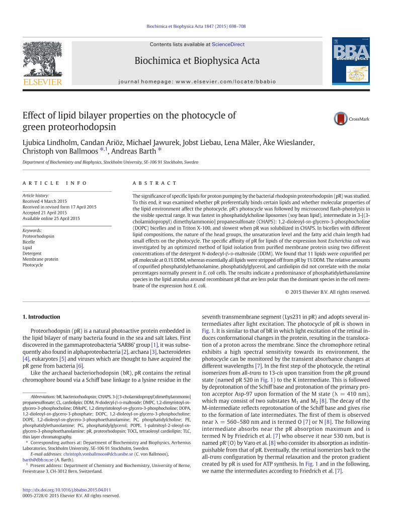
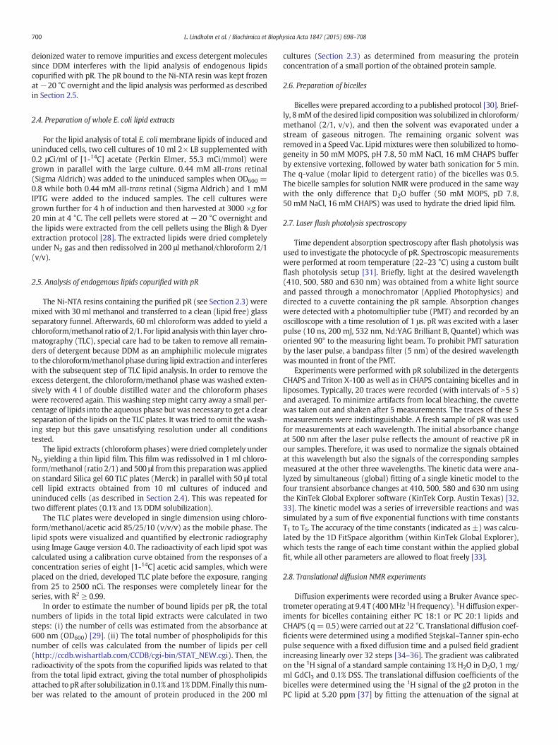
seventh transmembrane segment (Lys231 in pR) and adopts several in-termediates after light excitation. The photocycle of pR is shown inFig. 1. It is similar to that of bR in which light excitation of the retinal in-duces conformational changes in the protein, resulting in the transloca-tion of a proton across the membrane. Since the chromophore retinalexhibits a high spectral sensitivity towards its environment, thephotocycle can be monitored by the transient absorbance changes atdifferent wavelengths [7]. In the first step of the photocycle, the retinalisomerizes from all-trans to 13-cis upon transition from the pR groundstate (named pR 520 in Fig. 1) to the K intermediate. This is followedby deprotonation of the Schiff base and protonation of the primary pro-ton acceptor Asp-97 upon formation of the M state (λ = 410 nm),which may consist of two substates M1 and M2 [8]. The decay of theM-intermediate reflects reprotonation of the Schiff base and gives riseto the formation of late intermediates. The first of them is observednear λ = 560–580 nm and is termed O [7] or N [8]. The followingintermediate absorbs near the pR absorption maximum and istermed N by Friedrich et al. [7] who observe it near 530 nm, but isnamed pR′(O) by Varo et al. [8] who consider its absorption as indistin-guishable from that of pR. Eventually, the retinal isomerizes back to theall-trans configuration by thermal relaxation and the proton gradientcreated by pR is used for ATP synthesis. In Fig. 1 and in the following,we name the intermediates according to Friedrich et al. [7].
Biochimica et Biophysica Acta 1847 (2015) 698–708
Abbreviations: bR, bacteriorhodopsin; CHAPS, 3-[(3-cholamidopropyl)dimethylammonio]propanesulfonate; CL, cardiolipin; DDM,N-dodecyl-β-D-maltoside; DMPC, 1,2-dimyristoyl-sn-glycero-3-phosphocholine; DMoPC, 1,2 dimyristoleoyl-sn-glycero-3-phosphocholine; DOPA,1,2-dioleoyl-sn-glycero-3-phosphate; DOPC, 1,2-dioleoyl-sn-glycero-3-phosphocholine;DOPE, 1,2-dioleoyl-sn-glycero-3-phosphoethanolamine; PC, phosphatidylcholine; PE,phosphatidylethanolamine; PG, phosphatidylglycerol; POPE, 1-palmitoyl-2-oleoyl-sn-glycero-3-phosphoethanolamine; pR, proteorhodopsin; TOCL, tetraoleoyl cardiolipin; TLC,thin layer chromatography.⁎ Corresponding authors at: Department of Biochemistry and Biophysics, Arrhenius
Laboratories, Stockholm University, SE-106 91 Stockholm, Sweden.E-mail addresses: [email protected] (C. von Ballmoos),
[email protected] (A. Barth).1 Present address: Department of Chemistry and Biochemistry, University of Berne,
Freiestrasse 3, CH-3012 Bern, Switzerland.
http://dx.doi.org/10.1016/j.bbabio.2015.04.0110005-2728/© 2015 Elsevier B.V. All rights reserved.
Contents lists available at ScienceDirect
Biochimica et Biophysica Acta
j ourna l homepage: www.e lsev ie r .com/ locate /bbab io
The native lipid environment of green absorbing proteorhodopsin(Uniprot accession code: Q9F7P4) is not precisely known as pR studiedso far has been recombinantly overexpressed in Escherichia coli (E. coli).The E. coli membrane is a valid starting point for testing the lipid effecton pR's photocycle because the marine bacteria, in which pR was origi-nally discovered, have a similar phospholipid composition as E. coli [2,9–14]. The head group composition of phospholipids in E. coli remainsconstant over a broad spectrum of growth conditions [15]. Theaminophospholipid phosphatidylethanolamine (PE) is the major phos-pholipid in E. coli constituting 70–80% of the total phospholipid content.It is zwitter-ionic at physiological pH due to the protonated aminogroup and the negatively charged phosphate group. PE is accompaniedin the membrane by the two anionic lipids, phosphatidylglycerol (PG)and cardiolipin (CL), accounting for 15–20% and !5% of the membranephospholipids, respectively [16].
In order to understand more about the conditions affecting pR'sphotocycle, detailed knowledge about the interactions of wild typegreen pRwithmembrane lipids is important. It is well known that lipidscan affect the activity of membrane proteins through stable or dynamicinteractions. As an example, charged protein residues can readily inter-act with lipids through electrostatic interactions and form hydrogenbonds (H-bonds) with lipid head groups [17]. It has been observedthat the hydrophobic thickness of the lipid bilayer is an important factorfor the correct topology and insertion of a membrane protein and con-sequently for its optimal function [18,19]. Many membrane proteinsare known to prefer specific lipids, for example the Ca2+ ATPase prefersphosphatidylcholine (PC) over PE for optimumactivity [20], the respira-tory complexes II–IV are CL dependent [21], and KcsA requires anioniclipids for optimum activity [22]. The activity of bR, is reported to beinfluenced by several lipids [23,24], and a brief treatment of the purplemembrane with low concentrated detergent causes changes of the bRphotocycle without disrupting the trimer structure of bR [24] indicatinga strong dependence of bR activity on native membrane lipids. Thisassertion is verified by the recovery of normal photocycle behavior byincubating the disrupted membranes with a total extract of the nativelipids from the purple membrane [24].
pR has earlier been studied in different lipid and detergent environ-ments [7,25]. However, no systematic investigation has been done re-garding the effects of lipids on the photocycle. Therefore, the aim ofour researchwas to identify and understand how the lipophilic and am-phiphilic environment shapes pR activity. In this study, we have mainlyused bicelles, which are also known as mixed micelles, consisting of amixture between CHAPS and a phospholipid. Specifically, we testedthe influences of different types of lipid head groups, various fatty acidchain-lengths and the extent of saturation in the fatty acid chain onthe pR photocycle. Furthermore, we analyzed lipids that are copurified
with pR using two different detergent concentrations in order to detectwhether pR specifically binds certain lipids. Taken together, we ob-served that an intact membrane is beneficial for optimum function butthat particular lipid properties have only small effects.
2. Materials and methods
2.1. Chemicals and buffers
Most lipids used in our experiments were purchased from AvantiPolar Lipids (Alabaster, AL). Soy bean type II-S lipids and CHAPS werepurchased from Sigma Aldrich and N-dodecyl-β-D-maltoside (DDM)fromAffymetrix. Other chemicalswere all obtained from SigmaAldrich.
2.2. Cloning, growth, purification and reconstitution of unlabelled pR
The wild type green pR gene was synthesized by Eurofins MWGOperon (Ebersberg, Germany) and cloned into a pET27b+ (Invitrogen)vector using NdeI and XhoI as cloning sites [7]. A C-terminal His6-tagwas included in this construct to facilitate pR purification with Ni-NTAaffinity purification. C43 (DE3) [26] was used as an expression host forpR. pR was expressed and purified as described by Pfleger et al. [27]with the exception that DDM was exchanged for CHAPS during theNi-NTA purification step for pR intended for bicelle studies.
Proteoliposomes from soy bean phosphatidylcholine were preparedby a freeze–thaw procedure. In brief, 30 mg lipids dissolved in chloro-form were dried in a round bottomed flask by a stream of nitrogenand 2 h of vacuum. The lipids were then resuspended in 3 ml reconsti-tution buffer (50 mM MOPS, pH 7.5, 50 mM NaCl) and vortexed for10 min at room temperature under nitrogen atmosphere. Liposomeswere made unilamellar and homogeneous in size by a tip sonicator onice (Sonics, VCX130PB, 6 cycles, 30 s pulse, 30 s pause, 40% output).For reconstitution, 100 μl of 1 mg/ml pR was added to 1 ml of liposomesuspension and frozen in liquid nitrogen and thawed in water. Thefreeze–thaw cycle was repeated once. For flash photolysis measure-ments, 100 μl of liposomeswas dilutedwith 400 μl reconstitution buffer.
We checked our reconstitution of pR into liposomes using a pH sen-sitive dye on the inside of the liposomes. The light induced protonpumping experiments indicate that pR is functionally incorporatedand that the majority of pR molecules are oriented such that theypump protons to the inside of the liposomes.
2.3. Overexpression and purification of pR for the analysis of tightly boundlipids
In order to analyze lipids that bind to pRwith high affinity, phospho-lipids were radioactively labeled during pR expression by supplying thegrowthmediumwith [1-14C] sodium acetate (Perkin-Elmer). Twoml ofovernight culture was used to inoculate 200 ml of 2! Luria Bertani (LB)medium supplemented with 50 μg/ml kanamycin and 1 μCi/ml 14C ace-tate (Large culture). The culture was grown at 37 °C at 200 rpm. WhenOD600 reached 0.8, isopropyl-β-D-1-thiogalactopyranoside (IPTG) andall-trans retinal (Sigma-Aldrich) in ethanol were added to final concen-trations of 1mMand0.44mM, respectively. The cellswere kept at 37 °C,200 rpm for 4 h and harvested by centrifugation (3000!g) for 20min at4 °C. The cells were suspended in 50 mMMES buffer pH 6.0, containing300 mM NaCl, and disrupted mechanically with a glass homogenizer.The cell lysate was centrifuged for 1 h at 3700 !g in a top bench centri-fuge at 4 °C and themembrane pellet was further solubilized for 48 h in50 ml of 50 mMMES pH 6.0 buffer, containing 300mMNaCl, 5 mM im-idazole and either 1% or 0.1% DDM.
Detergent solubilized protein was obtained by centrifugation at150,000 !g for 45 min at 4 °C and the supernatant was incubated with4 ml of Ni-NTA beads overnight at 4 °C. After binding of pR to theresin, the resin beads were washed with wash buffer containing 0.1%Triton and 50 mM imidazole, followed by a second wash with 900 ml
Fig. 1. Simplified photocycle of pR according to [8] in the terminology of [7]. Numbers de-note the absorbance maximum of the respective state. The photocycle starts with excita-tion of the ground state of pR which is named pR 520 in the above scheme.
699L. Lindholm et al. / Biochimica et Biophysica Acta 1847 (2015) 698–708
deionized water to remove impurities and excess detergent moleculessince DDM interferes with the lipid analysis of endogenous lipidscopurified with pR. The pR bound to the Ni-NTA resin was kept frozenat"20 °C overnight and the lipid analysis was performed as describedin Section 2.5.
2.4. Preparation of whole E. coli lipid extracts
For the lipid analysis of total E. coli membrane lipids of induced anduninduced cells, two cell cultures of 10 ml 2! LB supplemented with0.2 μCi/ml of [1-14C] acetate (Perkin Elmer, 55.3 mCi/mmol) weregrown in parallel with the large culture. 0.44 mM all-trans retinal(Sigma Aldrich) was added to the uninduced samples when OD600 =0.8 while both 0.44 mM all-trans retinal (Sigma Aldrich) and 1 mMIPTG were added to the induced samples. The cell cultures weregrown further for 4 h of induction and then harvested at 3000 !g for20 min at 4 °C. The cell pellets were stored at "20 °C overnight andthe lipids were extracted from the cell pellets using the Bligh & Dyerextraction protocol [28]. The extracted lipids were dried completelyunder N2 gas and then redissolved in 200 μl methanol/chloroform 2/1(v/v).
2.5. Analysis of endogenous lipids copurified with pR
The Ni-NTA resins containing the purified pR (see Section 2.3) weremixed with 30 ml methanol and transferred to a clean (lipid free) glassseparatory funnel. Afterwards, 60 ml chloroform was added to yield achloroform/methanol ratio of 2/1. For lipid analysiswith thin layer chro-matography (TLC), special care had to be taken to remove all remain-ders of detergent because DDM as an amphiphilic molecule migratesto the chloroform/methanol phase during lipid extraction and interfereswith the subsequent step of TLC lipid analysis. In order to remove theexcess detergent, the chloroform/methanol phase was washed exten-sively with 4 l of double distilled water and the chloroform phaseswere recovered again. This washing step might carry away a small per-centage of lipids into the aqueous phase but was necessary to get a clearseparation of the lipids on the TLC plates. It was tried to omit the wash-ing step but this gave unsatisfying resolution under all conditionstested.
The lipid extracts (chloroform phases) were dried completely underN2, yielding a thin lipid film. This film was redissolved in 1 ml chloro-form/methanol (ratio 2/1) and 500 μl from this preparationwas appliedon standard Silica gel 60 TLC plates (Merck) in parallel with 50 μl totalcell lipid extracts obtained from 10 ml cultures of induced anduninduced cells (as described in Section 2.4). This was repeated fortwo different plates (0.1% and 1% DDM solubilization).
The TLC plates were developed in single dimension using chloro-form/methanol/acetic acid 85/25/10 (v/v/v) as the mobile phase. Thelipid spots were visualized and quantified by electronic radiographyusing Image Gauge version 4.0. The radioactivity of each lipid spot wascalculated using a calibration curve obtained from the responses of aconcentration series of eight [1-14C] acetic acid samples, which wereplaced on the dried, developed TLC plate before the exposure, rangingfrom 25 to 2500 nCi. The responses were completely linear for theseries, with R2 # 0.99.
In order to estimate the number of bound lipids per pR, the totalnumbers of lipids in the total lipid extracts were calculated in twosteps: (i) the number of cells was estimated from the absorbance at600 nm (OD600) [29]. (ii) The total number of phospholipids for thisnumber of cells was calculated from the number of lipids per cell(http://ccdb.wishartlab.com/CCDB/cgi-bin/STAT_NEW.cgi). Then, theradioactivity of the spots from the copurified lipids was related to thatfrom the total lipid extract, giving the total number of phospholipidsattached to pR after solubilization in 0.1% and 1%DDM. Finally this num-ber was related to the amount of protein produced in the 200 ml
cultures (Section 2.3) as determined from measuring the proteinconcentration of a small portion of the obtained protein sample.
2.6. Preparation of bicelles
Bicelles were prepared according to a published protocol [30]. Brief-ly, 8mMof the desired lipid compositionwas solubilized in chloroform/methanol (2/1, v/v), and then the solvent was evaporated under astream of gaseous nitrogen. The remaining organic solvent wasremoved in a Speed Vac. Lipid mixtures were then solubilized to homo-geneity in 50 mM MOPS, pH 7.8, 50 mM NaCl, 16 mM CHAPS bufferby extensive vortexing, followed by water bath sonication for 5 min.The q-value (molar lipid to detergent ratio) of the bicelles was 0.5.The bicelle samples for solution NMR were produced in the same waywith the only difference that D2O buffer (50 mM MOPS, pD 7.8,50 mM NaCl, 16 mM CHAPS) was used to hydrate the dried lipid film.
2.7. Laser flash photolysis spectroscopy
Time dependent absorption spectroscopy after flash photolysis wasused to investigate the photocycle of pR. Spectroscopic measurementswere performed at room temperature (22–23 °C) using a custom builtflash photolysis setup [31]. Briefly, light at the desired wavelength(410, 500, 580 and 630 nm) was obtained from a white light sourceand passed through a monochromator (Applied Photophysics) anddirected to a cuvette containing the pR sample. Absorption changeswere detected with a photomultiplier tube (PMT) and recorded by anoscilloscope with a time resolution of 1 μs. pR was excited with a laserpulse (10 ns, 200 mJ, 532 nm, Nd:YAG Brilliant B, Quantel) which wasoriented 90° to the measuring light beam. To prohibit PMT saturationby the laser pulse, a bandpass filter (5 nm) of the desired wavelengthwas mounted in front of the PMT.
Experiments were performed with pR solubilized in the detergentsCHAPS and Triton X-100 as well as in CHAPS containing bicelles and inliposomes. Typically, 20 traces were recorded (with intervals of N5 s)and averaged. To minimize artifacts from local bleaching, the cuvettewas taken out and shaken after 5 measurements. The traces of these 5measurements were indistinguishable. A fresh sample of pR was usedfor measurements at each wavelength. The initial absorbance changeat 500 nm after the laser pulse reflects the amount of reactive pR inour samples. Therefore, it was used to normalize the signals obtainedat this wavelength but also the signals of the corresponding samplesmeasured at the other three wavelengths. The kinetic data were ana-lyzed by simultaneous (global) fitting of a single kinetic model to thefour transient absorbance changes at 410, 500, 580 and 630 nm usingthe KinTek Global Explorer software (KinTek Corp. Austin Texas) [32,33]. The kinetic model was a series of irreversible reactions and wassimulated by a sum of five exponential functions with time constantsT1 to T5. The accuracy of the time constants (indicated as ±) was calcu-lated by the 1D FitSpace algorithm (within KinTek Global Explorer),which tests the range of each time constant within the applied globalfit, while all other parameters are allowed to float freely [33].
2.8. Translational diffusion NMR experiments
Diffusion experiments were recorded using a Bruker Avance spec-trometer operating at 9.4 T (400MHz 1H frequency). 1H diffusion exper-iments for bicelles containing either PC 18:1 or PC 20:1 lipids andCHAPS (q= 0.5) were carried out at 22 °C. Translational diffusion coef-ficients were determined using a modified Stejskal–Tanner spin-echopulse sequence with a fixed diffusion time and a pulsed field gradientincreasing linearly over 32 steps [34–36]. The gradient was calibratedon the 1H signal of a standard sample containing 1% H2O in D2O, 1 mg/ml GdCl3 and 0.1% DSS. The translational diffusion coefficients of thebicelles were determined using the 1H signal of the g2 proton in thePC lipid at 5.20 ppm [37] by fitting the attenuation of the signal at
700 L. Lindholm et al. / Biochimica et Biophysica Acta 1847 (2015) 698–708
increasing pulsed field gradient strengths to the Stejskal–Tanner equa-tion. Viscosity differences were accounted for bymeasuring the transla-tional diffusion coefficient of HDO in the bicelle sample and comparingit to its diffusion in H2O [38]. The obtained factor was used to normalize,i.e. viscosity-correct, the bicelle diffusion coefficient. The hydrodynamicradius was calculated with the Stokes–Einstein equation including ashape factor of 1.2 that accounted for the non-spherical shape of bicelles[39]. Experimental errors were estimated by repeating all diffusionexperiments 5 times. The obtained diffusion coefficients for the PC lipidsreflect the translational diffusion of the bicelles since the solubility of18:1 and 20:1 PC in water is negligible. Moreover, the tumbling of thebicelles is fast compared to thediffusion delay (typically 0.4 s) averaginglateral diffusion of lipids in the bilayer to zero [40].
3. Results
3.1. Membrane lipids in pR's native hosts
In order to identify relevant lipids thatmight affect the activity of pR,we reviewed available lipid data and analyzed the genomeof organismscontaining the pR gene. Three approaches were applied: (i) for well-established pR genes from uncultivated organisms or ones lacking com-plete genome information, the lipid composition in closely related phy-logenetic organisms was checked [2,9]. (ii) From organisms withoutlipid data but with completely sequenced genomes containing a pRgene, genes for key phospholipid enzymes were searched by BLASTusing the corresponding amino acid sequences from E. coli as probes(genomes fromDokdonia sp.MED134 [11] and gammaproteobacteriumHTCC2207). (iii) In addition, the experimentally determined lipid com-positions from several proteobacteria (having a pR gene) [10,13,14,41]were also considered.
The threemajor E. coli phospholipids PE, PG, and CLwere either pre-dicted or are the dominating lipids found experimentally. However, forthe organisms for which experimental data are available, the propor-tions between PE, PG and CL vary. In addition, some other,minor specieshave also been observed. Hence, given this coherent composition andthe functional expression of pR in E. coli, the PE, PG and CL lipids seemto be physiologically relevant candidates for the analysis of pR's lipiddependence.
3.2. Analysis of lipids that tightly bind to pR overexpressed in E. coli
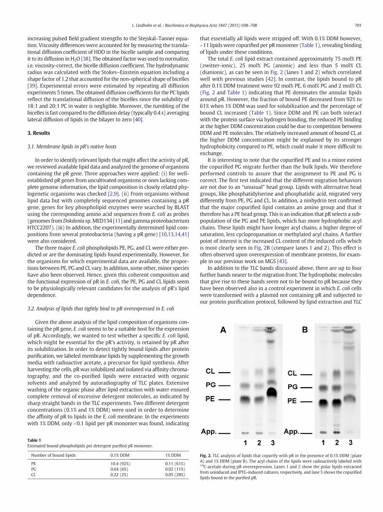
Given the above analysis of the lipid composition of organisms con-taining the pR gene, E. coli seems to be a suitable host for the expressionof pR. Accordingly, we wanted to test whether a specific E. coli lipid,which might be essential for the pR's activity, is retained by pR afterits solubilization. In order to detect tightly bound lipids after proteinpurification, we labeledmembrane lipids by supplementing the growthmedia with radioactive acetate, a precursor for lipid synthesis. Afterharvesting the cells, pRwas solubilized and isolated via affinity chroma-tography, and the co-purified lipids were extracted with organicsolvents and analyzed by autoradiography of TLC plates. Extensivewashing of the organic phase after lipid extraction with water ensuredcomplete removal of excessive detergent molecules, as indicated bysharp straight bands in the TLC experiments. Two different detergentconcentrations (0.1% and 1% DDM) were used in order to determinethe affinity of pR to lipids in the E. coli membrane. In the experimentswith 1% DDM, only ~0.1 lipid per pR monomer was found, indicating
that essentially all lipids were stripped off. With 0.1% DDM however,~11 lipidswere copurified per pRmonomer (Table 1), revealing bindingof lipids under these conditions.
The total E. coli lipid extract contained approximately 75 mol% PE(zwitter-ionic), 25 mol% PG (anionic) and less than 5 mol% CL(dianionic), as can be seen in Fig. 2 (lanes 1 and 2) which correlatedwell with previous studies [42]. In contrast, the lipids bound to pRafter 0.1% DDM treatment were 92 mol% PE, 6 mol% PG and 2 mol% CL(Fig. 2 and Table 1) indicating that PE dominates the annular lipidsaround pR. However, the fraction of bound PE decreased from 92% to61% when 1% DDM was used for solubilization and the percentage ofbound CL increased (Table 1). Since DDM and PE can both interactwith the protein surface via hydrogen bonding, the reduced PE bindingat the higher DDM concentration could be due to competition betweenDDM and PE molecules. The relatively increased amount of bound CL atthe higher DDM concentration might be explained by its strongerhydrophobicity compared to PE, which could make it more difficult toexchange.
It is interesting to note that the copurified PE and to a minor extentthe copurified PG migrate further than the bulk lipids. We thereforeperformed controls to assure that the assignment to PE and PG iscorrect. The first test indicated that the different migration behaviorsare not due to an “unusual” head group. Lipids with alternative headgroups, like phosphatidylserine and phosphatidic acid, migrated verydifferently from PE, PG and CL. In addition, a ninhydrin test confirmedthat the major copurified lipid contains an amine group and that ittherefore has a PE head group. This is an indication that pR selects a sub-population of the PG and PE lipids, which has more hydrophobic acylchains. These lipids might have longer acyl chains, a higher degree ofsaturation, less cyclopropanation or methylated acyl chains. A furtherpoint of interest is the increased CL content of the induced cells whichis most clearly seen in Fig. 2B (compare lanes 1 and 2). This effect isoften observed upon overexpression of membrane proteins, for exam-ple in our previous work on MGS [43].
In addition to the TLC bands discussed above, there are up to fourfurther bands nearer to themigration front. The hydrophobic moleculesthat give rise to these bands seem not to be bound to pR because theyhave been observed also in a control experiment in which E. coli cellswere transformed with a plasmid not containing pR and subjected toour protein purification protocol, followed by lipid extraction and TLC
Table 1Estimated bound phospholipids per detergent purified pR monomer.
Number of bound lipids 0.1% DDM 1% DDM
PE 10.4 (92%) 0.11 (61%)PG 0.64 (6%) 0.02 (11%)CL 0.22 (2%) 0.05 (28%)
Fig. 2. TLC analysis of lipids that copurify with pR in the presence of 0.1% DDM (plateA) and 1% DDM (plate B). The acyl chains of the lipids were radioactively labeled with14C-acetate during pR overexpression. Lanes 1 and 2 show the polar lipids extractedfrom uninduced and IPTG-induced cultures, respectively, and lane 3 shows the copurifiedlipids bound to the purified pR.
701L. Lindholm et al. / Biochimica et Biophysica Acta 1847 (2015) 698–708
analysis. The TLC plates showed the bands above CL but not the phos-pholipid bands seen in Fig. 2 (lane 3 in panels A and B).
3.3. Bicelle model system
In order to test the influence of a phospholipid environment on thephotocycle of pR, bicelles with a defined detergent to lipid ratio wereused. Such systemshave proven to beuseful for a variety of applications.They are bilayer like organizations [30,44] and have the advantage overliposomes of reduced light scattering and therefore enable a bettersignal to noise ratio in the optical flash photolysis experiments. In thebicelles, the ratio between the detergent CHAPS (16 mM) and otherlipids (8 mM) was kept constant but the types of lipids differed fordifferent preparations. CHAPS, a non-ionic detergent, was chosen forthis study because it is known to form bicelles in the presence ofphospholipids.
A main advantage of bicelles for our study was the reduced lightscattering as compared to turbid liposomes samples in spectroscopicmeasurements. Furthermore, the convenient preparation of pR–bicellecomplexes by mixing purified pR solubilized in CHAPS and pre-formed lipid preparations ensured controlled and reproducible condi-tions. In the following, different lipid properties were tested for theireffect on the photocycle comprising head group charge, fatty acidchain length and degree of unsaturation. For most lipids tested, wechose fatty-acyl chainswith one cisdouble bond as these occur naturallyin E. coli. In addition, this ensured that the lipids were in the fluid phasewhen the experiments were carried out at room temperature.
3.4. PC 18:1 and 20:1 form bicelles together with CHAPS
To characterize the CHAPS/lipid mixtures, we used pulsed fieldgradient diffusion NMR. The translational diffusion coefficient is linkedto the hydrodynamic radius of the aggregate via the Stokes–Einsteinequation. The diffusion coefficients and hydrodynamic radii formixtures containing either 18:1 PC (DOPC) or 20:1 PC (1,2-dieicosenoyl-sn-glycero-3-phosphocholine) lipids are given in Table 2.The hydrodynamic radii of both types of mixtures are typical for small,isotropic bicelles made with CHAPS [44], smaller than bicelles withDHPC (q=0.5), but significantly larger than CHAPSmicelles at the con-centration used here (20 mM) [45,46]. Moreover, we see that 18:1PC/CHAPS bicelles are somewhat smaller than 20:1 PC/CHAPS bicelles.We conclude that both lipids are incorporated into CHAPS containingbicelles, and form structures that are larger than CHAPS micelles.
To assure that the incorporation of pR into the bicelles did not alterthe morphology of the CHAPS/lipid mixtures, diffusion experimentswere carried out also on bicelles with added protein. Table 2 reportsthe diffusion coefficients and hydrodynamic radii for bicelles with andwithout pR. The hydrodynamic radius was in all cases around 3 nm.The addition of pR leads only to minor changes in the hydrodynamicradii of the bicelles, as expected since the protein concentration isaround an order of magnitude lower than that of bicelle assemblies.The most likely explanation for the observed slight decreases in thehydrodynamic radii is that there are two populations of bicelles, themajority being bicelles with a higher content of detergent, which
makes them smaller, together with a few larger ones containing pR.This would overall lead to a slight reduction in average size. Mostimportantly, the differences are small andwe conclude that the additionof protein does not lead to a major change in bicelle morphology.
3.5. Contribution of photocycle intermediates to the transient absorbancechanges
pR's photocycle was studied by flash photolysis with purified pRreconstituted in detergents, bicelles, and liposomes. This techniquehas been proven to be very useful to reliably monitor the photocycleof bR and pR. After the sample had been excited by a short laser pulseat 532nm(close to themaximal absorption at 520nm), transient absor-bance changes were measured at 410, 500, 580, and 630 nm. Spectra ofthe photocycle intermediates have been published [8] and can be usedto estimate the contribution of each intermediate to the absorption atthe four wavelengths used. In our discussion we will adopt the termi-nology of Friedrich et al. [7].
At 410 nm, the M intermediate absorbs 3–4 times stronger than theother intermediates and thepR ground state. Therefore, thiswavelengthreflects predominantly the formation and decay of the M intermediate.At all other wavelengths used in this study, the absorption of the Mintermediate is negligible.
At 500 nm, the pR ground state and the last intermediate N absorbequally and about twice as strong as the other intermediates. Thus, thesignal at this wavelength will be dominated by the time-dependent con-centrations of the ground state and of the N intermediate.
At 630 nm, O and K intermediates absorb strongest, whereas the pRgroundstate and the N intermediate have negligible absorbance. Thusthe signal will be dominated by the concentration of K in the earlyphase of the photocycle and by that of O in the late phase. The situationis similar at 580 nm, but pR and N also contribute to the signal sincetheir absorbance is 1/3 of that of the O intermediate.
3.6. The photocycle in detergents, bicelles and liposomes
In the first set of experiments, we tested the influence of differentmembrane mimetic environments on the general turnover of pR, asestimated from reformation of the ground state (observed at 500 nm)(Fig. 3A). Due to light scattering, the trace of the transient absorbancechange obtained with liposomes shows larger noise.
The transient absorbance change at 500nm is dominated by the con-centrations of the ground state and the N intermediate (see Section 3.5)and reflects therefore the overall turnover of thephotocycle. It exhibits asudden drop in absorbance immediately after the excitation flash due todepletion of the ground state. This initiation of the photocycle cannot beresolved kinetically with our method. Subsequently, the absorbance in-creases back to its initial value because theN intermediate is formed andthe ground state is restored. Since the N intermediate and the groundstate have similar absorption spectra (see Introduction andSection 3.5), the absorbance increase is probably dominated by theformation of the N intermediate. The subsequent decay of N and thereformation of the pR ground state are expected to have a smaller influ-ence on the signal.
The absorbance increase due to N formation and restoration of theground state can be fitted to a two-step reaction, if no reverse reactionis allowed. The faster reaction contributed to N80%. The reactionmodel was fitted to the data at individual wavelengths in this section.Therefore the time constants in this section should not be comparedwith the time constants obtained by global fitting to all 4 wavelengthssimultaneously, which are listed in Tables 3 and 4.
As depicted in Fig. 3A, formation of the late N intermediate and res-toration of the ground state are fastest for pR reconstituted in soy beanliposomes (time constants ~12 and ~100ms) followed by CHAPS/DOPCbicelles (~20 and ~140 ms), Triton X-100 (~40 and ~500 ms) andslowest in CHAPS (~80 and 1000 ms). Accordingly, turnover turned
Table 2Normalized translational diffusion coefficients Dt
a and hydrodynamic radii rH for 18:1 PCand 20:1 PC in bicelles with and without incorporation of pR.
18:1 PCb 18:1 PCb + pR 20:1 PCc 20:1 PCc + pR
Dt [10"11 m2 s"1] 7.2 ± 0.1 7.8 ± 0.1 6.4 ± 0.1 7.2 ± 0.2rH [nm] 2.8 2.6 3.2 2.8a Determined using the g2 1H signal in PC at 5.20 ppm and viscosity-corrected based on
the diffusion of HDO in the sample.b DOPC.c 1,2-dieicosenoyl-sn-glycero-3-phosphocholine.
702 L. Lindholm et al. / Biochimica et Biophysica Acta 1847 (2015) 698–708

out to be fastest in the “native” environment of a liposome. DOPC lipo-somes have also been tested and gave similar results to soy bean lipids(data not shown).
The slowdown of the turnover rate in CHAPS compared to Triton X-100 is not surprising, as CHAPS is considered a stiff detergent due to therigid steroid ring structure, possibly impairing themotional flexibility ofpR. However, upon mixing the slow CHAPS preparation with lipids inorder to form bicelles, turnover was accelerated up to 4-fold, reaching60% of the activity in soy bean liposomes. Thus the addition of lipids tothe CHAPS detergent micelles makes the lipid environment of pRmore similar to that in liposomes. Most likely, this is due to a replace-ment of CHAPS by lipids in the immediate vicinity of pR.
In the following, we compare the kinetics of several photocycleintermediates in the different reconstitution forms. Above pH 7, forma-tion of the M state is observed at 410 nm (Fig. 3B) as an absorbanceincrease due to deprotonation of the Schiff base. The following absor-bance decrease indicates the decay of the M intermediate and isinterpreted as the re-protonation of the Schiff base from the P-side, pre-sumably coupled to proton uptake from the medium. The Triton traceshows a very rapid M formation (~8 μs) and an M decay with a timeconstant of around ~0.5ms.With CHAPS, M formation is slightly slower(17 μs), the M state persists for a longer time and decays considerablyslower (~13ms) thanwith Triton. The kinetics of liposomes and bicellesare very similar, with M formation times of ~70–80 μs and M decaytimes of ~1–2 ms. The slow M decay in the stiff detergent CHAPS indi-cates that reprotonation of the Schiff base, which takes place during Mdecay, requires conformational mobility of the protein. This mobilityseems to be hampered in CHAPS, but seems to be recovered whenCHAPS is supplemented by lipids in our bicelles.
The absorption at 580 nmmonitors predominantly the K and the Ostate with contributions from the N and the ground state (seeSection 3.5). The signal (Fig. 3C) rises instantaneously due to the forma-tion of the K intermediate. Its following decay attenuates the absor-bance. Formation of the late N and O intermediates leads to a secondincrease in absorbance and their decay to thefinal decrease of the signal.Again, traces from pR reconstituted in soy bean liposomes or DOPCbicelles display a faster formation of the late intermediates with timeconstants of ~2 ms and 3 ms (time constants derived from fits to the580 nm trace only), respectively, while the two detergent samplesdisplay a distinctly later formation of these intermediates (~6–7 ms).
Taken together, we have observed that CHAPS delays the overallphotocycle drastically. In contrast, the rates and curve shapes of pR inbicelles are closer to those in liposomes and we can conclude thatbicelles are a suitable system for a systematic study on the influenceof lipid properties on the photocycle of pR.
3.7. Effect of lipid head groups on the pR photocycle
The influence of lipid head group properties on the photocycle of pRwas also studied. Here and in the following sections, the data were an-alyzed by global fitting, i.e. the data at the four different wavelengths
Fig. 3. Time-resolved spectroscopy of detergent (Triton X-100 and CHAPS) solubilized pR,pR in bicelles with the zwitter-ionic lipid DOPC and pR in soy bean liposomes at three dif-ferent wavelengths: (A) 500 nm, (B) 410 nm, and (C) 580 nm.
Table 3Time constants of the pR photocycle in different lipid environments obtained with prepa-ration 1. The time constants T1 to T5 were determined as described in the Materials andmethods. TwopRpreparations inCHAPSwere used. The results for preparation 1 are listedin this table and those for preparation 2 in Table 4. Results with the same preparation aredirectly comparable, whereas slightly different pR/CHAPS/lipid ratios in the two prepara-tions might affect the rates and thus the comparability between the two sets ofexperiments.
T1/μs T2/μs T3/ms T4/ms T5/ms
18:1 PG (DOPG) 44 ± 3 680 ± 110 1.7 ± 0.2 19.6 ± 1.8 91 ± 816:0–18:1 PE (POPE) 61 ± 10 530 ± 140 2.2 ± 0.2 17.5 ± 1.7 91 ± 816:0–18:1 PC (POPC) 39 ± 4 420 ± 50 2.8 ± 0.2 22.7 ± 1.5 108 ± 1320:1 PCa 41 ± 9 790 ± 310 1.9 ± 0.8 18.2 ± 2.3 91 ± 1118:1 PC (DOPC) 39 ± 4 490 ± 120 3.0 ± 0.2 20 ± 2 91 ± 816:1 PCb 38 ± 2 680 ± 200 2.0 ± 0.3 19 ± 2 111 ± 1114:1 PC (DMoPC) 33 ± 2 580 ± 100 2.1 ± 0.1 31 ± 4 167 ± 1314:0 PC (DMPC) 26 ± 1 480 ± 60 3.2 ± 0.2 37 ± 4 169 ± 16a 1,2-Dieicosenoyl-sn-glycero-3-phosphocholine.b 1,2-Dipalmitoleoyl-sn-glycero-3-phosphocholine.
Table 4Time constants of the pR photocycle in different lipid environments obtained with prepa-ration 2. See Table 3 for further information.
T1/μs T2/μs T3/ms T4/ms T5/ms
DOPC 39 ± 8 410 ± 120 2.9 ± 0.5 21 ± 2 95 ± 11DOPC/DOPG (9/1) 51 ± 12 440 ± 140 3.5 ± 0.4 26 ± 4 120 ± 18DOPC/DOPA (9/1) 53 ± 7 590 ± 140 3.5 ± 0.3 26.3 ± 3.5 114 ± 15DOPC/TOCL (9/1) 47 ± 3 490 ± 100 3.6 ± 0.2 28.7 ± 2.3 135 ± 16
703L. Lindholm et al. / Biochimica et Biophysica Acta 1847 (2015) 698–708
were fitted simultaneously to a sum of five exponential functions withdifferent time constants. A global fit is a mathematical solution (with afixed set of parameters) to observed traces and it is difficult to assignthe results to specific chemical reactions, as the obtained mathematicalsolution is often not unique. However, the following approximate as-signment can serve as a rough guideline: the smallest time constant T1is related to the decay of the early K intermediate and formation ofthe M intermediate. Time constants T2 and T3 are associated with thedecay of the M intermediate (see below for further discussion). Mainlytime constant T3 relates to formation of the late N and O intermediatesand time constants T4 and T5 correspond to their decay and the returnto the pR ground state. The time constants are listed in Tables 3 and 4.
The E. coli membrane consists mostly of PE (~75%) and PG (~25%)(see Section 3.2). In addition to these lipids, we have included PC inour study of head group effects, because it is also a zwitter-ionic lipidlike PE that is often used in functional studies as a replacement for PE.While PE is cone shaped, PC due to its larger head group has a more cy-lindrical shape and thusmuchbetter bilayer forming properties than PE.In contrast to PE and PC, the second most abundant lipid in E. coli PG isnegatively charged.
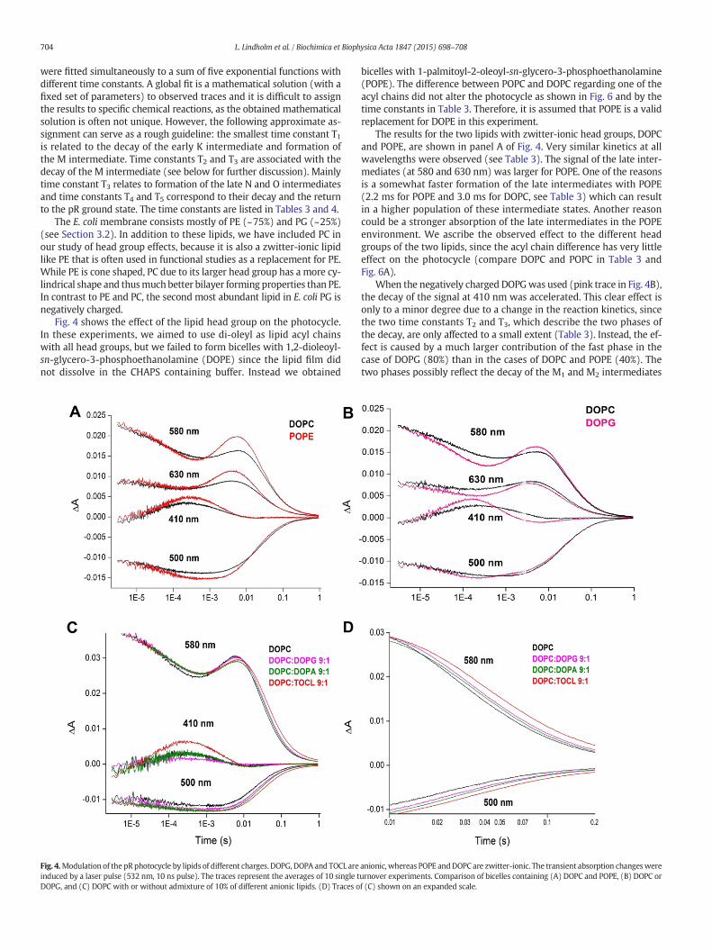
Fig. 4 shows the effect of the lipid head group on the photocycle.In these experiments, we aimed to use di-oleyl as lipid acyl chainswith all head groups, but we failed to form bicelles with 1,2-dioleoyl-sn-glycero-3-phosphoethanolamine (DOPE) since the lipid film didnot dissolve in the CHAPS containing buffer. Instead we obtained
bicelles with 1-palmitoyl-2-oleoyl-sn-glycero-3-phosphoethanolamine(POPE). The difference between POPC and DOPC regarding one of theacyl chains did not alter the photocycle as shown in Fig. 6 and by thetime constants in Table 3. Therefore, it is assumed that POPE is a validreplacement for DOPE in this experiment.
The results for the two lipids with zwitter-ionic head groups, DOPCand POPE, are shown in panel A of Fig. 4. Very similar kinetics at allwavelengths were observed (see Table 3). The signal of the late inter-mediates (at 580 and 630 nm) was larger for POPE. One of the reasonsis a somewhat faster formation of the late intermediates with POPE(2.2 ms for POPE and 3.0 ms for DOPC, see Table 3) which can resultin a higher population of these intermediate states. Another reasoncould be a stronger absorption of the late intermediates in the POPEenvironment. We ascribe the observed effect to the different headgroups of the two lipids, since the acyl chain difference has very littleeffect on the photocycle (compare DOPC and POPC in Table 3 andFig. 6A).
When the negatively charged DOPGwas used (pink trace in Fig. 4B),the decay of the signal at 410 nm was accelerated. This clear effect isonly to a minor degree due to a change in the reaction kinetics, sincethe two time constants T2 and T3, which describe the two phases ofthe decay, are only affected to a small extent (Table 3). Instead, the ef-fect is caused by a much larger contribution of the fast phase in thecase of DOPG (80%) than in the cases of DOPC and POPE (40%). Thetwo phases possibly reflect the decay of the M1 and M2 intermediates
Fig. 4.Modulation of the pR photocycle by lipids of different charges. DOPG, DOPA and TOCL are anionic, whereas POPE andDOPC are zwitter-ionic. The transient absorption changeswereinduced by a laser pulse (532 nm, 10 ns pulse). The traces represent the averages of 10 single turnover experiments. Comparison of bicelles containing (A) DOPC and POPE, (B) DOPC orDOPG, and (C) DOPC with or without admixture of 10% of different anionic lipids. (D) Traces of (C) shown on an expanded scale.
704 L. Lindholm et al. / Biochimica et Biophysica Acta 1847 (2015) 698–708
[8] and the different contributions of the two phases in different lipidsystems might indicate a different equilibrium constant between M1
and M2.In the next measurements, we investigated further the influence of
negatively charged lipids on the photocycle of pR. These experimentswere performed with a new pR preparation (preparation 2). Thebicelles contained DOPC as lipid matrix and different anionic lipids ina 9/1 ratio. At the pH of our measurements (pH 7.8), the charge ofDOPG is "1, that of 1,2-dioleoyl-sn-glycero-3 phosphate (DOPA)approximately "1.5, and that of tetraoleoyl cardiolipin (TOCL) "2.The time constants and rates for these systems are listed in Table 4.The experimental data are shown in Fig. 4C and on an expanded scalealso in Fig. 4D. They reveal small but systematic effects upon the addi-tion of negatively charged lipids. This is most clearly seen in Fig. 4D:the return to the ground state absorbance at 500 and 580 nm is slowerwhen the negative charge of theminor lipid increases. The effect can beexplained by the slightly different time constants for T4 and T5 obtainedin the fit (Table 4). However, most of them are equal within the errorlimits, indicating that the reaction kinetics are essentially unchanged.
3.8. Effect of membrane thickness on the pR photocycle
To test the influence of membrane thickness on the photocycle, pRwas reconstituted in bicelles with lipids having a PC head group and
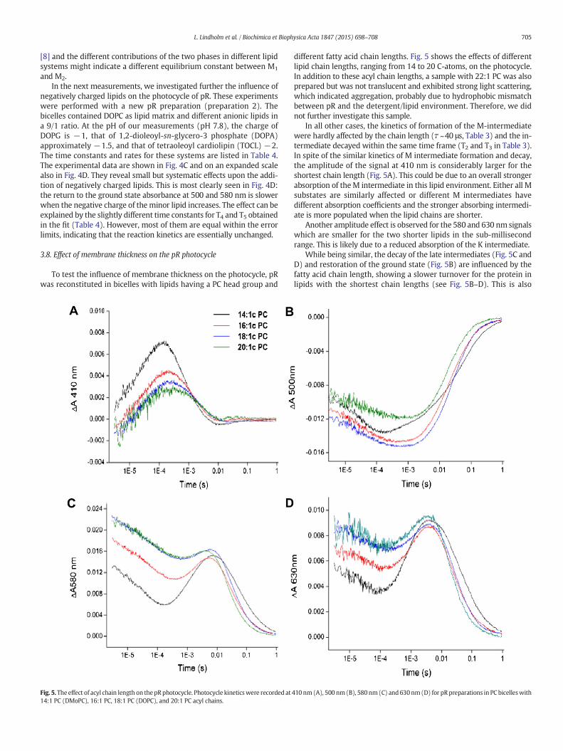
different fatty acid chain lengths. Fig. 5 shows the effects of differentlipid chain lengths, ranging from 14 to 20 C-atoms, on the photocycle.In addition to these acyl chain lengths, a sample with 22:1 PC was alsoprepared but was not translucent and exhibited strong light scattering,which indicated aggregation, probably due to hydrophobic mismatchbetween pR and the detergent/lipid environment. Therefore, we didnot further investigate this sample.
In all other cases, the kinetics of formation of the M-intermediatewere hardly affected by the chain length (! ~40 μs, Table 3) and the in-termediate decayed within the same time frame (T2 and T3 in Table 3).In spite of the similar kinetics of M intermediate formation and decay,the amplitude of the signal at 410 nm is considerably larger for theshortest chain length (Fig. 5A). This could be due to an overall strongerabsorption of theM intermediate in this lipid environment. Either all Msubstates are similarly affected or different M intermediates havedifferent absorption coefficients and the stronger absorbing intermedi-ate is more populated when the lipid chains are shorter.
Another amplitude effect is observed for the 580 and 630 nm signalswhich are smaller for the two shorter lipids in the sub-millisecondrange. This is likely due to a reduced absorption of the K intermediate.
While being similar, the decay of the late intermediates (Fig. 5C andD) and restoration of the ground state (Fig. 5B) are influenced by thefatty acid chain length, showing a slower turnover for the protein inlipids with the shortest chain lengths (see Fig. 5B–D). This is also
Fig. 5. The effect of acyl chain length on thepRphotocycle. Photocycle kineticswere recorded at 410 nm(A), 500 nm(B), 580nm(C) and 630nm(D) for pRpreparations in PC bicelleswith14:1 PC (DMoPC), 16:1 PC, 18:1 PC (DOPC), and 20:1 PC acyl chains.
705L. Lindholm et al. / Biochimica et Biophysica Acta 1847 (2015) 698–708
reflected in the time constants T4 and T5 (Table 3) which describe thereturn to the ground state. They are 31 and 167 ms in the presence ofthe 14:1 lipid, but near 20 and 100 ms with longer acyl chains. Thephotocycle is apparently slower with the 16 and 18 C chains than withthe 20 C chains (see Fig. 5B–D), but this is not revealed in the time con-stants. Therefore, the effect must be due to a larger proportion of thefaster phase (described by T4) than of the slowest phase (described byT5) in the 20 C system. Possible reasons for the different contributionsof the two phases are different absorption spectra of the intermediatesand different equilibrium constants between the intermediates in thedifferent lipid systems.
3.9. Effects of saturation vs unsaturation of fatty acid chains
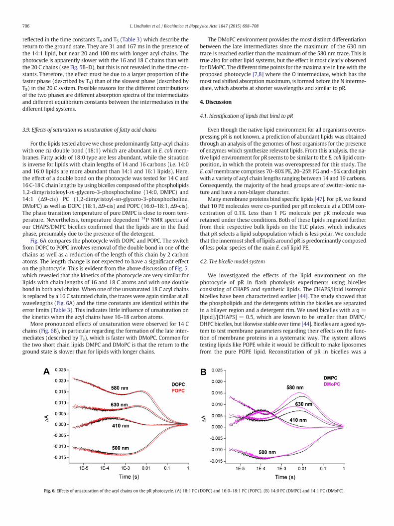
For the lipids tested abovewe chose predominantly fatty-acyl chainswith one cis double bond (18:1) which are abundant in E. coli mem-branes. Fatty acids of 18:0 type are less abundant, while the situationis inverse for lipids with chain lengths of 14 and 16 carbons (i.e. 14:0and 16:0 lipids are more abundant than 14:1 and 16:1 lipids). Here,the effect of a double bond on the photocycle was tested for 14 C and16C-18 C chain lengths byusing bicelles composed of the phospholipids1,2-dimyristoleoyl-sn-glycero-3-phosphocholine (14:0, DMPC) and14:1 ($9-cis) PC (1,2-dimyristoyl-sn-glycero-3-phosphocholine,DMoPC) as well as DOPC (18:1, $9-cis) and POPC (16:0–18:1, $9-cis).The phase transition temperature of pure DMPC is close to room tem-perature. Nevertheless, temperature dependent 31P NMR spectra ofour CHAPS/DMPC bicelles confirmed that the lipids are in the fluidphase, presumably due to the presence of the detergent.
Fig. 6A compares the photocycle with DOPC and POPC. The switchfrom DOPC to POPC involves removal of the double bond in one of thechains as well as a reduction of the length of this chain by 2 carbonatoms. The length change is not expected to have a significant effecton the photocycle. This is evident from the above discussion of Fig. 5,which revealed that the kinetics of the photocycle are very similar forlipids with chain lengths of 16 and 18 C atoms and with one doublebond in both acyl chains. When one of the unsaturated 18 C acyl chainsis replaced by a 16 C saturated chain, the traces were again similar at allwavelengths (Fig. 6A) and the time constants are identical within theerror limits (Table 3). This indicates little influence of unsaturation onthe kinetics when the acyl chains have 16–18 carbon atoms.
More pronounced effects of unsaturation were observed for 14 Cchains (Fig. 6B), in particular regarding the formation of the late inter-mediates (described by T3), which is faster with DMoPC. Common forthe two short chain lipids DMPC and DMoPC is that the return to theground state is slower than for lipids with longer chains.
The DMoPC environment provides the most distinct differentiationbetween the late intermediates since the maximum of the 630 nmtrace is reached earlier than the maximum of the 580 nm trace. This istrue also for other lipid systems, but the effect is most clearly observedfor DMoPC. The different time points for themaxima are in linewith theproposed photocycle [7,8] where the O intermediate, which has themost red shifted absorptionmaximum, is formed before the N interme-diate, which absorbs at shorter wavelengths and similar to pR.
4. Discussion
4.1. Identification of lipids that bind to pR
Even though the native lipid environment for all organisms overex-pressing pR is not known, a prediction of abundant lipids was obtainedthrough an analysis of the genomes of host organisms for the presenceof enzymes which synthesize relevant lipids. From this analysis, the na-tive lipid environment for pR seems to be similar to the E. coli lipid com-position, in which the protein was overexpressed for this study. TheE. colimembrane comprises 70–80% PE, 20–25% PG and ~5% cardiolipinwith a variety of acyl chain lengths ranging between 14 and 19 carbons.Consequently, the majority of the head groups are of zwitter-ionic na-ture and have a non-bilayer character.
Many membrane proteins bind specific lipids [47]. For pR, we foundthat 10 PE molecules were co-purified per pR molecule at a DDM con-centration of 0.1%. Less than 1 PG molecule per pR molecule wasretained under these conditions. Both of these lipids migrated furtherfrom their respective bulk lipids on the TLC plates, which indicatesthat pR selects a lipid subpopulation which is less polar. We concludethat the innermost shell of lipids around pR is predominantly composedof less polar species of the main E. coli lipid PE.
4.2. The bicelle model system
We investigated the effects of the lipid environment on thephotocycle of pR in flash photolysis experiments using bicellesconsisting of CHAPS and synthetic lipids. The CHAPS/lipid isotropicbicelles have been characterized earlier [44]. The study showed thatthe phospholipids and the detergents within the bicelles are separatedin a bilayer region and a detergent rim. We used bicelles with a q =[lipid]/[CHAPS] = 0.5, which are known to be smaller than DMPC/DHPC bicelles, but likewise stable over time [44]. Bicelles are a good sys-tem to test membrane parameters regarding their effects on the func-tion of membrane proteins in a systematic way. The system allowstesting lipids like POPE while it would be difficult to make liposomesfrom the pure POPE lipid. Reconstitution of pR in bicelles was a
Fig. 6. Effects of unsaturation of the acyl chains on the pR photocycle. (A) 18:1 PC (DOPC) and 16:0–18:1 PC (POPC). (B) 14:0 PC (DMPC) and 14:1 PC (DMoPC).
706 L. Lindholm et al. / Biochimica et Biophysica Acta 1847 (2015) 698–708
convenient route to test a variety of lipids, keeping the light scattering inthe sample to a minimum, which is beneficial for spectroscopicexperiments.
4.3. Photocycle summary
The effects of lipids on the photocycle were followed spectroscopi-cally at 410 nm (monitoring formation and decay of the M state), at500 nm (photoisomerization, formation and decay of N, and reforma-tion of the ground state) and at 580 and 630 nm, where mainly the Kand O intermediates of the cycle are observed. Complete photocycleswere obtained for all pR preparations. The photocycle was fastest in li-posomes, intermediate in bicelles and Triton X-100 and slowest inCHAPS. These data indicate that pR activity is indeed dependent onthe lipid environment. CHAPS, known as a stiff detergent, is expectedto restrict the protein motion and the drastically slowed downphotocycle indicated that motional flexibility is required for maximalpR activity. Noteworthy, bicelles containing a mixture of CHAPS andlipids restored much of the activity. This indicates that pR is now in a li-pidic environment and is in accordance with the notion of spatial sepa-ration between lipids and detergent in bicelles mentioned above.However, no bicelle sample had a similar turnover speed as pRreconstituted in liposomes. This can indicate that residual CHAPS mole-cules are still bound to pR, that the packing of the bilayer is different inbicelles and in liposomes, or that the oligomeric state of pR is different.
Different lipids in the bicelles affect the photocycle to some extentand lead to a variation of all five time constants by a factor of 2. This cor-responds to a rather small variation in activation energies of the respec-tive partial reactions, which amounts to less than 2 kJ/mol. This energydifference is much less than the energy of a typical hydrogen bond (10–20 kJ/mol), indicating that the kinetic effects are brought about byminor structural adjustments. The limited variation in the time con-stants demonstrates that pR is a robust protein which works in manydifferent lipid environments. Apart from the time constants, the absor-bance spectra of the intermediates or the equilibria between them areaffected by the lipid environment. This indicates that themembrane en-vironment canmodify the structure of pR and that some of these chang-es influence the electronic states of the retinal.
4.4. Effects of lipid head groups on the photocycle of pR
Different lipid head groups have little effect on the kinetics of thephotocycle as demonstrated by the similar time constants in Tables 3and 4. The negatively charged DOPG seems to modify the weight ofthe slow and the fast phase of M decay (pink trace in Fig. 4B), possiblybecause of a shifted equilibrium between the two M states M1 and M2.
4.5. Effects of bilayer thickness on the photocycle of pR
Another characteristic of the cell membrane is the thickness of thebilayer, which is determined by the length of the fatty acid chains ofthe phospholipids. The experiments on bicelles with different bilayerthicknesses revealed that functional pR requires bilayers with a hydro-phobic thickness of 20 carbons or less. When pR was reconstituted ina fixed pR/bicelle ratio, a higher incorporation of pR in shorter chainlength lipids was observed with the highest incorporation found for14 C lipids (data not shown). Lipidswith chains shorter than 14 carbonswere not tested as they are not common in the membranes of the or-ganisms where pR naturally occurs. Chains longer than 14 C atoms ac-celerate the decay of the late intermediates and the overall turnover.Additionally, the absorbance spectrum of the early K and M intermedi-ates is affected. These results indicate that pR adapts its conformation tofit the bilayer thickness without dramatically changing its reactionkinetics.
4.6. Effects of lipid unsaturation on the photocycle of pR
Unsaturation was found to have little effect on the photocycle whenthe acyl chains are long (18 carbons). For shorter chains a particularlyinteresting effect of unsaturation is that the O intermediate is formedsignificantly earlier than the N intermediate. This good separation ofthe time window of accumulation of these two intermediates impliesthat a convenient system to study theO toN transitionwould be bicellescontaining unsaturated lipids with short chains.
4.7. Comparison to previous work
While ourwork is thefirst systematic approach to test the lipid influ-ence on pR using a bicelle system, information on lipid effects on the pRphotocycle has been obtained by other groups [25], where theyreconstituted pR into nanodiscs and found an about two-times slowerphotocycle in DMPC than in DOPC, matching our findings (Table 3). Di-rect comparison of the rates is not possible because of different mathe-maticalmodels. Recently,Mörs et al. [48] have compared the photocycleof pR in liposomes and nanodiscs composed of DMPC/DMPA= 9/1 andfound time constants that are very similar to ours. The time constants ofour DMPC bicelle preparation are very similar to both preparations forthe fast reactions andonly ~1.5 times slower for the two slowest phases,reinforcing the validity of our system as lipid mimetic.
5. Conclusions
Predominantly lipidswith PE head group that are less polar than thecorresponding bulk lipids were found to associate with pR. They do notbind with high affinity since they can be removed by increased deter-gent concentration. Therefore, the retained PE at low detergent concen-trationmost likely represents the inner shell of lipids aroundpR in E. colimembranes. In spite of pR's preference for certain types of lipid, it doesnot seem to require particular lipids for function. Thisfinding is differentfrom results obtained for bR, which requires certain types of lipids foroptimum activity [24]. In the case of pR, lipid properties have insteadonly a mild influence on its function. They modulate the kinetics ofthe photocycle and the absorption of the intermediates to some extent,with chain length having themost pronounced effect. We conclude thatpR thrives in many different lipid systems, in particular in the E. colimembrane environment, which consists predominantly of thezwitter-ionic phospholipid PE with chain lengths of 16 to 18 carbons.In our experiments, these conditions sustain the fastest turnover rates.
The photocycle turnoverwas fastest in liposomes, intermediate in allinvestigated bicelles and in the detergent Triton X-100 and slowest inthe detergent CHAPS. This difference between bicelles and liposomesmight be due to remaining bound CHAPS molecules, due to a differentoligomeric organization of pR, or point towards the importance of col-lective bilayer properties. Nevertheless, the bicelle is a convenient sys-tem to study the function of pR, since it is closer to the native bilayermembrane environment than detergent solubilized pR, since thephotocycle involves all intermediates, and because of the reducedlight scattering as compared to liposomes.
Dedication
We would like to dedicate this work to Professor Åke Wieslander,who sadly passed away before the submission of this manuscript. Hisenthusiasm and knowledge were a source of inspiration for allcoauthors.
Transparency document
The Transparency document associated with this article can befound in the online version.
707L. Lindholm et al. / Biochimica et Biophysica Acta 1847 (2015) 698–708

Acknowledgements
Carl Tryggers Foundation provided a post-doctoral stipend to LjubicaLindholm and the Swedish Research Council (2006-4818) supportedCandan Ariöz. Christoph von Ballmoos was holder of a fellowship foryoung researchers from the Swedish Research Council. We thankNikolaos Daskalakis for help with the light driven proton pumpingassay.
References
[1] O. Béjà, L. Aravind, E.V. Koonin, M.T. Suzuki, A. Hadd, L.P. Nguyen, S.B. Jovanovic,C.M. Gates, R.A. Feldman, J.L. Spudich, E.N. Spudich, E.F. DeLong, Bacterial rhodopsin:evidence for a new type of phototrophy in the sea, Science 289 (2000) 1902–1906.
[2] J.R. de la Torre, L.M. Christianson, O. Beja, M.T. Suzuki, D.M. Karl, L. Heidelberg, E.F.DeLong, Proteorhodopsin genes are distributed among divergent marine bacterialtaxa, Proc. Natl. Acad. Sci. 100 (2003) 12830–12835.
[3] N.U. Frigard, A. Martinez, T.J. Mincer, E.F. DeLong, Proteorhodopsin lateral genetransfer between marine planktonic Bacteria and Archaea, Nature 439 (2006)847–850.
[4] J.M. Gonzalez, B. Fernandez-Gomez, A. Fernandez-Guerra, L. Gomez-Consarnau, O.Sanchez, M. Coll-Llado, J. Del Campo, L. Escuadero, R. Rodrigez-Martinez, L.Alonso-Saez, M. Latasa, I. Paulsen, O. Nedeshkovskaya, I. Lukenberri, J. Pinhassi, C.Pedro-Alio, Genome analysis of the proteorhodopsin-containing marine bacteriumPolaribacter sp. MED152 (Flavobacteria), Proc. Natl. Acad. Sci. 105 (2008)8724–8729.
[5] C. Sanchez, Horizontal gene transfer: eukaryotes under a new light, Nat. Rev.Microbiol. 9 (2011) 228–229.
[6] N. Yutin, E.V. Konin, Proteorhodopsin genes in giant viruses, Biol. Direct 7 (2012)34–37.
[7] T. Friedrich, S. Geibel, R. Kalmbach, I. Chizhov, K. Ataka, J. Heberle, M. Engelhard, E.Bamberg, Proteorhodopsin is a light-driven proton pump with variable vectoriality,J. Mol. Biol. 321 (2002) 821–838.
[8] G. Varo, L.S. Brown, M. Lakatos, J.K. Lanyi, Characterisation of the photochemical re-action cycle of proteorhodopsin, Biophys. J. 84 (2003) 1202–1207.
[9] C.L. Dupont, D.B. Rusch, S. Yooseph, M. Lombardo, R.A. Richter, R. Valas, M. Novotny,J. Yee-Greenbaum, J.D. Selengut, D.H. Haft, A.L. Halpern, R.S. Lasken, K. Nealson, R.Friedman, J.C. Venter, Genomic insights to SAR86, an abundant and uncultivatedmarine bacterial lineage, ISME J. 6 (2012) 1186–1199.
[10] E.P. Ivanova, N.V. Zhukova, V.I. Svetashev, N.M. Gorshkova, V.V. Kurilenko, G.M.Frolova, V.V. Mikhailov, Evaluation of phospholipid and fatty acid compositions aschemotaxonomic markers of Alteromonas-like proteobacteria, Curr. Microbiol. 41(2000) 341–345.
[11] J.M. Gonzalez, J. Pinhassi, B. Fernandez-Gomez, M. Coll-Llado, M. Gonzalez-Velazquez,P. Pigbo, S. Jaenicke, L. Gomez-Consarnau, A. Fernandez-Guerra, A. Goesmann, C.Pedroz-Alio, Genomics of the proteorhodopsin-containing marine flavobacteriumDokdonia sp. strain MED134, App. Environ. Microbiol. 77 (2011) 8676–8686.
[12] Y. Jiang, D.Y. Sorokin, R. Kleerebezem, G. Muyzer, M. van Loosdrecht,Plasticicumulans acidovorans gen. nov., sp. nov., a polyhydroxyalkonoate-accumulating gammaproteobacterium from a sequencing-batch bioreactor, Int. J.Syst. Evol. Microbiol. 61 (2011) 2314–2319.
[13] M. Fahrbach, J. Kuever, M. Remesh, B.E. Huber, P. Kampfer, W. Dott, J. Hollender,Steroidobacter denitrificans gen. nov., sp. nov., a steroidal hormone-degradinggammaproteobacterium, Int. J. Syst. Evol. Microbiol. 58 (2008) 2215–2223.
[14] N. Tanaka, L.A. Romanenko, T. Iino, G.M. Frolova, V.V. Mikhailov, Cocleimonas flavagen. nov., sp. nov., a gammaproteobacterium isolated from sand snail (Umboniumcostatum), Int. J. Syst. Evol. Microbiol. 61 (2011) 412–416.
[15] W. Dowhan, Molecular basis for membrane phospholipid diversity: why are thereso many lipids, Annu. Rev. Biochem. 66 (1997) 199–232.
[16] Y. Kanemasa, Y. Akamatsu, S. Nojima, Composition and turnover of the phospho-lipids in Escherichia coli, Biochim. Biophys. Acta 144 (1967) 382–390.
[17] A.N. Bondar, C. del Val, S.H. White, White Rhomboid protease dynamics and lipid in-teractions, Structure 17 (2009) 395–405.
[18] O.S. Andersen, R.E. Koeppe, Bilayer thickness and membrane protein function: anenergetic perspective, Annu. Rev. Biophys. Biomol. Struct. 36 (2007) 107–130.
[19] M.Ø. Jensen, O.G. Mouritsen, Lipids do influence protein function— the hydrophobicmatching hypothesis revisited, Biochim. Biophys. Acta 1666 (2004) 205–226.
[20] A.P. Starling, K.A. Dalton, J.M. East, S. Oliver, A.G. Lee, Effects of phosphatidylethanol-amines on the activity of the Ca2+-ATPase of sarcoplasmic reticulum, Biochem. J.320 (1996) 309–314.
[21] K. Pfeiffer, V. Gohil, R.A. Stuart, C. Hunte, U. Brandt, M.L. Greenberg, H. Schagger,Cardiolipin stabilizes respiratory chain supercomplexes, J. Biol. Chem. 278 (2003)52873–52880.
[22] F.I. Valiyaveetil, Y. Zhou, R. MacKinnon, Lipids in the structure, folding, and functionof the KcsA K+ channel, Biochemistry 41 (2002) 10771–10777.
[23] M.K. Joshi, S. Dracheva, A.K. Mukhopadhyay, S. Bose, R.W. Hendler, Importance ofspecific native lipids in controlling the photocycle of bacteriorhodopsin, Biochemis-try 37 (1998) 14463–14470.
[24] A.K. Mukhopadhyay, S. Dracheva, S. Bose, R.W. Hendler, Control of the integralmembrane proton pump, bacteriorhodopsin, by purple membrane lipids ofHalobacterium, Biochemistry 35 (1996) 9245–9252.
[25] M.J. Ranaghan, C.T. Schawall, N.N. Alder, R.R. Birge, Green proteorhodopsinreconstituted into nanoscale phospholipid bilayers (nanodiscs) as photoactivemonomers, J. Am. Chem. Soc. 45 (2011) 18318–18327.
[26] B. Miroux, J.E. Walker, Over-production of proteins in Escherichia coli: mutant hostthat allows synthesis, J. Mol. Biol. 260 (1996) 289–298.
[27] N. Pfleger, M. Lorch, A.C. Woerner, S. Shastri, C. Glaubitz, Characterisation of Schiffbase and chromophore in green proteorhodopsin by solid-state NMR, J. Biomol.NMR 40 (2008) 15–21.
[28] E.G. Bligh, W.J. Dyer, A rapid method of total lipid extraction and purification, Can. J.Biochem. Physiol. 37 (1959) 911–917.
[29] G. Sezanov, D. Joseleau-Petit, R. D'Ari, Escherichia coli physiology in Luria–Bertanibroth, J. Bacteriol. 189 (2007) 8746–8749.
[30] J. Cladera, J. Rigaud, J. Villaverde, M. Dunach, Liposome solubilization andmembraneprotein reconstitution using CHAPS and CHAPSO, Eur. J. Biochem. 243 (1997)798–804.
[31] M. Branden, A. Hamslauer, R.B. Gennis, P. Adelroth, P. Brzezinski, On the role of theK-proton transfer pathway in cytochrome c oxidase, Proc. Natl. Acad. Sci. 98 (2001)5013–5018.
[32] K.A. Johnson, Z.B. Simpson, T. Blom, Global kinetic explorer: a new computer pro-gram for dynamic simulation and fitting of kinetic data, Anal. Biochem. 387(2009) 20–29.
[33] K.A. Johnson, Z.B. Simpson, T. Blom, FitSpace explorer: an algorithm to evaluate mul-tidimensional parameter space in fitting kinetic data, Anal. Biochem. 387 (2009)30–41.
[34] E.O. Stejskal, J.E. Tanner, Spin diffusion measurements: spin echoes in the presenceof a time-dependent field gradient, J. Chem. Phys. 42 (1965) 288–292.
[35] P.T. Callaghan, M.E. Komlosh, M. Nyden, High magnetic field gradient PGSE NMR inthe presence of a large polarizing field, J. Magn. Reson. 133 (1998) 177–182.
[36] E.V. Meerwall, M. Kamat, Effect of residual field gradients on pulsed-gradient NMRdiffusion measurements, J. Magn. Reson. 83 (1989) 309–323.
[37] K. Nomura, M. Lintuluoto, K. Morigaki, Hydration and temperature dependence of13C and 1H NMR spectra of the DMPC phospholipid membrane and complete reso-nance assignment of its crystalline state, J. Phys. Chem. B 115 (2011) 14991–15001.
[38] L. Longsworth, The mutual diffusion of light and heavy water, J. Phys. Chem. 64(1960) 1914–1917.
[39] C.R. Cantor, P.R. Schimmel, Biophysical Chemistry: Part II: Techniques for the Studyof Biological Structure and Function 2, Macmillan, 1980.
[40] H. Biverståhl, J. Lind, A. Bodor, L. Mäler, Biophysical studies of the membrane loca-tion of the voltage-gated sensors in the HsapBK and KvAP K+ channels, Biochim.Biophys. Acta 1788 (2009) 1976–1986.
[41] C. Yang, M. Chen, A.B. Arun, C.A. Chen, J. Wang, W. Chen, Endozoicomonasmontiporae sp. nov., isolated from the encrusting pore coral Montiporaaequituberculata, Int. J. Syst. Evol. Microbiol. 60 (2010) 1158–1162.
[42] W. Dowhan, Molecular genetic approaches to defining lipid function, J. Lipid Res. 50(2009) S305–S310.
[43] C. Ariöz, H. Götzcke, L. Lindholm, J. Eriksson, K. Edwards, D.O. Daley, A. Barth, A.Wieslander, Heterologous overexpression of a monotopic glycosyltransferase(MGS) induces fatty acid remodeling in Escherichia coli membranes, Biochim.Biophys. Acta 1838 (2014) 1862–1870.
[44] A. Andersson, L. Mäler, Magnetic resonance investigations of lipid motion in isotro-pic bicelles, Langmuir 21 (2005) 7702–7709.
[45] J. Lipfert, L. Columbus, V.B. Chu, S.A. Lesley, S. Doniach, Size and shape of detergentmicelles determined by small-angle X-ray scattering, J. Phys. Chem. B 111 (2007)12427–12438.
[46] X. Quin, M. Liu, D. Yang, X. Zhang, Concentration-dependent aggregation of CHAPSinvestigated by NMR spectroscopy, J. Phys. Chem. B 114 (2010) 3863–3868.
[47] C. Hunte, Specific protein–lipid interactions in membrane proteins, Biochem. Soc.Trans. 33 (2005) 938–942.
[48] K. Mörs, C. Roos, F. Scholz, J. Wachveitl, V. Dötsch, F. Bernhard, C. Glaubitz, Modifiedlipid and protein dynamics in nanodiscs, Biochim. Biophys. Acta 1828 (2013)1222–1229.
708 L. Lindholm et al. / Biochimica et Biophysica Acta 1847 (2015) 698–708