Multilayer hydrogel coatings to combine hemocompatibility and antimicrobial activity
Upload
khangminh22Category
view
2download
0
HAL Id: hal-02330544https://hal.archives-ouvertes.fr/hal-02330544
Submitted on 19 Nov 2020
HAL is a multi-disciplinary open accessarchive for the deposit and dissemination of sci-entific research documents, whether they are pub-lished or not. The documents may come fromteaching and research institutions in France orabroad, or from public or private research centers.
L’archive ouverte pluridisciplinaire HAL, estdestinée au dépôt et à la diffusion de documentsscientifiques de niveau recherche, publiés ou non,émanant des établissements d’enseignement et derecherche français ou étrangers, des laboratoirespublics ou privés.
Bilayer Thin Films That Combine Luminescent and SpinCrossover Properties for an Efficient and Reversible
Fluorescence SwitchingAlin-Ciprian Bas, Xavier Thompson, Lionel Salmon, Christophe Thibault,Gábor Molnár, Oleg Palamarciuc, Lucie Routaboul, Azzedine Bousseksou
To cite this version:Alin-Ciprian Bas, Xavier Thompson, Lionel Salmon, Christophe Thibault, Gábor Molnár, et al.. Bi-layer Thin Films That Combine Luminescent and Spin Crossover Properties for an Efficient andReversible Fluorescence Switching. Magnetochemistry, MDPI, 2019, 5 (2), pp.28. �10.3390/magneto-chemistry5020028�. �hal-02330544�

magnetochemistry
Article
Bilayer Thin Films That Combine Luminescent andSpin Crossover Properties for an Efficient andReversible Fluorescence Switching
Alin-Ciprian Bas 1,2, Xavier Thompson 1, Lionel Salmon 1, Christophe Thibault 2,Gábor Molnár 1, Oleg Palamarciuc 3,4, Lucie Routaboul 1,* and Azzedine Bousseksou 1,*
1 LCC-CNRS, Université de Toulouse, CNRS, 31077 Toulouse, France; [email protected] (A.-C.B.);[email protected] (X.T.); [email protected] (L.S.);[email protected] (G.M.)
2 LAAS-CNRS, Université de Toulouse, CNRS, 31077 Toulouse, France; [email protected] Department of Chemistry, Moldova State University, MD-2009 Chisinau, Moldova; [email protected] Polivalent-95 SRL, MD-2028 Chisinau, Moldova* Correspondence: [email protected] (L.R.); [email protected] (A.B.)
Received: 12 March 2019; Accepted: 9 April 2019; Published: 1 May 2019�����������������
Abstract: We report on the vacuum thermal deposition of bilayer thin films of the luminescent complexIr(ppy)3, tris[2-phenylpyridinato-C2,N]iridium(III), and the spin crossover complex [Fe(HB(tz)3)2],bis[hydrotris(1,2,4-triazol-1-yl)borate]iron(II). Switching the spin state of iron ions from the lowspin to the high spin state around 337 K leads to a reversible jump of the luminescence intensity,while the spectrum shape and the luminescence lifetime remain unchanged. The luminescencemodulation occurs due to the different UV light absorption properties of the iron complex in thetwo spin states and its magnitude can therefore be precisely adjusted by varying the film thickness.These multilayer luminescence switches hold potential for micro- and nanoscale thermal sensing andimaging applications.
Keywords: spin crossover; luminescence; thin films
1. Introduction
Fluorescence switches are systems, which contain a light-emitting fragment whose emission canbe quenched reversibly through an external parameter [1]. Fluorescence switches are very usefulnotably for detection, imaging and optical storage devices. Therefore, they have applications in alarge range of fields, from health to informatics via environment [2–5]. The concept of fluorescenceswitches requires a way to controllably and reversibly modify the luminescence intensity. To obtain amodification of the luminescence activity, an external stimulus (chemical, thermal, mechanical, etc.) isapplied to the system [6–9].
In this context of luminescence switching, an attractive strategy consists of fabricating a hybridmaterial, which combines a luminophore species with a spin crossover (SCO) molecule. The latterare transition metal complexes, which can be reversibly switched between the low spin (LS) and thehigh spin (HS) states of the central metal ion [10–13]. The SCO is trigged by the application of anexternal stimulus, such as an increase or decrease of pressure or temperature, light irradiation (LIESSTeffect [14]), or the presence of an intense magnetic field [15]. The SCO phenomenon leads to a significantchange in the chemical and physical properties of the metal complex including the modification of itsmagnetic susceptibility, dielectric permittivity, mass density, color, refractive index, and elastic moduli.SCO complexes have recently received increasing interest because they offer an opportunity to fabricateswitchable nanomaterials and devices [16,17]. When combined with a luminophore, the presence
Magnetochemistry 2019, 5, 28; doi:10.3390/magnetochemistry5020028 www.mdpi.com/journal/magnetochemistry

Magnetochemistry 2019, 5, 28 2 of 14
of the SCO complex permits the transformation of a stimulus which has no (or moderate) effect onthe luminescent property, into a stimulus, which strongly influences the emission intensity [18]. Forexample, the luminescence intensity in general slowly decreases at increasing temperatures due tothe thermal activation of non-radiative decay channels [19]. However, this trend can be inversed byassociating the luminophore to an SCO complex, which will strongly quench the luminescence in thelow temperature (LS) state.
The combination of a luminophore with a SCO complex can lead to a luminescence modulationfundamentally by three different pathways: (i) the luminescence emission can be selectively re-absorbedby either the HS or the LS state, depending on the overlap between the emission spectrum of theluminophore and the absorbance of the complex in a given spin state. This will lead to a modulationof the luminescence intensity and, in certain cases, to spectral shifts, but the excited state lifetimeof the luminophore will remain obviously unaffected. For example, this ‘emission—reabsorption’mechanism has been proposed to be dominant in refs [20,21]. (ii) Alternatively, the excited state energyof the luminescent molecule can be transferred to the metal complex via Förster-type dipole–dipoleinteractions [19] without photon emission. Since this resonant process is also governed to a large extentby the spectral overlap between the complex and the luminophore, it will occur selectively in one orthe other spin state. Consequently, the luminescence emission intensity can be drastically quenched,but in contrast to the re-absorption scenario, this will be also accompanied by the modulation of theluminescence lifetime. On the other hand, no significant spectral shifts are expected for such energytransfer process. To our best knowledge, there is no literature report wherein this mechanism could beunambiguously demonstrated for a luminescent SCO material. (iii) Finally, various ‘environmentaleffects’ may also lead to the modulation of the luminescence upon the SCO process [19]. Theseeffects include the change of the polarity and stiffness of the local environment, the modulation ofintermolecular distances as well as charge transfer phenomena. Contrary to the re-absorption andenergy transfer phenomena, the occurrence and manifestation of this last category of effects remainsdifficult to predict and interpret. The change of local environment involves in most cases not only themodulation of emission intensity, but also a change of the non-radiative decay rate and spectral shifts.The most obvious diagnostic feature of these phenomena remains, however, the feeble correlationbetween the luminescence modulation and the spectral overlaps. We believe that these ‘environmentaleffects’ play a non-negligible role in most reported examples of luminescent SCO materials.
Previous literature, which associate SCO and luminescent properties in a hybrid material, reportsclassification according to four categories [18]. (i) In the first case, the SCO complex contains aluminescent ligand [22–36]. Albeit elegant, one main challenge of this ‘luminescent-SCO complex’strategy is the possibility to lose the SCO and/or the luminescent properties, which remain ratherdifficult to predict for a rational design of new complexes. In addition, the high concentration ofluminophores may lead to self-quenching. (ii) The second strategy consists of mixing (doping) the SCOmaterial with a fluorophore [21,37–41]. This can usually be achieved in a straightforward manner byadding a few percent of luminescent species to the SCO material during the synthesis of the latter. Theadvantage of this approach is its simplicity and efficiency. However, it is often difficult to control thehomogeneity of the material and to locate the luminophore within the mixture. (iii) To overcome thelatter problem one might also partially substitute different components of the SCO material (e.g., thecounter-ions, the ligands or even the metal ion) by a chemically similar, but luminescent species. Thisway one may obtain ‘co-crystals’ of the SCO and the luminescent species [42–44]. This ‘substitutionapproach’ can be seen also as a combination of the first two strategies. Yet, it is still not guaranteedthat the substituted material will conserve intact the properties of each constituent. Indeed, theintroduction of the luminescent molecule into the SCO material, even in low percentage, can lead to asignificant change of the SCO properties and, vice-versa, the luminescence may also be drasticallyaltered by the SCO crystal lattice. (iv) In a fourth strategy, the luminophore and the SCO parts arejuxtaposed in a spatially controlled assembly [45–50]. This strategy was mainly applied to the synthesisof nanoparticles with a SCO core and a luminescent shell. Notably, [Fe(Htrz)2(trz)]BF4 (trz = triazolato)

Magnetochemistry 2019, 5, 28 3 of 14
SCO nanoparticles were covered by a thin silica layer and luminescent molecules were then grafted onthe oxide surface [46]. This way the luminophores are well localized, while they are not expected tointerfere substantially with the SCO properties of the core-shell particles.
In this article, we present a novel approach for luminescence modulation by SCO, based onbilayer film deposition, which is conceptually similar to the last strategy of ‘spatially controlledassembly’. Indeed, SCO thin films have recently received much attention for their possibletechnological applications [16,17]. In a few cases, SCO thin films were even endowed withluminescent properties [38–40]. Yet, the mechanism of the luminescence switching is completelynew in the present work: we use the SCO film as a switchable UV filter to modulate the excitationlight intensity. We show that by an appropriate combination of materials their properties can bepreserved by this straightforward approach and it becomes therefore possible to modulate, in a fullyreversible and predictable manner, the emission intensity of a luminescent film using a thin, transparentSCO top-coating.
2. Experimental Section
Reagents and solvents used in this study are commercially available. The bulk powder of[Fe(HB(tz)3)2] was synthesized as described in Reference [51]. The Ir(ppy)3 powder was obtainedfrom Sigma Aldrich and used without any further purification. The fused silica substrates werepurchased from SCHOTT AG and cleaned with acetone and 2-propanol to remove contaminants.Multilayer films were grown by thermal evaporation in a PREVAC thermal deposition system at abase pressure of ca. 2 × 10⁻⁷ mbar. The SCO (luminescent) complex was heated until 250 ◦C (280 ◦C)in a quartz crucible and evaporated at a rate of 0.07 Å s−1 ( 0.03 Å s−1). For co-deposition, the twocomplexes were evaporated simultaneously at a pressure of ca. 5 × 10⁻⁷ mbar. The deposition rate was0.2 Å s−1 and 0.03 Å s−1 for the [Fe(HB(tz)3)2] and Ir(ppy)3 molecules, respectively. After deposition,the [Fe(HB(tz)3)2] films were annealed for 10 min at room temperature in ca. 80% relative humidity airin order to achieve a better crystallinity. The evaporation rate and film thickness were monitored in situusing a quartz crystal microbalance. The final determination of the film thickness and topography wascarried out using a Cypher ES atomic force microscope (AFM) from Oxford Instruments in amplitudemodulation mode in ambient air using OMCLAC160TS-R3 (Olympus, Tokyo, Japan) probes.
Grazing incidence X-ray diffraction (GIXRD) experiments were carried out in a PANalytical X’PertPRO MPD system using Cu-Kα radiation (45 kV and 40 mA) with a parallel-beam configuration. Theincident beam optics consisted of a mirror with a 1/32◦ divergence slit. A parallel plate collimator(0.18◦) and Soller slits (0.04◦) were mounted on the path of the diffracted beam. An X’Celeratordetector in receiving slit mode was used for X-ray collection. Temperature-dependent absorbancespectra of the films were collected at wavelengths between 200 and 800 nm using a Cary 50 (AgilentTechnologies, Santa Clara, CA, USA) spectrophotometer and a Linkam FTIR-600 heating/coolingstage (equipped with fused silica windows). The sample chamber was purged by dry nitrogen andspectra were acquired in the 293–393 K range with 1 K/min scan rate. A Fluoromax-4 (Horiba, Tokyo,Japan) spectrofluorimeter equipped with a xenon lamp source and an Optistat DN-V liquid nitrogencryostat connected to an ITC601 temperature controller (Oxford Instruments, Abingdon-on-Thames,UK) was used to acquire fluorescence excitation and emission spectra as a function of temperature.Fluorescence spectra were corrected for the instrumental response as implemented in the software.Luminescence lifetime measurements were performed using the time-correlated single photon counting(TCSPC) technique by means of a DeltaFlex (Horiba, Tokyo, Japan) instrument equipped with a 303 nm(pulse duration ~1 ns) as well as with a 280 nm (pulse duration > 100 ns) electroluminescent diode.Variable temperature lifetime measurements were conducted using the Optistat DN-V cryostat. Fittingsand lifetime calculations were performed using DAS6 fluorescence decay analysis software (Horiba,Tokyo, Japan).

Magnetochemistry 2019, 5, 28 4 of 14
3. Results and Discussion
On a transparent substrate, we aim to stack a thin layer of luminescent molecules covered by alayer of SCO molecules in order to achieve a luminescence intensity modulation driven by the spin stateswitching phenomenon. Obviously, the choice of the two types of molecules is crucial. We decided tocombine the bis[hydrotris(1,2,4-triazol-1-yl)borate]iron(II) SCO compound ([Fe(HB(tz)3)2] complex 1)with the tris[2-phenylpyridinato-C2,N]iridium(III) luminophore ((Ir(PPy)3)-complex 2) (Scheme 1).
Magnetochemistry 2019, 5, 28 4 of 14
3. Results and Discussion
On a transparent substrate, we aim to stack a thin layer of luminescent molecules covered by a layer of SCO molecules in order to achieve a luminescence intensity modulation driven by the spin state switching phenomenon. Obviously, the choice of the two types of molecules is crucial. We decided to combine the bis[hydrotris(1,2,4-triazol-1-yl)borate]iron(II) SCO compound ([Fe(HB(tz)3)2] complex 1) with the tris[2-phenylpyridinato-C2,N]iridium(III) luminophore ((Ir(PPy)3)-complex 2) (Scheme 1).
Scheme 1. Chemical structure of the iron and iridium complexes.
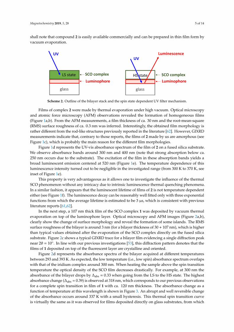
Recently, we have carried out a deep investigation of the SCO complex 1 in different forms (single crystal, powder and thin film) and it appears to us as an excellent candidate to perform this study [51–58]. Indeed, this compound is a rare example of sublimable spin crossover complexes (globally neutral with low molecular weight), therefore SCO films of complex 1 can be prepared by vacuum evaporation on a substrate. In these films, the SCO phenomenon is similar to the one observed in the solid state: they display a full and abrupt transition around 337 K (64 °C) with a narrow hysteresis loop of ca. 1 K width. These films can potentially be used for optical applications since sub-micrometric layers of 1 are transparent in the visible and near-infrared spectral ranges in both spin states. In addition, when using an appropriate annealing procedure they form a homogenous nano-crystalline texture with smooth surface topography, hence not only absorption, but also scattering losses are very small. On the other hand, complex 1 in the LS state absorbs much more UV light than in the HS state. For example, at 318 nm the absorption coefficient (3 × 104 cm−1), related most probably to a singlet-singlet charge transfer transition between the metal center and the ligand, vanishes completely when switching the complex from the LS (1A) to the HS (5T) state. It is this huge optical density change that we would like take advantage of in modulating the intensity of the luminescence. Indeed, complex 1 in the LS state will strongly absorb and thus block UV irradiation. Whereas, in the HS state, it is quasi-transparent in the UV, so the main part of the irradiation will reach and excite the luminophore layer underneath (Scheme 2). Therefore, we expect that the luminescent intensity of the material with complex 1 in the LS state should be substantially weaker than with 1 in HS state. Obviously, this strategy implies that UV irradiation should be used to excite the luminescence emission. To this aim we have chosen the luminescent complex Ir(PPy)3, which is probably one of the most studied molecules in the Organic Light-Emitting Diode (OLED) field and deep investigations have been performed in order to understand its luminescent properties [59–62]. This compound is well known to absorb light in the UV between 250 and 320 nm, which has been assigned to singlet-singlet π-π* transitions of the ligand [62]. It is well documented that its excitation under UV irradiation leads to a prominent green emission centered at 515 nm, which is attributed to the radiative decay of the triplet 3MLCT (metal ligand charge transfer) state to the ground state [62]. As the SCO film is transparent in visible region, this green emission of complex 2 will not significantly be attenuated by the presence of a thin layer of complex 1. Besides these
Complex 1 Complex 2
Scheme 1. Chemical structure of the iron and iridium complexes.
Recently, we have carried out a deep investigation of the SCO complex 1 in different forms(single crystal, powder and thin film) and it appears to us as an excellent candidate to perform thisstudy [51–58]. Indeed, this compound is a rare example of sublimable spin crossover complexes(globally neutral with low molecular weight), therefore SCO films of complex 1 can be preparedby vacuum evaporation on a substrate. In these films, the SCO phenomenon is similar to the oneobserved in the solid state: they display a full and abrupt transition around 337 K (64 ◦C) with a narrowhysteresis loop of ca. 1 K width. These films can potentially be used for optical applications sincesub-micrometric layers of 1 are transparent in the visible and near-infrared spectral ranges in bothspin states. In addition, when using an appropriate annealing procedure they form a homogenousnano-crystalline texture with smooth surface topography, hence not only absorption, but also scatteringlosses are very small. On the other hand, complex 1 in the LS state absorbs much more UV light than inthe HS state. For example, at 318 nm the absorption coefficient (3 × 104 cm−1), related most probably toa singlet-singlet charge transfer transition between the metal center and the ligand, vanishes completelywhen switching the complex from the LS (1A) to the HS (5T) state. It is this huge optical densitychange that we would like take advantage of in modulating the intensity of the luminescence. Indeed,complex 1 in the LS state will strongly absorb and thus block UV irradiation. Whereas, in the HSstate, it is quasi-transparent in the UV, so the main part of the irradiation will reach and excite theluminophore layer underneath (Scheme 2). Therefore, we expect that the luminescent intensity ofthe material with complex 1 in the LS state should be substantially weaker than with 1 in HS state.Obviously, this strategy implies that UV irradiation should be used to excite the luminescence emission.To this aim we have chosen the luminescent complex Ir(PPy)3, which is probably one of the moststudied molecules in the Organic Light-Emitting Diode (OLED) field and deep investigations have beenperformed in order to understand its luminescent properties [59–62]. This compound is well knownto absorb light in the UV between 250 and 320 nm, which has been assigned to singlet-singlet π-π*transitions of the ligand [62]. It is well documented that its excitation under UV irradiation leads to aprominent green emission centered at 515 nm, which is attributed to the radiative decay of the triplet3MLCT (metal ligand charge transfer) state to the ground state [62]. As the SCO film is transparent invisible region, this green emission of complex 2 will not significantly be attenuated by the presence of athin layer of complex 1. Besides these favorable and very well documented luminescent properties, we
Magnetochemistry 2019, 5, 28 5 of 14
shall note that compound 2 is easily available commercially and can be prepared in thin film form byvacuum evaporation.
Magnetochemistry 2019, 5, 28 5 of 14
favorable and very well documented luminescent properties, we shall note that compound 2 is easily available commercially and can be prepared in thin film form by vacuum evaporation.
Scheme 2. Outline of the bilayer stack and the spin state dependent UV filter mechanism.
Films of complex 2 were made by thermal evaporation under high vacuum. Optical microscopy and atomic force microscopy (AFM) observations revealed the formation of homogeneous films (Figure 1a,b). From the AFM measurements, a film thickness of ca. 30 nm and the root-mean-square (RMS) surface roughness of ca. 0.3 nm was inferred. Interestingly, the obtained film morphology is rather different from the rod-like structures previously reported in the literature [62]. However, GIXRD measurements indicate that, contrary to those reports, the films of 2 made by us are amorphous (see Figure 1c), which is probably the main reason for the different film morphologies.
Figure 1d represents the UV-vis absorbance spectrum of the film of 2 on a fused silica substrate. We observe absorbance bands around 300 nm and 400 nm (note that strong absorption below ca. 250 nm occurs due to the substrate). The excitation of the film in these absorption bands yields a broad luminescent emission centered at 520 nm (Figure 1e). The temperature dependence of this luminescence intensity turned out to be negligible in the investigated range (from 300 K to 370 K, see inset of Figure 1e).
This property is very advantageous as it allows one to investigate the influence of the thermal SCO phenomenon without any intricacy due to intrinsic luminescence thermal quenching phenomena. In a similar fashion, it appears that the luminescent lifetime of films of 2 is not temperature dependent either (see Figure 1f). The luminescence decay can be reasonably well fitted only with three exponential functions from which the average lifetime is estimated to be 3 µs, which is consistent with previous literature reports [61,62].
glass
Luminophore SCO complex
Luminescence
HS state
UV
glass
Luminophore SCO complexLS state
UV
Scheme 2. Outline of the bilayer stack and the spin state dependent UV filter mechanism.
Films of complex 2 were made by thermal evaporation under high vacuum. Optical microscopyand atomic force microscopy (AFM) observations revealed the formation of homogeneous films(Figure 1a,b). From the AFM measurements, a film thickness of ca. 30 nm and the root-mean-square(RMS) surface roughness of ca. 0.3 nm was inferred. Interestingly, the obtained film morphology israther different from the rod-like structures previously reported in the literature [62]. However, GIXRDmeasurements indicate that, contrary to those reports, the films of 2 made by us are amorphous (seeFigure 1c), which is probably the main reason for the different film morphologies.
Figure 1d represents the UV-vis absorbance spectrum of the film of 2 on a fused silica substrate.We observe absorbance bands around 300 nm and 400 nm (note that strong absorption below ca.250 nm occurs due to the substrate). The excitation of the film in these absorption bands yields abroad luminescent emission centered at 520 nm (Figure 1e). The temperature dependence of thisluminescence intensity turned out to be negligible in the investigated range (from 300 K to 370 K, seeinset of Figure 1e).
This property is very advantageous as it allows one to investigate the influence of the thermalSCO phenomenon without any intricacy due to intrinsic luminescence thermal quenching phenomena.In a similar fashion, it appears that the luminescent lifetime of films of 2 is not temperature dependenteither (see Figure 1f). The luminescence decay can be reasonably well fitted only with three exponentialfunctions from which the average lifetime is estimated to be 3 µs, which is consistent with previousliterature reports [61,62].
In the next step, a 107 nm thick film of the SCO complex 1 was deposited by vacuum thermalevaporation on top of the luminophore layer. Optical microscopy and AFM images (Figure 2a,b),clearly show the change of surface morphology and reveal the formation of some islands. The RMSsurface roughness of the bilayer is around 3 nm (for a bilayer thickness of 30 + 107 nm), which is higherthan typical values obtained after the evaporation of the SCO complex directly on the fused silicasubstrate. Figure 2c shows a typical GIXRD trace for a bilayer film evidencing a single diffraction peaknear 2θ = 10◦. In line with our previous investigations [53], this diffraction pattern denotes that thefilms of 1 deposited on top of the fluorescent layer are crystalline and oriented.
Figure 2d represents the absorbance spectra of the bilayer acquired at different temperaturesbetween 293 and 393 K. As expected, the low temperature (i.e., low spin) absorbance spectrum overlapswith that of the iridium complex around 300 nm. When heating the sample above the spin transitiontemperature the optical density of the SCO film decreases drastically. For example, at 300 nm theabsorbance of the bilayer drops by ∆abs = 0.33 when going from the LS to the HS state. The highestabsorbance change (∆abs = 0.39) is observed at 318 nm, which corresponds to our previous observationsfor a complete spin transition in film of 1 with ca. 120 nm thickness. The absorbance change as afunction of temperature at this wavelength is shown in Figure 3. An abrupt and well reversible changeof the absorbance occurs around 337 K with a small hysteresis. This thermal spin transition curveis virtually the same as it was observed for films deposited directly on glass substrates, from which

Magnetochemistry 2019, 5, 28 6 of 14
we can infer that the SCO properties of complex 1 are not significantly modified by the presence ofthe luminescent film underneath. This result corroborates our previous observations [53] about thenegligible role of the nature of the substrate on the properties of films of 1. Figure 2e depicts theluminescence emission spectra of the bilayer film at different temperatures. Overall, these spectraclosely resemble that of the neat fluorescent film, despite the excimer shoulder around 600 nm which issomewhat more intense. Remarkably, in the bilayer, the luminescence intensity changes drasticallybetween ca. 333 and 350 K, in harsh contrast with the quasi-constant luminescent intensity of thefilm of complex 2 in the same temperature range. On the other hand, the luminescent lifetime of 2is not significantly altered by the presence of the SCO layer and the decay curves remain virtuallytemperature independent (Figure 2f).Magnetochemistry 2019, 5, 28 6 of 14
Figure 1. Characterization of films of Ir(PPy)3. Typical optical microscopy (a) and AFM topography (b) images, (c) GIXRD pattern, (d) UV-vis absorption spectrum, (e) luminescence emission spectrum (excitation: 300 nm, high pass filter λemission > 400 nm) and its temperature dependence in insert and (f) variable temperature luminescent decay curves (excitation: 303 nm, emission 520 nm) recorded over the first 800 ns of the decay.
In the next step, a 107 nm thick film of the SCO complex 1 was deposited by vacuum thermal evaporation on top of the luminophore layer. Optical microscopy and AFM images (Figure 2a,b), clearly show the change of surface morphology and reveal the formation of some islands. The RMS surface roughness of the bilayer is around 3 nm (for a bilayer thickness of 30 + 107 nm), which is higher than typical values obtained after the evaporation of the SCO complex directly on the fused silica substrate. Figure 2c shows a typical GIXRD trace for a bilayer film evidencing a single diffraction peak near 2θ = 10°. In line with our previous investigations [53], this diffraction pattern denotes that the films of 1 deposited on top of the fluorescent layer are crystalline and oriented.
(c)
10 15 20 25 30 35 400
50
100
150
200
250
300
350
Inte
nsity
(a.u
)
2θ (°)
(b) (a)
1 µm
(e)
(d)
(f)
Figure 1. Characterization of films of Ir(PPy)3. Typical optical microscopy (a) and AFM topography(b) images, (c) GIXRD pattern, (d) UV-vis absorption spectrum, (e) luminescence emission spectrum(excitation: 300 nm, high pass filter λemission > 400 nm) and its temperature dependence in insert and(f) variable temperature luminescent decay curves (excitation: 303 nm, emission 520 nm) recorded overthe first 800 ns of the decay.

Magnetochemistry 2019, 5, 28 7 of 14
Magnetochemistry 2019, 5, 28 8 of 14
AFM/quartz balance (~800 nm) and XRD/UV-absorbance (~500 nm) can be explained by a formation of an SCO film with a partial crystallinity and thus partial spin transition.
Figure 2. Characterization of bilayer films of Ir(PPy)3 and [Fe(HB(tz)3)2]. Typical optical microscopy (a) and AFM topography (b) images, (c) GIXRD pattern, (d) variable temperature UV absorption spectra, (e) variable temperature luminescence emission spectra (excitation: 300 nm, high pass filter λemission > 400 nm) and (f) variable temperature luminescence decay curves (excitation: 303 nm, emission 520 nm) recorded over the first 800 ns of the decay.
1 µm
(a)
(c)
10 15 20 25 30 35 400
10000
20000
30000
40000
50000
60000
Inte
nsity
(cou
nts)
2θ (°)
(e)
450 500 550 600 650 7000
1
2
3
4
5 370 K (HS)
Lum
ines
cenc
e in
tens
ity (a
.u)
Wavelength (nm)
300 K (LS)
(f)
(d)
(b)
Figure 2. Characterization of bilayer films of Ir(PPy)3 and [Fe(HB(tz)3)2]. Typical optical microscopy(a) and AFM topography (b) images, (c) GIXRD pattern, (d) variable temperature UV absorptionspectra, (e) variable temperature luminescence emission spectra (excitation: 300 nm, high pass filterλemission > 400 nm) and (f) variable temperature luminescence decay curves (excitation: 303 nm,emission 520 nm) recorded over the first 800 ns of the decay.

Magnetochemistry 2019, 5, 28 8 of 14Magnetochemistry 2019, 5, 28 9 of 14
Figure 3. Luminescence modulation: Normalized temperature dependence (over two heating-cooling cycles) of the UV absorbance (318 nm) and of the luminescence intensity (excitation: 300 nm, emission 520 nm) for a bilayer film of 30 + 107 nm thickness.
The results obtained with the three different SCO film thickness are summarized in Figure 4. The spin transition curves are comparable for each sample in terms of spin transition temperatures, though slight differences in the shape of the curves can be depicted (Figure 4a).
Figure 4. Correlation between the absorbance of [Fe(HB(tz)3)2] and the luminescence of Ir(PPy)3. (a) Normalized temperature dependence of the UV absorbance (at 330 nm) for three bilayer samples with different SCO layer thicknesses (107, 214 and 765 nm). (b) Absorbance changes associated with the SCO for different SCO film thickness. (c) Normalized temperature dependence of the luminescence intensity for different SCO film thickness. (d) Plot of the luminescence intensity ratio vs. the UV transmittance ratio between the HS and LS states. The corresponding SCO film thickness is also shown.
(c) (d)
(b) (a)
Figure 3. Luminescence modulation: Normalized temperature dependence (over two heating-coolingcycles) of the UV absorbance (318 nm) and of the luminescence intensity (excitation: 300 nm, emission520 nm) for a bilayer film of 30 + 107 nm thickness.
In Figure 3 we compare the temperature dependences of the absorbance (λ = 318 nm) and theluminescence intensity (λexcitation = 300 nm, λemission = 520 nm) for the bilayer film over two thermalcycles between 300 and 380 K. The drastic increase in the intensity of the luminescence above ca. 335 Kcan be unambiguously attributed to the spin transition phenomena (note that the slight differencein SCO temperatures observed in absorbance and fluorescence experiments can be attributed to thedifferent heating-cooling equipment used to control the temperature of the sample). It may be worthto note also the good overall reproducibility of these measurements over successive heating-coolingcycles, despite a small, continuous decrease of the luminescence intensity due to a slow (hours scale)photo-bleaching phenomena.
At this point, we can conclude that the intrinsic properties of each compound, luminescentand SCO, are not significantly affected by the presence of each other. Yet, we observe a reversiblemodulation of the luminescence intensity at the SCO, which we can attribute to the different UVabsorbance properties of the iron complex in its two different spin states: either fully transmitting (HSstate) or partially blocking (LS state) the light beam used for luminescence excitation. This hypothesisis supported by the fact the promotion of the iron complex from the LS to the HS state doubles theluminescence intensity of our bilayer film, while its UV transmittance (λ = 300 nm) doubles as well.In the same time, the luminescence spectral shape and lifetime remain temperature and spin-stateindependent, which is also fully compatible with the proposed mechanism.
In order to obtain further proof for this explanation and to verify the scalability of the luminescencemodulation amplitude, we have prepared two other bilayer samples with SCO layers of ca. 200 nmand 800 nm thickness. For the thickest sample, we had to overcome a difficulty concerning theformation of crystalline films. As mentioned above, oriented, crystalline SCO films of 1 are obtainedby annealing the pristine evaporated film in humid air with ca. 80% relative humidity. However, thismethod is not fully efficient when the thickness of the film is greater than ca. 200 nm. To preparethe sample with 800 nm of SCO thickness, we have therefore first deposited an SCO layer of 200 nmon top of a 30 nm layer of 2 and we have annealed the film as usual. Then, we have put back theannealed sample in the evaporator to grow an additional 600 nm thick layer of SCO molecules. Postdeposition AFM measurements agree with the value indicated by the quartz balance and confirmthat the thickness of the SCO layer is around 800 nm. X-ray diffraction on this thick film revealedonly one significant diffraction peak near 2θ = 10◦ indicating crystallinity and preferred orientation.However, the XRD intensity and the UV absorbance of this film correspond to a film thickness of onlyca. 500 nm. The significant difference between the film thickness values of AFM/quartz balance (~800nm) and XRD/UV-absorbance (~500 nm) can be explained by a formation of an SCO film with a partialcrystallinity and thus partial spin transition.

Magnetochemistry 2019, 5, 28 9 of 14
The results obtained with the three different SCO film thickness are summarized in Figure 4. Thespin transition curves are comparable for each sample in terms of spin transition temperatures, thoughslight differences in the shape of the curves can be depicted (Figure 4a).
Magnetochemistry 2019, 5, x; doi: FOR PEER REVIEW www.mdpi.com/journal/magnetochemistry
Article Bilayer Thin Films That Combine Luminescent and Spin Crossover Properties for an Efficient and Reversible Fluorescence Switching
(a) (b)
(c) (d)
Figure 4. Correlation between the absorbance of [Fe(HB(tz)3)2] and the luminescence of Ir(PPy)3. (a) Normalized temperature dependence of the UV absorbance (at 330 nm) for three bilayer samples with different SCO layer thicknesses (107, 214 and 765 nm). (b) Absorbance changes associated with the SCO for different SCO film thickness. (c) Normalized temperature dependence of the luminescence intensity for different SCO film thickness. (d) Plot of the luminescence intensity ratio vs. the UV transmittance ratio between the HS and LS states. The corresponding SCO film thickness is also shown.
Figure 4. Correlation between the absorbance of [Fe(HB(tz)3)2] and the luminescence of Ir(PPy)3.(a) Normalized temperature dependence of the UV absorbance (at 330 nm) for three bilayer sampleswith different SCO layer thicknesses (107, 214 and 765 nm). (b) Absorbance changes associated withthe SCO for different SCO film thickness. (c) Normalized temperature dependence of the luminescenceintensity for different SCO film thickness. (d) Plot of the luminescence intensity ratio vs. the UVtransmittance ratio between the HS and LS states. The corresponding SCO film thickness is also shown.
In response to the absorbance change between the LS and HS states (∆ALH) (Figure 4b), animportant and reversible change of the luminescence intensity is also observed around 337 K (Figure 4c)in each bilayer sample. As expected, the extent of the luminescence modulation (IHS/ILS) increaseswith the thickness of the SCO layer. Changes from ca. 200% to 1300% (with reference to the roomtemperature value) from the thinnest to the thickest film are obtained (see Table 1). The most remarkablepoint, as illustrated both in Table 1 and Figure 4d, is the perfect one-to-one correlation between theluminescence modulation IHS/ILS in the film of 2 and the transmittance change TrHS/TrLS in the film of1. In other words, the UV transmittance of the SCO films gives an accurate scale of modification of theluminescence intensity of the Ir complex. This means that one can modulate the luminescence intensity‘at will’ by adjusting the thickness of the SCO layer. The results in Figure 4 provide irrefutable proofof the mechanism of luminescence modulation in the bottom layer, which occurs via the modulationof the exciting light intensity by the spin-state dependent UV absorbance of the top layer. (It may beworth to note that by changing the thickness of the luminescent layer, we possibly change the ratio ofmonomer/excimer emissions, which may alter the SCO modulation amplitude.)

Magnetochemistry 2019, 5, 28 10 of 14
Table 1. Comparison of the UV absorption and luminescent properties of bilayer films with differentthickness of SCO layers.
Thickness of SCO Layer∆Abs = AbsLS − AbsHS Trmax/Trmin at 300 nm Imax/Imin
At 300 nm At 318 nm
107 nm 0.331 0.392 2.14 1.95214 nm 0.582 0.755 3.82 3.59765 nm 1.118 1.204 13.05 13.12
We have also investigated if a closer contact between the SCO and luminescent molecules ina mixed film could allow for an efficient luminescence modulation. Indeed, a few examples in theliterature demonstrate that a luminescent molecule can interact strongly and selectively with SCOcomplexes in either the low or the high spin state [18]. Obviously, the probability of an efficientelectronic energy transfer between the triplet emitting state of the luminophore and the week singletd-d absorption band of the LS complex is extremely low. Nevertheless, one might expect a significantamplification or quenching of the luminescence emission via ‘environmental effects’ related to thestructural changes which accompany the SCO. In any case (i.e., electronic or structural effects), aclose proximity between the two types of molecules is required. To investigate this possibility weco-evaporated the SCO and luminescent molecules to form mixed films with ca. 12%, 15% and21% nominal luminophore content. Overall, we observed that for increasing doping levels the SCOproperties of the films degrade and the luminescence modulation thus becomes negligible. Figure 5summarizes the results obtained for the lowest doping (12%). As shown in Figure 5a, the X-raydiffraction spectrum of the doped film can be ascribed to an amorphous/semi-crystalline film withoutany particular texture. This means that the presence of the luminophore molecules within the depositinhibit the recrystallization of the SCO film. Consequently, the spin transition curves obtained fromvariable temperature absorbance measurements on the mixed films (Figure 5b) are very different fromwhat we generally observe for the pure films of 1. In particular, the absorbance change associated withthe SCO is very low (∆A = 0.057); it is approximately ten times smaller than what is expected for apure SCO film with the same thickness. As it can be expected, we observe thus only a very weak effectof the spin transition on the luminescence emission intensity ratio (IHS/ILS = 1.2) (Figure 5b).
Magnetochemistry 2019, 5, 28 11 of 14
Figure 5. Characterization of a [Fe(HB(tz)3)2] film doped with 12% Ir(PPy)3. (a) Representative GIXRD pattern. The inset shows the outline of the sample. (b) Temperature dependence of the UV absorbance (318 nm) and of the luminescence intensity (excitation: 300 nm, emission 520 nm) on heating and cooling.
4. Conclusions
In this manuscript, we outlined an efficient approach to fabricate hybrid materials, which combine luminescent and spin crossover molecules in a bilayer film stack. Crucially, we have shown that the intrinsic properties of the two layers were not affected by the presence of the other. Yet, the luminescence intensity reversibly increased when the spin crossover molecules were switched from the LS to the HS state in the uppermost layer. We found a strict one-to-one correlation between the luminescence modulation (IHS/ILS) and the UV transmittance change with the spin transition (TrHS/TrLS). In the same time, neither the spectral shape nor the decay time of the luminescence emission were altered. These results prove that the origin of the luminescence modulation is a direct result of the different attenuation of the exciting UV light in the two spin states. These results also show that luminescence can be a very sensitive indicator of the thermally induced spin transition, which could be exploited for micro- and nanoscale thermal sensing and imaging applications [21,63,64].
Author Contributions: ACB, XT, LR and GM carried out sample synthesis and characterization. All authors contributed to the conceptualization, data analysis and writing the paper.
Funding: This work was funded by the Région Occitanie (Contract No. 15050450), by the European Commission through the SPINSWITCH project (H2020-MSCA-RISE-2016, Grant Agreement No. 734322), the EMERGENCE@INC2018 project (LR) and by the Louisiana State University LSAMP International Research Experience Project for Undergraduates (NSF Grant #1560390).
Conflicts of Interest: The authors declare no conflict of interest.
References
1. Amendola, V.; Di Casa, M.; Fabbrizzi, L.; Licchelli, M.; Mangano, C.; Pallavicini, P.; Poggi, A. Mechanical switches of fluorescence. J. Incl. Phenom. Macrocycl. Chem. 2001, 41, 13–18.
2. Pischel, U. Molecular switches as platforms for information processing. Chimia 2014, 68, 505–511. 3. Cheng, Y.; Jie, W.; Chao, G.; Xin-Ge, L.; Bin, D.; Yan, L. A facile water-stable MOF-based “off-on”
fluorescent switch for label-free detection of dopamine in biological fluid. J. Mater. Chem. B 2017, 5, 2524–2535.
4. Yang, L.; Jianping, W.; Liang, Y.; Cheng, Z.; Ruilong, Z.; Zhongping, Z.; Bianhua, L.; Changlong, J. Fluorescent paper sensor fabricated by carbazole-based probes for dual visual detection of Cu2+ and gaseous H2S. RSC Adv. 2016, 6, 5638456391.
10 15 20 25 30 35 400
200
400
600
800
1000
1200
1400
1600
Inte
nsity
(cou
nts)
2θ (°)
Spin crossover complex
(a) (b)
Figure 5. Characterization of a [Fe(HB(tz)3)2] film doped with 12% Ir(PPy)3. (a) RepresentativeGIXRD pattern. The inset shows the outline of the sample. (b) Temperature dependence of the UVabsorbance (318 nm) and of the luminescence intensity (excitation: 300 nm, emission 520 nm) on heatingand cooling.

Magnetochemistry 2019, 5, 28 11 of 14
4. Conclusions
In this manuscript, we outlined an efficient approach to fabricate hybrid materials, which combineluminescent and spin crossover molecules in a bilayer film stack. Crucially, we have shown that theintrinsic properties of the two layers were not affected by the presence of the other. Yet, the luminescenceintensity reversibly increased when the spin crossover molecules were switched from the LS to theHS state in the uppermost layer. We found a strict one-to-one correlation between the luminescencemodulation (IHS/ILS) and the UV transmittance change with the spin transition (TrHS/TrLS). In thesame time, neither the spectral shape nor the decay time of the luminescence emission were altered.These results prove that the origin of the luminescence modulation is a direct result of the differentattenuation of the exciting UV light in the two spin states. These results also show that luminescencecan be a very sensitive indicator of the thermally induced spin transition, which could be exploited formicro- and nanoscale thermal sensing and imaging applications [21,63,64].
Author Contributions: A.-C.B., X.T., L.R. and G.M. carried out sample synthesis and characterization. All authorscontributed to the conceptualization, data analysis and writing the paper.
Funding: This work was funded by the Région Occitanie (Contract No. 15050450), by the EuropeanCommission through the SPINSWITCH project (H2020-MSCA-RISE-2016, Grant Agreement No. 734322),the EMERGENCE@INC2018 project (LR) and by the Louisiana State University LSAMP International ResearchExperience Project for Undergraduates (NSF Grant #1560390).
Conflicts of Interest: The authors declare no conflict of interest.
References
1. Amendola, V.; Di Casa, M.; Fabbrizzi, L.; Licchelli, M.; Mangano, C.; Pallavicini, P.; Poggi, A. Mechanicalswitches of fluorescence. J. Incl. Phenom. Macrocycl. Chem. 2001, 41, 13–18. [CrossRef]
2. Pischel, U. Molecular switches as platforms for information processing. Chimia 2014, 68, 505–511. [CrossRef][PubMed]
3. Cheng, Y.; Jie, W.; Chao, G.; Xin-Ge, L.; Bin, D.; Yan, L. A facile water-stable MOF-based “off-on” fluorescentswitch for label-free detection of dopamine in biological fluid. J. Mater. Chem. B 2017, 5, 2524–2535. [CrossRef]
4. Yang, L.; Jianping, W.; Liang, Y.; Cheng, Z.; Ruilong, Z.; Zhongping, Z.; Bianhua, L.; Changlong, J. Fluorescentpaper sensor fabricated by carbazole-based probes for dual visual detection of Cu2+ and gaseous H2S. RSCAdv. 2016, 6, 5638456391. [CrossRef]
5. Xin, L.; Tao, R.R.; Hong, L.-J.; Cheng, J.; Jiang, Q.; Lu, Y.-M.; Liao, M.-H.; Ye, W.-F.; Lu, N.-N.; Han, F.; et al.Visualizing peroxynitrite fluxes in endothelial cells reveals the dynamic progression of brain vascular injury.J. Am. Chem. Soc. 2015, 137, 12296–12303.
6. Marc, V.; Benet, M.; Mena, S.; Rabih, O.; Al-Kaysi, J.H.; Guirado, G. Multistimuli-responsive fluorescentswitches based on spirocyclic meisenheimer compounds: Smart molecules for the design of optical probesand electrochromic materials. J. Org. Chem. 2018, 83, 9166–9167.
7. Bao-Hua, D.; Chen, Y.-L. Recent progress in organic mechanoluminescent materials. Chin. Chem. Lett. 2018,29, 245–251.
8. Jaume, G.-A.; Swaminathan, S.; Sortino, S.; Raymo, F.M. Plasmonic activation of a fluorescentcarbazole-oxazine switch. Chem. Eur. J. 2014, 20, 10276–10284.
9. Gareau, D.; Desrosiers, A.; Vallee-Belisle, A. Programmable quantitative DNA nanothermometers. Nano Lett.2016, 16, 3976–3981. [CrossRef] [PubMed]
10. Bousseksou, A.; Molnar, G.; Salmon, L.; Nicolazzi, N. Molecular spin crossover phenomenon: Recentachievements and prospects. Chem. Soc. Rev. 2011, 40, 3313–3335. [CrossRef] [PubMed]
11. Garcia, Y.; Gutlich, P. Thermal spin crossover in Mn(II), Mn(III), Cr(II), and Co(III) coordination compounds.Top. Curr. Chem. 2004, 234, 49–62.
12. Halcrow, M.A. Spin crossover materials. In Properties and Applications; Halcrow, M.A., Ed.; John Wiley &Sons Ltd.: Hoboken, NJ, USA, 2013.
13. Bousseksou, A. (Ed.) Spin crossover phenomenon. C. R. Chim. 2018, 12, 1055–1300.

Magnetochemistry 2019, 5, 28 12 of 14
14. Decurtins, S.; Gütlich, P.; Köhler, C.P.; Spiering, H.; Hauser, A. Light-induced excited spin state trapping ina transition-metal complex: The hexa-1-propyltetrazole-iron (II) tetrafluoroborate spin-crossover system.Chem. Phys. Lett. 1984, 105, 1–4. [CrossRef]
15. Bousseksou, A.; Boukheddaden, K.; Goiran, M.; Consejo, C.; Boillot, M.L.; Tuchagues, J.P. Dynamic responseof the spin-crossover solid Co(H2(fsa)2en)(py)2 to a pulsed magnetic field. Phys. Rev. B Cover. Condens.Matter. Mater. Phys. 2002, 65. [CrossRef]
16. Kuppusamy, S.K.; Ruben, M. Emerging trends in spin crossover (SCO) based functional materials anddevices. Coord. Chem. Rev. 2017, 346, 176–205.
17. Molnar, G.; Sylvain, R.; Salmon, L.; Nicolazzi, W.; Bousseksou, A. Spin crossover nanomaterials: Fromfundamental concepts to devices. Adv. Mater. 2018, 30. [CrossRef]
18. Shepherd, H.; Carlos, J.; Quintero, M.; Molnar, G.; Salmon, L.; Bousseksou, A. Luminescent Spin-CrossoverMaterials; John Wiley & Sons Ltd.: Hoboken, NJ, USA, 2013; pp. 347–373.
19. Marfunin, A.S. Spectroscopy, Luminescence, and Radiation Centers in Minerals; Springer: Berlin, Germany,1979; 352p.
20. Suleimanov, I.; Molnar, G.; Salmon, L.; Bousseksou, A. Near-infrared luminescence switching in aspin-crossover polymer nanocomposite. Eur. J. Inorg. Chem. 2017, 3446–3451. [CrossRef]
21. Salmon, L.; Molnar, G.; Zitouni, D.; Quintero, C.; Bergaud, C.; Micheau, J.-C.; Bousseksou, A. A novelapproach for fluorescent thermometry and thermal imaging purposes using spin crossover nanoparticles.J. Mater. Chem. 2010, 20, 5499–5503. [CrossRef]
22. Piguet, C.; Rivara-Minten, E.; Hopfgartner, G.; Buenzli, J.-C.G. Molecular magnetism and iron(II) spin-stateequilibrium as structural probes in heterodinuclear d–f complexes. Helv. Chim. Acta 1995, 78, 1651–1672.[CrossRef]
23. Wang, C.F.; Li, R.F.; Chen, X.Y.; Wei, R.J.; Zheng, L.S.; Tao, J. Synergetic spin crossover and fluorescence inone-dimensional hybrid complexes. Angew. Chem. Int. Ed. 2015, 54, 1574–1577. [CrossRef]
24. Lochenie, C.; Wagner, K.G.; Karg, M.; Weber, B. Modulation of the ligand-based fluorescence of 3d metalcomplexes upon spin state change. J. Mater. Chem. C 2015, 3, 7925–7935. [CrossRef]
25. Wang, C.F.; Sun, M.J.; Guo, Q.J.; Cao, Z.X.; Zheng, L.S.; Tao, J. Multiple correlations between spin crossoverand fluorescence in a dinuclear compound. Chem. Commun. 2016, 52, 14322–14325. [CrossRef] [PubMed]
26. Estrader, M.; Salinas Uber, J.; Barrios, L.A.; Garcia, J.; Lloyd-Williams, P.; Roubeau, O.; Teat, S.J.; Aromi, G.A magneto-optical molecular device: Interplay of spin crossover, luminescence, photomagnetism, andphotochromism. Angew. Chem. Int. Ed. 2017, 56, 15622–15627. [CrossRef] [PubMed]
27. Kuppusamy Senthil, K.; Salitros, I.; Moreno-Pineda, E.; Ruben, M. Spacer type mediated tunable spincrossover (SCO) characteristics of pyrene decorated 2,6-bis(pyrazol-1-yl)pyridine (bpp) based Fe(II) molecularspintronic modules. Dalton Trans. 2017, 46, 9765–9768.
28. Schaefer, B.; Bauer, T.; Faus, I.; Wolny, J.A.; Dahms, F.; Fuhr, O.; Lebedkin, S.; Wille, H.-C.; Schlage, K.;Chevalier, K.; et al. A luminescent Pt2Fe spin crossover complex. Dalton Trans. 2017, 46, 2289–2302.[CrossRef]
29. Wang, J.-L.; Qiang, L.; Yin-Shan, M.; Xin Liu, H.; Zheng, Q.S.; Chun-Ying, D.; Tao, L. Fluorescence modulationvia photoinduced spin crossover switched energy transfer from fluorophores to FeII ions. Chem. Sci. 2018, 9,2892–2897. [CrossRef]
30. Lochenie, C.; Schoetz, K.; Panzer, F.; Kurz, H.; Maier, B.; Puchtler, F.; Agarwal, S.; Koehler, A.; Weber, B.Spin-crossover iron(II) coordination polymer with fluorescent properties: Correlation between emissionproperties and spin state. J. Am. Chem. Soc. 2018, 140, 700–709. [CrossRef]
31. Hasegawa, M.; Franz, R.; Hara, T.; Kikuchi, Y.; Fukuda, Y.; Okubo, J.; Hoshi, T.; Linert, W. Fluorescencespectra of Fe(II) spin crossover complexes with 2,6-bis(benzimidazole-2’-yl)pyridine. Chem. Phys. 2002, 277,21–30. [CrossRef]
32. Garcia, Y.; Robert, F.; Naik, A.D.; Zhou, G.; Tinant, B.; Robeyns, K.; Michotte, S.; Piraux, L. Spin transitioncharted in a fluorophore-tagged thermochromic dinuclear iron(II) complex. J. Am. Chem. Soc. 2011, 133,15850–15853. [CrossRef]
33. Gonzalez-Prieto, R.; Fleury, B.; Schramm, F.; Zoppellaro, G.; Chandrasekar, R.; Fuhr, O.; Lebedkin, S.;Kappes, M.; Ruben, M. Tuning the spin-transition properties of pyrene-decorated 2,6-bispyrazolylpyridinebased Fe(II) complexes. Dalton Trans. 2011, 40, 7564–7570. [CrossRef]

Magnetochemistry 2019, 5, 28 13 of 14
34. Santoro, A.; Kershaw Cook, L.J.; Kulmaczewski, R.; Barrett, S.A.; Cespedes, O.; Halcrow, M.A. Iron(II)complexes of tridentate indazolylpyridine ligands: Enhanced spin-crossover hysteresis and ligand-basedfluorescence. Inorg. Chem. 2015, 54, 682–693. [CrossRef]
35. Carine, E.; Piguet, C.; Bunzli, J.-C.G.; Hopfgartner, G. High-spin iron(II) as a semitransparent partner fortuning europium(III) luminescence in heterodimetallic d-f complexes. Chem. Eur. J. 2001, 7, 3014–3024.
36. Piguet, C.; Rivara-Minten, E.; Bernardinelli, G.; Buenzli, J.-C.G.; Hopfgartner, G. Noncovalent lanthanidepodates with predetermined physicochemical properties: Iron(II) spin-state equilibria in self-assembledheterodinuclear d-f supramolecular complexes. J. Chem. Soc. Dalton Trans. 1997, 421–433. [CrossRef]
37. Quintero, C.M.; Gural’skiy, I.A.; Salmon, L.; Molnar, G.; Bergaud, C.; Bousseksou, A. Soft lithographicpatterning of spin crossover complexes. Part 1: Fluorescent detection of the spin transition in singlenanoobjects. J. Mater. Chem. 2012, 22, 3745–3751. [CrossRef]
38. Matsuda, M.; Hikaru, I.; Hiroyuki, T. Electroluminescence quenching caused by a spin-crossover transition.Chem. Lett. 2008, 37, 374–375. [CrossRef]
39. Matsuda, M.; Hikaru, I.; Hiroyuki, T. Reproducible on-off switching of the light emission from theelectroluminescent device containing a spin crossover complex. Thin Solid Films 2008, 517, 1465–1467.[CrossRef]
40. Matsuda, M.; Keita, K.; Ryoma, U.; Nobuaki, K.; Hiroyuki, T. Characteristics of organic light-emitting devicesconsisting of dye-doped spin crossover complex films. Thin Solid Films 2013, 531, 451–453. [CrossRef]
41. Gural’skiy, I.A.; Quintero, C.M.; Abdul-Kader, K.; Lopes, M.; Bartual-Murgui, C.; Salmon, L.; Zhao, P.;Molnar, G.; Astruc, D.; Bousseksou, A. Detection of molecular spin-state changes in ultrathin films byphotonic methods. J. Nanophotonics 2012, 6, 63517. [CrossRef]
42. Tovee, C.A.; Kilner, C.A.; Thomas, J.A.; Halcrow, M.A. Co-crystallizing two functional complex molecules ina terpyridine embrace lattice. Cryst. Eng. Comm. 2009, 11, 2069–2077. [CrossRef]
43. Kershaw Cook, L.J.; Halcrow, M.A. Doping ruthenium complexes into a molecular spin-crossover material.Polyhedron 2015, 87, 91–97. [CrossRef]
44. Matsukizono, H.; Keita, K.; Nobuo, K. Self-assembly-directed spin conversion of iron(II) 1,2,4-triazolecomplexes in solution and their effect on photorelaxation processes of fluorescent counter ions. Chem. Lett.2008, 37, 446–447. [CrossRef]
45. Titos-Padilla, S.; Herrera, J.M.; Chen, X.-W.; Delgado, J.J.; Colacio, E. Bifunctional Hybrid SiO2 NanoparticlesShowing Synergy between Core Spin Crossover and Shell Luminescence Properties. Angew. Chem. Int. Ed.2011, 50, 3290–3293. [CrossRef]
46. Suleimanov, I.; Kraieva, O.; Molnar, G.; Salmon, L.; Bousseksou, A. Enhanced luminescence stability witha Tb-spin crossover nanocomposite for spin state monitoring. Chem. Commun. 2015, 51, 15098–15101.[CrossRef] [PubMed]
47. Suleimanov, I.; Kraieva, O.; Sanchez Costa, J.; Fritsky, I.O.; Molnar, G.; Salmon, L.; Bousseksou, A. Electroniccommunication between fluorescent pyrene excimers and spin crossover complexes in nanocompositeparticles. J. Mater. Chem. C 2015, 3, 5026–5032. [CrossRef]
48. Kraieva, O.; Suleimanov, I.; Molnar, G.; Salmon, L.; Bousseksou, A. CdTe quantum dot fluorescencemodulation by spin crossover. Magnetochemistry 2016, 2, 11. [CrossRef]
49. Luo, Y.-H.; Jing-Wen, W.; Wen, W.; Xiao-Tong, H.; Dan-Li, H.; Chen Chen, T.X.; Qiyue, S.; Bai-Wang, S.Bidirectional photoswitching via alternating NIR and UV irradiation on a core-shell UCNP-SCO nanosphere.ACS Appl. Mater. Interfaces 2018, 10, 16666–16673. [CrossRef]
50. Herrera, J.M.; Titos-Padilla, S.; Pope, S.J.A.; Berlanga, I.; Zamora, F.; Delgado, J.J.; Kamenev, K.V.; Wang, X.;Prescimone, A.; Brechin, E.K.; et al. Studies on bifunctional Fe(II)-triazole spin crossover nanoparticles:Time-dependent luminescence, surface grafting and the effect of a silica shell and hydrostatic pressure on themagnetic properties. J. Mater. Chem. C 2015, 3, 7819–7829. [CrossRef]
51. Rat, S.; Ridier, K.; Vendier, L.; Molnar, G.; Salmon, L.; Bousseksou, A. Solvatomorphism and structural-spincrossover property relationship in bis[hydrotris(1,2,4-triazol-1-yl)borate]iron(II). Cryst. Eng. Comm. 2017, 19,3271–3280. [CrossRef]
52. Shalabaeva, V.; Mikolasek, M.; Manrique-Juarez, M.D.; Bas, A.-C.; Rat, S.; Salmon, L.; Nicolazzi, W.; Molnar, G.;Bousseksou, A. Unprecedented size effect on the phase stability of molecular thin films displaying a spintransition. J. Phys. Chem. C 2017, 121, 25617–25621. [CrossRef]

Magnetochemistry 2019, 5, 28 14 of 14
53. Shalabaeva, V.; Rat, S.; Manrique-Juarez, M.D.; Bas, A.-C.; Vendier, L.; Salmon, L.; Molnar, G.; Bousseksou, A.Vacuum deposition of high-quality thin films displaying spin transition near room temperature. J. Mater.Chem. C 2017, 5, 4419–4425. [CrossRef]
54. Manrique-Juarez, M.D.; Mathieu, F.; Shalabaeva, V.; Cacheux, J.; Rat, S.; Nicu, L.; Leichle, T.; Salmon, L.;Molnar, G.; Bousseksou, A. A bistable microelectromechanical system actuated by spin-crossover molecules.Angew. Chem. Int. Ed. 2017, 56, 8074–8078. [CrossRef]
55. Ridier, K.; Rat, S.; Salmon, L.; Nicolazzi, W.; Molnar, G.; Bousseksou, A. Scan-rate and vacuum pressuredependence of the nucleation and growth dynamics in a spin-crossover single crystal: The role of latent heat.Phys. Chem. Chem. Phys. 2018, 20, 913–945. [CrossRef]
56. Shalabaeva, V.; Ridier, K.; Rat, S.; Manrique-Juarez, M.D.; Salmon, L.; Seguy, I.; Rotaru, A.; Molnar, G.;Bousseksou, A. Room temperature current modulation in large area electronic junctions of spin crossoverthin films. Appl. Phys. Lett. 2018, 112. [CrossRef]
57. Mikolasek, M.; Manrique-Juarez, M.D.; Shepherd, H.J.; Ridier, K.; Rat, S.; Shalabaeva, V.; Bas, A.-C.;Collings, I.E.; Mathieu, F.; Cacheux, J.; et al. Complete set of elastic moduli of a spin-crossover solid:Spin-state dependence and mechanical actuation. J. Am. Chem. Soc. 2018, 140, 8970–8979. [CrossRef]
58. Ridier, K.; Rat, S.; Shepherd, H.J.; Salmon, L.; Nicolazzi, W.; Molnar, G.; Bousseksou, A. Spatiotemporaldynamics of the spin transition in [Fe(HB(tz)3)2] single crystals. Phys. Rev. B 2017, 96, 134106. [CrossRef]
59. Adachi, C.; Baldo, M.A.; Forrest, S.R.; Thompson, M.E. High-efficiency organic electrophosphorescentdevices with tris(2-phenylpyridine)iridium doped into electron-transporting materials. Appl. Phys. Lett.2000, 77, 904–906. [CrossRef]
60. Kawamura, Y.; Brooks, J.; Brown, J.J.; Sasabe, H.; Adachi, C. Intermolecular interaction and a concentration-quenching mechanism of phosphorescent Ir(III) complexes in a solid film. Phys. Rev. Lett. 2006, 96, 017404.[CrossRef]
61. Taiju, T.; Aljaroudi, N. Energy transfer between Ir(ppy)3 molecules in neat film and concentration quenchingof phosphorescence. Opt. Mater. 2008, 30, 1375–1381.
62. Namdas, E.B.; Ruseckas, A.; Samuel, I.D.W.; Lo, S.-C.; Burn, P.L. Photophysics of Fac-Tris(2-Phenylpyridine)iridium(III) cored electroluminescent dendrimers in Solution and films. J. Phys. Chem. B 2004, 108, 1570–1577.[CrossRef]
63. Low, P.; Kim, B.; Takama, N.; Bergaud, C. High-spatial-resolution surface-temperature mapping usingfluorescent thermometry. Small 2008, 4, 908–914. [CrossRef]
64. Vetrone, F.; Naccache, R.; Zamarron, A.; Juarranz de la Fuente, A.; Sanz-Rodriguez, F.; Martinez Maestro, L.;Martin Rodriguez, E.; Jaque, D.; Sole, H.G.; Capobianco, J.A. Temperature Sensing Using FluorescentNanothermometers. ACS Nano 2010, 4, 3254–3258. [CrossRef] [PubMed]
© 2019 by the authors. Licensee MDPI, Basel, Switzerland. This article is an open accessarticle distributed under the terms and conditions of the Creative Commons Attribution(CC BY) license (http://creativecommons.org/licenses/by/4.0/).
Copyright © 2022 FDOKUMEN