Anti-obesity and anti-tumor pro-apoptotic peptides are sufficient to cause release of cytochrome c...
13
Anti-obesity and anti-tumor pro-apoptotic peptides are sufficient to cause release of cytochrome c from vesicles Cristina M. Sandoval, Bridget Salzameda, Kristine Reyes, Taylor Williams, Valerie S. Hohman, and Leigh A. Plesniak * Department of Chemistry and Biochemistry, University of San Diego Abstract Peptides that target tissue for an apoptotic death have potential as therapeutics in a variety of disease conditions. The class of peptides described herein enters the cell through a specific receptor-mediated interaction. Once inside the cell, the peptide migrates toward the mitochondria, where the membrane barrier is disrupted. These experiments demonstrate that upon treatment with these short peptides large unilamellar vesicles are not lysed, a graded mode of leakage is observed and the transient pores formed by these peptides are large enough to release entrapped cytochrome c from the vesicles. Keywords Apoptosis; vesicles; leakage; cytochrome c; cell penetrating peptides 1. Introduction Peptide ligands that interact with cell surface receptors and induce apoptosis can be valuable lead compounds into the design of targeted therapy for a variety of disease conditions. Recent studies report high-throughput screening for peptide-induced apoptosis via mimicry of the TRAIL:tumor necrosis (TNF) factor interaction[1] and other cell surface receptors [2,3]. The class of peptides described herein, hunter killer peptides (HKPs) use receptor interactions as a vehicle for intracellular delivery and have been reported for targeting angiogenic tumor vasculature[4], white adipose tissue[5], and arthritic tissue[6]. These pro-apoptotic peptides contain a variable amino terminal homing sequence, which mediates interactions with cell surface receptors, and a common positively charged carboxyl terminal apoptosis sequence motif that is connected through a glycinyl glycine linker (Figure 1). Once inside the cell, the peptide initiates apoptosis through a membrane perturbing interaction with the mitochondria [4]. The apoptotic sequence is comprised of D-amino acids to increase the in vivo stability. NMR derived structures in the presence of lipid micelles have been reported[7,8] for two peptides that target angiogenic tumor cells for apoptosis but the mechanism of their interaction with lipid bilayers is unknown. Cell penetrating peptides (CPPs) are another class of cationic peptides that include penetratin, tat, and transportan, which cross the cell membrane. Several mechanisms of intracellular transport have been proposed for these peptides[9,10]. CPPs have been modified for delivery * Corresponding Author 5998 Alcala Park, San Diego, CA 92110, U.S.A. Telephone (619) 260-4031, Fax (619) 260-2211, [email protected]. Publisher's Disclaimer: This is a PDF file of an unedited manuscript that has been accepted for publication. As a service to our customers we are providing this early version of the manuscript. The manuscript will undergo copyediting, typesetting, and review of the resulting proof before it is published in its final citable form. Please note that during the production process errors may be discovered which could affect the content, and all legal disclaimers that apply to the journal pertain. NIH Public Access Author Manuscript FEBS Lett. Author manuscript; available in PMC 2008 November 27. Published in final edited form as: FEBS Lett. 2007 November 27; 581(28): 5464–5468. NIH-PA Author Manuscript NIH-PA Author Manuscript NIH-PA Author Manuscript
-
Upload
independent -
Category
Documents
-
view
1 -
download
0
Transcript of Anti-obesity and anti-tumor pro-apoptotic peptides are sufficient to cause release of cytochrome c...
Anti-obesity and anti-tumor pro-apoptotic peptides are sufficientto cause release of cytochrome c from vesicles
Cristina M. Sandoval, Bridget Salzameda, Kristine Reyes, Taylor Williams, Valerie S.Hohman, and Leigh A. Plesniak*Department of Chemistry and Biochemistry, University of San Diego
AbstractPeptides that target tissue for an apoptotic death have potential as therapeutics in a variety of diseaseconditions. The class of peptides described herein enters the cell through a specific receptor-mediatedinteraction. Once inside the cell, the peptide migrates toward the mitochondria, where the membranebarrier is disrupted. These experiments demonstrate that upon treatment with these short peptideslarge unilamellar vesicles are not lysed, a graded mode of leakage is observed and the transient poresformed by these peptides are large enough to release entrapped cytochrome c from the vesicles.
KeywordsApoptosis; vesicles; leakage; cytochrome c; cell penetrating peptides
1. IntroductionPeptide ligands that interact with cell surface receptors and induce apoptosis can be valuablelead compounds into the design of targeted therapy for a variety of disease conditions. Recentstudies report high-throughput screening for peptide-induced apoptosis via mimicry of theTRAIL:tumor necrosis (TNF) factor interaction[1] and other cell surface receptors [2,3]. Theclass of peptides described herein, hunter killer peptides (HKPs) use receptor interactions as avehicle for intracellular delivery and have been reported for targeting angiogenic tumorvasculature[4], white adipose tissue[5], and arthritic tissue[6]. These pro-apoptotic peptidescontain a variable amino terminal homing sequence, which mediates interactions with cellsurface receptors, and a common positively charged carboxyl terminal apoptosis sequencemotif that is connected through a glycinyl glycine linker (Figure 1). Once inside the cell, thepeptide initiates apoptosis through a membrane perturbing interaction with the mitochondria[4]. The apoptotic sequence is comprised of D-amino acids to increase the in vivo stability.NMR derived structures in the presence of lipid micelles have been reported[7,8] for twopeptides that target angiogenic tumor cells for apoptosis but the mechanism of their interactionwith lipid bilayers is unknown.
Cell penetrating peptides (CPPs) are another class of cationic peptides that include penetratin,tat, and transportan, which cross the cell membrane. Several mechanisms of intracellulartransport have been proposed for these peptides[9,10]. CPPs have been modified for delivery
* Corresponding Author 5998 Alcala Park, San Diego, CA 92110, U.S.A. Telephone (619) 260-4031, Fax (619) 260-2211,[email protected]'s Disclaimer: This is a PDF file of an unedited manuscript that has been accepted for publication. As a service to our customerswe are providing this early version of the manuscript. The manuscript will undergo copyediting, typesetting, and review of the resultingproof before it is published in its final citable form. Please note that during the production process errors may be discovered which couldaffect the content, and all legal disclaimers that apply to the journal pertain.
NIH Public AccessAuthor ManuscriptFEBS Lett. Author manuscript; available in PMC 2008 November 27.
Published in final edited form as:FEBS Lett. 2007 November 27; 581(28): 5464–5468.
NIH
-PA Author Manuscript
NIH
-PA Author Manuscript
NIH
-PA Author Manuscript
of cargo including peptides [11] and nucleic acids[12-14] to intracellular targets; however,cellular uptake and toxicity have been shown to be cargo dependent [15-17]. Hunter-Killerpeptides (HKPs) have some similarity to CPPs, but transport into cells is targeted by thereceptor-specific homing sequence. HKPs have demonstrated approximately ten-fold selectivetoxicity for targeted cells over cells not expressing the receptor [5] and selective reduction inbody mass in white adipose tissue [4,5].
The potential therapeutic activity of HKPs is based upon their ability to induce apoptosis.Release of cytochrome c into the cytoplasm stimulates assembly of the apoptosome ,subsequent activation of caspase-9[18,19] and apoptosis. In this work, we report experimentsthat characterize interactions between HKPs with two different homing sequences and largephospholipid unilamellar vesicles (LUVs). These experiments show that when vesicles aretreated with peptide, the vesicles are not lysed and that transient pores with defined size areformed. The ability of these peptides to induce efflux of cytochrome c is investigated as wellas the impact of swapping homing sequences. Relative insensitivity to the homing sequencewill suggest the potential for wider application of these peptides for potential therapeutictreatment of disease.
2. Materials and Methods2.1 Materials
Egg phosphatidylglycerol (PG) and egg phosphatidylcholine (PC), were purchased fromAvanti Polar Lipids (Alabaster, AL). Fitc-Dextrans of 4 kD (FD4), 10 kD (FD10), 20 kD(FD20), 40 kD (FD40), 70 kD (FD70) size, bee venom mastoparan and bovine heartcytochrome c were purchased Sigma-Aldrich (St. Louis, MO). Immunoblotting was done onProtran® BA83, 0.2 μm membrane (VWR, Cat. No. 89026-732) using a Minifold II System(Whatman, Schleicher & Schuell). The membrane was probed with a 1:1000 dilution of mouseanti-cytochrome c monoclonal antibody (Clone 2B5.F8) purchased from Assay Designs Inc.
2.2 Creation of large unilamellar vesiclesLUVs were made by drying down phospholipids from a chloroform solution and vacuum driedovernight. The lipids were suspended with 70 mM calcein, 75 mg/ml FITC-Dextrans (FD) or20 mg/ml cytochrome c in 20 mM Hepes buffer pH 7.5 to a phospholipid concentration of 35mM. Suspensions were vortexed and placed in a water bath at 45°C for 5 min and repeateduntil the phospholipids were suspended. Five freeze/thaw cycles in N2 (l) were performed. Thesample was placed in an extruder containing 2 stacked membranes (100 nm) and then passed11 times. The vesicles were passed over a 60 ml Sephacryl S-300 or Sephracyl S-100 (GEHealthcare) column to separate the vesicles from dye, Fitc-Dextrans or cytochrome c. Totalphospholipid concentration was assayed by a modified phosphate assay [20]. Vesicles elutedfrom gel filtration generally contained approximately 1 mM phospholipid.
2.3 Vesicle leakage studiesAll fluorescence measurements were carried out on a Jasco FP6600 spectrofluorometerequipped with Pelletier temperature controller at 25°C. Leakage experiments with calcein andFitc-dextran entrapped vesicles were performed as described previously[8,21]. Baselinefluorescence (IO) was measured on samples containing 20 mM Hepes buffer and 30uL of thecollected vesicles diluted to 3 mL. Peptide was then added and another fluorescencemeasurement (IP) was taken. Total leakage (IT) was measured after addition of 5% triton X-100to a final concentration of 0.01%. Percent leakage was calculated by the equation: %L= (IP -IO) / (IT-IO) × 100. Phospholipid assays were carried out after leakage experiments [20].Leakage assays were carried out within 24 hours of creation of the vesicles.
Sandoval et al. Page 2
FEBS Lett. Author manuscript; available in PMC 2008 November 27.
NIH
-PA Author Manuscript
NIH
-PA Author Manuscript
NIH
-PA Author Manuscript
2.4 Mode of leakageCalcein entrapped LUVs with 70:30 PC:PG molar ratio were created and separated from thefree dye as described above. The bee venom peptide, mastoparan was used as a positive control.Details of these experiments and graded mode of leakage by mastoparan X and magainin havepreviously been reported [22,23]. These vesicles were diluted ten-fold in 20 mM Hepes, pH7.0. Mastoparan or HKP1 was added to a concentration of 4 μM to vesicles at a peptide:lipidratio of 0.04 to induce leakage. For a negative control, buffer was added in place of peptide.From the peptide treated samples and the control, 300 μl was removed for a ten-fold dilutionupon which leakage was quantified via fluorescence measurements. Triton X-100 was addedto determine 100% leakage. The quench factor (It/Io) was determined for the vesicles prior toleakage. The remaining leakage sample was loaded onto gel filtration column to separate thefree dye formed from leakage from the vesicles. The vesicle eluent was collected and It/Io wasremeasured.
2.5 Comparison of hydrodynamic radius of fitc-dextrans and cytochrome cThe stokes radius of the fitc-dextrans are reported by the manufacturer (Sigma), and the stokesradius of cytochrome c is reported [24]. Because dextrans are linear and cytochrome c isglobular, we carried out experiments that would provide relative hydrodynamic size of FD4,FD10, FD20 and cytochrome c under the identical conditions using gel filtrationchromatography. The column (1.5 cm diameter) was packed with Sephacryl-S100 resin (GEHealth Sciences) with a bed volume of 55 ml and equilibrated with three column volumes of50 mM Tris, 150 mM NaCl pH 7.5 (column buffer). Fitc-Dextrans (FD4, FD10 and FD20)were loaded in samples containing 15 mg in 500 μl of column buffer. Cytochrome c samplescontaining 10 mg/ml in 500 μl of column buffer were loaded onto the column. The columnflow rate was 0.9 ml/min for all samples. Elution was monitored by absorbance at 280 nm.
2.6 Detection and analysis of cytochrome c leakage from LUVsCytochrome c loaded vesicles were treated with HKP1 to final concentrations of 4.6 μM, 11.5μM, 23 μM and 46 μM. Untreated vesicle samples were used as the controls to verify stabilityof the vesicles. Total leakage of cytochrome c was demonstrated by addition of triton X-100to a final concentration of 1.1%. At the higher concentrations required for these experiments,the vesicle suspensions became turbid with treatment of peptide, suggesting fusion of vesicles.In order to avoid interference with gel filtration, HKP1 treated vesicles were centrifuged at13000 × g for 30 seconds at room temperature prior to column separation to sediment fusedvesicles. Vesicles and free cytochrome c in the supernatant were separated by gel filtration.Fractions were collected and immunoblotted with cytochrome c antibody. The vesicle pelletswere resuspended in 300 mM NaCl, 20 mM HEPES, pH 7.5, monitored for relative turbidityby OD 280 nm and transferred to a nitrocellulose membrane, which was probed with amonoclonal anti-cytochrome c antibody followed by alkaline phosphatase detection to verifyretention of cytochrome c in the fused vesicles.
3. Results3.1 Graded mode of leakage
Initial experiments (Table 1) indicated the HKP1 undergoes graded leakage and that thevesicles remain intact after treatment with the peptide. The experiment was performed inparallel with mastoparan (Mst), a pore forming peptide with a graded mode of leakage that wehave observed previously. Graded mode of leakage [23] has been reported for Mastoparan X.The relative fluorescence (It/Io) is not expected to increase for the control sample, but may bethe result of incomplete separation of vesicles from free calcein in the initial purification ofvesicles, which are at 10 fold higher concentration than subsequent separations. The quench
Sandoval et al. Page 3
FEBS Lett. Author manuscript; available in PMC 2008 November 27.
NIH
-PA Author Manuscript
NIH
-PA Author Manuscript
NIH
-PA Author Manuscript
factor(It/Io) was plotted as a function of peptide concentration and leakage. Figure 2 showsthat calcein self-quenching decreases as a function of HKP1 concentration until 7 μM HKP1,where quenching ceases because 100 % leakage has occurred. The quench factor is linear withleakage, demonstrating that the vesicles lose a portion of their contents when they undergoleakage (Figure 2B). This graded mode of leakage is estimated to have an upper limit of 5 msfor the lifetime of the pore, through which the contents diffuse[25].
3.2 Hydrodynamic radius of molecules released from vesicles by HKPsHKP1 and HKPao can induce leakage of fluorescent dextrans from vesicles that have a largerhydrodyanmic radius than cytochrome c (Figure 3A). Similar size dependence profiles suggestthat the size of the transient pores formed by both these peptides are the same. Fitc-dextranshave self-quenching properties[26-28], and fluorescence intensity increases as the localconcentration of dye decreases with leakage from vesicles. FD4 and FD10 both leak fromvesicles with high efficiency under the same peptide:phospholipid ratios that have beenpreviously reported with calcein-entrapped vesicles[8]. The size of the pore appears to becomelimiting for molecules with size between FD10 and FD20, which have a stokes radius of 2.3nm and 3.3 nm [29]. Minimal leakage occurs for dyes larger than FD20. Gel filtrationchromatography verified the relative hydrodynamic radii of FD10, FD4 and cytochrome c(Figure 3B), demonstrating that cytochrome c is of intermediate size between the 1.4 nm and2.3 nm reported for FD4 and FD10.
3.3 HKP1 treatment promotes cytochrome c leakage from LUVsTreatment of PC:PG vesicles with HKP1 initiated release of entrapped cytochrome c (Figure4) in a concentration dependent fashion at peptide:lipid ratios ranging from 0.007 to 0.07, inthe same range as the fitc-dextran experiments described earlier. Additionally, at the millimolarconcentrations of phospholipid required for these experiments, an increase in sample turbiditywas observed upon addition of peptide, suggesting vesicle fusion or aggregation is also inducedby HKP1. Chromatographic separation of the vesicles (11 ml elution volume) from releasedcytochrome c (33 ml) is shown in Figure 4A-D as a function of HKP1 concentration. Littlechange in the elution profile was observed relative to untreated vesicles in the presence of 4.6μM (data not shown). At 11.5 μM of HKP1, the vesicle peak area is 50% of the untreatedsample (Fig. 4B) accompanied by a small increase in absorbance at the cytochrome c peakarea. Sample turbidity is observed at this HKP1 concentration and higher, probably due tovesicle aggregation. Increasing the HKP-1 concentration of the vesicles to 23 μM and 46 μMresults in a decrease in the vesicle elution profile and further increase in the peak area of releasedcytochrome c (Fig. 4C and D). Precipitation of cytochrome c can be excluded as the source ofturbidity because the similar results were obtained with vesicles with entrapped buffer lackingcytochrome c and treatment of free cytochrome c with peptide did not result in precipitation.At 46 μM HKP1 approximately 60% of the cytochrome c is released, suggesting that asignificant portion is retained in the fused vesicles. Immunoblotting of the resuspended pelletverified the presence of cytochrome c. An alternate explanation for the turbidity is that underthe conditions required for these experiments, the vesicles are lysed but the retention of theapproximately 40% cytochrome c in the fused vesicles suggests that this is not the case.
4. DiscussionHKPs are synthetic peptides designed to target specific tissue for programmed cell death fortherapeutic purposes. A common pro-apoptotic peptide sequence is used and homing sequencesare swapped to mediate specific cell surface receptor interactions. In these experiments, wehave shown that HKPs are sufficient to cause the release of cytochrome c from phospholipidvesicles. Two peptides with different homing sequences form similar size pores, suggesting acommon mechanism and adaptability of these peptides to a variety of tissues. The vesicles
Sandoval et al. Page 4
FEBS Lett. Author manuscript; available in PMC 2008 November 27.
NIH
-PA Author Manuscript
NIH
-PA Author Manuscript
NIH
-PA Author Manuscript

retain interior contents after treatment with peptide, and are therefore not lysed. Fusion oraggregation of vesicles may occur. Future experiments will examine the ability of thesepeptides to fuse vesicles.
Acknowledgements
The work was supported by NIH R15 grant GM068431-02A1.
References1. Okochi M, Nakanishi M, Kato R, Kobayashi T, Honda H. High-throughput screening of cell death
inducible short peptides from TNF-related apoptosis-inducing ligand sequence. FEBS Lett2006;580:885–9. [PubMed: 16427631]
2. Bussolati B, Grange C, Tei L, Deregibus MC, Ercolani M, Aime S, Camussi G. Targeting of humanrenal tumor-derived endothelial cells with peptides obtained by phage display. J Mol Med2007;85:897–906. [PubMed: 17384922]
3. Kalie E, Jaitin DA, Abramovich R, Schreiber G. An interferon alpha2 mutant optimized by phagedisplay for IFNAR1 binding confers specifically enhanced antitumor activities. J Biol Chem2007;282:11602–11. [PubMed: 17310065]
4. Ellerby HM, et al. Anti-cancer Activity of Targeted Pro-Apoptotic Peptides. Nature Medicine1999;5:1032–1038.
5. Kolonin MG, Saha PK, Chan L, Pasqualini R, Arap W. Reversal of obesity by targeted ablation ofadipose tissue. Nat Med 2004;10:625–32. [PubMed: 15133506]
6. Gerlag DM, Borges E, Tak PP, Ellerby HM, Bredesen DE, Pasqualini R, Ruoslahti E, Firestein GS.Suppression of Murine Collagen-Induced Arthritis by Targeted Apoptosis of Synovial Neovasculature.Arthritis Research 2001;3:357–361. [PubMed: 11714389]
7. Plesniak LA, Parducho JI, Ziebart A, Geierstanger BH, Whiles JA, Melacini G, Jennings PA.Orientation and helical conformation of a tissue-specific hunter-killer peptide in micelles. Protein Sci2004;13:1988–96. [PubMed: 15273301]
8. Sandoval CM, et al. Structural evaluation of a novel pro-apoptotic peptide coupled to CNGRC tumorhoming sequence by NMR. Chemical Biology & Drug Design 2006;67:417–24. [PubMed: 16882316]
9. Palm C, Jayamanne M, Kjellander M, Hallbrink M. Peptide degradation is a critical determinant forcell-penetrating peptide uptake. Biochim Biophys Acta 2007;1768:1769–76. [PubMed: 17499577]
10. Thoren PE, Persson D, Isakson P, Goksor M, Onfelt A, Norden B. Uptake of analogs of penetratin,Tat(48-60) and oligoarginine in live cells. Biochem Biophys Res Commun 2003;307:100–7.[PubMed: 12849987]
11. Pero SC, Shukla GS, Cookson MM, Flemer S Jr, Krag DN. Combination treatment with Grb7 peptideand Doxorubicin or Trastuzumab (Herceptin) results in cooperative cell growth inhibition in breastcancer cells. Br J Cancer 2007;96:1520–5. [PubMed: 17426702]
12. Abes R, Arzumanov AA, Moulton HM, Abes S, Ivanova GD, Iversen PL, Gait MJ, Lebleu B. Cell-penetrating-peptide-based delivery of oligonucleotides: an overview. Biochem Soc Trans2007;35:775–9. [PubMed: 17635146]
13. Marshall NB, Oda SK, London CA, Moulton HM, Iversen PL, Kerkvliet NI, Mourich DV. Arginine-rich cell-penetrating peptides facilitate delivery of antisense oligomers into murine leukocytes andalter pre-mRNA splicing. J Immunol Methods. 2007
14. Moschos SA, Williams AE, Lindsay MA. Cell-penetrating-peptide-mediated siRNA lung delivery.Biochem Soc Trans 2007;35:807–10. [PubMed: 17635153]
15. El-Andaloussi S, Jarver P, Johansson HJ, Langel U. Cargo dependent cytotoxicity and deliveryefficacy of cell-penetrating peptides: a comparative study. Biochem J. 2007
16. Keller S, Bothe M, Bienert M, Dathe M, Blume A. A simple fluorescence-spectroscopic membranetranslocation assay. Chembiochem 2007;8:546–52. [PubMed: 17330902]
17. Esbjorner EK, Lincoln P, Norden B. Counterion-mediated membrane penetration: cationic cell-penetrating peptides overcome Born energy barrier by ion-pairing with phospholipids. BiochimBiophys Acta 2007;1768:1550–8. [PubMed: 17466938]
Sandoval et al. Page 5
FEBS Lett. Author manuscript; available in PMC 2008 November 27.
NIH
-PA Author Manuscript
NIH
-PA Author Manuscript
NIH
-PA Author Manuscript

18. Adrain C, Martin SJ. The mitochondrial apoptosome: a killer unleashed by the cytochrome seas.Trends Biochem Sci 2001;26:390–7. [PubMed: 11406413]
19. Bouchier-Hayes L, Lartigue L, Newmeyer DD. Mitochondria: pharmacological manipulation of celldeath. J Clin Invest 2005;115:2640–7. [PubMed: 16200197]
20. Chen PS, Toribara TY, Warner H. Microdetermination of Phosphorus. Analytical Chemistry1956;28:1756.
21. Medina ML, Chapman BS, Bolender JP, Plesniak LA. Transient Vesicle Leakage Initiated by aSynthetic Peptide Derived from the Death Domain of Neurotrophin Receptor, p75NTR. Journal ofPeptide Research 2002;59:149–158. [PubMed: 11972750]
22. Matsuzaki K, Murase O, Tokuda H, Funakoshi S, Fujii N, Miyahima K. Orientational andAggregational States of Magainin 2 in Phospholipid Bilayers. Biochemistry 1994;33:3342–3349.[PubMed: 8136371]
23. Matsuzaki K, Yoneyama S, Murase O, Miyajima K. Transbilayer Transport of Ions and LipidsCoupled with Mastoparan X Translocation. Biochemistry 1996;35:8450–8456. [PubMed: 8679603]
24. Ackers GK. Molecular exclusion and restricted diffusion processes in molecular-sievechromatography. biochemistry 1964;3:723–30. [PubMed: 14193644]
25. Rex S, Schwarz G. Quantitative Studies on the Melittin-Induced Leakage Mechanism of LipidVesicles. Biochemistry 1998;37:2336–45. [PubMed: 9485380]
26. Rex S. Pore formation induced by the peptide melittin in different lipid vesicle membranes. BiophysChem 1996;58:75–85. [PubMed: 8679920]
27. Stutzin A. A fluorescence assay for monitoring and analyzing fusion biological membrane vesiclesin vitro. FEBS Lett 1986;197:274–80. [PubMed: 2419164]
28. Stutzin A, Cabantchik ZI, Lelkes PI, Pollard HB. Synexin-mediated fusion of bovine chromaffingranule ghosts. Effect of pH Biochim Biophys Acta 1987;905:205–12.
29. Sigma. , editor. Sigma. Product Information Fluorescein Isothiocyanate-Dextran. 1997.
AbbreviationsFD4
fitc-dextran 4 kD
FD10 fitc-dextran 10 kD
FD20 fitc-dextran 20 kD
FD40 fitc-dextran 40 kD
FD70 fitc-dextran 70 kD
fitc fluorescein isothiocyanate
HKP hunter-killer peptide
LUVs large unilamellar vesicles
Mst mastoparan
PC
Sandoval et al. Page 6
FEBS Lett. Author manuscript; available in PMC 2008 November 27.
NIH
-PA Author Manuscript
NIH
-PA Author Manuscript
NIH
-PA Author Manuscript

phosphatidylcholine
PG phosphatidylglycerol
TNF tumor necrosis factor
TRAIL TNF-related apoptosis-inducing ligand
Sandoval et al. Page 7
FEBS Lett. Author manuscript; available in PMC 2008 November 27.
NIH
-PA Author Manuscript
NIH
-PA Author Manuscript
NIH
-PA Author Manuscript
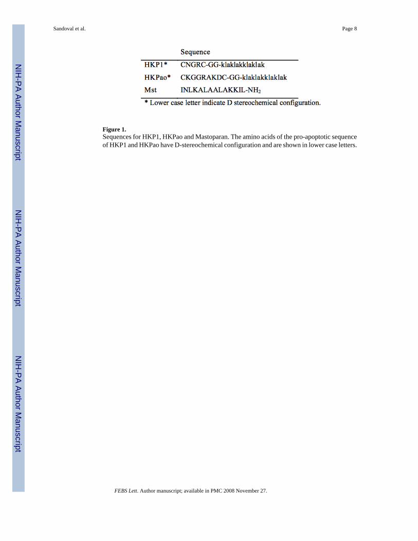
Figure 1.Sequences for HKP1, HKPao and Mastoparan. The amino acids of the pro-apoptotic sequenceof HKP1 and HKPao have D-stereochemical configuration and are shown in lower case letters.
Sandoval et al. Page 8
FEBS Lett. Author manuscript; available in PMC 2008 November 27.
NIH
-PA Author Manuscript
NIH
-PA Author Manuscript
NIH
-PA Author Manuscript

Figure 2.A plot of It/Io against peptide concentration (A) and leakage (B). Decreasing It/Io with leakageindicates partial leakage of vesicle contents, and therefore survival of vesicles to treatment withpeptide.
Sandoval et al. Page 9
FEBS Lett. Author manuscript; available in PMC 2008 November 27.
NIH
-PA Author Manuscript
NIH
-PA Author Manuscript
NIH
-PA Author Manuscript

Figure 3.(A) For the determination of size of molecules that can leak from vesicles with treatment ofpeptide, efflux of entrapped FD dyes of 4 kD, 10 kD, 20 kD and 70 kD size from 70:30 molarratio PC:PG vesicles was monitored. Efflux of the fitc-dextrans was detected via loss offluorescence self-quenching[26] at 520 nm for 5 minutes. Excitation was at 490 nm with
Sandoval et al. Page 10
FEBS Lett. Author manuscript; available in PMC 2008 November 27.
NIH
-PA Author Manuscript
NIH
-PA Author Manuscript
NIH
-PA Author Manuscript
excitation and emission slit widths set to 1 nm. The assay concentration of peptide andphospholipid were 150 nM and 14 +/- 3 μM, respectively for all experiments. The stokes radiusof cytochrome c (1.7 nm)[24] is represented by the dotted line. (B) Elution of FD4 , FD10 andcytochrome c from gel filtration chromatography demonstrates relative hydrodynamic radiusunder identical conditions.
Sandoval et al. Page 11
FEBS Lett. Author manuscript; available in PMC 2008 November 27.
NIH
-PA Author Manuscript
NIH
-PA Author Manuscript
NIH
-PA Author Manuscript

Figure 4.Leakage of cytochrome c from vesicles was evaluated by gel filtration. The profiles areidentified as (A), untreated vesicles (B), 11.5 μM HKP-1 (C), 23 μM HKP1 and (D), 46 μMHKP1. Vesicles elute at 11 ml and released cytochrome c elutes at 33 ml. (E) Relative peakarea of the vesicle (open circles) and cytochrome c (solid squares) peaks were plotted as afunction of HKP1 concentration. The increase in turbidity (solid circles) was monitored bylight scattering at 280 nm of the resuspended pellet.
Sandoval et al. Page 12
FEBS Lett. Author manuscript; available in PMC 2008 November 27.
NIH
-PA Author Manuscript
NIH
-PA Author Manuscript
NIH
-PA Author Manuscript

NIH
-PA Author Manuscript
NIH
-PA Author Manuscript
NIH
-PA Author Manuscript
Sandoval et al. Page 13
Table 1Determination that homing pro-apoptotic peptides induce graded mode of leakage.
HKP1 Mst Control
It/Io before 4.4 4.4 4.4L 63% 69% 0%It/Io after 2.3 2.3 5.7
FEBS Lett. Author manuscript; available in PMC 2008 November 27.