Endothelial progenitor cell-derived extracellular vesicles protect from complement-mediated...
13
Nephrol Dial Transplant (2014) 0: 1–13 doi: 10.1093/ndt/gfu364 Original Article Endothelial progenitor cell-derived extracellular vesicles protect from complement-mediated mesangial injury in experimental anti-Thy1.1 glomerulonephritis Vincenzo Cantaluppi 1 , Davide Medica 1 , Claudio Mannari 2 , Giulia Stiaccini 2 , Federico Figliolini 1 , Sergio Dellepiane 1 , Alessandro Domenico Quercia 1 , Massimiliano Migliori 3 , Vincenzo Panichi 3 , Luca Giovannini 2 , Stefania Bruno 1 , Ciro Tetta 4 , Luigi Biancone 1 and Giovanni Camussi 1 1 Nephrology, Dialysis and Kidney Transplantation Unit and Center for Experimental Medical Research (CeRMS), Department of Medical Sciences, University of Torino, Torino, Italy, 2 Department of Pharmacology, University of Pisa, Pisa, Italy, 3 Nephrology and Dialysis Unit, Ospedale Versilia, Camaiore, LU, Italy and 4 EMEALA Medical Board, Fresenius Medical Care, Bad Homburg, Germany Correspondence and offprint requests to: V. Cantaluppi; E-mail: [email protected] ABSTRACT Background. Endothelial progenitor cells (EPCs) are known to induce tissue repair by paracrine mechanisms including the release of growth factors and extracellular vesicles (EVs), nano- particles able to carry proteins and genetic information to target cells. The aim of this study was to evaluate whether EVs derived from EPCs may protect from complement-mediated mesangial injury in experimental anti-Thy1.1 glomerulonephritis. Methods. EVs were isolated by serial ultracentrifugation from supernatants of cultured human EPCs and characterized for their protein and RNA content. In vivo, EVs were injected i.v. in the experimental rat model of mesangiolytic anti-Thy1.1 glomerulonephritis evaluating renal function, proteinuria, complement activity and histological lesions. In vitro, the bio- logical effects of EPC-derived EVs were studied in cultured rat mesangial cells incubated with anti-Thy1.1 antibody and rat or human serum as complement source. Results. After i.v. injection in Thy1.1-treated rats, EVs localized within injured glomeruli and inhibited mesangial cell activation, leucocyte infiltration and apoptosis, decreased pro- teinuria, increased serum complement haemolytic activity (CH50) and ameliorated renal function. EV treatment de- creased intraglomerular deposition of the membrane attack complex (MAC or C5b-9) and expression of smooth muscle cell actin and preserved the endothelial antigen RECA-1 and the podocyte marker synaptopodin. The protective effect of EVs was significantly reduced by pre-treatment with a high dose of RNase (1 U/mL), suggesting a key role for EV-carried RNAs in these mechanisms. Indeed, EPC-derived EVs contained differ- ent mRNAs coding for several anti-apoptotic molecules and for the complement inhibitors Factor H, CD55 and CD59 and the related proteins. The in vitro experiments aimed to investigate the mechanisms of EV protection indicated that EVs trans- ferred to mesangial cell mRNAs coding for Factor H, CD55 and CD59 and inhibited anti-Thy1.1 antibody/complement- induced apoptosis and C5b-9/C3 mesangial cell deposition. Conclusions. EVs derived from EPCs exert a protective effect in Thy1.1 glomerulonephritis by inhibition of antibody- and complement-mediated injury of mesangial cells. Keywords: apoptosis, complement inhibitors, endothelial progenitor cells, extracellular vesicles, glomerulonephritis INTRODUCTION Thy1.1 glomerulonephritis is characterized by complement- mediated mesangial cell injury and endothelial cell loss [1]. Several studies addressed the role of bone marrow-derived stem cells in the repair of injured glomeruli [2, 3]. Endothelial progenitor cells (EPCs) circulating bone marrow-derived pre- cursors are able to localize within sites of vascular injury. EPCs exert protective effects in experimental models of myo- cardial infarction and hind limb ischaemia [4]. In Thy1.1 © The Author 2014. Published by Oxford University Press on behalf of ERA-EDTA. All rights reserved. 1 NDT Advance Access published December 8, 2014 at University of Torino on January 7, 2015 http://ndt.oxfordjournals.org/ Downloaded from
-
Upload
independent -
Category
Documents
-
view
0 -
download
0
Transcript of Endothelial progenitor cell-derived extracellular vesicles protect from complement-mediated...

Nephrol Dial Transplant (2014) 0: 1–13doi: 10.1093/ndt/gfu364
Original Article
Endothelial progenitor cell-derived extracellular vesiclesprotect from complement-mediated mesangial injuryin experimental anti-Thy1.1 glomerulonephritis
Vincenzo Cantaluppi1, Davide Medica1, Claudio Mannari2, Giulia Stiaccini2, Federico Figliolini1,
Sergio Dellepiane1, Alessandro Domenico Quercia1, Massimiliano Migliori3, Vincenzo Panichi3,
Luca Giovannini2, Stefania Bruno1, Ciro Tetta4, Luigi Biancone1 and Giovanni Camussi1
1Nephrology, Dialysis and Kidney Transplantation Unit and Center for Experimental Medical Research (CeRMS), Department of Medical
Sciences, University of Torino, Torino, Italy, 2Department of Pharmacology, University of Pisa, Pisa, Italy, 3Nephrology and Dialysis Unit,
Ospedale Versilia, Camaiore, LU, Italy and 4EMEALA Medical Board, Fresenius Medical Care, Bad Homburg, Germany
Correspondence and offprint requests to: V. Cantaluppi; E-mail: [email protected]
ABSTRACT
Background. Endothelial progenitor cells (EPCs) are known toinduce tissue repair by paracrine mechanisms including therelease of growth factors and extracellular vesicles (EVs), nano-particles able to carry proteins and genetic information to targetcells. The aim of this study was to evaluate whether EVs derivedfrom EPCs may protect from complement-mediated mesangialinjury in experimental anti-Thy1.1 glomerulonephritis.Methods. EVs were isolated by serial ultracentrifugation fromsupernatants of cultured human EPCs and characterized fortheir protein and RNA content. In vivo, EVs were injected i.v.in the experimental rat model of mesangiolytic anti-Thy1.1glomerulonephritis evaluating renal function, proteinuria,complement activity and histological lesions. In vitro, the bio-logical effects of EPC-derived EVs were studied in cultured ratmesangial cells incubated with anti-Thy1.1 antibody and rator human serum as complement source.Results. After i.v. injection in Thy1.1-treated rats, EVs localizedwithin injured glomeruli and inhibited mesangial cellactivation, leucocyte infiltration and apoptosis, decreased pro-teinuria, increased serum complement haemolytic activity(CH50) and ameliorated renal function. EV treatment de-creased intraglomerular deposition of the membrane attackcomplex (MAC or C5b-9) and expression of smooth muscle cellactin and preserved the endothelial antigen RECA-1 and thepodocyte marker synaptopodin. The protective effect of EVs
was significantly reduced by pre-treatment with a high dose ofRNase (1 U/mL), suggesting a key role for EV-carried RNAs inthese mechanisms. Indeed, EPC-derived EVs contained differ-ent mRNAs coding for several anti-apoptotic molecules and forthe complement inhibitors Factor H, CD55 and CD59 and therelated proteins. The in vitro experiments aimed to investigatethe mechanisms of EV protection indicated that EVs trans-ferred to mesangial cell mRNAs coding for Factor H, CD55 andCD59 and inhibited anti-Thy1.1 antibody/complement-induced apoptosis and C5b-9/C3 mesangial cell deposition.Conclusions. EVs derived from EPCs exert a protective effectin Thy1.1 glomerulonephritis by inhibition of antibody- andcomplement-mediated injury of mesangial cells.
Keywords: apoptosis, complement inhibitors, endothelialprogenitor cells, extracellular vesicles, glomerulonephritis
INTRODUCTION
Thy1.1 glomerulonephritis is characterized by complement-mediated mesangial cell injury and endothelial cell loss [1].Several studies addressed the role of bone marrow-derivedstem cells in the repair of injured glomeruli [2, 3]. Endothelialprogenitor cells (EPCs) circulating bone marrow-derived pre-cursors are able to localize within sites of vascular injury.EPCs exert protective effects in experimental models of myo-cardial infarction and hind limb ischaemia [4]. In Thy1.1
© The Author 2014. Published by Oxford University Presson behalf of ERA-EDTA. All rights reserved.
1
NDT Advance Access published December 8, 2014 at U
niversity of Torino on January 7, 2015
http://ndt.oxfordjournals.org/D
ownloaded from

nephritis, intra-renal injection of EPCs was shown to reduceendothelial injury and mesangial cell activation [5]. The regen-erative effect of EPCs has also been ascribed to paracrine me-chanisms including the release of growth factors and ofextracellular vesicles (EVs) [6, 7]. EVs include exosomes andshedding vesicles that have been recently shown to play a keyrole in the intercellular cross-talk through the transfer of pro-teins and genetic information [8]. We previously showed thatEVs released from EPCs activated angiogenesis by horizontaltransfer of mRNAs [7] and protected the kidney from ischae-mic acute injury by delivering pro-angiogenic microRNAs [9].
The aim of this study was to evaluate whether EPC-derivedEVs may have a protective effect in anti-Thy1.1 glomerulo-nephritis through RNA transfer to injured mesangial cells.
MATERIALS AND METHODS
Isolation and characterization of EPCs andEPC-derived EVs
EPCs were isolated and characterized from peripheral bloodmononucleated cells of healthy donors by density centrifugation[7, 9]. EVs were obtained from supernatants of EPCs [10]. EVshape and size were evaluated by transmission electron micros-copy and Nanosight analysis [9]. The expression of Factor H,CD55 and CD59 both in EPCs and EVs were studied by fluores-cent activated cell sorter (FACS), western blot and confocal mi-croscopy. RNA was extracted from EPCs and EVs by themirVana isolation kit (Ambion, Austin, TX) and analysed byAgilent 2100 Bioanalyzer (Agilent Tech. Inc., Santa Clara, CA).Nucleic acid content of EVs was studied by GUAVA FACS ana-lysis using acridine orange (Sigma Aldrich, St Louis, MO) [11].Reverse Transcription Polymerase Chain Reaction (RT-PCR)was performed by the High-Capacity cDNA Reverse Transcrip-tion Kit (Applied Biosystems, Foster City, CA). Sequence-specificoligonucleotide primers were designed using Primer Express(Applied Biosystems); human Factor H: forward, 50-TTACTGGGATCACATTCATTGCA-30; reverse, 50-TGGCAGGCAACGTCTATAGATTT-30; human CD55: forward, 50-TTGAAGAGTTCTGCAATCGTAGCT-30; reverse, 50-CTGTAACCTGGACGGCACTCAT-30; human CD59: forward, 50-TCTTCTGCCATTCAGGTCATAGC-30; reverse, 50-TGGTAATGAGACACGCATCAAAA-30. The relative expression of different mRNAswas detected by SYBR green method using relative Ct quantifica-tion analysis (Ct = Ct target –Ct control). In selected experi-ments, EVs were labelled with the red fluorescent dye PKH26(Sigma Aldrich) or treated with a high dose (1 U/mL) of RNase(Ambion) for 1 h at 37°C. Bioanalyzer and quantitativeRT-PCR (qRT-PCR) were used to evaluate the total RNAcontent and the presence of selected mRNAs (Factor H, CD55and CD59) and microRNAs (miR-126, miR-296) in unstimu-lated or RNase-treated EVs. EVs isolated from human fibro-blasts were used as control in all experiments.
Experimental anti-Thy1.1 glomerulonephritis:functional and histological studies
Animal care and treatment were conducted in conformitywith the National Institutes of Health Guide for animal care.
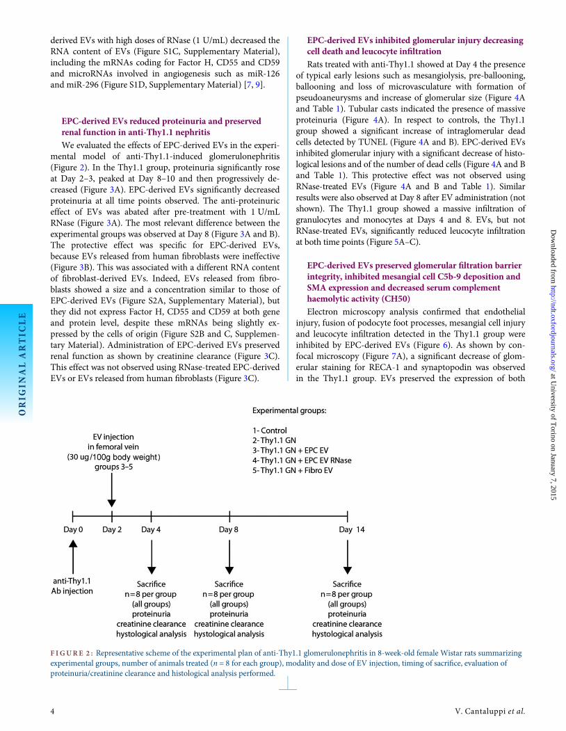
Briefly, 8-week-old female Wistar rats were subjected to i.v.injection (femoral vein) of 0.5 mL of 0.9% saline containingvehicle alone or 400 µg of anti-Thy1.1 antibody (Cedarlane,Ontario, Canada). At Day 2 after anti-Thy1.1 injection, animalswere divided as follows: (i) control (untreated); (ii) Thy1.1(Thy1.1 + 0.9% saline); (iii) Thy1.1 + EPC EVs (Thy1.1 + 30 µg/100 g body weight EPC EVs); (iv) Thy1.1 + EPC EV RNase(Thy1.1 + 30 µg/100 g body weight EPC EVs pre-treated with 1U/mL RNase) and (v) Thy1.1 + fibroblast EVs (Thy1.1 + 30 µg/100 g body weight EVs derived from human fibroblasts). Tenanimals for each group were housed in ventilated cages and sacri-ficed after 4, 8 and 14 days. Proteinuria was measured daily on24-h urine by an automated method (Bio-Rad LaboratoriesGmbH, München, Germany). Serum and urine creatinine weredetermined by commercially available optical densitometric kits(Arbor Assays, Ann Arbor, MI). For histological analysis, 5 µm-thick paraffin kidney sections were stained with haematoxylinand eosin stain (H&E; Merck, Darmstadt, Germany) to definethe percentage of glomeruli affected by the following typical al-terations: pre-ballooning, ballooning, mesangiolysis, microaneur-ysms, podocyte bridging and tuft adhesion [12]. Morphology ofthe glomerular filtration barrier was also evaluated by transmis-sion electron microscopy [13]. Terminal deoxynucleotidyl trans-ferase dUTP nick end labeling (TUNEL) assay (Chemicon Int.,Temecula, CA) for the detection of apoptotic cells was per-formed. Leucocyte infiltration was evaluated by immunoperoxi-dase staining with an anti-monocyte (Chemicon Int.) or anti-granulocyte (Serotec, Oxford, UK) antibody. The expansion ofmesangial areas was quantified by immunoperoxidase stainingwith an anti-smooth muscle cell actin (SMA) antibody (Dako,Glostrup, Denmark). Confocal microscopy analysis was per-formed on frozen sections to evaluate the expression of synap-topodin (Progen Biotechnik, Heidelberg, Germany), RECA-1(Serotec) or C5b-9 (Santa Cruz Biotech, Santa Cruz, CA) andto evaluate biodistribution of PKH26 red-labelled EVs 2 hafter injection. Complement haemolytic activity (CH50) on ratsera was evaluated by Enzyme-Linked ImmunoSorbent Assay(ELISA) (Bioassay Technology Laboratory, Shanghai, China).
Rat and human glomerular cell isolation,characterization and culture
Rat mesangial cells (RMCs) were purchased from ATCC(Manassas, VA, USA) and cultured in DMEM medium with 4mM L-glutamine and 15% fetal bovine serum. Primary cul-tures of human mesangial cells, glomerular endothelial cells(GECs) and podocytes were isolated from glomeruli as previ-ously described [14, 15].
In vitro internalization of EPC-derived EVsinto different glomerular cells
RMCs, human mesangial cells, endothelial cells or podocyteswere seeded on six-well plates and incubated with different dosesof PKH26-labelled EVs for 2 h. EV internalization was evaluatedby confocal microscopy (Zeiss LSM 5 PASCAL, Jena, Germany)or FACS in the presence or absence of 1 µg/mL of differentblocking antibodies directed to αVβ3-integrin (BioLegend), α4-integrin, α6-integrin (Chemicon Int.), CD29, L-selectin (BectonDickinson) or control irrelevant cytokeratin 19 (Dako) [9].
ORIG
INALARTIC
LE
2 V. Cantaluppi et al.
at University of T
orino on January 7, 2015http://ndt.oxfordjournals.org/
Dow
nloaded from

In vitro studies on RMCs
We recreated in vitro the complement-mediated damageof RMC culturing cells in the presence of anti-Thy1.1 antibodyfor 1 h in serum-free Roswell Park Memorial Institutemedium at 37°C and with the subsequent addition of a lyticdose (33%) of normal rat or human AB serum as complementsource for 40 min at 37°C [16]. After complement activation,the effects of EVs on RMCs were evaluated by the followingassays.
Cell damage. RMCs (5 × 10⁴) were cultured on 24-well andincubated with different stimuli and 250 µg/mL 2,3-bis-(2-methoxy-4-nitro-5-sulfophenyl)-2H-tetrazolium-5-carboxa-nilide (XTT) (Sigma Aldrich). Samples were analysed in anautomatized ELISA reader at a wavelength of 450 nm [17].
Cell death. RMCs were subjected to TUNEL assay (ChemiconInt.) and analysed under a fluorescence microscope to detectgreen-stained apoptotic cells in 10 non-consecutive micro-scopic fields [18, 19]. The same samples were analysed byMUSE assay to detect the co-staining for Annexin V and7-aminoactinomycin D (7-AAD) [20, 21].
RT-PCR, immunofluorescence and FACS studies. Afterincubation with anti-Thy1.1 + normal rat serum and differenttypes of EVs, total RNA was extracted from RMCs to performRT-PCR for human Factor H, CD55 and CD59. RMCs werestained with specific antibodies directed to rat or human C5b-9 (Santa Cruz or Dako, respectively), C3, Factor H (Abcam),CD55 or CD59 (Becton Dickinson): cells were then incubated
with appropriated fluorescein isothiocyanate or phycoerythrinconjugated secondary anti-IgG antibodies and examined by im-munofluorescence and FACS.
Statistical analysis
All data of different experimental procedures are expressedas average ± SD. Statistical analysis was performed by Student’st-test or ANOVA with the Newman–Keuls multicomparisontest. For FACS, the Kolmogorov–Smirnov nonparametric statis-tical test was performed.
RESULTS
Characterization of EPC-derived EVs and expressionof complement inhibitors
Purified EVs showed a homogenous pattern of spheroidparticles with a size ranging from 60 to 130 nm as seen by elec-tron microscopy and confirmed by Nanosight analysis(Figure S1A, Supplementary Material). Acridine orange stain-ing showed that EVs derived from EPCs were positive forRNA but negative for DNA (Figure S1B, SupplementaryMaterial). We found by RT-PCR that EPCs expressed mRNAscoding for the complement inhibitors Factor H, CD55 andCD59 (Figure 1A). The expression of these complement inhi-bitors was confirmed at the protein level in EPCs by FACS andimmunofluorescence studies (Figure 1B). EPC-derived EVscarried both mRNAs coding for Factor H, CD55 and CD59(Figure 1A) and the related proteins (western blot and FACSanalysis in Figure 1C and D, respectively). Treatment of EPC-
F IGURE 1 : Expression of complement inhibitors Factor H, CD55 and CD59 by EPCs and EPC-derived EVs. (A) Representative qRT-PCRshowing the expression of mRNAs coding for Factor H, CD55 and CD59 in both EPCs and EPC-derived EVs (left panels). (B) FACS andconfocal microscope analysis showing Factor H, CD55, CD59 expression in EPCs (EPC, central panels). (C and D) Western blotting (C, EPCwas used as control) and FACS analysis (D) detecting Factor H, CD55, CD59 in EPC-derived EVs (EV, right panels). For FACS analysis,control-matched isotype was used as experimental control (open curves). Kolmogorov–Smirnov statistical analysis was performed. For westernblot analysis of Factor H, α-actin was used as positive control. Three independent experiments were performed with similar results.
ORIG
INALARTIC
LE
E P C e x t r a c e l l u l a r v e s i c l e s a n d g l o m e r u l a r i n j u r y 3
at University of T
orino on January 7, 2015http://ndt.oxfordjournals.org/
Dow
nloaded from
derived EVs with high doses of RNase (1 U/mL) decreased theRNA content of EVs (Figure S1C, Supplementary Material),including the mRNAs coding for Factor H, CD55 and CD59and microRNAs involved in angiogenesis such as miR-126and miR-296 (Figure S1D, Supplementary Material) [7, 9].
EPC-derived EVs reduced proteinuria and preservedrenal function in anti-Thy1.1 nephritis
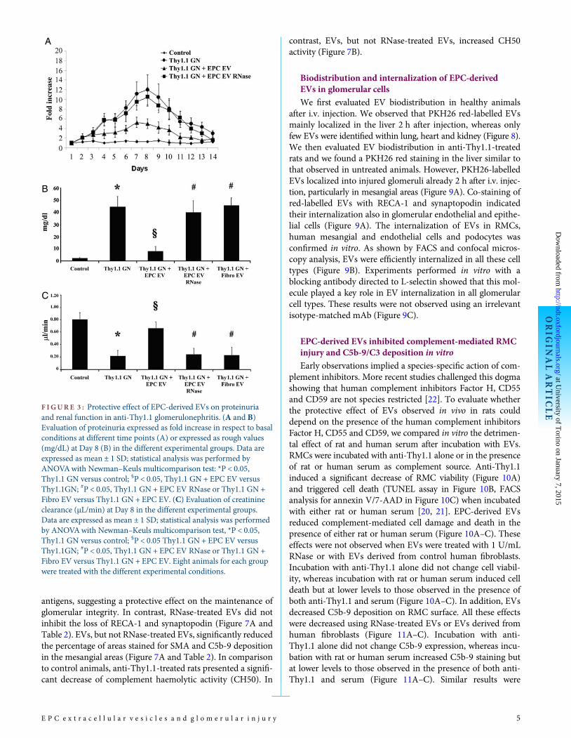
We evaluated the effects of EPC-derived EVs in the experi-mental model of anti-Thy1.1-induced glomerulonephritis(Figure 2). In the Thy1.1 group, proteinuria significantly roseat Day 2–3, peaked at Day 8–10 and then progressively de-creased (Figure 3A). EPC-derived EVs significantly decreasedproteinuria at all time points observed. The anti-proteinuriceffect of EVs was abated after pre-treatment with 1 U/mLRNase (Figure 3A). The most relevant difference between theexperimental groups was observed at Day 8 (Figure 3A and B).The protective effect was specific for EPC-derived EVs,because EVs released from human fibroblasts were ineffective(Figure 3B). This was associated with a different RNA contentof fibroblast-derived EVs. Indeed, EVs released from fibro-blasts showed a size and a concentration similar to those ofEPC-derived EVs (Figure S2A, Supplementary Material), butthey did not express Factor H, CD55 and CD59 at both geneand protein level, despite these mRNAs being slightly ex-pressed by the cells of origin (Figure S2B and C, Supplemen-tary Material). Administration of EPC-derived EVs preservedrenal function as shown by creatinine clearance (Figure 3C).This effect was not observed using RNase-treated EPC-derivedEVs or EVs released from human fibroblasts (Figure 3C).
EPC-derived EVs inhibited glomerular injury decreasingcell death and leucocyte infiltration
Rats treated with anti-Thy1.1 showed at Day 4 the presenceof typical early lesions such as mesangiolysis, pre-ballooning,ballooning and loss of microvasculature with formation ofpseudoaneurysms and increase of glomerular size (Figure 4Aand Table 1). Tubular casts indicated the presence of massiveproteinuria (Figure 4A). In respect to controls, the Thy1.1group showed a significant increase of intraglomerular deadcells detected by TUNEL (Figure 4A and B). EPC-derived EVsinhibited glomerular injury with a significant decrease of histo-logical lesions and of the number of dead cells (Figure 4A and Band Table 1). This protective effect was not observed usingRNase-treated EVs (Figure 4A and B and Table 1). Similarresults were also observed at Day 8 after EV administration (notshown). The Thy1.1 group showed a massive infiltration ofgranulocytes and monocytes at Days 4 and 8. EVs, but notRNase-treated EVs, significantly reduced leucocyte infiltrationat both time points (Figure 5A–C).
EPC-derived EVs preserved glomerular filtration barrierintegrity, inhibited mesangial cell C5b-9 deposition andSMA expression and decreased serum complementhaemolytic activity (CH50)
Electron microscopy analysis confirmed that endothelialinjury, fusion of podocyte foot processes, mesangial cell injuryand leucocyte infiltration detected in the Thy1.1 group wereinhibited by EPC-derived EVs (Figure 6). As shown by con-focal microscopy (Figure 7A), a significant decrease of glom-erular staining for RECA-1 and synaptopodin was observedin the Thy1.1 group. EVs preserved the expression of both
F IGURE 2 : Representative scheme of the experimental plan of anti-Thy1.1 glomerulonephritis in 8-week-old female Wistar rats summarizingexperimental groups, number of animals treated (n = 8 for each group), modality and dose of EV injection, timing of sacrifice, evaluation ofproteinuria/creatinine clearance and histological analysis performed.
ORIG
INALARTIC
LE
4 V. Cantaluppi et al.
at University of T
orino on January 7, 2015http://ndt.oxfordjournals.org/
Dow
nloaded from
antigens, suggesting a protective effect on the maintenance ofglomerular integrity. In contrast, RNase-treated EVs did notinhibit the loss of RECA-1 and synaptopodin (Figure 7A andTable 2). EVs, but not RNase-treated EVs, significantly reducedthe percentage of areas stained for SMA and C5b-9 depositionin the mesangial areas (Figure 7A and Table 2). In comparisonto control animals, anti-Thy1.1-treated rats presented a signifi-cant decrease of complement haemolytic activity (CH50). In
contrast, EVs, but not RNase-treated EVs, increased CH50activity (Figure 7B).
Biodistribution and internalization of EPC-derivedEVs in glomerular cells
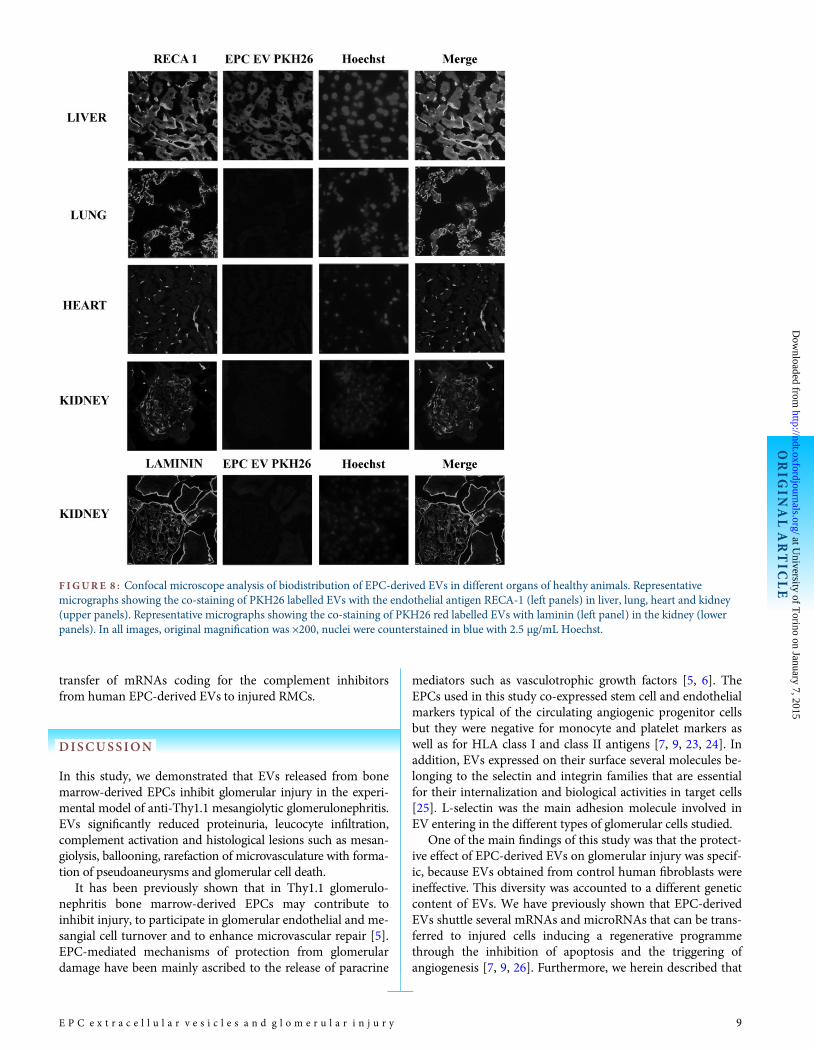
We first evaluated EV biodistribution in healthy animalsafter i.v. injection. We observed that PKH26 red-labelled EVsmainly localized in the liver 2 h after injection, whereas onlyfew EVs were identified within lung, heart and kidney (Figure 8).We then evaluated EV biodistribution in anti-Thy1.1-treatedrats and we found a PKH26 red staining in the liver similar tothat observed in untreated animals. However, PKH26-labelledEVs localized into injured glomeruli already 2 h after i.v. injec-tion, particularly in mesangial areas (Figure 9A). Co-staining ofred-labelled EVs with RECA-1 and synaptopodin indicatedtheir internalization also in glomerular endothelial and epithe-lial cells (Figure 9A). The internalization of EVs in RMCs,human mesangial and endothelial cells and podocytes wasconfirmed in vitro. As shown by FACS and confocal micros-copy analysis, EVs were efficiently internalized in all these celltypes (Figure 9B). Experiments performed in vitro with ablocking antibody directed to L-selectin showed that this mol-ecule played a key role in EV internalization in all glomerularcell types. These results were not observed using an irrelevantisotype-matched mAb (Figure 9C).
EPC-derived EVs inhibited complement-mediated RMCinjury and C5b-9/C3 deposition in vitro
Early observations implied a species-specific action of com-plement inhibitors. More recent studies challenged this dogmashowing that human complement inhibitors Factor H, CD55and CD59 are not species restricted [22]. To evaluate whetherthe protective effect of EVs observed in vivo in rats coulddepend on the presence of the human complement inhibitorsFactor H, CD55 and CD59, we compared in vitro the detrimen-tal effect of rat and human serum after incubation with EVs.RMCs were incubated with anti-Thy1.1 alone or in the presenceof rat or human serum as complement source. Anti-Thy1.1induced a significant decrease of RMC viability (Figure 10A)and triggered cell death (TUNEL assay in Figure 10B, FACSanalysis for annexin V/7-AAD in Figure 10C) when incubatedwith either rat or human serum [20, 21]. EPC-derived EVsreduced complement-mediated cell damage and death in thepresence of either rat or human serum (Figure 10A–C). Theseeffects were not observed when EVs were treated with 1 U/mLRNase or with EVs derived from control human fibroblasts.Incubation with anti-Thy1.1 alone did not change cell viabil-ity, whereas incubation with rat or human serum induced celldeath but at lower levels to those observed in the presence ofboth anti-Thy1.1 and serum (Figure 10A–C). In addition, EVsdecreased C5b-9 deposition on RMC surface. All these effectswere decreased using RNase-treated EVs or EVs derived fromhuman fibroblasts (Figure 11A–C). Incubation with anti-Thy1.1 alone did not change C5b-9 expression, whereas incu-bation with rat or human serum increased C5b-9 staining butat lower levels to those observed in the presence of both anti-Thy1.1 and serum (Figure 11A–C). Similar results were
F IGURE 3 : Protective effect of EPC-derived EVs on proteinuriaand renal function in anti-Thy1.1 glomerulonephritis. (A and B)Evaluation of proteinuria expressed as fold increase in respect to basalconditions at different time points (A) or expressed as rough values(mg/dL) at Day 8 (B) in the different experimental groups. Data areexpressed as mean ± 1 SD; statistical analysis was performed byANOVAwith Newman–Keuls multicomparison test: *P < 0.05,Thy1.1 GN versus control; §P < 0.05, Thy1.1 GN + EPC EV versusThy1.1GN; #P < 0.05, Thy1.1 GN + EPC EV RNase or Thy1.1 GN +Fibro EV versus Thy1.1 GN + EPC EV. (C) Evaluation of creatinineclearance (µL/min) at Day 8 in the different experimental groups.Data are expressed as mean ± 1 SD; statistical analysis was performedby ANOVAwith Newman–Keuls multicomparison test, *P < 0.05,Thy1.1 GN versus control; §P < 0.05 Thy1.1 GN + EPC EV versusThy1.1GN; #P < 0.05, Thy1.1 GN + EPC EV RNase or Thy1.1 GN +Fibro EV versus Thy1.1 GN + EPC EV. Eight animals for each groupwere treated with the different experimental conditions.
ORIG
INALARTIC
LE
E P C e x t r a c e l l u l a r v e s i c l e s a n d g l o m e r u l a r i n j u r y 5
at University of T
orino on January 7, 2015http://ndt.oxfordjournals.org/
Dow
nloaded from
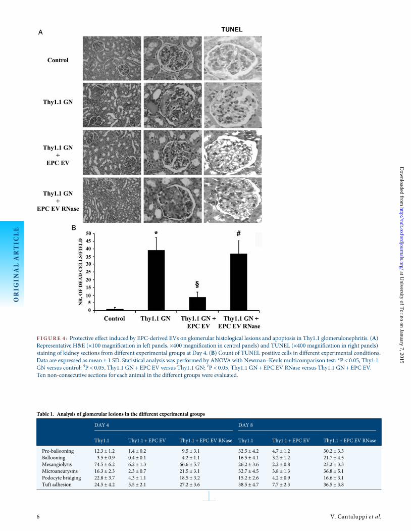
F IGURE 4 : Protective effect induced by EPC-derived EVs on glomerular histological lesions and apoptosis in Thy1.1 glomerulonephritis. (A)Representative H&E (×100 magnification in left panels, ×400 magnification in central panels) and TUNEL (×400 magnification in right panels)staining of kidney sections from different experimental groups at Day 4. (B) Count of TUNEL positive cells in different experimental conditions.Data are expressed as mean ± 1 SD. Statistical analysis was performed by ANOVAwith Newman–Keuls multicomparison test: *P < 0.05, Thy1.1GN versus control; §P < 0.05, Thy1.1 GN + EPC EV versus Thy1.1 GN; #P < 0.05, Thy1.1 GN + EPC EV RNase versus Thy1.1 GN + EPC EV.Ten non-consecutive sections for each animal in the different groups were evaluated.
Table 1. Analysis of glomerular lesions in the different experimental groups
DAY 4 DAY 8
Thy1.1 Thy1.1 + EPC EV Thy1.1 + EPC EV RNase Thy1.1 Thy1.1 + EPC EV Thy1.1 + EPC EV RNase
Pre-ballooning 12.3 ± 1.2 1.4 ± 0.2 9.5 ± 3.1 32.5 ± 4.2 4.7 ± 1.2 30.2 ± 3.3Ballooning 3.5 ± 0.9 0.4 ± 0.1 4.2 ± 1.1 16.5 ± 4.1 3.2 ± 1.2 21.7 ± 4.5Mesangiolysis 74.5 ± 6.2 6.2 ± 1.3 66.6 ± 5.7 26.2 ± 3.6 2.2 ± 0.8 23.2 ± 3.3Microaneurysms 16.3 ± 2.3 2.3 ± 0.7 21.5 ± 3.1 32.7 ± 4.5 3.8 ± 1.3 36.8 ± 5.1Podocyte bridging 22.8 ± 3.7 4.3 ± 1.1 18.5 ± 3.2 15.2 ± 2.6 4.2 ± 0.9 16.6 ± 3.1Tuft adhesion 24.5 ± 4.2 5.5 ± 2.1 27.2 ± 3.6 38.5 ± 4.7 7.7 ± 2.3 36.5 ± 3.8
ORIG
INALARTIC
LE
6 V. Cantaluppi et al.
at University of T
orino on January 7, 2015http://ndt.oxfordjournals.org/
Dow
nloaded from

observed for C3 deposition (Figure 12A–C). As shown byRT-PCR analysis, RMCs incubated with EPC-derived EVs ex-pressed mRNAs coding for the human complement inhibitors
Factor H, CD55 and CD59 not detectable in basal conditionsor when EVs were pre-treated with RNase (Figure S3, Supple-mentary Material). These results suggested the putative
F IGURE 5 : EPC-derived EVs reduced leucocyte infiltration in anti-Thy1.1 glomerulonephritis. (A) Representative micrographs(original magnification, ×400) of infiltrating granulocytes and monocytes in different experimental groups at Day 4. (B and C) Counts of infil-trating granulocytes (B) and monocytes (C) in different experimental groups at Day 4 (black columns) and at Day 8 (white columns). Data areexpressed as mean ± 1 SD. Statistical analysis was performed by ANOVAwith Newman–Keuls multicomparison test. In respect to control, theThy1.1 group showed a significant increase of leucocyte infiltration (*P < 0.05, Thy1.1 GN versus control). EVs significantly decreased intraglo-merular granulocyte and monocyte infiltration (§P < 0.05, Thy1.1 GN + EPC EV versus Thy1.1 GN). The decrease of leucocyte infiltration wasnot observed when EVs were treated with RNase (#P < 0.05, Thy1.1 GN + EPC EV RNase versus Thy1.1 GN + EPC EV). Ten non-consecutivesections for each animal in the different groups were evaluated.
F IGURE 6 : Electron microscopy analysis of kidney sections from rats with Thy1.1 nephritis treated or not with EPC-derived EVs. EVspreserved the integrity of glomerular architecture characterized by a decrease of endothelial injury, fusion of podocyte foot processes and mesan-gial cell activation. Original magnification was ×7000.
ORIG
INALARTIC
LE
E P C e x t r a c e l l u l a r v e s i c l e s a n d g l o m e r u l a r i n j u r y 7
at University of T
orino on January 7, 2015http://ndt.oxfordjournals.org/
Dow
nloaded from
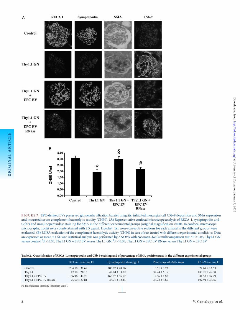
F IGURE 7 : EPC-derived EVs preserved glomerular filtration barrier integrity, inhibited mesangial cell C5b-9 deposition and SMA expressionand increased serum complement haemolytic activity (CH50). (A) Representative confocal microscope analysis of RECA-1, synaptopodin andC5b-9 and immunoperoxidase staining for SMA in the different experimental groups (original magnification ×400). In confocal microscopemicrographs, nuclei were counterstained with 2.5 µg/mL Hoechst. Ten non-consecutive sections for each animal in the different groups wereevaluated. (B) ELISA evaluation of the complement haemolytic activity (CH50) in sera of rats treated with different experimental conditions. Dataare expressed as mean ± 1 SD and statistical analysis was performed by ANOVAwith Newman–Keuls multicomparison test: *P < 0.05, Thy1.1 GNversus control; §P < 0.05, Thy1.1 GN+ EPC EV versus Thy1.1 GN; #P < 0.05, Thy1.1 GN + EPC EV RNase versus Thy1.1 GN+ EPC EV.
Table 2. Quantification of RECA-1, synaptopodin and C5b-9 staining and of percentage of SMA positive areas in the different experimental groups
RECA-1 staining FI Synaptopodin staining FI Percentage of SMA areas C5b-9 staining FI
Control 204.10 ± 31.69 200.97 ± 48.56 0.51 ± 0.77 22.69 ± 12.53Thy1.1 42.10 ± 28.16 42.04 ± 33.22 32.24 ± 6.15 185.76 ± 47.38Thy1.1 + EPC EV 156.96 ± 44.78 168.97 ± 56.77 7.56 ± 4.87 41.53 ± 39.99Thy1.1 + EPC EV RNase 25.50 ± 27.81 38.71 ± 32.44 36.23 ± 3.65 197.91 ± 36.56
FI, Fluorescence intensity (arbitrary units).
ORIG
INALARTIC
LE
8 V. Cantaluppi et al.
at University of T
orino on January 7, 2015http://ndt.oxfordjournals.org/
Dow
nloaded from
transfer of mRNAs coding for the complement inhibitorsfrom human EPC-derived EVs to injured RMCs.
DISCUSSION
In this study, we demonstrated that EVs released from bonemarrow-derived EPCs inhibit glomerular injury in the experi-mental model of anti-Thy1.1 mesangiolytic glomerulonephritis.EVs significantly reduced proteinuria, leucocyte infiltration,complement activation and histological lesions such as mesan-giolysis, ballooning, rarefaction of microvasculature with forma-tion of pseudoaneurysms and glomerular cell death.
It has been previously shown that in Thy1.1 glomerulo-nephritis bone marrow-derived EPCs may contribute toinhibit injury, to participate in glomerular endothelial and me-sangial cell turnover and to enhance microvascular repair [5].EPC-mediated mechanisms of protection from glomerulardamage have been mainly ascribed to the release of paracrine
mediators such as vasculotrophic growth factors [5, 6]. TheEPCs used in this study co-expressed stem cell and endothelialmarkers typical of the circulating angiogenic progenitor cellsbut they were negative for monocyte and platelet markers aswell as for HLA class I and class II antigens [7, 9, 23, 24]. Inaddition, EVs expressed on their surface several molecules be-longing to the selectin and integrin families that are essentialfor their internalization and biological activities in target cells[25]. L-selectin was the main adhesion molecule involved inEV entering in the different types of glomerular cells studied.
One of the main findings of this study was that the protect-ive effect of EPC-derived EVs on glomerular injury was specif-ic, because EVs obtained from control human fibroblasts wereineffective. This diversity was accounted to a different geneticcontent of EVs. We have previously shown that EPC-derivedEVs shuttle several mRNAs and microRNAs that can be trans-ferred to injured cells inducing a regenerative programmethrough the inhibition of apoptosis and the triggering ofangiogenesis [7, 9, 26]. Furthermore, we herein described that
F IGURE 8 : Confocal microscope analysis of biodistribution of EPC-derived EVs in different organs of healthy animals. Representativemicrographs showing the co-staining of PKH26 labelled EVs with the endothelial antigen RECA-1 (left panels) in liver, lung, heart and kidney(upper panels). Representative micrographs showing the co-staining of PKH26 red labelled EVs with laminin (left panel) in the kidney (lowerpanels). In all images, original magnification was ×200, nuclei were counterstained in blue with 2.5 µg/mL Hoechst.
ORIG
INALARTIC
LE
E P C e x t r a c e l l u l a r v e s i c l e s a n d g l o m e r u l a r i n j u r y 9
at University of T
orino on January 7, 2015http://ndt.oxfordjournals.org/
Dow
nloaded from
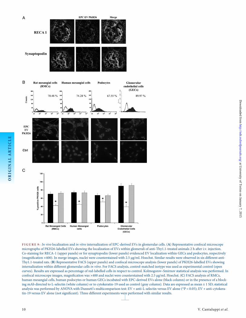
F IGURE 9 : In vivo localization and in vitro internalization of EPC-derived EVs in glomerular cells. (A) Representative confocal microscopemicrographs of PKH26-labelled EVs showing the localization of EVs within glomeruli of anti-Thy1.1-treated animals 2 h after i.v. injection.Co-staining for RECA-1 (upper panels) or for synaptopodin (lower panels) evidenced EV localization within GECs and podocytes, respectively(magnification ×400). In merge images, nuclei were counterstained with 2.5 µg/mL Hoechst. Similar results were observed in six different anti-Thy1.1-treated rats. (B) Representative FACS (upper panels) and confocal microscope analysis (lower panels) of PKH26-labelled EVs showinginternalization within different glomerular cells in vitro. For FACS analysis, control-matched isotype was used as experimental control (opencurves). Results are expressed as percentage of red-labelled cells in respect to control. Kolmogorov–Smirnov statistical analysis was performed. Inconfocal microscope images, magnification was ×400 and nuclei were counterstained with 2.5 µg/mL Hoechst. (C) FACS analysis of RMCs,human mesangial cells, human podocytes or human GECs incubated with EPC-derived EVs alone (black column) or in the presence of a block-ing mAb directed to L-selectin (white column) or to cytokeratin-19 used as control (gray column). Data are expressed as mean ± 1 SD; statisticalanalysis was performed by ANOVAwith Dunnett’s multicomparison test: EV + anti-L-selectin versus EV alone (*P < 0.05); EV + anti-cytokera-tin-19 versus EV alone (not significant). Three different experiments were performed with similar results.
ORIG
INALARTIC
LE
10 V. Cantaluppi et al.
at University of T
orino on January 7, 2015http://ndt.oxfordjournals.org/
Dow
nloaded from

EPC-derived EVs but not fibroblast-derived EVs carriedmRNA coding for the complement inhibitors Factor H, CD55and CD59 as well as the correspondent proteins. Complement
Factor H is a key regulator of the alternative pathway whosedeficiency is associated with different human diseases includ-ing haemolytic uraemic syndrome, membranoproliferativeglomerulonephritis, lupus nephritis and transplant rejection[27, 28]. The lack of Factor H leads to a massive C3 activationwith consequent accumulation in the glomerular structures.The treatment with human Factor H has been shown to
F IGURE 1 0 : Protective effect of EPC-derived EVs on RMC damageand death induced by anti-Thy1.1 Ab and complement activation.(A) XTT-based assay evaluating RMC damage in different experi-mental conditions. Results are expressed as average optical density(OD) ± 1 SD generated by viable RMCs. (B) TUNEL assay showingRMC death in different experimental conditions. Results are ex-pressed as mean number of dead cells ± 1 SD/microscope field (×100magnification). (C) FACS analysis after dual staining with Annexin Vand 7-AAD of RMCs treated with different experimental conditions.Results are expressed as percentage of apoptotic cells ± 1 SD. Threeexperiments were performed with similar results. Statistical analysiswas performed by ANOVAwith Newman–Keuls multicomparisontest. Either 33% rat or human serum induced RMC injury and death(*P < 0.05, serum versus vehicle). This effect was significantly en-hanced by addition of 4 µg/mL anti-Thy1.1 Ab (*P < 0.05, Thy1.1Ab + serum versus vehicle; °P < 0.05 Thy1.1 Ab + serum versus serumalone). EPC EV significantly reduced RMC injury and death (§P <0.05, Thy1.1 Ab + serum + EPC EV versus Thy1.1 Ab + serum). Thiseffect was not observed using RNase-treated EPC EV or fibroblast-derived EV (Fibro EV) (#P < 0.05, Thy1.1 Ab + serum + EPC EVRNase or Fibro EV versus Thy1.1 Ab + serum + EPC EV).
F IGURE 1 1 : EPC-derived EVs significantly reduced C5b-9 depos-ition on RMCs. (A and B) Representative immunofluorescence mi-crographs of C5b-9 deposition on RMCs treated with differentexperimental conditions. Rat (A) or human (B) serum was used ascomplement source, respectively. Three experiments were performedwith similar results. Nuclei were counterstained with 2.5 µg/mLHoechst; original magnification was ×400. (C) FACS analysis of C5b-9 deposition on RMCs treated with different experimental conditions.Rat (black columns) or human (white columns) serum was used ascomplement source, respectively. Statistical analysis was performedby ANOVAwith Newman–Keuls multicomparison test. Either 33%rat or human serum induced C5b-9 expression on RMCs (*P < 0.05,serum versus vehicle). This effect was significantly enhanced by add-ition of 4 µg/mL anti-Thy1.1 Ab (*P < 0.05, Thy1.1 Ab + serumversus vehicle; °P < 0.05, Thy1.1 Ab + serum versus serum alone).EPC EV significantly reduced C5b-9 staining (§P < 0.05, Thy1.1 Ab +serum + EPC EV versus Thy1.1 Ab + serum). This effect was not ob-served using RNase-treated EPC EV or fibroblast-derived EV (FibroEV) (#P < 0.05, Thy1.1 Ab + serum + EPC EV RNase or Fibro EVversus Thy1.1 Ab + serum + EPC EV).
ORIG
INALARTIC
LE
E P C e x t r a c e l l u l a r v e s i c l e s a n d g l o m e r u l a r i n j u r y 11
at University of T
orino on January 7, 2015http://ndt.oxfordjournals.org/
Dow
nloaded from
reverse renal complement deposition in Factor H-deficientmice [29]. CD55 or decay-accelerating factor (DAF) is a com-plement regulator widely expressed in mammalian tissueswhich is known to inhibit the classical and the alternativepathways by targeting C3 convertase. CD55 deficiency leads toan increased susceptibility to develop anti-glomerular base-ment membrane nephritis or focal and segmental glomerulo-sclerosis [30, 31]. In acute toxic nephritis, CD55 has beenshown to confer protection from complement-mediated
podocyte injury [32]. CD59 is a regulatory protein that inhibitsC5b-9, the terminal effector of cell damage able to triggerglomerular cell death [32, 33]. Several studies demonstratedthat complement activation mediates the typical histologicallesions of experimental Thy1.1 glomerulonephritis. In particu-lar, the mesangial injury has been ascribed to cell death conse-quent to C5b-9 formation. Indeed, C5b-9 has been shown toinduce the release of mediators able to worsen mesangial celldamage such as inflammatory cyotokines and nitric oxide[34]. Moreover, C5b-9 can trigger mesangial cell apoptosis viaa caspase-dependent manner [35]. In this study, we observed areduced deposition of C5b-9 within glomeruli of rats treatedwith EVs. This could depend on the reduced accumulation ofinflammatory cells and/or on the delivery of inhibitors of com-plement activation by EVs. The Factor H, CD55 and CD59complement inhibitors carried by EPC-derived EVs are ofhuman origin and therefore according to the dogma of speciesspecificity action of complement inhibitors should not beactive in rats. However, this dogma has been recently chal-lenged as it has been shown that there is considerable cross-species activity for these membrane regulators of complementactivation [22]. Studies aimed to investigate the capacity ofhuman, rat and mouse analogues of CD55 to regulate homolo-gous and heterologous complement activation revealed theabsence of species restriction [22, 36]. Similar results were ob-served for CD59 [22, 37]. In this study, we compared theability of human EPC-derived EVs to inhibit in vitro Thy1.1-induced RMC injury in the presence of rat or human serumas complement source. The inhibitory effect of EVs on me-sangial cell injury was observed not only using rat but alsohuman serum. Therefore, the results of this study suggestthat the protective effect of EPC-derived EVs may depend onthe transfer of mRNAs coding for the human complementinhibitors Factor H, CD55 and CD59 as well as the corres-pondent proteins to injured RMCs. This transfer resulted ina significant reduction of C5b-9 and C3 deposition andconsequent complement-mediated cell injury. Moreover, theexpression of SMA by mesangial cells was inhibited by EPC-derived EVs, suggesting a limitation of myofibroblastic differ-entiation. The loss of the protective effect of EVs both in vivoand in vitro after RNAse treatment further suggests that thebiological effects of EVs are mainly related to the transfer ofRNAs.
In conclusion, EVs released from EPCs decrease protein-uria and inhibit glomerular injury by transferring specificRNAs in experimental anti-Thy1.1 glomerulonephritis. Thisprotective effect may depend on the limitation of cell deathand inflammatory cell recruitment induced by antibody- andcomplement-mediated injury of mesangial cells.
ACKNOWLEDGEMENT
This work was supported by Italian Government MIUR PRINproject, ‘Regione Piemonte, Piattaforme BiotecnologichePiSTEM project and Ricerca Finalizzata and Local UniversityGrants (“ex-60%”)’.
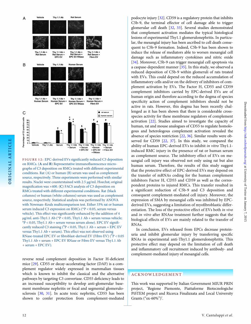
F IGURE 1 2 : EPC-derived EVs significantly reduced C3 depositionon RMCs. (A and B) Representative immunofluorescence micro-graphs of C3 deposition on RMCs treated with different experimentalconditions. Rat (A) or human (B) serum was used as complementsource, respectively. Three experiments were performed with similarresults. Nuclei were counterstained with 2.5 µg/mL Hoechst; originalmagnification was ×400. (C) FACS analysis of C3 deposition onRMCs treated with different experimental conditions. Rat (blackcolumns) or human (white columns) serum was used as complementsource, respectively. Statistical analysis was performed by ANOVAwith Newman–Keuls multicomparison test. Either 33% rat or humanserum induced C3 expression on RMCs (*P < 0.05, serum versusvehicle). This effect was significantly enhanced by the addition of 4µg/mL anti-Thy1.1 Ab (*P < 0.05, Thy1.1 Ab + serum versus vehicle;°P < 0.05, Thy1.1 Ab + serum versus serum alone). EPC EV signifi-cantly reduced C3 staining (§P < 0.05, Thy1.1 Ab + serum + EPC EVversus Thy1.1 Ab + serum). This effect was not observed usingRNase-treated EPC EV or fibroblast-derived EV (Fibro EV) (#P < 0.05Thy1.1 Ab + serum + EPC EV RNase or Fibro EV versus Thy1.1 Ab+ serum + EPC EV).
ORIG
INALARTIC
LE
12 V. Cantaluppi et al.
at University of T
orino on January 7, 2015http://ndt.oxfordjournals.org/
Dow
nloaded from
CONFLICT OF INTEREST STATEMENT
None declared.
REFERENCES
1. Nakamura K, Oka M, Shirai M et al. Source of reactive oxygen species inanti-Thy1 nephritis. Ren Fail 1998; 20: 399–405
2. Kunter U, Rong S, Djuric Z et al. Transplanted mesenchymal stem cellsaccelerate glomerular healing in experimental glomerulonephritis. J AmSoc Nephrol 2006; 17: 2202–2212
3. Rampino T, Gregorini M, Bedino G et al. Mesenchymal stromal cellsimprove renal injury in anti-Thy 1 nephritis by modulating inflammatorycytokines and scatter factors. Clin Sci 2011; 120: 25–36
4. Zampetaki A, Kirton JP, Xu Q. Vascular repair by endothelial progenitorcells. Cardiovasc Res 2008; 78: 413–421
5. Uchimura H, Marumo T, Takase O et al. Intrarenal injection of bonemarrow-derived angiogenic cells reduces endothelial injury and mesangialcell activation in experimental glomerulonephritis. J Am Soc Nephrol2005; 16: 997–1004
6. Yang Z, von Ballmoos MW, Faessler D et al. Paracrine factors secreted byendothelial progenitor cells prevent oxidative stress-induced apoptosis ofmature endothelial cells. Atherosclerosis 2010; 211: 103–109
7. Deregibus MC, Cantaluppi V, Calogero R et al. Endothelial progenitor cellderived microvesicles activate an angiogenic program in endothelial cellsby a horizontal transfer of mRNA. Blood 2007; 110: 2440–2448
8. Ratajczak J, Wysoczynski M, Hayek F et al. Membrane-derived microvesi-cles: important and underappreciated mediators of cell-to-cell communi-cation. Leukemia 2006; 20: 1487–1495
9. Cantaluppi V, Gatti S, Medica D et al. Microvesicles derived from endo-thelial progenitor cells protect the kidney from ischemia-reperfusioninjury by microRNA-dependent reprogramming of resident renal cells.Kidney Int 2012; 82: 412–427
10. Théry C, Regnault A, Garin J et al. Molecular characterization of dendriticcell-derived exosomes. Selective accumulation of the heat shock proteinhsc73. J Cell Biol 1999; 147: 599–610
11. Figliolini F, Cantaluppi V, De Lena M et al. Isolation, characterization andpotential role in Beta cell-endothelium cross-talk of extracellular vesiclesreleased from human pancreatic islets. PLoS One 2014; 9: e102521
12. Kriz W, Hähnel B, Hosser H et al. Pathways to recovery and loss of ne-phrons in anti Thy-1 nephritis. J Am Soc Nephrol 2003; 14: 1904–1926
13. Bussolati B, Bruno S, Grange C et al. Isolation of renal progenitor cellsfrom adult human kidney. Am J Pathol 2005; 166: 545–555
14. Conaldi PG, Bottelli A, Baj A et al. Human immunodeficiency virus-1 tatinduces hyperproliferation and dysregulation of renal glomerular epithe-lial cells. Am J Pathol 2002; 161: 53–61
15. Conaldi PG, Bottelli A, Wade-Evans A et al. HIV-persistent infection andcytokine induction in mesangial cells: a potential mechanism for HIV-associated glomerulosclerosis. AIDS 2000; 14: 2045–2047
16. Nauta AJ, Daha MR, Tijsma O et al. The membrane attack complex ofcomplement induces caspase activation and apoptosis. Eur J Immunol2002; 32: 783–792
17. Cantaluppi V, Biancone L, Romanizzi GM et al. Macrophage stimulatingprotein may promote tubular regeneration after acute injury. J Am SocNephrol 2008; 19: 1904–1918
18. Bruno S, Grange C, Collino F et al. Microvesicles derived from mesenchy-mal stem cells enhance survival in a lethal model of acute kidney injury.PLoS One 2012; 7: e33115
19. Cantaluppi V, Weber V, Lauritano C et al. Protective effect of resin ad-sorption on septic plasma-induced tubular injury. Crit Care 2010; 14: R4
20. Cragg MS, Howatt WJ, Bloodworth L et al. Complement mediated celldeath is associated with DNA fragmentation. Cell Death Differ 2000; 7:48–58
21. Golay J, Introna M. Mechanism of action of therapeutic monoclonal anti-bodies: promises and pitfalls of in vitro and in vivo assays. Arch BiochemBiophys 2012; 526: 146–153
22. Morgan BP, Berg CW, Harris CL. ‘Homologous restriction’ in comple-ment lysis: roles of membrane complement regulators. Xenotransplant-ation 2005; 12: 258–265
23. Prokopi M, Pula G, Mayr U et al. Proteomic analysis reveals presence ofplatelet microparticles in endothelial progenitor cell cultures. Blood 2009;114: 723–732
24. Yoon CH, Hur J, Park KW et al. Synergistic neovascularization by mixedtransplantation of early endothelial progenitor cells and late outgrowthendothelial cells: the role of angiogenic cytokines and matrix metallopro-teinases. Circulation 2005; 112: 1618–1627
25. Biancone L, Cantaluppi V, Duò D et al. Role of L-selectin in the vascularhoming of peripheral blood-derived endothelial progenitor cells. JImmunol 2004; 173: 5268–5274
26. Cantaluppi V, Biancone L, Figliolini F et al. Microvesicles derived fromendothelial progenitor cells enhance neoangiogenesis of human pancreaticislets. Cell Transplant 2012; 21: 1305–1320
27. Noris M, Remuzzi G. Translational mini-review series on comple-ment factor H: therapies of renal diseases associated with complementfactor H abnormalities: atypical haemolytic uraemic syndrome and mem-branoproliferative glomerulonephritis. Clin Exp Immunol 2008; 151:199–209
28. Bao L, Haas M, Quigg RJ. Complement factor H deficiency accelerates de-velopment of lupus nephritis. J Am Soc Nephrol 2011; 22: 285–295
29. Fakhouri F, de Jorge EG, Brune F et al. Treatment with human comple-ment factor H rapidly reverses renal complement deposition in factorH-deficient mice. Kidney Int 2010; 78: 279–286
30. Sogabe H, Nangaku M, Ishibashi Y et al. Increased susceptibility of decay-accelerating factor deficient mice to anti-glomerular basement membraneglomerulonephritis. J Immunol 2001; 167: 2791–2797
31. Bao L, Haas M, Pippin J et al. Focal and segmental glomerulosclerosisinduced in mice lacking decay-accelerating factor in T cells. J Clin Invest2009; 119: 1264–1274
32. Lin F, Salant DJ, Meyerson H et al. Respective roles of decay-acceleratingfactor and CD59 in circumventing glomerular injury in acute nephrotoxicserum nephritis. J Immunol 2004; 172: 2636–2642
33. Huang Y, Qiao F, Abagyan R et al. Defining the CD59-C9 binding inter-action. J Biol Chem 2006; 281: 27398–27404
34. Lovett DH, Haensch GM, Goppelt M et al. Activation of glomerular me-sangial cells by the terminal membrane attack complex of complement. JImmunol 1987; 138: 2473–2480
35. Liu L, Qiu W, Wang H et al. Sublytic C5b-9 complexes induce apoptosisof glomerular mesangial cells in rats with Thy-1 nephritis through role ofinterferon regulatory factor-1-dependent caspase 8 activation. J Biol Chem2012; 287: 16410–16423
36. Harris CL, Spiller OB, Morgan BP. Human and rodent decay-acceleratingfactors (CD55) are not species restricted in their complement-inhibitingactivities. Immunology 2000; 100: 462–470
37. Powell MB, Marchbank KJ, Rushmere NK et al. Molecular cloning,chromosomal localization, expression, and functional characterization ofthe mouse analogue of human CD59. J Immunol 1997; 158: 1692–1702
Received for publication: 25.9.2014; Accepted in revised form: 30.10.2014
ORIG
INALARTIC
LE
E P C e x t r a c e l l u l a r v e s i c l e s a n d g l o m e r u l a r i n j u r y 13
at University of T
orino on January 7, 2015http://ndt.oxfordjournals.org/
Dow
nloaded from