signaling in mesangial cells - grown in three-dimensional
195
SIGNALING IN MESANGIAL CELLS GROWN IN THREE-DIMENSIONAL CULTURE ROY ZENT A thesis submitted in conformity with the requirements for the degree of Doctor of Philosophy in the Institute of Medical Science University of Toronto O Copyright by Roy Zent i 997
-
Upload
khangminh22 -
Category
Documents
-
view
1 -
download
0
Transcript of signaling in mesangial cells - grown in three-dimensional

SIGNALING IN MESANGIAL CELLS GROWN IN THREE-DIMENSIONAL
CULTURE
ROY ZENT
A thesis submitted in conformity with the requirements for the degree of
Doctor of Philosophy in the
Institute of Medical Science University of Toronto
O Copyright by Roy Zent i 997

National Library of Canada
Bibliothéque nationale du Canada
Acquisitions and Acquisitions et Bibliographie Services services bibliographiques
395 Wellington Street 395. rue Wellington OttawaON K1AON4 OttawaON K l A O N 4 Canada Canada
The author has granted a non- exclusive licence allowing the National Library of Canada to reproduce, Ioan, distribute or seil copies of this thesis in microfonn, paper or electronic formats.
The author retains ownershp of the copyright in ths thesis. Neither the thesis nor substantial extracts fkom it may be printed or otherwise reproduced without the author's permission.
L'auteur a accordé une licence non exclusive permettant à la Bibliothèque nationale du Canada de reproduire, prêter, distribuer ou vendre des copies de cette thèse sous la forme de microfiche/film, de reproduction sur papier ou sur format électronique.
L'auteur conserve la propriété du droit d'auteur qui protège cette thèse Ni la thèse ni des extraits substantiels de celle-ci ne doivent être imprimés ou autrement reproduits sans son autorisation.

SIGNALING BY MESAYGIAL, CELLS G R O M IN THREE-DIMEXSIONAL CULTURE, Doctor of Philosophy, 1997 by Roy Zent, institute of Medical Science University of Toronto.
Mesangial cells grown as monolayers exhibit a "proIiferative" phenotype which differs
from the non proliferative state of these cells in vivo. .Mesangial cells embedded in matrix
exhibit a phenotype more characteristic of cells in vivo. The purpose of this study was to
characterize the ce11 s ignahg associated with mesangial cell-extracellular interactions, in
both physiological and disease models, utilizing the gel contraction assay.
Mesangial cells embedded in collagen and exposed to fetal bovine semm (FBS),
fibronectin, lysophospharidic acid (LPA) and platelet denved growth factor P (PDGF-
BB) cause contraction of collagen gels. Although each of these substances initiates
contraction of the collagen gel via different mechanisms, they al1 induce phosphorylation
of a similar set of tyrosine kinases. When PDGF-BB induces gel contraction.
autophosphorylation of the PDGF-P receptor is minimal, but a similar tyrosine - - - - - - - - - - - - - - - - - - - - - - - - - - - - - -
phosphorylation cascade to that caused by FBS is initiated. In contrast. mesangial cells
grown in 2-dimensional culture and exposed to the same concentration of PDGF result in
a different tyrosine phosphorylation pattern demonstrating that the response of rnesangial
cells to PDGF-BB is altered by the phenotype of the cell.
Low concentrations of H,O,, endogenous mesangial ce11 production of reactive oxygen
species (ROS) in response to aminotriazole or puromycin. and ROS produced by

neutrophils induce a biphasic response on mesangial cell-collagen gel contraction. n ie gel
contraction is associated with a tyrosine kinase phosphorylation pattern similar to that
observed foilowing FBS-induced collagen gel contraction. By contrast, mesangial cells
exposed to the same concentrations of &Ol in 2-dimensional cultures, demonstrate
significant differences in tyrosine phosphorylation, once again demonstrating that the
response of cells to stimuli is altered by the phenotype of the cell.
Glornerular epithelial ceIl injury is thought to be the primary reason for the development of
proteinuria in puromycin aminonucleoside nephrosis (P.AN) and mesangial cells are
considered resistant to the effects of pummycin. When mesangial cells are exposed to
simila. concentrations of puromycin that cause PAN in vivo, FBS-induced collagen gel
contraction is uihibited in a dose dependent manner. Antioxidants counteract the effect of
puromycin on gel contraction. These results imply that mesangial cells may play a
previously unrecogized role in PAN.
Taken together these studies suggest that growth factors and ROS alter rnesangial cell-
extracellular mamx interactions and the cellular responses of mesangial cells to these
growih factors and ROS is detemined, at least in part, by the phenotype of the mesangial
cell.

-ACrCVOWLEDGEMENTS
The work that resulted in this thesis was performed under the guidance and heip of Dr.
blelvin Silverman and Dr. Menachem Ailenberg. Your patience. enthusiasm, insight and
generosity are greatly appreciated. I would also like to diank Dr. Catharine Uihireside and
Dr. Gregory Downey for their useh1 comments and suggestions. My colleagues Yong
uan were a constant Song, Bryan Lo. Pam Speight, Paula C l a p a n and Sandy Mc Gu,
inspiration.
FinaIIy. 1 would like to thank Miles Canada and The ffidney Foundation of Canada for
kllowship support.
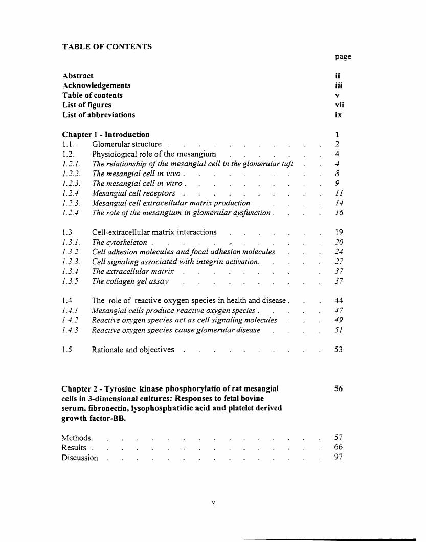
Abstract .4cknowledgements Table of contents List of figures List of abbreviations
ii iii v vii ix
Cbapter 1 - Introduction 1 1.1. GlomeruIar structure . - 7
1.3. Physiological role of the rnesangium . - 4 1.2- 1. n e relationship of the mesangial cell in the glornendar ti<Ji . 4 1 . 2 . 71iemesangialcellinvivo. . . 8 1.2.3. 71te mesangial cell in vitro . . 9 1.2.4 lblesangialcellreceptors . . I I 1.1.3. Mesangial cell extracellular mamir production . . 14 - 4 n e role of the mesangium in glomerular dvssfunction . . . 16
1.3 Cell-extracellular matrix interactions . - 19 1-3.1. The cytoskeleton . .. . . 20 1-32 Ce11 adhesion molendes and focal odhesion molecules . 24 1.3.3. Ce11 signaling associared with integrin activation. . - 27 1.3.4 Dte e-xtracelltdar nratrir . . 37 1.3. j The cohgetl gel assu-v . . 37
1.4 The role of reactive oxygen species in health and disease . . 4 4 1.4. I Mesangial cells produce reactive o-ygen species . . . 47 1.4.2 Reactive O-ygen species act os cell signaling nioleades . . 49 1.4.3 Reacrive oxygen species cause glornentlar diseuse . . 51
1.5 Rationale and objectives . . 53
Chapter 2 - Tyrosine kinase phosphorylatio of rat mesangial ceils in 3-dimensional cultures: Responses to fetal bovine serum, fibronectin, lysophosphatidic acid and platelet derived growth factor-BB.
Methods. . Results . Discussion . . .

Chapter 3 - Reactive oxygen species modulate mesangial cell-extracellular rnatrix interactions
Methods. . . 103 Results - - LOO Discussion . . 133
C hapter 1 - Puromycin aminooucleoside in hibits mesangial cell-induced contraction of collagen gels by stimulating production of reactive o l g e n species
blethods. . Results . Discussion -
Chapter 5 - Sumrnary and conclusions
References

LIST OF FIGURES
Figure 1. Figure 2. Figure 3. Figure 4. Figure 5 . Figure 6. Figure 7. Figure S. Figure 9. Figure 10. Figure I 1. Figure 12. Figure 13. Figure 14. Figure 15. Figure 16. Figure 17. Figure 1 5. Figure 19. Figure 20. Figure 2 1. Figure 22. Figure 23. Figure 24. Figure 25. Figure 26. Figure 77. Figure 28. Figure 29. Figure 30. Figure 3 1. Figure 32. Fisure 33. Figure 34. Figure 35. Figure 36. Figure 37 Figure 38. Figure 39. Figure 40.
An overview of the glomerulus . . Relationship betweeen the mesangium and capillaries. - Ce11 signaling associated with integrin activation . . 1Methods on how to make collagen gels . Mechanisrns of gel contraction . . FBS induces mesangial cell-collagen gel contraction . Tyrosine phosphorylation and FBS-induced gel contraction . Gel contraction inhibited by lavendustin .4 . Gel contraction inhibited by genistein - Lavendustin A inhibits FBS-induced tyrosine phosphorylation . Genistein inhibits FBS-induced tyrosine phosphorylation . Fibronectin induces gel contraction . . Tyrosine phosphorylation and fibronectin-induced gel contraction Tyrosine phosphorylation in 2- and 3-dimensional culture . FAIS phosphorylation and FBS-induced gel contraction . LMAPK and ce11 at tachent . PD 098959 inhibits MAPK - PD 098959 does not inhibit FBS-induced gel contraction .
LPA induces gel contraction . Tyrosine phosphoryiation and LPA-induced gel contraction .
PDGF-BB induces gel contraction . Tyrosine p hosphorylation and PDGF-B B-induced gel contraction PDGF-induced tyrosine phosphorylation in 2-dimensionai culture PDGF-P receptor phosphorylation . Wortmanin inhibits PDGF-BB-induced gel contraction . Wormianin inhibits PDGF-BB-induced tyrosine phosphorylation High concentrations of PDGF-BB disrupt actin filaments .
HzOz induces tyrosine phosphorylation in 3-dimensional culture H 2 0 2 induces tyrosine phosphorylation in 2-dimensional culture HIOl induces tyrosine phosphorylation in trypsinized cells . H,02 induces phosphorylation of F.4K in attached cells . Puromycin-induced gel contraction reversed by catalase . Puromycin-induced gel contraction reversed by DPI . Arninotriazole-induced gel contraction reversed by catalase . Aminotriazole-induced gel contraction reversed by DPI .
HIOz induces collagen gel contraction . Puromycin induces tyrosine phosphoryaltion. . .4minotnazole induces tyrosine phosphoryaltion . Puromycin and aminotriazole induce FAK phosphylation . . FMLP induces gel contraction .
vii

Figure 4 1. Figure 12. Figure 43. Figure 44. Figure 45. Figure 46. Figure 47. Figure 48. Figure 49. Figure 50. Figure 5 1. Figure 52. Fisure 53.
FMLP induces tyrosine phosphoryaltion. . . 134 Neutrophils induce gel contraction . . 126 Neutrophil-induced gel contraction is reversed by catalase . . 128 Neutrophil-induced gel contraction is reversed by DPI . . 139 Neutrophils induce mesangial ce11 tyrosine phosphorylation . l X Activated neutrophils induce mesangial ce11 FAK phosphorylation 13 l Puromycin inhibits FBS-induced gel contraction . - 142 Aminotriazole inhibits FBS-induced gel contraction - . 144 ROS scavengers induce gel contraction . . 145 .Mesangial cell viability as determined by the MTT assay . . 14? ROS scavengers inhibit effects of puromycin on gel contraction 148 DPI inhibits the effect of purornycin on gel contraction . . 119 Puromycin stimulated mesangial cells produce ROS . . 151

LIST OF .ABBREVIATIONS
. IDP
.OIP -4TP BOC DAG D %lEM DMSO DPI ECL F -.UC F AT FBS FGF F'rlLP G.U' GB M GFR Gd72 HBSS IL PI LDL LPA bLUK b t u x s brEM MTT bIT->rnIP P A 1 P .AG E P LLC' PBS PDGF PI-3K PIP PIP, PKC PLA. PLC PLCy P X 4 ?MSF
adenosine diphosphate adenosine monophosphate adenosinetriphosphate BOC-X- fomy lmethionyl leucine pheny lalanine diacy lglycerol Dulbecco's minimal essentiai medium dirnethy I-sulfoxide dipheny lene iodonium cnhanced chemiluminescnce focal adhesion kinase focai adhesion targeting sequence fetal bovine senun fibroblast growth factor Y- fomy imethiony 1 leucine pheny lalanine GTPase activating protein glomerular basernent membrane $ornerular filtration rate
factor receptor-bound protein *
Hanks ' baianced salts solution interleukin inositol trisphosphate low density Lipoprotein Iysop hosp hatidic acid rnitogen activared protein kinases myisto lated alanine-ric h C kinase substrate minimal essential medium [3.(4.5-dimerhylthiazol-1-y 1)l.j-dipheny ltenuolium bromide] membrane-type matrk metaloproteinase p lasminogen activator inhibitor poly acrylamide gel electrophoresis puromycin arninonucleoside nephrosis phosphate buffered saline platelet derived gowth factor phosphatidylinositol-3 kinase phosphatidylinositol phosphate phosphatidylinositol biphosphate protein kinase C phospholipase A2 phospholipase C phospholipase C gamma phorbol 12-rnyistate 1 3-acetate p heny 1-msthy lsulphony 1 fl uoride

PPP RGD RGE ROS SDS SW SOD SOS t-P.4 TBST TC .\ TGF
piatelet poor plasma argge-glycine -aspartic acid argimne-glycine -glutamine reactive oxygen species sodium dodec y 1 sulfate src homolgy superoxide dimutase son of sevenless tissue-type plasminogen activator tris buffered saline with rween trichIoracetic acid transforming growth factor tissue inhibitor of metalloproteinases turnor necrosis factor

Chapter 1 - In troducîion
The mesangium serves as a functional suppon system for the capillary network of the
glomcnilar tufi. Mesangal cells adhere to each other, or to extracellular rnatrix. in a
highly organized Mimensional array. The functionai linkage beween mesangial cells
and their surrounding extracellular matrix is by ce11 adhesion molecdes and it is the
interactions between these components that maintain this 3-dimensional structure as a
Eûnctional unit.
Over the yem. the principal in vitro expenmental method to study cell-ce11 and cell-
extracellular rnatrix interactions in the mesangium has consisted of culture systems in
which rnesangial cells are gown as rnonolayers anached to plasric or some other
subsnate. Cnder these conditions. mesangial cells exhibit a *'proliferative" phenotype
which is different fkom the phenotype of these cells in vivo. Evidence has been
accumularing that when cells are g o w n embedded in matrix rather than anached to
matris (so caiied 3-dimensional compared to 2-dimensional), they exhibit a phenotype
more charactenstic of'cells in vivo.
Ln this thesis we have investigated interactions benveen mesangial cells and their
extracellular rnatrix urilizing a floating 3-dimensional collagen gel assay and have
systematically studied the molecular events involved in the response of rnesangial cells in
;-dimensional culture to various physioloeic agonists. Whiie the work has tocused on
important questions in ceIl biology of mesangial cells we hope to demonstrate that the 3-

dimensional culture system. in particular the collagen gel contraction assay. c m become a
highly informative in vitro mode1 that can be exploited to investigate the role of
mesangial cells in regulating glomerular h c t i o n in both health and disease.
To place this work in contest. the smicture and h c t i o n of the g1omerulus will be bnetly
descnbed.
1.1 Glomerular Structure
The ~lomerulus is a compiex capil l iq bed responsible for ultrafiltering plasma the first
s t q e in urine formation. The filtration barrier of the glornerulus is composed of a
capillary nenvork lined by a t h n layer of endothelial cells. visceral epithelial cells and an
intentening basement membrane which is a synthetic product of the hvo ceil types. The
capillaries are supported centrally by the mesangium whch is composed of mesangial
cells and their surrounding matrix.
A fenestrated vascutar endothelium lines glomemlar capillaries forming the initial banier
to the passage of blood consrituents from the capillary lumen to the u r i n q space. The
fenestrations, about 700 nm in diarneter, are small enough to prevent the passage of blood
cells into the subendothelia1 space. Outside the capillary wall are the large visceral
epithelial ceils or podocyes. These cells have foot processes extending from their bodies
that arborize and contact the ~lomerular basement membrane. The distance benveen these
foot processes, referred to as the slit pore, varies h m 15 to 60 nrn and is bridged by the

d i t diaphragm (Tisher C.C. and 'iladsen K.M. 1991). Visceral epithelial cells are
responsible. at least in pan, for synthesis and maintenance of the glomerular bassrnent
membrane and are capable of producing collagenous proteins and glycosamino~i~cans
(Foidart f .B.. zt al., 1980: Striker G.E. and Striker L.J., 1985).
The glornerular basemenr membrane (GBM). interposed beween the visceral epithelial
cells and the endothelium. consists of a centrai dense Iayer. the lamina densa and iwo
thinner layen. the lamina rara interna and externa. The GBM is composed predominantly
of a procolla~en-like molecule associated with rnatrix glycoproteins. This molecule is
composed of dires identical u-chains rich in hydroxylysine. hydroxqproline. and glycine.
Lesser quantities of collagen IV and V. as well as laminin. fibronectin and other
extracellular matrix components are also found in the GBM (Courtoy P.S.. et al.. 1982:
Dean D.C., et al., 1933; Mminez-Hernandes .\.S., et al., 1981: Tischsr C C and Madsen
K.M.. 1991). The GBM has fixed nejatively charged anionic sites that consist of
protroglycans. predominantly perlecan. Size and charge selsctivity of the glomerular
filtration banier. which effectively resrricts the passage of molecules of the size of
alburnin or larger into the ultrafiltrate. is largely determined by the G B M
m e capillary loops are supponed by the mesangiurn and project into a space bounded by
Bouman's capsule. of which the outer layer is formed by thm parietal epithslial cells and
their basernent membrane. At the urinary pole the capsulu cells are in direct contact u-ith

cells lining the early proximal convoluted tubule. At the vascular pole these capsuiar ceils
are continuous with visceral epitheIial ceIIs (fi,aure I ).
Although the mesangium itself does not constitute part of the filtration barrier it does play
a major role in rnodulating its function.
io lo~i 1 1.2. Phvs
1.2.1. The relationshi~ of the mesangai cell in the olomenilar t ~ f i
The mesangium forms a central s tak that supports the anastomosing capillary nenvork of
the glomemlar nifi (Mene P.. et al., 1989). The capillaries are w~apped about the
mesangial stalk and are bordered by the mesangium and GBM for their entire len-gh
(figare 2 ) . The GBM encircles most of the circurnference of the glomemlar capillary.
however there is a small portion of the capillary where no GBM is present. Over this area
the mesangium attaches directly to the capillary wall. Mesangial cells aiso anach ro the
GBM at the "mesangal angles" (points where the GBM deflects from a purely
pcncapillary course to envelop the mesangium) (Sakai T. and K n z W.. 1987) and have
multiple GBM attachrnents within the axial gIomenilus (Lemley K.V., et ai.. 1992).
Because of the multiple at tachent sites of mesangial cells to the GBM. as well as the
fact that the GBM encircles most of the glomerular capilla., the GBM is believed ro be
the pnmary site where mesangial cells exen their effect on giomerular capillary lumenal
diameter. It is also postulated that mesangial cells play a role in counteracting glomsrular
capillary pressure (Knz W., et al.. 1990). Through such mechanisms, i t has been

Figure 1
The g!omerulus. Blood enten via afferent (.LA) and leaves via efferent (EX) artenoles.
Capillaries are lined by endothelial cells (EX) and covered by foot proceeses (F) of
visceral epithelial celis (EP) attached to the glomerular basement membrane (GBM).
Parietal epithelial cells form Bowman's capsule (B) whch joins the epitheliurn of the
proximal tubule (PT). Ultrafiltrate collects in Bowman's space (BS). (blodified from
Roushanpour E. and Knz. W., 1986)

Figure 2
Diagram of die mrsangial region. The rnesangiurn. consisting of mesangial ceils (hl) and
matrix (MM), is in the centre of capillaries. Parts of the rnesangium are covered 6y
basement membrane that consists of the lamina rara interna (LN), lamina densa (LD) md
lamina rara cxtema (LRE). The LW is continuous with the rnesangium and foot processes
of epithelial cells (Ep) anach to the LRE. Capillaries are lined by endothelial cells (En)
with fenestrations (F) allowing plasma to move into intracellular channels ( ICI.
(Modified fkom Lata H., 1993)

proposed that mesangsal cells regulate giornenilar filtration rate in vivo (Kiahr S.. et al..
198% Kreisberg J.. et al. 1985).
Rzcently. a study has demonsnated that Ij06 of capillary loops are totally surromdeci by
mesaqial cells (Inkyo-Hayasaka K.. et al., 1996). This indicates that mesangiai cells in
thtir owvn right may have a siwficant influence on the distribution of blood Bow in the
domerulus. Mesangial cells therefore can play a direct role in regulating jlomenilar V
filtration rate (GFR) through a combination of isometric and isotonic contractili~. Static
or îsomcmc contraction of mesangial cells is suggested by the cytoskeletaI composition
of rnçsangial cells in that they contain both actin and myosin. Dyamic or isotonic
contraction of mesangial cells is suggested by findings in mesangial cells p w n in 2-
dimensional culture and isolated jlomeruli showing contraction in response to vasoacrive
substances (.AusielIo DA, et al.. 1980: Fujiwara Y.. et al.. 199 1; Kreisbers J.I.. et al..
1985: Schlondorff D.. 1987; Singhal P.C.. et al., 1986). If mesangal cells dernonstrate
static contraction. the mesangial loops c m fuiction as a safety device against expansion
of the capillaries due ro excessive inuaglornexular pressure. Thus rnesangial cells
themselves can regulate the distention of the capillary walls. Dwamic contracrion of
mesangial cells on the other hand can bnng about contraction of about 1 j06 of rhe
olomemlar capillaries with consequent redistribution of intraglomenilar blood flow which -
effectively decreases filtration surface area (uikyo-Hayasaka K.. et al.. 1996).

It is far from clear how the mesangium controls glomerular filtration. However it is
evident at the cellular level one of the mechanisms whereby mesangial cells exen their
control on glornerular filtration is through mesangial ceil-extracellular matrix
interactions. This reaiization has very significant implications because it means that
mesangial cells g o w n in 2-dimensional systerns, traditionally used to explore the ce11
biology of the rnesangial ce11 in vitro, are probably inadequate and instead require the
more complex 3-dimensional culture system.
1.2.2. The mesangial ceII in vivo
In vivo. the majority of mesangial cells (85-90%) are irregular in shape and have a dense
nucleus. Their cytoplasmic processes, rich in microfilarnents, attach to the capillary
lumen or GBM via ceil adhesion molecules (Drenckhahn D., et al., 1990). Mesangial
cells contain a network of c ytoskeletal contractile elements including ac tin, myosin, u-
actinin, and tropomyosin (Xndrew P.M. and Coffey A.K., 1952; Becker C.G., 1972).
however no a-smooth muscle-type actin is found normally in these cells in vivo (Elger
M., et al., 1993; Kimura K., et al., 1995). Therefore, although mesangial cells have been
compared to vascular smooth muscle cells and pericytes, the composition of contractile
proteins in these two ce11 types differ (Schlondorff D., 1987). Mesangial cells only appear
to express a-smooth muscle actin when they change phenotype in glornerular disease
(Alpers C.E., ct al., 1992; Elger M., et al., 1993; Floege J., et al., 1992; Johnson R.J., et
al., 1997). Mesangial cells also produce large amounts of extracellular matnx
characterizing them more aiun to myofibroblasts (Johnson R.J, et al., 1992b).

The other 10-15% of mesangial cells. are denved fiom bone rnarrow and have
characteristics of monocytes/macrophages (Schreiner G.. 1992). These cells possess
phagacytic ability, express Fc recepton and display the comrnon leukccyte antigen. the
Ia antigen and C3b receptors (Schreiner G. and Unanue E.F., 1984). These cells. as well
as resident mesangial cells. are thought to be involved in the uptake and clearance of
macromolecules bom the glomerulus (Davies M., 1994).
1.2.3 The mesanrriaI ce11 in vitro
The majority of mesangial cells used in vitro are grown on plastic in a 2-dimensional
system. These cells are grown out fiom whole glorneruli, plated on plastic culture dishes
and maintained in medium containing high amounts of fetal bovine S e m (FBS).
Cultured mesangal cells exhibit rnany of the characteristics of mesangial cells that are
found in vivo; however, the phenotype is altered in that u-smooth muscle actin is
expressed (Elger M., et al., 1993). In 2-dimensional culture. mesansial crlls are
charactenzed as intrinsic mesangial cells by immunofluorescent staining which reveals
prominent intracellular cytoskeletal fibrils of actin and smooth muscle myosin, desmin
and vimentin arranged along the axis of the cell, and the absence of factor VIII,
cytokeratins and angiotensin-converting enzyme activity and common leukocyte antigen
(Davies M., 1994). As cells grow to confluency they resemble smooth muscle celk.
Although rnuch important information has been gained Eom cells gown in these 2-
dimensional cultures, there are a number of issues that need to be taken into consideration

when interpreting results from these culture systems before applying this information to
the in vivo situation. In vivo. mesancjal cells exist within a 3-dimensional environrnent
whcre the composition of extracellular matrix has been implicated in the control of ce11
differenriation, gene expression. migration, and proliferatxon of cells (Bomkldt K.E.. er
al.. 1995: Lin C.Y. and Grinnell F., 1993; Marx M.. et ai., 1993: Mars M.. et ai.. 1994:
Rankin S. and Rozengurt E., 1994; Zhu X. and Assoian R.K.. 1995). Thus. the spatial
organization of the components of the mesangal matrix is iikely to play a cntical role in
maintaking the normal rnesangial ce11 phenotype. that is. the non-prolifenting ceil
defined in morphological studies (Davies M.. 1994). Mesangid cells rnust b r passaged a
number of times (4-5) to obtain suficient numbers for cxperimental purposes. Rcsults
from experiments using cultured cells rnust be interpreted in the context of this
dedifferentiation. A relativety high concentration of FBS is needed to maintriin and
establish cells in culture systems. FBS provides the cells with fibronectin. larninin and
sowth factors necessary for cells to proliferate. -4s a consequence of these serurn factors. -
tells in 2-dimensional culture probably represent activated mesangial cells compared to
non-proliferating mesangial cells found in the glomerulus.
1.2.4 Mesmoial ce11 recentors
Despite limitations, mesangial cells grown in 2-dimensional culture. have increased our
knowledge O t' the ce11 biology and pathophysio l o g of mesangium. Numerous receptors
for ligands. which include eicosanoids, vasoactive peptides. neuropeptides. gowth
factors, and extracellular rnarrix, have been identified on mesangial cells (Davies !VI.,

1994). Cellular responses to these ligands have been investigated in terms of mesangial
contractility. ce11 proliferation and production of extracellular matnx. In this section the
vasoactive growth factor receptors are bnefiy discussed and integrins and their
interactions with extracellular matrix are reviewed as a separate section.
Vasoactive Receptors
In ?-dimensional ce11 culture systems, vasoactive peptides, which include angiotensin II.
vasopressin, norepinephrine, endothelin-1, thromboxane, adenosine. serotonin and
leukotrienes have been dernonstrated to induce mesangial ce11 contraction (Xusiello D., et
al,, 1980; Mathieu P.R.. et al., 1980; Mene P., et al., 1989). The ce11 signaling associated
with contraction of cells induced by these substances appears to have a common
mechanism. Ligand binds to its G protein coupled transmembrane receptor whic h
activates phospholipase C (PLC) leading to cleavage of phosphatidylinositol
bisphosphate (PIP,) with the formation of the second messengers inositol trisphosphare
(PI) and diacylglycerol (DAG). The IP, stimulates release of ca2- from intracellular
stores. ca2- binds to calmodulin and activates, arnongst other e n z p e s , myosin light
chain kinase with subsequent phosphorylation of myosin light chain leading to
contraction of the cell.
When rnesenchymal cells are gram in a 3-dimensional floating collagen gel, they spread
and with time contract the collagen gel. The mechanism will be discussed later. The
addition of vasoconstrictive agents. like dibuteryl CALIP. to these cells gown in 3-

dimensional gels will cause the cells to "round up" (Ailenbers ,M., et al., 1990) resulting in
inhibition of cell-induced gel contraction. These terminologies create a great deal of
confusion as collagen gel contraction in 3-dimensional culture systerns is caused by ce11
attachent and spreading, and inhibition of gel contraction is produced by rounding up of
cells (often descnbed as ce11 contraction in 2-dimensional culture systems).
Gro~vth Factor Receptors
Many semm factors alter the growth capabilities of mesangial cells grown in 2-
dimensional cultures. iMost growth factors are mitogenic with a few, like TGFP, having
predominantly anti-rnitogenic actions. To discuss the specific effects of al1 the factors is
beyond the scope of this review and only the groowth factors relevant to this thesis. in
particular PDGF-BB, wiil be emphasized.
PDGF
PDGF is a farnily of closely related proteins of approximately 30 kDa which are
synthesized as dimers of A and B chains. PDGF acts locally in an autocrine or paracrine
Fashion as it is rapidly inactivated by binding to a 2 rnacroglobulin (Ross R., et al., 1986).
The levels and type of PDGF recepton expressed by target cells determine the cellular
response to PDGF (Abboud H., 1993; Daniel T.O. and Kumjian D A , 1993). The PDGF-
p receptor recognizes predorninantly the B chah of PDGF and the PDGF-a receptor the
A chain. Both receptors transduce the rnitogenic signal of PDGF. However, in fibroblasts
actin reorganization and chernotaxis are mediated only by the P receptor (Heldin C.H..

1991). Binding of PDGF to the PDGF receptor induces receptor dimerization which
resuIts in ïeccptor autophosphorylation and provides a t tachent sites for subscrate
proteins. PDGF receptors associate with several SH3 domains containing substrates for
protein tyrosine krnases inciuding phosphatidylinositol-3 kinase (PI-3K). GTPase
activating protein (GAP). PLC-y, and src (VaIius hl. and Kazlauskas -4.. 1993). At l e m
one of these pathways converges on the RasjM4PK signaiing ultimatefy ieading to
activation of the .2P-1 transcription factor (Wennstrom S.. a ai.. 1994). PI-3K and GAP
mediated cell signaling pathways have been associated with cytoskeletal reorganization in
fibroblasts (Rankin S. and Rozengurt E.. 1994: M c Glade J.. et al.. 199;). The role of
PDGF in modulating cytoskeletal function in the $omerular mesangium is largely
PDGF protein as well as PDGF receptors are expressed in large quantities by mesanria
cells grown in 2-dimensionai culture systems (Abboud H.. 1993: Floese J.. et al.. 1993
Floege J.. et al.. 199 1 ) relative to expression in normal adult kidneys (Abboud H.. 19951.
In contrast. mesangial cells g o w n in 3-dimensional culture express approsirnareiy LOOh
of PDGF receptors compared to mesangial cells groown in 2-dimensional culture (.LLm
41.. et al.. 19933, which approximates the receptor expression in vivo. Most in vitro
research on the effects of PDGF on the mesangium is performed on rnesangial ceils
gown on 2-dimensional plastic plates. Mesangial cells ;ro\vn in this manner have
increased receptor expression and data collected from this system demonstrating
rnitogenic activity by PDGF cannot be applied to the in vivo situation. Cclls grown in 2-

dimensional culture systems change to a proiiferative phenotype (Bornfeldt K.E., et al..
1995; Charnbley-Campbell J.H.. et al., 198 1) and these changes allow ce1 1s to respond to
mitogens.
It is for this reason that 3-dimensiona1 culture systems are probably a more realistic
mode1 system of the mesangial ce11 in vivo. This mode1 will be used to delineate some of
the PDGF-induced ce11 signaling pathways that alter mesangial cell-exnacellular manix
interactions.
1.2.5 lMesanoial ce11 extracellular matrix production
Mesangial cells produce extracellular mahix components which combine in an orjanized
manner to make up the rnesangial matrix. .Mesagial rnanix supports the glomemlar tufi
and may also affect the flux of macromolecules through the mesangiurn. The major
constituents of mesangial matrix are secreted by the rnesangial ce11 (Couchman J.R.. et
al.. 1994). These extracellular marrix cornponents are continuously synthesized and
degaded and it is imbalance of these regulated processes that results in extracellular
matrix deposition which characterizes sclerotic glomemli found in end stage renal
disease. Soluble mediators such as growth factors. (e.3. TGF-P and PDGF), cytokines
(e-g. M - a ) and hormones (e-g. angiotensin II and vasopressin) stimulate the excess
production of extracellular matrix (Rupprecht H.D.. et al., 1996). In addition extracellular
matrix production is regulated by metabolic factors, e.g. high glucose causes diabetic

giornerulosclerosis. and reactive oxygen species (ROS) are associated with puromycin
nephropathy (Jones CL. . et al.. 1992).
The degradation of exnacellular matrix is reguiatrd by proteinases and their specific
inhibitors. Two classes of proteinases identified in mesangial cells both in vivo and in
viuo are the metalloproteinases and plasrninogen activators. -Metalloproteinase. a 72 kDa
protein. is released in a latent f o m and acnvated by membrane-type rnatrix
rnetalloproteinase (MT-MMP) resulting in degradation of extracellular mamx
rneralloproteinase (Alenberg, _LI. and Silverman. M.. 1996). Mesangial cells also secrets
tissue inhibitor of metalloproteinases (TDIP) 1 and 2, inhibirors of this T2 D a
rneralloproteinase. Culmed mesangal cells secrete sznall amounts of tissue-type
plasminogen activator (t-PA) and a 100-fold geater amount of tissue plasrninogen
activator inhibitor ( P X ) . t-PA is maintained in an inactive complex with PA-1 (Lacave
R.. et al.. 1989) and P.AI and t-PA synthesis is modulated by substances found in serum.
For example W - u selectively stimulates PAi syndiesis (Meulders Q.. er al.. 1992).
u-hile thrombin stimulates both [-PA and PAU (Villamediana L.M .. et al.. 1990).
The control of cxtracellular matrix deposition is complex. It is presently unclerir as ro the
relative contributions of rnatrix accumulation by synthesis, degradatisn and inhibiton of
degadative enzymes in the mesangium. Change in activity of any of these rnechanisrns
c m alter the amount and composition of extracellular matrix and ultimately influence
mrsangiai cell-rnatrk interactions and their biological effects in the glomenilus.

1.2.6 The role of the rnesangurn in domerular dvshnction
In patients with glomenilar disease, the usual clinicai presentation is either that of the
nephrotic or the nephritic syndrome which rnay or rnay not be associated with an
alteration in glomerular filtration rate. These conditions are associated with proliferation
of cells and/or accumulation of extracellular matrix in many instances. However, in
minimal change glomerulonephritis there are no changes in the glomemlus that can be
visualized on light rnicroscopy and only epithelial foot process effacement is seen on
electron microscopy.
Mesangial cells appear to play a role in a11 three pathological forms of glomemiar disease
and the role of the mesangiurn in proliferative glomenilonephntis as well as glomemlar
disease associated with mesangial expansion will be discussed in this section. Minimal
change glomerulonephritis and its mode1 systems will be discussed in geater detail latrr
in the introduction as much of the work in the thesis relates to this disease process.
Proliferation of glomemlar mesangial cells occurs in a nurnber of human glomerular
diseases including IgA nephropathy, membranoproliferative glomenilonephntis. some
forms of focal segmental glomemlosclerosis, lupus nephritis. difkse proliferative
nephntis associated with streptococcal infection, and possibly diabetic nephropathy
(Floege J., et al., 1993b). Although the initial stimuli resulting in this proliferative
response Vary considerably, immune complex deposition in the mesangium is one of the

common initiating events in proliferative glornenilonephntis. Deposition of these
ues to complexes is a potent stimulus for the recniitment of neutrophils andior macropha,
the mesangium. These leukocytes as well as the mesangial cells thernselves are activated
through cell surface receptors resulting in the production of prostaglandins. growth
factors like PDGF, cytokines, fÏee oxygen radicals and activarion of the cornplcment
cascade (Abboud H.. 1995; LMatsurnoto K. and Hatano M., 1991; Santiago A.. et al..
1991; Sedor J.R. and Abboud H., 1986). al1 stimulating mesangial cell proliferation.
Direct mesangial damage due to ischernia or ROS may also be a stimulus for mesangial
cell activation and proliferation. In the majority of cases of human glornerulonephntis.
recognition of the precise stimuli for mesangial ce11 activation and recruitment of
macrophages and/or monocytes to the mesangun remains unclear.
in an effort to obtain a better understanding of the mechanisrns of ce11 proliferation in
human g1ornerulonephritis, numerous animal models simulating these disease processes
have been developed. These include. amongst others. experimental models of 1-
nephropathy (Rifai A., 1987), glomerular disrase induced by snake venoms (Bradfield
J.W.B.. et al., 1977), remnant kidney models (Floege J., et al.. 1992), and the anti-Thy 1.1
mode1 which ciosely mimics a mernbranoproliferative glornerulonephntis (Bagchus
W.M.. et al.. 1986; Johnson R.J., et al., 1990). Multiple facton. including cornplement.
cytokines, ROS. and growth factors have been implicated in the pathogenesis in these
rnodels. tn particular, the g r o ~ h facton PDGF and fibroblast growth factor (FGF) have
been implicated in the mesangial-proliferative response (Johnson R.J.. et al.. 1990).

Lnfominately these expenmental models do not lend themselves well to in vitro systems
and the cellular and molecular mechanisms which stimulate mesangial proliferation have
not been comprehensively snidied. [n addition. al1 the in vitro work perfomed on
mesangial ceIIs in these mode1 systems involved 2-dimensional culture.
.Llesangral errraceilulnr mnrrir expansion
Expansion of glomemlar extracellular matrix is the hallmark of man- glomerular
diseases. It is present in early srages of diabetic nephropathy and soms
olomentlonephntis and is the final common pathway of end stage renal dissase which is - charactenzed by small. shninken sclerosed glomeruli. Mesangial ce11 culture techniques
and rat models have been used to identify the mechanisms whereby extracellular marrix
metabolism is dysfhctional. For example, mesangial cells g o i n in high concenuations
of glucose secrete increased arnounts of type TV collasen. laminin and Fibronectin:
however the catabolism of these proteins remains unchanjed (Davies hl.. 1994. In
addition growth factors like TGF-P and PDGF have been impiicated in the pathogencsis
of increased mesangial expansion in both diabctes and glomerulonephntis (5akamura T..
et al.. 1993; Okuda S.. et al., 1990). Complement srimularion of mesangial crlls. in
concentrations that do not produce ce11 lysis. induces production of exnacellular manix in
addition to associated CO-inflamrnatory mediators iike interleukm 1. interleukin-6. tumor
necrosis factor and ROS. in normal conditions mesangial cells express classical recepton
for low densicp lipoprotein (LDL) and these are found in increased quantities in the
nephrotic syndrome. Exposure of mesansial cells to increased quantities of LDL for

prolonged penods results in upregulation of type IV collagen and fibronecrin gene and
proiein expression (Kim S.B., et al., 1992; Rovin B.H. and Tan L.C.. 1993).
Similar to studies used to investigate the proliferative response of rnesangial cells in
glomerulonephritis. almost al1 in vitro eexpenments snidying mesangial matrix expansion
have been performed in cells grown in 2-dimensional culture. -4s we were interested in
studying behavior of non-proliferative cultured mesangial cells with an in vivo phenotype
in a glomerulonephritis model, we elected to use a 3-dimensional culture system.
1.3 Cell-extracellular rnatrix interactions
Cs11 adhesion is vital to maintain individual celIs in three-dimensional tissues which
make up organs. Cells are organized into highly diverse and distinctive patterns and a
variety of ce11 adhesion mechanisms are responsible for attaching ceIls to each other and
to their surrounding extracellular matrix. Ce11 adhesion proteins are usuallp
transmembrane glycoproteins that rnediate binding interactions at the extracellular
surface which determine the specificity of cell-ce11 and cell-extracellular matrix
interactions. These proteins include superfamilies of intekgins, cadherins,
irnmuno~lobulins, selectins and proteoglycans. The ce11 adhesion receptors reco-pize and
interact with other ce11 adhesion molecules or with extracelluiar matrix. The estracellular
matrix proteins are large glycoproteins and include collagens, fibronectins, laminins and
proteoglycans that assembIe into cornplex macromolecular arrays. On the intraceilular
surface of the plasma membrane these adhesion recepton associate with cytoplasmic

proteins which link the adhesion system to the cytoskeleton, regulate the Functions of the
adhesion molecules and transduce signals initiated at the ce11 surface by the adhesion
receptors (Gumbiner B.M., 1996).
This thesis will concentrate on the interactions between mesangial ceils and extrace[luIar
manil. In particular the ce11 signaling associated with cell-extracellular matrix
interactions of mesangial cells grown in a floating 3-dimensional gel will be assessed.
This systern requires an intact cell-actin cytoskeleton, ce11 adhesion molecules.
extracellular matrk to which the cells attach and an intact ce11 signalin; system. In the
next section each of these components will be discussed.
I 2 . 1 The Cytoskeleton
The ability of cells to adopt a variety of shapes and carry out directed movement depends
on the cytoskeleton, a complex network of protein filaments that extends through the
cytoplasm of the cell. The activities of the cytoskeleton depend on three principal types of
protein filaments: microfilaments (actin filaments), microtubules and intermediate
filaments. These Filaments are cornposed of protein monomers that are assembled into
more complex structures by interacting wirh their associated proteins. These proteins
either link the filaments to one another or to other stnictures, for example with the ce11
membrane. Discussion will be limited to the actin cytoskeleton as its structure and
function has relevance to ce11 adherence and ce11 mobility. both vital components OF the
gel contraction assay.

Actin is one of the most abundant proteins in eukaryotic cells and is found throughout the
cytoplasm. There is an especially dense network of actin and its associated proteins.
found just beneath the plasma membrane, which gives mechanical strength to the cell.
Approximately half of the actin molecules found within a ce11 are unpolymerized and
exist as free monomers or srnall complexes with other proreins. A dynarnic equiiibnum
exists between these monomers and the formed actin filaments which helps to drive
changes in ce11 shape.
In order to understand the mechanisms whereby actin can alter ceil shape it is essentiai to
understand the mechanism of actin polymerization and depolymenzation. In a cell free
system globular subunits of actin (G-actin) polyrnerize to form Iinear polçmers (F-actin)
of several micrometers in length in a reversible reaction that is independent of other
proteins and is simply determined by the temperature and ionic characteristics of the
solution (Stossel T.P., et al.. 1985). Polymenzation of actin is dependent on salt
concentrations and spontaneous actin pol ymerization requires a concentration of at kasr
10 miLi of a monovalent salt (Maniyama K. and Tsukagoshi K., 198-1). In addition, actin
dimers are thermodynamically unstable and tend to disassociate. However when
aggeegates containing a critical number of monomers assemble. monomer addition
becomes more probable than disassociation, hence addition to these "nuclei" becomes
more rapid. Nucleus formation is the rate limiting step in the polymerization reaction and
filament length is inversely proponional to the number of fibrrs formed from these nuclei
(Stossel T.P., et al., 1985).

Actin polymerization is an energy dependent process. Actin monomers bind the adenine
nucleotides ATP and ADP. and actin polymerization is associated with hydrolysis of
actin-bound ATP ro ADP. The rate and extent of polperization varies geatly depending
on whether the nucleotide bound to the actin monomers is ATP or ADP. .As ATP
rnonomers disassociate slower f?om filaments than do ADP monomers. having ATP
monomers at the ends of the actin filaments retards monomer loss kom the filament ends.
Therefore when actin is assembling, the effect of delayed XTP hydrolysis is to promote
actin polymerization. Conversely when depolymerization is taking place. monomer loss
is facilitateci by exposure of A D P monomerç formerly in the interior of the filament
Wegner A.. 1985).
Actin filaments are bipolar and exhibit so called "barbcd and "pointed" ends. Actin
rnonomers add more rapidly to rhe barbed or (-) end than the pointed or ( - ) end. A
different concentration of monomers is required at either end ro rnaintain a steady state.
This difference of cntical concentmtions. dic tated b y the opposire ends of actin fi lamrnts.
is consistent with the idea that actin monomers cycle through the actin filament from the
( - ) to the (-) end by the process called treadmilling (Stossel T.P.. et al.. 1985). Cnder
ionic conditions, similar to those found in cells. the p w t h of the (-) end of an actin
filament is 5 to 10 times faster than the (-) end. therefore by anchoring the (-) end of a
filament in a sprcific orientation the ce11 c m determine rate and direction of the g row~h of
the filament.

Proteins that interact with actin. the so called actin binding proteins. can modulate the
polymerization of G-actin into F-actin. Some of the proteins. for example profilin, bind to
actin monomers but do not form stable complexes with actin filaments. The major effect
of these proteins is to retard the incorporation of rnonomers into actin filaments. hence
inhibiting filament extension. Other proteins, the so called "capping" proteins like
gelsolin. bind to one end of the actin filarnent and prevent the exchange of monomers at
that end therefore decreasing actin polymenzation. Actin filaments in cells are linked
togerher by cross-linking proteins that result in the formation of actin networks and
filament bundles. It is these actin filament bundles that bind to the plasma membrane and
form the so called focal contact or focal adhesion.
It is clear that actin and its interactions with actin binding proteins are essential for ce11
migration. however the rnechanism whereby this occurs is uncertain. The most
cornpelling hypothesis, based on evidence compiled from multiple oganisms, suggests
that the initiating event for actin-based ce11 motility is the exposure of actin filament
barbs that lie just below the plasma membrane. Two capping proteins, gelsolin and capZ.
which are c;* dependent, are likely targets for signal pathways that initiate this actin
filament rnobilization. Gelsolin binds and severs pre-existing actin fibers, increasing their
number while decreasing their length. These short filaments are then capped by gelsolin
and cap2 until the next phase, when following another signal, these capping proteins
disassociate from the filament end once again creating nucleation sites for actin assemblv.
Actin monorners, which are released korn profilin, are added to these nucleation sites and

actin polymerization at these nucleation sites results in formation of lamellipodia which
in tum allows for ceil movernent (Barkalow K. and Harhivig J.H.. 1995).
c o n 1.32 Ce 1 adhesi adhesi
Inte-gins are a distinct fami iy of a p transmembrane heterodimeric glycoproteins that
contain an a (120-1 80 ma) and a P (90- 1 10 D a ) c h a h They are subdivided into groups
based on the homologous P chah and certain a chains c m assosiate with more than one P
c h a h There are 14 distinct a subunits and more than 8 P subunits that c m combine in
various combinations (Juliano R.L. and Haskil1 S., 1993). The u and P chains do not share
any si-enificant similarity. They have a large extracellular dornain, a single membrane
spanning region and a short cytopiasrnic domain (Akiyama S.K.. et al.. 1990). The
extracellular domain contains a ligand containing pocket that binds with multiple
extracel lular ligands which include extracellular matrices ( fibronectin, vitronectin. collagen,
osteopontin, epiligrin and laminin), soluble proteins (Van Willebrand factor. fibrinogen.
fibronectin, vitronectin and the C3b component of complement), members of the
irnrnunoglobulin superfarnily intercellular adhesion molecules and vascular ce11 adhesion
molecules (Faull R. and Ginsberj M.H., 1995). Specificity for the extracellular matris
proteins is through specifc binding domains for exarnple the arginine-glycine-aspartate
(RGD) binding site.
It is becoming evident that integrins are not simply adhesion receptors, but play a larger role
in bidirectional signaling between the ce11 and its extracellular environment (Hynes R.O..

1992). htegrins are able to m s d u c e so called "outside-in-signaling" which refers to the
transduction of ce11 signaling events within the ce11 following ligand binding. X rypical
example of this form of sigaling is the aggregation of integnns into focal adhesions
following binding to Iibronectin (Bunidge K., et al., 1992). A second type of signaling is
"inside-out-signaling" whch refen to the fact that integrin affinity for ligand can be
dynamically reguiated hom the cytosol. This means that integins ur.derso changes in
conformation and binding amnity in response to extemal stimuli and intemal signal
transduction (Stuiver 1. and O'Toole T.E., 1995).
The P cytoplasmic subunit of integrins has been shown to associate with the cytoskeleton
through the cytoskeletal proteins a-actinin and talin (Honçitz A.F., et al.. 1986; Otey C.A..
et al., 1993). These associations are important for "outside-in- signaling" especially when
integins interact with ligand resulting in the formation of focal adhesions. Focal adhesions
consist of extracellular matrix components on the exterior, brought into close contact with
a macromolecular cytoskeletal complex consisting of cytoskeletaI proteins including u-
actinin. vinculin, talin, paxillin and tensin linked to actin filaments, by a clustenng of
in teans (Yamada K.M. and Miyarnoto S., 1995). These focal contacts not only provide
an important structural h e w o r k for die ce11 but also serve to localise proteins for
subsequent sigaling events (Sastry S.K. and Honvitz X.F., 1993).
The P, cytoplasmic tail is necessary for the formation of focal adhesions as integins lose
their ability to redistribute proteins like a-actinin or talin if this integrin tail is mincated

(Lewis J..M. and Schwartz -%I.A., 1995). Aithough the jutamembrane sites for focal
adhesion kinase (FAK) and a-actinin are present next to the inte-grin tails. clustering of
these proteins does not induce focal adhesions (Sastry S.K. and Horwitz X.F.. 1993;
Yamada K M . and Miyamoto S.. 1995) once again irnplying that the B, cytoplasrnic tail is
necessary for the formation of focal adhesions. Multiple signaling molecules such as
cytopiasmic tyrosine kinases, protein kinase C, Ras-GAP, inositol lipids, arachîdonic acid
denvatives and M o have been implicated in regulating the formation of focal adhesions
(Yarnada K.M. and Miyamoto S., 1995). Studies on cells grown in ?-dimensional culture
have demonstrated that focal adhesion sites are not constant in their composition and are
being continuously formed and disappearing with an associated change in the
composition of molecules.
Focal adhesions appear to be primarily an in vitro phenornenon and in vivo they have
only been found in endothelial cells present at sites of hi& hernodynamic stress (Yamada
K.M. and Miyamoto S., 1995). There are however "extracellular matrix contacts" in vivo
which consist of clusters of integnn recepton that bind to fibrils of extracellular matrix
(especially fibronectin) and have distinct associations with cytoskeletal proteins (Chen
W.T. and Singer S.J., 1982). Many integrin associated signaling molecules. for exarnple
son of sevenless (SOS) and Jun kinase are absent fiom focal adhesions but present in
these "extracellular matrix contacts" (Yamada K.M. and Miyamoto S.. 1995). At present.
al1 the in vitro information regarding the formation of focal adhesions has been collected
from 3-dimensional culture systerns.

1.3.3 CeII sigalirlg assoc'ated I with intemin activation
Inte-gin activation, usually induced by an interaction with ligand or anti-integrin antibodies,
initiates a number of ce11 signaling pathways. Tyrosine kinases are the best described ce11
signaling molecuies associated with integrin activation (Schlaep fer D.D .. et al.. 1 994).
however senne threonine kinases such as protein kinase C (Vuori K. and Ruoslahti E..
1993) and mitogen activated protein kinases (MAE'K) (Chen Q.M.. et al.. 1994) are also
activated by stimulated integms.
htegrins associate wiîh qvtoskelera f proreins leading ro &rosine p hosp ho~atiori
hlthough the integrin cytoplasmic domains do not in themselves possess cyrosine kinase or
phosphatase activity, their binding with extracellular matrix initiates multiple
phosphorylations of intncellular components. This phenornenon occun approximately 5 to
10 minutes after cells anach to their exiraceilular rnatrix (Burridge K.. et al.. 1992).
F S . a highly conserved protein, is the best described of these intemediary proteins and
appears &O play a central role in integrin-mediated signal transduction (Richardson A. and
Panons J.T., 1995). It appears as if information located in the f.3 cytoplasmic domain is
sufficient for FAK activation, however calcium transients and protein kinase C may b s
required as costirnulatory events (Shattil S., et al.. 1994; Vuon K. and Ruoslahti E., 1993).
The C terminal domain of this protein has a proximal focal adhesion targeting sequence
(FAT) necessary for the protein to localise to focal adhesions. The NH: teminal can
localise to numerous p tails of integins (Hildebrand J.D., et al.. 1993). F.4K is an

interesting protein in that it lacks noncatalytic motifs, like the src homolgy SH2 or SH3
domains. Found in other receptor proteins (Zachary 1. and Rozengurt E., 1992). Despite th~s.
F;U( appean to couple with other proteins Iike paxillin. that contain these SH2 and SH?
dornains, through distinct sequences localised to its tyrosine phosphorylation sites in the
COOH domain (Pawson T., 1995; Schaller M.D. and Panons J.T., 1994). It is throu& these
linkages that F A K is capable of integrating the signaling processes triggerred by integrin
activation.
Although F M is the best descibed of the tyrosine kinase molecules associated with inte-grin
activation. the mechanism by which FAK is "wired" into die integrin signaling pathway is
not kno~m. F.U( c m interact in vivo with peptides f?om P inte-gin subunits. however no
direct association behveen integrins and tyrosine kinase activity has been described in cell
lysates (Clarke E.4. and Brugge J.S.? 1995). Tyrosine kinases only interact indirectly with
integrins through associations with the cytoskeletal complexes of the focal adhesion which
are induced by activated integrins. This mechanisrn of activation is supported by the fact
that cytoskeletal disruption with cytochalasin D inhibits integrin-induced tyrosine
phophorylation (Burridge K.. et al., 1992).
The cytoskeletal proteins pavillin and tensin are substrates of tyrosine phosphorylation.
Paxillin colocalises with talin and vinculin to focal adhesions, where it enables vinculin to
tarset to focal adhesions. Tyrosine phosphorylation of paxillin may enable the recruinnent
to adhesion sites of molecules that posses SH2 domains (Pawson T. and Gish G.D.. 1997).

One of these is tensin. an actin capping and cross linking protein which may create a
nucleus for acrui polymensation (Davis S.. et al., 1991). Tyrosine phosphoryiation is
neccssary but not the only factor required for paxillin activation. Activated ceIl signaling
molecules like prorein kinase C and ligands that anach to G-protein linked receptors ie.g.
lgsophosphatidic acid and bombesin), result in pavillin phosphorylarion (Damsky C.H. and
Werb 2.. 1992; Ridley A.J. and Hall .A.. 1992: Ridley A.J.. et al.. 1993).
Adapcor proteins thar contain only S H 2 and SH3 domains have been knpiicatrd in inrenn
mediated tyrosine h a s e sipaling (figue 3). Specifically. gowrh factor receptor-bound
protein (Grb2) that links activated receptor ~ o s i n e kinases to SOS, m activator of the Ras
pathway. is involved (Schlaepfer D.D.. et al.. 1994). SOS is a nucleoride exchange factor
that converts inactive Ras GDP to active Ras GTP. When cells adhere to fibronectin. Grb?
and SOS associate widi F.4K in an integin-dependent manner sqjesting diat ùiese nvo
proteins 1m.k activated FAK with activation of the Ras signal transduction pathway
(Schlaqfer D.D.. et al.. 1994). Another adaptor proteîn Crk w h c h contains both SH2 and
SH3 domains may also play a role in integin-mediatrd Ras signaliny. This protein binds to
a putative nucleotide exchange factor for Ras called C3G through an SH3 domain found on
Crk (Tanaka S.. et al.. 1994). Crk associates with tyrosine phosphorylatred paviilin via m
SHI domain. Thus both of thrse adaptor proteins (Grb2 and Crk} may link integins and the
cytoskeieton with Ras signaling pathway rhrough interactions with F . K SOS. C3G and
paxillin.

ctin)
';L[APK. Arachidonic -4cid $,
Pathway Nucleus
Figure 3
associates with the integn cytoph~mlc tails phosphot-ylating both itself and paxillin. These
tyrosine phosphorylated proteins then serve as a scaffold to recruit other signalling proteins
like src, SOS and Grb2. This results in activation of other s ignalhg pathways includiny
the 4L-K and rhs phospholipase C 7 pathway. It is unclear at present how gowth factors
and Rho participate in inte-& sigaling.

When celIs attach to fibronectin there is tyrosine phosphorylation and activation of W K
a downsirearn target of the Ras pathway (Chen Q., et al.. 1994; Schlaepfer D.D.. et al. 1994).
The association of Grb2 and SOS with FlUC makes it likely that integrins activate M4PK
via the Ras pathway. This activation is dependent on an intact cytoskeleton once again
suggesting that cytoskeletal complexes are necessary for activation of the pathway
(Schlaepfer D.D.. et al., 1994). This pathway is identical to the Ras-MAPK pathway that is
activated by growth factors suggesting that integins and growth facton may play a
synergisitic role in activating the pathway.
M M K has the ability to phosphorylate and activate transcription factors and thus it might
be involved in the integrin regulation of gene expression (Hill C.S. and T reisman R., 1995).
In addition W K phosphorylates and activates cytoplasmic phospholipase Al (cPLhl)
resulting in the hydrolysis of glycerophospholipids into arachidonic acid and
lysophospholipid (Hill C.S. and Treisman R., 1995). Integrin activation ha been s h o w to
result in the release of arachidonic acid, prior to ce11 spreading (Chwi J.S. and Sacobson
B.S., 1992; Qiu Z.H. and LesLe C.C., 1994) and arachidonic acid-induced leukotriene
production is necessary for actin polymerisation (Peppelenbosch iM.P., et al., 1993). Thus
h L V K and cPLA, may play a role in regulating the cytoskeletal changes neccessaq for ceIl
spreading.

htegrins induce activation of phospholipid krneses. phospholipases and protein krnase C.
Phosphatidyiinositol-3 kinase ( P I - X ) is an enzyme that cataiyzes the phosphorylation of
PII4) phosphate (HP) or PI(4,j) biphosphate (PIP2) resulting in the formation of
phosphatidyIinositol(3,4) bisphosphate or phosphatidylinositol(3,4.5) trisphosp hate. PI-3 K
associates with integrin-associated cytoskeletal complexes and CO-precipitates with F.4.K
through the S E domain of its p85 subunit (Chen H.C. and Guan G.L.. 1994: Zhuig I.. et
al.. 1993). The role of this kinase in ce11 function is not understood, however inhibition of
the enzyme blocks growth factor-induced actin cytoskeleton polymerization, suggesting that
it has a role in integin-regulated cytoskeletai reanangement (Wymann M. and .*car0 A..
1994).
Other PI kinases have been irnplicated in integrin signaling processes. There is increased
intracellular production of PIP2 when cells attach to extracellular matrices via inte- and
h s production of PIPz decreases when the cells disensage f?om their substratr (Chong L..
et al.. 1994; McNamee H., et al., 1993). rtte increased level of P P , in _olornenilar epithelial
cells is due to enhanced levels of PIP kinase activity stimulated by ceIl adhesion to
exnacellular matrices via inte-gins (Cybulsky A.V., et ai., 1996). Since PIP, is neccesary
for activation of actin binding proteins iike profilin, P P z may be important for ceII
anachment and spreading whic h requires actin polymerization (Xderem A., L 99 2; Therio t
I.A. and Mitchison T.J., 1993). PIP, is also a preferred substnte for PLC which is activated
by integrin attachent to collagen thmugh a tyrosine phosphorylation dependent pathwy
( B q S.T. and Critcley DR., 1994; Blake R.A., et al.. 1994). hcreased DAG and PI

concentrations. formed f?om PIP,, enhances activation of multiple forms of PKC which in
nun have been sho~vn to localise in focal adhesions (Woods -4. and Couchman IR.. 1993).
PKC appean to be necessq for ce11 spreading and FAK phosphorylation under certain
circumstances. It may also regulate actin-membrane interactions through phosphoxylation of
the PKC specific subsnate myristolated alanine-nch C kinase substrate (?IURCKS). a
protein that localises to focal adhesion-like sites (Aderem A.. 1992).
inregrin ac rivarion induces calcium mobilisation
integrin activation induces an increase in intracellular calcium which appean to be specific
to the cell type. integrin and extracellular matrix. (Juliano R.L. and Haskil1 S., 1993; Kanner
S.B.. et al.. 1993; Schwartz -MA. and Denninghoff K., 1994). The mechanisms have not
been elucidated but appear to be related to Pj-mediated calcium mobilisation (Kanner SB..
et al., 1993). These changes in concentrations may be important in actin
polymenzation which results in ce11 rnovernent as ca2- activates the actin-binding proteins
cap2 and gelsolin.
lnregrating the integrin-mediared signaling path ways
From the preceding review of ce11 signaling processes associated with integnn activation. i t
should be apparent that multiple interconnecting pathways are present. One of the major
challenges is to determine how integrin activated signaling is functionally coupled with
signaling pathways transduced by other ce11 surface receptors. In the next section, some of

the grow-th factors that interact with inte-gin-rnediated signaling as well as some of the
c~oskeleral proteins that may modulate this "cross talk" will be discussed.
One of the families of proteins associated wirh the cross talk behveen yowth factor- and
inte-gin-induced pathways are the Rho proteins. These proteins belong to the Ras
s ~ p e ~ a r n i l y of small GTP-binding proteins whch consists of three smaller subfamilies
Rho. Rac and CDC 42 (Takai Y.. et al., 1995). Rho activation IS induced following ligand
interaction with heterodimeric G proteins or tyrosine h a s e receptors and leads to
conversion of GDP-Rho to GTP-Rho (Takai Y.. et ai.. 1995). Rho appears to play a
regdatory role in ce11 morphololy by its interaction wirh actin. Rho also reglares several
e q m e s including PI-3K. and phospholipase D implying that Rho rnay conno1
cytoskeletal reorganization through the fomation of phospholipid merabolites. Tyrosine
h a s e receptors. for example PDGF-B, also activate Rho which is associated with actin
polyncrisation (Ridley A.J. and Hall -4.. 1994). The exact functional relationship benveen
Rho. the cytosksleton. tyrosine kinases and phospholipids is unclear at present and will only
likely be ssrablished when the direct target rnolecule for Rho is identified.
Ra-GAP is also proposed as an effector of growth factor mediated changes of ce11 shape
and adhesion. Overexpression of t h s protein results in disruption of actin stress fibres.
decreased focal adhesion formation and adherence to fibronection (Mc Glade J.. et al..
1 993). This protein is constitutively bound to a protein p 190 that has G M activity toumrds
Rho. Thu G M c m regulate cytoskeletal remangements which are adhesion dependent

through pl90 and Rho (Mc Glade J., et al., 1993). GAP, iike Rho, is stimulated by tyrosine
kinase receptors and as such these wo proteins can integrate the ce11 sigaling pathways of
integrins and gowth factors.
An interesting example of how diese proteins can modulate the effects of a growth factor on
the cytoskeleton is in the case of PDGF-BB. The signaling pathways involved in PDGF-
BB associated cytoskeletal changes include Ras-GAP, PI-3K, PLC-7 and src (We~strorn
S.. et al.. 1994). A stnking difference in PDGF concentration is required to induce
tyrosine phosphorylation of Pi-3K cornpared to that of Ras-GAP (Rankin S. and
Rozengurt E., 1994). PI-3K is stimulated at low concentrations of PDGF and is
associated with membrane niming in porcine aortic endothelial cell lines (PAE)
(Wennstrorn S., et al., 1994) and phosphorylation of FAK and paxillin in 3T 3 cells gown
on plastic (Rankin S. and Rozengun E.. 1994). Ln contrast to PI-3K, Ras-GAP tyrosine
phosphorylation is induced by high concentrations of PDGF (Rankin S. and Rozengun
E., 1994). Under such conditions PDGF forms a complex between GAP and p l9O
resulting in disorganization of actin stress fibers and focal adhesions (Mc Glade I.. et al..
1993).
Stimuli induced through some G-coupled receptors can enhance integrin-dependent
tyrosine phosphorylation by unknown mechanisms (Chrzanowska-Wodnicka M. and
Bumdge K., 1994). One of the substrates that cm do this is lysophosphatidic acid (LP.4).
a platelet-derived phospholipid found in semm, that activates cells through its own Cr-

protein coupled receptor, leading to stimulation of PLC, inhibition of adenylate cyclase
and the formation of p2 lN-GTp in a pertussis sensitive manner (Moolenaar WH., 1995).
Following ce11 exposure to LPA, W K phosphorylation is mediated by the p21N
pathway, while FAK and other tyrosine kinases like paxillin are activated through the
GTP binding protein Rho by PLC (Ridley A.J. and Hall A.. 1994; Van der Bend R.L.. et
al.. 1992). This phosphorylation of FAK rapidly induces stress fiber and focal adhesion
formation (Chrzanowska-Wodnicka M. and BuiTidge K., 1994). Once again the signaling
by LPA is mediated through Rho, indicating that this protein may have a vital role in
"inte-grating" the integin-mediated signaling pathways.
Uhen ail these observations discussed above are taken together it appears as if integnns
probably integrate a variety of different signaling pathways activated by both extracellular
rnatrk and growth factors by promoting formation of a specialized cytoskeletal scaffold that
foms the backbone of focal adhesions. Tt is this s c ~ o l d that orients the chemical signaling
molecules that mediate these transduction events in close proximity. hence providing
efficient cross-talk and signal integration (Plopper G.E., et al, 1995).
Despite this large arnount of information regarding integin-mediated signaling in 2-
dimensional culture systerns surnmarised in figure 3, there are few data on ce11 signaling
associated with cells grown in 3-dimensional cultures. As these cells express a different
phenotype to cells grown in Zdimensional culhue, their integrin-mediated sigaling
responses are likely di fferent.

1.3.4 The extracellular rraacrriy
Since this thesis will concentrate primady on mesangial ce11 signaling and alterations of
rnesangial ce11 h c t i o n in the collagen gel contraction assay the role of other extracellular
m a r k components. although important for mesangial ce11 fimction, will not be discussed in
any detail. Sufice to Say that extracellular rnaaix components have specific sites for
integin binding. The best described of these binding sites is the RGD sequence which 1s
present on collagens and fibronectin, the two extracellular matrices that we have used in
experiments.
1.33 n e collagen gel contraction assay
As discussed in the preceding section, there is increasing evidence at the cellular level fiom
experimental tissue culture that (Bornfeldt K.E., et al., 1995; Lin C.Y., and Grinnell F..
1993; Manc M., et al., 1993; Mm M., et al., 1994; Rankin S. and Rozengurt E.. 1994; Zhu
X. and .&soian R.K., 1995): (i) the proliferative behavïour of cells g o w n in 7- dimensional
systems (either on plastic or rnatrix) is dramatically increased relative to cells ernbedded in
a 3-dimensional culture system, (ii) responses to PDGF are different in cells rnaintained in
the 3-dimensional state and possibly in vivo when cornpared ro cells in the proliferative 2-
dimensional state, iii) responses to stimulation by extemal mediaton such as g o i h factors
or components of the extracellular matrk are dictated by anchorage dependent events
involving cytoskeletal interactions.

The rnolecular basis of cellular responses in the 2-dimensional culture and in particular
the extracellular matrix-cell signaling diat occurs following ce11 anachment has been well
chancterized. By contrast. the signaling events that occur when the same cells are placed
in 3-dimensional collagen are largely unknown. Nevertheless, the 3-dimensional culture
system such as the coilagen gel contraction and the "floating" collagen gel assays have
been extremely informative in helping to understand the ce11 biological mechanisms
underlying a variety of physiological and pathological states involving complex cell-
extracellular matrix interactions (Kitamura M., et al., 1992; Kitamura M., et al., 199 1 ; Lin
C.Y. and Grinneil F., 1993; M m M.. et al., 1993; M m M.. et al.. 1994; Montesano R.
and Orci L.. 1988).
The floating contractible collagen gel assay is performed by placing cells with migratory
potential into soluble collagen which is allowed to gelate. The gels are then exposed to
various agonists which result in the cells physically contracting the collagen gel- The
new diameter of the contracted gel is measured at particular time points and the area
calculated (figure 4).
The cell biotogy involved in this assay is well docurnented (figure 5 ) . When cells are
placed in a contractible collagen gel they attach to type 1 collagen via integins afîer
which they spread and re-arrange the collagen fibrils in a mechanical fashion resulting in
gel contraction (Eberhard Klein C., et al., 1991; Schiro J.A., et al.. 1991). Cells anach to,
and become embedded in the collagen gel within 30 minutes of being plated (Gnmeil. F

Figure 4
Collagen gels are made in duplicate by placing mesangial cells in soluble collagen that is
allowed to gelate. ünstimulated gels contract minimally and FBS stimulated gels contracr
rnauimally. Agonist induced gel contraction is presented as a percentage of maximal gel
contraction. Percentage gel contraction reflects the physical contraction of the gel induced
by the embedded mesangial cells.

Figure 5
Migratory cultured cells plated on plastic substratum (S) spread and generate vectorial
forces (V.4. VA') which originate boom the elastic nature of a c ~ filaments. These forces
are transmitted to the substratum via integins (B 1, 82). The rigidity of the plastic in tum
generates vectonal forces (VC,VC') that oppose the forces ~enerated by the actin filaments.
These two forces are opposite in direction and equal in magnitude (VA - VA' = VC
VC'). Thus, any reduction in the forces of VC and VC' brought about by plating tells on a
flexible substrate (colla~en 1 in this study) results in VA - VA' > VC + VC', leding to sel
conrraction. This contraction is achieved by a newly created attachment point (B') following
the migratory movernent (arrow M) of the cell. The forces gnerated by actin filaments pull
the newly established attachent (B') to its onginal position (BI) , while the ce11 remains
attached to the substratum. Hence, in order to achieve gel contraction, al1 the following
requirements mut be fulfilled: 1. Ce11 Spreading by elaborating actin filaments that
produce contracting forces. 2. Ce11 migration that permits formation of new cell-subsminuri
contacts. 3. Cell-substratum attachment that jenerate the necessary binding site for
transmission of forces to substratum. 4. Flexible substratum that allows shnnkage of the
substratum. Perturbation of any of the above four requirements will inhibit gel contraction.
P - Plasma membrane, N - Nucleus.


and Larnke CR.. 1984) and by four hours they begin to spread and put out
lammellipodia. -4s cells spread. the actin cytoskeleton jenerates force which is
rransrnined to the extracellular matrix via integins. resulting in extracellular matnx
remodeling (Ehrlich H.P. and Wyler D.J.. 1983; Mochiate K., et ai.. 199 1 : Stopak D. and
Hams A.K. 1982). Once the extracellular matrix is remodeled. cells migrate and form
new cell-substratum attachments.
Migration is also integrin-dependent. Integins are preferentially transported to the
leading edge of lammellipodia compared to the rear and slowiy neadrnilled backwards at
a rate which closely corresponds to the rate of actin treadrnilling (Schmidt C.E.. et al..
1993). This inregin association and stabilization with the cytoskeleton in lammellipodia
is associated with focal adhesion formation (Bunidge K., et al.. 1997).
-4s integins play a central in the gel contraction assay it is not surprisin% that anribodies
to B i integins and various u subunits have resulted in infubition of the assay in a number
of different ce11 types (Eberhard Klein C.. et al., 199 1: Hunt R.C., et al.. 1994: Kupper
T.S. and Ferguson A., 1993; Schiro J.A.. et al., 199 1).
Cells that are very motile do not induce gel contraction as diey are not attached to the
extracellular matrix for sufficient tirne to spread and rernodel the marris before they
continue migating. in contrast, cells that result in collagen gel contraction exert stronger
forces on the extracellular matrix in which they are embedded (Tucker R.P., et al., 1985).
This variation in migratory behavior may be due to cytoplasmic integrin domains
associating with different sets of cytoskeletal or cytoplasmic proteins (Schiro J. A.. et al..
1991).
in previous work carried out in 3-dimensional culture with mesenchymal and epitheIial
ce11 types, nurnerous substances including serum, fibronectin and various groowth factors
such as PDGF have each been s h o w to induce gel contraction (Gullberg D.. et al., 1990;
HaIl KG., et al., 1982; Montesano R. and Orci L., 1985). Serurn is the most potent
known inducer of gel contraction by promoting ce11 spreading and protein synthesis via
unknown mechanisms (Gillery P.. et al., 1986; Guidry C. and Grinnell F., 1985). The
mechanism of fibronectin-induced collagen gel contraction is largely unknown but may
be related to increased ceIl adhesion associated with RGD-dependent and RGD
independent receptor binding (Gullberg D., et al., 1989; Tung P. and Fritz 1.. 199 1 ). LPA-
induced collagen gel contraction (Lin C.Y. and Grime11 F., 1995) probably occurs as a
consequence of cytoskeletal changes through the Rho pathway. The mechanism of
PDGF-induced gel contraction in neonatal rat cardiac fibroblasts rernains unknown
(Gullberg D., et al.. 1990).
In order to maximize the utility of the gel contraction mode1 for investigating normal and
disease processes which involve cell-extracellular matrix interactions, it is necessary to
identi- the molecular ce11 signaling mechanisms that occur in gel contraction. As this has

not been done to date in any ce11 system, we set out to define some of the tyrosine kinase
"outside-in" signaling pathways linking integrin binding with extracellular matrix in the
gel contraction assay. Ln addition, the responses of mesangial cells in 3-dimensional
culture to different types of stimuli that c m moduiate these ce11 signaling pathways and
ultimately mesangial cell-exnacellular matrix interactions have not been determined.
1 .-t The role of reactive omgen SD * . ecies in health and disease
Molecules that have one or more unpaired elecnons are called free radicals. Ground state
diatomic oxygen O2 falls into this category as it has two unpaired electrons which have
the same spin quantum number, meaning that they both spin in parallel. I f 0: attempts to
bind with another molecule and oxidize it by accepting a pair of electrons from it, both
electrons need to be spinning in parallel to fit into the vacant space in the O2 orbitals. in
most biomolecules, the atoms forming covalent bonds have opposite spins and occupy the
same molecular orbital. Hence the reaction of oxygen with biomolecules is spin
restricted. This spin restriction is beneficial for life in an oxygen rich environment as it
slows d o m the reaction of molecular oxygen with these biological molecules. Despite
this. oxidation does occur as a result of normai cellular metabolism leading to the
formation of potentially darnaging ROS. The effects of the ROS are counteracted by
naturally occumng antioxidants and scavenger molecules. Transition metals, for exampie
~e ' - , accept and donate single electrons overcorning the spin restriction of oxygen and
consequently are particularly important in this regard (Halliwell B., 1989).

ROS are tormed in a several different ways. Singlet oxygen States arise when an input of
energy. for example light exposure of a pigment system Iike porphyîns, results in
alleviation of the spin restriction.
Superoxide (O,? is a far more important biological ROS than singlet oxygen and is
produced by addition of a single electron to molecular oxygen. 0:- is forrned in al1
aerobic ce11 systems primarily by leakage of electrons ont0 O2 fkom cellular electron
transport chains in mitochondna When oxidative phosphorylation occun, the
mitochondrial cytochrome enzyme system links production of ATP to controlled
tetravalent reduction of molecular oxygen to water (0: - 4H- - le-+ ZH20). In this
reaction the partially reduced oxygen-kee intemediates are iightly bound to the active
sites of the enzyme and present no threat to the ceil. At other cellular sites, sequential
univalent reduction of molecular oxygen by these hee electrons produces non bound
partially reduced oxygen free radicals that can disrupt normal ce11 function. Examples of
such sites include xanthine oxidase, aldehyde oxidase, tlavin dehydrogenases, peroxidase
systerns and membrane bound cytochromes (Baud L. and Ardaillou R., 1956).
Superoxide undergoes a dismutation reaction, catalyzed by the enzyme superoxide
dismutase. This enzyme causes Oz- to rapidly reduce (dismute) itself, alfowing one
superoxide radical to interact with another to fonn hydrogen peroxide (Hz02) according
to the following reaction: 0,- + Oz' + ZH* + Hz02 + Oz (Miller R.A. and Britgan B.E..
1995) .

The addition of an electron to Hz02 foms the h i a y reactive hydroxyl radical (OH-]
through the rnetal catalyzed Haber- Weiss reacrion (Halliweil B.. 199 1 ). In this reaction
the metal, for example ~e ' - , donates an electron to HzOz resulting in the following
reaction: ~ e ' + + HzOz + ~ e ' - + OH' + OH -.
By acquinng another proton the hydroxyi radical is convened to water. Two e v s
systems exist to cztalyze breakdown of Hz02. .4t low concentrations hydrogen peroxide is
removed by reacting with reduced glutathione to form oxidized glutathione and water and
this reaction is catalyzed by glutathione peroxidase. At hi$ concentrations. the e m e
catalase is more important in H202 removal. It catalyses the reaction 2 HIOz + 2 H 2 0 -
0 2 .
Superoxide, Hz& and OH- exhibit reactivity towards biological compounds. Superoxide
is only moderately reactive but can difhse sigificant distances in biological systems
and can react at sites distant to where it was forrned. Because 0:- is charged it does not
readily difhse through cellular membranes. If O?- is eenerated outside cells it can reach
intracellular locations by diffusing through anion channels (Miller R A . and Britgan B.E..
1995). The principal marner by which Oz- exerts its toxicity is by acting as a precursor
for the generation of H20z and OH'.
Hydrogen peroxide difhses through ce11 membranes easily where it causes peroxidation
of ce11 membranes, DNA damage and oxidation of cellular enzymes.

Hydroxyl radicals are the most potent of the radicals and cm oxidize a large number of
biological compounds. The reaction rate of OH* with these biological substrates is
diffusion-iimiied and once OH' is formed in a biologic system it is unlikely to travel v e q
fâr before it encounters a substrate it can oxidize. Thus it must be generated in close
proximity to its cellular target (Miller R.A. and Britgan E.E.. 1995). The main
mechanism whereby OH' produce their injurious effects is via lipid peroxidation which in
tum alten membrane properties such as fluidity, ion transport and enzyme activity
(Maridonneau 1.. et al., 1983).
1.1.1 Mesanoial cells produce reactive oxyeen s~ec ies
ROS have been implicated in many glomemlar diseases. In these conditions the source of
ROS rnay be either the rnesangial ce11 itself or extrinsic cells. for example. neutrophils
andior monocytes. Mesanpial cells produce ROS via an intrinsic M P H oxidase system
(Radeke H.H.. et al., 1991) in response to inflarnrnatory stimuli such as cytokines
(Radeke H.H.? et al., 1990). Neutrophils and monocytes/macrophages. which have been
implicated in leukocyte-dependent glomemlonephntis, exhibit a similar but much more
potent NADPH system (Shah S.V., 1995). The NADPH oxidase system in both
neutrophils and macrophages has a similar biochemical structure. It consists of two
membrane bound proteins with a redox function, namely an NADPH flavodehydrogenase
and a low potential cytochrome b558 contained on a single protein subunit along with
other membrane-bound and cytosolic proteins (Doussiere I. and Vignais P X , 1 99':
O'Donneil V.B., et ai., 1993; Radeke H.H, et al., 1991). Electrons are donated from
NADPH to the cytochrome via FAD and ultimately reduce oxygen to fom superoxide.
The production of superoxide radicals by ths mechanism can be inhibited by diphenyiene
iodonium (DPI) which binds to the flavoprotein thereby inhibiting oxidase activity
(Doussiere J. and Vignais P.V., 1992; Doussiere J. and Vignais P.V.. 1991; Ellis J.A.. et
al.. 1958; Gatley S.J. and Sherratî H.S.A.. 1976; O'Donnell V.B.. et al.. 1993; Radeke
H.H.. et al. 1991). This property of DPI has been exploited as a rool in the study of the
effects of ROS on biological systems (Doussiere 5. and Vignais P.V.. 1991; Ellis J.A., et
al.. 1988; Rand M.J. and Li C.G., 1993; Salvemini D., et al., 1991).
Despite the structural similarity between the mesangial ce11 and neutrophil NXDPH
oxidase systems, there are differences in stimulus-response coupling of the two. Phorbol
12-myistate 13-acetate (PMA) or senun-treated zymosan, both potent activators of
neutrophil NADPH oxidase, have only minimal or marginal effects on mesangial cells
(Radeke H.H., et al., 1990). On the other hand. interleukin- 1 (IL- 1) and tumor necrosis
factor (M) induce significant ROS production by mesangial cells. It is evident however
that at rnavimal stimulation mesangial cells only produce about 10% of the maximal
production of neutrophil ROS (Radeke H.H., et al., 1991). The production of ROS by
mesangial cells gown on plastic and stimulated by IL-1 continues over a penod of 5
houn in connast to the short "respiratory burst" of unanached neutrophils.
ROS in the mesangial ceIl are also produced by the enzyme xanthine oxidase. a cytosolic
enzyme that generates superoxide during the reduction of O? to HzO associated with the

formation of uric ncid (Hansson R., et al.. 1982). This enzyme is especially irnponruit 3s
an oxidase in ischemia. Phospholipases A? and C catalyse the release of anchidonic acid
From membrane associated phospholipids. The enzymatic oxidation of this unsanirated
falty acid via cycloxygenase and lipoxygenase pathways is associated with the production
of ROS (Kuehl FA.. et al., 1977: Smith R.L. and Weidemann J.. 1980).
1 Reactivr oxvgen soecies act as ce11 signaling molecules
Although ROS may result in cell injury and death when produced in excess. they do exist
in the physiological state. As already discussed above for the case of rnesangial cells rhey
are produced at a basai level either throua the process of respiration or by enzymatic or
chernical reactions. There is increasing evidence that ROS produced under basal
conditions in different cells may act as physiological regdators. For exarnple under these
conditions ROS induce cell gowth by affecting growh factors. alter gene expression. for
example the transcription factors >if-&. c-fos and c-jun (Lo Y.Y. and Cruz T.F.. 1995)
and moduiate signal ûansduction pathways like PLAI . phosphatases and ~ o s i n r kinases
( B a s A S . and Berk B.C.. 1995; Brume11 J.H.. et ai.. 1995; Fialkow L., et al.. 1994).
These ROS effects are due to either direct oxidation of critical protein sulfhydryl groups
or to the formation of transition metal complexes.
High concentrations of ROS are used as phosphatase inhibitors and the addition of
sodium vanadare and HzOt together induces formation of focal adhesions (Chrzanowska-
Wodnicka M. and Bumdge K.. 1994). ROS also result in tyrosine phosphoqlation of

growth factor receptors (Huanj R., et al., 1996) as well as participants of the RasNAPK
pathway. in vascular smooth muscle cells and neutrophils (Baas A.S. and Berk B.C..
1995; Bnunell J.H., et al., 1995; FiaIkow L., et al., 1994). In addition. both the
proliferative and migratory effects of PDGF on vascular smooth muscle cells. are at ieast
in part. dependent on the production of ROS. in these cells different thresholds of
intracellular HzOl are required for maximal PDGF stimulation of the pathways that lead
to mitogenesis or migration (Sundaresen M., et al., 1993). in culnired mesangial cells
ROS have been demonstrated to induce tyrosine phosphorylation of the PDGF receptor as
well as src-60 (Gonzales-Rubio M.S., et al., 1996). Al1 the work done on mesangial cells
has been performed on cells grown in 2-dimensional culture systems and as yet there are
no data availabie about the effects of ROS on cell-extracellular matrix interactions in 3-
dimensional culture systems.
The mechanisms whereby ROS act as ce11 signaling molecules has not as yet bcen wcll
investigated. Theoretically, they are good candidates for sigaling molecules in cellular
processes as they are small, rapidly diffusible and highly reactive. In addition. both the
intra- and extracellullar concentrations of ROS can be tightly controlled and rapidly
terminated by several enzymes including superoxide dismurase, catalase and glutathione
peroxidase. allowing for rapid termination of ce11 signals. These molecules may well
function as intracellular signals in a similar fashion to the now well descnbed SO system
which has been shorvn to play a role in regulation of vascular tone and ce11 mcdiated
responses.

1 A.3 Reactive ov(yeen s~ec ies cause domemlar disease
Glomerular disease is characterked by varying degrees of proteinuria or change in
glomemlar filtration rate and is usually accompanied by alterations in glomerular
rnorphology. Each of these features of glomerular dysfunction can be produced in animal
disease rnodels using agents like PMA or snake venoms to activate neutrophils to produce
ROS (Rehan A., et al., 1984; Rehan A., et ai., 1986; Shah S.V., et al.. 1987) or by
injection of hydrogen peroxide or toxins known to induce ROS production by mesangial
cells (Diamond J.R., et al., 1986; Kawamura T., et al.. 199 1; Shah S.V., et al.. 2987). In
animal models of glomemlar dysfunction induced by ROS production, the effects can be
reversed by the infbsion of antioxidants.
One of the best snidied animal models of glomenilar disease in which ROS have been
strongly implicated as a pathogenic factor is the rat model of puromycin arninonucleoside
nephropathy .
Pitromycin nephropathy
Purornycin aminonucleoside nephrosis (PAN) in rats has been extensively used as a model
to understand the pathogenesis of nephrotic syndrome in humans. particularly minimal
change glomerulonephntis. Glomerular epithelial cells appear io be the pnmary cellular
target in PAN. Within 21 houn of parenteral administration of puromycin, alburninuria
occurç and proteinuria ensues by 48 houn (Whiteside C.I., et al., 1993). There are
detectable alterations of the width of the podocyte foot processes within 24 hours and afler

48 hours. ioss of foot process structure is marked. Podocyte cytoskeletal disaggregation and
detachment of glomeruiar epithelial cells f7om the basement membrane is rnost severe on
day 5 (Whiteside C.I.. et al.. 1993). These morphological changes are accompanied by a
decrease in proteoglycan and increased collagen 1, laminin and fibronectin &.NA level
expression in the glomenilar basement membrane within the first 48 hours (Nakamura T.. et
al.. 1991). Cultured ~lomerular epithelial cells are also affected by puromycin in both a tirne
and dose dependant manner with increasingly severe ce11 injury noted at concentrations
benveen 25 ugml and 400 ugml (Kawaguchi M., et al., 1992).
Reacrive oxygen species have been implicated in the pathogenesis of PM4 both in vitro and
in vivo (Shah S.V., 1989). Increased amounts of ROS are found in whole glomemli in
adnarnycin-induced nephrotic syndrome which may be analogous to PAX (Ueda N., et al.,
1994) and ROS are also produced by cultured kidney slices exposed to puromycin @cardo
S.D.. et al., 1994). Cultured glomerular epitheliai cells exposed to puromycin produce H202
and toxicity produced by this oxidizing agent can be reversed by the antioxidants catalase
and desfemoxamine (Shah S.V., 1989). In the rat puromycin model, administration of the
antioxidants, SOD and allopurino1, reduced proteinuria and prevented glomerular injury
(Diamond J.R., et al., 1986). Taken together, there is substantial evidence implicating ROS
production in the pathogenesis of PAN with the glornenilar epithelial cell as the main target.
Mesangial cells on the other hand are generally considered to be resistant to puromycin
nephrotoxicity. Despite the fact that mesangial cells are weli known to produce relatively
large arnounts of ROS under a variety of expenmental disease conditions (Shah, S.V..

1995) comparatively little information is available on the potemial role of rnesangial ceils
in the pathogenesis of PX%.
1.5 Rationale and obiectives
1 -5.1 Rationale
As discussed in detail in previous sections. although there is a broad consensus t
mesangid cells play an important reglatory role in glomenilar function particularly with
respect to connol of glornemlar filtration rate. it is far from dear how this is
accomplished. Most believe that mesangid cells are able ro reglate glomemla. Filtrarion
rate through a combination of isorneaic and isotonic conaactility (Inkyo-H-asaka K.. et
al.. 1996). At the cellular level. such changes must reflect the responsiveness of
mesangiai ctlls exhibitin% an in vivo phenospe and their interactions with txtracehlar
matrix. nerefore to make k h e r progress in discovering the mechanism by which
mesangial cells control glomerular filtration rate. it is essential to be able to investigate
the cd1 biological determinants of mesangial cells b a t exhibit this in vivo phcnotype
This requirement is met by the in vitro 3-dimensional collagen culrure systern. In order to
make progress in unraveling the molecular basis of mesangial cell h c t i o n it is necessary
to characterize the extracellular maûix-ce11 siqaling pathways of mesangid cells that
express this in vivo phenotype. As outlined in the precedin; historical review. there are
several indicaton that sigmficant differences in rnesangial ce11 signaling in 3-dimensional
culture cornpared to '-dimensional culnire are to be expected.

Hypothesis 1: Mesangial ce11 signaling pathways involving ty~osine kinases in 3-
dimensional culture are different than in 2-dimensional culture.
Reactive oxygen species are implicated as causative agents in glomerular disease. The
source of ROS may either be the mesangid ce11 itself or "passengei' leukocytes such as
neutrophils, monocytes or macrophages. Aithough ROS are highly reactive molecules
with the potential to cause cell injury, al1 cells generaie a basal level of ROS through the
normal process of respiration or by enzyrnatic or chernical reactions. There is evidence
that ROS repulate ce11 signaling pathways in rnesangial cells. However the relevant work
has been carried out in a 2-dimensional culture system.
Hypothesis 11: Reactive oxygen çpecies act as signaling molecules that induce changes
in mesangial cell behaviour in 3-dimensional collagen gels.
In ?dimensional culture studies tyrosine lunase ce11 signaling is initiated through ce11
attachment. To test hypotheses 1 and II it becomes necessary to set up an equivalent assa?
for 3-dimensional mesangial ce11 cultures. The collagen gel contraction assay is highly
informative for this purpose because stimulation of gel contraction implies that mesangial
cells attach to collagen, spread and migrate through the collagen. Therefore, the response
of collagen gels following exposure to various agonists (e .g. extracellular matris
cornponents, gowth factors and stimulators of G-protein coupled receptors) in a sensc
creates a profile of specific ce11 signaling pathways of these integrin dependent events.

The specific objectives of this study are to:
(i) characterize the molecular basis of the gel contraction assay in response to a panel of
agonists including FBS, LPA, fibronectin and PDGF-BB.
( i i ) examine how ROS affect mesangial cell-induced gel contraction and associated
tyrosine kinase signaling responses.
The investigations carried out as part of this thesis are reported in three separate sections:
(i) Tyrosine kinase phosphorylation of rat mesangial cells in 3-dimensional cultures:
Responses to FBS. fibronectin, LPA and PDGF-BB.
(ii) modulation of mesangial cell-extracellular matrix interactions, in 3-dimensional
coliagen gel cultures by ROS.
(ii i) Studies of the effects of puromycin aminonucleoside on mesangial cell-induced
contraction of collagen gels.
Chapter 2 Tyrosine kinase phosphorylation of rat mesangial cells in 3-dimensional cultures: Responses to fetal bovine serum, fibronectin, Iysop hosp hatidic acid and platelet derived growth factor-BB
Fetal bovine semm is the rnosr potent inducer of collagen gel contraction, however the
rnanner by which FBS exerts its effects is not fully undentood (Grinneil. F. and Lamke
C. R., 1984; Guidry, C. and Grimeil F., 1985). It is likely due to more than one
mechanism as FBS contains many growth factors, bioactive lipids and extracellular
rnatnx components. Fibronectin hduced-gel conîraction is probably mediated by
increasing integn-mediated attachent. spreading and migration of cells in the collagen.
In ?-dimensional culture systems this cell attachent to fibronectin via integins has been
associated with formation of focal adhesions and increased tyrosine phosphorylation of a
number of proteins including both F A K and W K (Burridge, K. et al., 1992; Chen Q..
e t al., 1994). LPA, a potent bioactive lipid that has mitogenic properties. induces gel
contraction by fibroblasts grown in 3-dimensional culture (Lin Y.C. and GrinneIl F..
1995). The mechanism whereby LPA mediates this effect is unknown, howe~~er senun
s t w e d fibroblasts gro~own on plastic that are treated with LPA form focal adhesions which
are associated with phosphorylation of tyrosine kinases. specifically FAK (Chrzanowska-
Wodnika M. and Burridge K., 1994). The growth factor PDGF-BB, implicated in many
forms of glomemlar disease, also induces gel contraction in rat cardiac fibroblasts. In 2-
dimensional culture PDGF-BB induces focal adhesions associated with phosphorylation
of F A N and paxillin in 3T3 cells gown on plastic (Gullbers D., et ai., 1990; Rankin, S.
and Rozengun E., 1994).

In this chapter experiments have been performed to test whether rnesangial cell-coliagen
gel contraction induced by FBS. fibronectin, LPA and PDGF-BB is accompanied by
tyrosine phosphorylation. Tyrosine phosphorylation associated with agonist-induced gel
contraction is compared to tyrosine phosphorylation induced by integnn-rnediated ce11
attachment to extraceIlular matrix in a 2-dimensional culture system.
Methods
M u terials
klinirnal essential medium (MEM), Hanks balanced salts solution (HBSS), letal bovine
s e m (FBS) and hurnan plasma fibronectin were supplied by Gibco (Grande Island. m.
USA). Type 1 collagen, collagenase type la, poly-L-lysine, 3 ,(4.5-dimethy tthiazo 1-2-
y 1)2,j-diphenyltetraz01ium bromide (MTT), protein-G 4B sepharose. ARG-GLY -ASP-
SER and ARG-GLY-GLU-SER were obtained Born Sigma laboratones (St. Louis, MO.
USA). Monoclonal anti-phosphotyrosine antibodies, platelet derived growth factor BI3
and M M kinase substrate composed of a Parnino acid synthetic sequence
(APRTPGGRR) which includes the arnino acids 95-98 of myelin basic protein were
purchased from Upstate Biotechnologies (Lake Placid NY, USA). Polyclonal anti-MAPK
antibodies (12 kDa) and polyclonal anti-FAK antibodies were purchased From Santa Cruz
(La Jolla, CA, USA). The tyrosine kinase inhibitors lavendustin A. lavendustin B,
genistein and daidzein and the PI-3K inhibitor wortmanin were obtained Born
Calbiochem (La Jolla, CA, USA). 1-oleol-2-hydroxy-sn-glycero-3-phosphate.
monosodium salt o f lysophosphatidic acid (LPA) was obtained fiorn Avanti (Albaster,

M. USA) and rhoda-e-conjugated phalloidin was purchaseti korn 41oiecular Probes
(Eugene. OR). The 4 E K inhibitor PD 098959 was donated by the research division of
Parke Davis.
Tissue culsure
Primary cultures of rat rnesangial cells were established according to previous methods
( Harper P .A., et al.. 198 4). l k e e week-old, fluothane-anaesdietized, male Sprague
Dawley rats were killed by cervical dislocation and the kidneys were removed and
placed in icr cold KBSS. The cortex was dissecred kom the kidneys and pressed through
a sieve widi 750 un pores after whîch it was snained throua nylon sieves of 105 and 62
um respectively. Glomemli on the sieve (95% pure) were collected into KBSS and nnsed
several times. Isolated glomeruli were then plated in Dulbeco's minimal essential
medium (DMEM) with l5?6 FBS and anribiotics (prnicillin and strepromycin). Inirially
the predominant ce11 type in culiwe was of epithelial orisjn as determined by the
cobb lestone-like appearance and srainins for cytokeratin. Withn seven days rnesangial
cells appeared in the culture. These cells were spindle-like in appearance and sraincd
positively for desmin and vimentin. By day 21 the epithelial cells had roundrd up and
detachcd Ieaving a "pure" grou-th of mesangial cells.
Cy-tokeratin. vimentin and desmin were charactenzed by growing the mesan_oial crlls on
4ass plates, afier which they were incubated with anti-cytokeratin. ami-virnentin and s

anti-desmin antibodies conjugated to avidin-biotin. Staining of the cells was visualized
using an inverted phase-contrast microscope.
The rnesangial cells grew to confluency on the 100 mm plates after which they were
trypsinized using a 0.05% hypsinEDTA solution. This procedure involved washing the
cells three times in HBSS after which they were exposed to trypsin for 20 minutes at 37
degrees centigrade. The cells were then detached by gentle agitation of the Petri dish.
collected in a 15 ml test tube and centrifuged. The pellet was resuspended in DhIEM and
154'0 FBS and replated on plastic Petri dishes. Expenments were pertormed on cells
between passages 5 and 15.
Gel contracrion assa))
The gel contraction assay, whereby cells are placed in a mixture of coilagen type 1 and
'VIEM, is based on previously described methods (Ailenberg M.. et al.. 1990). Briefly.
mesangial celIs w r e trypsinized off the Petn dishes as descnbed in the section above.
The cells were counted in a counting charnber and placed in a solution of MELUI and type
1 collagen. This solution was prepared f h m stock solutions kept on ice: (1) I part of
10X MEM, (2) 1 part 0.26 M NaHCO, buffer, (3) 4 parts 0.5% collagen solution that was
solubilized by dissolving it ovemight in 0.1% acetic acid at 1 degrees centigrade.
Solutions 1-3 were mixed and titrated with 0.1M NaOH to pH 7.1. Mesangial cells
suspended in MEM were added to this mixture, and the solution was made up to 10 parts-
The final concentration of mesangial cells was 100 000 mesangial cellsiml. Depending on

the a s s q s perforrned, either 0.5 ml or 1 ml aliquots of the suspension were added to 21-
well tissue culture plates previously coated with 0.67% agarose. For some experiments 2
ml aliquots were added to 6-well tissue culture plates. The dishes containing the gels
were incubated at 37 degrees centigrade in a humidified atmosphere of 95% air and 5%
CO?. Gelation occurred within 3 hours and an equai volume oC;WM with the substances
being tested was added to the wells and incubated overnight. The following day
contraction of the gels was induced by either the addition of FBS, soluble fibronectin.
PDGF-BB or LPA. In one set of experirnents, fibronectin was added to the gels at the
time when rnesangial cells were placed in the solution of MEM and type I collagen.
Since FBS is the most potent known stimulator of gel contraction, we sou&t to
standardize and quanti5 the gel contraction induced by these different agonists relative to
FBS in each expenmental condition. The degree of contraction was rneasured after an
additional 24 hours and quantitated. nie longest and shortest axes of the gel were
measured using a dissecting microscope (Wild Leitz) at 16 times magnifrcation, and the
percentage OF maximal contraction was calcuiated using the following formula.
(area MEM treated gel - area test substance treated gel) O/* gel contraction = 100 times ------ -------------------*--------------------- ----------------
(ares MEM treated gel - area FBS treated gel)
Ce11 via bili'y srrrdies
Cell viability was assessed by the tetrazolium salt (MTT) method as well as trypan blue
dye exclusion.

:\UT assa-v
This assay was rnodified kom previously described methods (Hansen M.B.. et al., 1989).
Cells were cultured on 24 welled plastic plates in a concentration of 105 cells~well and
allowed to grow ovemight in LMEM and 15% FBS. The following day they were washed
3 times in HBSS and incubated with the agents tested for 24 houn. Cells were washed
with HBSS and incubated with iMTT at a concentration of I rng/rnl for 2 h at 37 degrees
centigade. The cells were treated with dimethyl-sulfoxide (DMSO) for 30 minutes and
the lysates were piaced in 96 well counting plates. The plates were read on a Du Pont
rnultiwell spectrophotometer at a wavelength of 560 nm. The results were expressed as a
percentage of the optical density of control cells.
T y p a n Blue .4ssay
Cells were prepared in a simila. fashion to those described above. The cells were
trysinized. removed Erom the plate, stained with trypan blue in a concentration of 1
mgtml and counted in a cell counting charnber usinj a phase contrasr microscope.
lrnrnrtttoblatting
Collagen gels were prepared in a similar fashion to those described above. The gels were
treated by the various substances tested for specific penods of tirne, after which the cells
embedded in collagen gels were isolated by dissolving the collagen with collagenase type
1 (20 ug/rnl) mixed with the phosphatase inhibitors 1 mM NaJO,, 30 mM sodium
pyrophosphate and 50 rnM NaF. Cells were then lysed for 20 minutes at 4 degrees

centigade in a lysis buffer (10 miM tris pH 8.0, 137 rnM NaCl, 10% glycrrol. Ioh Triton
X- 100. 1 mM phenyl-methylsulphony1 fluoride (PMSF), O. 15 U/ml aprotonin and 1 mM
Na,VO,) . The lysates were cleared by centrifugation at 10 OOOG for 10 minutes at 4
degrees centigrade after which the supematants were collected and equalized for protein
loading by the Bradford protein assay using the Biorad kit. The lysis buffer used in these
experiments resulted in formation of a triton-soluble supernatant. The triton-insoluble
&actions were discarded. The proteins were dissolved in a 5X SDS-PAGE sample buffer
and electrophoresed on a 10% polyacrylamide gel. Proteins were transferred
electrophoretically to nitrocellulose and blocked (0.250h gelatin, 1 O O h ethanolamine.
0.1M tris) overnight. The nitrocellu[ose was then probed with either an anti-
phosphotyrosine (4G10). anti-FAK or a n t i - W K antibody at a concentration of 1 5 000
for 2 hours followed by an hour incubation with a 1:s 000 dilution of a horse radish
peroxidase-conjugated sheep anti-mouse antibodies. Between exposures to the various
antibodies the nitrocellulose was washed with tris buffered saline with 0.1°6 tween
(TBST). Immunoreactive bands were visualized by enhanced chemiluminescnce (ECL)
as described by the manufacturers (Amersharn).
Ln al1 the gels negative and positive controls were performed and are shown with rach
substance tested in the results section. In assays utilizing cells grown in 3-dimensional
gels the negative control was cells maintained in MEM and the positive control cells
exposed to FBS for a period of 15 minutes. As FBS is known to induce maximal gel
contraction. the tyrosine phosphorylation induced by other agonists is always described

relative to FBS-induced tyrosine phosphorylation. These controls are done in wery
expenment as the patterns of tyrosine phosphorylation V a r y boom cxpenment to
expenment. due to differences in conditions in each gel contraction assay. Although
precautions were taken to reduce proteolysis by including appropriate proteol'ic
inhibiton in buffers. there is some variation in appearance of phosphorylated bands
(espescially low molecular weight markers) in some experiments. .Ill the lanes of
imrnunoblots and irnmunoprecipitations shown in the photopphs were run on die same
slab gels. Ln many of the figures the lanes of these slab gels were cut and pasted togethrr
in order to illustrate the points being made more clearly.
Imrntrnoprecipiration
Cells were isolated in a similar fashion to those for imrnunobloning and lysed in a
modified RIPA buffer (Tris-HCL pH 7.1. W-10 1?h, sodium deoxycholate 0.1j06, NaCl
150 m!I. EGTA 1mM. PMSF lm\[. aprotinin 1 ugml, leupeptin 1 ugml. pepstatin 1
ug ml. Ya,VO, I n V and NaF ImM) for 10 minutes at 4 degees centigrade after which
they were cleared by centrifugarion at 10 OOOG for 10 minutes at 1 degrees centigrade.
The supernatants were collected and equalized for protein loading (Bradford protein
assay). Equal volumes of the lysates were immunoprecipitated wirh antibody for at l e s t
an h o u at 4 degrets centigrade after which they were incubated with protein-G
sepharose beads (precleared by 0.2546 ovalbumin) for a lûrther 2 h3urs.
Imrnunoprecipitates were washed three times in the lysis buffer, electrophoreszd on a

109.6 polyacrylarnide gel and then analyzed by immunoblotting using an anti-
phosphotyrosine, anti-MMK or a n t i - F . . antibody as described in the previous section.
The assay described by Upstate Biotechnology was used. In brief cells were lysed in
modified RiPA buffer, imrnunoprecipitated and bound to protein G sepharose as
described in the previous section. The irnmunoprecipitates were washed three times with
modified RIPA buffer and the supernatant was removed. The irnmunoprecipitates were
incubated widi 10 ul of reaction buffer (100 nbI Tris-HC1, pH 7.0. 0.4 mbl EGTX. 0.4
mM 'la,VO,, 40 mM Mg acetate), 10 u1 of l m i i stock solution of MXPK substrate
peptide (final concentration 250 uLL1assay) and 10 ul of cold adenosine 5'-triphosphate
and ["PI ATP mixture (10 ul 1 mCii100 ul (3000 CCmMole) ["PI ATP dilured in an
aliquot of 90 ul O.4mbl unlabeled ATP) at room temperature for 10 minutes. After
incubation, 20 ul 10% TCA was added to the mixture and incubated for a further 5
minutes. The reaction was stopped by placing a 20 ul aliquot on PSI filter paper. The
paper was washed three times in 0.757% phosphoric acid and then rinsed with acetone, air
dned. and radioactivity was determined by Iiquid scintillation counting. To confirm that
equal arnounts of W K were irnrnunoprecipitated, the MMK irnrnunoprecipitates were
electrophoresed and immunobIotted with an anti-MMK antibody.

.-î crin Filament Sraining
Mesangjal ceIl F-actin was localised using rhodamine-conjugated pballoidin. Czlls plated
and cultured on coverslips were fixed with 2% paraformaldehyde for 10 minutes and
permeabilized with acetone (-20 degrees centigrade) for 10 minutes. Cells were air dned
and then rehydrated with PBS. This was followed by a half hour incubation with in PBS
containing 1.5 X 10% rhodamine phalloidin. The cells were then washed several times in
PBS and mounted in 50°h glycerol containing 0.1M n-propyl gallate for examination by
confocal fluorescent microscop y.
Sratistics
AI1 experiments were performed in duplicate on at least three different occasions. The
results of the gel contraction assays were normalized as s h o w in the methods section.
The means and standard errors of the pooled data of the contraction assays were
calculated and shown graphcally . When multiple points were assayed, the regession
coefficients of the cuves was derermined and the significance of these regession
coeficirnts was compared by the students'-t test. When single points were measured, the
mran and standard error of the assays were calculated and their significance compared
using the students-t test.

Resuits
%osine phosphoq.darion is associated with FBS induced gel contraction
In 2-dimensional culture, ceil attachment to extracellular r n a h via integrins induces
tyrosine phosphorylation of multiple proteins (Burridge K., et al.. 1992). -4s gel
contraction is dependent on integrin mediated ce11 attachent. spreading and migration
we hypothesized that gel contraction is also associated with tyrosine phosphorylation.
FBS is the most potent known stimulus for mesangial cell-induced gel contraction and is
used as a reference to quantitate gel contraction induced by other iess potent agonists (see
methods section). We therefore set out to determine whether tyrosine phosphorylation is
associated with FBS-induced contraction.
Gels were incubated overnight in MEM, then exposed to 3% FBS after which gel
contraction was measured at 30 minutes, 1 hou. 2 hours, 4 hours and 24 hours. To
identiQ tyrosine kinase phosphorylation associated with contraction, gels were prepared
and exposed to 3% FBS in a fashion similar to that described above, afier which they
were dissolved in collagenase, the cells Iysed, subjected to SDS-PAGE and
imrnunoblotted with an anti-phosphotyosine antibody at the same time points. As
demonstrated in figure 6, gel contraction was minimal an hour after addition of hlEM
containing 3% FBS and 24 hour incubation of gels in FBS was required for maximal sel
contraction to occur. Ln contrast, inspection of figure 7 shows that in response to FBS,
tyrosine kinase phosphorylation peaked at 30 minutes and retumed to basal levels by 2 1
hours. The major protein bands involved in the tyrosine kinase phosphorylation pathway

tirne (hours)
Figure 6
FBS induces mesangial cell-collagen gel contraction. Mesangial cells were ernbedded in
collagen and incubated in MEM ovemight. 3% FBS was added to the gels and % gel
contraction was measured and calculated as descnbed in the methods section at the
corresponding times.

Figure 7
FBS-inchced - c d l a ~ e ~ gel-cent~acrion k açseciated wiîh ty-msinephosgho-yI_atio~.,
Mesangial cells were embedded in collagen and incubated in ,SEM ovemight aAer which
they were extracted frorn die gels, lysed and subjected to SDS-PAGE followrd by
irnrnunoblotting using a monoclonal antiphosphotyrosine antibody. Lane I dernonstrates
basal tyrosine phosphorylation of mesangial cells. The change in pattern of tyrosine
phosphorylation after addition of FBS is dernonstrated in lanes 2-6 at times 30 seconds.
30 minutes, L hou, 4 hours and 23 houn respectively. The arrows on the right indicatr
rhe regions in which major changes in tyrosine phosphorylarion were seen.

migrate at 170. 120, 70. 60.12 and 36 kDa When cells were grown on plastic and serum
starved ovemi&t, there was no increase in tyrosine phosphorylation at sirnilar time points
afier the addition of 3 % FBS (data not shown).
Frorn these data it is evident that gel contraction is associated with tyrosine
phosphorylation. This tyrosine phosphorylation precedes the onset of gel contraction and
the gels are still contracting after tyrosine phosphorylation r e m s to basal levels.
Tyrosine phosphorylation is associated with gel contraction rather than simply the
addition of FBS to mesangial cells.
To demonstrate that tyrosine kinases are essential for gel contraction the tyrosine kinase
inhibitors. lavendustin A (O-4OuM) or genistein (0-5ug/ml), were added to LMEM in the
presence of FBS. As demonstrated in figures 8 and 9 lavendustin A and genistein but not
their inactive isoforms lavendustin B and daldzein inhibited gel contraction in a dose
dependent rnanner. There was no dmg-induced ce11 death at the tesred concentrations as
verified by MTT (data not shown) and aypan blue exclusion assays.
Inspection of figures 10 and 11 confirm that in the presence of the tyrosine kinase
inhibiton lavendustin A and genistein, but not their inactive isoforms lavendustin B and
daldzein, there is decreased tyrosine phosphorylation associated with gel contraction.

- - -
Figure 8
Lavendustin A + but not lavendustin B + decrcases gel contraction. Mesansial
cells embedded in collagen gels were treated with increasinj concentrations of both
substances ovemight and then exposed to 396 FBS for a further 24 hours. Percent gel
contraction was calculated as described in the methods section. The data points represent
die mean -i- the standard error of h e e expenments performed in duplicate. * drnotes a
si-yificant (peO.02) difference in inhibition of gel contraction by lavendustin h when
compared to lavendustin B.


Figure 10
Lavendustin A inhibits FBS-induced tyrosine phosphorylation. Mesangial cells were
prepared in a sirnilu fashion to that descnbed in figure 7. Lane 1 demonstrates basal
tyrosine phosphorylation of rnesangial cells and lane 2 shows maximal tyrosine
phosphorylation 45 minutes afier addition of 3% FBS. Lavendustin A (40 MM) (lane 3)
but not lavendustin B (40 mM) (lane 4) inhibits FBS induced tyrosine phosphorylation.

Figure 1 I
Genistein inhibits FBS-induced tyrosine phosphorylation. Mesanjial cells were prepared
in a similar fashion to that descnbed in figure 7. Lanes 1 and 7 dernonstrate respecrively
basal and rnavirnally stimulated tyrosine phosphorylation by 396 FBS. Genistein ( 5
ug'rnl) (lane 3) but nor dddzein ( 5 ugiml) (lane 1) inhibits FBS-induced tyrosine
phosphorylation.

.Vesangral cd-collugen gel conrroc~zon is induced 6-v Jbrmecrin and inhibueci & RGD
Once WC had established that FBS-induced sel contraction is associated with tyrosine
phosphorylation we were interested in detemining whether fibmnectin. known to
promote cet1 attachrnent, migration and spreading via integins. would induce mesangid
cell-collagen gel contraction and its associated tyosine p hosphorylation. According ly
fibronectin (80 ugml) was mixed with liquid collagen I and gelation was allowed ro
procred for three hours. Following this. MEM was added and the percentage of gel
contraction was deremiined relative to control gels in which neither fibronectin or
FBS were added. As shown in figure 13, addition of fibronectin resulted in approsimately
half the maximal gel contraction that occurred with FBS. Also shown in figure 12 is that
fibronec~in induced gel contraction was sigificantly inhibited by addition of soiublr
RGD peptide to the gel at a concentration of 0.5 mgml but not by the control I inactive)
peptide. RGE.
Figure 13 demonstrates that addition of fibronectin to mesaqial cells in 3-dimensional
gels (lane 3) results in increased tyrosine phosphorylation when compared to - unstimulated mesangial cells (lane 1). The pattern of tyrosine phosphorylation is similu.
but not identical to. maximal phosphorylation induced by the addition of FBS (lanr 2 ) .
Somr of the differences b e ~ e e n fibronectin- and FBS-induced tyrosine p hosp horylation
noted in figure 13 may represent hue differences in tyrosine phosphorylation. or perhaps
in the Iower molecular weight range may reflect some proteolytic activity.

FBS Fibronectin Fi bronectin + RGD
Fibronectin + RGE
Figure 12
Fibronectin induces mesangial cell-collagen gel contraction. Mesangial cells were
embedded in collagen mixed together with fibronectin 80 ugfml either alone or in the
presence of RGD or the control peptide RGE. Contraction of gels is expressed as a
percentage of maximal gel contraction induced by FBS. The bars represent the mean +/-
the standard error of three expenments perfonned in duplicate. * A significant @< 0.02)
reduction in fibronectin induced contraction occurred in the presence of RGD but not
RGE.

Figure 13
Fibronectin-induced collagen gel contraction is associated with tyrosine phosphorylation.
Mesangial cells were placed in collagen gels and subjected to imrnunoblotting as
described in the legend of figure 7. Lane 1 demonstrates basal tyrosine phosphorylation.
The changes in tyrosine phosphorylation at 45 minutes after the addition of 30h FBS and
fibronectin (80 ug/ml) are shown in Ianes 2 and 3 respectively. The thick dark band in
lane 1 just below the 42 kDa rnarker is probably as a result of protzolytic degradation.

Gel conrracrion and c d arrachrnenr induce similar pattenis of nrosine pliosphoniutio~i
Nzxr we sought to direcrly cornpare the tyrosine phosphorylation induced by ce11
anachment in a 2-dimensional mode1 system with that observed in the 3-dimensional gel
contraction system. Accordingly mesangal cells were senun starved overnigh~ir and
wpsinized after which they were allowed to attach to collagen 1 for 45 minutes (2-
dimensional system). Cells suspended in 'IIEM for 45 minutes served as controls. As
shown in figure 1-4, rnesangial ce11 attachment to collagen in 2-dimensions resulted in
tyrosine phosphorylation of a similar set of proteins as occurred during gel conrraction
induced by FBS (compare lane 2 and lane 1).
Taken together these results suggest that tyrosine kinase phosphorylation associated with
gel contraction in 3-dimensional culture is similar to that observed when cells undergo h
attachment to extracellular matrices in 2-dimensional culture (Bumidge K.. et al.. 1992;
Schlaepfer D.D.. et aI.. 1994).
Gel conrracrion and rnesungtal ce22 arrachmenr ru collagen resrtlrs NI F.4K
phosphor$ation.
F.4.K is one of the key molecules phosphorylated when cells anach to fibronectin vin
integins. As gel contraction is dependent on ce11 attachment. spreading and migration we
assessed whether €.a is phosphorylated with gel contraction. To do this. rnesangial cells
embedded in collagen gels were lysed 45 minutes afier exposure to either fibronectin or
FB S. The lysates were immunoprecipitated with anti-F;U( antibody and immunob lotted

Figure 14
FBS-induced gel connaction exhibits a similar pattern of tyrosine phosphorylation as
cells that attach to collagen in ?-dimensions. h one set of experiments mesangial cells
were placed in collagen sels and subjected to irnmunoblotting as described in the legend
of figure 7. In another set. mesangial cells were nysinized and either left in MEM or
placed on type 1 collagen for 45 minutes. lysed and subjected to SDS-PAGE and
irnrnunoblotted using an antiphosphotyrosine antibody. Lane 1 demonstrares basal
tyrosine phosphorylation of cells in a collagen gel and lane 2 demonstrates rhe changes in
tyrosine phosphorylation seen 45 minutes after the addition of 39'0 FBS. Lane 3
demonstrates the phosphorylation pattem of cells that are trypsinized off plastic plates for
45 minutes and lane 4 the increase in tyosine phosphorylation when cells are allowed to
anach to collagen type i for 45 minutes.

with anti-phosphotyrosine antibody and then cornpared with similarly treated lysates of
cells that had been allowed to attach to collagen in 7-dimensions for 15 minutes or to
control cells lefi in MEiM. immunoblots using anti-Fa% antibody were run concurrently
to verify that the loading was constant. As shown in figure 15, tyrosine phosphorylation
o f FAK increased in attached (lane 2) compared to unattac hed controls (lane I ). S imi lady
phosphorylation of FAK was increased in mesangial cells Town in 3-dimensions in
which gel contraction was induced by addition of either FBS (lane 5) or fibronectin (lane
4) compared to MEM controls (lane 3).
Role of I t W K in mediating gel contracrion
To determine whether b W K phosphorylation occurred following rnesan_oial ce11
anachment to collagen or following mesangial cell-induced gel contraction, ceil lysates
from each system were immunoprecipitated with an anti b t U K (47 kDa isofom)
antibody. hspection of figures 16 and 17 demonstrates there is increased tyrosine
phosphorylation of W.WK from mesangial cells that either anach to collagen or contract
gels following the addition of the agonist FBS. Further, these immunoprecipitates
e'diibited increased M M K enzyme activity in parailel with the observed increze in
tyrosine phosphorylation.
uI contrat to the inhibitory action of the tyrosine kinase inhibitors genistein and lavendustin
.A. addition of the MEK inhibitor PD 098959 (kîndly donated by P a r k Davis) (Alessi D.R..
et al., 1995) to collajen gels for one hour prier to addition of 3% FBS did not inhibit FBS

Figure 15
FBS-induced gel contraction results in phosphorylation of F M . blesangial cells were
trypsinized and either left in MEM (lane 1) or placed on collagen type I (lane 2) for one
hour. Cells were then Iysed and immunoprecipitated with an anti F A X antibody following
which they were subjected to SDS-PAGE and Western blotting using an
antiphosp ho tyrosine antibody (upper panel). Lysates were also irnmunoblotted with an
anti F M antibody (lower panel). In lanes 3-5 mesangial cells were placed in collagen
gels as described in the legend of figure 7, but were fint imrnunoprecipitated with an anti
FAK antibody and then subjected to Western blotting with an antiphosphoposine
antibody (upper panel). The lower panel demonstrates immunoblots of the
immunoprecipitate with anti F a antibody. Lane 3 demonstrates the basal
phosphorylation of FAK and lanes 4 and 5 phosphorylation associated with addition of
fibronectin (80 @ml) and 3% FBS respectively.

Attached
Figure 16
Mesangial cells activate MAPK when they attach to collagen. Mesangial cells were
wsin ized and either left in MEM or allowed to attach to collagen type 1 for one hou.
CeIls were chen bsed anci immunopeeipttaie&v6;itka~ mtL%MK(Q D a ) a n t i b a d ~
The bars in the graph demonsû-ate the activity of W K as a ?G relative ~to unattached
cells assessed by the MAPK assay described in rnethods. The lower panel demonstrates
the immunoprecipitate immunoblotted with an antiphosphotyrosine antibody and in the
upper panel the immunoprecipitate immunoblotted with an antibody to the 11 kDa
isoform of MAPK is shown. The graphs represent the mean -/- the standard of three
expenments performed in duplicate. * includes a significant (pc 0.01) increass in h I M K
activity above basal.

- MEM FBS MEK lnhibitor
Figure 17
FBS-induced gel contraction is associated with k W K activation. Mesangial cells were
placed in collagen gels as described in the legend of figure 7. The bars of the gaph - - - - - - - - - - - - - - - - - - - - - - - - - - - - - -
demonstrate basal - W K activity of cells treated with 396 FBS or preneated with the
MEK inhibitor (40mM) for one h o u and then FBS expressed as a 9 6 basal bL*K
activity of control gels left in MEM alone. Upper and lower panels show irnrnunoblots of
the irnmunoprecipitates with a n t i - W K and antiphosphotyosine antibodies
respectively. The graphs represent the mean -i- the standard deviation of rhree
expenments performed in duplicate. * indicates a si,~ficant ( ~ ~ 0 . 0 2 ) increase in M.VK
activity above basal levels.

induced gel contraction (figue 19). Furthemore. as s h o w in f iwe 117. in the presence of
the MEK inhibitor. both MAPK phosphorylation and kinase activity were decreased. Thus
MAPK activation and presumably secondary üanscriptional events are not necessary for gel
conmction.
~ophosphar id i c acid induces gel conrrncrion rhar is assocïared wirh @rosine kniase
phosphot=vZmzon
LPA is a well described platelet-derived phospholipid found in s e m that activates cells
through its own G-prorein-coupled receptor. LPA-induced gel contraction rnay be as a
result of its ability to rapidly induce stress fiber and focal adhesion formation with
phosphorylation of F A X . We therefore assessed whether LP.4-induced gel contraction
resulted in increased tyrosine phosphorylation. specificaIly F.-U( phosphorylation.
L h e n LPA is addrd to die gel contraction assay at a concen~ation less rhan 1.35 ugml
no effecr is produced. However over the range 1.25-10 uglml LPA induces rapid gel
contraction wirh onser occumng wirhin the fint four hours. As shown in figure 19 gel
contraction in the presence of LPA ( 5 uyrnl) reaches only 3j0& of rhat induced by FBS
with no hrther increase taking place after addition of the canier protein fa@ acid-hee
bovine serum albumin (O. 1 - 10 mgjrnl). Inspection of figure 20 shows that LP- induc rd
zel contraction is associated with increased tyrosine kinase phosphorylation. including C
increased phosphorylation of FAK as demonstrated by immunoprecipitation (lane 3). The

FBS FBS + PD 098959
Figure 18
PD 098959 does not inhibit gel contraction. blesangial cells wsre embedded in collagen
and left for diree hours after which MEbI kvas added to the gels and they were allowed to
incubate ovenipht. The following day some gels were exposed to PD 098959 for an hour
aRer which 3% FBS was added to both sets of gels. The graphs represent the mean +- the
standard deviation of three expenments performed in duplicate.

FBS LPA
Figure 19
LPA induces gel contraction. Mesangid cells were embedded in collagen and incubated
in MEM ovemight. Either 39% FBS or 5 u g m l LPA was used to induce gel contraction.
LPA-induced gel contraction is expressed as a percentage of maximal gel conaction
induced by FBS. The graphs represent the mean -:- the standard deviation of three
experiments performed in dupIicate.

Figure 20
LPA-induced gel contraction is associated with tyrosine kinase phosphorylation. in the
lower panel, gels were prepared as descnbed in figure 7 and an immunoblot of the cell
lysates was performed utilizing an antiphosphotyrosine antibody. For the upper and
middle panels collagen gels were prepared in a similar fashion to figure 15. These panels
demonstrate respectively the FAK immunoprecipitate imrnunoblotred with an
antiphosphotyrosine and ant i -FM antibody. Lane 1 demonstrates basal tyrosine and
F K phosphorylation of mesangial cells whereas maximal phosphorylation following
exposure to 3% FBS for 45 minutes is shown in Iane 2. .4ddition of LPA (5 ug'ml) results
in a similar pattern of tyrosine phosphorylation (lane 3) as produced by FBS.

pattern of tyrosine phosphorylation associated witb addition of LP.4 to mesangial cells
gown in 3-dimensional culture is very similar to that induced by FBS.
PDG& BB induces gel contracfzon
PDGF-BB has previously been shown to induce gel contraction and is aiso known to
cause phosphorylation of FAK in 3T3 ceil gown in 2-dimensional culture. We therefore
assessed whether PDGF-BB would stimulate mesangial cell-induced gel contraction and
whether this contraction is associated with tyrosine phosphorylation.
As shown in figure 2 1 when mesangiai ceils grown in a 3-dimensional culture system are
exposed to PDGF-BB a 'Bell shaped" mesangiai cell-induced gel contraction c w e is
observed with mêuimal contraction reached at L O np/mI. Contraction is reduced to control
levels by 80 ngjrnl. Changes in tyrosine phosphorylation that accompany exposure to
PDGF-BB were assessed by lysing cells within the gel 15 minutes afier PDGF-BB
stimulation. Similar to the "bel1 shaped c w e of gel contraction. at 10 @ml PDGF-BB
(tigure 22) tyrosine phosphorylation was maximal and similar to that seen when FBS was
added to the collagen gels (compare lanes 2 and 3). However at the high PDGF-BB
concentration of 80 ng/ml, tyrosine phosphorylation more closely resembled controls
rreated with MEiM alone (lane 4). Of significance, inspection of figure 22 reveals linle
tyrosine phosphorylation at the 170 kDa position which would correspond to the PDGF
receptor at either of the PDGF concentrations tested. Immunoprecipitation of FAK in gels

Figure 2 I
PDGF-BB induces gel contraction. Mesangial cells were embedded in collagen and
incubated in MEM ovemight. PDGF-BB-induced gel contraction is expressed as a
percentage of maximal FBS-induced gel contraction.

Figure 22
PDGF-BB induced gel contraction is associated with tyrosine phosphoryIation. In the
lower panel, gels were prepared as described in figure 7 and imrnunobiots of ce11 lysates
was performed utilizing an antiphosphotyrosine antibody. For the upper and middle
panels collagen gels were prepared in a similar fashion to figure 15. These panels
demonstrate respectively the FAK irnmunoprecipitate irnrnunobloned with an
antiphosphotyrosine and anti-FAK antibody. Lane 1 demonstrates basal tyrosine
phosphorylation and FAK phosphoryiation of mesangid cells within the gel. The changes
in the pattern of tyrosine and FAK phosphorylation that occurs with the addition of 394
FBS (lane 2). PDGF-BB 10 ng'ml and 80 ng/ml for 15 minutes are s h o w in lanes 3 and
4 respectively.

treared with 10 ngml o f PDGF-BB reveals increased phosphorylation. but by cornparison
there was litile phosphorylation of FAK in gels treated with 80 ng/ml.
By contrat when mesangial cells were grown as a monolayer on either plastic or collagen
type 1 (Zdimensional) and then exposed to 10 ngiml PDGF-BB there was strong
phosphorylation of a band at 170 kDa as well as ai 42 kDa with milder phosphoqhtion at
170 kDa and 66 kDa (figure 23).
To confirm that there was indeed a discrepancy in expression and phosphorylation of the
PDGF-P receptor in mesangial cells placed in a 3-dimensional collagen gel compared to
plastic (2-dimensional) we immunoprecipitated the PDGF-P receptor after mesangiai
cells were exposed to either 10 ngml or SO @ml of PDGF-BB using a specific PDGF-P
receptor antibody (figure 24). As previously shown (Marx M.. et al.. 1993) vie f o n d that
phosphorylation of the PDGF-P receptor was markedly decreased in the 3-dimensional
ce1 compared to cells grown on plastic at equivalent concentrations. Moreover. Y
phosphorylation induced by 80 ng/ml of the PDGF-P receptor in 3-dimensional culture
was only slightly increased compared to serum-starved cells g o w n on plastic. but not
exposed to PDGF-BB.
To investigaie the signaling pathway activated by low concentrations of PDGF-BB in
collagen gels we exposed rnesangial cells placed in 3-dimensional culture to increasing
(non-toxic) concentrations of the PI-3 kinase inhibitor wortmanin at the same time as we

Figure 23
PDGF-BB induces a different pattern of tyrosine phosphorylation when cells are g o u m in
2-dimensional culture. Cells grown on either plastic or a thin layer of collagen type 1 were
senun starved overnight and then exposed to 10 ng/mI PDGF-BB for 45 minutes. Cells
were scraped fkorn the plates, lysed and subjected to SDS-PAGE followed by
irnrnunoblotting with an antiphosphotyrosine antibody. Lanes 1 and 2 demonstrate
unstimulated and stimulated cells respectively grown on plastic. Lanes 3 and 4
demonstrate unstimulated and stimulated cells respectively grown on collagen type 1.

Figure 23
PDGF-P recep tor phosphorylation di ffers in cells culnired in 3-dimensional sysrems
compared to ?-dimensional systerns. In one set of expenments. gels were prepared and
allowed to stand in MEM overnight. f hey were then either lefi in MEM (lane 1 ) or
exposed to PDGF-BB 10 ng/ml (lane 7) or 80 ng'ml (lane 3) for 45 minutes after which
they were dissolved in collasenase, lysed and immunoprecipitated with an anti-PDGF
receptor antibody and immunoblotted with an anti-phosphotyrosine antibody. In another
set of experiments, an equal nurnber of cells that were embedded in collagen, were left on
plastic culture dishes (lanes 4-6) and growth arrested by s e m - s t m a t i o n ovemight.
Similar to the mesangial cells maintained in the collagen gels, they were then either left
in MEM (lane 1) or exposed to PDGF-BB 10 ngml (lane 5 ) or SO ngm1 (Iane 6 ) for 45
minutes after which they were lysed and imrnunoprecipitated with anti-PDGF receptor
antibody and immunoblotted with an antiphosphotyrosine antibody.

added PDGF-BB at a concentration of 10 nsjml. In the presence of womnanin PDGF-
BB-induced gel contraction was inhibited in a dose dependent rnanner. but wortmanin
had no effect on FBS induced gei contraction (figure 15). Wortmanin aiso inhibited
tyrosine phosphorylation induced by PDGF-BB 10 ngml. but had no effcct on FBS-
induced tyrosine phosphorylarion (figure 16).
PDGF-BB has been reported to disrupt the actin cytoskeleron of fibroblasts in culture in
concentrations above 2.5 ng/ml. To test for this effect in mesangial cells we allowed cells
to grow on giass cover slips for 24 hours afier which they were exposed to a
concentration of either 2.5 ngrnl. 5 ngml, 10 ngml or SO nyml PDGF-%B. .At higher
concentrations of PDGF-BB (90 ngml) we observed a tendency for cells to detach kom
the glass coverslips. .Afier six hours of exposure to this concentration of PDGF-BB rhose
mesangial cells sri11 anached to the coverslips were fixed. permeabilized. stained with
rhodamine phalloidin and visualized using confocal microscopy. -4s shown in Figure 2: a1
the h@er conceniration of PDGF-BE3 (80 ngrnl) approximately IO-XO/o of mesansial
cells roundrd up and this was accompanied by changes in the panem of the actin
filamenrs when compared to cells exposed to lower concentrations of PDGF-BB.
Taken together these results dernonstrate that exposure of mesangial cells in 3-
dimensional culture to low concentrations of PDGF-BB in the range of 10 nglml initiates
a PI-3K-dependent recmltment of F .M as well as phosphorylarion of the other tyrosine
kinases with linle apparent increase in autophosphorylation of the PDGF-P receptor.

80 - O -- Li)
O 2 u C 0 60 - U
Figure 35
Wortmanin inhibits PDGF-BB-induced gel contraction. Collagen gels were prepared in a
similar fashion to the legend in figure 1. Cells were incubated ovemight in wonmanin
(6.25 -100 nM) and contraction induced by PDGF-BB (10 ng/ml) + was inhibited
in a dose dependent rnanner. FBS induced gel contraction was not inhibited by
wortmanin (6.25 -100 ni t ) + . Percent gel contraction was calculated as described
in the methods section. The data points represent the mean -i- the standard error of rhree
experiments performed in duplicate.

Figure 36 - - - -
Womnanin inhibits PDGF-BB-induced tyrosine phosphorylation. Collagen gels were
prepared in a similar fashion to those in figure 7. Lane 1 demonstrates basal ryosine
phosphorylation. Lanes 2 demonstrates maximal tyrosine phosphorylation following
addition of 3% FBS and lane 3 tyrosine phosphorylation of cells treated with wortmanin
(100 niM) overnight and then exposed to 3% FBS. Lanes 1 and 5 demonstrate tyrosine
phosphorylation associated with PDGF-BB (10 ng/ml) in the absence and presencr of
wortrnanin ( 100 mLI) respectively.

Figure 27
High concentrations of PDGF-BE3 aiter the actin cytoskeleton. Cells were _orovin on glass
cover slips, senun starved and exposed to PDGF-BB in varying concentrations for 6
hours. Crlls were stained with rhodarnine phalloidin and examined by confocal
microscopy. Normal stress fibers were visualized in cells exposed to MELM (a), and 10
@ml (b) PDGF-BB respectively. At 80 n g h l of PDGF-BB (c), 20-3O?6 of cells became
more round in appearance with an alteration in the actin filament pattern.

Discussion
Previous investigation of the mechanism of wound healing and artenal wall injury have
utilized 3-dimensional ce11 culture systems in the form of the floating gel contraction, to
investigate ce11 biologic responses of fibroblasts and vascular smooth muscle cells to
injury (Bomfeldt K.E. et al., 1995; GrinneIl F. and Larnke CR., 1984). Extracellular
matrix-rnesangial ce11 interactions have also been studied in 3-dimensional culture
(Kitarnura M., et al., 1992; Kitarnura M., et al., 1991; Marx M., et al., 1993). A major
advantage of the floating gel contraction systern is that cells prown in 3-dimensional
culture appear to exhibit more closely an in vivo phenotype than the usual 2-dimensionai
culture ( M m M., et al., 1993). Although rnuch work has been done in this field, the
rnolecular b a i s for the signaling mechanisms that regulate the behavior of cells in 3-
dimensional culture was previously unknown.
The results of the present study clearly show thar gel contraction by rnesangial cells
embedded in collagen (induced by FBS) is associated with a similar tyrosine kinase
signaling cascade that is stimulated when rnesangial cells attach to collagen in a 2-
dimensional culture system, including the phosphorylation of FAK and L W K (Bumdge
K., et al.. 1992; Roeckel D. and Krieg T., 1994; Schlaepfer D.D., et al, 1994). Moreover
the tyrosine phosphorylation cascade that accompanies induction of gel contraction
appears to be similar to that seen with the formation of focal adhesions, in a 2-
dimensional culture system (Burridge K., et al., 1992; Polanowska-Grabowska R., et al..
1993; Schlaepfer D.D., et al., 1994).

A novel finùing of the present snidy is that k L V K activation is not n e c e s s e for
mesanJial ceil-collagen gel contraction. This is based on the obsenration that the specifk
MEK inhibitor, blocked activation of the 42 kDa isoform of M M K (Alessi D.R.. et al..
1995) but did not inhibit mesangial ce11 collasen gel contraction. This finding indicates
that although bLQK may play a pivotal role in adhesion-dependent gene activation Lt is
not essential for sel contraction. Other components of the tyrosine kinase cascade
initiated by gel contraction remained intact in the presence of the MEK inhibitor
indicating that bLQK dependent events are probably not relevant.
The ability of RGD to inhibit fibronectin-induced gel contraction. supporrs the work of
Tung P. and Fritz 1.. (1 991) who postulated that fibronectin increased cell adhcsion
through RGD-dependent inte-gins as well RGD-independent inte-in recepton.
Previousiy published work has demonstmted that LPA. a platelet-denved phospholipid
found in serum. activates cells through its own G-protein-coupled receptor. leriding to
stimulation of PLC. inhibition of adeny iate cyclase and the formation of p l 1 lY-GT P in a
pertussis-sensitive rnanner (Moolenaar W.H.. 1995). Following cell exposure ro LPA.
b L U K phosphorylation is mediated by the p2 1 pathwavay. while F;U( and othrr tyrosine
kinases are activated through the GTP-binding protein Rho by PLC (Ridley .LI. and Hall
A.. 1994). This phosphorylation of F.AK rapidly induces stress fiber formation and focal
adhesions (Chrzanowska-Wodnickn 41. and Bunidse K.. 1994). This previous work
would explain why LPA-induced mesangial ce11 collagen gel contraction took place

rapidly and was virnially complete within the first hour. Although the initiating event
leading to gel contraction by LPA is likety to differ frorn fibronectin and FBS. these
results show that the pattern of wosine phosphorylation induced by L P h is sirnilar to
fibronectin and FBS. This suggesrs that following LPA-induced ce11 surface interactions.
there is likely recruitment of a common effector pathway involving formation of focal
adhesions, leading to gel contraction.
Probably the most interestins aspect of the present smdy concerns the role of PDGF-BB
as a replator of mesangial ce11 behavior. .-Ithough known primarily for its mitosenic
acrivity. PDGF-BB has aiso been shown in other ceIl types to reguiate cytoskeletal
architecture by inducing phosphorylation of myosin light chah kinase (Bockus B. and
Stiles C.D., 1984) and the formation of membrane ruffles in fibroblasts due to Rac-
dependent actin filament polymenzation (Ridiey A..! and Hal! A., 1992: Ridky X.J.. et
al.. 1992). PDGF-BB has aIso been shown to increase ce11 attachenr to fibronectin
(Shiba Y., et al.. 1989), induce ce11 migration (Abedi H. and Zachary 1.. 1995) and
çtimulate collagen gel contraction (Gullbcrj D., et al., 1990). The signalino, pathways
involved in PDGF-BB-associated cytoskeietal changes have been characterized in rnany
ce11 types and include Ras-GAP. PI-3-K. PLC-;/ and src (Wnnstrom S.. et al.. 1994). O f
particular relevance to this study was the smking difference in PDGF concentration
required to induce tyrosine phosphorylation of PI-3K compared to that of Ras-GAP
(Rankin S. and Rozengun E., 1994). PI-3K is stirnulated at low concentrations of PDGF
whcre it causes membrane ruffling in P.= ce11 lines ( W e ~ s t r o r n S.. et al.. 1994) and

phosphorylation of F.W and paxillin in 3T3 cells g r o w on plastic (Rankin S. and
Rozengun E.. 1994). in conaast to PI-3 kinase, Ras-GAP tyosine phosphorylation is
induced by concentrations of PDGF higher than 10 ngml (Rankin S. and Rozengurt E..
1994). Under such conditions PDGF forms a cornplex between GAP and p 190 resulting
in disorganization of actin stress fibers and focal adhesions (Mc Glade I.: et al., 1993).
The findings that the PI-3K inhibitor wortmanin, inhibits gel contraction and associated
tyrosine phosphorylation strongly supports the idea that in rnesangial cell-collagen gels.
low concentrations of PDGF-BB activates PI-3K producing actin reorganization and focal
adhesion formation which induces gel contraction and its associated tyrosine kinase
phosphorylation cascade (including FAK). With increasing concentrations of PDGF-BB,
ir is possible that Ras-GAP is increasingly phosphorylated resulting in interference of
both sness fiber and focal adhesion formation with consequent inhibition of gel
contraction and a corresponding decrease of tyrosine phosphorylation of FXK and other
phosphotyrosine proteins.
These results with PDGF-BB are different from those of Gu llberg who observed a
progressive increase in gel contraction with PDGF-BB up to a concentration of 50 ngml
in neonatal rat cardiac fibroblasts (Gullberg D. et al., 1990). But this discrepancy is likely
due to the fact that the response of various ce11 types to PDGF differs. For example,
similar ro our expenence with rnesangial cells, PDGF-BB stimulation of 313 cells g r o w
on plastic results in a bell-shaped pattern of FAK and paxillin phosphorylation as PDGF-

BB concentrations increase ( R a n h S. and Rozengurt E., 1994). However. when smoorh
muscle ceils gown on plastic are stimulated with increasing concentrations of PDGF-BB
(100 @ml) there is a linear increase of F.4K phosphorylation (Xbedi H. and Zachary 1..
1995).
It is apparent from the present study that ceils embedded in 3-dimensional collagen gels
respond to stimulation by PDGF-BB in a different rnanner f?om cells go-n in die
traditional 2-dimensional systems (on plastic or collagen type 1). Similar to other
investigators, we found that there is decreased expression and tyrosine phospboryfation of
mesangial cell PDGF P receptors in the collagen gel system (Man 41.. et al.. 1993). Wc
funher dernonstrated that despite this apparent decreased receptor autophosphoryiation
response to PDGF-BB at 10 ng'ml, PDGF P receptor occupancy leads to activation of
tyrosine phosphorylation cascade associated with gel contraction. The pattern of tyrosine
phosphorylation observed after exposure to low concentrations of PDGF-BB is very
similar to that seen in FBS-. LP.4- and fibronectin- induced gel contraction. This
indicates that the tyrosine phosphorylation cascade initiated by PDGF-BI3 may be
associated with formation of focal adhesions subsequent to PDGF-induced actin
polymerization.
In the normal kidney relatively small arnounts of PDGF protein and PDGF receptors are
expressed and i t is only in the diseased and developing kidney that PDGF and PDGF
recepton are markedly upregulated (Abboud H., 1993). The results of this investigation

In the normal kidney relatively small arnounts of PDGF protein and PDGF receptors are
expressed and it is only in the diseased and developing kidney that PDGF and PDGF
receptors are markedly upreguiated (Abboud H.' 1995). The results of this investigation
suggest that the role of PDGF in the physiological sening may be to regulate ceil-
extracellular matrix interactions rather than the proliferative response of mesangial cells.
[n pathologcal conditions. like forms of glomeruionephritis, there is etidence that collagen
and extracellular matrix will be degraded by proteases leading to aiterations of cell-
extracellular matrix interactions (Couchman J.R., et al., 1994). Such a change could
potentially alter the phentoype of the mesangial cell to a proliferarive form capable of
responding to mitogenic circulating s e m factors Like PDGF and L P A The mesangial ce11
response in glomerular infiammatory States is probably analagous to mi-=tory and
proliferative changes observed in vascular smooth muscle cells followhg artenal wall
injury or in fibroblast repair during wound heaiing momfeldt K.E.. et al., 1995; Grinnell
F. and Lamke CR.. 2 984).

Chapter 3 Reactive oxygen species moduiate mesangial ceIl-extracellular matrix
in teractions
In the previous chapter it was s h o w that tyrosine kinases were phosphorylated by
mesangial cells in response to substances that induce mesangial cell-collagen gel
contraction. It is now possible to investigate whether mesangial cell-induced gel
contraction and its associated tyrosine phosphorylation is affected by molecules like ROS
that can act as ce11 signaling molecules as well as cause parhoiogical conditions.
In this chapter we delineate the phosphotyrosine kinase ce11 signaling associated with
mesangial cells g o ~ m in either 3-dimensional culture. 2-dimensional culture or
mesangial cells trypsinized off plastic. that are exposed to &Oz WC investigate the
effects of low concentrations of ROS. produced endogenousiy by mesangial cells treated
with puromycin or arninotriazole. on rnesangial cd-cxtracellular matnx interactions
utilizing the collagen gel contraction assay and assess whether tyosine kinase
phosphorylation is associated with the gel contraction. Furthemore we drscribe the
e ffects of esogenously produced ROS by neuü-ophils on mesangial cell-induced gel
contraction and the tyosine phosphorylation associated with this contraction.
Methods
Mzrerials
Purornycin arninonucleoside, aminotriazole. N-fomylmethionyl leucine phenylalmine
(FMLP) and BOC- N- fortnylrnethionyl leucine pheny lalanine (BOC) ivere obtained korn

Siama labotatories (St. Louis, MO. USA). Diisopropyl Eluorophosphate was from
Calbiochem (La JolIa, CA, USA). Catalase was obtained from Worthington laboratories.
Diphenylene iodoniurn (DPI) was a gifl kom Dr Sergio Grinstein (Hospital for Sick
Chikiren, Toronto, Ontario. Canada).
.Vezttrophil prepararion
Human neutrophils (more than 98% pure) were isoiated f?om cinated whole blood
obtained by venipuncture. using dextran sedirnentation and discontinuous plasma-Percoll
gradients as previously descnbed (Haslett C.. et al., 1985). Briefly. 1 0 ml. whole blood
was aspirated from hurnan subjects and placed in stenle polypropylene tubes containing
3.8?6 citrate as an anticoagulant. The tubes were centrifuged at room temperature at 175 x
g for 20 minutes afler which the platelet-rich plasma was removed following which 5 ml
of 696 Dextran T-500 (containing 0.9% NaCl) was added to the lower leukocyte
containing Fraction and made up to 50 mi. The cells were allowed to stand at room
temperature for half an hour and then the leukocyte rich Iayer was removed after which it
was centrifuged at i l ? x g for a hinher 6 minutes. In the mean time the platelet-rich
plasma is underlaid with 9006 Percoll and centrifuged at 1000 x g for 50 min. The top
layer, which is platelet poor plasma (PPP), is removed. The pellet of cells formed from
the centrihigation procedure is resuspended with 2 ml PPP and transferred to a 15 ml tube
where it is underlaid with 42% mixture of plasmdPercol1 and this layer is fkther
underlaid with a 5 1% mixture of plasrna/Percoll and the gradients are centifuged for 10
minutes at 180 x g. Three layers are left in the 15 ml tube after this procedure: red blood

cells at the bottom, neutrophils in the middle and monocytes/macrophages in the middle.
The neutrophils are removed and washed with PPPlPercoll initially and later by KRPD
(0.9% NaCl. 0.05?6 dextrose, 12.5 mM NaPO, dibasic, 3.1 mil NaPO, monobasic, 4.8
rmM KCI and 1.2 rnM MgSOa). Cells were piaced in KRPD at a concentration of 8
milIion/mI. To minimize proteolysis of mesangial cells following extraction in detergent,
the neutrophils were pretreated for 30 minutes with 5 m i l diisopropyl fluorophosphate, a
serine proteinase inhibitor, and washed three tirnes in PBS prior to use.
Tissue culture
Mesangial cells were isolated and maintained as described in the previous chapter.
Experiments were performed on cells between passages 5 and 15.
Gel contraction assay
This assay was performed in an identical fashion to that described in chapter 2.
Cell viabiiiy srzrciies
This assay was performed in an identical fashon to that descnbed in chapter 2.
lmrnunoblotting and Irnrnunoprecipitation
These assays were performed in an identical fashion to that described in chapter 2.

Results
Shorr rem erposure of rnesangial cells grown in 3-dimensional cuhre sysrems ro hzgh
concentrations of -0, induces îyrosine phospho~latzon.
To determine if exposure of mesangial cells to HzOl in 3-dimensional culture resulted in
tyosine phosphoryiation. collagen gels were prepared as described in thz methods secrion
and left in MEM overnighr. The following day the gels were exposed CO încreasing
concentrations of H,02 (2.5 m!l-10 miM) for 45 minutes after which the cells were lysed
and either electrophoresed and imrnunoblotted with an antiphosphotyosine antibody or
fint immunoprecipirated with a po lyclond anti-FAK antibody and then immuno blorted
with an antiphosphotyrosine antibody. As shown in figure 78 exposure of mesangai cells
in 3-dimensional culture to H202 induced tyrosine phosphorylation in a dose dependent
rnanner of several proteiw (lanes 3-5). .Moreover the pattern of ty~osine phosphorylation
was not only similar to that seen in FBS-induced gel contraction. but also more intense
(lane 2). in a second prorocol, cells were goum on plastic (2-dimensional culture system)
and senim starved for 48 hours. after which they were either exposed to H201 (3.5-LO
mil) while still on plastic for 45 minutes or trypsinized, suspendrd in ,LE?VI. and then
exposed to HIOz for 45 minutes. Attached cells, as well as cells in suspension. were lysed
and immunob loned with antiphosphotyosine antibodies. As shown in figure 29, the
results obtained in 2-dimensional culture differed fkom mesangial cells g o w n in 3-
dimensional culture. In 2-dimensional culture compared to 3-dimensional c u l ~ e , there
ivas a more prominent band at 170 kDa. By cornparison unanached rnesangial cells
maintained in bLEM and then exposed to H 2 0 2 exhibited rnarkedly decreased tyrosine

Figure 28
H202 induces tyosine kinase phosphorylation of mesangial cells in 3-dimensional
culture. Mesangial cells were embedded in collagen and incubated in MEM ovemight
after which they were extracted fi-orn the gels, lysed and subjected to immunoblotting
with monoclonal antiphosphoty-rosine antibody (see methods). Lane 1 demonstrates basal
tyrosine phosphorylation of mesangial cells in the presence of MEM. The change in
pattern of tyrosine phosphorylation 45 minutes after the addition of 3% FBS is
demonstrated in Iane 2. Lanes 3-5 demonstrate the tyrosine phosphorylation that occurs
45 minutes after the addition of 2.5 miLi, 5 mM and 10 miM of H201 respectively.

Figure 29
H707 indugs- tyrosine - - - - - - - kinase phosphorylation of mesangial cells in 2-dimensional - - - - - - - - - - - - - - - - - - - - - -
culture. Mesangiai cells were grown to confluency on plastic culture dishes and semm
starved for 48 hours. The cells were then scraped kom the plates and subjected to
immunoblotting using monoclonal antiphosphotyrosine antibody. Lane 1 demonstrates
basal tyrosine phosphorylation of mesangial cells in the presence of' MEM, lane 2 the
change in pattern of tyrosine phosphorylation 45 minutes after the addition of 304 FBS
and lanes 3-5 the tyrosine phosphorylation that occurs 45 minutes after the addition of
2.5 mM, 5 mh1 and 10 mi' of HZ02 respectively.

markedly decreased tyrosine phosphorylation relative to cells in either 1 or 3-dimensional
culture (figure 30). The 120 kDa band, which was strongly phosphorylated in cells grown
in both 2 and 3-dimensional culture systems exposed to 10 m l H202> was shown to be
F.2K b y immunoprecipitation. This was demonstrared b y first irnrnunoprecipitating ce1 l
lysates with polyclonal anti-FAK antibody, and then immunoblofling them with an anti-
phosphotyrosine antibody or a monoclonal anti-FAK antibody. As s h o w in figure 3 1.
lysates in each lane contained the same arnount of F K but the degree of phosphorylation
varied in response to H201 as descnbed in the legend.
ROS indrtce gel contraction.
Since exposure of mesangial cells in 3-dimensional culture systems to H,02 results in
enhanced tyrosine phosphorylation, we hypothesized that exposure of mesangial cells to
ROS would result in gel contraction. Endogenous production of ROS by mesangial cells
is increased by both puromycin (Zent R.. et al., 1995) and arninotriazole. a substance
which inhibits H,O- - - breakdovin by the enzyme catalase (Stark- P.E. and Farber J.L..
1985; Ceda N., et al., 1994). We utilized both these substances to investigate the effects
of endogenously produced ROS on mesangal cell-induced gel contraction. Gels were
prepared and incubated overnight in MEM then exposed to either arninotriazole (0.625- 10
mb1) or puromycin (2.5-20 ugiml). The percentage of gel contraction w s rnrasured at 6
hours and expressed as a percentage of maximal gel contraction induced by 3% FBS.
Figures 32, 33. 34 and 35 demonstrate that over a six h o u time period. both puromycin
and arninotriazole respectively induce gel contraction in a dose dependent fashion. This

Figure 30
H,O: induces lcss tyrosine phosphorylation in nypsinized cells. Mcsangial cells were
zrown to confluency on plastic culture dishes, serum starved for 19 hours. nypsinized and r
the suspended cells were treated with either FBS or varying concentrations of HzO2 for 45
minutes afier which they were subjected to imunoblotring using monoclonnl
antip hosp hotyrosine antibody. Lane 1 demonsirates basal tyrosine phosphorylation of
mesangial cells in MEM. lane 2 the change in pattern of tyrosine phosphorylation after
addition of FBS and lanes 3-5 the tyrosine phosphorylation that occurs afier the addition
of 2.5 ml.!, 5mM and 10 mi of H202 respectively.

Figure 31
H2O2 induces phosphorylation of F a in attached cells. Cells prepared in a similar
fashon to each of the three protocols described in figs. 28, 29 and 30 were either left in
MEM or exposed to 10 miLi of Hz02 for 45 minutes. Cells were then lysed and
irnrnunoprecipitated with an anti-FAK antibody following which they were
immunoblotted with an antiphosphotyrosine antibody (lower panel) or an anti-FAK
antibody (upper panel). Lanes I and 2 respectively demonstrate the basai level of FAK
phosphorylation of cells grown in 3-dimensional collagen gels and the increased
phosphorylation that occurs following exposure to 10 mihl H201. Lanes 3 and 4
respectively show FAK phosphorylation of cells groown on plastic in both the basal state
and after exposure to 10 f i 1 HzOz Lanes 5 and 6 dernonstrate respectively that basal
F.4K p hosphorylation of trypsinized mesangial cells increases minimal1 y after addition O f
10 H,02.

Figure 32
Puromycin induced-gel contraction is reversed by catalase. Mesangial cells were
embedded in collagen gels and left ovemight in MEM. The following moming the gels
were exposed to increasing concentrations of puromycin only + or puromycin and
catalase (12.5 ug/rnl) + . The gels were measured 6 hours after exposure and percent
gel contraction was calculated as described in the methods section. Contraction induced
by puromycin is expressed as percentage of maximal FBS-induced gel contraction.

Figure 33
Puromycin induced-gel conaaction is reversed by DPI. Gels were prepared in an identical
fashion to those descnbed in figure 32 and exposed to either puromycin alone + or
purornycin and DPI (0.615 mM) + . Contraction induced by puromycin is
expressed as percentage of maximal FBS-induced gel contraction.

- llc' II-
Figure 34
hninotriazole induced-sel contraction is revened by catalase. Gels were prepared in the
same manner as figure 32 and exposed to increasins concentrations of arninomazoie + or aminoh-iazole and catalase (12.5 ugml ) + . Percentage gel contraction was
calculated and expressed in the same way as in fiawe 32.

Figure 35
Arninotnazole induced-gel contraction is reversed by DPI. Gels were prepared in an
identical fashion to those described in figure 32 and exposed to arninotriazole alone
or arninotriazole and DPI (0.625 mi!) + . Percentage gel
contraction was calculated and expressed in the sarne way as in figure 32.

gel contraction is inhibited by the addition of the ROS scavenger catalase (12.5 ugiml)
and the flavoprotein inhibitor DPI (0.625 uM).
To test directly whether the ROS themselves results in gel contraction. experimenrs were
performed to investigate the direct effects of H201 on mesangial cell-induced collagen gel
contraction. In these experiments gels need to be exposed to HzOz for a prolonged period
of time (at least 6 hours) and under these conditions the concentrations of Hz02. whch
stimulate tyrosine phosphorylation within 45 minutes, are toxic as determined by the
MTT assay. Therefore we developed a protocol in which collagen gels were exposed to
low concentrations of HzO, (75-800 uiLi) augmented by the simultaneous presence of the
catalase inhibitor arninotriazole (0.625 miM). Under these conditions, gel contraction
occurred in a "bel1 shaped" manner with maximal p l contraction at 100 uM HzOl
retuming to basal levels at 800 ULM H,02 (figure 36).
Taken together these graphs indicate that ROS have a differential effect on mesangial ce11
induced contraction, which is biphasic. When cells are exposed to low concentrations of
ROS for a relatively prolonged period of time gel contraction is induced. As the ROS
concentration is increased, this effect dissipates.

Figure 36
H102 induces mesangial cell-collagen gel contraction. Mesangial cells were embedded in
collagen and incubated in MEM ovemight as described in the methods section. The gels
were incubated with increasing concentrations of HtOz and arninotnazole (0.625 ml ) for
6 hours. Percent gel contraction was calculated as described in the methods section and is
expressed as a percentage of môuimal FBS-induced gel contraction.

Puronycin and aminoirïazole induce prosine phosphon>larzon associared wirlz gel
c o n ~ c t z a n .
To determine whether aminoniazole- or puromycin-induced gel contraction initiates
tyrosine phosphorylation similar ro H1O2, mesangial cells in collagen gels were cxposed
to puromycin (5 uJml) or aminotriazole (2.5 nbf) for one hour following which the cells
were lysed, electrophoresed and immunobloned with an anti-phosphotyrosine antibody.
As s h o w in figures 37 and 38. exposure to puromycin and arninotriazole resulted in
tyrosine phosphorylation. Addition of DPI (0.625 LM). an inhibitor of ROS formation.
and catalase ( 12.5 ugiml) reversed both the aminoniazole- and puromycin-induced
enhanced ~ o s i n e p hosphory lation. Ln addition. immunoprecipitation with anti- F.U(
revealed thar both puromycin ( 5 ug/ml) and arninotrîazole (2.5 ntibl) cause increased
phosphorylation of F-UC (figure 39).
F M P induces gel con~ucrion and gwoszne knase phospltogdarion
Ln neutrophils, the fonny l peptide N- fomy lmethiony 1 leucine pheny lalanine (FMLP)
stimulates the NADPH oxidase via seven-membrane-spanning domain receptors which
are functionally linked to heteroûimenc G proteins. In addition to activation of the
NADPH oxidase system, stimul~tion of these recepton c m activate downstream effector
pathways including kinases and phosphatases (Bokoch G.M., 1 995). Mesangial cells
express low potential cytochrome b558 u- and p subunits, a 45- or 66-kDa flavoprotein.
and a 47-~DÎ. phosphoprotein, the three essential components of a plasma membrme-
associated NADPH oxidase systern. similar to that described in neutrophils (Radeke

Figure 37
Low concentrations of purornycin induce wosine phosphorylation. Mesangial cells were
prepared in a similar fashion to figure 28. Lane 1 demonstrates the basal tyrosine
phosphorylation of mesangial cells, lane 2 tyrosine phosphorylation associated with 45
minute exposure of 3% FBS, lane 3 tyrosine phosphorylation induced by puromycin
(5uJ/rnl), and phosphorylation inhibition by DPI (0.625 mi ) and catalase (12.5 ug'ml)
(lanes 4 and 5 respectively).

Fbufe-3S - - - - - - - - - - - -
b - - - - - - - - - - - - - - - - - -
Law concentrations of arninotrkzole induce tyrosine phosphorylation. .Llesangial celis
were prepared in a similar fashion to figure 37. Lane 1 demonstrates the basal tyosine
phosphorylation of mesangid cells. Iane 2 tyrosine phosphorylation arsociared with 45
minute exposure of 3% FBS, lane 3 tyrosine phosphorylation induced by arninotriazole
(2.5 mM), and phosphorylation inhibition by DPI (0.675 mi) and catalase (17.5 ugml)
(lanes 4 and 5 respectively).

Figure 39
Purornycin and aminotriazole induce tyrosine phosphorylation of F M . Lysates were
taken from mesangial cells prepared in a similar fashion to figures 37 and 38 and the
lysates were imrnunopreciptated with an anti-FAK antibody following which they were
subjected to SDS-PAGE and immunoblotting using an anti-phosphotyrosine antibody
(lower panel) or anti-FAK antibody (upper panel). Lane 1 demonstrated basal F M
phosphorylation and lanes 2-1 phosphorylation after 15 minute incubation with 3%
FBS, purornycin Smg/ml or aminotnazole 2.5 rnM.

H.H.. et al.. 1991). To test if FMLP had any effect on 3-dimensional mesan$al ce11
cultures. gels were prepared and allowed to incubate ovemight in ME41 and then exposed
ro FMLP. The gel contraction induced by F'Vl'LP (O. I LM) was rapid and gels connacted
within an hour of its addition. By 6 houn the gels were mavimally contracted (figure 40).
Contraction of gels induced by FMLP was prevented by the inactive receptor competitor
t-BOC F N P (0.1 uiil). Addition of either the ROS scavenger catalase or DPI inhibited
no more than 5096 of the FMLP-induced gel contraction indicating that FMLP-induced
oel contraction is mediated by other cell sigaling pathways in addition to those linked to - generation of ROS.
-4s shown in figure 41 similar to aminoûiazole and puromycin. FMLP-induced
contraction is associared with tyrosine p hosphory lation. inspection of figure 41 ais0
indicated that the FMLP induced tyrosine phosphorylation is only partially revened by
catalrise and DPI consîsrent with the finding that catalase and DPI only partially reverse
the FMLP induced gel contraction.
.Vetirrophiis induce rnesangial cell-collagen gel con~racrion thar is associnred wirh
iyosilre ph osphoc fariotz
Neutrophils have been implicated in the pathogenesîs of various forms of
glomemlonephritis through secretion of degradative enzymes and cationic proteins and
through the activation of the XADPH oxidase system which results in the generation of
toxic metabolites of oxygen (Shah S.V.. 1995; Bokoch G.M. 1995). To assess whether

FBS - -
BOC - - -
FMLP - - -
Cat FMLP+ DPI FMLP+ - - C a t - - - D P I -
Figure 40
FMLP induces gel contraction. Mesangial cells were embedded in collagen geis and l e f i
overnight in MEM afier which they were either treated with FSLLP (0.1 uh.1). BOC-
FMLP (0.1 uM), FMLP (O. I LM) and catalase ( 1 2.5 u g h l ) or EXLP (0.1 uiM) and DPI
(0.625 ULM). Gel contraction was measured 6 hours after the addition of the stimulants and
is expressed as a Oh of maximal gel contraction induced by 3% FBS.

- - - - - - - - - - - - - - - - - - - - - - - - - -
- - - - - - - -
Figure 41
FMLP induces tyrosine phosphory lation. Collagen gels were prepared in a similar
fashion to those in figure 28. Lane 1 demonstrates basal tyrosine phosphorylation. Lane 2
demonstrates maximal tyrosine phosphorylation induced by a 4.5 minute incubation in 39.b
FBS. Lanes 3-5 demonstrate the induction of tyrosine phosphorylation by 45 minute
exposure to FMLP (0.1 LM), FMLP (0.1 uM) and catalase (12.5 ug'ml) or FkILP (0.1
uM) and DPI (0.625 UM) respectively.
non-activated or activated neutrophils could induce gel contraction through ROS delivery
to mesangial cells. collagen gels were prepared and left in MEM ovemighr. The
foollowing day neutrophils (2~10"'rnl) were added to the gels either alone or in the
presence of FMLP and the amour of gel connachon measured and compared ro maximal
gel contraction induced by 3% FBS.
When neutrophils were added there was appmximately 6 3 6 of the maximal gel
contraction caused by FBS after 6 hours. Neutrophils and mesangial cells activated by
addition of F4lL.P induced approximately 8j0/a of maximal seel contraction (figure 42).
The gel contraction induced by FMLP acrivated and non activated neutrophils was
significantly reduced by addition of the ROS scavengers cataiase and DPI. As noted in
fi,we 42. catalase and DPI complerely reversed neutrophil-induced gel contraction. but
sirnilar to die observation with FMLP alone. in the presence of both FMLP and
neutrophils a component of gel contraction was not reversed by caralase and DPI. To
determine whether gel contraction was due to secretion of factors other than ROS
produced by neutrophils. we carried out experiments with conditioned medium from
neutrophils. 'ieunophils were first activated by FMLP and kepr in MEM for 14 hours
afier whch this medium was added to mesangial cell-collagen gels. in contrast to gel
contraction induced by activated neutrophils, no gel contraction resulted from addition of
the conditioned medium (Data not stiown). These data indicate that gel contraction is
induced by the production of ROS by neutrophils as well as the effect of FMLP on
mesangial cells.

FBS Neut Neut+ Neut+ Neut+ Neut+ Neut+ FMLP Cat FMLP+ DPI FMLP+
Cat DPI
Figure 42
Neutrophils induce gel contraction. Collagen gels were prepared and allowed to incubate
in MEM oovernight. The followin~ rnorning freshly prepared human neutrophils il?( 10'
/ml) were added to the gels either alone or in the presence of catalase ( 1 2.5 ugml) or DPI
(0 .63 uii). Other gels had neutrophils ( 2 ~ 1 0 ~ . ' r n l ) stirnulated with FSILP (0.1 f i l )
added to them either alone. or in the presence of catalase (1 2.5 u ~ m l ) or DPI (0.625 LM).

To determine whether increasing the concentration of neutrophi 1s affec ted the mesangial
cell-induced gel contraction assay, collagen gels were allowed to incubate in MEM
ovemight and then exposed to increasing numbers of activated neutrophils in the presence
or absence of DPI or catalase. Gel contraction was measured at 6 hours. As s h o w in
figures U and 44 neutrophil-induced gel contraction produced a "bell shaped" curve with
increasing concentrations of cells. Maximal contraction is observed in the presence of
approsimately 2?( 1 ~ ~ / r n l neutrophils. When concentrations of 8x1 06/rnl or more
neutrophils are used the contraction is rnarkedly impaired. ROS inhibitors block the
neutrophil induced gel contraction at low concentrations of neutrophils. However as the
concentration of the neutrophils increases catalase and DPI decrease the inhibitory effects
of ROS on mesangial cells in the gels.
To study the tyrosine phosphorylation induced by neutrophils, mesangial cells embedded
in collasen gels were exposed to either neutrophils alone or neutrophils and FMLP for
one hour. The mesangial cells fkom the gels were lysed, the protein electrophoresed and
irnmunob loned with an anti-phosphotyrosine antibody. Neutrophiis alone induced
tyrosine phosphorylation. When neutrophils and FMLP were both added there was
increased phosphorylation compared to neutrophiIs alone. The tyrosine phosphorylation
was reversed by the addition of catalase and DPI (figure 45). irnmunoprecipitarion with
anti-FAK antibody revealed increased phosphorylation of FAK in mesangial ce11 induced
gel contraction by FMLP alone, neutrophils and neutrophils plus FMLP (figure 46).

Figure 43
Neutrophil induced-gel contraction is revened by catalase. Gels were prepared and
incubated ovemight after which they were exposed to neutrophils activated with FMLP
(0.1 uM) in increasing concentrations either alone or in the presence of
catalase (1 2.5 @ml) + .

.- - - - - - -
O 5000 10000 15000 20000 25000 30000
neutrophils X 1000
Figure 44
Neurrophii-induced collagen gel contraction is reversed b y D PI. Co Hagen gels were
preparedin the sarne manner as those in figure 4-4 and exposed to neutrophils activated
with FMLP (O. 1 uii) in increasing concenmtions either alone or in the
presence of DPI (0.625 uiM) + .

Figure 45 - - - - -
- - - - - - - - - - - - - - - - -
Neutrophils induce mesangial cell tyrosine p h o ~ p h ~ i T a r i ~ f i . Colhgengëls wereprepared-
in a sirnilar fashion to those descrïbed in fi-me 28. Lane 1 demonsua<es the basal
tyrosine phosphorylation activity of the cells, lane 2 maximal tyrosine kinase
phosphorylation induced by a 45 minute incubation with 396 FBS. Lanes 3 and 1 show
tyrosine phosphorylation 45 minutes after the addition of neutrophils (1~ l0~ i rn l ) or
neutrophilç ( 7 ~ 1 0 ~ h l ) and I3E.P (0.1 uii). Lanes 5 and 6 show that tyosine
phosphorylation induced by activated neutrophils can be inhibited by the addition of
catalase (12.5 ugml) and DPI (0.625 UM) respectively.

Figure 46
FMLP and neutrophils induce FM phosphorylation in mesagial cells Collagen gels
were prepared in a similar fashion to those in figure 3 1. Lane 1 dernonstrates the basal
phosphory lation of F M . Lanes 2-1 demonstrate the increasing phosphory larion of F AK
widi a 45 minute incubation w i h FMLP(0.I UV) alone. neu~ophilç ( 2 ~ 1 0 ~ h l l ) alone or
with both neutrophils and EWLP together. The lower panel demonstrates the FAK
immunoprecipitates irnmunoblotted with an anti-phosphotyrosine antibody and the upper
panel the immunoprecipitates immunobIotted with anti-F.M antibody.

Taken together these results imply that neutrophils produce ROS which in Iow
concentrations induce gel contraction accompanied by tyrosine phosphorylation.
However when large ceil concentrations of neutrophils are added they produce substances
including ROS which inhibit gel contraction. Thus ROS produced by neutrophils appear
to modulate mesangial cell h c t i o n in a concentration dependent manner.

Discussion
Until recently the effects of ROS in biological systems have been evaluated pnncipally
in terms of their potential to cause ce11 damage because of their high reactivity. [t is now
becorning evident that ROS may play a role as signaling molecules influencing cellular
Functions. The present snidy supports the regdatory role for ROS in mesangial cells
goum in 3-dimensional collagen gels. The ROS-induced mesangial cell-collagen gel
contraction exhibited a "bel1 shaped c w e in which at low concentrations gel contraction
is stimulated and as concentrations increase contraction is inhibited. A tyrosine
phosphorylation-dependent cascade, which inc ludes phosphory lation of FXK. is ac tivated
and appears to be similar to the pathway induced by other asonists like FBS, fibronectin.
LPA and PDGF-BB that induce contraction of collagen gels by mesangial cells.
It has recently been shown that exposure of mesangial cells grown in 2-dimensional culture
to H202 increases tyrosine phosphorylation of the PDGF receptor and pp60C''K (Gonzales-
Rubio M., et al., 1996). Furthemore it was demonstrated that short term exposure of
mesangial cells in Z-dirnensional culture to relatively high concentrations of H202 increased
their mitogenic response. The results of the present investigation confirm the increase of
tyrosine phosphorylation when mesangial cells are grown in 2-dimensional culture and
exposed to high concentrations of H202. Fuithemore we have shown that this reponse in 2-
dimensional culture is different to that obtained when mesangial cells are g o w in 3-
dirnensional collagen systems and exposed to high concentrations of H202, indicating that
the effect of ROS on mesangial cells is dependent on ce11 phenotype. The presence of a

170 kDa band following mesangid ce11 exposure to Hz02 in 2-dimensional compared to 3-
dimensional culture is likely related to the fact rhat there is much greater expression of the
PDGF recepton in cells gown in 2-dimensionai culture when compared to mesangid cells
grown in 3-dimensional culture (Marx M., et al.. 1993). Furthemore. our observation that
tyosine phosphorylation did not occur when trypsinized cells were exposed to H20-
implies that the cellular response to H20 is aitachementdependent. It is also clear fkom
work of others that exposure of cells in a 7-dimensional culture system to hi&
concentrations of H1O, aibeit in the presence of vanadate (another phosphatase inhibitor).
results in formation of focal adhesions (Chrzanowska-Wodnicka MLI. and Burridge K..
L 994).
From the results of the presenr smdy there are several possible mechanisms to explain how
ROS stimulate mesangial-extracellular maàu interactions and thereby induce gel
conmction. H20Z is a broad spscmim phosphatase lnhibitor (Xecht D. and Zick Y.. 1993)
and this phosp hatase inhibition may result in tyrosine phosphorylation of FAK, paxillin.
pp60"= or tensin leading to focal adhesion formation, subsequent ce11 attachment,
spreadin; and rni_gration. It is also possible that &O, has a direct effect on inre_&s.
causing a conformation change in either the extracellular or intracellular domain leading to
integrin-mediated ce11 attachment to ligand and subsequent focal adhesion formation with
resultant tyrosine phosphorylation (outside-in signaiing). Finally. it is concrivable that
ROS activate some other surface receptor which is functionally linked to recruitment and
phosphorylation of F.4K and other tyosule kinases.

The present study has shown that endogenous generation of ROS following exposure to low
concen~tions of puromycin and arninotriazole induce collagen gel contraction, presumably
b y increasing attac hment, spreading and migration of mesangial cells b y mec hanisms
similar to those of gel contraction induced by FBS, fibronectin, LPA and PDGF-BB. When
endogenous ROS production by mesangial cells is increased by exposure to higher
concentrations of puromycin or arninotrîazole, mesangial cells lose the ability to contract
collagen gels and will eventually result in inhibition of even maximal FBS-induced gel
contraction (Zent R., et al., 1995). Under these conditions (descnbed in chapter 4), although
the mesangal cells were found to be viable as tested by MTT and nypan blue assays it is
obvious that many metabolic bctions, including that of reorganizing collagen gels, are
affected by ROS but salvaged by simultaneous administration of catalase and DPI. It is
possible that as concentrations of puromycin or aminotriazole are increased eventually the
oxidative stress induced by ROS production becomes toxic perhaps inducing apoptosis
through nuclear transcription stress factors like NF-KB (Eluttke T.B. and Sandstrom PA.,
1 994).
A sirnilar situation may be occurring in the kidney in the rat puromycin nephrotoxicity
model. In response to puromycin adminstration, rnesangial cells produce large amounts of
ROS. These molecules are able to diffise to the adjacent visceral parietal epithelial cells
which are more sensitive to ROS cytoxicity. Therefore at a tirne when mesangial ce11
Function rnay be virtually normal or only exhibiting rninor alterations in mesanpial cell-

extracellular manix interactions. ce11 damage resulting in foot process effacement could be
occuning in the the epithelial layers. Evennially with c o n ~ u e d exposure to puromycin the
mesangiai cells themselved may be induced to undergo apoptosis or simply succumb to
severe necrosis.
The results obtained when mesanpial cells were CO-cultured with neutophils and exposed to
a cellular scource of ROS paraileiled our findings wwhen endogenous ROS production is
increased by puromycin and aminotriazole. When F-VILP was added to mesangal ceils
alone. it resulted in gel contraction which was partidly inhibited by either DPI or catalase.
Therefore the mechanism by which FMLP induces gei contraction in mesangid ce11 3-
dimensional cultures is in part thou& NADPH oxidase-stimulated ROS production.
whch is identical to the mechanism by whch FMLP stimulates neutrophl ROS production.
As DPI and cataiase only partidly inhibit the FMLP affect, other signalhg mechanisms
must be invoked following FMiP exposure.
W heil low concentrations of activated neuaophils were added to 3-dimensional mesan~al
ce11 cultures, gel contraction was induced and this effect was only partially reversed by
cataiase and DPI. These results can be explained by the fact that concentrations of DPI used
were too low to fully ùihibit ROS production by neu~ophils. Concentrations of 7uh,I of DPI
are required to hlly inhibit ROS producrion by neutrophils (Brumell, J. H., et ai., 1993),
however these concentrations are toxic to mesangal cells. ho the r explanation is that

FMLP has an additive effect to the ROS produced by the neuaoptuls by acting on the
mesangxai cells via a mechanism that is ROS independent.
ROS generated by activated neutrophils are most likeiy able to diffise into the mesangid
cells and stimulate their attachment and migration, leading to gel contraction. When the
concentration of neutrophils was progressively increased as would probably occur in
certain forms of proliferative or necrotising glornenilonephntis, mesangiai cells lost their
ability to contract collagen gels. This inhibitory effect on gel contraction was predorninantly
due to ROS production suice the effects of the activated neutrophils was partially reversed
by the addition oEDPI and catalase and gel contraction was re-established.
The resuits of the present study indicate that at low concentrations ROS stimulate mesanpial
cell-induced gel contration and at higher concentrations ROS inhibit gel contraction. Thessr
results are consistent with animal models of glornerulonphritis whrre a large body of
evidence indicates that increased production of ROS, generated either enzyrnaticaily or in
an autocrine fashion or by passenger neutrophils, affects glomerular filtration rate
(Cybulsky A.V., et al., 1987; Lianos E.A., et ai., 1983; Shah S.V., 1995). The mechanism
whereby this occurs is still unclear. It may be due to dynamic contraction of the mesangial
cells that surround the capillary loops or related to contraction of the biomechmical unit
that the mesangium foms with the glomerular basement membrane or some combination of
these nvo mechanisms. The present study also points to the fact that ROS have the potsntial
to act as regulators of tyrosine phosphorylation in the glomemlar mesangium.

Chapter 4 Puromycin aminonucleoside inhibits mesangial dl-induced contraction
of coilagen gels by stirnulating production of reactive O-xygen species
In this chapter the studies on ROS interactions with mesangial cells in 3-dimensional
culture have been expanded and the effect of increasing endogenous mesangal ce11 ROS
production stimulated by higher doses of puromycin and aminotriazole examined. It
previously thought that mesangial cells were resistant to the effects of puromycin and that
epithelial ce11 foot process injury was the primary reason for the development of proteinuria
in puromycin arninonucleoside nephrosis.
[n the previous chapter it was demonstrated that ROS, the prirnary effector of this epithelial
ce11 injury, does in fact modulate mesangial cell-extracellular matrix interactions. in low
concentrations ROS produced by mesangial cells following exposure to purornycin induces
collagen gel contraction and in higher concentrations it appean to inhibit gel coniraction.
The purpose of this part of the thesis was to investigate whether puromycin, in higher but
sri11 non-cytotoxic concentrations, caused mesangial ce11 dyshnction with particular
reference to cell-extracellular matrix interactions. Similarly we wished to investigaie
whether exposure of concentrations of puromycin that induced gel contraction continued
to exert this effect if the exposure time was prolonged to 21 houn, which is the approximatr
time ihat proteinuria begins to occur in the rat mode1 of puromycin aminonucleoside
nephrosis.

Methods
LC.iaterials
Catalase, superoxide disrnutase (SOD), colchicine, desfemoxamine B mesylate and
allopurinol were obtained kom Sigma Laboratones (St Louis, MO). Rhodamine-conjugated
p halloidin and dihydrorhodamine 123 were O btained fiom Mo lecular Probes (Eugene, OR).
Detection of ROS P roduced b-v iMesangial Cells
Production of ROS fiom puromycin-treated mesangial cells was examined using
dihydrorhodamine whch can be oxidised to the fluorescent product rhodarnine 123
(Hendenon L.M. and Chappe11 J.B., 1993; Rohe GA. et al., 1988). A stock solution of
2mM of dihydrorhodamine was prepared in dimethyl sulfoxide. Mesangid cells cultured at
a density of 10~lrnl in 24 weil plates were incubated for 24 hours in MEM with 30 U/ml
honeradish peroxidase and ZmM of dihydrorhodamine at 37°C. Increasing concenûxtions
of puromycin kom 20 ugml to 160 ug/rnl were added to the cells. The inhibitory effect of
DPI at a concentration of 0.625 mM on cells treated with puromycin was also assessed. The
cells were ûypsinized and filtered through a 35 urn filter. The generation of rhodarnine 123
was analyzed on a Becton Dickinson FXCscan with excitation at 488nm and the
fluorescence rneasured at 525 nm.
Tissue Culture
Tissue culture was performed in an identical fashion to that described in chapter 2.

731e Gel Contraction .4ssay
This assay was perfomed in an identical fashion to that described in chapter 2.
Cell Viabiliy Studies
These studies were perfonned in an identical fashion as descnbed in chapter 2.
Acrin Filament Smining
These studies were performed in an identical fashion as described in chapter 2.
The effect of puromyin on rnesangtai cell-induced collagen gel contraction
Collagen gels were prepared in a sirnilar fashion to that described in chapter 2 and allowed
to gelate for three hours after which ;MM with or without the puromycin was added.
Twelve houn later, the gels were exposed to FBS and gel contraction was assessed 21 hours
later. As demonstrated in figure 47, when puromycin was added to MEM in the presence of
FBS there was a dose dependent inhibition of the FBS-induced gel contraction. In the
concentration range of 5 ugml to 160 ugml no ce11 death was noted (confhmed by MTT
assay and nypan blue exclusion, data not shown) suggesting that the inhibition of gel
contraction was not simply due to impairment of celi viability.
As an intact cytoskeleton is necessary for gel contraction (Ailenberg M.. et al., 1990), we
determined whether puromycin neatment of mesangal cells disnipted the actin filaments.
Cells were plated on cover slips. treated with puromycin in concentrations up to 160 ug'rnl
and stained with rhodamine phalloidin. The actin filaments of mesangial cells were intact
and identical to conaols under these conditions (data not shown). In other experirnents.
colchicine, in concentrations up to S nM, did not inhibit gel contraction (data not shown),
indicating that diis assay is not dependent on microtubular function.
High concen&ra&ions of aminotriazole in hibzt FBS-induced gel con traction
Our next objective was to determine if exposure of mesangial cells to ROS for 24 hours,
would inhibit FBS-induced gel contraction in a dose dependent manner. For this purpose

uglml
Figure 47
The dose dependent mhibitory effect of puromycin on FBS-induced mesangal cell-collagen
gei contraction. Mesangid cells, embedded in collagen gels, were aeated with increasing
concentrations of puromycin for 24 hours and then e'xposed to 396 FBS for a fbrther 74
hours. $6 gel contractility was calculated using the formula outlined in the methods section.
ï h e data points represent the mean +/- the standard enor of three experirnents performed
in duplicate.

we snidied the effect of aminotriazole on mesangial cells embedded in collagen gels. As
shown in figure 48. when aminotriazole (5 rnM - 40 mM) was added to .MEM in the
presence of FBS, there was a dose dependant inhibition of the FBS-induced gel contraction
in the absence of ce11 death (confirmed by MTT assay and hypan blue exclusion, data not
shown). Th~s inhibitory effect of aminotnazole on gel contraction was completely reversed
by concomitant addition of catalase 0.4 m g h l confïrming that inhibition of gel contraction
was due to an increase in endogenously produced H202.
ROS produced b-v mesangial cells under normal conditions for prolonged perïods of grne
(24 hours) inhibit gel coniraction
To hrther confirm that ROS played a role in rnesangial cell collagen gel contraction we
demonstrated that the addition of antioxidants to the system induced gel contraction in the
absence of FBS. As shown in figure 49, DPI, catalase and the combination of cataiase and
SOD aach caused significant gel contraction in the absence of FBS.
Taken together these results c o n h not only that ROS inhibit the ability of mesangial cells
to contract collagen gels, but aIso that endogenous ROS produced by mesangial ceils under
the present culture conditions are acting to oppose collagen gel contraction.
The effecr of ROS scavengers on puromycin toxiciy
Although al1 the gel contraction assays were perfomed in the non-cyiotoxic range of
puromycin, we wished to determine whether ROS scavengerj prevent cytotoxic injury to

Figure 48
The inhibitory effect of aminotrkzole + on FBS-induced rnesan@al cell-collagen gel
contraction and its cornplete reversal by the addition of O.? m s h i catdase + .
Mesangid cells were embedded in coilagen gels and % contractility cdculated as described
in the methods section. The data points represent the mean +i- the standard error of three
experirnents performed in duplkate.

MEM SOD DPI CAT
Figure 49
induction of gel contraction by ROS scavengers SOD, catalase or DPI in the absence of
FBS. Mesangial cells were embedded in collagen as described in the legend for figure 47
however no FBS was added afier 24 hours. Contraction is expressed as a % relative to a gel
with the addition of M M alone. The data points represent the mean +- the standard m o r
of three experirnents performed in duplicate. * indicates a signifiant (p < 0.02) change in
contraction induced by catalase, DPI and catalaçe/SOD when compared to MEM ahne.

mesangial cells in a manner similar to that demonstrated in cultured glornerular epithelial
cells (Kawaguchi M., et al.. 1993). Cell viability. using bfTT assays (see rnethods) on
mesangai ceils in the presence of puromycin done or when puromycin is accompanied by
addition of ROS scavengen, was assessed. To ensure that the antioxidants tested did not
interfere with the optical density reading of' the assay. controls using these substances in the
absence of purornycin were performed. Mesangid cells treated with the antioxidants alone
gave optical density readings similar !O the control mesangid cells (data not shown). The
data shown in fiame 50 indicate that up to 160 ugirnl there is no observable toaicity of
puromycin. For concentrations greater than 160 us$ml there is progressive puromycin
cpotoxicity which is larsely reversed by ROS scavengers. The data of the SlTT as-
correlated with the mesangid ce11 cyotoxicity observed with ûgan blue exclusion staining
(data not shown). These data m e r suggest that puromycin treatment of mesangial cells in
vitro causes increased ROS production whch in nim affects cellular funution and viability.
nie efecr of ROS scavengers on pto-ornyczn inhibzrion of gel contrucnort
We ne= detemiined whether or not the inhibitory effect of puromycin on collagen gel
contraction was actualiy due to puromycin-induced ROS production. -4s a first step the
e ffect of ROS scavengers on puromycin-treated coilagen gels was assessed. Results shown
in fiawe 5 1 demonstrate that addition of catalase (0.4 mgirnl) and SOD (1000 uiml) to
purornycin-treated mesangid cell-collagen gels, aione or in combination. decreased the
inhibitory effect of puromycin. .As demonstrated in fiagure 57. DPI which inhibits
flavoproteins in the concentration 0.625 UV, also reversed the inhibitos effect of

Figure 50
Mesangid ce11 viability rneasured by MTT absorbance assay. Puromycin alone . T- ,
puromycin and SOD , puromycin and catalase - v- - , puromycin, catalase and
SOD + , and puromycin and DPI are cornpared to control cells. Cells were
plated and treated with varying concentrations of puromycin with or without ROS
scavengers for 24 houn after which a MTT assay was done. Data points represent the mean
+/- standard error of three duplicate experirnents. * indicates a significant @ < 0.07)
difference Li viability between cells treated with puromycin alone and with ROS inhibitors.

50 1 I I 1 1 1 I l 1
O 20 40 60 80 100 120 140 160 180
uglml
Figure 51
Reversal of the inhibitory effect of puromycin + on FBS-induced MC collagen gel
contraction by the addition of antioxidants SOD -)_ , catalase - - and SOD and
catalase - - . % mesangial ce11 contraction is calculated as described in the methods
section. SOD (1000 dml), catalase (0.4 rng/ml) and the combination of the two reverse the
effect of puromycin in a dose dependent marner. Data points represent the mean +/- the
standard error of rhree duplicate experiments performed. * indicates a significant @ < 0.03)
difference in inhibition of gel contraction by purornycin when treated with ROS inhibitors
SOD and catalase.

Revenal of the inhibitory effect of puromycin + on FBS-induced collagen gei
contraction by the addition of DPI (0.625 uM) + . Mesangial cells were embedded
in collagen gels and % contractility calculated as descnbed in the methods section. The data
points represent the rnean +/- the standard error of three expenments performed in
duplicate. * indicates a signifiant @ < 0.02) difference in inhibition of gel contraction by
puromycin when treated with DPI.

purornycin. in contrast neither desfemoxamine (0. 5 mM - 8 rnM) nor allopurinol (0.5n.M -
2 niM) reversed the inhibitory effect of puromycin on the collagen gels (data not shown).
ROS production by mesangial celk ireated with Puromyczn
The reversai of puromycin inhibition of gel contraction by the ROS scavengers cataiase and
SOD (figure 5 1) and DPI (figure 52) provides indirect evidence that the effect of puromycin
is rnediated by increased mesangid ce11 ROS production. To examine directly whether
purornycin stirnulated mesangial cells to produce increased ROS, we made use of the
fluorescent probe dihydrorhodamine 123 menderson L.M. and Chappe11 J.B, .1993; Rothe
GA., et al., 1988) to measure directly ROS production over 24 hours: a tirne M e similar
to the gel conû;icîion assay. To ensure that the increase in fluorescence of rhodamine 123
was due to ROS production by mesangial cells rather than due to the puromycin or DPI,
similar expenments were carried out in a ce11 kee system initially with mesangial cells
being placed in the medium for only half an hour prior to flow cytometry. The
dihydrorhodamine fluorescence changes in the initially ce11 fkee system with DPI (0.625
nM) and puromycin (5 ugml - 160 ug/ml) were similar to non-stimulated mesangial cells.
However inspection of figure 53 reveals that in mesangial cells treated with increasing
concentrations of puromycin there is a progressive increase of rhodarnine 113 fluorescence.
Moreover, also shown in figure 53, is the fact that DPI inhibits the ROS production
stimulated by the addition of puromycin to mesangial cells. These results provide saong
evidence that puromycin increases production oCROS by mesangial cells.

Figure 53
Mesangial ce11 induced dihydrorhodamine fluorescence changes following puromycin (5
ug/ml- 160 ug/rnl) treatment + and afler the addition of DPI (0.625 h l )
+ The data points represent the mean +/- the standard error of three
experiments done in duplicate. * indicates significant @ c 0.02) difference in fluorescence
b y puromyc in-treated mesangial cells compared to cells treated wi th puromycin and DP 1.

Discussion
Previous studies on the pathogenesis of PAN have tended to focus almost exclusively on
puromycin-hduced changes in glomerular epithelial cells or the glomerular epithelial ce11 -
glomerular basement membrane interaction. as being primatily responsible for proteinuria.
The mesangial ce11 in contrast is considered to be "resistant" to these concentrations of
puromycin. In iact puromycin sensitivity is widely used to distinguish between mesangial
cells and glornenilar epithelial cells under primary culture conditions (Krrisberg J.I. and
Wilson P.D., 1988). Notwithstanding these considerationç, in the present snidy we have
demonstrated that puromycin, in concentrations similar to those that cause pathological
changes to glomerular epithelial cells in culture and in vivo also result in mesangial ce11
dyshction. in particular the results of the present study show that puromycin inhibits
FBS-induced mesangial cell-collagen gel contraction, an assay which reflects the integrity
of the interactions between the ce1 cytoskeleton and cell-substratum attachrnents. Moreover
these effects of puromycin on mesangial cells are caused by a puromycin-mediated
induction of increased ROS production.
In the present study no morphologie evidence of puromycin-induced changes in actin
filaments were found. In addition, microtubules do not appear to be involved in the gel
contraction assay. lnstead, the most likely cause of puromycin inhibition of FBS-induced
contraction is alterations at the level of mesangial cell-extracellular rnaûix attachent or
alterations in the cell signaling processes that govem the mesangial cell-extracellular marrix
interaction.
The inhibitory effect of amuloaiazole on FBS-induced mesangial cell-collagen gel
contraction and its revend by catalase as well as the induction of gel contraction by DPI
and catalase in the absence of FBS demonstrate that endogenous mesangial ce11 ROS play a
modulatory role in mesangial cell-collagen gel interactions under normal steady state
resring culture conditions. ROS have been shown to act as modulaton of ce11 signaling in
both mesangial cells (chapter 3) and other cell types. They have been shown to regulaie
tyrosine kinases in neutrophils (Fialkow L., et al.,I994) and induce various gene products
such as bcl-2 protein which in turn modulates the toxic effects of ROS (Hockenbury
D.M.Z., et al., 1993).
Although H20Z would appear to be implicated, we have not identified unequivocally which
ROS is actually responsible for the inhibitory effect on collagen gel contraction. The
hding that DPI reverses the purornycin changes implicates ROS formed by the NADPH
oxidase system as being at least in part responsible for the inhibitory effect of purornycin on
gel contraction. In contrast to the hdings of Diamond et al (Diarnond J.R., et al., 1986)
ailopurinol did not reverse the effect produced by puromycin. Consequently it is unlikely
that production of ROS by the xanthine oxidase pathway piays a signinificant role in our
mesangial ce11 studies.
Although the effects of puromycin in the gel assays were demonstrated in the non-cytotoxic
range, our study confimis that mesangial cells in culture are more resistant to the cytotoxic

effects of puromycin than glomenilar epithelial cells. Cytotoxicity was noted at
concentrations p a t e r than 160 ug'ml in mesangial cells compared to 25 ugml in
,oiornenilar cpitheliai cells (Kawaguchi M., et ai., 1991). At concentrations greatter than 160
u g n l the puromycin-induced cytotoxicity of mesangiai cells was reversed by the addition
of ROS mhibiton. This observation is similar to the earlier reponed effects of puromycin on
glom.nuIar epithelial cells in culture suggesting that die rnechanisrn of injury in mesangal
cells is sk i la r to that of glomerular epithelial cells albeit at higher concena;itions of
puromycin.
Rcsring mesangial cells produce basal rates of HIOl of approxirnately 0.4 m i per 1 0 ~ cells
per h o u (Baud L. and Ardaillou R.. 1986) whch is si-ificantly higher than glomerular
epitheliai cells whch produce almost no detectable H20. levels in the resting srate (Baud L.
and Xrdaillou R.. 1986; Kawaguchi W.. et al., 1992). Since mesangid cells have a much
geater potential to produce ROS than glomenilar epithelial cells. they may in fact be a -
more important source of ROS than the glomerular epithelial cells in PX% nephrosis.
Consequently the effects of rnhanced mesangial ce11 ROS production may not just be
limiteci to changining mesangial cell-extraceilular maûix interactions but could instead cause
secondary damage to adjacent jlomerular epithelial cells.
Puromycin has been shown to increase platelet activating-factor and tutnour necrosis factor
in cultured rnesangial cells (Gomez-Chiarri M., et al.. 1994), however little else is known
about the role of these cells in P,LV. We have shown that puromycin affects mesangal cell-

interactions with collagen gels. For gel contraction to occur mesangd cells must be able to
produce a force because cytoskeletal dimption, for example by cytochalasin D, uihibits gel
contraction (Ailenberg M., et al., 1994). Ce11 migration is also a sigificant parameter and
there m u t be an intact cell-extracellular rnatrix interface in the form OF a ce11 adhesion
molecule in order that the generated force be transmitted to the gel (Kupper T.S., and
Ferguson A., 1993). From the work described in chapter 3 it is evident the tyrosine kinase
ce11 signaling pathways involved in gel contraction are modulated by ROS produced by
short terni exposure oflow conentrations of puromycin. However in this thesis we have not
demonstrated how higher concentrations of ROS inhibit gel contraction. ROS may have a
subtle effect on actin, integnns or the ce11 signaling pathways, or continued exposure to
puromycin rnay result in mesangial celis undergoing apoptosis or succurnbing to
cytotoxicity that is not detected by the MTT assay.
In conclusion, we have demonstrated that purornycin affects mesangial cell-collasen gel
contractility in concentrations sirnilar to those that cause morphological and cytotoxic
changes of glomerular epithelial cells in culture and that this effect is mediated by ROS
production. The effect of ROS production by mesangial cells d e r exposure to purornycin
rnay not just be limited to the mesangial ce11 itself, but may also influence the adjacent
glomemlar epithelial cells causing glomerular epithelial ce11 toxicity and possibly
glomemlar epithelial cell detachment. Thus mesangal cells may play a more important role
in the pathogenesis of PAN nephrosis than has heretofore been recognised.

Chapter 5 Summary and conclusions
The aim of this thesis was to study aspects of ce11 biology and pathophysiology of cultured
mesangial cells that closely resemble the in vivo phenotype by utilizing a 3-dimensional
culture system. We have demonstrated that mesangial cell-collagen gel contraction is
assosiated a tyrosine phosphorylation pathway similar to that activated when cells attach to
extrace~lular matrix in 2-dimensional culture systems. FAK appean to play a central role in
the ce11 signaling cascade while i W K , although phosphorylated, is not required for gel
contraction.
Of special interest was the work examining the effect of PDGF-BB, an important mediator
of _plomerular fûnction in heaith and disease. Mesangid cells p w n in 3-dimensions
respond to PDGF-BB in a different manner to the same mesangial cells g r o m in 2-
dimensions. There is less expression and phosphorylation of the PDGF-P receptor in cells
g r o w in 3-dimensional gels and the pattern of tyrosine h a s e phosphorylation d e r
stimulation by 1 Ongiml PDGF-BB is different in cells grown in 2-dimensionai culture when
compared to mesangial cells grown in 3-dimensionai gels. In cells grown in 7-dimensional
system, besides phosphorylation of the receptor, there is increased tyrosine phosphorylation
of proteins migrating at 175 kDa, 68 kDa, and 42 D a . In contmt, cells g r o w in 3-
dimensional culture showed liîtle phosphorylation of the receptor and a tyrosine
phosphorylation pattem that resembled that of FBS-induced collagen gel contraction.
Despite diminished phosphorylation of the PDGF-P receptor. PDGF-BB in a concentration
of 10 ng/mi, induced gel contraction. The ce11 signaling pathway involved in this activation

of gel contraction appean to be a PI-3K pathway, which is activated at low levels of PDGF-
p receptor phosphorylation. These results indicate that although cells goum in 3-
dimensional culture do not have a mitogenic response to low concentrations of PDGF-BB
( M m M., et al., 1993). this growth factor affects cytoskeleton and cell-extracellular matrix
interactions.
The phosphorylation pattern associated with FBS, PDGF-BB, LP.4 , fibronectin and ROS
induced gel contraction was similar despite the fact that ai1 of these substances initiate gel
contraction by different mechanism. This implies that the pattern of tyrosine
phosphorylation obsewed when mesanghl cells contract gels is related to a final common
pathway, most likely with formation of focal adhesions.
The results of this thesis become significant when they are considered in the light that the
mesangial cells in vivo probably exen their physiological effecü by altenng cell-
extracellular matrix interactions. Mesangial cells interact with the glomerular basement
membrane forming a biomechanical structure which interfaces with a sigificant hction of
the capillary surface. In addition mesangal cells and extracellular manix surround
approximately 15?6 of glomemlar capillary loops. Thus mesangial cells may reglate
glomemlar filtration rate through a combination of i s o m e ~ c and isotonic contraction. Sincr
it is the in vivo phenotype of the rnesangial ce11 that ultimately detemines the isornetric and
isotonic contractility States, this provides a strong rationale for using the Mimensional
culture contraction assay as a mode1 of glomemlar functional regulation.

The sel contraction assay can also be used to help explain some of the in vivo phenomenan
observed in models of pathophysiology. Mesangal cells probably respond to injury in a
similar fashon to vascular smooth muscle ceils in a modei of vascular trauma caused by
angioplasty. Ln this modei a clear role for gowth factors like PDGF in rnediating migation
has been established (Ferns G.A.A., et al.. 1991 ; Jawien -4.. et al.. 1992; Jackson CL.. et al..
1993). Vascular injury is established by a balloon catheter which denudes the endothelid
layer and injures smooth muscle cells located in the the media of the artery. Within a day
there is proliferation of the vascular smooth muscie cells foilowed by their migration kom
the media through the inremal elastic lamina into the intima after whch proliferation of the
intimal smooth muscle occm. Together these events result in the formation of a neointima
whch is as thick as the media nvo weeks after injury (Clowes A.. et al.. 1983)-
Administration of anti-PDGF antibodies decreased the intimal thickening prirnarily by
decreasing vascuiar srnooth muscle migration to the intima fkom the media (Jackson C.L.. et
al.. 1993.
in the =lorneruIar disease, injury is usually associated with immune cornplex deposition in
the mesangium. Deposition of immune cornplex is a potent stimulus for the recruitment
of neutrophils and/or macrophages to the mesangiurn. The ieukocytes and mesangial cells
themselves are activated through ce11 surface recepton, resulting in the production of
prostaglandins, gowth factors like PDGF and TGF-P. cytokines. free oxysen radicals and
activation of the complement cascade (Abboud H., 1995; .Çlatsumoto K. and Hatano 41..

199 1 ; Santiago A., et al., 199 1 ; Sedor J.R. and Abboud K., 1986). Similar to the vascuhr
muscle cells in the angioplasty model, these stimuli may result in mesangial ce11
migration to the site of injury where they rnay change phenotype and proliferate in part
due to altered cell-extracellular matrix interactions.
It is unclear at present under what circumstances growd~ factors induce directed ce11
migration rather than proliferation in vivo. Results kom in vitro snidies such as this thesis
show that regulation of expression of molecules involved in intracellukir signahg is
altered with different ce11 phenotypes. Thus in disease processes the net effect of g r o ~ h
factors rnay be determined by the relationship between expression of different s ignahg
molecules in the rnigratory and proliferative phenoypes of cells.
We were also able to use the gel contraction assay to study the effects of ROS on mesangal
cell-extracellular matrix interactions. These molecules are primarily irnplicated in
glomemlar diseases and their physiological role in the glornemlus is unexplored. We
drmonstrated a biphasic response of mesangial cells to exposure to ROS. In low
concentntions ROS induce mesangial ceIl collagen gel contraction. However, as the
concentration of ROS increases this stimulatory effect decreases and eventually becomcs
inhbitory to mesangial cell-collagen gel contraction. The effect of ROS on mesangial cell-
collagen gel interactions was the sarne no matter whether the ROS was produced
endogenously or added fiom an exogenous scource in the f o m of activated neunilphils or
H202.

In addition ta the work perfomed in this thesis. there is a growing body of evidence that
ROS act as physiologcal mediaton of cellular responses at tow concentrations in many
different cell types. HZ02 rnimics the activity of insulin in glucose transport and in iipid
synthesis in rat adipocytes (Wdcherjee S.P.. et al, 1980; May J.M. and De Haen C., 1979).
H2O2 plays a role a in cellular uon metabolimi (Panropoulas K. and Hentze MU;.. 1993)
and ROS stimulate a wlde range of immediate response genes such as c-jun. c-fos and c-
myc (Luna MC., et d., 1994). in addition ROS have been implicated in regulation of
gon-th. for example by inducing phosphorylation of gr0w.h factor recepton like the PDGF
receptor (Gonzales-Rubio, M., et al., 1996). W l e n this body of evidence is added to the
fact that ROS are found in every ce11 type and their intracellular levels can be precisely
regulated by multiple enzymes the hypothesis that ROS act as second messengers is
attractive and appropriate.
There are a nurnber of different mechanisms whereby hi& concentrations of ROS e x m
their effects on cells. Exposure to low doses of H102 ( 10- 100 LM) induces aporosis whereas
higher concentrations of this oxidant induce ce11 necrosis (Lennon S.V., et al.. 199 1; Bunkr
T'.AM. and Sandstrom PA.. 1994). Many antineoplastic agents Iike doxorubicin, cisplatinum
and ether-linked lipids, that increase inü-acellular levels of ROS, induce apoptosis and
oxidative damage in sensitive cells (Benchekroun M..M., et al., 1993; Diomede L.. et. al..
1993). Oxidative stress induced by decreasing the ability of a ce11 to scavenge or detosify
ROS, for exarnple by the utilization of aminotriazole to inhibit catalase activity or depleting

intracellular stores of glutarhione, c m render cells more susceptible to oxidative stress-
induced apoptosis (Lanick LW. and Wright S C , 1990). Progammed ce11 death induced by
oxidative stress is also re-.-ersed by the addition of antioxidants. For example T'Tu
stimulation of a nurnber of ce11 types results in a rapid rise in levels of intracellular ROS
(Mathews 'i., et al., 1987). SOD-deficienr T cells have been shown to be more susceptible
to TZiF and the decision as to whether a M-sensitive cell undersoes apoptosis or necrosis
depends on the extent of ROS formation and the cell's ability to buffer the "oxidative"
burst (Hirose K.. et al., 1993). This body of evidence indicares that exposure of mesangal
cells to relativeiy hi& concentrations of exogenously or endogenously produced ROS may
induce apoptosis. Thus the specûum of dysfunction caused by ROS in collagen gel
contraction may range £iom mild ceil dysfunction to apoptosis (Burtke T.B. and Sandstrom
P.A. 1994) and ce11 necrosis.
The mechanism whereby ROS induce apoptosis is not well undersrood however ir is
know that mitochondnal production of ROS is particularly important in apoptosis as
cellular rnetabolism is dependent on oxidative phosphrylation (Halliwel B. and Gutteridge
I.M.. 1984: Ramasarma T., 1982). in programmed ce11 death the electron transfer chain ni-
become uncoupled resulting in the direct reduction of O? by electrons which would
normally be directed towards oxidative phosphorylation. Hence the level of ROS generated
during oxidative phosphorylation determines whether a ce11 wi11 proli ferate or underso
apoptosis. Mitochondnal an&or cytosolic ROS levels are regulated in a cornplex lastuon bp
a nurnber of proteins, for example the bcl-2 gene product whch prevents apoptosis bÿ

interacting with mitochondrial SOD (Hockenbery D., et al., 1990). ROS-mediated damage
per se nay not cause apoptosis and ROS may simply cause an oxidative shift in the cellular
redox state which modifies the nature of stimulatory signals resulting in ce11 death as
opposed to proliferation. This shifl in oxidative state may result either fiom suppressicin of
celluar antioxidants or a depletion of cellular reductants by ROS.
This thesis has increased our knowledge about mesangial ceII-extracellular mahx
interactions utilizing an in vitro system where the mesangal cells exhibit a phenotype more
akm to the in vivo situation. We have demonstrated significant differences in cell responses
to extemal stimuli in the 3-dimensional culture system and this system should be used more
commonly to investigate rnesangial cell-exbacellular matrix interactions. However. like al1
tissue culture systems, it may be difficult to reconcile the results corn our studies with the
hchoning glomeni1us. Under normal conditions in vivo, mesangial cells are in contact
with type IV collagen and not type 1 collagen. However due to the considerable expense of
type N collagen the experiments described in ttus thesis were performed in type 1 collagen.
In addition, despite the advantaje that 3-dimensional culture of mesangal cells results in a
phenotype that resembles mesangial cells found in vivo, the system is technica!ly difficult
to work with. There is C O ~ M U O U S ~ ~ a fuie balance between adding too many cells to gels
which results in spontaneous gel contraction and not having sufficient cells to perform the
biochemical experiments. It is also difficult to obtain ce11 lysates that are kee of collagen,
which physicaily interferes with the electrophoresis and subsequent immunoblotting.
Finally, during gel contraction, a number of ce11 biological processes like ce11 anachment,

ce11 spreading and cell migration are taking place simultaneously and the assay cannot be
used to study specific interactions behveen cells and extracellular rnatrîx.
Future implications
The culture techniques used in thk thesis have explored the interactions between mesangial
ceils and extraceIIular matrix, however the results are difficult to interpret in a more
physiological setting. As such, one of the the Future challenges is to atternpt to devise a 3-
dimensional culture system in which the forces exerted by rnesangia! cells on capillaries can
be measured. It is only when systems such as these are devised that we will be able to
undentand how cell-extracellular matrix interactions within the mesangium control
glomerular filtration rate.
Another hteresting aspect that emerged eom this work is the effect mesangid cells may
have on glomerular epithelial cells in disease processes like P A N The cross talk between
these ce11 types can be investigated in CO-culture experiments using dual culture chambers.
The principle of this system is to determine the effects of other ce11 types, for example
mesangial cells, on the permeability of the epithelial barrier by detennining the trans-
epithelial flux of radiolabelled substances of known rnolecular weight and charse.
Studies in this thesis have implied that ROS have a physiological effect on mesangid cells
whch progresses to various degrees of cellular dysfûncnction. It is possible that the pathology
seen in pummycin aminonucleoside is caused by ROS induction of apopotosis of both

glomerular epithelial and mesangial cells. It would be of interest to investigate dus
possbibility with particular reference to upregulation of antioxidant factors, like bcl-2.
From a cell biology point of view one of the most interesting observations made is that the
effects of ROS on mesangid cells are attachent-dependent. The mechanism whereb y this
occurs is unknown. ROS may be changing the confonnational state of integins or may be
acting as phosphatase inhibitors resulting in tyrosine phosphorylation of molecules that
induce focal adhesion formation. The mechanism is most likely cornplex and will requise
sophisticated cell biology and biochemical techniques to elucidate.

References
Abboud, H. 1 993. Growth factors in glomerulonephritis. Kidnq Inr. 43 :?Z-X?.
Abboud, H. 1995. Role of platelet-derived growth factor in renal injury. Annu. Rev. Physioi. 57297-309.
Abedi. K. and 1. Zachary. 1995. Differential efffects of platelet-derived growth factor BB on pl25 focal adhesion kinase and paxillin tyrosine phosphoryiation and on ce11 migration in rabbit aonic vascular smooth muscle cells and Swiss 3T3 fibroblasts. J. Bioi- Chem. 270: 1 1367-1 1376.
Aderem, A. 1992. Signal transduction and the actin cytoskeleton: the roles of hIARCKS and profilin. Trends Biochem. Sci. 1 7:438-443.
Ailenberg, M., P. Tung, and 1. Fritz. 1990. Transforming growth factor-b elicits shape changes and increases contractility of testicular peritubular cells. Bioi. Reprod. 42499- 509.
Ailenberg, M., T. Weinstein, 1. Li, and M. Silverman. 1994. Activation of procollagenase IV by cytochalasin D and concanavlin A in culnired rat mesangial cells: Linkage to cytoskeletal reorganisation. J. Am. Soc. Nephroi. 4: 1760-1 770.
Ailenberg, M. and M. Silverman. 1996. Cellular activation of jelatinase -4 by cytochalasin D is accompanied by enhanced mRNA expression of both gelatinase -4 and its membrane-associated gelatinase A activaon (MT-MbIP). Biochem. J. 3 13579-884.
..\kiyama, S. K., K. Nagata and K. M. Yamada. 1990. CeIl surface receptors for extracellular matrix components. Biockim. l3iophy.s. .4cra. 103 1 :9 1 - 1 10.
Alessi, D. R., A. Cuenda, P. Cohen, D. Dudley, and A. Saltiel. 1995. PD 098059 is a specific inhibitor of the activation of MAP kinase kinase in vitro and in vivo. J. Bioi. Cllent. 270:27489-27494.
Alpers, C. E., K. L. Hudkins, A. M. Gown, and R. S . Johnson. 1992. Enhanced expression of "muscle specific" actin in glomerulonephritis. Kidney Inr. 41 : 1 134- 1 112.
Andrew, P. M. and .4. K. Coffey. 1982. Cytoplasmic contractile elements in glomenilar cells. Fed. Proc. 423046-3052-
Ausiello, D. A., J. 1. Kreisberg, C. Roy, and M. J. Kamovsky. 1980. Contraction of cultured rat glomenilar cells of apparent rnesangial origin aRer stimulation with angiotensin II and arginine vasopressin. J. Clin. Invest. 65:754-760.

Baas. A. S . and B. C. Berk. 1995. Differential activation of mitogen-activated protein kinases by H2Oz and Oz- in vascular smooth muscle cells. Circ. Res. 77129-36.
Bagchus. W. Y., P. Hoedemaeker. J. Rozing, and W. W. Bakksr. 1986. GLornemlonephntis indced b y monoclonal anti-Thy 1.1 antibodies. Lah. Invest. 55 :68O- 687.
Barkalow, K. and J. H. Hartwig. 1995. The role of actin filament barbsd-end exposure in cytoskeletal dynamic and ce11 mobility. Bichem. Soc. Trans. 2 3 3 5 1-456.
Barry. S. T. and D. R. Cntcley. 1994. The RhoA dependent assembly of focal adhesions in Swiss 3T3 cells is associated with increased tyrosine phosphorylation and die recruitment of both p p l î 5 F . X and protein kinase c-6 to focal adhesions. J. Cell Sci. 1072033-2045.
Baud. L. and R. Ardaillou. 1986. Reactive oxygen species:production in the kidney. .-lm. J. Physioi. 25 1 : F'?6j-F776.
Benchekroun. M.N.. P. Pourquier, B. Schott and J. Robert. 1993. Doxonibicin-induced lipid peroxidation and glutathione peroxidase activity in turnor celi lines selected for resistance to doxombicin. Erir. J. Biochem. 2 1 1 : 14 1 - 146.
Becker. C. G. 1972. Demonstration of actinomyosin in in mesangial cells of the rend glomeruius. Am. J. Paihol. 66:97-110.
Blake. R. A., G. L. Schieven. and S. P. Watson. 1994. Collagen stimulates tyrosine phosphorylation of phospholipase C-gamma 2 but nor phospholipase C-gamma 1 in human platelets FEBS Letters 3 5 3 2 11-2 16.
Bockus. B. and C. D. Stiles. 1984. Reguiariori of cytoskeletal architecture by platelet- derived growth factor, insulin and epidermal gowth factor. E-v. CeIl Res. 153: 186- 1%.
Bokoch. G. M. 1 995. Chernoamactant signaling and leukocyle activation. Blood 86: 1649- 1660.
Bornfeldt, K. E., E. W. Raines, L. M. Graves, M. P. Skinner, E. G. Krebs. and R. Ross. 1995. Platelet-denved gowth factor. Distinct signal transduction pathways associated with migration versus proliferation. .4nnals ilew York dcad. Sei. 766:416-430.
Bradfield, J. W. B., V. Cattell, and J. Smith. 1977. The mesangiai ce11 in glomerulonephntis. Mesanjial ce11 proliferation cased by Habu snake venom in the nt. Lab. Invest. 36:457-494.

Bnimell, J. H.. A. L. Burkhardt. J. B. Bolen, and S. Grinstein. 1995. Endogenous reactive intermediates activate tyrosine kinases in human neutrophils. J. Biol. Chem. 271 :145j- 1.46 1 .
Bumdge. K., C. E. Turner. and L. H. Romer. 1992. Tyrosine phosphorylation of paxillin and pp 1 2 5 ~ : ' ~ accornpanies ce11 adhesion to extracellular rnatrix: A roie in cytoskeletal assmbly. J. Cell Biol. 1 19993-903.
Buttke, T. B. and P. A. Sandstmm. 1994. Oxidative stress as a mediator of apoptosis. Immz<nol. Today 1 3 339 5-496.
Chambley-Campbell, J. K., G. R. Campbell, and R. Ross. 198 1. The smooth muscle ce11 in culture. Physiol. Rev. 59: 1-55.
Chen, H. C. and G. L. Guan. 1994. Association of focai adhesion kinase with its potential substrate phosphatidylinositol 3-kinase. Proc. Na'ar. Acad. Sci LTSA 9 1 : 10 148- 10 152.
Chen, Q., M. S. Kinch, T. H. Lin. K. Burridge, and R. L. Juliano. 1994. Integrin-mediated ce11 adhesion activates mitogen-activated protein kinases. J. Biol. Chem. 26926602- 26605.
Chen. W. T. and S. J. Singer. 1982. Immunoelectron rnicroscopic studies of the sites of cell-sustratum and cell-cell contacts in cultured fibroblasts. J. CeZI Biol. 95205-722.
Chong, L. D., A. Traynor-Kaplan, G. M. Bokoch, and M. A. Schwartz. 1994. The small GTP-binding protein Rho regulates a phosphatidylinositol Cphosphate 5-kinase in mammalian cells. Cell79:507-5 13.
Chrranowka-Wodnicka hl. and K. Burrïdge. 1994. Tyrosine phosphorylation is involved in reorganisation of the actin cytoskeleton in response to s e m or LPA stimulation. J. Cell Sci. 1 O7:3643-3654.
Chun, J- S. and B. S. Jacobsen. 1992. Spreading of HeLa cells on a collagen substratum requires a second messenger formed by the lipoxygenase metabolism of arachadonic acid released by collasen receptor clustering. Mol. Biol. Cell3:18 l-J92.
Clarke, E. A. and J. S. Bmgge. 1995. Integnns and signal transduction pathways: The road taken. Science 368233-239.
Clowes. A. W., M.A. Reidy and M.M. Clowes. 1983. Kinetics of cellular proliferation afier arterial injury. Smooth muscle growth in the absence of endothelium. Lab. hvesr. 491327-333.
Couchman, J. R., L. A. Beavan, and K. J. McCarthy. 1994. Glomerular matrix: Synthesis. turnover and role in mesangial expansion. Kidney Int. 15:318-335.

Counoy, P. I., R. Timpl, and M. G. Farquhar. 1982. Cornparivie distribution of laminin type IV collagen, and fibronectin in the rat glomerulus. J. Hisrochem. Qrochem. 30374- 556.
Cybulsky, A. V., W. Lieberthal, R. J. Quigg, H. G. Rennke, and D. J. Salant. 1987. A role for complement in thromboxane-rnediated glomenilar injury. Am. J. Parhoi. 118:45-5 1 .
Cybulsky. A. V., A.J. Mc Tavish. J. Papillon. 1996. Extracellular matrix stimulates production and breakdown of inositol phospholipids. Am J. Ph-vsiol. 27 1 :F579-F587.
Damsky, C. H. and 2. Werb. 1992. Signal transduction by integrin receptors for extracellular matrix: cooperative processing of extracellular information. Gwr. Opin. Crll Biol. 4:772-78 1 .
Daniel, T. 0. and D. A. Kumjian. 1993. Platelet-derived growth factor in renal development and disease. Semin. Nephrol. 13:87-95.
Davies. M., J. Evfartin. G. Thomas. and D. H. Loven. 1992. Proteinases and glomerular matrix turnover. Kidney h r . 4 1 :67 1-678.
Davies. M. 1993. The mesangial cell: A tissue culture view. K i d n q [nt. 43:320-377.
Davis, S., M. L. Lu, S. H. Lo, S. Lin, J. A. Butler, B. J. Dmker, T. M. Roberts, Q. An, and L. B. Chen. 1991. Presence of an SU2 domain in the actin-binding protein tensin. Science X?:7 12-7 15.
Dean, D. C., J. F. Barr, J. W. Freita;, and B. G. Hudson. 1983. Isolation of type IV procollagen-Iike polypeptides korn glomerular basement membrane. J. Biol. Chem. 258:590-596.
Diarnond, J. R., J. F. Bonventre, and M. J. Karnovsky. 1986. A role for free oxygen radicals in arninonucleoside nephrosis. Kidnq Inr. 29:478-183.
Diomede, L., F. Colotta, B. Piovani, F. Re, E.J. Modest and M. Salmona. 1993. Induction of apoptosis in human Ieukemic cells by the ether lipid 1-octadecyi-2-methyl-rac-glycero- 3-phosphocholine. A possible basis for its selective action. Int. J Cancer. 53; 124- 130.
Doussiere, I. and P. V. Vignais. 199 1. Inhibition of O,' generating oxidase of neutrophils by iodonium biphenyl in a ce11 fiee system: Effect of the redox state of the oxidase complex. Biochenl. Biophys. Res. Cornm. 175: 143- 15 1.
Doussiere, J. and P. V. Vignais. 1992. Diphenylene iodonium as an inhibitor of the NADPH complex of bovine neutrophils. Eur. J. Biochem. 208 :6 1-7 1.

Drenckhahn. D.. H. Schnittler, R. Nobiling, and W. Knz. 1990. Ultrastructural organisation of contractile proteins in rat glornemlar mesangial cells. clm. J. Pathoi. 137: 1343-1351.
Eberhard Klein, C., D. Dressel, T. Steinmayer, C. Mauch, B. Eckes. T. Krieg, R. B. Bankert. and L. Weber. 1991. hte-gin a$, is upregulated in fibroblasts and highly aggressive rnelanoma cells in rhree- dimensional collagen lattices and mediates the reorganization of collagen I fibnls. J. Cell B i d 1 1 5: 1427- 1436.
Ehrlich, H. , P. and D. J. Wyler. 1983. Fibroblast contraction of collagen lattices in vitro: Inhibition by chronic inflammatory ce11 mediators. J. C d . Phys. 1 16:345-35 1.
Elger, M., D. Drenckhahn, R. Nobiling, P. Mundel, and W. Knz. 1993. Culnired rat rnesangial cells contain smooth muscle alpha-actin not found in vivo. -4m. J. Pathol. 4111 134-1 142.
Ellis. J. A., S. J. Mayer, and 0. T. G. Jones. 1985. The effect of the NADPH oxidase inhibitor diphenyleneiodonium on aerobic and anaerobic microbicidal activities of human neutrophils. Biochem. J. 25 1 :887-89 1.
Faull. R. .J. and H. Ginsberg.M. 1995. Dynamic regulation of integins. Stem Ceiis 1338- 46.
Ferns.G.A.A.. E. W. Raines. K.H. Sprugel, AS. Motani, MA. Reidy and R. Ross. 199 1. Inhibition of neointmal smooth muscle accumulation after angioplasty by an antibody to PDGF. Science. 353: 1 129- 1 132.
Fialkow, L.. C. K. Chan, D. Rotin. S. Grinstein, and G. P. Downey. 1994. Activation of the mitogen-activated protein kinase signaling pathway in neutrophils. Role of oxidants. J. Biol. Chem. 269:3 1234-3 1247.
Floege, J., N. Topley, J. Hoppe, T. B. Bartlett, and K. Resch. 1991. Mitogenic effect of platelet-denved growth factor in human glomemlar mesangial cells : Modulation and/or suppression by inflammatory cytokines. Clin. E.rp Immzrnology 86:334-34 1.
Floege, J., M. W. Burns, C. E. Alpers, A. Yoshimura, P. Pritzl. K. Gordon, R. A. Seifert. D. F. Bowen-Pope, W. G. Couser, and R. J. Johnson. 1992. Glomerular ce11 proliferation and PDGF expression precedes glomerulosclerosis in the remnant kidney model. Kid>ie~ Int. 4 1 297-309.
Floege. J., E. Eng, B. A. Young, C. E. Alpers, T. B. Barrett, D. F. Bowen-Pope, and R. J. Johnson. 1993a. Infhion of platelet derived growth factor or basic fibroblast growth

facror induces selective glomemlar mesangial cell proliferation and rnatrix accumulation in rats. J. Cllrr. Invesr. 9629522962.
Floege. I.. E. Eng, B. A. Young, and R. J. Johnson. 1993b. Factors involved in the regulation of mesangial ce11 proliferation in vitro and in vivo. Kidney inr . 42:SU-S54.
Foidart. .J. B., Y. S. Pirard. R. J. Winand. and P. R. Mathieu. 1980. Tissue culture of nomai rat glorneruli: Glycosaminoglycan biosynthesis by homogenous cultures of rat giomerular epithelial cells. Renal Pksioi. 3 : 163- 170.
Fujiwara. Y.. E. Kitamun, S. Ochi. S. Shin. hl. Fukunaga M. Yokoyama Y. Fukuhara. N. Ceda T. Kamada and Y. Onta 1991. Isotopic mesurernent of glornemlar inracapillq volume as a quantitative index for mesan_gial ce11 contractility. Conrrib. .Vephrol. 95: 12-2 1.
Gatley. S. J. and K. S. A. Sherran. 1976. Relation of binding of diphenyleneiodonium on mirochondnal reactions. Biochem. J. 1 58:307-3 15.
Gillery. P.. F. Maquart. and I. Borel. 1986. Fibronectin dependence of the conrracrion of collagen lanices by human skin fibroblasts. Ekp. Ceil Res. 167:79-l7.
Gomez-Chiani. M.. A. Oniz. J. L. Lerma J. L. Lopes - . hada F. Marnpaso. E. Gonzales. and J. Egido. 1994 involvement of tumor necrosis factor and platelet- activating facror in the pathogenesis of expenmental nephrosis in rats. Lub. hvest. 7O:U9-J59.
Gonzales-Rubio. 41.. S. Voit. D. Rodnguez-Puyol. M. Weber. and 41. Mars. 1996. Osidative stress induces tyrosine phosphorylation of PDGF a- and b-receptors and pp60" 5rC in measngial cells. Kidney Int. 50: 164- 1 73.
Gnnnell. F. and C. R. Larnke. 1984. Reorganization of hydrated collasen gels by human skin fibroblasts. J. Cell. Sci. 6 6 5 1-63.
Guidry. C. and F. Grinnell. 1385. Studies on the rnechanism of hydrated collagen gel reorynization by human skin fibroblasts. J. Ceil Sci. 79:67-81.
Gullberg, D.. L. Terracio, T. Borg,K.. and K. Rubin. 2989. Identification of integin-like matrix receptors with affiniiy for interstitial collagens. J. Biol. Cltem. 764: 12686- 12694.
Gullbers, D.. -4. Tingstrom, .A. Thuresson. L. Olsson. L. Terracio. T. K. Borg, and K. Rubin. 1990. P,integin-mediated collagen gel contraction is stimulated by PDGF. Ekp. Ceil Res. 186264272.

Gurnbiner. B. LM. 1996. Ce11 adhesion: The molecular basis of tissue architecture mci morphogenesis. CeZZ 84345-357.
Hall. €3. G., D. A. Farson. and .M. J. Bissell. 1982. Lumen fornation by epithelial ce11 lines in response to collagen overlay: a morphogenetic mode1 in culture. Proc. .Vat/. -4cad. Sci. USA. 79:4672-4676.
Halliwell. B. 199 1. Reactive oxygen species in living systems: Scowce. biochemistry. and role in human. Am. J. Med 9 1 : 14s-22s.
Halliwell, B. and J.M.C. Guttendge. 1984. Role of iron in oxygen radical reactions. Meth Etqvm. 1 Oj :U- j6 .
Halliwell. B. and J.M.C. Gutteridge. 1989. Free radicies in biology and medicine.
Hansen, M. B., S. E. Nielson, and K. Berg. 1989. Re-examination and hrther development of a precise and rapid dye method for measuring ce11 g r o w ~ k d l . J. Immrrnol. Methods 1 19203-2 10.
Hansson, R., R. Gustafsson, S. Ionsson, S. Lunstam, T. Petterson. T. Schersten, and J. Waldenstrom. 1982. Effect of xanthine oxidase inhibition on renal circulation after ischemia. Transplant. Proc. 5 1-33.
Harper. P. A., J. M. Robinson, R. L. Hoover, T. C. Wrisht, and M. J. Kamovshy. 1981. Improved methods for culturing rat glomemlar cetls. K i d n q Inr. 26:875-880.
Haslett. C., L. A. Guthrie. M. M. Kopaniak, R. B. Johnston, and P. M. Henson. 1985. Modulation of multiple neutrophil functions by preparative methods or trace concentrations of bacterial lipopolysaccharide. Am. J. Parhol. 1 19: 10 1 - 1 10.
Hecht, D. and Y. Zick. 1992. Selective inhibition of protsin tyrosine phosphatase activities by H20Z and vanadate in vitro. Biochem. Biophys. Res. Comm. 188: 773-779.
Heldin, C. H. 1992. Structural and functional studies on platelet-derived grow-th factor. EACfBO. J. 1 1 :425 1-4259.
Kenderson, L. M. and J. B. Chappell. 1993. Dihydrorhodamine 123: A fluorescent probe for superoxide generation? Ezrr. J. Biochem. 2 17:973-980.
Hildebrznd, J. D., LM. D. Schailer, and J. T. Parsons. 1993. Identification of sequences required for the efficient localization of the focal adhesion kinase, pp 125FAK. to cellular focaI adhesions. J. Cell Biol. 123 :993- 1005.
Hill. C- S. and R. Treisman. 1995. Transcriptional regulation by exnacellular signals:mechanisms and specificity. Cell80: 199-2 1 1.

Hirose, K., D.L. Longo. J.J. ûppenheim and K. Matsushima. 1993. Overexpression of mitochondrial manganese superoxide dismutase promotes the survival of tumor cells exposed to interleukin-1, tumor necrosis factor. selected anticancer drugs, and ionizing radiation. F.4SEB J. 7:36 2 -368.
Hockenbery, D.. G. Nunez, C. Milliman. R.D. Schreiber and S.J. Korsmeyer. 1990. Bcl-2 is an inner mitochondrial membrane protein that blocks progrmmed ce11 death. :Votrtre. 338:334-336.
Hockenbury. D. M.. Z. N. Oltavi, X. Yin, C. L. Miiliman. and S. J. Korsmeyer. 1993. Bcl-2 functions in an oxidant pathway to prevent apoptosis. Cell 7 5 : M -25 1.
Honuitz, A. F., K. Duggan, C. Buck, and M. C. Beckerle. 1986. Interaction of plasma membrane fibronectin receptor with talin-a transmembrane link. Natzrre 32053 1-533.
Huang. R.. J. Wu. and E. D. Adarnson. 1996. LW activates growth receptors via reactive oxygen intermediates. J. Cell Biol. 133:Xl-220.
Hunt, R. C., V. A. Pakalnis, P. Choudhury, and E. P. Black. 1994. Cytokines and s e m cause u$, integrin mediated contraction of collagen gels by cultured retinal pigment epithelial cells. Invest. Opthalmol. Vis. Sci. 35:955-963.
H y e s , R. . 0. 1992. Integins. versatility, modulation and cell signaling in ce11 adhesion. Ce21 69: 1 1-25.
Inkyo-Hayasaka K.. T. Sakai, X. Kobayashi, 1. Shirato, and Y. Tomino. 1996. Three- dimensional analysis of the whole mesangiurn in the rat. Kidney Inl. 50:673-683.
Jackson, C.L.. E.W. Raines, R. Ross and M.A. Reidy. 1993. Role of endojenous platelet- dreived growth factor in arterial smooth muscle ce11 migration after balloon catheter injury. Atherosclerosis Thrombosis. 13: 12 18- 1226.
Jawien, A.D., D.F. Bowen-Pope, V. Lindner, S.M. Schwartz and A. Clowes. 1992. Platelet-denved growth factor promotes smooth muscle ce11 migration and intima1 thickening in a rat mode1 of balloon angioplasty. J. Clin. Invesr. 89507-3 1 1 .
Johnson, R. J., R. L. Garcia, P. Pritzl, and C. E. Alpers. 1990. Platelets mediate glornerular ce11 proliferation in immune cornplex nephriris induced by anti-mesangial ce11 antibodies in the rat. Am. J. Pathol. 136:369-374.
Johnson. R. J., C. E. Alpen, A. Yoshimura, D. Lornbardi, P. Pritzl, J. Floege, and S. M. Schwartz. 1992a. Renal injury fiom angiotensin-II mediated hypertension. &pertension 1 9 :&&t'?a.

Johnson. R. J.. J. Floege. A. Yoshimun, H. Iida. W. G. Couser. and C. E. Alpers. 1992b. The activated rnesangial cell: .A ~olomerular "myofibroblasr"? J. .-lm. Soc. .Vephro/. 2:s 190-S 197.
Jones. C. L.. S. Buch. M. Post, L. McCulloch. E. Liu, and A. Eddy. 1992. Renal extracellula. rnanix accumulation in acute puromycin aminonucleoside nephrosis in rats. .-lm. J. Pathol. 141:1381-1396.
Suliano. R. L. and S. Haskill. 1993. Signal transduction frorn the extracellular mamx. J. CelZ Bioi. 120577-585.
Kmer , S. B., L. S. Grosmaire. J. A. Ledbetter, and N. K. Damie. 1993. Beta 2-integrin LFA- 1 signaling through phospholipase C-gamma 1 activation. Proc. .Vud .-Icad. Sci. CSA. 90: 7099-7 1 03.
Kawaguchi. 41.. M. Yamada H. Wada. and T. Okigalu. 1992. Roles of active oxygen species in glomemlar epithelial ce11 injury in vitro caused by puromycin aminonucleoside. Toxicologv 72329-340.
Kawamura, T.. T. Yoshioka T. Bills. A. Fogo. and 1. Ichikawa. 1991. Glucocorticoid raises antioxidant enzymes and protects glomemli Eom oxidant injuries. K i d n q Ott.
-lO:29 1-30 1.
Kim. S. B., S . Park. K. Y. Song, Y. J. Cho. H. 1. Cheong, J. S. Park. and C. D. Hong. 1992. Effects of low density lipoprotein (LDL) on type CV collagen production by rat rnesangial cells. J. .-lm. Soc. LVephroi. 2:635
&mura. K.. R. Na~ai. T. Sakai. M. h k a w a ;CI. Kuro-o. 3. Kobayashi. I. Shirato. T. Inagami. M. Oshi, N. Suzuki, S. O b a N. Mise. A. Tojo. Y. Hirata. A. Goto. Y. Yazaki. and 41. Ornata. 1995. Diversity and variability of smooth muscle phenotypes of renal artenoles as revealed b y myosin isoform expression. Kidtzgv Inr. 48:37?-357.
Kitamura M., T. .Mitarai, N. Mamyama. R. Nagasawa H. Yoshida and T. Sakai. 1991. Mesangial ce11 behaviour in a three-dimensional extracellular matrix. K i d ~ i q [nt. 40:653- 661.
Kitarnura M.. Y. Maruyama, T. Mitarai. R. Nagasawa, H. Yoshida. and 0. Sakai. 1992. Exnacellular maûix contraction by cultured mesangial cells: Modulation by transforminy orowth factor p and maû-ix components. Ekp. Mol. Pathol. 56: 1 32- 1-12. - Klahr, S.. G. Schreiner. and I. Ichikawa. 1985. The progression of rend disease. .V. E~igi. J. .bled. 3 18: 1657-1666.

Koushanpour. E. And Kri2.W. 1986. Formation of glornentlar ultrafiltrate. In Renal physiology. Pnnciples, structure, and hnction. Spnnger-Verlag, New York. 5 1 -72.
Kreisberg. .J. 1.. M. Venkatachaiam. and D. Troyer. 1985. Contractile propenies of cultured glomerular mesangial cells. .lm. J. Physiol. 249:F457-F463.
Kreisberg, J. 1. and P. D. Wilson. 1988. Renal cell culture. J. Elecfron Microscopy. Tech. 9:23i5-263.
Kriz, W., M. Elger, K. V. Lemley, and T. Sakai. 1990. Mesangial cell-glornerular basement membrane connections counteract glomerular capihry and mesangium expansion. .-lm. J. Nephrol. I O:S4-S 13.
Kuehl, F. A., I. L. Humes, R. W. Egan, E. A. Ham, C. Beveridge, and C. G. Van .%man. 1977. Role of prostoglandin endoperoxide PGG? in infiammatory processes. iVarure 2651170-173.
Kupper, T. S. and T. Ferguson,X. 1993. A potential pathophysioiogic role for u$! intesgin in human eye diseases involving vitreoretinal contraction. F.4SB 7: 1 -IO 1 - 1106.
Lacave, R.. E. Rondeau, S. Ochi, F. Delarue, W. D. Schkuning, and J. D. Sraer. 1989. Charactensation of a plasma activator and its inhibitor in human mesangial cells. Kidne~. Int. 35906-811.
Larrick. J.W. and S.C. Wright. 1990. Cytotoxic mechanism of tumor necrosis factor- alpha. F.4SEB J. 4:32 15-3223.
Latta, H. 1992. An approach to the stnicnire and function of the glomerular mesangiurn. J. .4m. Soc. Nephrol. 2:S65-S73.
Lemley, K. V., LM. Elger, I. Koeppen-Hagemann. M. Kretzler, M. Xagata T. Sakai. S. Uiker. and W. Kriz. 1992. The glomexuiar mesangiurn:capillary support f i c t i o n and its failure under experimental conditions. Clin. Invest. 70343-856.
Lemon, S.V., S.J. Martin and T.G.Cotter. 199 1. Dose-dependent induction of apoptosis in human tumour ce11 lines by widely diverging stimuli. Cell Prolif: 21:203-2 11.
Lewis, J. M. and M. A. Schwartz. 1995. Mapping in vivo cytoplasmic associations of cfloplasmic proteins with integrin b, cytoplasmic domain mutants. iW01. Btol. Cell 6 : 15 l - 160.
Lianos, E. A., G. A. Andres, and M. J. Dunn. 1983. Glomerular prostoglandin and thromboxane synthesis in rat nephrotoxic seum nephritis. J. Clin. Invesr. 72: l-W- LUS.

Lin. C. Y. and F. GrinneII. 1993. Decreased level of PDGF-stimulated receptor autophosphorylation by fibroblasts in mechanically relaxed collagen matrices. J. Cell Biol. 122:663-672.
Lin. C. Y. and F. GrimeII. 1995. Treatment of human fibroblasts with vanadats and platelet-denved gowth factor in the presence of s e m inhibits collagen matrix contraction. Exp. CeU Res. 22 1 :73-82.
Lo, Y. Y. and T. F. Cruz. 1995. involvement of reactive oxygen species in cytokine and growth factor induction of c-fos expression in chondrocytes. J Biol. Chern. 270: 1 1 727- 1 1730.
Luna M.C.. S. Wong and C.I. Corner. 1994. Photodynarnic therapy mediated induction of early response genes. Cancer Res. 54: 1 3 74- 13 80.
>faridomeau. I., P. Braquet. and R. P. Garay. 1983. Na' and K- transport dam-e induced by oxygen fYee radicals in human red cell membranes. J. Biol. Chern. 3 8 : 3 107- 3113.
klartinez-Hernandes, -4. S.. S. Gay. and E. I. Miller. 1982. Wtrastrucniral localization o f type V collagen in rat kidney. J. Cell Biol. 92343-349.
Maruyarna. K. and K. Tsukagoshi. 1984. Effects of KCI, .LIgCII. and CaCl: concentrations on the monomer-pol ymer equilibrium of ac tin in the presence and absence of cytochalasin D. Biochem. 96:605-6 1 1.
Matthews. N., M L . Neale. S.K. Jackson and J.M. Stark. 1957. Tumour ce11 killing by nunour necrosis hctor: inhibition by anaerobic conditions, fiee-radical scavengers and inhibitors of arachidonate metabolismJmmunol. 62: 153- 155.
.Llar?<. M.. T. O. Daniel, M. Kashgarian, and .J. A. >ladri. 1993. Spatial organization o f the extracellular matrix modulates the expression of PDGF-receptor subunits in mesangial cells. Kidney Int. 43: 1027- 1 O4 1.
Marx, M.. R. A. Perlmutter, and J. A. Madn. 1994. Modulâtion of platelet-derived g o ~ h factor receptor expression in microvascular endothelial cells durinj in vitro angiogenesis. J Clin. h e s r . 93131-139.
Mathieu. P. R., J. B. Foidart. C. H. Dubois, C. A. Dechenne, and J. Deheneffe. 1980. Tissue culture of normal rat glorneruli. Contractile activity of the cultured mesangial cells. In~pesr. Ceil. Pathol. 3: 12 1 - 128.
Matsumoto, K. and >ILI. Hatano. 199 1. Soluble immune complexes stimulate production of interleukin- 1 by cultured rat mesangial cells. Am. J. i é p ï o l . 1 1 : 138- 143.

May. J.M. and C. de Haen. 1979. The insulin-like effect of hydrogen peroxide on pathways of lipid synthesis in rat adipocytes. J. Bioi. Chem. 254:9017-902 1.
Mc Glade. 1.. B. Brunkhorst, D. Anderson, G. IvIbamalu, J. Settleman. S. Dedhar. M. Rozakis-Adcock, L. B. Chen, and T. Pawson. 1993. The N-terminal region of G.Q regulates cytoskeletal structure and ce11 adhesion. EMBO. J. 123073-308 1.
McNamee. H. P., D. Ingber, and M. A. Schwartz. 1993. Adhesion to fibronectin stimulates inositol lipid synthesis and enhances PDGF-induced inositol lipid breakdow. J- Cd1 Bioi. 12 1 :673-678.
Mene. P.. M. S. Simonson, and M. J. Dunn. 1989. Physiology of the mesangial cell. Physiol. Rev. 69: 1347- 1324.
Meuiders, Q., C. J. He, C. Adida, ICI. N. Peraldi, W. D. Schleuning, and J. D. Sraer. 1992. Tumor necrosis factor alpha increase antifibrinolytic activity of cultured human rnesangial cells. Kidney Int. 42327-334.
Miller, R. A. and B. E. Britgan. 1995. The formation and bioiogic significmce of phagocyte-denved oxidants. J. Clin. Invest. 4239-49.
Mochiate. K., P. Pawelek. and F. Gnnnell. 199 1. Stress relaxation of contracted collagen gels: Disruption of actin filament bundles, release of ce11 surface fibronectin, and d o m - regulation of DNA and protein synthesis. Exp. Cell. Res. 193: 198-207.
Montesano, R. and L. Orci. 1988. Transforming growth factor P srimulates collagen- matnx contraction b y fibrob lasts: Implications for wound healing. Proc. !Var- dcad. Sci- USA 85 :WU--l897.
Evioolenaar. W. H. 1995. Lysophosphatidic acid, a multifimctional phospholipid messenger. J. Bioi. Chem. 270: 12949- 12953.
Muckhejee, S.P., C. Muckhejee and W.S. Lynn. 1980. Activation of p p v a t e dehydrogenase in rat adipocytes by concanavlin A: evidence for insulin-like effect mediated by hydrogen peroxide. Biobhern. Bzophys. Res. Commun. 9:36-11.
Nakamura, T., 1. Ebiharo, 1. Shirato, Y. Tomino, and H. Koide. 199 1. Modulation of basernent membrane component gene expression in glomeruli of aminonucleoside neplirosis. Lab. bivesr. 64640-647.
Nakamura, T.. M. Fukui, 1. Ebihara, S. Osada 1. Nagaoka, Y. Tomino. and H. Koidr. 1993. mRNA expression of growth factors in sJorneruli Erom diabetic rats. Diaberes 32:173-181.

O'DonneIl, V. B.. D. G. Tew, O. T. G. Jones. and P. J. England. 1993. Studies on the inhibitory mechanism of iodonium compounds with special reference to neutrophil XADPH oxidase. Biochem. 290:41-39.
Okuda, S., L. R. Languino, E. Ruoshlati. and W. A. Border. 1990. Elevated expression of transforming growth factor P and proteoglycan production in expenmental glomemlonephntis. J. Clin. Invest. 86453362.
Otey. C. A., G. B. Vasquez, K. Burridge, and B. N. Enckson. 1993. Mapping of the a- actinin within the b, integrin cytoplasrnic domain. J. Biol. Chem. 268:? 1 193-2 1 197.
Pantopoulos, K. and M.W. Hentze. 1995. Rapid responses to oxidative stress mediated by iron regulated protein. EMBO J. l4:29 1 7-2924.
Pawson, T. 1995. Protein molecules and signaling networks. Xzture 373373-580.
Peppelenbosch, M. P., L. G. Tertoolen, W. J. Hage, and S. W. de Laar. 1993. Epidermal mowth factor-induced actin remodellinj is regulated by 5-lipoxygenase cylooxygenase "
products. Ce11 74:563-5 75.
Plopper, G.E.. H.P. McNarnee. L.E. Dike, K. Bojanowski, D E . hgber. 1993. Conver;ence of integnn and growdi factor receptor signaling pathways within the focal adhesion complex. Mol. Biol. CeU. 6: 1349- 1365.
Polanowska-Grabowska, R., M. Geanacopoulos, and A. R. L. Gear. 1993. Platelet adhesion to collagen via the alPl integrin under artenal flow condition causes rapid tyrosine phosphorylation of pp 1 2jFeAK . Biochem. J. 296543-317.
Qiu, 2. H. and C. C. Leslie. 1994. Protein kinase C-dependent and -independent pathways of mitogen-activated protein kinase activation in macrophages by stimuli that activate phospholipase At. J. Biol. Chem. 269: 19480- 1948%
Radeke, H. H., B. Meier, N. Topley, I. Floge. G. G. Habermehl. and K. Resch. 1990. Interleukinl-a and turnor necrosis factor -a induce oxygen radical production in mesangial cells. Kidney [nt. 3 7: 767-775.
Radeke, H. H., A. R. Cross, J. T. Hancock, O. T. G. Jones, M. Nakamura, V. Kawer, and K. Resch. 199 1. Functional expression of N M D H oxidase cornponents (alpha- and beta- subunits of cytochrorne b55S and 45-kDa flavoprotein) by intrinsic human glomemiar mesangial celi. J. Biol. Chem. 266:2 lOE-?1029.

Ramasma, T. 1982. Generation of H20Z in biomembranes. Bzochim. et Bioph-vs. .Icsa. 94:69-93.
Rand, M. J. and C. G. Li. 1993. The inhibition of nitric oxide-rnediated relaxations in rat aorta and anococcygeus muscle by diphenylene iodoniurn. Clin. Exp. Phannacol. P l ~ s . 20: 141-148.
Rankin, S. and E. Rozengurt. 1994. Platelet-derived growth factor modulation of focal adhesion kinase ( ~ 1 2 5 ~ ~ ~ ) and paxillin tyrosine phosphorylation in Swiss 3T3 cells. J. Biol. Chent. 269:704-7 10.
Rehan, A., K. I. Johnson, R. C. Wiggins. R. G. Kunkei, and P. A. Ward. 1981. Evidence for the role of oxygen radicals in acute nephrotic nephriiis. Lab. Invest. 5 1 :396-403.
Rehan. A.. R. C. Wiggins, R. G. Kunkel, G. O. Till, and K. J. Johnson. 1986. Glomemlar injury and proteinuria after intrarenal injection of cobra venom factor. Am. J. Purhoi- 123:57-66.
Ricardo, S. D., J. F. Bernam. and G. B. Ryan. 1994. Reactive oxygen species in arninonucleoside nep hrosis. Kidney Int. 45 : 105 7- 1 069.
Richardson. A. and I. T. Parsons. 1995. Signal transduction through inregrins: a central role for focal adhesion kinase? Bioessays 17229-236.
Ridey, A. .le, H. F. Paterson, C. L. Johnston, D. Diekman, and A. Hall. 1992. The small GTP-binding protein rac regulates growth factor-induced membrane ruffling. Ceil 7O:4O 1-410.
hdley, A. J. and A. Hall. 1992. The small GTP-binding protein rho regulates the assembly of focal adhesions and actin stress fibres in response to growth Factors. Cefi 70:389-399.
hdley. A. J. and A. Hall. 1994. Signai transduction pathways regulating Rho-mediated stress fibre formation: requirement for a tyrosine kinase. EMBO. J. 13 2600-26 10.
Rifai. A. 1987. Experimental models for IgA-associated nephrophntis. Kidney lm. 3 1: 1 - 7 .
Roeckel, D. and T. Krieg. 1994. Three dimensional contact with type 1 collagen rnediates tyrosine phosphorylation in primary human fibroblasts. Ekp. Cell Res. 21 1 :42-48.
ROSS, R., E. W. Raines, and D. F. Bowen-Pope. 1986. The biology o f platelet derived grovrith factor. Ce1145 : 155- 169.

Rothe, G.. -4. Oser, md G. Valet. 1988. Dhydrorhodamine 133: .A new flow photometric indicator for respiratory burst activity in neutrophil granulocytes. ~Varuunvissenschafre~z 75:354-355.
Rovin, B. H. and L. C. Tan. 1993. LDL stimulates mesangal fibronectin production and chernoattractant expression. Kidnq in^ 43 :2 1 5-225.
Rupprecht. H. D., H. Schocklmann,O.. and R. B. Sterzel. 1996. Cell-matrix interactions in the glomerular mesangiun. K i d n q Inï. 49: 1 575- 1582,
Sager. S.. R. J. Bosch. and .J. C. Pelayo. 1991. Distinct modulatory action of plarelet- derived g o ~ h factor (PDGF) B-chah on the rat renal microcirculation. J. dm. Soc. .Vephrol. 3526
Sakai. T. and W. Knz. 1987. The structural relationstup between mesangial cells and basement membrane of the renal ~lomerulus. -4nar. E m b y i . 176373-356.
Salvemini. D.. W. Radziszewslu. V. Mollace, A. Moore. D. Willoughby. and J. Vane. 199 1. Diphenylene iodonium, an inhibitor of i?ee radical formation. inhibits platelet aggreegation. European J. Pharmacol. 1 99: 15- 1 5.
Santiajo. -4.. T. Mori, I. Satriano, and D. Schlondodf. 1991. Replation of Fc recepton for IGg on culnired rat mesangial cells. Kidnq Inr. 3837-94
S a s q . S . K. and A. F. Horwitz. 1993. Integnn cytopiasmic domains:mediators of cytoskeietai linkages and extra- and intracellular initiated tirnsmembrane si-aling. Crcrr. Opin. CelZ Biol. 5 : 8 1 9-83 1.
Schaller. M. D. and I. T. Parsons. 1994. Focal sdhesion kinase and associared prorrins. Curr. Opin. Cefl Biol. 6:7Oj-Ï 10.
Schiro, J. A., B. M. C. Chan, W. T. Roswit, P. D. Kasçner, A. P. Pentland. A. 2. Eisen, and T. S. Kupper. 1 99 1. Integrin a ' ~ , (MA-?) mediateç reorganisation and contraction of collagen matrices by human ceils . Cell67:403410.
Schlaepfer. D. D., S. K. Hanks, T. Hunter, and P. van der Geer. 1994. Integnn-mediated signal transduction Iinked to Ras pathway by GRB? binding to focal adhesion kinase. :I;arure 372786-79 1.
Schlondorff, D. 1987. The glomerular mesangal cell: .kn expanding role for a specialised pencyte. F.4SEB J 1272-38 1.

Schmidt. C. E.. A. F. Horwitz. D. A. Lauffenbqer, and M. P. Sheetz. 1993. Integrin- cfloskeletal interactions in rnigrating fibroblasts are dynarnic. assymetric and regulated. J Ce12 Biol. 123 :977-99 1.
Sc hreiner, G. 1992. The mesangal p hogocyte and its replation of contractile biolog. J. dm. Soc. Nephrol. 2: S 74S82 .
Schreiner. G. and E. F. Cinanue. 1984. The ongin of the rat mesangial phagocyte and its expression of the leukocyte common antigen. Lab. Invesr. 5 l :j 15-523-
Schwartz. M. A. and K. Denninghoff. 1994 Aipha v integrins mediate the rise in innacellular calcium in endotheliai cells on fibronectin wen diough they play a minor roIe in adhesion. J, Biol. Chem. 269: 1 1 133-1 1 137.
Sedor, J. R. and H. Abboud. 1986. Hydrogen peroxide stimulates PGEl synthesis by cultured rat mesanpial cells. Kidney Inr. 2929 1
Shah, S. V., W. H. Baricos. and A. Basci. 1957. Degradation of human glomemlar basement membrane by stimulated neutrophils. Activation of mrtalloproteinase~ s b y reactive oxygen metabolites. J. Clin. Invesr. 7925-3 1 .
Shah. S. V. 1989. Role o f reactive oxygen merabolires in experirnental glomenilar disease. Kidnq lnr. 35: 1093-1 106.
Shah. S. V- 1995. The role of oxygen metabolites in glornemlar disease. dnnu. Rev. PhysioL 57245-262.
Shanil. S. 1.. K. Ginsberg.hI. and J. S. Brugge. 1994. Adhesive signalhg in platelcts. Crlw. Opin. Ceil Bioi. 6595-704.
S h b a Y.. Y. Sasaki, Y. Kanno, and F. Grinnell. 1989. Enhuiced binding o f fibronectin coated latex beads to 3T3-Llcells is correlated with escape kom g o w h anest. E-rp. Cell Res. 152:lU-151.
Sinaal, P. C., L. A. Scarschmidt, 'i. Gibbons, and R. 34. Hays. 1956. Mesangial ceil contraction and relaxation on sillicone rubber surface. K i d n q Inr. 30:561-573.
Smith, R. L. and M. WeidernannJ. 1980. Reactive oxygen production associated with arachadonic metabolism by peritoneal macrophages. Biochem. Biophy. Res. Cornm. 66AOWl4.
Srarke. P. E. and 1. L. Farber. 1985. Endogenous defences against the cytotoxicip of hydrogen peroxide in cultured rat hepaocytes. J. Biol. Chem. 260:56-92.

Stopak, D. and .4. K. Harris. 1982. Connective tissue morphogenesis by Fibroblast traction. Deveiop. Biol90:383-398.
stossei. T. P., C. Chaponnier, R. M. Ezzell, J. H. Hartwig, P. A. Janmey, S. E. Kwiatkowsky, S. E. Lind, D. B. Smith, F. S. Southwick, H. L. Yin, and K. S. Zaner. 1 985. Nonmuscle acting-binding proteins. Ann. Rev. Cell Biol. 1 :3 53-40?.
Stnker, G. E. and L. J. Striker. 1985. Biology of disease: Glomerular ce11 culture. Lab. Invest. 53122-131.
Stuiver, 1. and T. E. O'Toole. 1995. Regulation of integnn function and cellular adhesion. Stem Cells 13 250-362.
Sundaresan, M., 2. Yu, V.J. Ferrans. K. irani, T. Finkel. 1993. Requirement for generation of HZOz for platelet derived growth factor signal transduction. Science 2701296-299.
Takai, Y., T. Sasaki, K. Tanaka, and H. Nakanishi. 1995. Rho as a regulator of the cytoskeleton. Trendr Biochem. Scz. 20227-23 1 .
Tanaka, S.. T. Morishita, Y. Hashimoto, S. Hattori, S. Nakamura, M. Shibuya, K. Matuoka, T. Takenawa, T. Kurata, and K. Nagashirna. 1994. C3G, a guanine nucleotide releasing protein expressed ubiquitously, binds to the src homolgy 3 domains of CRK and GRB7 /ASH proteins. Proc. Natl. Acad. Sei. USA 9 1 :3443-3447.
Theriot, J. A. and T. J. Mitchison. 1993. The h e e faces of profillin. Cell75:335-838.
Tisher. C.C. and K.M. Madsen. 1991. Anatomy of the kidney. in The Kidney. B.M. Brenner and F.C. Rector, editon. W.B. Saunders Company, Philadelphia. 3- 13 1.
Tucker, R. P., B. F. Edwards, and C. A. Enckson. 1985. Tension in the culture dish:micro filament organisation and migratory behaviour of quai1 neural crest cells. Cell Motil. 5:225-237.
Tung, P. and I. Fritz. 1991. Transforming growth factor-p and platelet derived growth facror synergisitcally stimulate contraction by testicular peritubular cells in culture in serum- free medium. J. Cell. Physiol. l16:3 86-393.
Ueda, N., B. Guidet, and S. V. Shah. 1994. Measurement of intraceilular generation of hydrogen peroxide by rat glomenili in vitro. Kidney Inr. 49788-793.
Valius, M. and A. Kazlauskas. 1993. Phospholipase C-gamma and phosphaiidyiinositol 3 kinase are the downstream mediators of the PDGF receptors mitogenic signal. Cell 73:32 1-334.

Van der Bend, R. L., J. Brumer, K. Jalink, E. J. van Corven, W. K. Moolenaar, and W. J. van Blittenwijk. 1992. Identification of a putative membrane receptor for the bioactive phospholipid, tysophosphatidic acid. EMBO. J. 1 1 : X W - X O 1.
Villamediana, L. M., E. Rondeau, C. J. He, R. L. Medcalf, M. N. Peraldi, R. Lacave, F. Delanie, and J. D. Sraer. 1990. Thrombin regulates components of the fibrinolytic system in human mesangial cells. Kidney Int. 38:956-96 1.
Vuon. K. and E. Ruoslahti. 1993. Activation of protein kinase C precedes alpha 5 beta 1 integrin-rnediated ce11 spreading on fibronectin. J. Biol. Chem. 2 6 8 2 1 459-2 1462.
Wegner, A. 1985. Subtleties of actin assembly. Narure 3 13:97-98.
Wennsnom, S., A. Siegbahn, K. Yokote, A. Arvidsson, D. Heldin, S. Mon, and L. Cksson-Welsh. 1994. Membrane ruffling and chernotaxis transduced by the PDGF P- receptor require the binding site for phosphatidylinositol 3' kinase. Oncogene 9:65 1-660.
Whiteside, C. I., R. Carneron, S. Munk, and J. Levy. 1993. Podocyte cytoskeletal disaggregation and basement-membrane detachment in puromycin aminonucleoside nephrosis. Am. J. Parhol. 142: 1641-1653.
Woods, A. and J. R. Couchman. 1992. Protein kinase C involvement in focal adhesion formation. J. Ce11 Sci. 10 1 :277-290.
W p a n n , M. and A. Arcaro. 1994. Platelet-denved growth factor-induced p h p ho tidylinositol 3-kinase activation mediates actin rearrangement in fibrob lasts. Biochem. J. B 8 : j 17-520.
Yamada, K. M. and S. Miyamoto. 1995. Integrin transmernbrane sigaling and cytoskeletal control. Cuw. Opin. Cell Biol. 7:68 1-689.
Zachary, 1. and E. Rozengurt. 1992. Focal adhesion kinase @l?jFAK): A point of convergence in the action of neuropeptides, integins and oncogenes. Ce11 7 1 :89 1-894.
Zent. R., M. Ailenberg, and M. Silverman. 1995. Puromycin aminonucleoside inhibits mesangial cell-induced gel contraction of collagen gels by stimulating production of reactive oxygen species. Kidney Inr. 47:8 1 1-8 17.
Zhang, J., W. J. King, S. Dillon, A. Hall, L. Feig, and S. E. Rittehouse. 1993. Activation of platelet phosphatydilinositide 3-kinase requires the small GTP-binding protein Rho. J. Biol. Chern. 268225 1-2254.

Zhu, X. and R. K. Assoian. 1995. Integrin-dependent activation of MAP kinase: A liri to shape dependent proli feration. Mol. Biol. Cell6273-282.

IMAGE EVALUATION TEST TARGET (QA-3)
APPLIED A IMAGE. lnc 1653 East Main Street - .=: Rochester. NY 14609 USA -- ,--=. Phone: 71 W482-0300 -- -- - - Fax: 7161288-5989
9 1993. Applied Image. lnc.. Ai1 Rights Reserved