Anti-cancer efficacy of intravenously administered tumor-targeted vesicles entrapping tocotrienol
25
TITLE Anti-cancer efficacy of intravenously administered tumor-targeted vesicles entrapping tocotrienol AUTHORS NAMES AND AFFILIATION Ju Yen Fu and Christine Dufès * Strathclyde Institute of Pharmacy and Biomedical Sciences, University of Strathclyde, 161 Cathedral Street, Glasgow G4 0RE, United Kingdom RUNNING TITLE Tumor-targeted tocotrienol for cancer therapy * CORRESPONDING AUTHOR Christine Dufès Strathclyde Institute of Pharmacy and Biomedical Sciences, University of Strathclyde, 161 Cathedral Street, Glasgow G4 0RE, United Kingdom Phone: 44 -141 548 3796 Fax: 44 -141 552 2562 E-mail: [email protected]
-
Upload
strathclyde -
Category
Documents
-
view
2 -
download
0
Transcript of Anti-cancer efficacy of intravenously administered tumor-targeted vesicles entrapping tocotrienol
TITLE
Anti-cancer efficacy of intravenously administered tumor-targeted vesicles entrapping
tocotrienol
AUTHORS NAMES AND AFFILIATION
Ju Yen Fu and Christine Dufès*
Strathclyde Institute of Pharmacy and Biomedical Sciences, University of Strathclyde, 161
Cathedral Street, Glasgow G4 0RE, United Kingdom
RUNNING TITLE
Tumor-targeted tocotrienol for cancer therapy
* CORRESPONDING AUTHOR
Christine Dufès
Strathclyde Institute of Pharmacy and Biomedical Sciences, University of Strathclyde, 161
Cathedral Street, Glasgow G4 0RE, United Kingdom
Phone: 44 -141 548 3796
Fax: 44 -141 552 2562
E-mail: [email protected]
2
ABSTRACT
Despite its potent in vitro anti-cancer activity, the vitamin E extract tocotrienol has its
therapeutic potential hampered by its poor bioavailability and by its inability to reach tumors
in a specific way after intravenous administration. One possibility to overcome this issue
would be to entrap tocotrienol within vesicles bearing transferrin, whose receptors are present
in abundance on many cancer cell types. In this study, we demonstrated that the systemic
administration of tocotrienol entrapped within transferrin-bearing vesicles led to tumor
suppression of 20% of A431 epidermoid carcinoma tumors and 50% of B16-F10 melanoma
tumors at the end of the treatment. The survival of animals treated with these vesicles was
improved by more than 20 days in comparison with the controls, for the two cancer models
tested. Animals did not show any secondary effects following administration of the treatment.
The entrapment of tocotrienol within transferrin-bearing vesicles is therefore a promising
therapeutic strategy, which could result in tumor suppression after systemic administration of
this delivery system.
3
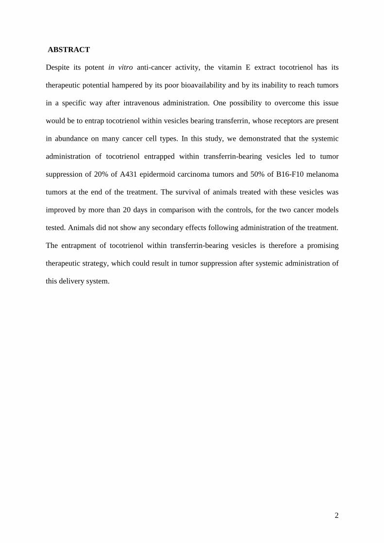
GRAPHICAL ABSTRACT
Bioluminescence imaging of the tumor regression resulting from the intravenous
administration of transferrin-bearing vesicles entrapping tocotrienol, in a mouse bearing
subcutaneous B16-F10-luc tumors. (Controls: untreated tumors).
KEYWORDS
Cancer therapy; delivery system; in vivo; tocotrienol; transferrin; tumor targeting
Days
1 3 5 7 9 11
Days
1 3 5 7 9 11
4
INTRODUCTION
Tocotrienol, a compound of the vitamin E family, has recently gained considerable attention
due to its potent anti-cancer activity displayed in vitro on many types of cancer, including
prostate, breast, pancreatic, colorectal, lung and liver cancer [1- 4]. It has also been used as a
therapeutic adjuvant, leading to synergistic anti-proliferative effects when associated with
various anti-cancer drugs such as tamoxifen, celecoxib and gemcitabine [5-9]. However, its
therapeutic use against cancer in vivo has been prevented by its poor bioavailability and its
inability to specifically reach tumors. In order to overcome these problems, we recently
entrapped the tocotrienol-rich fraction (TRF) of palm oil in tumor-targeted vesicles bearing
transferrin [10]. Transferrin (Tf) is a particularly interesting ligand for active tumor targeting,
as its receptors are expressed in abundance on many types of cancers [11]. Transferrin-
bearing vesicles provides a tumor-selective targeting strategy in addition to its passive
targeting, resulting from the enhanced permeability and retention of particulate delivery
systems in tumors [12]. Transferrin has been widely used as a tumor-targeting agent for
tumor-targeted drug and gene delivery [11, 13-17]. We recently demonstrated that the
intravenous injection of transferrin-conjugated vesicles entrapping TRF resulted in regression
of the tumors shortly after administration of the treatment. However, this therapeutic effect
was short-lived and only lasted for the duration of the treatment. We now seek to increase this
therapeutic effect by optimizing the frequency of administration of the TRF vesicle
formulation. The objectives of this work are therefore to assess the in vivo therapeutic efficacy
of this optimized treatment.
5
MATERIALS AND METHODS
Materials
Tocotrienol Rich Fraction (TRF) of palm oil corresponds to a mixture of various tocotrienol
isomers (17.6% α-tocotrienol, 15.1% δ-tocotrienol and 23.1% γ-tocotrienol), 15.3% α-
tocopherol, and other tocotrienol-related compounds. TRF was a kind gift from Dr. Abdul
Gapor (Malaysian Palm Oil Board, Kuala Lumpur, Malaysia).
Solulan C24 came from Amerchol (Edison, NJ). A431 human epidermoid carcinoma and
Bioware® B16-F10-luc-G5 mouse melanoma expressing the firefly luciferase, were
respectively obtained from the European Collection of Cell Cultures and from Caliper Life
Sciences (Hopkinton, MA). Media needed for the culture of these cells was purchased from
Invitrogen (Paisley, UK). All other reagents and chemicals that are not specifically mentioned
below were purchased from Sigma Aldrich (Poole, UK).
Preparation of transferrin-bearing vesicles entrapping TRF
Transferrin-conjugated vesicles entrapping TRF were prepared and characterized as
previously described [10]. To prepare control vesicles entrapping TRF, a mixture of sorbitan
monostearate (Span 60; 65 mg), cholesterol (58 mg), Solulan C24 (54 mg) in 2 mL TRF
solution (0.5 mg/mL, prepared in dimethylsulfoxide) was shaken at 60oC for 1 hour, before
being probe sonicated with a Soniprep 150 (MSE, United Kingdom) for 4 min. Tf-bearing
vesicles were then prepared by conjugating Tf (6 mg) to the control vesicles (2 mL), using
dimethylsuberimidate as a cross-linker [10].
Unentrapped TRF, dimethylsulfoxide, free Tf and dimethylsuberimidate were removed by
ultracentrifugation (150 000 g for 1h). The drug loaded vesicle pellet was then resuspended in
2 ml PBS.
6
TRF loading in the vesicles was measured by spectrofluorimetry (λex 295nm, λem 325nm)
using a Varian Cary eclipse fluorescence spectrophotometer (Agilent Technologies, CA) after
disruption of the vesicles with isopropanol.
Cell culture
A431 and B16-F10-luc-G5 cancer cell lines overexpressing transferrin receptors were
respectively cultivated in DMEM or RPMI-1640 medium supplemented with 10% (v/v) fetal
bovine serum, 1% (v/v) L-glutamine and 0.5% (v/v) penicillin-streptomycin, in a humidified
atmosphere of 5% CO2 at 37oC.
In vivo tumoricidal activity
The in vivo experiments in this study were approved by the local ethics committee and were
conducted according to the UK Home Office regulations.
A431 and B16-F10-luc-G5 cells were subcutaneously injected to both flanks of female
immunodeficient BALB/c mice (1 x 10 6 cells per flank). Vascularized tumors, with a typical
diameter of 5 mm, were palpable 6 days after implantation of the cells. The mice were then
intravenously injected via the tail vein with TRF entrapped in Tf-bearing vesicles, control
vesicles or in solution (10 µg TRF per injection for all treatments, 5 mice per treatment). The
administration of the treatment was done once daily for 20 days.
The weight of the animals was measured daily as a surrogate marker of toxicity. The volume
of the tumors was also determined daily, by caliper measurements (volume = d3 x π/6). At the
end of the experiment, the tumor response to treatment was classified according to the
Response Evaluation Criteria in Solid Tumors [18]: progressive disease, when the relative
tumor volume has increased by at least 1.2-fold compared to its volume at the start of the
experiment, stable disease when the relative tumor volume is comprised between 0.7 and 1.2-
7
fold compared to its starting volume, partial response when the relative tumor volume is
smaller or equal to 0.7-fold, and complete response when the tumor has completely
disappeared.
The therapeutic efficacy of the treatments was also assessed by bioluminescence imaging,
using an IVIS Spectrum (Caliper Life Sciences, MA). Mice bearing subcutaneous B16-F10-
luc-G5 tumors were intravenously injected with the treatments described above. Ten minutes
before imaging, they were intraperitoneally injected with D-luciferin, the substrate of
luciferase (150 mg D-luciferin /kg body weight) and anaesthetized by isoflurane inhalation on
Days 1, 3, 5, 7, 9 and 11 of the experiment. D-luciferin–expressing bioluminescent tumors
were able to emit some light, which was detected for a constant duration of 2 min using
Living Image® software. All images were acquired using the same illumination settings.
Statistical Analysis
Results were expressed as means ± standard error of the mean (S.E.M). Statistical
significance was assessed by one-way analysis of variance (ANOVA) and Tukey multiple
comparison post-test (Minitab® software, State College, PE). Differences were considered
statistically significant for P values lower than 0.05.
8
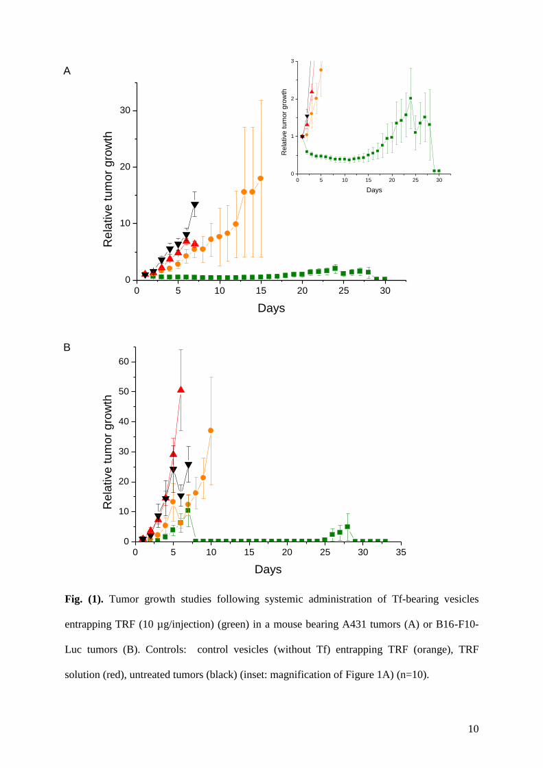
RESULTS
Intravenously administered TRF entrapped in Tf-bearing vesicles led to tumor regression
within 72 hours on A431 tumors (Fig. 1A). This effect was maintained for 15 days, allowing
the majority of the tumors to reach a size about half their initial size. From Day 15, while
some tumors kept regressing, others started growing, which led to an overall slowdown of
A431 tumor growth in comparison to the tumor growth observed following administration of
the other treatments. From Day 24, the mice bearing growing tumors reaching the maximum
allowed size (13 mm) had to be euthanized. The other mice, with tumors smaller than 13 mm
diameter, were kept in the study. By contrast, none of the other treatments resulted in any
A431 tumor regression. The effects on tumor growth observed following treatment with Tf-
bearing vesicles entrapping TRF were significantly different from those resulting from the
other treatments.
The replacement of A431 tumors by B16-F10-Luc tumors resulted in a different pattern of
tumor growth. The treatment with TRF entrapped in Tf-bearing vesicles led to the decrease of
all tumors for the 3 first days of the experiment (Fig. 1B). From Day 3, although some tumors
kept regressing, others started growing, which led to an overall B16-F10 tumor growth similar
to that observed following treatment with control vesicles until Day 7. From Day 7, the mice
bearing tumors that reached the maximum allowed size had to be euthanized. The overall
tumor size was decreasing from Day 7 to Day 25, until two of the tumors which were
regressing started growing again, resulting in the sacrifice of the mouse carrying them at Day
28. The experiment was prolonged by 3 days compared to the A431 tumor growth
experiment to assess if any other tumors would start growing again, which was not the case.
For this cell line as well, none of the other treatments resulted in tumor regression. The effects
on tumor growth observed following treatment with Tf-bearing vesicles entrapping TRF were

9
significantly different from those resulting from the other treatments from Day 8 to Day 33
(end of the experiment).
10
Fig. (1). Tumor growth studies following systemic administration of Tf-bearing vesicles
entrapping TRF (10 µg/injection) (green) in a mouse bearing A431 tumors (A) or B16-F10-
Luc tumors (B). Controls: control vesicles (without Tf) entrapping TRF (orange), TRF
solution (red), untreated tumors (black) (inset: magnification of Figure 1A) (n=10).
A
B
0 5 10 15 20 25 30
0
10
20
30R
ela
tive
tu
mo
r g
row
th
Days
0 5 10 15 20 25 30 35
0
10
20
30
40
50
60
Re
lative
tu
mo
r g
row
th
Days
0 5 10 15 20 25 30
0
1
2
3
Re
lative
tu
mo
r g
row
th
Days
11
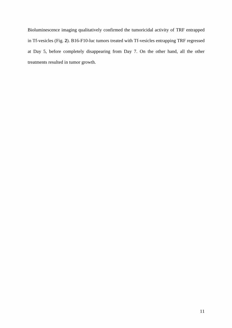
Bioluminescence imaging qualitatively confirmed the tumoricidal activity of TRF entrapped
in Tf-vesicles (Fig. 2). B16-F10-luc tumors treated with Tf-vesicles entrapping TRF regressed
at Day 5, before completely disappearing from Day 7. On the other hand, all the other
treatments resulted in tumor growth.
12
Fig. (2). Bioluminescence imaging of the tumoricidal activity of TRF entrapped in Tf-
bearing vesicles in mice bearing subcutaneous B16-F10-luc tumors. Controls: TRF
administered as entrapped in control vesicles or in solution, untreated tumors. The scale
indicates surface radiance (photons/s/cm2/steradian).
13
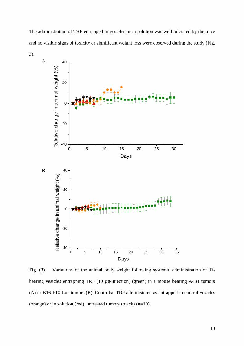
The administration of TRF entrapped in vesicles or in solution was well tolerated by the mice
and no visible signs of toxicity or significant weight loss were observed during the study (Fig.
3).
Fig. (3). Variations of the animal body weight following systemic administration of Tf-
bearing vesicles entrapping TRF (10 µg/injection) (green) in a mouse bearing A431 tumors
(A) or B16-F10-Luc tumors (B). Controls: TRF administered as entrapped in control vesicles
(orange) or in solution (red), untreated tumors (black) (n=10).
0 5 10 15 20 25 30 35
-40
-20
0
20
40
Re
lative
ch
an
ge
in
an
ima
l w
eig
ht
(%)
Days
0 5 10 15 20 25 30
-40
-20
0
20
40
Re
lative
ch
an
ge
in
an
ima
l w
eig
ht
(%)
Days
A
B
14
On the last day of the experiment, treatment with Tf-bearing vesicles entrapping TRF resulted
in complete tumor disappearance for 20% of A431 tumors and tumor regression for another
20% of A431 tumors (Fig. 4A). Treatment with control vesicles also led to tumor regression,
but with the less positive outcome of 10% partial response and 10% stable response. On the
other hand, all the tumors treated with TRF solution or left untreated were growing.
The tumor response to treatment was more successful for B16-F10 tumors. On the last day of
the experiment, 50% of B16-F10 tumors treated with Tf-bearing vesicles entrapping TRF had
completely disappeared, while another 10% of tumors had regressed (Fig. 4B). By contrast,
all the other treatments resulted in 100% tumor growth.

15
Fig. (4). Overall tumor response to treatments following systemic administration of Tf-
bearing vesicles entrapping TRF (10 µg/injection) in a mouse bearing A431 tumors (A) or
B16-F10-Luc tumors (B). Controls: TRF entrapped in control vesicles or in solution,
untreated tumors (n=10).
Tf ves. Control ves. Free TRF Untreated
0
20
40
60
80
100
Re
sp
on
se
(%
)
Treatments
Progressive
Stable
Partial
Complete
Tf ves. Control ves. Free TRF Untreated
0
20
40
60
80
100
Re
sp
on
se
(%
)
Treatments
Progressive
Stable
Partial
Complete
A
B

16
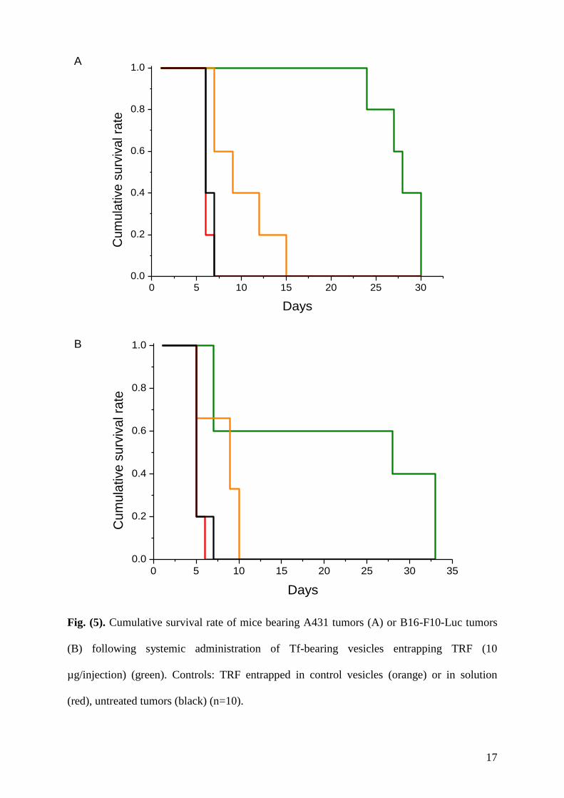
As a result of this improved therapeutic effect, the survival of A431 tumors-bearing mice
treated with Tf-bearing and control vesicles was extended by 23 days and 8 days respectively,
in comparison with the mice receiving no treatment (Fig. 5A). The survival was even
improved on B16-F10 tumors-bearing mice, as it was respectively extended by 26 days and 3
days after treatment with Tf-bearing and control vesicles, compared to untreated mice (Fig.
5B). However, the administration of TRF solution did not extend the survival of the animals
compared to untreated mice with both tumor cell lines.
17
Fig. (5). Cumulative survival rate of mice bearing A431 tumors (A) or B16-F10-Luc tumors
(B) following systemic administration of Tf-bearing vesicles entrapping TRF (10
µg/injection) (green). Controls: TRF entrapped in control vesicles (orange) or in solution
(red), untreated tumors (black) (n=10).
0 5 10 15 20 25 30 35
0.0
0.2
0.4
0.6
0.8
1.0
Cu
mu
lative
su
rviv
al ra
te
Days
0 5 10 15 20 25 30
0.0
0.2
0.4
0.6
0.8
1.0
Cu
mu
lative
su
rviv
al ra
te
Days
A
B
18
DISCUSSION
Although highly promising in vitro, the use of the tocotrienol for the treatment of cancer has
mainly been hampered by the inability of this drug to specifically reach tumors following
systemic injection. To overcome this issue, we hypothesized that entrapping TRF in drug
delivery systems bearing transferrin, whose receptors are present in abundance on cancer
cells, would increase the amount of drug delivered to cancer cells and therefore increase its
therapeutic efficacy.
In this study, the intravenous injection of tocotrienol entrapped in transferrin-bearing vesicles
led to a complete tumor disappearance of 20% of A431 tumors and 50% of B16-F10 tumors.
Consequently, tumor-bearing mice treated with this formulation showed an extended survival
of more than 20 days compared to untreated mice.
This work corresponds to a major improvement of the therapeutic efficacy of this drug for the
treatment of tumors. Tocotrienol co-encapsulated with simvastatin in lipid nanoparticles had
already been shown to have an improved anti-proliferative effect against +SA mammary
epithelial cancer cell line, in comparison with control α-tocopherol nanoparticles (with
respective IC50 of 0.52 µM and 17.7 µM) in in vitro experiments. However, no in vivo results
have been reported with this delivery system so far [19]. In our previous studies with the
Solulan-based vesicles, we demonstrated that the systemic administration of TRF entrapped in
Tf-bearing vesicles were able to decrease the size of A431 tumors, but only for the duration of
the treatment, which was 10 days [10]. All the tumors then grew back after halting the
treatment. None of them disappeared at any time when treated with only 10 injections. By
contrast, our current regimen of administration of 20 injections increased the therapeutic
effect on both tested tumors, resulting in complete disappearance of 20% and 50% of them,
respectively. In addition, this therapeutic effect was maintained after halting the treatment.
The results obtained in this current study were even improved compared with those obtained
19
with our intravenously administered Tf-conjugated, tocopheryl-based multilamellar vesicles
entrapping tocotrienol, which resulted in tumor eradication of 20% of A431 tumors and 40%
of B16-F10 tumors [16]. These results demonstrate that tocotrienol is able to exert an anti-
cancer therapeutic effect in vivo, not only as a tumor growth inhibitor or as a therapeutic
adjuvant as previously described [1, 20], provided it is targeted to the tumors. In addition, this
targeted nanomedicine was able to reach subcutaneous tumor models overexpressing
transferrin receptors, following intravenous injection and should therefore have the potential
to reach metastatic tumors disseminated in the body.
The mechanism of action used by tocotrienol to exert its therapeutic effect in our study is still
unknown. Tocotrienol has been shown to be able to activate p53, induce apoptosis and
modulate Bax/Bcl-2 ratio [21]. In addition, it can inhibit angiogenesis by down regulating the
expression of the vascular endothelial growth factor receptor [22]. It also exerts its anti-cancer
effect by inhibiting DNA polymerase and telomerase, enzymes involved in the proliferation
of cancer cells [23-24]. The extent of the therapeutic effect observed in our study made us
suggest that many of these mechanisms were involved in the anti-cancer effect observed.
There may be scope for further improvements in the in vivo activity of these nanomedicines
by understanding the mechanisms behind the variability of the response between individual
tumors, hopefully resulting in a further optimized therapeutic effect against tumors.
These therapeutic effects and the good tolerability of the treatments potentially make the
transferrin-conjugated vesicles entrapping tocotrienol a highly promising nanomedicine,
which will be further investigated.
20
CONCLUSION
The entrapment of tocotrienol in transferrin-conjugated vesicles significantly increased the
therapeutic efficacy of tocotrienol in vivo compared to the drug solution. The systemic
injection of tocotrienol entrapped in these targeted vesicles resulted in the disappearance of
20% of A431 tumors and 50% of B16-F10 tumors on the last day of the experiment.
Consequently, the survival of the tumor-bearing mice was extended by more than 20 days
compared to untreated mice, on both cancer cell lines. Transferrin-vesicles entrapping
tocotrienol is therefore a highly promising therapeutic system which deserves further
investigation.

21
CONFLICT OF INTEREST
The authors have no competing interests.
ACKNOWLEDGEMENTS
This work was supported by a Malaysian Palm Oil Board Studentship to Ju Yen Fu. The IVIS
was funded with an equipment grant (No. ME0442) from The Wellcome Trust.
22
REFERENCES
[1] Nesaretnam K. Multitargeted therapy of cancer by tocotrienols. Cancer Lett 2008;
269: 388-395.
[2] Sun W, Wang Q, Chen B, et al. Gamma-tocotrienol-induced apoptosis in human
gastric cancer SGC-7901 cells is associated with a suppression in mitogen-activated
protein kinase signalling. Br J Nutr 2008; 99: 1247-1254.
[3] Schauss AG. Tocotrienols: a review. In: Watson RR, Preedy VR, Eds. Tocotrienols:
Vitamin E beyond tocopherols, CRS Press Taylor & Francis Group: Boca Raton, Fl
2009; pp. 3-12.
[4] Xu WL, Liu JR, Liu HK, et al. Inhibition of proliferation and induction of apoptosis
by gamma-tocotrienol in human colon carcinoma HT-29 cells. Nutrition 2009; 25:
555-566.
[5] Kunnumakkara AB, Sung B, Ravindran J, et al. Gamma-tocotrienol inhibits pancreatic
tumors and sensitizes them to gemcitabine treatment by modulating the inflammatory
microenvironment. Cancer Res 2010; 70: 8695-8705.
[6] Shirode AB, Sylvester PW. Synergistic anticancer effects of combined gamma-
tocotrienol and celecoxib treatment are associated with suppression in Akt and NF-
kappaB signaling. Biomed Pharmacother 2010; 64: 327-332.
[7] Yang Z, Lee MJ, Zhao Y, et al. Metabolism of tocotrienols in animals and synergistic
inhibitory actions of tocotrienols with atorvastatin in cancer cells. Genes Nutr 2012;
7(1): 11-8.
[8] Sylvester PW, Wali VB, Bachawal SV, et al. Tocotrienol combination therapy results
in synergistic anticancer response. Front Biosci 2011; 17: 3183-3195.
[9] Dufès C. Delivery of the vitamin E compound tocotrienol to cancer cells. Ther Deliv
2011; 2(11): 1385-1389.
23
[10] Fu JY, Blatchford DR, Tetley L, et al. Tumor regression after systemic
administration of tocotrienol entrapped in tumor-targeted vesicles. J Control Release
2009; 140: 95-99.
[11] Calzolari A, Oliviero I, Deaglio S, et al. Transferrin receptor 2 is frequently
expressed in human cancer cell lines. Blood Cells Mol Dis 2007; 39: 82-91.
[12] Maeda H. The tumor blood vessel as an ideal target for macromolecular anticancer
agents. J Control Release 1992; 19: 315-324.
[13] Daniels TR, Delgado T, Helguera G, et al. The transferrin receptor part II: targeted
delivery of therapeutic agents into cancer cells. Clin Immunol 2006; 121: 159-176.
[14] Dufès C, Schätzlein AG, Tetley L, et al. Niosomes and polymeric chitosan based
vesicles bearing transferrin and glucose ligands for drug targeting. Pharm Res 2000;
17 (10): 1250-1258.
[15] Dufès C, Muller JM, Couet W, et al. Anticancer drug delivery with transferrin
targeted polymeric chitosan vesicles. Pharm Res 2004; 21: 101-107.
[16] Fu JY, Zhang W, Blatchford DR, et al. Novel tocotrienol-entrapping vesicles can
eradicate solid tumors after intravenous administration. J Control Release 2011; 154:
20-26.
[17] Lemarié F, Chang CW, Blatchford DR, et al. Anti-tumor activity of the tea
polyphenol epigallocatechin gallate encapsulated in targeted vesicles after
intravenous administration. Nanomedicine 2013; 8(2): 181-192.
[18] Eisenhauer EA, Therasse P, Bogaerts J, et al. New response evaluation criteria in
solid tumours: revised RECIST guideline (version 1.1). Eur J Cancer 2009; 45: 228-
247.

24
[19] Ali H, Shirode AB, Sylvester PW, et al. Preparation, characterization and anticancer
effects of simvastatin-tocotrienol lipid nanoparticles. Int J Pharm 2010; 389: 223-
231.
[20] Wada S, Satomi Y, Murakoshi M, et al. Tumor suppressive effects of tocotrienol in
vivo and in vitro. Cancer Lett 2005; 229: 181-191.
[21] Agarwal MK, Agarwal ML, Athar M, et al. Tocotrienol-rich fraction of palm oil
activates p53, modulates Bax/Bcl2 ratio and induces apoptosis independnent of cell
cycle association. Cell Cycle 2004; 3: 205-211.
[22] Nakagawa K, Eitsuka T, Inokuchi H, et al. DNA chip analysis of comprehensive
food function: inhibition of angiogenesis and telomerase activity with unsaturated
vitamin E tocotrienol. BioFactors 2004; 21: 5-10.
[23] Eitsuka T, Nakagawa K, Miyazawa T. Down-regulation of telomerase activity in
DLD-1 human colorectal adenocarcinoma cells by tocotrienol. Biochem Biophys Res
Commun 2006; 348: 170-175.
[24] Mizushina Y, Nakagawa K, Shibata A, et al. Inhibitory effect of tocotrienol on
eukaryotic DNA polymerase 1 and angiogenesis. Biochem Biophys Res Commun
2006; 339: 949-955.
25
FIGURE LEGENDS
Fig. (1). Tumor growth studies following systemic administration of Tf-bearing vesicles
entrapping TRF (10 µg/injection) (green) in a mouse bearing A431 tumors (A) or B16-F10-
Luc tumors (B). Controls: control vesicles (without Tf) entrapping TRF (orange), TRF
solution (red), untreated tumors (black) (inset: magnification of Figure 1A) (n=10).
Fig. (2). Bioluminescence imaging of the tumoricidal activity of TRF entrapped in Tf-
bearing vesicles in mice bearing subcutaneous B16-F10-luc tumors. Controls: TRF
administered as entrapped in control vesicles or in solution, untreated tumors. The scale
indicates surface radiance (photons/s/cm2/steradian).
Fig. (3). Variations of the animal body weight following systemic administration of Tf-
bearing vesicles entrapping TRF (10 µg/injection) (green) in a mouse bearing A431 tumors
(A) or B16-F10-Luc tumors (B). Controls: TRF administered as entrapped in control vesicles
(orange) or in solution (red), untreated tumors (black) (n=10).
Fig. (4). Overall tumor response to treatments following systemic administration of Tf-
bearing vesicles entrapping TRF (10 µg/injection) in a mouse bearing A431 tumors (A) or
B16-F10-Luc tumors (B). Controls: TRF entrapped in control vesicles or in solution,
untreated tumors (n=10).
Fig. (5). Cumulative survival rate of mice bearing A431 tumors (A) or B16-F10-Luc tumors
(B) following systemic administration of Tf-bearing vesicles entrapping TRF (10
µg/injection) (green). Controls: TRF entrapped in control vesicles (orange) or in solution
(red), untreated tumors (black) (n=10).