Peptides from conserved regions of paramyxovirus fusion (F) proteins are potent inhibitors of viral...
6
Proc. Natl. Acad. Sci. USA Vol. 93, pp. 2186-2191, March 1996 Medical Sciences Peptides from conserved regions of paramyxovirus fusion (F) proteins are potent inhibitors of viral fusion D. M. LAMBERT*, S. BARNEY, A. L. LAMBERT, K. GUTHRIE, R. MEDINAS, D. E. DAVIS, T. BUCY, J. ERICKSON, G. MERUTKA, AND S. R. PETrEWAY, JR.t Trimeris, Inc., P.O. Box 13963, Research Triangle Park, NC 27709 Communicated by Maurice R. Hilleman, Merck & Co., Inc., West Point, PA, November 8, 1995 (received for review October 6, 1995) ABSTRACT The synthetic peptides DP-107 and DP-178 (T-20), derived from separate domains within the human immunodeficiency virus type 1 (HIV-1) transmembrane (TM) protein, gp4l, are stable and potent inhibitors of HIV-1 infection and fusion. Using a computer searching strategy (computerized antiviral searching technology, C.A.S.T.) based on the predicted secondary structure of DP-107 and DP-178 (T-20), we have identified conserved heptad repeat domains analogous to the DP-107 and DP-178 regions of HIV-1 gp4l within the glycoproteins of other fusogenic viruses. Here we report on antiviral peptides derived from three representative paramyxoviruses, respiratory syncytial virus (RSV), human parainfluenza virus type 3 (HPIV-3), and measles virus (MV). We screened crude preparations of synthetic 35-residue pep- tides, scanning the DP-178-like domains, in antiviral assays. Peptide preparations demonstrating antiviral activity were purified and tested for their ability to block syncytium for- mation. Representative DP-178-like peptides from each paranyxovirus blocked homologous virus-mediated syncy- tium formation and exhibited EC50 values in the range 0.015- 0.250 ,uM. Moreover, these peptides were highly selective for the virus of origin. Identification of biologically active pep- tides derived from domains within paramyxovirus F1 proteins analogous to the DP-178 domain of HIV-1 gp4l is compelling evidence for equivalent structural and functional features between retroviral and paramyxoviral fusion proteins. These antiviral peptides provide a novel approach to the develop- ment of targeted therapies for paramyxovirus infections. The Paramyxoviridae family encompasses three genera: the Paramyxovirus group, including parainfluenza types 1-4, mumps, Newcastle disease, Sendai, and simian type 5 viruses; the Morbillivirus group, including measles, canine distemper, and rinderpest viruses; and the Pneumovirus group, including human and bovine respiratory syncytial viruses (RSVs) and mouse pneumonia virus (1, 2). Many of these viruses are responsible for human disease. All members of the family contain two major surface glycoproteins, a receptor-binding protein (HN, H, or G), which facilitates attachment to cells, and a fusion (F) protein, which enables penetration of the virus genome into the host cell. Fusion of the paramyxovirus enve- lope with the cell membrane occurs at neutral pH and devel- opment of extensive syncytia in tissue culture is the charac- teristic cytopathologic effect (CPE) observed for many mem- bers of this virus family. Fusion of the viral envelope or infected cell membranes with uninfected cell membranes is an essential step in the virus life cycle (3-5). The F glycoprotein mediates this function in paramyxoviruses, whereas in retroviruses fusion is mediated by the transmembrane (TM) protein. Both types of fusogenic proteins require activation by a proteolytic cleavage event mediated by a host cell enzyme (6, 7). Certain conserved The publication costs of this article were defrayed in part by page charge payment. This article must therefore be hereby marked "advertisement" in accordance with 18 U.S.C. §1734 solely to indicate this fact. 2186 features are common among the fusion proteins of different fusogenic viruses (3). First, since these are class I integral membrane proteins, a hydrophobic sequence near the C terminus anchors the protein in the virus envelope; second, a hydrophobic stretch of amino acids at the N terminus serves as the fusion domain and anchors the protein in the host cell membrane during the fusion process (3, 5). Interestingly, the fusion domains at the amino termini of RSV F, and human immunodeficiency virus type 1 (HIV-1) TM show a high degree of sequence homology (8). Two intervening amphipathic heptad repeat regions have been identified in the fusion proteins of orthomyxoviruses, paramyxoviruses, and retroviruses (9, 10). Recently, muta- tional analyses and binding experiments have provided evi- dence for the interaction of these peptide regions in HIV-1 (11-13). These general domains within retrovirus TM pro- teins, paramyxovirus F proteins, coronavirus peplomer pro- teins, and influenza virus HA2 proteins have been reported to contain primary amino acid sequences predicted to form a-helices or coiled coils (5, 9-11, 14-16). The report of Gallaher et al. (14) first predicted the helical domains of the HIV TM protein (gp4l). This work led to the discoveries of Wild et al. (11, 15, 17), who demonstrated that synthetic peptides derived from these potentially helical regions within the TM protein of HIV-1 were potent inhibitors of viral fusion and infection. One such peptide, DP-107, forms a helical coiled coil in aqueous solution at neutral pH. A second peptide, DP-178, demonstrates little or no helical structure in solution but exhibits potent and specific inhibition of HI V-1 fusion and infection (11). Similar results have been independently reported for an overlap- ping peptide (18, 19). We used a motif searching strategy, based on the known sequences of gp4l helical domains, that accurately predicted similar regions within the fusion proteins of a wide variety of enveloped viruses, including the paramyxovirus fusion proteins reported here. Because of the functional and structural similarities between the HIV-1 TM protein and the F1 subunit of the paramyxovirus fusion glycoproteins, we investigated the possibility that func- tional homologues of DP-107 or DP-178-like peptides could be derived from similar domains within the F1 glycoproteins. Identification of similarly active peptides in the F proteins would provide additional evidence for conserved structural and functional features between the fusion proteins of retro- viruses and paramyxoviruses. We report here the identification of functional domains within the F1 proteins of RSV, human parainfluenza virus 3 (HPIV-3), and measles virus (MV) that correspond to the DP-107 and DP-178 domains of HIV-1. We also describe peptides derived from paramyxovirus F1 regions comparable to DP-178 that possess extremely potent and selective antiviral Abbreviations: CPE, cytopathic effect; DMSO, dimethyl sulfoxide; HIV, human immunodeficiency virus; HPIV, human parainfluenza virus; MV, measles virus; RSV, respiratory syncytial virus. *To whom reprint requests should be addressed. tPresent address: Bayer Corporation, Pharmaceutical Division, P.O. Box 507, Clayton, NC 27520.
-
Upload
independent -
Category
Documents
-
view
1 -
download
0
Transcript of Peptides from conserved regions of paramyxovirus fusion (F) proteins are potent inhibitors of viral...
Proc. Natl. Acad. Sci. USAVol. 93, pp. 2186-2191, March 1996Medical Sciences
Peptides from conserved regions of paramyxovirus fusion (F)proteins are potent inhibitors of viral fusionD. M. LAMBERT*, S. BARNEY, A. L. LAMBERT, K. GUTHRIE, R. MEDINAS, D. E. DAVIS, T. BUCY, J. ERICKSON,G. MERUTKA, AND S. R. PETrEWAY, JR.tTrimeris, Inc., P.O. Box 13963, Research Triangle Park, NC 27709
Communicated by Maurice R. Hilleman, Merck & Co., Inc., West Point, PA, November 8, 1995 (received for review October 6, 1995)
ABSTRACT The synthetic peptides DP-107 and DP-178(T-20), derived from separate domains within the humanimmunodeficiency virus type 1 (HIV-1) transmembrane (TM)protein, gp4l, are stable and potent inhibitors of HIV-1infection and fusion. Using a computer searching strategy(computerized antiviral searching technology, C.A.S.T.) basedon the predicted secondary structure of DP-107 and DP-178(T-20), we have identified conserved heptad repeat domainsanalogous to the DP-107 and DP-178 regions of HIV-1 gp4lwithin the glycoproteins of other fusogenic viruses. Here wereport on antiviral peptides derived from three representativeparamyxoviruses, respiratory syncytial virus (RSV), humanparainfluenza virus type 3 (HPIV-3), and measles virus (MV).We screened crude preparations of synthetic 35-residue pep-tides, scanning the DP-178-like domains, in antiviral assays.Peptide preparations demonstrating antiviral activity werepurified and tested for their ability to block syncytium for-mation. Representative DP-178-like peptides from eachparanyxovirus blocked homologous virus-mediated syncy-tium formation and exhibited EC50 values in the range 0.015-0.250 ,uM. Moreover, these peptides were highly selective forthe virus of origin. Identification of biologically active pep-tides derived from domains within paramyxovirus F1 proteinsanalogous to the DP-178 domain of HIV-1 gp4l is compellingevidence for equivalent structural and functional featuresbetween retroviral and paramyxoviral fusion proteins. Theseantiviral peptides provide a novel approach to the develop-ment of targeted therapies for paramyxovirus infections.
The Paramyxoviridae family encompasses three genera: theParamyxovirus group, including parainfluenza types 1-4,mumps, Newcastle disease, Sendai, and simian type 5 viruses;the Morbillivirus group, including measles, canine distemper,and rinderpest viruses; and the Pneumovirus group, includinghuman and bovine respiratory syncytial viruses (RSVs) andmouse pneumonia virus (1, 2). Many of these viruses areresponsible for human disease. All members of the familycontain two major surface glycoproteins, a receptor-bindingprotein (HN, H, or G), which facilitates attachment to cells,and a fusion (F) protein, which enables penetration of the virusgenome into the host cell. Fusion of the paramyxovirus enve-lope with the cell membrane occurs at neutral pH and devel-opment of extensive syncytia in tissue culture is the charac-teristic cytopathologic effect (CPE) observed for many mem-bers of this virus family.
Fusion of the viral envelope or infected cell membranes withuninfected cell membranes is an essential step in the virus lifecycle (3-5). The F glycoprotein mediates this function inparamyxoviruses, whereas in retroviruses fusion is mediated bythe transmembrane (TM) protein. Both types of fusogenicproteins require activation by a proteolytic cleavage eventmediated by a host cell enzyme (6, 7). Certain conserved
The publication costs of this article were defrayed in part by page chargepayment. This article must therefore be hereby marked "advertisement" inaccordance with 18 U.S.C. §1734 solely to indicate this fact.
2186
features are common among the fusion proteins of differentfusogenic viruses (3). First, since these are class I integralmembrane proteins, a hydrophobic sequence near the Cterminus anchors the protein in the virus envelope; second, ahydrophobic stretch of amino acids at the N terminus serves asthe fusion domain and anchors the protein in the host cellmembrane during the fusion process (3, 5). Interestingly, thefusion domains at the amino termini of RSV F, and humanimmunodeficiency virus type 1 (HIV-1) TM show a highdegree of sequence homology (8).Two intervening amphipathic heptad repeat regions have
been identified in the fusion proteins of orthomyxoviruses,paramyxoviruses, and retroviruses (9, 10). Recently, muta-tional analyses and binding experiments have provided evi-dence for the interaction of these peptide regions in HIV-1(11-13). These general domains within retrovirus TM pro-teins, paramyxovirus F proteins, coronavirus peplomer pro-teins, and influenza virus HA2 proteins have been reported tocontain primary amino acid sequences predicted to forma-helices or coiled coils (5, 9-11, 14-16). The report ofGallaher et al. (14) first predicted the helical domains of theHIV TM protein (gp4l). This work led to the discoveries ofWild et al. (11, 15, 17), who demonstrated that synthetic peptidesderived from these potentially helical regions within the TMprotein of HIV-1 were potent inhibitors of viral fusion andinfection. One such peptide, DP-107, forms a helical coiled coilin aqueous solution at neutral pH. A second peptide, DP-178,demonstrates little or no helical structure in solution but exhibitspotent and specific inhibition of HIV-1 fusion and infection (11).Similar results have been independently reported for an overlap-ping peptide (18, 19). We used a motif searching strategy, basedon the known sequences of gp4l helical domains, that accuratelypredicted similar regions within the fusion proteins of a widevariety of enveloped viruses, including the paramyxovirus fusionproteins reported here.
Because of the functional and structural similarities betweenthe HIV-1 TM protein and the F1 subunit of the paramyxovirusfusion glycoproteins, we investigated the possibility that func-tional homologues of DP-107 or DP-178-like peptides could bederived from similar domains within the F1 glycoproteins.Identification of similarly active peptides in the F proteinswould provide additional evidence for conserved structuraland functional features between the fusion proteins of retro-viruses and paramyxoviruses.We report here the identification of functional domains
within the F1 proteins of RSV, human parainfluenza virus 3(HPIV-3), and measles virus (MV) that correspond to theDP-107 and DP-178 domains of HIV-1. We also describepeptides derived from paramyxovirus F1 regions comparableto DP-178 that possess extremely potent and selective antiviral
Abbreviations: CPE, cytopathic effect; DMSO, dimethyl sulfoxide;HIV, human immunodeficiency virus; HPIV, human parainfluenzavirus; MV, measles virus; RSV, respiratory syncytial virus.*To whom reprint requests should be addressed.tPresent address: Bayer Corporation, Pharmaceutical Division, P.O.Box 507, Clayton, NC 27520.
Proc. Natl. Acad. Sci. USA 93 (1996) 2187
activity in vitro against HPIV-3, RSV, and MV. Identificationof paramyxovirus inhibitory peptides expands the initial ob-servations of Wild et al. (15, 17) and demonstrates thatantiviral peptides derived from discrete fusion protein do-mains can be found in other enveloped viruses. In addition, thepotential of these peptides to be antiviral agents for medicallyimportant paramyxoviruses is discussed.
MATERIALS AND METHODSCells and Virus. Human RSV (Long strain), human MV
(Edmonston strain), and HPIV-3 (strain NIH 47885) wereobtained from the American Type Culture Collection(ATCC). The cell lines HEp2, Vero, and CV-1 were alsoobtained from ATCC. HEp2 cell monolayers persistentlyinfected with HPIV-3 were made as described (20). Stocks ofHIV-1LA1, CEM, and MOLT-4 cells were kindly provided byT. Matthews (Duke University Medical Center).
Computerized Antiviral Searching Technology (C.A.S.T.).The primary amino acid sequences of the F proteins of RSV,HPIV-3, and MV were analyzed with a computer searchstrategy designed with the aid of a commercially availablesoftware package, PC/GENE. Starting with the amino acidsequences of HIV-1 gp4l peptides, DP-107 and DP-178, motifswere designed to search for amino acid sequences with similarcharacteristics within viral surface proteins, includingparamyxovirus fusion proteins. Available viral protein se-quences found in the Swiss-Prot database were scanned formatching motifs with the PC/GENE program PESEARCH.
Cytotoxicity and Viral CPE Assays. Crude peptides weretested for their ability to protect cell monolayers from viralCPE and to evaluate cytotoxicity in uninfected cells. The CPEand cytotoxicity assays were developed for RSV, HPIV-3, andMV and are based on cellular metabolism of a tetrazolium salt(XTT) to a water-soluble formazan dye (21).
Cell Fusion Assays. Virus-induced cell-cell fusion assayswere used to assess the ability of peptides to block fusion. Cellswhich were acutely infected with either RSV, HPIV-3, or MV(multiplicity of infection, 3-5) were added to uninfected cellmonolayers in the presence of peptides. Infected cells weredispersed at 24 hr postinfection by Versene (EDTA) treatmentand washed, and a predetermined number of cells were addedto uninfected cell monolayers in 96-well plates to give repro-ducible numbers of syncytia (generally 50-75 per well) inuntreated control wells. Dilutions of peptides in completemedium were added to uninfected cell monolayers in 96-wellplates, then infected cells were added to each well. After 18-24hr of incubation at 37°C in a 5% CO2 atmosphere, monolayerswere fixed and stained with 0.5% (wt/vol) crystal violet in100% methanol. Stained syncytial plaques were counted witha stereo dissecting microscope. A reduction in formation ofsyncytia (infectious centers) in comparison with an infectedcell control containing no peptide is indicative of antiviralactivity. A variation of this assay which used cell-free virions(50-100 plaque-forming units) in place of acutely infected cellsgave similar results. Either procedure allows for quantitativemeasurement of cell-to-cell spread of infection mediated bythe F protein of the respective paramyxovirus. Dose-responsecurves were generated and the 50% effective concentration(EC5o) for each peptide was determined. Fusion assays withHIV-1LA1 were performed as described (22).
Peptide Synthesis. Peptides were synthesized on a RaininSymphony Multiplex multiple peptide synthesizer. Standardsolid-phase synthesis techniques using fluorenylmethoxycar-bonyl-protected amino acids were used (23, 24). All peptideswere acetylated at the N terminus and amidated at the Cterminus to enhance the biological half-life of the peptides(25). Cleavage of peptides from the resin and removal ofside-chain blocking groups were automatically performed onthe instrument with trifluoroacetic acid and the appropriate
scavengers (5% thioanisol, 5% water, 2.5% ethanedithiol, 0.8M phenol) (26). After cleavage, the peptide was precipitatedwith 4 volumes cold ether for 20 min, collected by centrifu-gation, washed twice with cold ether, and dried under vacuumfor 24 hr. Lyophilized peptides were stored desiccated andpeptide solutions were made in water or phosphate-bufferedsaline (PBS) or, if necessary, dimethyl sulfoxide (DMSO).
Peptide Purification and Characterization. Peptides werepurified by reverse-phase HPLC on a Waters DeltaPak C18column (25 mm x 300 mm, 15-,um particles) using a water andacetonitrile gradient containing 0.1% trifluoroacetic acid. Allpeptides were >95% pure by analytical reverse-phase HPLC.Amino acid analysis and electrospray mass spectroscopy con-firmed the composition of the peptides.
Circular Dichroism. CD measurements were obtained withan Aviv Associates model 62DS spectropolarimeter calibratedwith a standard solution of 10-camphorsulfonic acid (27).Samples contained 10-50 ,tM peptide in 0.1 M NaCl/10 mMpotassium phosphate, adjusted to pH 7.0. For samples con-taining trifluoroethanol, the aqueous buffer was diluted 1:1(vol/vol) with trifluoroethanol. Spectra were collected at 1°Cwith a 1.5-nm bandwidth, a 0.5-nm step size, and a 2.0-sec timeconstant in either 1- or 10-mm-path-length cells. Blank cor-rected data were smoothed with a third-order polynomialfunction. Sample temperature was regulated by a thermoelec-tric cell holder accurate to within 1C.
RESULTSA four-stage process was employed to identify biologicallyactive peptides from the F proteins of paramyxoviruses. First,we used C.A.S.T. to analyze the primary amino acid sequencesof the F proteins for domains that contained sequences;similarin potential secondary structure characteristics to the DP-107and DP-178 peptides of HIV-1 gp4l. Second, we synthesizedoverlapping 35-residue peptides from the identified DP-178-like domains and tested the crude preparations for antiviralactivity. Third, the peptides from active crude preparationswere purified and retested for antiviral activity in virus fusionassays. Fourth, peptides were analyzed for helical structure byCD spectroscopy.Computer Searches to Identify Heptad Repeat (HR) Do-
mains. Based on the previously identified amino acid charac-teristics of peptides DP-107 and DP-178 from HIV-1 gp4l (15,17), motifs were designed to search for amino acid sequenceswhich are predicted to form helical secondary structure inother fusogenic viral proteins. The computer motif searchselectively identified domains within gp4l that overlaid theamino acid sequences of DP-107 and DP-178 in all availableHIV-1 sequences.
This approach allowed identification of biologically activepeptides within both the HR1 and HR2 domains of the F1proteins from RSV, HPIV-3, and MV (Fig. 1). The followingDP-107-like (HR1) and DP-178-like (HR2) regions wereidentified: RSV Fl, aa 149-206 (HR1) and aa 474-523 (HR2);HPIV-3 Fl, aa 119-183 (HR1) and aa 438-493 (HR2); MV,aa 116-191 (HR1) and aa 438-488 (HR2).The identified sequences within HR1 and HR2 domains
shared several characteristics with DP-107 and DP-178. Therelative positions of these two domains-located next to thefusion peptide domain and the transmembrane anchor, re-spectively-were similar to the comparable domains in gp4l(Fig. 1). Within the RSV, HPIV-3, and MV F1 glycoproteins,the search motif identified HR1 domains corresponding inposition to the DP-107 coiled-coil domain of HIV-1, imme-diately downstream from the predicted fusion peptide se-quences at their respective N termini. Similarly, regions cor-responding to the DP-178 domain of HIV-1 TM were identi-fied proximal to the predicted transmembrane region of eachF, glycoprotein. The motif search also identified a unique
Medical Sciences: Lambert et al.
2188 Medical Sciences: Lambert et al.
i1
.T.2
DI)P1 DPI)P 78 (T-20)
113 Doinir i KIIDomaiJin )1R2 Donmia;in
T-1 18
v ll - I T-IX |I84T-205
4RH
T-2547
Active Peptides from HR2 DomainsSeguence
T-I I 8Ac-.9s1-FDASIASIVNSLKlINOSI.Al;IRKSDEIl.1.HNVNAGKST-I1-205A%c-.:>.IDI)ISIEI.NKAKSDI.)l I-:-SKFWIRRSNQKLDSIGNW;H.-lI-257 Ac- 5L1lIRIDI.GPPISI I-FRKI .I)VGTINKN.GNAIAKLEDAKE,LL.
region within the RSV F2 protein designated HR3 (aa 53-100;Fig. 1). No comparable HR3 domains were identified in eitherHPIV-3 or MV F2 glycoproteins.
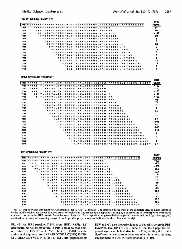
Peptide Scanning Across HR2 Domains to Locate ActivePeptides. The two identified F1 heptad repeat domains rangein length from 47 to 75 aa and represent general regions withinthe F1 proteins (Fig. 1). We chose a peptide scanning approachwhich required synthesis of overlapping peptides covering theHR1 and HR2 regions. Thirty-five amino acids (i.e., fiveheptad repeats) was chosen as a standard length for thepeptides because the DP-107 and DP-178 peptides were ofsimilar length (38 and 36 aa, respectively) and because thislength was more efficiently synthesized than single peptidescovering the entire region (i.e., 47-75 aa). Peptide scans acrossthe HR1 domains were assayed and several demonstratedmoderate antiviral activity (data not shown). Overlappingcrude 35-aa peptides scanning across the HR2 domainsshowed significantly more potent antiviral activity when ana-lyzed for their ability to protect cultures from viral CPE (Fig.2). Note that peptides starting from the N-terminal area ofeach viral HR2 domain showed little or no activity (>100,g/ml), whereas potent but variable antiviral activity was seenwith the remaining peptides. We had previously determinedthat crude preparations of DP-107 and DP-178 were only 3-5times less active than pure peptides and that these preparationswere not cytotoxic at concentrations of up to 100 ,tg/ml,indicating that initial screening for active peptides would notrequire purification (data not shown). More than 95% of thecrude preparations were soluble (1 mg/ml) in phosphate-buffered saline at pH 7. Less soluble peptides were dissolvedin DMSO prior to analysis. No differences in antiviral activitywere observed when DMSO was used to dissolve peptideswhich were also soluble in PBS (data not shown).
Antiviral Activity of Peptides Derived from HR2 Domains ofF Proteins. Crude peptide preparations from HR2 wereanalyzed for their ability to prevent CPE in infectivity assayswith RSV, HPIV-3, and MV. As shown in Fig. 2, peptidesexhibited a wide range of antiviral activity in these assays.Because the crude preparations were synthesized and handledsimultaneously, the inactive peptides served as negative con-trols. Several peptides from all three viruses demonstratedsignificant antiviral activity. Active peptide preparations fromeach virus were chosen for further study: T-1 18 from RSV,T-205 from HPIV-3, and T-257 from MV.
rIansmembraneanchor
CIfCOfH H IV-1I
FIG. 1. Linear protein maps of HIV-1TM compared with RSV, HPIV-3, and
I O(0ff RSV!' MV fusion (F) proteins. Designated DP-
107-like (HR1) and DP-178-like (HR2)domains in the paramyxovirus F proteins
i oo(l HPIV-3 are designated by amino acid number. Anx )0 additional F2 domain, designated HR3,
was identified in RSV but not in HPIV-3or MV. The predicted fusion peptide (FP)
t~t{)I1 M;IV domains located at the N-terminal ends ofthe F1 proteins are designated by theiramino acid numbers and overlap with theN-terminal ends of predicted HR1 do-mains. The HR2 peptide sequences cho-
*!N1)
sen for purification and further study for(j-)1 each virus are shown at the bottom. Pep-
l->n-N l1)0)5 l tides from each virus were tested in either
[Y-NF[1 0.01 5 antifusion assays or plaque reduction as-
.4---NHI, 0.068 says to determine the EC5o values (,uM).
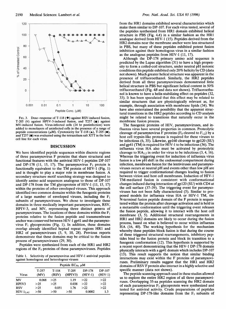
Active peptides were resynthesized, purified, and tested ininfectious-center assays for their ability to block viral fusion.Fig. 3 shows the antiviral titration curves and cytotoxicityprofiles of a representative active peptide for each virus: T-118(RSV), T-205 (HPIV-3), and T-257 (MV). EC50 values (,AM)were calculated for each peptide from triplicate assays. Arepresentative dose-response curve is shown for each peptideand indicates that the antiviral activity seen is peptide depen-dent. Results of repeated assays showed that for RSV, peptideT-118 was the most active, with EC50 values of 0.050 ,uM.Several other peptides from this region had antiviral activitywithin the range 0.05-0.15 ,uM. The HPIV-3 peptide, T-205,inhibited fusion and infection at EC50 values as low as 0.015,uM. Against MV, T-257 exhibited antifusion activity with EC50values as low as 0.042 ,uM.
Selectivity and Specificity of Antiviral Peptides. Threerepresentative active peptides from the HR2 domains ofthese viruses were highly selective for their viruses of origin(Table 1). The EC50 values for selected peptides against theirhomologous virus were compared with values obtained whenthe peptides were tested against heterologous viruses. Asshown in Table 1, no cross-inhibitory activity of any of theparamyxovirus DP-178-like peptides (up to about 25 ,uM)was detected for HIV-1. Likewise, neither DP-107 norDP-178 was active against the paramyxoviruses. PeptideT-118 (RSV) and peptide T-257 (MV) were not activeagainst HPIV-3, nor were their peptides active against eachother at concentrations up to 100 ,tg/ml. However, theHPIV-3 peptide, T-205, was active against RSV at 1.76 ,uM,an -46-fold higher concentration than its activity againstHPIV-3 (0.038 ,tM). T-205 blocked MV fusion at 1.19 ,uM,an -31-fold higher concentration than against HPIV-3.None of the peptides tested exhibited sufficient cytotoxiceffects to determine 50% cytotoxic concentration (CCso)values (i.e., >500 ,ug/ml). Thus, these peptides exhibitselectivity index values (CC5o/EC50) of >1900 to >3000against their homologous viruses.
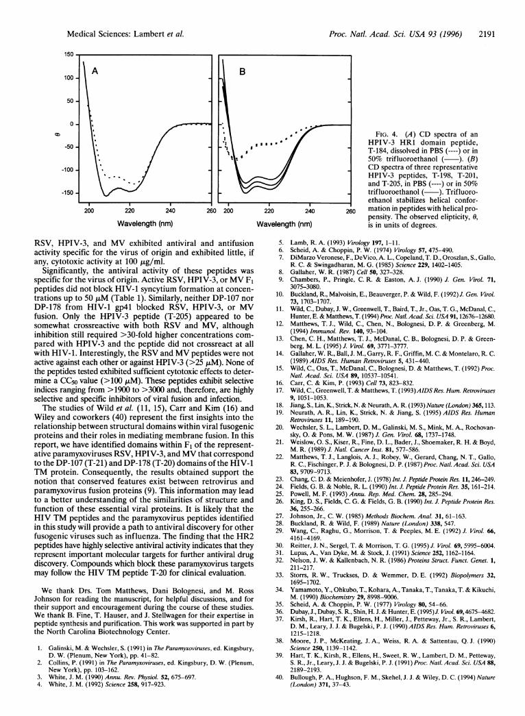
Structural Analysis of HR1 and HR2 Peptides. Becausethe peptides described above were derived from F1 domainspredicted to have helical characteristics similar to DP-107and DP-178, we analyzed representative peptides from eachdomain by CD for helical content. Purified peptide samplesfrom HR1 and HR2 peptides were analyzed in PBS and theCD spectra for purified HPIV-3 peptides are shown in
sUJ4- "pl12)
Ni1
Pl(ptidle TM
-
Proc. Natl. Acad. Sci. USA 93 (1996)
Medical Sciences: Lambert et al. Proc. Natl. Acad. Sci. USA 93 (1996) 2189
RSV DP-178-LIKE REGION (F1)ECSO
HlW: G E P I I N F Y O P L V F P S D E F D A S I S Q V N E K I N Q S L A F I R K S D E L L H N V N A G K S T T| UhtlT-104 I N F Y D P L V F P S D E FD A S I S Q V N E K I N Q S LA F I R K 91T-105 I N F Y D P L V F P S D E FDA 8 I S Q V N E K N Q SL A F I R K S 93T-106 N F Y D P L V F P S D E F DA S I S Q V N E K I N Q SL A F I R K S D p100T-107 F Y D P L V F P S D E F DA S I S Q V N E K I N Q SL A F I R K S D E 20T-10S Y D P L V F P S D E F DA S I S Q V N E K I N Q SLA F I R KS8D E L 6T-10S D P L V F P S D E FD A S I S Q V N E K I N Q S L A F I R K S D E L L 8T-110 P L V F P S D E F DA S I S Q V N E K I N Q S L A F I R K S D E L L H 30T-111 L V F P S D E F DA S I S Q V N E K I N Q SL A F I R K S D E L L HN 9T-112 V F P S D E F DA S I S Q V N E K I N Q S L A F R K S D E L L HN V 19T-113 F P S D E F DA S I S Q V N E K I N Q S L A F I R K S D E L L HN V N 8T-114 P S D E F DA S I S Q V N E K I N Q S L A F I R K S D E L L HN V N A 6T-11S SD E F DA S I S Q V N E K I N Q S L A F I R K S D E L L HN V N A G 6T-116 D E F DA S I S Q V N E K I N Q S L A F I R K S D E L L HN V N A G K 12T-117 E F DA S I S Q V N E K I N Q S L A F I R K S D E L L HN V N A G K S 13T-11 FDA S I S Q V N E K I N Q S LA F I R K SD E L L HN V N A O K S T 6T-119 DA S I S Q V N E K I N Q S L A F I R K S D E L L HN V N A G K S T T 8
HPIV3 DP178-LIKE REGION (Fl)EC6O
Hf Y T P N D0I T L N N S V A L D P I D I S I E L N K A K S D L E E S K E W I R R S N Q K L D S I G N W H Q S S T (ml)
T-189 Y T P N D I T L N N S V A L D P I DI S I E L N K A K S D L E ES K E p100T-190 T P N D I T L N N S VA L D P I D I S I E L N K A K S D L E ES K E W p100T-191 P N D I T L N N S V A L D P I D I S I E L N K A K S D L E ES K E W I >100T-192 N D I T L N N S V A L D P I D I S I E L N K A K S D L E ES K E W I R p10.0T-193 D I T L N N S V A L D P I D I S I E L N K A K S D L E ES K E W I R R %100T-14 I T L N N S V A L D P I 0 I S I E L N K A K S D L E ES K E W I R R S 62T-195 T L N N S V A L D P I D I S I E L N K A K S D L E ES K E W I R R S N 72T-196 L N N S V A L D P I D I S I E L N K A K S D L E ES K E W I R R S N Q 1T-197 N N S V A L D P I D I S I E L N K A K S D L E E S K E W I R R S N Q K 6T-196 N S V A L D P I D I S I E L N K A K S D L E ES K E W I R R S N Q K L 02T-190 S V A L D P I D I S I E L N K A K S D L E ES K E W I R R S N Q K LD 2T-200 V A L D P I D I SI E L N K A K S D L E ES K E W I R R S N Q K LD S IT-201 A L D P I D I S I E L N K A K S D L E ES K E W I R R S NIK L D I 0.1T-202 L D P I D I S I E L N K A K S D L E ES K E W I R R S N Q K LD S IG 0.03T-203 D P I D I S I E L N K A K S D L E ES K E W I R R S N Q K LD S IG N 0.2T-2D4 P I D I S I E L N K A K S D L E ES K E W I R R S N Q K LD S IG N W 0.07T4W I D I S I I L N KAK 8 D LEES KEW I R RSNQ K LDS IO NWN 0.11T-206 D I S I E L N K K S D L EESK E W I R R SNQK LDS IG NWHMQ 2T-2D7 I S I E L N K A K S D L E ES K E W I R R S N Q K LD S IG N W H Q S 2T-208 S I E L N K A K S D L E ES K E W I R R S N Q K LDS IG N W H Q S IT-200 I E L N K A K S D L E ES K E W I R R S N Q K LD S IG N W H Q S S T 2T-210 E L N K A K S D L E ES K E W I R R S N Q K LD S IG N W H Q S S T T 2.4
MV DP178-LIKE REGION (Fl)ECGO
NR2 P D A V Y L H R I D L P P I S L E R L D VG T N L N A I A K L E D A K E L L ESS DQ I L R S M K (unT-252 P D A V Y L H R I D L G P P I L E R L D V G T N L G N A I A K L E D p100T-253 DA V Y L H R I D L Q P P I S L E R L D V G T N L Q N A I A KL E D A p100T-254 AVY L H R D L G P P I S L E R L D VOT N L N A I AKEAK LE100T-255 V Y L H R I D L G P P I S L E R L D V G T N L O N A I A KL E D A K E 85.3T-256 Y L H R I D L G P P I S L E R L D V G T N LG N A I A KL E D A K E L 90.7T4S L N R I D L PP I S L E R L D V O T N L O N A I A K L E D A K I L L 1.5T-258 H R I D L G P P I S L E R L D V G T N L G N A I A KL E DA K E L L E 2.2T-250 R I D L G P P I S L E R L D V O T N L G N A I A KL E D A K E L L E S 1.7T-26D I D L G P P I S L E R L D V O T N L G N A I A KL E D A K E L L E S S 4.9T-261 D L G P P I S L E R L D V G T N L G N A I A KL E DA K E L L E S S D 5.7T-2t2 L G P P S L E R L D V G T N L G N A I A KL E D A K E L L E S S DQ 6.5T-263 G P P I S L E R L D V G T N L G N A I A KL E D A K E L L E S S DQ 1 10.1T-264 P P I S L E R L D V G T N L G N A I A K L E D A K E L L E S S D Q L 1.1T-265 P S L E R L D V G T N L G N A I A KL E DA K E L L E S S D ILR 3.1T-206 I S L E R L D V G T N L G N A I A KL E D A K E L L E S S D ILR S 13.0T-267 S L E R L D V G T N L G N A I A KL E DA K E L L E S S D ILR S M 12.3T-268 L E R L D V G T N L G N A A KL E D A K E L L E S S DQ I LR S M K 7.3
FIG. 2. Peptide walks through the HR2 domains of RSV, HPIV-3, and MV. The amino acid sequences of the complete HR2 domains identifiedby the motif searches are shown in boxed areas for each virus. Sequential 35-aa peptides (shifting by 1 aa from the N termini) were synthesizedto scan across the entire HR2 domain for each virus as indicated. Each peptide is designated by its compound number and the EC50 values (,ug/ml)obtained in the antiviral screening assays of crude peptide preparations are indicated in the column at the right.
Fig. 4B. An HR1 peptide, T-184, from HPIV-3 (Fig. 4A) RSV and MV also showed evidence of helical structure in PBS.demonstrated helical structure in PBS similar to that dem- However, like DP-178 (11), none of the HR2 peptides dis-onstrated for DP-107 of HIV-1 TM (15). T-184 has the played significant helical structure in PBS, but they did exhibitamino acid sequence: Ac-LKEAIRDTNKAVQSVQSSIGN- significant helical content when examined in a helix-inducingLIVAIKSVQDYVNK-NH2 (aa 147-181). HR1 peptides from environment of 50% trifluoroethanol (Fig. 4B).
2190 Medical Sciences: Lambert et al.
81
6'
4'
2'
FIG.T-205MV-in4added Ipeptideand T2cell lin
We haof thr(functi(and Efuncti(and isseconfidentifand Dwithinidentiiof a IsubunidomaiHPIVparamproteiianchovirusoverlaHR2demoiprocesPep
region
Table Iagainst
ViruMVHPIV3RSVHIV-11
o from the HR1 domains exhibited several characteristics whichmake them similar to DP-107. For each virus tested, several ofthe peptides synthesized from HR1 domain exhibited helical
° \.f a 's a;vv v + + + + + - structure in PBS (Fig. 4A) in a similar fashion as the HR1
Ae;.' A analogue derived from HIV-1 (15). Peptides derived from theA8 >XgA - iHR2 domains near the membrane anchor were less structured
0 in PBS, but many of these peptides exhibited potent fusioninhibition against their homologous virus in a similar fashionas the analogous peptides from HIV-1 (11, 17).
0 Although the DP-178 primary amino acid sequence ispredicted by the Lupas algorithm (31) to have a high propen-
o '\ + sity to form a coiled-coil structure, under neutral pH isotonicconditions this peptide exhibited only 20% helicity by CD (datanot shown). Much greater helical structure was apparent in the
o X presence of trifluoroethanol. Similarly, the HR2 peptidesderived from all three paramyxoviruses demonstrated littlehelical structure in PBS but significant helical content in 50%
0 "'i _X.,4' $ trifluoroethanol (Fig. 4B and data not shown). Trifluoroetha-nol is known to have a helix-stabilizing effect on peptides (32,33). It has been speculated that this effect may be related tosimilar structures that are physiologically relevant as, for
0.001 0 01 0.1 1 10 100 example, through association with membrane lipids (34). WePeptide Conc. (ptM) have also entertained the possibility that the apparent struc-
tural transitions in the HR2 peptides according to CD analysis3. Dose-response of T-118 (*) against RSV-induced fusion, might be related to transitions that naturally occur in the(v) against HPIV-3-induced fusion, and T257 (.) against membrane fusion process.duced fusion. Virus-infected cells (24 hr postinfection) were The fusogenic proteins of HIV, paramyxoviruses, and in-to monolayers of uninfected cells in the presence of a range of fluenza virus have several properties in common. Proteolytic> concentrations (,tM). Cytotoxicity for T-118 (A), T-205 (0), cleavage of paramyxovirus F proteins (Fo cleaved to F1,2) by a57 (-) was evaluated using the tetrazolium salt XTT in the host host cell trypsin-like protease is required for these viruses toe for each virus. be infectious (6, 35). Likewise, gpl60 cleavage to gpl20 (SU)
DISCUSSION and gp4l (TM) is required for HIV-1 to be infectious (36). Theinfluenza virus HA also must be activated by proteolytic
ve identified peptide sequences within discrete regions cleavage to HA1,2 in order for virus to be infectious (3, 4, 16).ee paramyxovirus F proteins that share structural and Whereas the triggering event for induction of influenza virusonal features with the antiviral HIV-1 peptides DP-107 fusion is a low pH shift in the endosomal compartment during)P-178 (11, 15, 17). The paramyxovirus F1 protein is infection, membrane fusion for the lentiviruses and paramyxo-onally equivalent to the TM protein of HIV-1 (Fig. 1) viruses occurs at neutral pH and events other than low pH are,thought to play a major role in membrane fusion. A required to trigger conformational changes leading to fusiondary structure motif searching strategy was designed to between virion and host cell membranes. Induction of HIV-1fy amino acid sequences analogous to those of DP-107 TM-mediated fusion is consistent with conformational'P-178 from the TM glycoprotein of HIV-1 (11, 15, 17) changes induced during interaction of gpl20 (SU) with CD4 atthe proteins of other enveloped viruses. This approach the cell surface (37-39). The triggering event for paramyxo-fied two common domains within the fusogenic proteins viruses has not been fully characterized (5). Similar to pro-large number of enveloped viruses, including the F1 posed models for influenza virus HA or HIV-1 TM, theits of paramyxoviruses. We chose to investigate these N-terminal fusion peptide domain of the F protein is seques-ins in three medically important paramyxoviruses, RSV, tered within the protein after cleavage activation and is held in-3, and MV, representing three distinct genera of a metastable conformation until the triggering event releasesiyxoviruses. The locations of these domains within the F1 the fusion peptide, allowing it to interact with the host cellns relative to the fusion peptide and transmembrane membrane (3, 5). Additional structural rearrangements inr was conserved between HIV-1 gp4l and the paramyxo- HR1 and HR2 domains are likely to occur during the fusionF1 glycoproteins (Fig. 1). In addition, these domains process, based on what is believed to occur within influenzap already identified heptad repeat regions HR1 and HA (16, 40). The working hypothesis for the mechanism
of ]paramyxoviruses 5, 9, 10, 28). Previous reports whereby these peptides block fusion is that during the courseofiparamyxoviruthses , , 0,
t . of these triggered structural rearrangements, inhibitory pep-istofparamyxtht isedomains maybs i tides bind to the fusion protein and block its transition to a
sof paramyxoviruses(z2,0o) fusogenic conformation (12). This hypothesis is supported bytides were synthesized from each of the HR1 and HR2 a recent report demonstrating that the HIV-1 DP-178 domain5Sof the F1 proteins of these paramyxoviruses. Peptides physically interacts with a gp4l domain which includes DP-107
1. Selectivity of paramyxovirus and HIV-1 antiviral peptides (13). This result supports the notion that similar bindinghomologous and heterologous viruses interactions may exist within the F proteins of paramyxovi-
ruses. Preliminary results suggest that the HR1 and HR2Peptide EC5o, ,uM domains of RSV F protein also interact in a highly selective and
T-257 T-118 T-205 DP-178 DP-107 specific manner (data not shown).is (MV) (RSV) (HPIV3) (HIV-1) (HIV-1) The peptide scanning approach used in these studies allowed
0.068V) >25V) 1.19V3) >22V-1) >22V-1) us to explore the entire HR2 region of all three paramyxovi-0.068 >25 1.19 >22 >22 ruses. Overlapping 35-aa peptides scanning the HR2 domain>25 >25 0.038 >22 >22 of each paramyxovirus F1 glycoprotein were synthesized and>25 0.051 1.76 >22 >22 tested for antiviral activity. Crude preparations of peptides
LAI >50 >50 >50 0.0002 1.3 representing DP-178-like domains from the F1 subunits of
12(
10(
0
C1)cu
ci
Proc. Natl. Acad. Sci. USA 93 (1996)
Proc. Natl. Acad. Sci. USA 93 (1996) 2191
150
100
50
0
-50
-100
-150
200 220 240 260 200 220 240
Wavelength (nm)260
Wavelength (nm)
FIG. 4. (A) CD spectra of anHPIV-3 HR1 domain peptide,T-184, dissolved in PBS (----) or in50% trifluoroethanol ( ). (B)CD spectra of three representativeHPIV-3 peptides, T-198, T-201,and T-205, in PBS (----) or in 50%trifluoroethanol ( ). Trifluoro-ethanol stabilizes helical confor-mation in peptides with helical pro-pensity. The observed elipticity, 0,is in units of degrees.
RSV, HPIV-3, and MV exhibited antiviral and antifusionactivity specific for the virus of origin and exhibited little, ifany, cytotoxic activity at 100 ,tg/ml.
Significantly, the antiviral activity of these peptides wasspecific for the virus of origin. Active RSV, HPIV-3, or MV F1peptides did not block HIV-1 syncytium formation at concen-trations up to 50 ,uM (Table 1). Similarly, neither DP-107 norDP-178 from HIV-1 gp4l blocked RSV, HPIV-3, or MVfusion. Only the HPIV-3 peptide (T-205) appeared to besomewhat crossreactive with both RSV and MV, althoughinhibition still required >30-fold higher concentrations com-pared with HPIV-3 and the peptide did not crossreact at allwith HIV-1. Interestingly, the RSV and MV peptides were notactive against each other or against HPIV-3 (>25 ,uM). None ofthe peptides tested exhibited sufficient cytotoxic effects to deter-mine a CC5o value (>100 ,uM). These peptides exhibit selectiveindices ranging from >1900 to >3000 and, therefore, are highlyselective and specific inhibitors of viral fusion and infection.The studies of Wild et al. (11, 15), Carr and Kim (16) and
Wiley and coworkers (40) represent the first insights into therelationship between structural domains within viral fusogenicproteins and their roles in mediating membrane fusion. In thisreport, we have identified domains within F1 of the represent-ative paramyxoviruses RSV, HPIV-3, and MV that correspondto the DP-107 (T-21) and DP-178 (T-20) domains of the HIV-1TM protein. Consequently, the results obtained support thenotion that conserved features exist between retrovirus andparamyxovirus fusion proteins (9). This information may leadto a better understanding of the similarities of structure andfunction of these essential viral proteins. It is likely that theHIV TM peptides and the paramyxovirus peptides identifiedin this study will provide a path to antiviral discovery for otherfusogenic viruses such as influenza. The finding that the HR2peptides have highly selective antiviral activity indicates that theyrepresent important molecular targets for further antiviral drugdiscovery. Compounds which block these paramyxovirus targetsmay follow the HIV TM peptide T-20 for clinical evaluation.
We thank Drs. Tom Matthews, Dani Bolognesi, and M. RossJohnson for reading the manuscript, for helpful discussions, and fortheir support and encouragement during the course of these studies.We thank B. Fine, T. Hauser, and J. Stellwagen for their expertise inpeptide synthesis and purification. This work was supported in part bythe North Carolina Biotechnology Center.
1. Galinski, M. & Wechsler, S. (1991) in The Paramyxoviruses, ed. Kingsbury,D. W. (Plenum, New York), pp. 41-82.
2. Collins, P. (1991) in The Paramyxoviruses, ed. Kingsbury, D. W. (Plenum,New York), pp. 103-162.
3. White, J. M. (1990) Annu. Rev. Physiol. 52, 675-697.4. White, J. M. (1992) Science 258, 917-923.
5. Lamb, R. A. (1993) Virology 197, 1-11.6. Scheid, A. & Choppin, P. W. (1974) Virology 57, 475-490.7. DiMarzo Veronese, F., DeVico, A. L., Copeland, T. D., Oroszlan, S., Gallo,
R. C. & Swingadharan, M. G. (1985) Science 229, 1402-1405.8. Gallaher, W. R. (1987) Cell 50, 327-328.9. Chambers, P., Pringle, C. R. & Easton, A. J. (1990) J. Gen. Virol. 71,
3075-3080.10. Buckland, R., Malvoisin, E., Beauverger, P. & Wild, F. (1992) J. Gen. Virol.
73, 1703-1707.11. Wild, C., Dubay, J. W., Greenwell, T., Baird, T., Jr., Oas, T. G., McDanal, C.,
Hunter, E. & Matthews, T. (1994) Proc. Natl. Acad. Sci. USA 91, 12676-12680.12. Matthews, T. J., Wild, C., Chen, N., Bolognesi, D. P. & Greenberg, M.
(1994) Immunol. Rev. 140, 93-104.13. Chen, C. H., Matthews, T. J., McDanal, C. B., Bolognesi, D. P. & Green-
berg, M. L. (1995) J. Virol. 69, 3771-3777.14. Gallaher, W. R., Ball, J. M., Garry, R. F., Griffin, M. C. & Montelaro, R. C.
(1989) AIDS Res. Human Retroviruses 5, 431-440.15. Wild, C., Oas, T., McDanal, C., Bolognesi, D. & Matthews, T. (1992) Proc.
Natl. Acad. Sci. USA 89, 10537-10541.16. Carr, C. & Kim, P. (1993) Cell 73, 823-832.17. Wild, C., Greenwell, T. & Matthews, T. (1993)AIDS Res. Hum. Retroviruses
9, 1051-1053.18. Jiang, S., Lin, K., Strick, N. & Neurath, A. R. (1993) Nature (London) 365,113.19. Neurath, A. R., Lin, K., Strick, N. & Jiang, S. (1995) AIDS Res. Human
Retroviruses 11, 189-190.20. Wechsler, S. L., Lambert, D. M., Galinski, M. S., Mink, M. A., Rochovan-
sky, 0. & Pons, M. W. (1987) J. Gen. Virol. 68, 1737-1748.21. Weislow, 0. S., Kiser, R., Fine, D. L., Bader, J., Shoemaker, R. H. & Boyd,
M. R. (1989) J. Natl. Cancer Inst. 81, 577-586.22. Matthews, T. J., Langlois, A. J., Robey, W., Gerard, Chang, N. T., Gallo,
R. C., Fischinger, P. J. & Bolognesi, D. P. (1987) Proc. Natl. Acad. Sci. USA83, 9709-9713.
23. Chang, C. D. & Meienhofer, J. (1978) Int. J. Peptide Protein Res. 11, 246-249.24. Fields, G. B. & Noble, R. L. (1990) Int. J. Peptide Protein Res. 35, 161-214.25. Powell, M. F. (1993) Annu. Rep. Med. Chem. 28, 285-294.26. King, D. S., Fields, C. G. & Fields, G. B. (1990) Int. J. Peptide Protein Res.
36, 255-266.27. Johnson, Jr., C. W. (1985) Methods Biochem. Anal. 31, 61-163.28. Buckland, R. & Wild, F. (1989) Nature (London) 338, 547.29. Wang, C., Raghu, G., Morrison, T. & Peeples, M. E. (1992) J. Virol. 66,
4161-4169.30. Reitter, J. N., Sergel, T. & Morrison, T. G. (1995) J. Virol. 69, 5995-6004.31. Lupas, A., Van Dyke, M. & Stock, J. (1991) Science 252, 1162-1164.32. Nelson, J. W. & Kallenbach, N. R. (1986) Proteins Struct. Funct. Genet. 1,
21 1-217.33. Storrs, R. W., Truckses, D. & Wemmer, D. E. (1992) Biopolymers 32,
1695-1702.34. Yamamoto, Y., Ohkubo, T., Kohara, A., Tanaka, T., Tanaka, T. & Kikuchi,
M. (1990) Biochemistry 29, 8998-9006.35. Scheid, A. & Choppin, P. W. (1977) Virology 80, 54-66.36. Dubay, J., Dubay, S. R., Shin, H. J. & Hunter, E. (1995)J. Virol. 69,4675-4682.37. Kirsh, R., Hart, T. K., Ellens, H., Miller, J., Petteway, Jr., S. R., Lambert,
D. M., Leary, J. J. & Bugelski, P. J. (1990) AIDS Res. Hum. Retroviruses 6,1215-1218.
38. Moore, J. P., McKeating, J. A., Weiss, R. A. & Sattentau, Q. J. (1990)Science 250, 1139-1142.
39. Hart, T. K., Kirsh, R., Ellens, H., Sweet, R. W., Lambert, D. M., Petteway,S. R., Jr., Leary, J. J. & Bugelski, P. J. (1991) Proc. Natl. Acad. Sci. USA 88,2189-2193.
40. Bullough, P. A., Hughson, F. M., Skehel, J. J. & Wiley, D. C. (1994) Nature(London) 371, 37-43.
Medical Sciences: Lambert et al.