
Cell Fusion: Optically Induced Cell Fusion Using Bispecific Nanoparticles (Small 22/2013)
7
1 © 2013 Wiley-VCH Verlag GmbH & Co. KGaA, Weinheim wileyonlinelibrary.com 1. Introduction Physical contact between cells is a key step toward the estab- lishment of various signal transduction pathways that take part in the complex intercellular communication system. An important example is the interaction between immune system effector cells and foreign cells, which involves cell attachment, specific recognition, and the execution of various mechanisms for destroying the foreign cell. [1] Today, redi- recting the immune system for the purpose of cancer cell elimination is a promising alternative to traditional therapies; various techniques have been studied in an attempt to pro- mote attachment between tumor cells and cytotoxic T lym- phocytes (CTLs) [2] or natural killer (NK) cells. [3] Coupling Optically Induced Cell Fusion Using Bispecific Nanoparticles Daniella Yeheskely-Hayon,* Limor Minai, Lior Golan, Eldad J. Dann, and Dvir Yelin between the different cell types is often achieved by using bispecific antibodies, i.e., molecules that possess high affinity to receptors on two different cell types. Bispecific antibodies were initially produced by chemical linkage [4,5] or heterohy- bridoma, [6] and were shown to successfully bind malignant cells to CTLs, [7,8] macrophages, [9] and NK cells; [10] however, such complex molecules are often difficult to synthesize and may have poor chemical stability. [11] In the last decade, advanced molecular engineering approaches have enabled the use of new and improved bispecific molecules, [12,13] which address some of the difficulties faced by previous methods by improving stability and potency. Recent studies have shown that dendritic cells (DCs) fused to primary tumor cells [14] from patients with ovarian carcinoma, [15] breast carcinoma, [16] malignant glioma, [17] leukemia, [18] and multiple myeloma [19] have stimulated strong CTL activity against autologous tumor cells. [20,21] The inte- gration of the cytoplasm from a tumor cell into the DC may facilitate the entry of tumor antigens into the DC’s antigen processing and presenting pathway. [22] Cell fusion for these applications most often involves the use of polyethylene glycol (PEG), [23] which replaces the water molecules in con- tact with the plasma membrane and compromises its integ- rity. Although this method is relatively straightforward, it often suffers from low efficiency and high toxicity. [24,25] Additional methods for promoting cell fusion involve the use of electroporation [26] and viruses. [27] The use of a tightly focused femtosecond laser beam [28] has also been proposed DOI: 10.1002/smll.201300696 Redirecting the immune system to eliminate tumor cells is a promising alternative to traditional cancer therapies, most often requiring direct interaction between an immune system effector cell and its target. Herein, a novel approach for selective attachment of malignant cells to antigen-presenting cells by using bispecific nanoparticles is presented. The engaged cell pairs are then irradiated by a sequence of resonant femtosecond pulses, which results in widespread cell fusion and the consequent formation of hybrid cells. The dual role of gold nanoparticles as conjugating agents and fusion promoters offers a simple yet effective means for specific fusion between different cells. This technology could be useful for a variety of in vitro and in vivo applications that call for selective fusion between cells within a large heterogenic cell population. Cell Fusion Dr. D. Yeheskely-Hayon, Dr. L. Minai, L. Golan, Prof. D. Yelin Department of Biomedical Engineering Technion-Israel Institute of Technology 32000 Haifa, Israel E-mail: [email protected] Prof. E. J. Dann Department of Hematology and Bone Marrow Transplantation Blood Bank and Aphaeresis Unit Rambam Medical Center and Bruce Rappaport Faculty of Medicine Technion-Israel Institute of Technology 31096 Haifa, Israel small 2013, DOI: 10.1002/smll.201300696
-
Upload
independent -
Category
Documents
-
view
1 -
download
0
Transcript of Cell Fusion: Optically Induced Cell Fusion Using Bispecific Nanoparticles (Small 22/2013)

Cell Fusion
Optically Induced Cell Fusion Using Bispecifi c Nanoparticles
Daniella Yeheskely-Hayon , * Limor Minai , Lior Golan , Eldad J. Dann , and Dvir Yelin
Redirecting the immune system to eliminate tumor cells is a promising alternative to traditional cancer therapies, most often requiring direct interaction between an immune system effector cell and its target. Herein, a novel approach for selective attachment of malignant cells to antigen-presenting cells by using bispecifi c nanoparticles is presented. The engaged cell pairs are then irradiated by a sequence of resonant femtosecond pulses, which results in widespread cell fusion and the consequent formation of hybrid cells. The dual role of gold nanoparticles as conjugating agents and fusion promoters offers a simple yet effective means for specifi c fusion between different cells. This technology could be useful for a variety of in vitro and in vivo applications that call for selective fusion between cells within a large heterogenic cell population.
1. Introduction
Physical contact between cells is a key step toward the estab-
lishment of various signal transduction pathways that take
part in the complex intercellular communication system.
An important example is the interaction between immune
system effector cells and foreign cells, which involves cell
attachment, specifi c recognition, and the execution of various
mechanisms for destroying the foreign cell. [ 1 ] Today, redi-
recting the immune system for the purpose of cancer cell
elimination is a promising alternative to traditional therapies;
various techniques have been studied in an attempt to pro-
mote attachment between tumor cells and cytotoxic T lym-
phocytes (CTLs) [ 2 ] or natural killer (NK) cells. [ 3 ] Coupling
© 2013 Wiley-VCH Verlag Gmb
DOI: 10.1002/smll.201300696
Dr. D. Yeheskely-Hayon, Dr. L. Minai, L. Golan,Prof. D. YelinDepartment of Biomedical Engineering Technion-Israel Institute of Technology 32000 Haifa, Israel E-mail: [email protected]
Prof. E. J. DannDepartment of Hematology and Bone Marrow Transplantation Blood Bank and Aphaeresis Unit Rambam Medical Center and Bruce Rappaport Faculty of Medicine Technion-Israel Institute of Technology31096 Haifa, Israel
small 2013, DOI: 10.1002/smll.201300696
between the different cell types is often achieved by using
bispecifi c antibodies, i.e., molecules that possess high affi nity
to receptors on two different cell types. Bispecifi c antibodies
were initially produced by chemical linkage [ 4 , 5 ] or heterohy-
bridoma, [ 6 ] and were shown to successfully bind malignant
cells to CTLs, [ 7 , 8 ] macrophages, [ 9] and NK cells; [ 10 ] however,
such complex molecules are often diffi cult to synthesize
and may have poor chemical stability. [ 11 ] In the last decade,
advanced molecular engineering approaches have enabled
the use of new and improved bispecifi c molecules, [ 12 , 13 ] which
address some of the diffi culties faced by previous methods by
improving stability and potency.
Recent studies have shown that dendritic cells (DCs)
fused to primary tumor cells [ 14 ] from patients with ovarian
carcinoma, [ 15 ] breast carcinoma, [ 16 ] malignant glioma, [ 17 ]
leukemia, [ 18 ] and multiple myeloma [ 19 ] have stimulated strong
CTL activity against autologous tumor cells. [ 20 , 21 ] The inte-
gration of the cytoplasm from a tumor cell into the DC may
facilitate the entry of tumor antigens into the DC’s antigen
processing and presenting pathway. [ 22 ] Cell fusion for these
applications most often involves the use of polyethylene
glycol (PEG), [ 23 ] which replaces the water molecules in con-
tact with the plasma membrane and compromises its integ-
rity. Although this method is relatively straightforward,
it often suffers from low effi ciency and high toxicity. [ 24 , 25 ]
Additional methods for promoting cell fusion involve the
use of electroporation [ 26 ] and viruses. [ 27 ] The use of a tightly
focused femtosecond laser beam [ 28 ] has also been proposed
1H & Co. KGaA, Weinheim wileyonlinelibrary.com
D. Yeheskely-Hayon et al.full papers
for promoting fusion between neighboring cells by rupturingtheir membranes at specifi c locations. [ 29 , 30 ] None of these
methods, however, is selective to specifi c types of cells, thus
preventing their potential use for in vivo therapy or for other
applications in which prior cell sorting or isolation is chal-
lenging or impractical.
Herein, we present a novel approach for specifi cally
engaging and fusing selected cells by using custom-designed,
bispecifi c nanoparticles and subsequent irradiation by a tai-
lored sequence of resonant femtosecond pulses. Our bispe-
cifi c nanoparticle comprises a gold sphere coated by a layer
containing two different antibodies having high affi nity to
two target cells. The nanoparticles play a dual role in pro-
moting fusion: fi rst, their dual affi nity to both the tumor cells
and the immune system cells effectively binds these cells
together; second, their unique optical properties at resonance
irradiation conditions allow intense laser pulses to locally
compromise the cells’ membranes. By using this technique,
we demonstrate effi cient selective coupling between malig-
nant B cells and antigen-presenting cells, followed by suc-
cessful fusion triggered by the irradiation of a few intense
laser pulses.
2. Results
2.1. Specifi c Attachment Using Dual-Affi nity Gold Nanoparticles
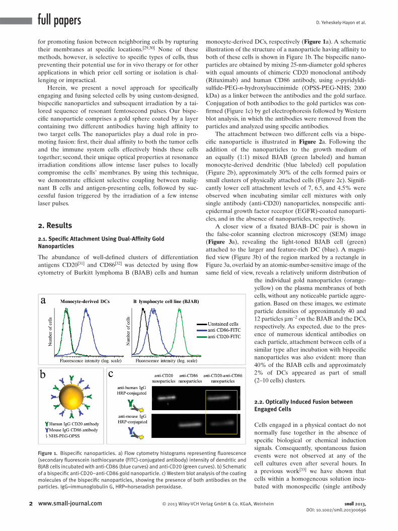
The abundance of well-defi ned clusters of differentiation
antigens CD20 [ 31 ] and CD86 [ 32 ] was detected by using fl ow
cytometry of Burkitt lymphoma B (BJAB) cells and human
2 www.small-journal.com © 2013 Wiley-VCH Ve
Figure 1 . Bispecifi c nanoparticles. a) Flow cytometry histograms represe(secondary fl uorescein isothiocyanate (FITC)-conjugated antibody) intensiBJAB cells incubated with anti-CD86 (blue curves) and anti-CD20 (green curof a bispecifi c anti-CD20–anti-CD86 gold nanoparticle. c) Western blot anamolecules of the bispecifi c nanoparticles, showing the presence of both particles. IgG = immunoglobulin G, HRP = horseradish peroxidase.
monocyte-derived DCs, respectively ( Figure 1 a). A schematic
illustration of the structure of a nanoparticle having affi nity to
both of these cells is shown in Figure 1 b. The bispecifi c nano-
particles are obtained by mixing 25-nm-diameter gold spheres
with equal amounts of chimeric CD20 monoclonal antibody
(Rituximab) and human CD86 antibody, using o -pyridyldi-
sulfi de-PEG- n -hydroxylsuccinimide (OPSS-PEG-NHS; 2000
kDa) as a linker between the antibodies and the gold surface.
Conjugation of both antibodies to the gold particles was con-
fi rmed (Figure 1 c) by gel electrophoresis followed by Western
blot analysis, in which the antibodies were removed from the
particles and analyzed using specifi c antibodies.
The attachment between two different cells via a bispe-
cifi c nanoparticle is illustrated in Figure 2 a. Following the
addition of the nanoparticles to the growth medium of
an equally (1:1) mixed BJAB (green labeled) and human
monocyte-derived dendritic (blue labeled) cell population
(Figure 2 b), approximately 30% of the cells formed pairs or
small clusters of physically attached cells (Figure 2 c). Signifi -
cantly lower cell attachment levels of 7, 6.5, and 4.5% were
observed when incubating similar cell mixtures with only
single antibody (anti-CD20) nanoparticles, nonspecifi c anti-
epidermal growth factor receptor (EGFR)-coated nanoparti-
cles, and in the absence of nanoparticles, respectively.
A closer view of a fi xated BJAB–DC pair is shown in
the false-color scanning electron microscopy (SEM) image
( Figure 3 a), revealing the light-toned BJAB cell (green)
attached to the larger and feature-rich DC (blue). A magni-
fi ed view (Figure 3 b) of the region marked by a rectangle in
Figure 3 a, overlaid by an atomic-number-sensitive image of the
same fi eld of view, reveals a relatively uniform distribution of
rlag GmbH & Co. KGaA
nting fl uorescence ty of dendritic and ves). b) Schematic lysis of the coating antibodies on the
the individual gold nanoparticles (orange-
yellow) on the plasma membranes of both
cells, without any noticeable particle aggre-
gation. Based on these images, we estimate
particle densities of approximately 40 and
12 particles μ m − 2 on the BJAB and the DCs,
respectively. As expected, due to the pres-
ence of numerous identical antibodies on
each particle, attachment between cells of a
similar type after incubation with bispecifi c
nanoparticles was also evident: more than
40% of the BJAB cells and approximately
2% of DCs appeared as part of small
(2–10 cells) clusters.
2.2. Optically Induced Fusion between Engaged Cells
Cells engaged in a physical contact do not
normally fuse together in the absence of
specifi c biological or chemical induction
signals. Consequently, spontaneous fusion
events were not observed at any of the
cell cultures even after several hours. In
a previous work [ 33 ] we have shown that
cells within a homogeneous solution incu-
bated with monospecifi c (single antibody
, Weinheim small 2013, DOI: 10.1002/smll.201300696

Optically Induced Cell Fusion Using Bispecifi c Nanoparticles
Figure 3 . Bispecifi c gold nanoparticles on the plasma membrane of a coupled BJAB–DC pair. a) SEM image of the dendritic (false color blue) and BJAB (false color green) cell pair. Scale bar represents 1 μ m. b) Magnifi ed view of the region marked by a rectangle in (a), overlaid by a back-scattering detection image that reveals the presence of gold nanoparticles (false color yellow). Scale bar represents 200 nm.
Figure 2 . Cell coupling using a bispecifi c nanoparticle. a) Schematic illustration of nanoparticle-mediated coupling between a malignant B cell and a DC. b) Fluorescence-phase contrast image of BJAB cells (green cytoplasm) attached to DCs (blue nuclei). Scale bar represents 10 μ m. c) Cell attachment effi ciency using different nanoparticles. ( ∗ ) indicates P < 0.0001.
coating) gold nanospheres were led to fuse following irra-
diation by a few high-intensity femtosecond pulses that were
tuned to the plasmonic resonance wavelength of the nano-
particles. The fusion effi ciency of cells of different types was
extremely low in these experiments due to the random and
transient physical contacts between the cells.
To demonstrate specifi c cell fusion, the entire volume of the
culture containing the coupled BJAB–DC pairs was washed
off the unbound nanoparticles and irradiated by a sequence
(ten pulses, 1 kHz repetition rate) of intense (12 mJ cm − 2 ),
ultrashort (50 fs), resonant (545 nm wavelength) laser pulses.
© 2013 Wiley-VCH Verlag Gmbsmall 2013, DOI: 10.1002/smll.201300696
Time-lapse fl uorescence-phase contrast imaging of two
exemplary fusion events between BJAB and DCs is shown
in Figure 4 a. Data analysis based on 1000 cells revealed that
fusion between BJAB and DCs was less common (7%) than
fusion between homogeneous pairs (or clusters) of BJAB
cells only (20%). Moreover, DC–DC fusion was not detected
at all, unless a BJAB cell was involved in the fusion process
into a DC–DC–BJAB cell hybrid. The irradiated BJAB cells
and DCs that were in physical contact showed fi rst signs of
morphological changes immediately after irradiation, toward
the formation of hybrid cells with a unifi ed cytoplasm after
20–30 min (Figure 4 a). A schematic model of the fusion pro-
cess between engaged cells irradiated by femtosecond pulses
is illustrated in Figure 4 b, which depicts the local disruption
of the cells’ plasma membranes initiated by the several tens
of nanometers in diameter [ 34 , 35 ] cavitation bubble that has
been formed around the particle. [ 36 ]
In addition to DCs, foreign or tumor antigens could also
be presented to the adaptive immune system by macrophages,
following attachment and fusion with the target cells. To dem-
onstrate the induction of fusion between macrophages and
malignant B cells, monocyte-derived macrophages expressing
3www.small-journal.comH & Co. KGaA, Weinheim

D. Yeheskely-Hayon et al.full papers
Figure 4 . Fusion between dendritic and BJAB cells. a) Two time sequences of fl uorescence-phase contrast images of fusing BJAB cells (green cytoplasm) and DCs (blue nuclei) following incubation with anti-CD20–anti-CD86 nanoparticles and irradiation by ten pulses. Scale bars represent 10 μ m. b) Schematic illustration of the fusion process between attached cells triggered by femtosecond pulses.
CD86 surface receptors were mixed with CD20-expressing
BJAB cells and incubated within a medium containing bispe-
cifi c anti-CD20–anti-CD86 nanoparticles. After 15 min of
incubation, nearly 50% of the macrophages in the culture
were found attached to BJAB cells ( Figure 5 a,b), a signifi -
cantly higher rate compared with coupling in a nanoparticle-
free medium (20%). In contrast, the use of monospecifi c
anti-CD86 nanoparticles resulted in 26% macrophage–BJAB
cell attachment and 30% macrophage–macrophage attach-
ment. After irradiation by a single sequence (ten pulses,
1 kHz repetition rate) of femtosecond pulses, approximately
9% of the macrophage–BJAB cell pairs were successfully
fused. A typical fusing pair of cells is shown in a time-lapse
image sequence in Figure 5 c, which reveals the plasma mem-
brane fusion (phase contrast, top row), cytoplasmic (green
fl uorescence, second row) transfer from the BJAB cell during
fusion, and gradual passage of the nuclear blue marker from
the macrophage nucleus to the BJAB cell nucleus (third
row). Consistently, the fused cells remained viable more than
24 h after irradiation in all fusion experiments.
3. Discussion
Although gold nanoparticles are relatively chemically inert
within biological tissue, they could strongly bind through
4 www.small-journal.com © 2013 Wiley-VCH Verlag GmbH & Co. KGaA
various ligands to targeting molecules,
thereby yielding simple, cost-effective, and
stable conjugates of substantial binding
specifi city. [ 37 , 38 ] Their targeting effi ciency
could be further enhanced by using mul-
tiple targeting molecules, [ 39 ] resulting in
an inherent ability to attach together cells
that express different receptors, through
strong chemical bonds. Once the cells are
attached, the unique optical properties
of the gold particles could be utilized to
promote fusion. While the exact mecha-
nism by which the irradiated nanoparticles
promote cell fusion is as yet unclear, it is
most likely triggered by the mechanical
effect around the irradiated nanoparticles,
which destabilizes the adjacent plasma
membranes, [ 40 ] thus leading to the for-
mation of a single hybrid cell with a
unifi ed cytoplasm. In our experiments,
when the fl uence of the femtosecond
pulse train exceeded a certain threshold
level (typically fi ve pulses of 20 mJ cm − 2
each), cells often underwent widespread
necrosis, most likely due to their inability
to effectively recover from the extensive
membrane destabilization or rupture.
Interestingly, cells that were undergoing
fusion were less prone to necrosis than
individual cells or unfused pairs of cells.
A partial possible explanation for this
observation is the lower surface area of
the fusion product, which allows its plasma
membrane to restore its integrity rapidly
and more effectively. Another interesting observation was
the mediating role of the BJAB cells in the fusion between
two DCs, which resulted in the formation of numerous DC–
DC–BJAB cell triplets. This property may potentially be used
in various future applications to increase fusion effi ciency
between nonfusogenic cells.
Fusing different cells having different complementary
properties into a single hybrid product of desired properties
would be extremely valuable for various applications in tissue
regeneration [ 41 ] and drug development, [ 42 ] and for the estab-
lishment of new therapeutic strategies. [ 43 , 44 ] In this proof-of-
concept study, we have demonstrated specifi c fusion between
tumor cells and DCs, the role of which in cancer vaccination
is well characterized, [ 21 ] as well as fusion between tumor cells
and macrophages, which are known to be involved in various
immune regulation pathways; [ 45 , 46 ] the macrophages’ exact
role in stimulating antitumor response following fusion with
cancer cells, however, is as yet unknown.
For specifi c applications, further research would be
required for testing the functionality of the hybrid cell prod-
ucts. One potential obstacle for some applications would be
the presence of remaining nanoparticles on the plasma mem-
branes of the fusion products, which could interfere with
their antigen presentation process. The remaining nanoparti-
cles could also inhibit immune regulation processes that are
, Weinheim small 2013, DOI: 10.1002/smll.201300696

Optically Induced Cell Fusion Using Bispecifi c Nanoparticles
Figure 5 . Macrophage–B cell coupling and fusion. a) Three selected regions of interest from a fl uorescence-phase contrast image of macrophages (blue nuclei) and BJAB cells (green cytoplasm) after incubation with anti-CD20–anti-CD86 nanoparticles. Bar chart: Macrophage–BJAB and macrophage–macrophage attachment effi ciency for different particles. ( ∗ ) indicates P < 0.0001. b) Time-lapse image sequence of fusing macrophage (top right) and BJAB (bottom left) cells following pulse irradiation. Phase contrast, green, and blue fl uorescence images (top, middle, and bottom panels, respectively) show the redistribution of the cells’ content during fusion. Scale bars represent 10 μ m.
usually initiated via the conjugated receptors. One could avoid
these diffi culties by developing a method for nondestructive
removal of the nanoparticles from the fusion products, or
alternatively, by minimizing the total particle concentration
during the course of the attachment–fusion process. Finally,
antibody detachment from the gold nanoparticles could be
harmful for various in vivo and ex vivo applications, due to
undesired triggering of complement-dependent cytotox-
icity. [ 47 ] The problem could be minimized by optimizing the
antibody-to-particle ratio within the bispecifi c nanoparticles,
thereby minimizing the amount of released antibodies.
4. Conclusion
In summary, we have presented a novel technique for selec-
tive fusion between antigen-presenting cells and tumor cells,
by using specifi cally designed nanoparticles for the attach-
ment of selected cells, and fusion of their plasma membranes
following irradiation by a short sequence of resonant femto-
second laser pulses. The low toxicity of the gold particles, high
specifi city, effi ciency, and relative simplicity of this approach
would make it useful for a wide range of biomedical appli-
cations, and open new possibilities in biotechnology and in
fundamental biological research. This approach would also
broaden the use of nanotechnology for various biomedical
applications by offering effective means for triggering and
controlling desired interactions on a nanometer scale, poten-
tially resulting in a more accurate research procedure and
less invasive medical intervention.
© 2013 Wiley-VCH Verlag Gmbsmall 2013, DOI: 10.1002/smll.201300696
5. Experimental Section
Cell Cultures : BJAB cells were grown at 37 ° C and 5% CO 2 in RPMI-1640 medium (Sigma, Israel) supplemented with 2 m M glu-tamine, 5 m M sodium pyruvate, and 10% heat-inactivated fetal bovine serum. Cells were maintained at a concentration below 10 6 cells per mL to allow logarithmic growth.
Isolation and Differentiation of Dendritic Cells and Mac-rophages : Peripheral blood mononuclear cells from a consenting healthy donor were isolated by Lymphoprep (Axis Shield, Norway) density centrifugation according to the manufacturer's pro-tocol, and cultured in RPMI-1640 with 5% autologous plasma for 1–2 h. Nonadherent cells were removed, and the adherent popu-lation was cultured for 5–7 days in RPMI-1640 containing only 5% autologous serum or RPMI-1640 containing 5% autologous serum, 500 units mL − 1 recombinant human interleukin-4 (BioVision, USA), and 1000 units mL − 1 granulocyte macrophage colony-stimulating factor (BioVision, USA), to yield macrophages and immature DCs, respectively. Half-volume medium replacement was performed every 3 days. DC maturation was obtained by adding 100 units mL − 1 of tumor necrosis factor- α (BioVision, USA) to the medium at day 7.
Nanoparticle Preparation : Gold nanoparticles were prepared using a citrate reduction protocol [ 48 ] resulting in an average particle diameter of 25 nm. Anti-CD20 (Rituximab, Roche Israel) and anti-CD86 (BioLegend, USA) coating of gold nanoparticles was carried out according to Weiss et al. [ 49 ] with some modifi cations. Briefl y, antibodies were incubated with OPSS-PEG2000-NHS (JenKem, USA) for 1 h in a mole ratio of 1:5000, followed by an additional 1 h of incubation with gold nanospheres in a mole ratio of 1:10 6
5www.small-journal.comH & Co. KGaA, Weinheim
D. Yeheskely-Hayon et al.
6
full papers
gold nanoparticles:OPSS-PEG2000-NHS. Glycine (30 m M ) was added for overnight incubation followed by three washes with phosphate-buffered saline (PBS). Conjugation of both anti-bodies to the gold particles was confi rmed by detaching the antibody coating layer from the gold nanospheres by using β -mercaptoethanol and analyzing the reduced solution using gel electrophoresis followed by Western blot analysis.Scanning Electron Microscopy : Cells were fi xed with 3% glu-taraldehyde and mounted on polylysine-coated silicon chips (Ted Pella, Inc.). Samples were then fi xed with 1% osmium and dehy-drated as described elsewhere. [ 50 ] The microscope system used (Zeiss Ultra Plus HRSEM) was equipped with a Schottky fi eld-emission electron gun and BalTec VCT100 cold stage maintained at − 150 ° C.
Specifi c Attachment and Fusion Experiments : BJAB cells were stained with 1 μ M calcein acetoxymethyl ester for 15 min at room temperature, followed by three PBS washes. DCs and macrophage nuclei were stained with Hoechst 33342 (2 μ g mL − 1 ; Sigma, Israel) for 15 min at room temperature followed by washes with PBS and 3 min of incubation with trypsin, in order to obtain nonadherent antigen-presenting cells (APCs). Cells were then mixed in a 1:1 ratio (APCs:BJAB cells) in the presence of antibody-coated gold nanoparticles (5 × 10 10 particles mL − 1 ) for 20 min at room tem-perature under moderate shaking. Following incubation, the cells were washed three times with PBS and seeded on eight-well chamber slides (Lab-Tek II, Thermo Scientifi c). Pairs of APCs and BJAB cells were then counted based on the cells' morphology and fl uorescent dyes. The statistical signifi cance of the results was assessed based on counting at least 500 cells and using a two-proportion z -test.
Laser Pulse Irradiation : A beam from a Ti:sapphire oscillator (Tsunami, Spectra Physics) was amplifi ed (Spitfi re Pro XP, Spectra Physics) and wavelength-tuned to 545 nm by using an optical par-ametric amplifi er (Topas-C, Spectra Physics). The pulse duration was 50 fs at a 1 kHz repetition rate. Cells were irradiated within eight-well chamber slides (Lab-Tek II, Thermo Scientifi c), which were placed within a microscope incubator (Okolab Inc.) at con-trolled temperature and CO 2 concentration. The irradiation pattern was a 35 × 35 rectangular array of 250- μ m-diameter spots (approx-imately 75 mm 2 total area). Multiple pulse irradiation per spot was achieved by scanning the beam at lower rates, so that each point was irradiated by several consequent overlapping spots.
Acknowledgements
We thank Prof. Yoram Reiter and Dr. Gili Bisker for enlightening discussions, Prof. Doron Melamed for providing the Burkitt lym-phoma cell line, and Dima Bourdetsky and Ravit Oren for technical assistance. This work was funded in part by a European Research Council starting grant (239986) and by the Lorry I. Lokey Interdisci-plinary Center for Life Sciences and Engineering.
[ 1 ] G. P. Dunn , L. J. Old , R. D. Schreiber , Immunity 2004 , 21 , 137 . [ 2 ] E. J. Roy , U. Gawlick , B. A. Orr , D. M. Kranz , Adv. Drug Delivery
Rev. 2004 , 56 , 1219 .
www.small-journal.com © 2013 Wiley-VCH V
[ 3 ] R. D. Jachimowicz , G. Fracasso , P. J. Yazaki , B. E. Power , P. Borchmann , A. Engert , H. P. Hansen , K. S. Reiners , M. Marie , E. P. von Strandmann , A. Rothe , Mol. Cancer Ther. 2011 , 10 , 1036 .
[ 4 ] B. Karpovsky , J. A. Titus , D. A. Stephany , D. M. Segal , J. Exp. Med. 1984 , 160 , 1686 .
[ 5 ] M. Brennan , P. F. Davison , H. Paulus , Science 1985 , 229 , 81 . [ 6 ] C. Milstein , A. C. Cuello , Nature 1983 , 305 , 537 . [ 7 ] U. D. Staerz , O. Kanagawa , M. J. Bevan , Nature 1985 , 314 , 628 . [ 8 ] P. Perez , R. W. Hoffman , S. Shaw , J. A. Bluestone , D. M. Segal ,
Nature 1985 , 316 , 354 . [ 9 ] P. K. Wallace , J. L. Romet-Lemonne , M. Chokri , L. H. Kasper ,
M. W. Fanger , C. E. Fadul , Cancer Immunol. Immunother. 2000 , 49 , 493 .
[ 10 ] A. Hombach , W. Jung , C. Pohl , C. Renner , U. Sahin , R. Schmits , J. Wolf , U. Kapp , V. Diehl , M. Pfreundschuh , Int. J. Cancer 1993 , 55 , 830 .
[ 11 ] S. Johnson , S. Burke , L. Huang , S. Gorlatov , H. Li , W. Wang , W. Zhang , N. Tuaillon , J. Rainey , B. Barat , Y. Yang , L. Jin , V. Ciccarone , P. A. Moore , S. Koenig , E. Bonvini , J. Mol. Biol. 2010 , 399 , 436 .
[ 12 ] A. Loffl er , P. Kufer , R. Lutterbuse , F. Zettl , P. T. Daniel , J. M. Schwenkenbecher , G. Riethmuller , B. Dorken , R. C. Bargou , Blood 2000 , 95 , 2098 .
[ 13 ] P. A. Baeuerle , P. Kufer , R. Bargou , Curr. Opin. Mol. Ther. 2009 , 11 , 22 .
[ 14 ] R. M. Steinman , Annu. Rev. Immunol. 1991 , 9 , 271 . [ 15 ] J. Gong , N. Nikrui , D. Chen , S. Koido , Z. Wu , Y. Tanaka ,
S. Cannistra , D. Avigan , D. Kufe , J. Immunol. 2000 , 165 , 1705 . [ 16 ] J. Gong , D. Avigan , D. Chen , Z. Wu , S. Koido , M. Kashiwaba ,
D. Kufe , Proc. Natl. Acad. Sci. USA 2000 , 97 , 2715 . [ 17 ] T. Kikuchi , Y. Akasaki , M. Irie , S. Homma , T. Abe , T. Ohno , Cancer
Immunol. Immunother. 2001 , 50 , 337 . [ 18 ] J. Galea-Lauri , D. Darling , G. Mufti , P. Harrison , F. Farzaneh ,
Cancer Immunol. Immunother. 2002 , 51 , 299 . [ 19 ] N. Raje , T. Hideshima , F. E. Davies , D. Chauhan , S. P. Treon ,
G. Young , Y.-T. Tai , D. Avigan , J. Gong , R. L. Schlossman , P. Richardson , D. W. Kufe , K. C. Anderson , Br. J. Haematol. 2004 , 125 , 343 .
[ 20 ] D. Avigan , B. Vasir , J. Gong , V. Borges , Z. Wu , L. Uhl , M. Atkins , J. Mier , D. McDermott , T. Smith , N. Giallambardo , C. Stone , K. Schadt , J. Dolgoff , J.-C. Tetreault , M. Villarroel , D. Kufe , Clin. Cancer Res. 2004 , 10 , 4699 .
[ 21 ] D. Avigan , Blood Rev. 1999 , 13 , 51 . [ 22 ] S. Koido , M. Ohana , C. Liu , N. Nikrui , J. Durfee , A. Lerner , J. Gong ,
Clin. Immunol. 2004 , 113 , 261 . [ 23 ] J. Gong , D. Chen , M. Kashiwaba , D. Kufe , Nat. Med. 1997 , 3 , 558 . [ 24 ] S. Schneiderman , J. L. Farber , R. Baserga , Somatic Cell Genet.
1979 , 5 , 263 . [ 25 ] E. Gottfried , R. Krieg , C. Eichelberg , R. Andreesen , A. Mackensen ,
S. W. Krause , Cancer Immun. 2002 , 2 , 15 . [ 26 ] U. Zimmermann , Biochim. Biophys. Acta 1982 , 694 , 227 . [ 27 ] S. Nagata , K. Yamamoto , Y. Ueno , T. Kurata , J. Chiba , Hybridoma
1991 , 10 , 369 . [ 28 ] A. Vogel , J. Noack , G. Hüttman , G. Paltauf , Appl. Phys. B: Lasers
Opt. 2005 , 81 , 1015 . [ 29 ] R. W. Steubing , S. Cheng , W. H. Wright , Y. Numajiri , M. W. Berns ,
Cytometry 1991 , 12 , 505 . [ 30 ] K. Kuetemeyer , A. Lucas-Hahn , B. Petersen , H. Niemann ,
A. Heisterkamp , J. Biomed. Opt. 2011 , 16 , 088001 . [ 31 ] T. F. Tedder , M. Streuli , S. F. Schlossman , H. Saito , Proc. Natl.
Acad. Sci. USA 1988 , 85 , 208 . [ 32 ] J. W. Young , L. Koulova , S. A. Soergel , E. A. Clark , R. M. Steinman ,
B. Dupont , J. Clin. Invest. 1992 , 90 , 229 . [ 33 ] L. Minai , D. Yeheskely-Hayon , L. Golan , G. Bisker , E. J. Dann ,
D. Yelin , Small 2012 , 8 , 1732 . [ 34 ] A. N. Volkov , C. Sevilla , L. V. Zhigilei , Appl. Surf. Sci. 2007 , 253 ,
6394 .
erlag GmbH & Co. KGaA, Weinheim small 2013, DOI: 10.1002/smll.201300696

Optically Induced Cell Fusion Using Bispecifi c Nanoparticles
[ 35 ] G. Bisker , D. Yelin , J. Opt. Soc. Am. B 2012 , 29 , 1383 . [ 36 ] E. Lukianova-Hleb , Y. Hu , L. Latterini , L. Tarpani , S. Lee ,
R. A. Drezek , J. H. Hafner , D. O. Lapotko , ACS Nano 2010 , 4 , 2109 . [ 37 ] X. Cai , S. Conley , M. Naash , Vision Res. 2008 , 48 , 319 . [ 38 ] A. Llevot , D. Astruc , Chem. Soc. Rev. 2012 , 41 , 242 . [ 39 ] S. Bhattacharyya , J. A. Khan , G. L. Curran , J. D. Robertson ,
R. Bhattacharya , P. Mukherjee , Adv. Mater. 2011 , 23 , 5034 . [ 40 ] G. Wu , A. Mikhailovsky , H. A. Khant , C. Fu , W. Chiu , J. A. Zasadz-
inski , J. Am. Chem. Soc. 2008 , 130 , 8175 . [ 41 ] C. A. Cowan , J. Atienza , D. A. Melton , K. Eggan , Science 2005 ,
309 , 1369 . [ 42 ] G. Kohler , C. Milstein , Nature 1975 , 256 , 495 . [ 43 ] Y. Guo , M. Wu , H. Chen , X. Wang , G. Liu , G. Li , J. Ma , M. S. Sy , Sci-
ence 1994 , 263 , 518 . [ 44 ] K.-W. Peng , C. J. TenEyck , E. Galanis , K. R. Kalli , L. C. Hartmann ,
S. J. Russell , Cancer Res. 2002 , 62 , 4656 .
© 2013 Wiley-VCH Verlag GmbHsmall 2013, DOI: 10.1002/smll.201300696
[ 45 ] A. Mantovani , P. Allavena , A. Sica , Eur. J. Cancer 2004 , 40 , 1660 .
[ 46 ] D. M. Mosser , J. P. Edwards , Nat. Rev. Immunol. 2008 , 8 , 958 . [ 47 ] G. Bisker , D. Yeheskely-Hayon , L. Minai , D. Yelin , J. Controlled
Release 2012 , 162 , 303 . [ 48 ] J. Turkevich , P. C. Stevenson , J. Hillier , J. Phys. Chem. 1953 , 57 ,
670 . [ 49 ] A. Weiss , T. C. Preston , J. Popov , Q. Li , S. Wu , K. C. Chou ,
H. M. Burt , M. B. Bally , R. Signorell , J. Phys. Chem. C 2009 , 113 , 20252 .
[ 50 ] L. Shaulov , R. Gruber , I. Cohen , A. Harel , J. Cell Sci. 2011 , 124 , 3822 .
Received: March 5, 2013 Revised: April 15, 2013Published online:
7www.small-journal.com & Co. KGaA, Weinheim