Roles of vaccinia virus EEV-specific proteins in intracellular actin tail formation and low...
11
Downloaded from www.microbiologyresearch.org by IP: 54.157.140.51 On: Sun, 10 Apr 2016 23:11:31 Journal of General Virology (1998), 79, 1415–1425. Printed in Great Britain ................................................................................................................................................................................................................................................................................... Roles of vaccinia virus EEV-specific proteins in intracellular actin tail formation and low pH-induced cell–cell fusion Christopher M. Sanderson, 1 Friedrich Frischknecht, 2 Michael Way, 2 Michael Hollinshead 1 and Geoffrey L. Smith 1 1 Sir William Dunn School of Pathology, University of Oxford, South Parks Road, Oxford OX1 3RE, UK 2 European Molecular Biology Laboratory, Postfach 10.2209, Meyerhofstrasse 1, 69117 Heidelberg, Germany During vaccinia virus (VV) morphogenesis intra- cellular mature virus (IMV) is wrapped by two additional membranes to form intracellular envel- oped virus (IEV). IEV particles can nucleate the formation of actin tails which aid movement of IEVs to the cell surface where the outer IEV membrane fuses with the plasma membrane forming cell- associated enveloped virus (CEV) which remains attached to the cell, or extracellular enveloped virus (EEV) which is shed from the cell. In this report, we have used a collection of VV mutants lacking individual EEV-specific proteins to compare the roles of these proteins in the formation of IEV and IEV-associated actin tails and fusion of infected cells after a low pH shock. Data presented here show that Introduction Vaccinia virus (VV) is a poxvirus that was used as the vaccine to eradicate smallpox (Fenner et al., 1988) and is the most extensively studied member of the orthopoxvirus genus (Moss, 1996). Like other poxviruses, VV replicates in the cytoplasm using its own enzymes for transcription and DNA replication and produces large, enveloped virions. Assembly of VV particles is complex and produces two structurally distinct infectious virions termed intracellular mature virus (IMV) and extracellular enveloped virus (EEV). IMV represents the majority of infectious virus and has been the form of virus studied by most investigators due to its abundance, ease of purification and robust structure. IMV and EEV bind to different cellular receptors (Vanderplasschen & Smith, 1997), enter cells by different mechanisms (Vanderplasschen et al., 1998) and EEV, unlike IMV, is resistant to neutralization by antibody (Ichihashi, 1996 ; Author for correspondence : Geoffrey L. Smith. Fax ›44 1865 275501. e-mail glsmith!molbiol.ox.ac.uk p45–50 (A36R) is not required for IEV formation or for acid-induced cell–cell fusion, but is required for formation of IEV-associated actin tails. In contrast, gp86 (A56R), the virus haemagglutinin, is not required for formation of either IEV or IEV- associated actin tails. Data presented also confirm that p37 (gene F13L), gp42 (B5R) and gp22–24 (A34R) are needed for formation of IEV-associated actin tails and for cell–cell fusion after low pH shock. The phenotypes of these mutants were not affected by the host cell type as similar results were obtained in a range of different cells. Lastly, comparisons of the phenotypes of VV strains Western Reserve, ΔA34R and ΔA36R demonstrate that actin tails are not required for low pH-induced cell–cell fusion. Vanderplasschen et al., 1997). IMV particles are formed by the envelopment of the viral genome in membranes that were proposed originally to be synthesized de novo (for review see Dales & Pogo, 1981), but more recently were reported to be derived from the intermediate compartment between the endoplasmic reticulum and the Golgi stacks (Sodeik et al., 1993). After maturation, some IMV particles are wrapped by a double membrane (Ichihashi et al., 1971 ; Morgan, 1976 ; Payne & Kristensson, 1979) derived from tubular endosomes (Tooze et al., 1993) or the trans-Golgi network (Hiller & Weber, 1985 ; Schmelz et al., 1994) to form an intracellular enveloped virus (IEV). IEV particles move to the cell surface where the outer membrane fuses with the plasma membrane to form cell- associated enveloped virus (CEV) which remains on the cell surface (Blasco & Moss, 1992), or EEV which is released from the cell. The movement of IEV can be assisted by the polymerization of actin tails (Cudmore et al., 1995, 1996), similar to those described for Shigella, Listeria and Rickettsia (for reviews see Tilney & Tilney, 1993; Cossart & Kocks, 1994; Cossart, 1995) ; however, EEV are still formed without actin tail formation (Wolffe et al., 1997 ; Mathew et al., 1998). EEV may also be formed by budding of IMV through the plasma 0001-5379 # 1998 SGM BEBF
-
Upload
uni-heidelberg -
Category
Documents
-
view
6 -
download
0
Transcript of Roles of vaccinia virus EEV-specific proteins in intracellular actin tail formation and low...

Downloaded from www.microbiologyresearch.org by
IP: 54.157.140.51
On: Sun, 10 Apr 2016 23:11:31
Journal of General Virology (1998), 79, 1415–1425. Printed in Great Britain. . . . . . . . . . . . . . . . . . . . . . . . . . . . . . . . . . . . . . . . . . . . . . . . . . . . . . . . . . . . . . . . . . . . . . . . . . . . . . . . . . . . . . . . . . . . . . . . . . . . . . . . . . . . . . . . . . . . . . . . . . . . . . . . . . . . . . . . . . . . . . . . . . . . . . . . . . . . . . . . . . . . . . . . . . . . . . . . . . . . . . . . . . . . . . . . . . . . . . . . . . . . . . . . . . . . . . . . . . . . . . . . . . . . . . . . . . . . . . . . . . . . . . . . . . . . . . . . . . . . . . . . . . . . . . . . . . .
Roles of vaccinia virus EEV-specific proteins in intracellularactin tail formation and low pH-induced cell–cell fusion
Christopher M. Sanderson,1 Friedrich Frischknecht,2 Michael Way,2 Michael Hollinshead1
and Geoffrey L. Smith1
1 Sir William Dunn School of Pathology, University of Oxford, South Parks Road, Oxford OX1 3RE, UK2 European Molecular Biology Laboratory, Postfach 10.2209, Meyerhofstrasse 1, 69117 Heidelberg, Germany
During vaccinia virus (VV) morphogenesis intra-cellular mature virus (IMV) is wrapped by twoadditional membranes to form intracellular envel-oped virus (IEV). IEV particles can nucleate theformation of actin tails which aid movement of IEVsto the cell surface where the outer IEV membranefuses with the plasma membrane forming cell-associated enveloped virus (CEV) which remainsattached to the cell, or extracellular enveloped virus(EEV) which is shed from the cell. In this report, wehave used a collection of VV mutants lackingindividual EEV-specific proteins to compare theroles of these proteins in the formation of IEV andIEV-associated actin tails and fusion of infected cellsafter a low pH shock. Data presented here show that
IntroductionVaccinia virus (VV) is a poxvirus that was used as the
vaccine to eradicate smallpox (Fenner et al., 1988) and is themost extensively studied member of the orthopoxvirus genus(Moss, 1996). Like other poxviruses, VV replicates in thecytoplasm using its own enzymes for transcription and DNAreplication and produces large, enveloped virions.
Assembly of VV particles is complex and produces twostructurally distinct infectious virions termed intracellularmature virus (IMV) and extracellular enveloped virus (EEV).IMV represents the majority of infectious virus and has beenthe form of virus studied by most investigators due to itsabundance, ease of purification and robust structure. IMVand EEV bind to different cellular receptors (Vanderplasschen& Smith, 1997), enter cells by different mechanisms(Vanderplasschen et al., 1998) and EEV, unlike IMV, isresistant to neutralization by antibody (Ichihashi, 1996 ;
Author for correspondence: Geoffrey L. Smith.
Fax 44 1865 275501. e-mail glsmith!molbiol.ox.ac.uk
p45–50 (A36R) is not required for IEV formation orfor acid-induced cell–cell fusion, but is required forformation of IEV-associated actin tails. In contrast,gp86 (A56R), the virus haemagglutinin, is notrequired for formation of either IEV or IEV-associated actin tails. Data presented also confirmthat p37 (gene F13L), gp42 (B5R) and gp22–24(A34R) are needed for formation of IEV-associatedactin tails and for cell–cell fusion after low pH shock.The phenotypes of these mutants were not affectedby the host cell type as similar results were obtainedin a range of different cells. Lastly, comparisons ofthe phenotypes of VV strains Western Reserve,∆A34R and ∆A36R demonstrate that actin tails arenot required for low pH-induced cell–cell fusion.
Vanderplasschen et al., 1997). IMV particles are formed by theenvelopment of the viral genome in membranes that wereproposed originally to be synthesized de novo (for review seeDales & Pogo, 1981), but more recently were reported to bederived from the intermediate compartment between theendoplasmic reticulum and the Golgi stacks (Sodeik et al.,1993). After maturation, some IMV particles are wrapped by adouble membrane (Ichihashi et al., 1971 ; Morgan, 1976 ; Payne& Kristensson, 1979) derived from tubular endosomes (Toozeet al., 1993) or the trans-Golgi network (Hiller & Weber, 1985 ;Schmelz et al., 1994) to form an intracellular enveloped virus(IEV). IEV particles move to the cell surface where the outermembrane fuses with the plasma membrane to form cell-associated enveloped virus (CEV) which remains on the cellsurface (Blasco & Moss, 1992), or EEV which is released fromthe cell. The movement of IEV can be assisted by thepolymerization of actin tails (Cudmore et al., 1995, 1996),similar to those described for Shigella, Listeria and Rickettsia (forreviews see Tilney & Tilney, 1993 ; Cossart & Kocks, 1994 ;Cossart, 1995) ; however, EEV are still formed without actin tailformation (Wolffe et al., 1997 ; Mathew et al., 1998). EEV mayalso be formed by budding of IMV through the plasma
0001-5379 # 1998 SGM BEBF

Downloaded from www.microbiologyresearch.org by
IP: 54.157.140.51
On: Sun, 10 Apr 2016 23:11:31
C. M. Sanderson and othersC. M. Sanderson and others
F-Actin D8L
rA3
6R
D A
36R
D A
34R
WR
Fig. 1. Distribution of F-actin and VV particles in BS-C-1 cells infected with WR (a and e), ∆A34R (b and f), ∆A36R (c and g)or rA36R (d and h) at 10 p.f.u. per cell. At 18 h p.i. viral particles were stained with MAb AB1.1 against the VV D8L protein(e–h), while TRITC–phalloidin was used to stain F-actin (a–d).
BEBG
Downloaded from www.microbiologyresearch.org by
IP: 54.157.140.51
On: Sun, 10 Apr 2016 23:11:31
Vaccinia virus EEV mutantsVaccinia virus EEV mutants
membrane, as observed late in infection with the IHD-W strainof VV (Tsutsui, 1983).
Six VV genes have been identified that encode proteinscontained within the outer membrane of EEV particles. Theseare : A56R [haemagglutinin (HA) gp86] (Payne & Norrby,1976 ; Shida, 1986), F13L (p37) (Hirt et al., 1986), B5R (gp42)(Engelstad et al., 1992 ; Isaacs et al., 1992), A34R (gp22–24)(Duncan & Smith, 1992), A36R (p45–50) (Parkinson & Smith,1994) and A33R (gp23–28) (Roper et al., 1996). None of theseproteins (excludingA33R,which has not yet been investigated)affect the formation of IMV, but they have differing affects onIMV wrapping, actin tail formation, EEV release and low pH-induced cell–cell fusion. F13L (Blasco & Moss, 1991) and B5R(Engelstad & Smith, 1993 ; Wolffe et al., 1993) are required forthe wrapping of IMV by intracellular membranes. Viruseslacking A34R (McIntosh & Smith, 1996 ; Wolffe et al., 1997) orthe short consensus repeat domains of B5R (Mathew et al.,1998) enable IEV formation and enhanced release of EEV, butprevent actin tail formation. This suggests that actin tailformation and retention of CEV particles on the cell surfacemay involve common viral components.
Cell–cell fusion can be induced by brief incubation of VV-infected cells at low pH and requires the p14 protein (geneA27L) that is present on the surface of IMV (Gong et al., 1989,1990). Fusion also requires the formation of enveloped viruses(IEV, CEV and EEV) since it does not occur with mutant viruseslacking F13L and B5R (Blasco & Moss, 1991 ; Wolffe et al.,1993). Interestingly, fusion is also blocked in ∆A34R-infectedcells (Duncan, 1992 ; Wolffe et al., 1997), although this virusproduces EEV particles (McIntosh & Smith, 1996).
In this study, we have used a collection of mutant viruseslacking individual EEV-specific proteins to study the roles ofthese proteins in the formation of actin tails and correlate thiswith the ability to retain CEV particles on the cell surface andcause low pH-induced cell–cell fusion.
Methods+ Cells and viruses. Monkey kidney BS-C-1 cells were grown inDulbecco’s modified Eagle’s medium [DMEM with 10% foetal bovineserum (FBS)]. HeLa cells were grown in minimum essential medium(MEM) containing 10% FBS.
The sources of VV strains WR and IHD-J were described previously(Alcamı! & Smith, 1992). VV strain WR mutants lacking genes F13L (p37)(Blasco & Moss, 1991), B5R (gp42) (Engelstad & Smith, 1993), A34R(gp22–24) (McIntosh & Smith, 1996) and A36R (p45) (Parkinson &Smith, 1994) have been described previously. A VV strain WR mutantlacking A56R (gp86) was constructed by transient dominant selection (G.L. Smith, unpublished data). For simplicity the mutant viruses are referredto here with names indicating the gene which has been deleted, namely∆F13L, ∆A34R, ∆A36R, ∆A56R and ∆B5R. Viruses were grown andtitrated by plaque assay on BS-C-1 cells as described previously (Mackettet al., 1985).
+ Immunocytochemistry
(i) BSC-1 cells. Virus was adsorbed for 1 h on ice using 10 p.f.u. percell. After adsorption, non-adherent viruses were washed away and
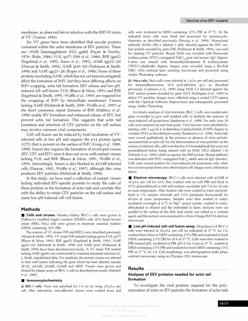
cells were incubated in MEM containing 2±5% FBS at 37 °C. At theindicated times cells were fixed and processed for immunocyto-chemistry as described previously (Herzog et al., 1994). Monoclonalantibody (MAb) AB1.1 (diluted 1 :300), directed against the IMV sur-face protein encoded by gene D8L (Parkinson & Smith, 1994), was usedfor staining VV particles. Bound MAb was revealed with fluoresceinisothiocyanate (FITC)-conjugated F(ab«)
#goat anti-mouse IgG (Sigma).
F-actin was stained with tetramethylrhodamine B isothiocyanate(TRITC)–phalloidin (Sigma). Images were recorded using a Bio-RadMRC 1024 confocal laser scanning microscope and processed usingAdobe Photoshop software.
(ii) HeLa cells. HeLa cells were infected at 1 p.f.u. per cell and processedfor immunofluorescence 10 h post-infection (p.i.) as describedpreviously (Cudmore et al., 1995) using MAb C3 directed against theIMV surface protein encoded by gene A27L (Rodriguez et al., 1985) todetect VV particles. Images were collected using a cooled CCD camerawith the OpenLab Software (Improvision) and subsequently processedusing Adobe Photoshop.
For kinetic analyses of viral infections, BS-C-1 cells were seeded ontoglass coverslips to give well isolated cells to facilitate the analyses ofvirus-induced cell projections (Sanderson et al., 1998). For each virus 30cells were analysed at each time-point. Viral factories were identified bystaining with 1 µg}ml 4«,6-diamidino-2-phenylindole (DAPI) (Sigma) tovisualize DNA as described previously (Sanderson et al., 1996). Actin tailswere scored qualitatively as the presence or absence of virus particle-associated tails in each cell. For the determination of virus particles on thesurface of infected cells, cells were fixed in 4% formaldehyde but were notpermeabilized before being stained with rat MAb 19C2 (diluted 1 :8)(Schmelz et al., 1994) which recognizes the B5R protein. Bound antibodywas detected with FITC-conjugated F(ab«)
#rabbit anti-rat IgG (Serotec).
Cells were scored positive for virus-induced cell projections only whentwo or more projections were observed per cell. Images were collected asabove.
+ Electron microscopy. BS-C-1 cells were infected with ∆A36R at10 p.f.u. per cell for 24 h, then washed with ice-cold PBS and fixed in0±5% glutaraldehyde in 200 mM sodium cacodylate (pH 7±4) for 30 minat room temperature. After fixation cells were washed in water and post-fixed in 1% osmium tetroxide and 1±5% potassium ferrocyanide for60 min at room temperature. Samples were then washed in water,incubated overnight at 4 °C in Mg#+-uranyl acetate, washed in water,dehydrated in ethanol and flat embedded in Epon. Sections were cutparallel to the surface of the dish, lead citrate was added as a contrastagent, and the sections were examined in a Zeiss Omega EM 912 electronmicroscope.
+ Low pH-induced cell–cell fusion assay. Monolayers of BS-C-1cells were infected at 10 p.f.u. per cell (as indicated) at 37 °C for 1 h,washed three times in MEM containing 2±5% FBS and incubated in freshMEM containing 2±5% FBS for 20 h at 37 °C. Cells were then washed inPBS (neutral pH), incubated in PBS (pH 5) for 3 min at 37 °C, washed inMEM containing 2±5% FBS and incubated in fresh MEM containing 2±5%FBS at 37 °C for 3 h. Cell morphology was photographed under phasecontrast microscopy using an Olympus CK2 microscope.
ResultsAnalyses of EEV proteins needed for actin tailformation
To investigate the viral proteins required for the poly-merization of actin on IEV particles the formation of actin tails
BEBH

Downloaded from www.microbiologyresearch.org by
IP: 54.157.140.51
On: Sun, 10 Apr 2016 23:11:31
C. M. Sanderson and othersC. M. Sanderson and others
F-Actin D8L
D A
56R
D B
5R
D F
13L
Fig. 2. Distribution of F-actin and VV particles in BS-C-1 cells infected with ∆A56R (a and d), ∆B5R (b and e) or ∆F13L (c andf ) at 10 p.f.u. per cell. At 18 h p.i. viral particles were stained with MAb AB1.1 against the VV D8L protein (d–f), whileTRITC–phalloidin was used to stain F-actin (a–c).
was analysed using a collection of VV mutants which lackindividual EEV proteins. The formation of VV-associated actintails in infected BS-C-1 cells was assessed by staining VVparticles with MAb AB1.1, directed against the D8L geneproduct (Parkinson & Smith, 1994), and actin tails withTRITC–phalloidin. Numerous virus particles were visible incells infected with each virus (Fig. 1 e–h). However, while actintails were detected in cells infected with WR virus (Fig. 1a),they were absent in cells infected with ∆A34R (Fig. 1b) or∆A36R (Fig. 1 c). The failure of ∆A34R-infected cells to form
actin tails confirmed a recent report (Wolffe et al., 1997). To becertain that the A36R protein was required for actin tailformation, a revertant virus (rA36R) in which the A36R genehad been re-inserted into the deletion mutant was examined.This virus was able to induce actin tail formation (Fig. 1d).Analyses of other viruses lacking specific EEV proteins showedthat cells infected with ∆B5R (Fig. 2b) and ∆F13L (Fig. 2 c) failedto make actin tails, confirming previous reports (Blasco &Moss, 1992 ; Cudmore et al., 1995), whereas cells infected with∆A56R induced actin tail formation (Fig. 2a). These data show
BEBI
Downloaded from www.microbiologyresearch.org by
IP: 54.157.140.51
On: Sun, 10 Apr 2016 23:11:31
Vaccinia virus EEV mutantsVaccinia virus EEV mutants
that A56R is the only EEV-specific protein examined that isdispensable for induction of actin-tail formation.
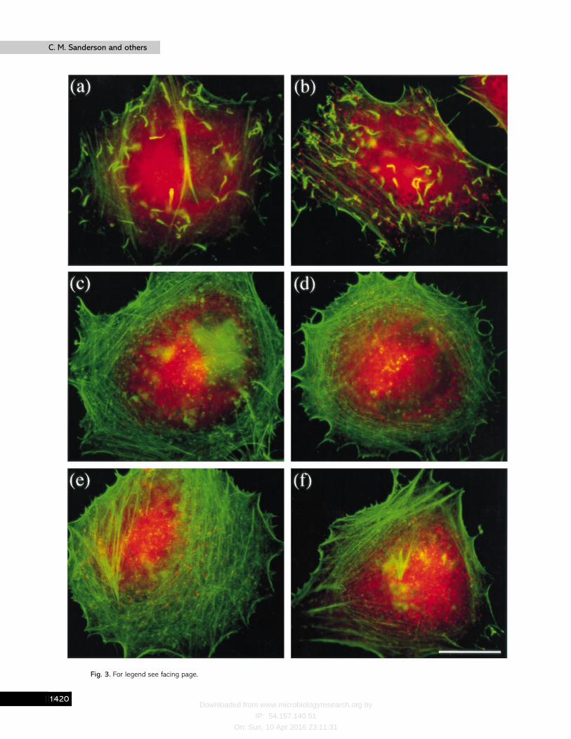
As host components are required for the formation of actintails on intracellular bacteria (Welch et al., 1997), it was possiblethat loss of VV-induced actin tail formation could be due toeither the virus genotype or the relative availability of host cellfactors. To address this, actin tail formation was examined inseveral different cell lines infected with the different viruses.Fig. 3 shows images of HeLa cells infected with the mutantviruses and stained with phalloidin to detect F-actin (greensignal) and MAb C3 against the VV p14 protein to show virusparticles (red signal). As previously noted (Cudmore et al.,1995), actin tails are visible with a single virus particle at the tipin WR-infected cells (Fig. 3a). Similarly, in cells infected with∆A56R many virus-associated actin tails are visible (Fig. 3b). Incontrast, cells infected with viruses ∆F13L, ∆B5R, ∆A34R or∆A36R failed to make actin tails although numerous virusparticles were present throughout the cell (Fig. 3 c–f ). Thisresult matches that observed in BS-C-1 cells and several othercell types examined (C2-C12, CV-1 and TK−143) (data notshown), confirming that it is the virus genotype rather than thehost cell that determines if actin tails are formed. However, itwas noticeable that the number of actin tails varied with celltype and HeLa cells produced appreciably more intracellularactin tails than BS-C-1 cells (compare Fig. 1a and 1d and Fig.2a with Fig. 3a and 3b).
It was possible that the lack of actin tails seen after infectionwith some viruses was due to altered kinetics of tail formationrather then an absolute inhibition. To test this possibility, theformation of actin tails was examined in cells infected witheither WR, ∆A34R, ∆A36R or ∆B5R. In each case 30 cells wereexamined at 2 h intervals between 8 and 18 h p.i. (Fig. 4). Asa control for the efficiency of infection, the formation ofcytoplasmic factories containing virus DNAwas also examinedby staining with DAPI. Cells infected with each virus containedcytoplasmic factories by 10 h p.i., but only WR-infected cellscontained virions with actin tails. Although ∆A34R- and∆A36R-infected cells failed to produce actin tails, most cellscontained surface virions by 12 h p.i. The slight delay in theappearance of surface virions compared to WR suggested thatalthough actin tails are not essential for EEV formation theymay accelerate the dispersal of particles to the cell surface. Itwas also evident that not all ∆A34R-infected cells containedsurface virions ; this might reflect the release of ∆A34R− virusfrom the cell surface rather than its retention as CEV (McIntosh& Smith, 1996).
Another parameter that was examined was the formation ofextended projections from cells that appear late duringinfection with VV (Sanderson et al., 1998). The formation ofthese projections requires the polymerization of actin(Sanderson et al., 1998) and therefore it was possible that insituations where the polymerization of actin to form in-tracellular actin tails was inhibited, there might be enhancedformation of cell projections late during infection. However,
examination of cells infected with WR, ∆B5R, ∆A34R or∆A36R showed that there appeared to be no direct correlationbetween the formation of cellular projections and intracellularactin tails (Fig. 4). Although virus-induced cell projectionsappeared a little sooner with ∆B5R and ∆A34R, a similarproportion of cells had developed cellular projections by 18 hp.i.
∆A36R forms both IEV and CEV particles
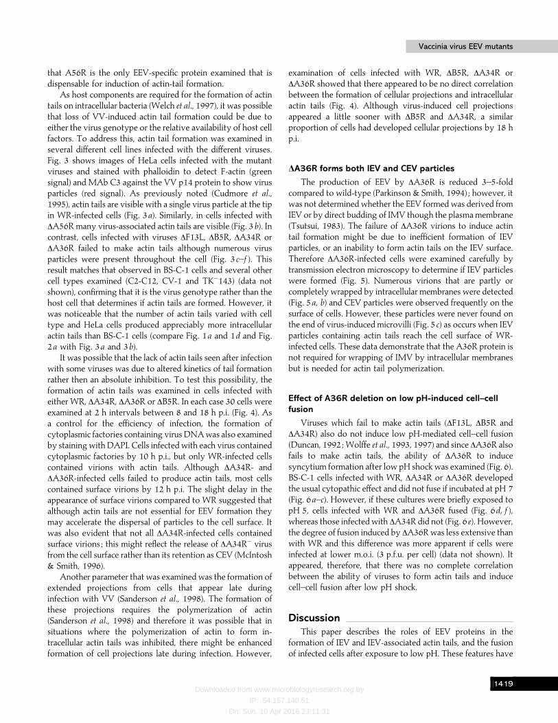
The production of EEV by ∆A36R is reduced 3–5-foldcompared to wild-type (Parkinson & Smith, 1994) ; however, itwas not determined whether the EEV formed was derived fromIEV or by direct budding of IMV though the plasma membrane(Tsutsui, 1983). The failure of ∆A36R virions to induce actintail formation might be due to inefficient formation of IEVparticles, or an inability to form actin tails on the IEV surface.Therefore ∆A36R-infected cells were examined carefully bytransmission electron microscopy to determine if IEV particleswere formed (Fig. 5). Numerous virions that are partly orcompletely wrapped by intracellular membranes were detected(Fig. 5a, b) and CEV particles were observed frequently on thesurface of cells. However, these particles were never found onthe end of virus-induced microvilli (Fig. 5 c) as occurs when IEVparticles containing actin tails reach the cell surface of WR-infected cells. These data demonstrate that the A36R protein isnot required for wrapping of IMV by intracellular membranesbut is needed for actin tail polymerization.
Effect of A36R deletion on low pH-induced cell–cellfusion
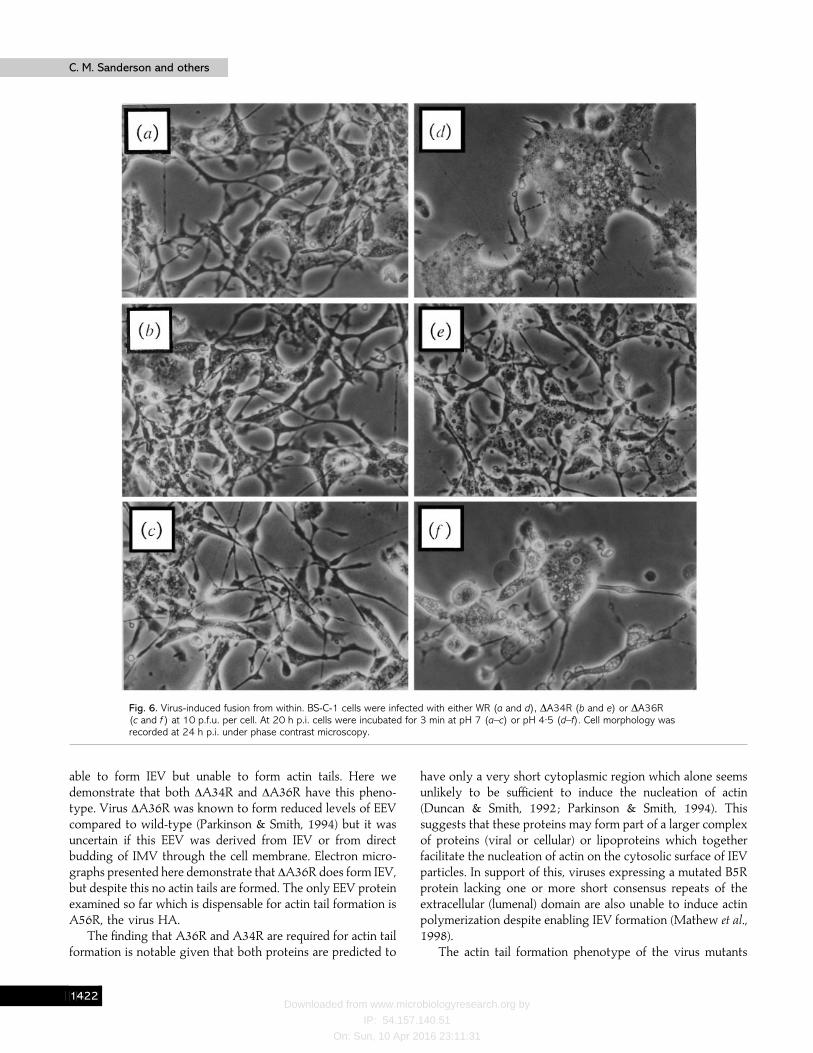
Viruses which fail to make actin tails (∆F13L, ∆B5R and∆A34R) also do not induce low pH-mediated cell–cell fusion(Duncan, 1992 ; Wolffe et al., 1993, 1997) and since ∆A36R alsofails to make actin tails, the ability of ∆A36R to inducesyncytium formation after low pH shock was examined (Fig. 6).BS-C-1 cells infected with WR, ∆A34R or ∆A36R developedthe usual cytopathic effect and did not fuse if incubated at pH 7(Fig. 6a–c). However, if these cultures were briefly exposed topH 5, cells infected with WR and ∆A36R fused (Fig. 6d, f ),whereas those infected with ∆A34R did not (Fig. 6 e). However,the degree of fusion induced by ∆A36R was less extensive thanwith WR and this difference was more apparent if cells wereinfected at lower m.o.i. (3 p.f.u. per cell) (data not shown). Itappeared, therefore, that there was no complete correlationbetween the ability of viruses to form actin tails and inducecell–cell fusion after low pH shock.
DiscussionThis paper describes the roles of EEV proteins in the
formation of IEV and IEV-associated actin tails, and the fusionof infected cells after exposure to low pH. These features have
BEBJ
Downloaded from www.microbiologyresearch.org by
IP: 54.157.140.51
On: Sun, 10 Apr 2016 23:11:31
C. M. Sanderson and othersC. M. Sanderson and others
Fig. 3. For legend see facing page.
BECA

Downloaded from www.microbiologyresearch.org by
IP: 54.157.140.51
On: Sun, 10 Apr 2016 23:11:31
Vaccinia virus EEV mutantsVaccinia virus EEV mutants
(a) (b) (c) (d)
Fig. 4. Kinetics of formation of actin tails, surface virions and cellularprojections. BS-C-1 cells were infected with the indicated virus at10 p.f.u. per cell and were fixed every 2 h between 8 and 18 h p.i. andprocessed as described in Fig. 2, except that DAPI was included to stainDNA. At each time-point 30 cells were analysed for the presence of viralfactories, actin tails, surface particles or virus-induced cellularprojections. Data are expressed as the percentage of cells positive forthe different parameters at each time-point.
Fig. 5. Transmission electron microscopyof BS-C-1 cells infected with ∆A36Rvirus. Cells infected with ∆A36R virus at10 p.f.u. per cell were fixed andprocessed for electron microscopy 24 hp.i. (a) An area of cytoplasm containingimmature viral particles (IV), IMV particles(IMV) and particles in the process ofwrapping by intracellular membranes (W).(b) A fully wrapped IEV particle. (c) ACEV particle bound to the outer surfaceof the plasma membrane. Bars : 200 nm(a) ; 100 nm (b and c).
been assessed using a collection of virus mutants which lackspecific genes.
Actin tails are formed on IEV but not IMV particles andtherefore mutant viruses which fail to make IEV do not induceactin tail formation, as was shown first with ∆F13L (Blasco &
Fig. 3. Formation of actin tails in HeLa cells. Cells were infected with WR (a), ∆A56R (b), ∆F13L (c), ∆B5R (d), ∆A34R (e) or∆A36R (f) at 1 p.f.u. per cell for 10 h. After fixation, cells were stained with a combination of MAb C3 against the A27L geneproduct on the surface of IMV particles (red signal), and FITC–phalloidin to show the distribution of F-actin (green signal).
Moss, 1992 ; Cudmore et al., 1995). The data presented hereconfirm this result and demonstrate that ∆B5R is also unable toinduce formation of actin tails, consistent with its defect inwrapping of IMV (Engelstad & Smith, 1993 ; Wolffe et al.,1993). Recently, Wolffe et al. (1997) reported that ∆A34R is
BECB
Downloaded from www.microbiologyresearch.org by
IP: 54.157.140.51
On: Sun, 10 Apr 2016 23:11:31
C. M. Sanderson and othersC. M. Sanderson and others
Fig. 6. Virus-induced fusion from within. BS-C-1 cells were infected with either WR (a and d), ∆A34R (b and e) or ∆A36R(c and f ) at 10 p.f.u. per cell. At 20 h p.i. cells were incubated for 3 min at pH 7 (a–c) or pH 4±5 (d–f). Cell morphology wasrecorded at 24 h p.i. under phase contrast microscopy.
able to form IEV but unable to form actin tails. Here wedemonstrate that both ∆A34R and ∆A36R have this pheno-type. Virus ∆A36R was known to form reduced levels of EEVcompared to wild-type (Parkinson & Smith, 1994) but it wasuncertain if this EEV was derived from IEV or from directbudding of IMV through the cell membrane. Electron micro-graphs presented here demonstrate that ∆A36R does form IEV,but despite this no actin tails are formed. The only EEV proteinexamined so far which is dispensable for actin tail formation isA56R, the virus HA.
The finding that A36R and A34R are required for actin tailformation is notable given that both proteins are predicted to
have only a very short cytoplasmic region which alone seemsunlikely to be sufficient to induce the nucleation of actin(Duncan & Smith, 1992 ; Parkinson & Smith, 1994). Thissuggests that these proteins may form part of a larger complexof proteins (viral or cellular) or lipoproteins which togetherfacilitate the nucleation of actin on the cytosolic surface of IEVparticles. In support of this, viruses expressing a mutated B5Rprotein lacking one or more short consensus repeats of theextracellular (lumenal) domain are also unable to induce actinpolymerization despite enabling IEV formation (Mathew et al.,1998).
The actin tail formation phenotype of the virus mutants
BECC

Downloaded from www.microbiologyresearch.org by
IP: 54.157.140.51
On: Sun, 10 Apr 2016 23:11:31
Vaccinia virus EEV mutantsVaccinia virus EEV mutants
was shown to be determined by the virus genotype rather thanthe cell type used for infection, since in each cell type examinedthe phenotype of the mutants was the same. However, it wasevident that the number of actin tails varied (HeLa cells gavemore than BS-C-1 cells) and this presumably reflects therelative abundance of some cellular factor(s) required for actinpolymerization on IEV particles.
The formation of actin tails on IEV particles impartsmotility (3 µm}min) (Cudmore et al., 1995, 1996) that aids theirdispersal through the cell to the plasma membrane. However,the ability of ∆A34R (Wolffe et al., 1997) (and Figs 1–3),∆A36R (Figs 1–3) and the mutants lacking the SCR domains ofB5R (Mathew et al., 1998) to make EEV despite not makingactin tails indicated that actin tails are not essential for IEVdispersal and EEV release. Rather, the function of actin tailsmight be to speed up this process, as suggested by the kineticmeasurements of virus appearance at the cell surface (Fig. 4),and to enable the direct spread of CEV into adjacent cells onthe tip of specialized cell surface microvilli (Cudmore et al.,1995). Evidently there are at least three ways that EEV can beformed : actin tail-dependent or -independent movement ofIEV particles, and direct budding of IMV through the plasmamembrane (Tsutsui, 1983). Possibly these have different rolesin different situations or cell types.
Is there a correlation between actin tail formation and EEVrelease or plaque size? Concerning EEV release, the mutantsthat are unable to form actin tails can produce either enhancedlevels of EEV, e.g. ∆A34R (¬25-fold) (McIntosh & Smith,1996) or the B5R SCR domain mutants (¬70-fold) (Mathewet al., 1998), or reduced levels of EEV, e.g. ∆A36R (3–5-foldlower) (Parkinson & Smith, 1994). Also, IHD-J forms both actintails and enhanced levels of EEV. Plainly no correlation can bemade. However, for plaque size, each mutant which is deficientin actin tail formation produces a small plaque. For viruseswhich fail to make IEV, e.g. ∆B5R and ∆F13L, this is presumablydue to the greatly diminished levels of CEV and EEV, but∆A34R, ∆A36R and the B5R SCR domain mutant viruses makeIEV and EEV yet the plaque size is small. Conversely, whenactin tails are made, e.g. ∆A56R, plaque size is normal (G. L.Smith unpublished data). Actin tail formation might thereforebe required for normal plaque size and a possible reason mightbe that actin tails facilitate the direct spread of CEV intoadjacent cells on the tip of specialized cell surface microvilli(Cudmore et al., 1995). Although enveloped virus is importantfor virus spread to neighbouring cells (Rodriguez & Smith,1990 ; Blasco & Moss, 1991), the absolute level of CEV is notcritical for plaque size since the IHD-J virus, which retains littleCEV, makes a plaque of similar size to WR which makes highCEV and low EEV.
Concerning cell–cell fusion, the data of Fig. 6 show thatthere is no correlation between the formation of actin tails andability to undergo low pH-induced cell–cell fusion : WR virusmakes actin tails and induces fusion, ∆A36R induces fusion butdoes not make actin tails, and ∆A34R neither makes tails nor
induces fusion. There also appears to be no direct correlationbetween cell–cell fusion and the release or retention of themajority of enveloped virus (EEV or CEV) : some viruses whichproduce enhanced levels of EEV, such as IHD-J and the B5RSCR domain mutants, induced cell–cell fusion after low pHshock but another virus, ∆A34R, does not (Blasco & Moss,1991 ; Wolffe et al., 1997 ; Mathew et al., 1998) (this paper anddata not shown). For ∆A34R the inability to induce cell–cellfusion might reflect the diminished infectivity of ∆A34R− EEV(McIntosh & Smith, 1996) or the requirement for this proteinin release of IMV from the wrapping membrane (Wolffe et al.,1997). Virus ∆A36R can induce fusion but this is less extensivethan seen with the other viruses ; possibly this might reflectreduced levels of EEV (Parkinson & Smith, 1994) and}or CEV,which was not examined.
The exact mechanism whereby p14 mediates fusion isuncertain. However, the recent demonstration that low pHdisrupts the EEV outer membrane (Vanderplasschen et al.,1998) suggests that this treatment would expose p14 presenton the surface of the IMV particles contained within CEVparticles emerging from infected cells or EEV particles whichrebind to the cell surface.
Lastly, we have examined the formation of cellularprojections which are produced by extending lamellipodia lateduring infection (Sanderson et al., 1998). The formation ofextending lamellipodia requires actin polymerization, andtherefore the production of these structures might beinfluenced by the formation of actin tails on IEV particles sincethis would affect the pool of actin available for polymerization.However, cell projection formation was found to be unaffectedby actin tail formation. Instead the number of these projectionsthat formed late during infection was influenced by the celltype (data not shown).
In summary, we have characterized a collection of virusmutants lacking specific EEV proteins for the formation of IEV,IEV-associated actin tails and cell–cell fusion. Of the five EEVproteins examined, only A56R is dispensable for actin tailformation. Loss of F13L or B5R prevents IEV formation, andloss of A34R or A36R prevents the formation of actin tails onIEV. Actin tail formation is not required for retention of CEVor for acid-induced cell–cell fusion.
Note added in proof. The requirement for the A36R protein in actintail formation is also reported by E. J. Wolffe, A. S. Weisberg & B. Moss(1998). Role for the vaccinia virus A36R outer envelope protein in theformation of virus-tipped actin-containing microvilli and cell-to-cellspread. Virology (in press).
This work was supported by the grants from The Wellcome Trust andthe Medical Research Council.
ReferencesAlcamı!, A. & Smith, G. L. (1992). A soluble receptor for interleukin-1β
encoded by vaccinia virus : a novel mechanism of virus modulation of thehost response to infection. Cell 71, 153–167.
BECD
Downloaded from www.microbiologyresearch.org by
IP: 54.157.140.51
On: Sun, 10 Apr 2016 23:11:31
C. M. Sanderson and othersC. M. Sanderson and others
Blasco, R. & Moss, B. (1991). Extracellular vaccinia virus formation andcell-to-cell virus transmission are prevented by deletion of the geneencoding the 37,000-Dalton outer envelope protein. Journal of Virology65, 5910–5920.
Blasco, R. & Moss, B. (1992). Role of cell-associated enveloped vacciniavirus in cell-to-cell spread. Journal of Virology 66, 4170–4179.
Cossart, P. (1995). Actin based bacterial motility. Current Opinion in CellBiology 7, 94–101.
Cossart, P. & Kocks, C. (1994). The actin-based motility of thefacultative intracellular pathogen Listeria monocytogenes. Molecular Micro-biology 13, 395–402.
Cudmore, S., Cossart, P., Griffiths, G. & Way, M. (1995). Actin-basedmotility of vaccinia virus. Nature 378, 636–638.
Cudmore, S., Reckmann, I., Griffiths, G. & Way, M. (1996). Vacciniavirus : a model system for actin-membrane interactions. Journal of CellScience 109, 1739–1747.
Dales, S. & Pogo, B. G. T. (1981). Biology of poxviruses. In VirologyMonographs, pp. 1–101. Edited by D. W. Kingsbury & H. zur Hausen.Berlin : Springer-Verlag.
Duncan, S. A. (1992). Analysis of three vaccinia virus genes, one of which isessential for plaque formation. D. Phil., University of Oxford, Oxford, UK.
Duncan, S. A. & Smith, G. L. (1992). Identification and characterizationof an extracellular envelope glycoprotein affecting vaccinia virus egress.Journal of Virology 66, 1610–1621.
Engelstad, M. & Smith, G. L. (1993). The vaccinia virus 42 kDaenvelope protein is required for envelopment and egress of extracellularvirus and for virulence. Virology 194, 627–637.
Engelstad, M., Howard, S. T. & Smith, G. L. (1992). A constitutivelyexpressed vaccinia virus gene encodes a 42 kDa glycoprotein related tocomplement control factors that forms part of the extracellular envelope.Virology 188, 801–810.
Fenner, F., Anderson, D. A., Arita, I., Jezek, Z. & Ladnyi, I. D. (1988).Smallpox and its Eradication. Geneva : World Health Organization.
Gong, S. C., Lai, C. F., Dallo, S. & Esteban, M. (1989). A single pointmutation of Ala-25 to Asp in the 14,000-M
renvelope protein of vaccinia
virus induces a size change that leads to the small plaque size phenotypeof the virus. Journal of Virology 63, 4507–4514.
Gong, S. C., Lai, C. F. & Esteban, M. (1990). Vaccinia virus induces cellfusion at acid pH and this activity is mediated by the N-terminus of the14-kDa virus envelope protein. Virology 178, 81–91.
Herzog, M., Draeger, A., Ehler, E. & Small, V. J. (1994).Immunofluorescence microscopy of the cytoskeleton : double and tripleimmunofluorescence. In Cell Biology : A Laboratory Manual, pp. 355–360.San Diego : Academic Press.
Hiller, G. & Weber, K. (1985). Golgi-derived membranes that contain anacylated viral polypeptide are used for vaccinia virus envelopment.Journal of Virology 55, 651–659.
Hirt, P., Hiller, G. & Wittek, R. (1986). Localization and fine structure ofa vaccinia virus gene encoding an envelope antigen. Journal of Virology58, 757–764.
Ichihashi, Y. (1996). Extracellular enveloped vaccinia virus escapesneutralization. Virology 217, 478–485.
Ichihashi, Y., Matsumoto, S. & Dales, S. (1971). Biogenesis ofpoxviruses : role of A-type inclusions and host cell membranes in virusdissemination. Virology 46, 507–532.
Isaacs, S. N., Wolffe, E. J., Payne, L. G. & Moss, B. (1992). Charac-terization of a vaccinia virus-encoded 42-kilodalton class I membraneglycoprotein component of the extracellular virus envelope. Journal ofVirology 66, 7217–7224.
McIntosh, A. A. G. & Smith, G. L. (1996). Vaccinia virus glycoproteinA34R is required for infectivity of extracellular enveloped virus. Journalof Virology 70, 272–281.
Mackett, M., Smith, G. L. & Moss, B. (1985). The construction andcharacterization of vaccinia virus recombinants expressing foreign genes.In DNA Cloning : A Practical Approach, pp. 191–211. Edited by D. M.Glover. Oxford : IRL Press.
Mathew, E., Sanderson, C. M., Hollinshead, M. & Smith, G. L. (1998).The extracellular domain of vaccinia virus protein B5R affects plaqueformation, EEV release and intracellular actin tail formation. Journal ofVirology 72, 2429–2438.
Morgan, C. (1976). Vaccinia virus reexamined : development andrelease. Virology 73, 43–58.
Moss, B. (1996). Poxviridae : the viruses and their replication. In FieldsVirology, pp. 2637–2671. Edited by B. N. Fields, D. M. Knipe & P. M.Howley. Philadelphia : Lippincott–Raven.
Parkinson, J. E. & Smith, G. L. (1994). Vaccinia virus gene A36Rencodes a M
r43–50 K protein on the surface of extracellular enveloped
virus. Virology 204, 376–390.
Payne, L. G. & Kristensson, K. (1979). Mechanism of vaccinia virusrelease and its specific inhibition by N
"-isonicotinoyl-N
#–3-methyl-4-
chlorobenzoylhydrazine. Journal of Virology 32, 614–622.
Payne, L. G. & Norrby, E. (1976). Presence of haemagglutinin in theenvelope of extracellular vaccinia virus particles. Journal of GeneralVirology 32, 63–72.
Rodriguez, J. F. & Smith, G. L. (1990). IPTG-dependent vaccinia virus :identification of a virus protein enabling virion envelopment by Golgimembrane and egress. Nucleic Acids Research 18, 5347–5351.
Rodriguez, J. F., Janezcko, R. & Esteban, M. (1985). Isolation andcharacterization of neutralizing monoclonal antibodies to vaccinia virus.Journal of Virology 56, 482–488.
Roper, R. L., Payne, L. G. & Moss, B. (1996). Extracellular vaccinia virusenvelope glycoprotein encoded by the A33R gene. Journal of Virology 70,3753–3762.
Sanderson, C. M., Parkinson, J. E., Hollinshead, M. & Smith, G. L.(1996). Overexpression of the vaccinia virus A38L integral membraneprotein promotes Ca#+ influx into infected cells. Journal of Virology 70,905–914.
Sanderson, C. M., Way, M. & Smith, G. L. (1998). Virus-induced cellmotility. Journal of Virology 72, 1235–1243.
Schmelz, M., Sodeik, B., Ericsson, M., Wolffe, E. J., Shida, H., Hiller, G.& Griffiths, G. (1994). Assembly of vaccinia virus : the second wrappingcisterna is derived from the trans Golgi network. Journal of Virology 68,130–147.
Shida, H. (1986). Nucleotide sequence of the vaccinia virus hemag-glutinin gene. Virology 150, 451–462.
Sodeik, B., Doms, R. W., Ericsson, M., Hiller, G., Machamer, C. E.,van’Hof, W., van Meer, G., Moss, B. & Griffiths, G. (1993). Assembly ofvaccinia virus : role of the intermediate compartment between theendoplasmic reticulum and the Golgi stacks. Journal of Cell Biology 121,521–541.
Tilney, L. G. & Tilney, M. S. (1993). The wily ways of a parasite :induction of actin assembly by Listeria. Trends in Microbiology 1, 25–31.
Tooze, J., Hollinshead, M., Reis, B., Radsak, K. & Kern, H. (1993).Progeny vaccinia viruses and human cytomegalovirus particles utilizeearly endosomal cisternae for their envelopes. European Journal of CellBiology 60, 163–178.
BECE
Downloaded from www.microbiologyresearch.org by
IP: 54.157.140.51
On: Sun, 10 Apr 2016 23:11:31
Vaccinia virus EEV mutantsVaccinia virus EEV mutants
Tsutsui, K. (1983). Release of vaccinia virus from FL cells infected withIHD-W strain. Journal of Electron Microscopy 32, 125–140.
Vanderplasschen, A. & Smith, G. L. (1997). A novel virus binding assayusing confocal microscopy : demonstration that the intracellular andextracellular vaccinia virions bind to different cellular receptors. Journal ofVirology 71, 4032–4041.
Vanderplasschen, A., Hollinshead, M. & Smith, G. L. (1997). Anti-bodies against vaccinia virus do not neutralize extracellular envelopedvirus but prevent virus release from infected cells and comet formation.Journal of General Virology 78, 2041–2048.
Vanderplasschen, A., Hollinshead, M. & Smith, G. L. (1998). Vacciniavirus extracellular enveloped virus and intracellular mature virus entercells by different mechanisms. Journal of General Virology 79, 877–887.
Welch, M. D., Iwamatsu, A. & Mitchison, T. J. (1997). Actin poly-merization is induced by Arp2}3 protein complex at the surface of Listeriamonocytogenes. Nature 385, 265–269.
Wolffe, E. J., Isaacs, S. N. & Moss, B. (1993). Deletion of the vacciniavirus B5R gene encoding a 42-kilodalton membrane glycoprotein inhibitsextracellular virus envelope formation and dissemination. Journal ofVirology 67, 4732–4741.
Wolffe, E., Katz, E., Weisberg, A. & Moss, B. (1997). The A34Rglycoprotein gene is required for induction of specialized actin-containingmicrovilli and efficient cell-to-cell transmission of vaccinia virus. Journal ofVirology 71, 3905–3915.
Received 8 December 1997; Accepted 6 February 1998
BECF