Macrophages and Dendritic Cells Use the Cytosolic Pathway to Rapidly Cross-Present Antigen from...
11
of May 5, 2016. This information is current as Antigen from Live, Vaccinia-Infected Cells Cytosolic Pathway to Rapidly Cross-Present Macrophages and Dendritic Cells Use the Maria Carmen Ramirez and Luis J. Sigal http://www.jimmunol.org/content/169/12/6733 doi: 10.4049/jimmunol.169.12.6733 2002; 169:6733-6742; ; J Immunol References http://www.jimmunol.org/content/169/12/6733.full#ref-list-1 , 24 of which you can access for free at: cites 59 articles This article Subscriptions http://jimmunol.org/subscriptions is online at: The Journal of Immunology Information about subscribing to Permissions http://www.aai.org/ji/copyright.html Submit copyright permission requests at: Email Alerts http://jimmunol.org/cgi/alerts/etoc Receive free email-alerts when new articles cite this article. Sign up at: Print ISSN: 0022-1767 Online ISSN: 1550-6606. Immunologists All rights reserved. Copyright © 2002 by The American Association of 9650 Rockville Pike, Bethesda, MD 20814-3994. The American Association of Immunologists, Inc., is published twice each month by The Journal of Immunology by guest on May 5, 2016 http://www.jimmunol.org/ Downloaded from by guest on May 5, 2016 http://www.jimmunol.org/ Downloaded from
-
Upload
independent -
Category
Documents
-
view
2 -
download
0
Transcript of Macrophages and Dendritic Cells Use the Cytosolic Pathway to Rapidly Cross-Present Antigen from...
of May 5, 2016.This information is current as
Antigen from Live, Vaccinia-Infected CellsCytosolic Pathway to Rapidly Cross-Present Macrophages and Dendritic Cells Use the
Maria Carmen Ramirez and Luis J. Sigal
http://www.jimmunol.org/content/169/12/6733doi: 10.4049/jimmunol.169.12.6733
2002; 169:6733-6742; ;J Immunol
Referenceshttp://www.jimmunol.org/content/169/12/6733.full#ref-list-1
, 24 of which you can access for free at: cites 59 articlesThis article
Subscriptionshttp://jimmunol.org/subscriptions
is online at: The Journal of ImmunologyInformation about subscribing to
Permissionshttp://www.aai.org/ji/copyright.htmlSubmit copyright permission requests at:
Email Alertshttp://jimmunol.org/cgi/alerts/etocReceive free email-alerts when new articles cite this article. Sign up at:
Print ISSN: 0022-1767 Online ISSN: 1550-6606. Immunologists All rights reserved.Copyright © 2002 by The American Association of9650 Rockville Pike, Bethesda, MD 20814-3994.The American Association of Immunologists, Inc.,
is published twice each month byThe Journal of Immunology
by guest on May 5, 2016
http://ww
w.jim
munol.org/
Dow
nloaded from
by guest on May 5, 2016
http://ww
w.jim
munol.org/
Dow
nloaded from

Macrophages and Dendritic Cells Use the CytosolicPathway to Rapidly Cross-Present Antigen from Live,Vaccinia-Infected Cells1
Maria Carmen Ramirez and Luis J. Sigal2
Professional APCs (pAPC) can process and present on their own MHC class I molecules Ags acquired from Ag donor cells (ADC).This phenomenon of cross-presentation is essential in the induction of CD8� T cell responses to viruses that do not infect pAPCand possibly contributes to the induction of CD8� responses to many other viruses. However, little is known about the mechanismsunderlying this process. In this study, we show that dendritic cells and macrophages cross-present a model Ag supplied by vacciniavirus-infected ADC via the cytosolic route. Strikingly, we also found that cross-presentation of Ags provided by vaccinia-infectedcells occurs within a couple of hours of pAPC/ADC interaction, that the duration of cross-presentation lasts for only 16 h, and thatcross-presentation can occur at early times of infection when the ADC are still alive. The Journal of Immunology, 2002, 169:6733–6742.
A ctivated CD8� T cells can have their target in any cellexpressing MHC class I and their cognate peptide. How-ever, the initiation of CD8� T cell responses requires Ag
presentation by bone marrow-derived professional APCs (pAPC)3
(1–4) most likely because they express costimulatory moleculesthat provide a crucial second signal to CD8� T cells (5–11), andbecause they can carry Ags from the sites of infection into sec-ondary lymphoid organs where T cell responses are initiated (12,13). Although the definitive identification of the pAPC that ini-tiates CD8� T cell responses in vivo is still unknown, in the pastfew years there has been a large body of literature assigning thisfunction to dendritic cells (DC) (14, 15). Nonetheless, macro-phages (M�) are also good candidates since they share many of thecharacteristics of DC including common precursors, expression ofcostimulatory molecules, and the ability to move to and from sitesof inflammation. In addition, they can reconstitute CTL responsesin vivo (16).
Because pAPC play such an important role in the orchestrationof antiviral CD8� T cell immunity, it becomes important to un-derstand the mechanisms whereby these cells present viral Ags. Aswith all other cells, infected pAPC can present endogenously syn-thesized viral Ags in vitro and in vivo (17). In this way, they caninitiate responses by a process known as direct presentation. How-ever, pAPC also have the unique ability to acquire Ag from ex-ogenous sources and present them on their own MHC class I mol-ecules. This ability of pAPC to present exogenous Ags can be
readily observed in vitro by coculturing DC or M� with manydifferent antigenic formulations, and also in vivo upon inoculationof mice with those same Ags (18).
In this paper, we use the term “cross-presentation” to refer ex-clusively to a subtype of exogenous Ag presentation that involvesthe transfer of Ag from an Ag donor cell (ADC) to pAPC. Theimportant physiological function of cross-presentation in viral in-fections became clear when we demonstrated that cross-presenta-tion is necessary and sufficient to initiate CTL responses to virusesthat do not infect pAPC (3). Moreover, it is also becoming appar-ent that cross-presentation is required to induce CTL responses toviruses that suppress MHC class I Ag presentation on infectedcells (19, 20). In addition, it is very likely that, concurrent withdirect presentation (17), cross-presentation contributes somewhatto the induction of CTL toward most other viruses. Significantly,it has been shown that cultured DC phagocytose apoptotic or ne-crotic cells that had been infected with influenza virus or irradiatedcells that had been infected with recombinant vaccinia virus andcross-present viral Ags supplied by these cells (21–23). However,in those same reports, M� phagocytosed the apoptotic/necroticcells, but were unable to cross-present the associated Ags. This ledthe authors to propose that cross-presentation of cell-associatedAgs is a function of DC and not of M�. Furthermore, they pro-posed that by competing for Ag uptake, M� actually inhibit DCcross-presentation. It should be noted that this model contrastedwith the ability of M� to present on MHC class I and also on MHCclass II molecules other types of exogenous Ags (not cell-associ-ated) such as soluble proteins, proteins bound to beads, or recom-binant proteins expressed by bacteria (24–27) and to reconstituteCTL responses in vivo (16). However, the view that M� cannot cross-present Ags supplied by virus-infected cells is very widely held.
To gain a better understanding of the mechanisms of cross-pre-sentation during the course of a viral infection, we developed an invitro assay of MHC class I Ag cross-presentation where Ag pro-duction is restricted to vaccinia virus-infected ADC, and Ag pre-sentation is restricted to pAPC (see Fig. 1 for a diagram of theassay). Using this assay, we show that M� as well as DC arecapable of cross-presenting viral Ags supplied by infected cells.This cross-presentation is the direct result of viral infection as itoccurs in the absence of further treatment of the ADC to induce
Fox Chase Cancer Center, Philadelphia, PA 19111
Received for publication August 20, 2002. Accepted for publication October11, 2002.
The costs of publication of this article were defrayed in part by the payment of pagecharges. This article must therefore be hereby markedadvertisement in accordancewith 18 U.S.C. Section 1734 solely to indicate this fact.1 This work was supported by National Institutes of Health Grant R01AI048849 (toL.J.S.).2 Address correspondence and reprint requests to Dr. Luis J. Sigal, Associate Mem-ber, Fox Chase Cancer Center, 7701 Burholme Avenue, Philadelphia PA 19111.E-mail address: [email protected] Abbreviations used in this paper: pAPC, professional APC; ADC, Ag donor cell;CRPMI, complete RPMI media; DC, dendritic cell; M�, macrophage;�-gal, �-ga-latosidase; ER, endoplasmic reticulum.
The Journal of Immunology
Copyright © 2002 by The American Association of Immunologists, Inc. 0022-1767/02/$02.00
by guest on May 5, 2016
http://ww
w.jim
munol.org/
Dow
nloaded from

apoptosis, necrosis, or to inactivate the virus. In addition, we showthat our model Ag follows the cytosolic route within the pAPC,since it requires proteasome processing, TAP transport, and is in-hibited by brefeldin A. Moreover, we also found that M� and DCcross-present Ag with fast kinetics since cross-presentation is de-tectable within 1 h of ADC-pAPC encounter, peaks between 2 and5 h, and decreases rapidly to almost nil within the next 10–15 h.Strikingly, cross-presentation does not require apoptosis or necro-sis of the ADC, since live-infected cells are capable of supplyingAgs to pAPC within few hours of becoming infected.
Materials and MethodsCells lines
All cells were maintained in complete RPMI media (CRPMI) that con-sisted of RPMI supplemented with 10% FCS (Atlanta Biologicals,Norcross, VA), 2 mM L-glutamine, penicillin-streptomycin, 0.01 MHEPES buffer and nonessential amino acids (all from Invitrogen, Carlsbad,CA), and 5 � 10�5 2-ME (Sigma-Aldrich, St. Louis, MO). A9 cells (H-2K)are an APRT and HPRT negative derivative of strain L cells (no. CCL-1.4;American Type Culture Collection (ATCC), Manassas, VA). A9-T7 cells(28, 29) are A9 cells stably transfected with T7 polymerase (a kind giftfrom Dr. B. Moss, National Institutes of Health, Bethesda, MD). B3Z (30)is a CTL hybridoma that produces �-galatosidase (�-gal) upon recognitionof the OVA epitope SIINFEKL in the context of the H-2Kb molecule (akind gift from Dr. N. Shastri, University of California, Berkeley, CA). Allcells were grown and assays incubated at 37°C in an atmosphere of 5%CO2 unless otherwise indicated.
Mice
All experiments using animals were performed under protocols approvedby the Institutional Animal Use and Care Committee. All the mice used inthe experiments were bred at Fox Chase Cancer Center’s Laboratory An-
imal Facility (Philadelphia, PA). TAP0/0 (B6-Tap1tp1Arp) breeders wereoriginally purchased from The Jackson Laboratory (Bar Harbor, ME).C57BL/6 (B6) mice were from the Fox Chase Cancer Center colony.
Viruses
The WR strain of vaccinia virus and WR expressing the phage T7 poly-merase was a gift from Dr. B. Moss. Vaccinia virus expressing the mini-gene (M)SIINFEKL was a kind gift of Dr. J. Yewdell (National Institutesof Health). All vaccinia stocks were grown in Hela S3 cells (no. CCL-2.2;ATCC) in T150 tissue culture flasks and virus titers determined using BS-C-1 cells (no. CCL-26; ATCC) growing in 6-well plates following pub-lished procedures (29). Virus titers were adjusted to 109 PFU in CRPMI.
Plasmids
We generated a PCR fragment of OVA197–386 preceded by a Kozac se-quence and ending in a stop codon using as template a plasmid containingthe full cDNA of OVA (a generous gift from Dr. L. Shen, University ofMassachusetts Medical Center, Worcester, MA). As forward and reverseprimers we used oligos with the sequences CACCATGCCTTTCAGAGTGACTGAGCA and TTAAGGGGAAACACATCTGCC, respectively. ThePCR fragment was directionally cloned into pCDNA 3.1-TOPO (Invitro-gen) following manufacturer’s instructions to obtain pCDNA-OVA 197–386.
The cloned fragment was excised from pCDNA-OVA197–386 with BamHIand NotI and inserted into the BamHI-NotI sites of plasmid pBlueScript IISK (pBS; Stratagene, La Jolla, CA) to generate pBS-OVA197–386. Tran-scription and translation from OVA197–386 generates a fragment of chickenOVA (residues 197–386) lacking the endoplasmic reticulum (ER) transfersignal sequence but containing the Kb-restricted OVA epitope SIINFEKL(amino acid single letter code) corresponding to positions 258–265.
ELISA
The presence of OVA197–386 was determined by a sandwich ELISA inlysates of ADC that had been prepared as described in the next section andlysed at 106 cells/ml in PBS 0.1% Triton X-100 buffer. For the ELISA,96-well flat-bottom RIA/ELISA plates (Costar; all other plasticware wasfrom BD Biosciences, Mountain View, CA) were incubated overnight with50 �l of a 1/2,500 solution of goat anti-OVA Ab (ICN Pharmaceuticals,Costa Mesa, CA), washed, and blocked with 200 �l ELISA buffer (PBS0.5% nonfat milk, 0.1% Triton X-100, 0.025% Tween 20) for 2 h at 37°C.A total of 50 �l of cell lysates in ELISA buffer were added to triplicatewells as sets of seven 10-fold serial dilutions and incubated at room tem-perature for 3 h. The plates were thoroughly washed and 50 �l of a1/16,000 dilution of rabbit anti-OVA (ICN Pharmaceuticals) diluted inELISA buffer was added and incubated at room temperature for 2 h. Theplates were washed again, incubated with 1/8,000 dilution of goat anti-rabbit IgG conjugated with HRP (Kirkegaard & Perry Laboratories, Gaith-ersburg, MD) in ELISA buffer, washed, incubated with tetramethylbenzi-dine substrate (Kirkegaard & Perry Laboratories) for 20 min and read at450 nm in a � Quant 96-well spectrophotometer (Bio-tek Instruments,Watford Herts, U.K.). As a positive control, we used purified chicken OVA(ICN Pharmaceuticals). However, the exact quantification of the amount ofOVA197–386 present in the cell lysates was not possible because the poly-clonal Abs that we used were raised against full-length OVA. Therefore, someepitopes present in the full-length OVA might be absent in OVA197–386, whichmay skew the signal. As a guide, the absorbance for 10 pg/well of purifiedOVA was 0.259 at 450 nm of Abs. All ELISA experiments were repeated atleast three times. Each data point represents the mean of triplicate wells for oneof the dilutions. The value of the signal decreased linearly with the decrease inthe concentration of the cell lysate or of the standard.
Ag cross-presentation assayPreparation of ADC
A9-T7 or A9 cells were seeded in 60-mm tissue culture dishes (1.2 � 106
cells/dish) or 6-well plates (4 � 105 cells/well). Following overnight in-cubation at 37°C, the cells were transfected with the indicated DNA usingLipofectamine 2000 (Invitrogen) as recommended by the manufacturer.Four or 6 h later, the monolayer was washed twice with PBS and the cellsinfected for 2 h with 10 PFU/cell (the number of cells was an estimationcalculated as twice the initial number of cells) of the indicated vacciniavirus in Optimem media (Invitrogen). Next, the monolayer was washedtwice with complete media and incubated in CRPMI for the indicatedtimes. When indicated, the cells were irradiated for 5 min with a short wave(254 nm) Spectroline germicidal UV lamp EF180 (Spectronics, Westbury,NY) placed at 15 cm over the cells. The cells were harvested with a rubber
FIGURE 1. Diagram of the cross-presentation assay. ADC cells are pre-pared by transfecting A9-T7 cells (H-2k) with a plasmid controlled by theT7 promoter (PBS-OVA197–386) and infecting with wild-type vaccinia. Weprepared M� and DC by culturing mouse bone marrow with M-CSF orGM-CSF, respectively, and responder cells were the B3Z hybridoma thatis reactive with the OVA peptide SIINFEKL in the context of H-2Kb andproduces �-gal when activated.
6734 MECHANISMS OF CROSS-PRESENTATION DURING VACCINIA INFECTION
by guest on May 5, 2016
http://ww
w.jim
munol.org/
Dow
nloaded from

policeman, centrifuged, and the pellet resuspended to an estimated con-centration of 106 or 107 cells/ml according to the requirements of the par-ticular experiment.
In some experiments (see Fig. 4), the ADC cells were split in aliquotsand frozen at �80°C. Aliquots were thawed at different times, vortexed,and used in the cross-presentation assay. In other experiments (see Fig. 5),the cells were transfected with the indicated PBS-OVA197–386 and the in-fections were performed at different times, the first infection being 4 hbefore transfection and the last infection just immediately before harvest-ing. In this way, all the ADC cultures containing cells that had been trans-fected at the same time but infected at different times were harvestedtogether.
Generation of bone marrow-derived pAPC
To obtain DC, bone marrow cells obtained from the C57BL/6 or TAP0/0
mice were incubated overnight in CRPMI. Nonadherent cells were col-lected, seeded at 106 cells/ml, and cultured for 5–6 days in CRPMI sup-plemented with 7 ng/ml GM-CSF (BD PharMingen, San Diego, CA) whichwas replaced every 2–3 days (BD PharMingen). To obtain M�, the pro-cedure was similar, but instead of GM-CSF, the media contained 20% ofL cell supernatant as a source of M-CSF (16).
Cross-presentation assays
In some experiments, pAPC were grown in 10-mm bacteriological gradepetri dishes (BD Biosciences), collected with a rubber policeman, washed,counted, and 105 placed at the indicated ADC:pAPC ratio in duplicatewells of white, flat-bottom 96-well plates (BD Biosciences). In other ex-periments, pAPC were grown in 24-well plates and cross-presentation wasinitiated by adding 5 � 105 ADC (107 cells/ml). In some cases, 20 minbefore adding the ADC, the media of the pAPC was replaced with 200 �lof CRPMI that contained 16 �M lactacystin, 1 �g/ml brefeldin A (bothfrom Sigma-Aldrich), or diluent. In this case ADC were added directly,without media replacement. To measure direct presentation by M� and DCinfected with vaccinia (M)SIINFEKL, 5 PFU/cell of virus stock was addedinstead of the ADC. To allow for cross-presentation or direct presentation,the ADC-pAPC cocultures or infected pAPC were incubated for 4 h or theindicated times, harvested using the plunger of a 1 ml syringe as a rubberpoliceman. When indicated, the harvested cells were spun and fixed for 15min in 200 �l of 0.5% paraformaldehyde, washed twice with PBS, neu-tralized for 20 min in 0.5 mM L-lysine in CRPMI, washed twice in CRPMI,and serially diluted in white 96-well plates. In some cases, the procedurewas similar but the cells were not fixed. The concentration of pAPC inthese conditions was estimated by counting the number of pAPC in wellsthat did not receive ADC. In other experiments, the process was reversedand the pAPC were added to the ADC cultures. For this purpose, the ADCwere prepared by transfecting and infecting A9-T7 cells in 6-well plates.After 9 h, any detached ADC cells were removed by gently washing themonolayer with CRPMI after which 106 pAPC that had been grown in100-mm petri dishes were added to the ADC that remained attached to thewells. Following a 4-h incubation, the pAPC-ADC cocultures were har-vested using a rubber policeman, fixed, and seeded in 96-well plates asbefore.
To determine cross-presentation, 105 B3Z responder cells were added toeach well of the 96-well plates above and incubated for an additional16–24 h to allow for hybridoma activation and �-gal production. To detect�-gal activity, we used the Galactostar chemiluminescent kit (Applied Bio-systems, Foster City, CA) following the instructions of the manufacturerexcept that we used 15 �l/well of lysis buffer and 40 �l/well of substratediluted 1/150 in reaction buffer. The assays were read using a Topcountinstrument (Packard Instrument, Meriden, CT).
All experiments of cross-presentation were repeated at least three timesand each figure corresponds to a representative experiment. Each data pointin the figures represents the average of duplicate wells. Positive controlsincluded pAPC incubated with 2-fold serial dilutions of SIINFEKL at aninitial concentration of 200 or 100 pM and a final concentration of 3.2 or1.56 pM. This range of concentrations induced a nonsaturating, linear re-sponse by the hybridoma. The maximal stimulation through cross-presen-tation fluctuated around the values obtained between 3.12 and 6.25 pM andnever reached the �-gal activity induced with 50 pM SIINFEKL.
Ab staining for flow cytometry
For cell surface staining 0.5 � 106 cells were incubated in a well of aU-bottom 96-well microtiter plate with 0.1 �g of the corresponding pri-mary labeled Ab in 50 �l FSB (PBS 2% FCS, 0.04% sodium azide). Thefollowing primary labeled Abs used were PE-labeled anti-MHC class II(I-Ab-specific clone AF6-120.1), FITC-labeled anti-CD11c (clone HL3),FITC-labeled anti-Vb8.1,8.2 (clone MR5-2), PE-labeled anti-CD8 (clone
53-6.7) from BD PharMingen, and FITC-labeled anti-CD11b (a kind gift ofDr. R. Hardy, Fox Chase Cancer Center). For immediate analysis, the cellswere washed with FSB three times and resuspended in 0.4 ml SB. Whenthe analysis was postponed, the cells were washed three times with PBSand resuspended in 0.2 ml PBS containing 0.5% paraformaldehyde.
Detection of cell deathEthidium bromide and acridine orange staining for microscopy
ADC well prepared in 6-well plates as above. After a 13-h infection, 10 �lof ethidium bromide (Sigma-Aldrich) and acridine orange (Fisher Scien-tific, Pittsburgh, PA) (both stocks at 100 �g/ml in PBS) were added, andthe cells were immediately observed under FITC (green) and Rhodamine(red) filters using a Nikon Diaphoto microscope (Nikon, Melville, NY).Pictures were taken with a Pixera Pro ISOES camera (Nikon) coupled to theNikon microscope at �100 magnification. The green and red images weremerged using Picture Publisher 10.0 software (Micrograph, Dallas, TX).
TUNEL assay for apoptotic death
Apoptotic death was assessed using the TUNEL kit (Roche DiagnosticSystems, Somerville, NJ). Briefly, ADC were prepared in 6-well plates,and following a 13-h infection ADC were washed in PBS, fixed in 4%paraformaldehyde, and permeabilized in 0.1% Triton X-100, 0.1% sodiumcitrate for 2 min on ice. Fixed and permeabilized ADC were washed withPBS and incubated for 1 h at 37°C in a solution containing 25 mM Tris (pH6.6), 200 mM cacodylate, 1 mM CoCl2, 0.6 nM fluorescein-12dUTP, and25 U of TdT (Roche Diagnostic Systems). ADC were analyzed by fluo-rescent microscopy in a drop of PBS using a FITC band path filter.
ResultsM� and DC can cross-present Ags shed byvaccinia-infected cells
To study cross-presentation in viral infections, it is absolutely nec-essary to restrict Ag synthesis to the ADC and Ag presentation tothe pAPC. Vaccinia can infect pAPC and cannot be used to studycross-presentation as a consequence of viral infection if the Ag isencoded within the viral genome because direct presentationwould mask cross-presentation. Therefore, to study cross-presen-tation as a consequence of vaccinia virus infection, we restrictedAg synthesis to the ADC by adapting a method originally used toproduce recombinant proteins in mouse cells. In this system,A9-T7 cells, a derivative of the mouse fibroblast cell line A9 (anHPRT and APRT negative mouse subclone of the L strain, H-2K)stably expresses the bacteriophage T7 polymerase. These cells pro-duce a protein encoded within a transiently transfected plasmidand controlled by the T7 promoter only under conditions of vac-cinia virus infection (28). The ability of vaccinia virus to induceexpression of a T7 controlled protein in the presence of T7 poly-merase has been attributed to the capacity of the virus to stabilizethe plasmid encoded RNA in the cytoplasm. For our purposes, wetransfected A9-T7 cells with pBS-OVA197–386 and infected withwild-type vaccinia virus. The cells were then incubated for 24 h,harvested, lysed, and the presence of OVA197–386 was determinedby ELISA. Fig. 2A shows that A9-T7 cells that had been trans-fected with pBS-OVA197–386 and infected with wild-type vacciniaproduced large amounts of OVA197–386 while transfection ofA9-T7 cells with pBS-OVA197–386 in the absence of vaccinia in-fection or parent A9 cells (T7 negative) that had been transfectedwith PBS-OVA197–386 and infected with wild-type vaccinia didnot produce OVA197–386. Further controls showed that A9 cellsproduced as much OVA as A9-T7 cells when they were trans-fected with pBS-OVA197–386 and infected with recombinant vac-cinia virus carrying the T7 polymerase, indicating that parent A9cells were an appropriate negative control since they could betransfected and infected with similar efficiency as A9-T7 cells. Inaddition, OVA197–386 was not detected in lysates of A9-T7 cellsthat had been infected with vaccinia wild type, but transfected with
6735The Journal of Immunology
by guest on May 5, 2016
http://ww
w.jim
munol.org/
Dow
nloaded from

vector alone (data not shown). In related experiments, we trans-fected A9-T7 and A9 cells with pBS-�-gal instead of pBS-OVA197–386 to find that most (80 –100%, varying with each ex-periment) of the A9-T7 cells that had been infected with vacciniawild type stained blue very rapidly (within a few minutes) and verystrongly with X-gal while no staining was found in A9 cells in-fected with wild-type vaccinia (Fig. 2B). In addition, strong stain-ing was observed in control A9-T7 and A9 cells that had beeninfected with vaccinia-T7 but not in uninfected A9-T7 cells (data notshown). Therefore, A9-T7 cells transfected with pBS-OVA197–386
and infected with wild-type vaccinia are an excellent source ofADC to study cross-presentation. In this system, OVA197–386 con-stitutes a model of a viral Ag as it is only produced upon vacciniainfection. In addition, ADC irradiation is not required because thetransfer of live virus to the pAPC or even unwilling transfection ofthe pAPC by carryover plasmid (an unlikely but theoretically pos-sible problem that may occur when using vaccinia-T7 instead ofA9-T7 cells) should not result in production and direct presenta-tion of OVA197–386 by the pAPC.
With a reliable method in hand to generate ADC, our first ob-jective was to determine whether different pAPC could cross-present viral Ags supplied by vaccinia-infected cells. As pAPC weused M� and DC prepared from bone marrow cells obtained fromC57BL/6 (B6) mice. The identity of the pAPC was confirmed bymicroscopic morphology (data not shown) and by flow cytometry,which indicated that the bone marrow cells cultured in the pres-ence of M-CSF expressed MHC class II but not CD11c as is ex-pected for M�, while the cells cultured in the presence of GM-CSFexpressed MHC class II and CD11c as expected for DC (Fig. 2C).It should be noted that we consistently found that the M� cultureswere highly homogeneous, while the DC were more heterogeneouswith day to day variations (40–70%) in the proportion of cellsexpressing CD11c. In addition, both cell types expressed CD11b,CD16, H-2Kb, and the M� scavenger receptor A. DC expressedmoderate levels of B7-2 and low B7-1 while M� expressed low
B7-2 and no B7-1. Both cell types increased MHC and B7 expres-sion upon activation with LPS or UV- irradiated vaccinia-infectedA9-T7 cells (data not shown). To determine cross-presentation,A9-T7 or A9 control ADC (H-2K) were transfected and infected asindicated in Materials and Methods, harvested, washed, and platedat different concentrations in 96-well plates. To these cells, B6-derived (H-2b) pAPC (M� or DC) and B3Z responder T cells(OVA-specific, H-2Kb-restricted) were added. The cells were in-cubated overnight and the activation of B3Z was determined bymeasuring �-gal activity. Our results show that both M� and DCcross-presented OVA197–386 when cocultured with A9-T7 cellsthat had been transfected with pBS-OVA197–386 and infected withwild-type virus (Fig. 2D). Importantly, no signal was detectedwhen we used similarly treated A9 cells that did not produceOVA197–386 as ADC controls. It should be noted that in this ex-periment as well as others, the cross-presentation signal obtainedwith M� was higher than with DC, typical of most but not all ourexperiments. Although an interpretation could be that M� cross-present better, it is more likely that the day to day variation in thepurity of the DC cultures results in this difference. In additionalcontrols, A9 cells and A9-T7 cells transfected with pBS-OVA197–386
and infected with recombinant vaccinia-T7 cross-presented similarly(data not shown). Also, no signal was detected when A9-T7 cells wereinfected with wild-type vaccinia and transfected with empty pBS vec-tor (see Fig. 2E). Furthermore, when we used ADC supernatants nocross-presentation was observed indicating that the cross-presentedAg is cell associated (see Fig. 2E). Altogether, these experimentsdemonstrate that M� can cross-present viral Ags from vaccinia-in-fected cells similar to DC, and that cross-presented Ags are cell as-sociated. It also demonstrates that vaccinia-infected cells provide Agfor cross-presentation without a requirement to artificially induce ap-optosis or necrosis of the infected cells. This cross-presentation ishighly efficient as only 0.06 ADC are required for each pAPC todetect cross-presentation, and 0.25 ADC per pAPC provides saturat-ing amounts of Ag (Fig. 2D).
FIGURE 2. M� and DC cross-present Ag fromvaccina-infected cells. A, A9-T7 or A9 cells (as indi-cated) were transfected with pBS-OVA197–386 and in-fected as indicated. The expression of OVA197–386 in50 �l of cell lysates (106 cells/ml) was determined byELISA. All samples in the figure were processed at thesame time. The A450 for 10 pg of purified OVA in 50�l was 0.259. B, X-gal staining of A9-T7 and A9 cellstransfected with pBS-�-gal and infected with wild-type vaccinia virus. C, Flow cytometry of M� and DCcultures using anti-CD11c-FITC and anti-MHC classII-PE mAbs. Results with control-irrelevant Abs arealso shown. D, A total of 105 M� or 105 DC wereincubated with different concentrations of A9-T7 cellsor control A9 cells that had been transfected with pBS-OVA197–386 and infected with wild-type vaccinia vi-rus, and with 105 B3Z hybridoma cells in duplicatewells of 96-well plates. The three-cell culture was in-cubated for 18 h after which �-gal production by theB3Z cells was measured by chemiluminescence. E, Atotal of 105 M� or DC were incubated with 105 A9-T7cells or their supernatants that had been transfectedwith pBS-OVA197–386 or pBS vector as indicated, andinfected with wild-type vaccinia virus. B3Z hybrid-oma and detection of �-gal activity as in D.
6736 MECHANISMS OF CROSS-PRESENTATION DURING VACCINIA INFECTION
by guest on May 5, 2016
http://ww
w.jim
munol.org/
Dow
nloaded from

Cross-presentation of OVA197–386 follows the cytosolic route
Previous work with proteins bound to beads and bacteria express-ing recombinant proteins led respectively to the discovery of thecytosolic and vacuolar routes of exogenous Ag presentation (26,31). The cytosolic route is characterized by Ags gaining access tothe cytosol of the pAPC and, similar to the presentation of endog-enous proteins (32–36), the Ags are degraded by the proteasomeand the resulting peptides transported by the TAP to the ER. In theER, the peptides bind to newly synthesized MHC class I moleculesand are transported to the cell surface via the secretory pathway. Inthe vacuolar route, the exogenous Ags do not gain access to thecytosol and are most likely degraded by proteases within endoso-mal or lysosomal compartments and the resulting peptides bind toMHC class I molecules that recycle from the cell surface (26, 37).We have previously shown that bone marrow chimeric mice withTAP-deficient pAPC were inefficient in generating CTL responsesto several viruses, including vaccinia, suggesting that the dominantroute of cross-presentation in viral infections is cytosolic (2, 3).However, the use of TAP deficiency as the single method to dem-onstrate that an Ag follows the cytosolic route has been criticizedbecause TAP deficiency reduces the number of MHC class I mol-ecules at the cell surface that could be available for recycling andcould indirectly affect the vacuolar route (38). Therefore, theseresults may require confirmation in a more controlled environmentwhere each of the steps of the cytosolic route can be individuallyevaluated. We first tested the role of TAP in the cross-presentationof Ags supplied by vaccinia-infected cells. For this purpose,A9-T7 ADC were prepared as before and pAPC were obtainedfrom either TAP-deficient or wild-type B6 mice as controls. Asseen in Fig. 3A, M� and DC from wild-type B6 mice were able tocross-present OVA197–386 delivered by vaccinia-infected cellswhile those from TAP-deficient mice did not cross-present,strongly suggesting a dominant role for the cytosolic route. Toconfirm this, we blocked other steps of the cytosolic route by treat-ing B6 M� and DC with lactacystin to inhibit the proteasome orwith brefeldin A to block the secretory pathway. To minimize anytoxic or nonspecific effects of the drugs on the pAPC or the T cellhybridoma, the pAPC were only exposed to the inhibitors for 20min before the incubation with the ADC and during the 4 h of theprocessing period. After this, the pAPC were fixed with parafor-
maldehyde and thoroughly washed before mixing with B3Z cells.These experiments showed that both lactacystin and brefeldin Acompletely inhibited cross-presentation (Fig. 3B). As a control, weinfected pAPC with recombinant vaccinia virus expressing theminigene (M)SIINFEKL which does not require proteasome pro-cessing but requires de novo MHC class I synthesis. As expected,we found that brefeldin A but not lactacystin inhibited direct pre-sentation by M� or DC (Fig. 3B, small insets). In additional con-trols we found that M� and DC still cross-presented when theywere not fixed and brefeldin A was removed by thorough washingbefore adding the B3Z responder cells (Fig. 3B). Altogether, thesedata demonstrate that M� and DC use the cytosolic route to cross-present OVA197–386 delivered by vaccinia-infected cells.
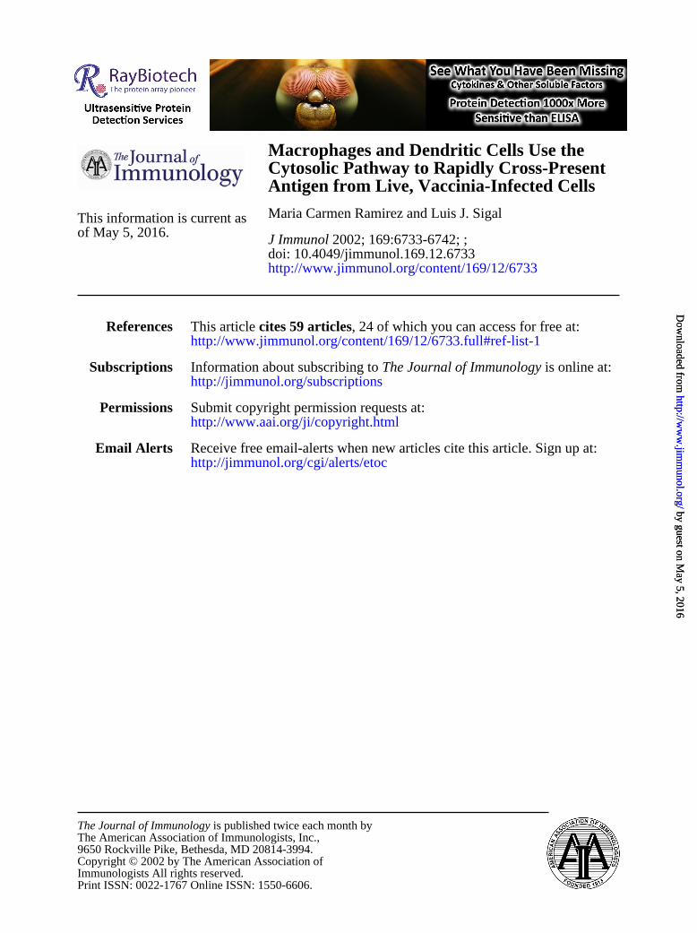
Cross-presentation by M� and DC is rapid and transient
To address how soon and for how long pAPC cross-present Agfollowing pAPC-ADC contact, we prepared ADC as before al-though they were frozen in aliquots which, at different times pointswithin a 16-h period, were thawed, and saturating numbers (1ADC:1 pAPC) were immediately added to cultures of M� or DC.All ADC-pAPC cocultures were harvested together at the end ofthe assay, fixed with paraformaldehyde to stop further Ag process-ing, thoroughly washed, and serially diluted in duplicate wells of96-well plates to which B3Z responder cells were added. Cross-presentation was determined following overnight incubation. Theresults indicated that M� and DC cross-presented Ag acquiredfrom vaccinia-infected cells with fast and similar kinetics (Fig.4A). Maximal cross-presentation by both pAPC was reached be-tween 2 and 5 h of ADC-pAPC contact and decreased thereafterwith very little Ag remaining at the surface of pAPC after 16 h ofADC-pAPC coculture. It was reasonable to think that cross-pre-sentation extinguished relatively quickly because vaccinia viruscarried over by the ADC may have infected the pAPC and mayhave affected their ability to present Ags at later time points ad-versely. To test for this possibility, we performed a set of exper-iments where carry-over virus was inactivated by irradiating theADC with UV light before dividing in aliquots and freezing whileall other conditions remained the same. This irradiation renderedADC that did not transfer virus since their lysates did not infectBSC-1 cells nor did they induce expression of �-gal in A9-T7 that
FIGURE 3. Cross-presentation of OVA197–386 fol-lows the cytosolic route. ADC were prepared by trans-fection of A9-T7 cells with PBS-OVA197–386 and in-fection with vaccinia wild type. A, Titrated numbers ofADC were coincubated with 105 B3Z cells and 105 M�or DC obtained from TAP or B6 mice. Cross-presen-tation was measured after a 24-h incubation. Only thedata representing the 1:1 ADC:pAPC ratio are shown.The small insets correspond to control pAPC pulsedwith X mM SIINFEKL. B, A total of 106 ADC (mainfigure) or 5 PFU/cell recombinant vaccinia virus ex-pressing (M)SIINFEKL as a minigene (small insets)were added to 106 B6 M� or DC that had been prein-cubated for 20 min with and in the presence of lacta-cystin (16 �M) or brefeldin A (1 �g/ml) as indicated.Following a 4-h incubation, the ADC-pAPC cocultureswere harvested, fixed with paraformaldehyde, thor-oughly washed, plated at different concentrations in du-plicate wells of 96-well plates, and used in cross-pre-sentation assays with B3Z cells as in Fig. 2C. Althoughmultiple dilutions of pAPC were analyzed, the mainfigure only depicts the data corresponding to wells con-taining 105 pAPC.
6737The Journal of Immunology
by guest on May 5, 2016
http://ww
w.jim
munol.org/
Dow
nloaded from
had been transfected with pBS-�-gal while lysates of infected,nonirradiated controls did (data not shown). In addition, M� andDC that had been incubated with irradiated ADC for 24 h remainedviable and became activated as revealed by increased expression ofMHC and B7 molecules by FACS similar to the activation inducedby incubation with LPS (data not shown). Despite virus inactiva-tion, the kinetics of cross-presentation remained the same (Fig.4B). From these results we conclude that M� and DC cross-presentviral Ag acquired from vaccinia-infected cells rapidly and tran-siently in the presence or in the absence of active virus. However,it should be noted that in these last experiments the ADC wereobviously dead as a consequence of the freeze-thaw and, in Fig.4B, the irradiation. Nonetheless, this was the only way to keep ourAg source identical from one data point to the other without havingto process each set of cells at different times and storing the fixedpAPC for prolonged times.
ADC provide Ag for cross-presentation at early times ofinfection
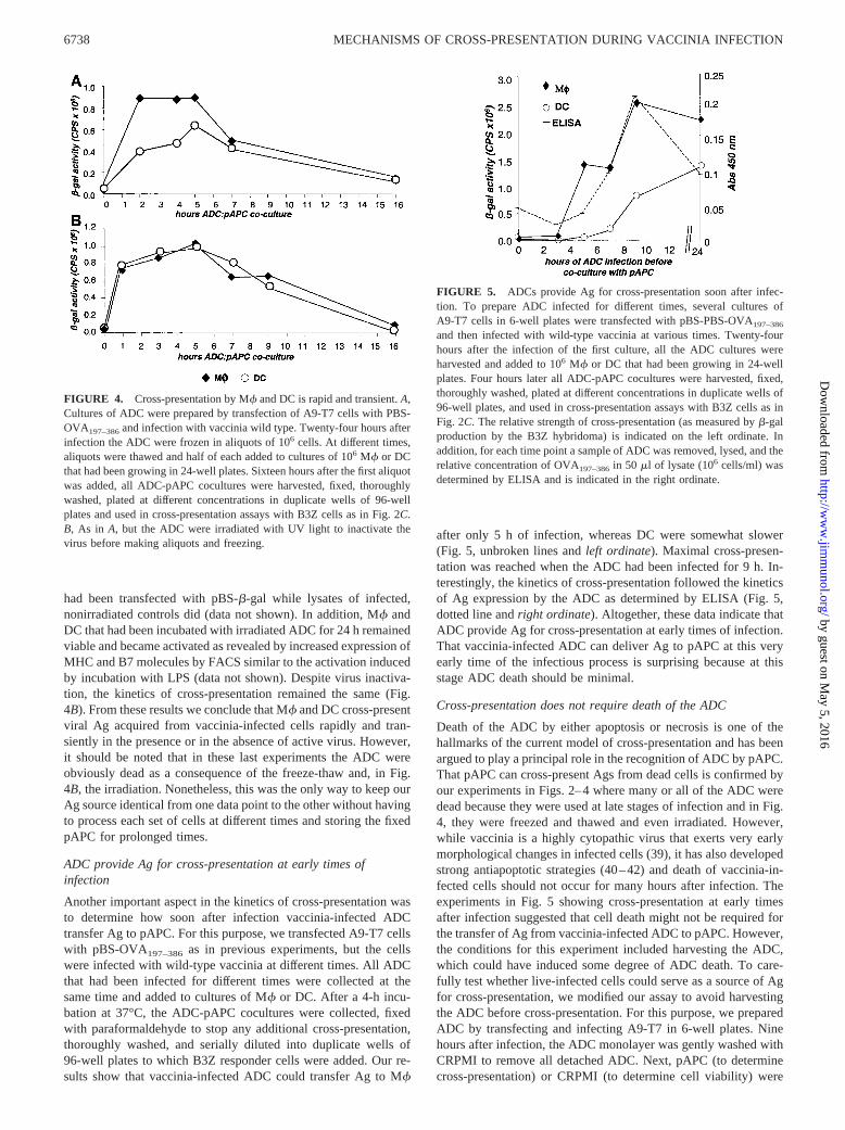
Another important aspect in the kinetics of cross-presentation wasto determine how soon after infection vaccinia-infected ADCtransfer Ag to pAPC. For this purpose, we transfected A9-T7 cellswith pBS-OVA197–386 as in previous experiments, but the cellswere infected with wild-type vaccinia at different times. All ADCthat had been infected for different times were collected at thesame time and added to cultures of M� or DC. After a 4-h incu-bation at 37°C, the ADC-pAPC cocultures were collected, fixedwith paraformaldehyde to stop any additional cross-presentation,thoroughly washed, and serially diluted into duplicate wells of96-well plates to which B3Z responder cells were added. Our re-sults show that vaccinia-infected ADC could transfer Ag to M�
after only 5 h of infection, whereas DC were somewhat slower(Fig. 5, unbroken lines and left ordinate). Maximal cross-presen-tation was reached when the ADC had been infected for 9 h. In-terestingly, the kinetics of cross-presentation followed the kineticsof Ag expression by the ADC as determined by ELISA (Fig. 5,dotted line and right ordinate). Altogether, these data indicate thatADC provide Ag for cross-presentation at early times of infection.That vaccinia-infected ADC can deliver Ag to pAPC at this veryearly time of the infectious process is surprising because at thisstage ADC death should be minimal.
Cross-presentation does not require death of the ADC
Death of the ADC by either apoptosis or necrosis is one of thehallmarks of the current model of cross-presentation and has beenargued to play a principal role in the recognition of ADC by pAPC.That pAPC can cross-present Ags from dead cells is confirmed byour experiments in Figs. 2–4 where many or all of the ADC weredead because they were used at late stages of infection and in Fig.4, they were freezed and thawed and even irradiated. However,while vaccinia is a highly cytopathic virus that exerts very earlymorphological changes in infected cells (39), it has also developedstrong antiapoptotic strategies (40–42) and death of vaccinia-in-fected cells should not occur for many hours after infection. Theexperiments in Fig. 5 showing cross-presentation at early timesafter infection suggested that cell death might not be required forthe transfer of Ag from vaccinia-infected ADC to pAPC. However,the conditions for this experiment included harvesting the ADC,which could have induced some degree of ADC death. To care-fully test whether live-infected cells could serve as a source of Agfor cross-presentation, we modified our assay to avoid harvestingthe ADC before cross-presentation. For this purpose, we preparedADC by transfecting and infecting A9-T7 in 6-well plates. Ninehours after infection, the ADC monolayer was gently washed withCRPMI to remove all detached ADC. Next, pAPC (to determinecross-presentation) or CRPMI (to determine cell viability) were
FIGURE 5. ADCs provide Ag for cross-presentation soon after infec-tion. To prepare ADC infected for different times, several cultures ofA9-T7 cells in 6-well plates were transfected with pBS-PBS-OVA197–386
and then infected with wild-type vaccinia at various times. Twenty-fourhours after the infection of the first culture, all the ADC cultures wereharvested and added to 106 M� or DC that had been growing in 24-wellplates. Four hours later all ADC-pAPC cocultures were harvested, fixed,thoroughly washed, plated at different concentrations in duplicate wells of96-well plates, and used in cross-presentation assays with B3Z cells as inFig. 2C. The relative strength of cross-presentation (as measured by �-galproduction by the B3Z hybridoma) is indicated on the left ordinate. Inaddition, for each time point a sample of ADC was removed, lysed, and therelative concentration of OVA197–386 in 50 �l of lysate (106 cells/ml) wasdetermined by ELISA and is indicated in the right ordinate.
FIGURE 4. Cross-presentation by M� and DC is rapid and transient. A,Cultures of ADC were prepared by transfection of A9-T7 cells with PBS-OVA197–386 and infection with vaccinia wild type. Twenty-four hours afterinfection the ADC were frozen in aliquots of 106 cells. At different times,aliquots were thawed and half of each added to cultures of 106 M� or DCthat had been growing in 24-well plates. Sixteen hours after the first aliquotwas added, all ADC-pAPC cocultures were harvested, fixed, thoroughlywashed, plated at different concentrations in duplicate wells of 96-wellplates and used in cross-presentation assays with B3Z cells as in Fig. 2C.B, As in A, but the ADC were irradiated with UV light to inactivate thevirus before making aliquots and freezing.
6738 MECHANISMS OF CROSS-PRESENTATION DURING VACCINIA INFECTION
by guest on May 5, 2016
http://ww
w.jim
munol.org/
Dow
nloaded from

added to the ADC that remained attached to the wells. After a 4-hincubation, the ADC-pAPC cocultures were harvested, immedi-ately fixed, and cross-presentation determined as usual. Fig. 6Ashows that both M� and DC cells effectively cross-presented Agunder these conditions. At the same time that the ADC-pAPC co-
cultures were harvested, the cells that received only CRPMI werestained with acridine orange and ethidium bromide or using theTUNEL assay. Acridine orange is a supravital dye that intercalatesin the DNA and stains the nuclei of all cells green. With thisstaining, apoptosis can be determined by comparing the morphol-ogy of the stained nuclei. Ethidium bromide stains the nuclei ofnonviable cells bright orange. Fig. 6B demonstrates that A9-T7cells that had been transfected and infected 13 h earlier (this timeincludes the 9 h of infection before adding the ADC to the cross-presentation wells and the 4 h of pAPC/ADC coculture) exhibitedthe typical rounding and membrane roughness of vaccinia-infectedcells. However, they were �97% viable as revealed by exclusionof ethidium bromide, and they were not apoptotic as revealed bythe morphology of the nuclei and the absence of nuclear fluores-cence in the TUNEL assay. For comparison, the staining of nor-mal, apoptotic, and saponin-permeabilized apoptotic cells are in-cluded in the figure. The low level of death (�3%) cannot explainthe cross-presentation observed, because these ADC inducedcross-presentation signals similar to those where 100% of the cellswere dead. Therefore, live-infected ADC can transfer Ag to pAPCfor cross-presentation.
Because Fig. 4 indicated that dead cells could transfer Ag topAPC following only 1 h of coculture, we tested whether the sameoccurred when using live-infected cells as ADC. Therefore, A9-T7cells were prepared as in Fig. 6 and cocultured with pAPC for 1 or2 h. The results in Fig. 7 show that, similar to what occurs withdead-infected ADC, pAPC can cross-present Ag from live-infectedADC within 1 h of coculture.
DiscussionCross-presentation is a kind of exogenous Ag presentationwhereby pAPC acquire Ags from other cells and present them ontheir own MHC class I molecules. This process might have itsforemost physiological importance in the generation of antiviralimmunity. Much of what is currently known about exogenous pre-sentation was obtained from the study of other types of exogenousAgs such as soluble and particulate proteins, immune complexes,and bacteria (24–27, 43). Although the studies with these antigenicformulations greatly advanced our knowledge about exogenouspresentation, cross-presentation of Ags delivered by virus-infectedcells involves greater complexity as it involves a multitude of pa-rameters that may affect the ADC, the pAPC, and the interactionsof both cell types with the virus. For example, cross-presentationmight be affected by the quantity and timing of Ag production,virus-induced ADC death, the production of cytokines or otheralarm signals by ADC, the kinetics with which pAPC acquire theAg from infected cells, the need for Ag to compete with a multi-tude of other Ags produced by the ADC, the ability of the virus to
FIGURE 6. Cross-presentation does not require death of the ADC:ADC were prepared with A9-T7 cells in 6-well plates that were transfectedwith pBS-OVA197–386 or empty pBS II vector as control, and infected withwild-type vaccinia virus. Nine hours after the infection, the ADC weregently washed to remove detached cells. A, the ADC cultures received 106
M� or DC and were incubated for an additional 4 h after which the ADC-pAPC cocultures were harvested, fixed, thoroughly washed, plated at dif-ferent concentrations in duplicate wells of 96-well plates, and used incross-presentation assays with B3Z cells as in Fig. 2C. B, The ADC cul-tures received CRPMI and 4 h later 10 �l stocks of acridine orange, asupravital dye that stains all cells green, and ethidium bromide that stainscells with permeable plasma membrane orange, or were stained for apo-ptosis using a TUNEL assay. Positive controls for apoptosis were irradiatedfor 10 min with UV light before the 4-h incubation period and positivecontrols for membrane permeability were treated with 0.2% saponin. Thecells were observed by phase contrast (left column), for acridine orange andethidium bromide fluorescence staining (center column) using green andred band path filters that were merged for the figure, and for TUNELfluorescence (right column) using the green band path filter. Experimentaland controls as indicated. Note that the dim fluorescence that is observedin the experimental “transfected, infected” cells by TUNEL is localized tothe cytoplasm and most likely labels viral DNA. This cytoplasmic fluo-rescence is also observed in the “transfected, infected, UV-treated” but notin the “UV-treated” controls both of which show strong nuclear staining.
FIGURE 7. Cross-presentation by M� and DC is also rapid when ADCare still alive. ADC were prepared as in Fig. 6 but they were coculturedwith pAPC for the indicated times instead of 4 h.
6739The Journal of Immunology
by guest on May 5, 2016
http://ww
w.jim
munol.org/
Dow
nloaded from

infect the pAPC, the route followed by the Ag within the pAPC,etc. A major problem in studying cross-presentation in viral infec-tions has been the difficulty in establishing reliable models whereone can be certain that the measured phenomenon is cross-presen-tation and not direct presentation. We have previously achievedthis in vivo by using a poliovirus system that restricted virus in-fection to non-pAPC and showed that cross-presentation is neces-sary and sufficient to induce CTL responses (3). However, thecareful dissection of many important aspects of cross-presentationsuch as those that we describe in this study requires a reliable invitro system that could limit Ag production to ADC and Ag pre-sentation to pAPC. The use of allogeneic ADC is a simple ap-proach to restrict Ag presentation to pAPC. However, restrictingthe synthesis of Ag to ADC is more challenging. Other authorshave used human monocytes infected with recombinant vacciniavirus as ADC. However, this required the UV irradiation of theADC to avoid transfer of live virus to the pAPC (23). Therefore,under these conditions cross-presentation was a result of irradia-tion and vaccinia was a vector for Ag production. In this study, wedesigned a powerful cross-presentation assay using vaccinia virusthat undoubtedly restricts Ag production to ADC and presentationto pAPC without the need to artificially kill the ADC. Therefore,it allows measuring cross-presentation while maintaining intact thecomplex interaction between the virus, the ADC, and the pAPC.Using this assay, we have confirmed that DC can cross-present Agshed by vaccinia-infected cells, and also demonstrated for the firsttime that M� can also cross-present Ag supplied by vaccinia-in-fected ADC with high efficiency. Moreover, our results show thatfor the mechanisms and kinetics of cross-presentation that we haveanalyzed, M� and DC are almost identical. In the past few years,there has been much emphasis placed on the exclusive role of DCas the primary cell type involved in Ag presentation while the ideaof M� as major player in this process has been almost abandoned.One possible reason for our results being different from those re-ported by Albert and coworkers (21, 22) is that human M� andmouse M� may differ in their ability to cross-present. It is alsopossible that M� of differing tissue origin (peripheral blood mono-cyte-derived vs bone marrow-derived) differ in their ability tocross-present. Another possibility for this difference is that differ-ent pAPC could handle diverse Ags or cells infected by variousviruses differently since we used OVA and vaccinia virus whileAlbert et al. (21, 22) used influenza virus. This notion is reinforcedby the demonstration that M� generated under similar conditionsas ours failed to present exogenous OVA in the form of immunecomplexes (44). Nonetheless, our finding that M� cross-presentAgs contradicts a model generalizing the view that cross-presen-tation of Ags provided by infected cells is an exclusive ability ofDC and that M� are scavengers with an enzymatic machinery thatdestroys these Ags rather than uses them to generate antigenicpeptides for MHC class I cross-presentation (22). Therefore, therole of M� as pAPC should be revisited. However, it should benoted that our assay measures Ag presentation and not priming ofnaive CD8� T cells. Therefore, it remains possible that the acti-vation of naive cells in the lymph nodes is an exclusive function ofDC. This would agree with the observation that DC but not M�can be found in the T cell areas of lymphoid organs (45). However,cross-presenting M� could be involved in the amplification of theresponse in the periphery, or in the rapid activation of memorycells in tissues during recall responses. Alternatively, there is theprovocative possibility that cross-presenting M� that localize tothe subcapsular region of lymph nodes (17, 45) could rapidly dif-ferentiate to DC and migrate to the lymph nodes to activate naiveT cells.
In this manuscript, we also show that OVA197–386 as a model ofa vaccinia Ag is cross-presented by pAPC using the cytosolicroute. This result agrees and confirms our previous work in vivousing poliovirus also expressing a truncated, cytosolic form ofOVA, and with the results from other laboratories using a differentvirus and Ag (22). Nevertheless, it is also interesting to note thatfor Ags delivered in the form of bacteria or protein bound to mi-crobeads, exogenous presentation of SIINFEKL followed the cy-tosolic route when expressed in the context of the native OVAsequence (as in our experiments). However, it could use the vac-uolar pathway when expressed in a recombinant form, fused veryclose to the C terminus of the Escherichia coli Crl protein. Whetherthese findings are mirrored when the exogenous Ag is provided byvaccinia infected cells is currently under investigation (26).
The current model of cross-presentation during viral infectionsis that DC acquire Ag from apoptotic- or necrotic-infected cells inthe periphery and migrate to lymphoid organs (13, 46, 47). Thisprocess is accompanied by the maturation of the DC, which in-volves a decreased ability to phagocytose and increased expressionof MHC, B7, and other molecules allowing for a more effectivepresentation of Ag to T cells (48, 49). Our experiments show thatpAPC cross-present Ags released by vaccinia-infected cells verysoon after contacting ADC, that cross-presentation lasts for a shortperiod of time, and that cross-presentation starts at early times ofinfection when the ADC are still alive.
That pAPC cross-present Ag from virus-infected cells very soonafter ADC-pAPC contact is in agreement with the finding thatcross-presenting cells migrate to the draining lymph nodes within6 h after scarification of the epidermis with HSV (50). However,our finding that cross-presentation of Ags from vaccinia-infectedcells is also short-lived even in the presence of saturating amountsof Ag and in the absence of pAPC infection is surprising. Indeed,it would be expected that direct presentation by pAPC infectedwith cytopathic viruses would be short-lived but not during cross-presentation when the pAPC infection is spared as when we irra-diated the ADC. Still, it is very likely that our results reflect theduration of cross-presentation for individual cells in vivo, and isconsistent with the measured half-life of MHC class I peptide com-plexes of high affinity at the surface of cells, which is �6–12 h(51, 52). If this is correct, it implies that the activation of pAPCand their ability to stimulate naive T cells should also occur veryfast, and that the activation of a sufficient number of precursorCTL in vivo may require the continuous arrival of pAPC from thesite of infection. An alternative explanation for the short time ofcross-presentation during vaccinia infection may be that vacciniamay produce cross-presentation inhibitory factors. Poxviruses areknown to encode multiple immune evasion molecules such as cy-tokine and chemokine-like proteins (53, 54). If this were the case,the immune evasion molecules acting in our assay should be trans-ferred with the infected cells because we discarded the superna-tants. Moreover, these factors should work in trans (i.e., producedin the ADC but functions in the pAPC) because virus inactivationdid not alter the kinetics of cross-presentation. To our knowledge,inhibition of MHC class I Ag presentation in trans has not yet beendescribed. If this were the reason for the short time that cross-presentation lasts, it might be of interest to identify the responsiblemolecules. Another alternative is that some mechanisms that pro-long Ag cross-presentation in vivo may be absent in our assay. Forexample, cytokines or inflammatory molecules could increase thehalf-life of the MHC class I peptide complexes at the cell surface(48) or retard the intracellular degradation of Ag within the pAPC.
It is widely held that cross-presentation in viral infections re-quired either necrotic or apoptotic cells as the source of Ag. There-fore, our present findings that cross-presentation occur at early
6740 MECHANISMS OF CROSS-PRESENTATION DURING VACCINIA INFECTION
by guest on May 5, 2016
http://ww
w.jim
munol.org/
Dow
nloaded from

times of vaccinia infection and without the need for cell death isstriking. It will now be important to identify the signals used by theinfected ADC to alert pAPC and to determine the mechanismswhereby pAPC incorporate the Ags supplied by live-infectedADC. One possible mechanism is that pAPC phagocytose and pro-cess whole live-infected ADC. It is also possible that M� and DCcells recognize signals imparted by the infected cells and kill themjust before phagocytosis. Other possibilities are that live-infectedADC release Ag-loaded exosomes (55) or antigenic peptidesbound to heat shock proteins (56). These could be taken up byphagocytosis or through receptor-mediated endocytosis and cross-presented by pAPC. We also speculate that the ability of pAPC toacquire Ag from live ADC at early times of infection could beadvantageous because at this time the replication cycle of the virusmight not have been completed and pAPC could avoid becominginfected themselves.
Together, our findings that cross-presentation during vacciniainfection occurs very rapidly following ADC-pAPC contact andthat ADC provide Ag before dying and at early phases of infectionshould contribute to rapid Ag presentation and a headstart in theinitiation of the CD8� T cell response. This timing could be crit-ical to tip the balance in favor of the host in its race against thevirus. Together, our findings and those of others (17, 50) point toa model where direct presentation and cross-presentation are bothfast and brief. This would also require pAPC-T cell contact to bebrief and occur at a very early phase of the immune response. Thismodel would also be in agreement with findings that a short ex-posure of CD8� T cells to Ag bearing pAPC is sufficient to starta program of maximal proliferation and differentiation into effectorand memory cells (57–60).
AcknowledgmentsWe thank the Fox Chase Cancer Center Laboratory Animal, DNA sequenc-ing, Fannie E. Rippel Biochemistry and Biotechnology, and the Flow-cy-tometry facilities for their support. We also thank Drs. Kerry Campbell,Glenn Rall, and David Wiest (Fox Chase Cancer Center) for their com-ments on the manuscript, and Dr. Roque Bort for his comments and hisvaluable help with the fluorescence microscope. We also thank Dr. LienjunShen (University of Massachusetts Medical Center) for OVA containingplasmids, Dr. Nilab Shastri for B3Z cells (University of California, LosAngeles, CA), and Dr. Bernard Moss (National Institutes of Health) forA9-T7 cells and viruses.
References1. Lenz, L. L., E. A. Butz, and M. J. Bevan. 2000. Requirements for bone marrow-
derived antigen-presenting cells in priming cytotoxic T cell responses to intra-cellular pathogens. J. Exp. Med. 192:1135.
2. Sigal, L. J., and K. L. Rock. 2000. Bone marrow-derived antigen-presenting cellsare required for the generation of cytotoxic T lymphocyte responses to virusesand use transporter associated with antigen presentation (TAP)-dependent and-independent pathways of antigen presentation. J. Exp. Med. 192:1143.
3. Sigal, L. J., S. Crotty, R. Andino, and K. L. Rock. 1999. Cytotoxic T-cell im-munity to virus-infected non-haematopoietic cells requires presentation of exog-enous antigen. Nature 398:77.
4. Zinkernagel, R. M., G. Kreeb, and A. Althage. 1980. Lymphohemopoietic originof the immunogenic, virus-antigen-presenting cells triggering anti-viral T-cellresponses. Clin. Immunol. Immunopathol. 15:565.
5. McAdam, A. J., E. A. Farkash, B. E. Gewurz, and A. H. Sharpe. 2000. B7costimulation is critical for antibody class switching and CD8� cytotoxic T-lymphocyte generation in the host response to vesicular stomatitis virus. J. Virol.74:203.
6. Sigal, L. J., H. Reiser, and K. L. Rock. 1998. The role of B7-1 and B7-2 co-stimulation for the generation of CTL responses in vivo. J. Immunol. 161:2740.
7. June, C. H., J. A. Bluestone, L. M. Nadler, and C. B. Thompson. 1994. The B7and CD28 receptor families. Immunol. Today 15:321.
8. Allison, J. P. 1994. CD28–B7 interactions in T-cell activation. Curr. Opin. Im-munol. 6:414.
9. Razi-Wolf, Z., G. Freeman, F. Galvin, B. Benacerraf, L. Nadler, and H. Reiser.1992. Expression and function of the murine B7 antigen, the major costimulatorymolecule expressed by peritoneal exudate cells. Proc. Natl. Acad. Sci. USA 89:4210.
10. Schwartz, R. 1992. Costimulation of T lymphocytes: the role of CD28, CTLA-4,and B7/BB1 in interleukin-2 production and immunotherapy. Cell 71:1065.
11. Freeman, G. J., F. Borriello, R. J. Hodes, H. Reiser, J. G. Gribben, J. W. Ng,J. Kim, J. M. Goldberg, K. Hathcock, G. Laszlo, et al. 1993. Murine B7-2, analternative CTLA4 counter-receptor that costimulates T cell proliferation andinterleukin 2 production. J. Exp. Med. 178:2185.
12. Mondino, A., A. Khoruts, and M. K. Jenkins. 1996. The anatomy of T-cell ac-tivation and tolerance. Proc. Natl. Acad. Sci. USA 93:2245.
13. Schumacher, T. N. 1999. Accessory to murder. Nature 398:26.14. Banchereau, J., and R. M. Steinman. 1998. Dendritic cells and the control of
immunity. Nature 392:245.15. Mellman, I., S. J. Turley, and R. M. Steinman. 1998. Antigen processing for
amateurs and professionals. Trends Cell Biol. 8:231.16. Debrick, J. E., P. A. Campbell, and U. D. Staerz. 1991. Macrophages as accessory
cells for class I MHC-restricted immune responses. J. Immunol. 147:2846.17. Norbury, C. C., D. Malide, J. S. Gibbs, J. R. Bennink, and J. W. Yewdell. 2002.
Visualizing priming of virus-specific CD8� T cells by infected dendritic cells invivo. Nat. Immunol. 3:265.
18. Yewdell, J. W., C. C. Norbury, and J. R. Bennink. 1999. Mechanisms of exog-enous antigen presentation by MHC class I molecules in vitro and in vivo: im-plications for generating CD8� T cell responses to infectious agents, tumors,transplants, and vaccines. Adv. Immunol. 73:1.
19. Basta, S., W. Chen, J. R. Bennink, and J. W. Yewdell. 2002. Inhibitory effects ofcytomegalovirus proteins US2 and US11 point to contributions from direct prim-ing and cross-priming in induction of vaccinia virus-specific CD8� T cells. J. Im-munol. 168:5403.
20. Gold, M. C., M. W. Munks, M. Wagner, U. H. Koszinowski, A. B. Hill, andS. P. Fling. 2002. The murine cytomegalovirus immunomodulatory gene m152prevents recognition of infected cells by M45-specific CTL but does not alter theimmunodominance of the M45-specific CD8 T cell response in vivo. J. Immunol.169:359.
21. Albert, M. L., S. F. Pearce, L. M. Francisco, B. Sauter, P. Roy, R. L. Silverstein,and N. Bhardwaj. 1998. Immature dendritic cells phagocytose apoptotic cells via�v�5 and CD36, and cross-present antigens to cytotoxic T lymphocytes. J. Exp.Med. 188:1359.
22. Albert, M. L., B. Sauter, and N. Bhardwaj. 1998. Dendritic cells acquire antigenfrom apoptotic cells and induce class I-restricted CTLs. Nature 392:86.
23. Larsson, M., J. F. Fonteneau, S. Somersan, C. Sanders, K. Bickham,E. K. Thomas, K. Mahnke, and N. Bhardwaj. 2001. Efficiency of cross presen-tation of vaccinia virus-derived antigens by human dendritic cells. Eur. J. Im-munol. 31:3432.
24. Kovacsovics-Bankowski, M., and K. L. Rock. 1994. Presentation of exogenousantigens by macrophages: analysis of major histocompatibility complex class Iand II presentation and regulation by cytokines. Eur. J. Immunol. 24:2421.
25. Kovacsovics-Bankowski, M., K. Clark, B. Benacerraf, and K. L. Rock. 1993.Efficient major histocompatibility complex class I presentation of exogenous an-tigen upon phagocytosis by macrophages. Proc. Natl. Acad. Sci. USA 90:4942.
26. Pfeifer, J. D., M. J. Wick, R. L. Roberts, K. Findlay, S. J. Normark, andC. V. Harding. 1993. Phagocytic processing of bacterial antigens for class I MHCpresentation to T cells. Nature 361:359.
27. Norbury, C. C., L. J. Hewlett, A. R. Prescott, N. Shastri, and C. Watts. 1995.Class I MHC presentation of exogenous soluble antigen via macropinocytosis inbone marrow macrophages. Immunity 3:783.
28. Elroy-Stein, O., and B. Moss. 1990. Cytoplasmic expression system based onconstitutive synthesis of bacteriophage T7 RNA polymerase in mammalian cells.Proc. Natl. Acad. Sci. USA 87:6743.
29. Elroy-Stein, O., and B. Moss. 1992. Gene expression using the vaccinia virus/T7RNA polymerase. In Current Protocols in Molecular Biology, Vol. 2. F. Ausubel,R. Brent, R. Kingston, J. Moore, J. Seidman, and K. Struhl, eds. Wiley Inter-science, New York, p. 16.19.1.
30. Karttunen, J., S. Sanderson, and N. Shastri. 1992. Detection of rare antigen-presenting cells by the lacZ T-cell activation assay suggests an expression cloningstrategy for T-cell antigens. Proc. Natl. Acad. Sci. USA 89:6020.
31. Kovacsovics-Bankowski, M., and K. L. Rock. 1995. A phagosome-to-cytosolpathway for exogenous antigens presented on MHC class I molecules. Science267:243.
32. Townsend, A. R., J. Bastin, K. Gould, and G. G. Brownlee. 1986. Cytotoxic Tlymphocytes recognize influenza haemagglutinin that lacks a signal sequence.Nature 324:575.
33. Powis, S. J., A. R. Townsend, E. V. Deverson, J. Bastin, G. W. Butcher, andJ. C. Howard. 1991. Restoration of antigen presentation to the mutant cell lineRMA-S by an MHC-linked transporter. Nature 354:528.
34. Townsend, A., and J. Trowsdale. 1993. The transporters associated with antigenpresentation. Semin. Cell Biol. 4:53.
35. Rock, K. L., C. Gramm, L. Rothstein, K. Clark, R. Stein, L. Dick, D. Hwang, andA. L. Goldberg. 1994. Inhibitors of the proteasome block the degradation of mostcell proteins and the generation of peptides presented on MHC class I molecules.Cell 78:761.
36. Pamer, E., and P. Cresswell. 1998. Mechanisms of MHC class I-restricted antigenprocessing. Annu. Rev. Immunol. 16:323.
37. Wick, M. J., and J. D. Pfeifer. 1996. Major histocompatibility complex class Ipresentation of ovalbumin peptide 257–264 from exogenous sources: protein con-text influences the degree of TAP-independent presentation. Eur. J. Immunol.26:2790.
38. Song, R., and C. V. Harding. 1996. Roles of proteasomes, transporter for antigenpresentation (TAP), and �2-microglobulin in the processing of bacterial or par-ticulate antigens via an alternate class I MHC processing pathway. J. Immunol.156:4182.
6741The Journal of Immunology
by guest on May 5, 2016
http://ww
w.jim
munol.org/
Dow
nloaded from

39. Bablanian, R., B. Baxt, J. A. Sonnabend, and M. Esteban. 1978. Studies on themechanisms of vaccinia virus cytopathic effects. II. Early cell rounding is asso-ciated with virus polypeptide synthesis. J. Gen. Virol. 39:403.
40. Wasilenko, S. T., A. F. Meyers, K. Vander Helm, and M. Barry. 2001. Vacciniavirus infection disarms the mitochondrion-mediated pathway of the apoptoticcascade by modulating the permeability transition pore. J. Virol. 75:11437.
41. Kettle, S., A. Alcami, A. Khanna, R. Ehret, C. Jassoy, and G. L. Smith. 1997.Vaccinia virus serpin B13R (SPI-2) inhibits interleukin-1�-converting enzymeand protects virus-infected cells from TNF- and Fas-mediated apoptosis, but doesnot prevent IL-1�-induced fever. J. Gen. Virol. 78:677.
42. Dobbelstein, M., and T. Shenk. 1996. Protection against apoptosis by the vacciniavirus SPI-2 (B13R) gene product. J. Virol. 70:6479.
43. Amigorena, S., and C. Bonnerot. 1999. Fc receptor signaling and trafficking: aconnection for antigen processing. Immunol. Rev. 172:279.
44. Rodriguez, A., A. Regnault, M. Kleijmeer, P. Ricciardi-Castagnoli, andS. Amigorena. 1999. Selective transport of internalized antigens to the cytosol forMHC class I presentation in dendritic cells. Nat. Cell Biol. 1:362.
45. Jenkins, M. K., A. Khoruts, E. Ingulli, D. L. Mueller, S. J. McSorley,R. L. Reinhardt, A. Itano, and K. A. Pape. 2001. In vivo activation of antigen-specific CD4 T cells. Annu. Rev. Immunol. 19:23.
46. Banchereau, J., F. Briere, C. Caux, J. Davoust, S. Lebecque, Y. J. Liu,B. Pulendran, and K. Palucka. 2000. Immunobiology of dendritic cells. Annu.Rev. Immunol. 18:767.
47. Guermonprez, P., J. Valladeau, L. Zitvogel, C. Thery, and S. Amigorena. 2002.Antigen presentation and T cell stimulation by dendritic cells. Annu. Rev. Immu-nol. 20:621.
48. Cella, M., M. Salio, Y. Sakakibara, H. Langen, I. Julkunen, and A. Lanzavecchia.1999. Maturation, activation, and protection of dendritic cells induced by double-stranded RNA. J. Exp. Med. 189:821.
49. Winzler, C., P. Rovere, M. Rescigno, F. Granucci, G. Penna, L. Adorini,V. S. Zimmermann, J. Davoust, and P. Ricciardi-Castagnoli. 1997. Maturation
stages of mouse dendritic cells in growth factor-dependent long-term cultures.J. Exp. Med. 185:317.
50. Mueller, S. N., C. M. Jones, C. M. Smith, W. R. Heath, and F. R. Carbone. 2002.Rapid cytotoxic T lymphocyte activation occurs in the draining lymph nodes aftercutaneous herpes simplex virus infection as a result of early antigen presentationand not the presence of virus. J. Exp. Med. 195:651.
51. Sigal, L. J., P. Goebel, and D. E. Wylie. 1995. Db-binding peptides from influ-enza virus: effect of non-anchor residues on stability and immunodominance.Mol. Immunol. 32:623.
52. Su, R. C., and R. G. Miller. 2001. Stability of surface H-2Kb, H-2Db, and peptide-receptive H-2Kb on splenocytes. J. Immunol. 167:4869.
53. Smith, G. L., J. A. Symons, A. Khanna, A. Vanderplasschen, and A. Alcami.1997. Vaccinia virus immune evasion. Immunol. Rev. 159:137.
54. Moss, B., and J. L. Shisler. 2001. Immunology 101 at poxvirus U: immuneevasion genes. Semin. Immunol. 13:59.
55. Wolfers, J., A. Lozier, G. Raposo, A. Regnault, C. Thery, C. Masurier,C. Flament, S. Pouzieux, F. Faure, T. Tursz, et al. 2001. Tumor-derived exosomesare a source of shared tumor rejection antigens for CTL cross-priming. Nat. Med.7:297.
56. Suto, R., and P. K. Srivastava. 1995. A mechanism for the specific immunoge-nicity of heat shock protein-chaperoned peptides. Science 269:1585.
57. Bevan, M. J., and P. J. Fink. 2001. The CD8 response on autopilot. Nat. Immunol.2:381.
58. Busch, D. H., K. M. Kerksiek, and E. G. Pamer. 2000. Differing roles of inflam-mation and antigen in T cell proliferation and memory generation. J. Immunol.164:4063.
59. Kaech, S. M., and R. Ahmed. 2001. Memory CD8� T cell differentiation: initialantigen encounter triggers a developmental program in naive cells. Nat. Immunol.2:415.
60. van Stipdonk, M. J., E. E. Lemmens, and S. P. Schoenberger. 2001. Naive CTLsrequire a single brief period of antigenic stimulation for clonal expansion anddifferentiation. Nat. Immunol. 2:423.
6742 MECHANISMS OF CROSS-PRESENTATION DURING VACCINIA INFECTION
by guest on May 5, 2016
http://ww
w.jim
munol.org/
Dow
nloaded from