Sanford's Radical Cure. HUNT PLEISAKI KILLS. COAL, - DigiFind-It
An actinomycete isolate from solitary wasp mud nest having strongantibacterial activity and kills the Candida cells due the shrinkage and thecytosolic loss
Vijay Kumar, Bindu Naik, Omprakash Gusain and Gajraj Singh Bisht
Journal Name: Frontiers in Microbiology
ISSN: 1664-302X
Article type: Original Research Article
Received on: 04 Jun 2014
Accepted on: 05 Aug 2014
Provisional PDF published on: 05 Aug 2014
www.frontiersin.org: www.frontiersin.org
Citation: Kumar V, Naik B, Gusain O and Bisht GS(2014) An actinomyceteisolate from solitary wasp mud nest having strong antibacterialactivity and kills the Candida cells due the shrinkage and thecytosolic loss. Front. Microbiol. 5:446.doi:10.3389/fmicb.2014.00446
Copyright statement: © 2014 Kumar, Naik, Gusain and Bisht. This is an open-accessarticle distributed under the terms of the Creative CommonsAttribution License (CC BY). The use, distribution or reproductionin other forums is permitted, provided the original author(s) orlicensor are credited and that the original publication in thisjournal is cited, in accordance with accepted academic practice.No use, distribution or reproduction is permitted which does notcomply with these terms.
This Provisional PDF corresponds to the article as it appeared upon acceptance, after rigorous
peer-review. Fully formatted PDF and full text (HTML) versions will be made available soon.
Antimicrobials, Resistance and Chemotherapy

Frontiers in Microbiology Original Research Date
An actinomycete isolate from solitary wasp mud nest having strong 1
antibacterial activity and kills the Candida cells due the shrinkage and 2
the cytosolic loss 3
4
Vijay Kumar 1
, Bindu Naik2, Omprakash Gusain
3, Gajraj Singh Bisht
4* 5
6 1Department of Food Technology, Doon P.G. College of Agriculture Science and Technology, 7
Selaqui, Dehradun, UK, India 8
9 2Department of Bioprocess and Food Engineering, Institute of Agriculture Sciences, Banaras Hindu 10
University, Varanasi, UP, India, 221005 11
3Department of Zoology and Biotechnology, H.N.B. Garhwal University, Srinagar, Uttarakhand, 12
India. 13 14 4Department of Microbiology, Sardar Bhagwan Singh Post Graduate Institute of Biomedical Sciences 15
and Research, Balawala, Dehradun, Uttarakhand, India, 248161 16
17
*Correspondence 18 GRS Bisht, Professor, 19
Department of Microbiology, Sardar Bhagwan Singh Post Graduate Institute of Biomedical Sciences 20
and Research, Balawala, Dehradun, Uttarakhand, India, 248161 21
: Email: [email protected] Tel.: +91-9719148874 22
23 Key words: Solitary wasp mud nest, Streptomyces sp., anti-candidal activity, violaceusniger 24 clade, polyphasic taxonomy 25
26 Abstract 27
An actinomycetes strain designated as MN 2(6) was isolated from the solitary wasp mud nest. The 28 isolate was identified using polyphasic taxonomy. It produced the extensive branched brown 29
substrate and white aerial hyphae that changed into grayish black. The aerial mycelia produced the 30 spiral spore chains with rugose spore surface. The growth was observed between temperature range 31 of 27-37°C, pH 8-10 and below salt concentration of 6% (w/v). The comparative analysis of 16S 32 rRNA gene sequence and phylogenetic relationship showed that strain MN 2(6) lies in clade with 33 Streptomyces hygroscopicus subsp. hygroscopicus NRRL 2387
T, Streptomyces sporocinereus NBRC 34
100766T and Streptomyces demainii NRRL B-1478
T with which it shares a 16S rRNA gene sequence 35
similarity of 99.3%. The strain MN 2(6) can be differentiated from type strains based on phenotypic 36 characteristics. The strain MN 2(6) showed most promising activity against Gram-positive, Gram-37 negative bacteria, acid-fast bacilli and Candida species suggesting broad-spectrum characteristics of 38 the active metabolite. Evaluation of anti-candidal activity of the metabolite of strain MN 2(6) by 39
scanning electron microscopy (SEM) revealed changed external morphology of yeast. It kills the 40
Candida cells due to the shrinkage and the cytosolic loss. However, further studies are required to 41
elucidate the structure of the active metabolite produced by the isolate MN 2(6) 42
Kumar et al. Antimicrobial activity of Streptomyces sp. MN 2(6)
2 This is a provisional file, not the final typeset article
43
1. Introduction 44
In recent years searching new antibiotics has increased worldwide because of the serious problem of 45 antibiotic resistance among the microbes. The need for new antibiotics has been met largely by the 46 semisynthetic tailoring of natural product scaffolds discovered in the middle of the 20
th century 47
(Clardy et al., 2006). The soil-derived microorganisms have been extensively screened for 48 therapeutically important molecules. However, the frequency of discovering structurally new 49 compounds is apparently decreasing these years and there is a need to seek unutilized 50 microorganisms from unexplored sources (Brady et al., 2002). Moreover, the diversity of secondary 51 metabolites depends more or less on the isolation source, namely, the habitat of the producers 52
(Igarashi, 2004). The recent discovery of the novel primary and secondary metabolites from 53 taxonomically unique population of actinomycetes suggest that these organisms could add a new 54 dimension to microbial natural product research. In this context, new actinomycetes strains producing 55
active compounds have been recently isolated from novel sources including saline, ocean, mangrove 56 forests and niche habitats such as caves, beehives, pristine forests, lakes, rivers, shallow bird, solitary 57 wasp mud nest and other wetlands (Mukku et al., 2000; Mitra et al., 2008; Promnuan et al., 2009; 58 Radhakrishnan et al., 2010; Poulsen et al. 2011, Kumar et al. 2012a). Assuming the above facts, the 59
actinomycetes of the solitary wasp with regard to the occurrence of novel microbial flora have been 60 studied previously (Kumar et al., 2012a, b). New species of the microorganisms have the potential to 61
produce new metabolites, which justifies the isolation of new species at pharmaceutical research 62 laboratories (Shomura, 1979). The present report highlights the taxonomy and antimicrobial activity 63 of a new actinomycete strain isolated from the solitary wasp mud nest, a rare habitat. 64
2. Materials and methods 65 66
2.1 Isolation, identification and characterization 67
Strain MN 2(6) was isolated from the solitary wasp mud nest collected from Dehradun, India (Kumar 68 et al., 2012a). Cultural characteristics of the strain MN 2(6) was examined every day grown on 69
various International Streptomyces project (ISP) media (Shirling and Gottlieb, 1966). 70 Micromorphology and sporulation were observed under light microscope by the inclined coverslip 71 technique (Williams et al. 1989) on ISP-4 medium after incubating at 27°C for 7 days. The spore 72
chain morphology and spore surface ornamentation were examined by scanning electron microscopy 73
(Zeiss EVO 40 EP) of 15-day old cultures grown on ISP-4 according to the method described 74 previously (Kumar et al., 2011). Physiological characteristics were examined according to the 75 methods described in the International Streptomyces project (Shirling and Gottlieb, 1966) and 76
Bergey's Manual of Systematic Bacteriology (Locci, 1989). Resistance to some antibiotics was 77 detected by disc diffusion method. The isomeric forms of diaminopimelic acid (DAP) and the 78 diagnostic sugar in the whole-cell hydrolysates were determined as described in MTCC Laboratory 79
manual, IMTECH (1998), Chandigarh, India. 80
2.2 Molecular Identification 81
Chromosomal DNA of the strain MN 2(6) was prepared from cells grown in nutrient broth for 2-3 82 days incubation according to the method described earlier (Kumar et al., 2010). PCR amplification of 83
the 16S rDNA of the strain MN 2(6) was done according to the methods described previously 84 (Kumar et al., 2012a). The identification of phylogenetic neighbors was initially carried out by the 85
Kumar et al. Antimicrobial activity of Streptomyces sp. MN 2(6)
3
BLASTN (Altschulet al., 1997) program against the database containing type strains with validly 86
published prokaryotic names and representatives of uncultured phylotypes (Kim et al., 2012). The top 87 thirty sequences with the highest scores were then selected for the calculation of pair wise sequence 88 similarity using the global alignment algorithm (Myer and Miller, 1988), which was implemented at 89 the EzTaxon-e server (http://eztaxon-e.ezbiocloud.net/; Kim et al., 2012). The isolate was identified 90
using the EzTaxon-e server (http://eztaxon-e.ezbiocloud.net/; Kim et al., 2012) based on 16S rRNA 91 sequence data. The evolutionary history was inferred using the Neighbor-Joining method (Saitou and 92 Nei, 1987). The percentage of replicate trees in which the associated taxa clustered together in the 93 bootstrap test (1000 replicates) are shown next to the branches (Felsenstein, 1985). The tree is drawn 94 to scale, with branch lengths in the same units as those of the evolutionary distances used to infer the 95
phylogenetic tree. The evolutionary distances were computed using the Maximum Composite 96 Likelihood method (Tamura et al., 2004) and are in the units of the number of base substitutions per 97
site. All positions containing gaps and missing data were eliminated from the dataset (Complete 98
deletion option). There were a total of 1376 positions in the final dataset. Phylogenetic analyses were 99 conducted in MEGA4 (Tamura et al., 2007). 100 101
2.3 Production, extraction and partial purification of metabolite 102 103 Production of metabolite was done in Glucose soybean meal medium (GS) as described in the 104
previous study (Kumar et al., 2012c). The extraction and partial purification of antimicrobial 105 metabolite from culture filtrate was done according to the method described previously (Kumar et al., 106 2012c). 107
108
2.4 Biological and Chemical Detection of Antimicrobial Compounds 109 110 Crude extract sample was subjected to thin-layer chromatography (TLC) and spotted onto silica gel 111
plates (Merck Art 5735, Kieselgel 60F254), and then developed with chloroform: methanol: 25 % 112 ammonia (1:7:4, v/v) as the solvent mixture. The numbers of antibacterial active fractions were 113 detected by bioautography (Odakura et al., 1984) on silica gel plates seeded with Micrococcus luteus 114
and Candida albicans. Clear zone of inhibition indicated the position of antimicrobial compounds on 115 the TLC plates, and the retention factor (Rf) value was calculated. The crude product was dissolved 116
in methanol, and the absorption spectrum was recorded at 200–498.8 nm using UV–VIS 117 spectrophotometer (Systronics double beam spectrophotometer). The partial chemical nature of 118 antifungal metabolite was determined using in vitro assay. The polyene like activity of isolated 119
antimicrobial metabolite was carried out by ergosterol agar plate method (Jain and Jain, 2006). The 120 antimicrobial compound was partially characterized by spraying with chemical reagents such as 10 % 121 KOH ethanolic reagent, Millon’s reagent, vanillin-HCl reagent, ninhydrin reagents, iodine vapours, 122 50 % ethanolic H2SO4, and Dragendorf reagent. 123
124
2.5 Determination of antimicrobial activity (MIC method) 125
Minimum Inhibitory Concentration was determined by the micro dilution method using a 96 well 126 plate according to NCCLS (2008). Micro dilution methods involve the use of plastic microtiter plates 127 (96 well). The two-fold serial dilution of the antibiotics was prepared from 512 µg/ml to 0.004 µg/ml 128
range (final concentration in Mueller Hinton broth). The MIC is the lowest concentration of the agent 129 which completely inhibits the growth. The control containing no agent should be turbid (negative 130
control) while control with standard antibiotic should be clear (positive control). 131
132
Kumar et al. Antimicrobial activity of Streptomyces sp. MN 2(6)
4 This is a provisional file, not the final typeset article
2.6 Scanning electron microscopy (SEM) for study of antifungal action of crude extract 133
The anti-fungal action of extract was carried out by SEM. Candida albicans was treated in vitro with 134 half of the concentrations of MIC crude extract. The cells were harvested after 48 h of incubation at 135 35
oC by centrifugation at 4
°C for 5 min and were washed three times with phosphate buffer saline 136
(PBS). The cells pellet was fixed in 3% (v/v) glutaraldehyde in PBS (pH 7.4) and dehydrated in 137
increasing concentrations of ethanol (10%, v/v, increments, to 100%) (Lemar et al., 2005). Cells were 138 mounted onto stubs. The upper surface of each stub was then coated, under vacuum, with a film of 139 gold. The gold coating process was completed in 15-20 min. Once coated with gold, the specimens 140 were ready for examination under scanning electron microscope (ZEISS EVO 40 EP). The gold 141 coated metal stubs were viewed on the SEM at an accelerating voltage of 15 kV, a probe diameter of 142
102 Pa, to obtain secondary electron images. The field was scanned at low magnification until the 143 line of growth was detected. Areas with clear and cells of yeasts were then selected for examination 144
at higher magnification. Suitable fields in the preparation were photographed. 145
3 Results 146
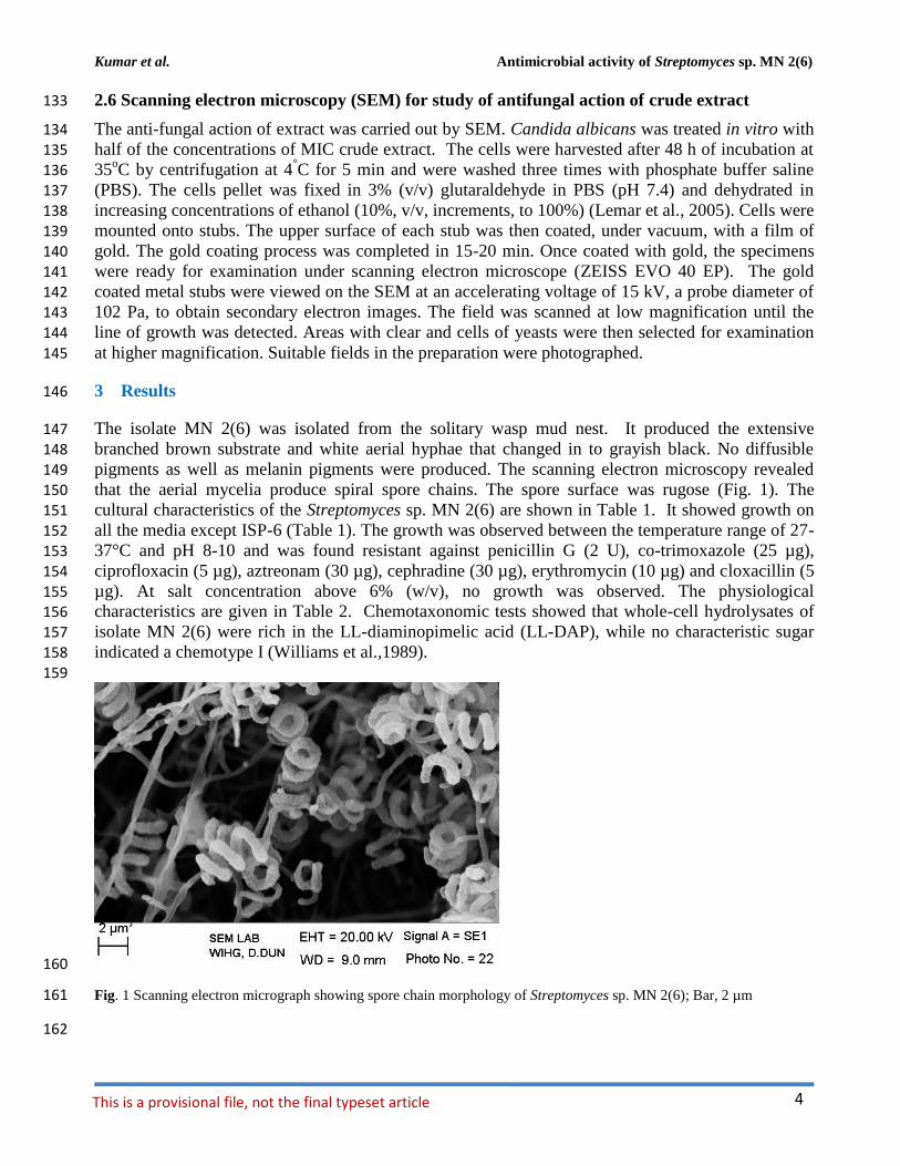
The isolate MN 2(6) was isolated from the solitary wasp mud nest. It produced the extensive 147 branched brown substrate and white aerial hyphae that changed in to grayish black. No diffusible 148 pigments as well as melanin pigments were produced. The scanning electron microscopy revealed 149
that the aerial mycelia produce spiral spore chains. The spore surface was rugose (Fig. 1). The 150 cultural characteristics of the Streptomyces sp. MN 2(6) are shown in Table 1. It showed growth on 151
all the media except ISP-6 (Table 1). The growth was observed between the temperature range of 27-152 37°C and pH 8-10 and was found resistant against penicillin G (2 U), co-trimoxazole (25 µg), 153
ciprofloxacin (5 µg), aztreonam (30 µg), cephradine (30 µg), erythromycin (10 µg) and cloxacillin (5 154 µg). At salt concentration above 6% (w/v), no growth was observed. The physiological 155 characteristics are given in Table 2. Chemotaxonomic tests showed that whole-cell hydrolysates of 156
isolate MN 2(6) were rich in the LL-diaminopimelic acid (LL-DAP), while no characteristic sugar 157 indicated a chemotype I (Williams et al.,1989). 158
159
160
Fig. 1 Scanning electron micrograph showing spore chain morphology of Streptomyces sp. MN 2(6); Bar, 2 µm 161
162

Kumar et al. Antimicrobial activity of Streptomyces sp. MN 2(6)
5
Table 1. Cultural characteristic of Streptomyces sp. MN 2(6) 163
+: Present, - : Absent 164
165
166
167
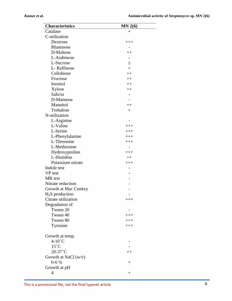
Table 2 Phenotypic characteristic of MN 2(6) 168
Characteristics MN 2(6)
Aerial mycelium White to Grayish black
Reverse Brown
Diffusible pigment -
Melanin pigment -
Sporulation Good
Spore chain Spirals
Starch hydrolysis +++
Casein hydrolysis -
Gelatin hydrolysis +++
Oxidase -
Medium Growth Aerial spore
mass
Revers
e color
Diffusible
pigment
Yeast extract-malt extract agar (ISP 2) Good White to gray Brown -
Oat meal agar (ISP 3) Good Grayish
white
Brown -
Inorganic salt starch agar (ISP 4) Good Whitish gray Green
brown
-
Glycerol asparagine agar base (ISP 5) Poor Gray Colorle
ss
-
Peptone yeast extract iron agar (ISP 6) Moderate Whitish gray Colorle
ss
-
Actinomycetes isolation agar (AIA) Good White to gray Brown -
Sabourad dextrose agar (SDA) Good Cream Brown
Kumar et al. Antimicrobial activity of Streptomyces sp. MN 2(6)
6 This is a provisional file, not the final typeset article
Characteristics MN 2(6)
Catalase +
C-utilization
Dextrose +++
Rhamnose -
D-Maltose ++
L-Arabinose -
L-Sucrose ±
L- Raffinose +
Cellobiose ++
Fructose ++
Inositol ++
Xylose ++
Salicin -
D-Mannose -
Mannitol ++
Trehalose +
N-utilization
L-Arginine -
L-Valine +++
L-Serine +++
L-Phenylalanine +++
L-Threonine +++
L-Methionine -
Hydroxyproline +++
L-Histidine ++
Potassium nitrate +++
Indole test -
VP test -
MR test -
Nitrate reduction -
Growth at Mac Conkey -
H2S production -
Citrate utilization +++
Degradation of
Tween 20 -
Tween 40 +++
Tween 80 +++
Tyrosine +++
Growth at temp.
4-10○C -
15○C -
20-37○C ++
Growth at NaCl (w/v)
0-6 % +
Growth at pH
4 +
Kumar et al. Antimicrobial activity of Streptomyces sp. MN 2(6)
7
Characteristics MN 2(6)
5 +
9 +
10 +
12 +
Growth in presence
Crystal violet (0.001,
w/v)
++
Phenol (0.1%, w/v) -
Pottasium terrulite
(0.001%, w/v)
++
(0.01%, w/v) ++
Sodium azide (0.01%,
w/v)
++
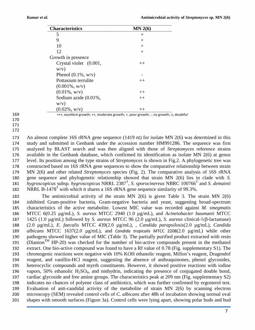
(0.02%, w/v) ++ +++, excellent growth; ++, moderate growth; +, poor growth; -, no growth; ±, doubtful 169 170 171 172
An almost complete 16S rRNA gene sequence (1419 nt) for isolate MN 2(6) was determined in this 173
study and submitted in Genbank under the accession number HM991286. The sequence was first 174 analyzed by BLAST search and was then aligned with those of Streptomyces reference strains 175
available in the Genbank database, which confirmed its identification as isolate MN 2(6) at genus 176
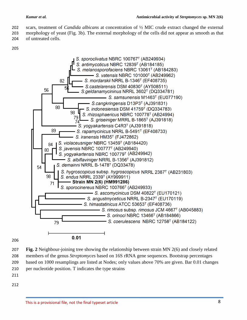
level. Its position among the type strains of Streptomyces is shown in Fig.2. A phylogenetic tree was 177
constructed based on 16S rRNA gene sequences to show the comparative relationship between strain 178 MN 2(6) and other related Streptomyces species (Fig. 2). The comparative analysis of 16S rRNA 179
gene sequence and phylogenetic relationship showed that strain MN 2(6) lies in clade with S. 180 hygroscopicus subsp. hygroscopicus NRRL 2387
T, S. sporocinereus NBRC 100766
T and S. demainii 181
NRRL B-1478T with which it shares a 16S rRNA gene sequence similarity of 99.3%. 182
The antimicrobial activity of the strain MN 2(6) is given Table 3. The strain MN 2(6) 183 inhibited Gram-positive bacteria, Gram-negative bacteria and yeast, suggesting broad-spectrum 184
characteristics of the active metabolite. Lowest MIC value was recorded against M. smegmatis 185 MTCC 6(0.25 µg/mL), S. aureus MTCC 2940 (1.0 µg/mL), and Acinetobacter baumanii MTCC 186 1425 (1.0 µg/mL) followed by S. aureus MTCC 96 (2.0 µg/mL), S. aureus clinical-1(β-lactamase) 187
(2.0 µg/mL), E. faecalis MTCC 439(2.0 µg/mL), , Candida parapsilosis(2.0 µg/mL), Candida 188
albicans MTCC 1637(2.0 µg/mL), and Candida tropicalis MTCC 2208(2.0 µg/mL) while other 189
pathogens showed higher value of MIC (Table 3). The partially purified product extracted with resin 190 (Dianion
TM HP-20) was checked for the number of bio-active compounds present in the methanol 191
extract. One bio-active compound was found to have a Rf value of 0.78 (Fig. supplementary S1). The 192 chromogenic reactions were negative with 10% KOH ethanolic reagent, Millon’s reagent, Dragendof 193 reagent, and vanillin-HCl reagent, suggesting the absence of anthraquinones, phenol glycosides, 194
heterocyclic compounds and myrrh constituents. However, it showed positive reactions with iodine 195 vapors, 50% ethanolic H2SO4, and ninhydrin, indicating the presence of conjugated double bond, 196 cardiac glycoside and free amine groups. The characteristics peak at 209 nm (Fig. supplementary S2) 197 indicates no chances of polyene class of antibiotics, which was further confirmed by ergosterol test. 198
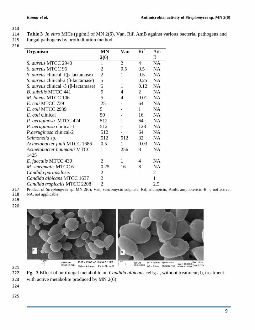
Evaluation of anti-candidal activity of the metabolite of strain MN 2(6) by scanning electron 199 microscopy (SEM) revealed control cells of C. albicans after 48h of incubation showing normal oval 200 shapes with smooth surfaces (Figure 3a). Control cells were lying apart, showing polar buds and bud 201
Kumar et al. Antimicrobial activity of Streptomyces sp. MN 2(6)
8 This is a provisional file, not the final typeset article
scars, treatment of Candida albicans at concentration of ½ MIC crude extract changed the external 202
morphology of yeast (Fig. 3b). The external morphology of the cells did not appear as smooth as that 203
of untreated cells. 204
205
206
Fig. 2 Neighbour-joining tree showing the relationship between strain MN 2(6) and closely related 207
members of the genus Streptomyces based on 16S rRNA gene sequences. Bootstrap percentages 208
based on 1000 resamplings are listed at Nodes; only values above 70% are given. Bar 0.01 changes 209
per nucleotide position. T indicates the type strains 210
211
212
Kumar et al. Antimicrobial activity of Streptomyces sp. MN 2(6)
9
213 Table 3 In vitro MICs (µg/ml) of MN 2(6), Van, Rif, AmB against various bacterial pathogens and 214 fungal pathogens by broth dilution method. 215 216
Organism MN
2(6)
Van Rif Am
B
S. aureus MTCC 2940 1 2 4 NA
S. aureus MTCC 96 2 0.5 0.5 NA
S. aureus clinical-1(β-lactamase) 2 1 0.5 NA
S. aureus clinical-2 (β-lactamase) 5 1 0.25 NA
S. aureus clinical -3 (β-lactamase) 5 1 0.12 NA
B. subtilis MTCC 441 5 4 2 NA
M. luteus MTCC 106 5 4 0.01 NA
E. coli MTCC 739 25 - 64 NA
E. coli MTCC 2939 5 - 1 NA
E. coli clinical 50 - 16 NA
P. aeruginosa MTCC 424 512 - 64 NA
P. aeruginosa clinical-1 512 - 128 NA
P.aeruginosa clinical-2 512 - 64 NA
Salmonella sp. 512 512 32 NA
Acinetobacter junii MTCC 1686 0.5 1 0.03 NA
Acinetobacter baumanii MTCC
1425
1 256 8 NA
E. faecalis MTCC 439 2 1 4 NA
M. smegmatis MTCC 6 0.25 16 8 NA
Candida parapsilosis 2 2
Candida albicans MTCC 1637 2 1
Candida tropicalis MTCC 2208 2 2.5 Product of Streptomyces sp. MN 2(6); Van, vancomycin sulphate; Rif, rifampicin; AmB, amphotericin-B, -, not active; 217 NA, not applicable; 218 219
220
221 Fg. 3 Effect of antifungal metabolite on Candida albicans cells; a, without treatment; b, treatment 222
with active metabolite produced by MN 2(6) 223
224
225
Kumar et al. Antimicrobial activity of Streptomyces sp. MN 2(6)
10 This is a provisional file, not the final typeset article
5 Discussion 226
Microorganisms have served as a source for the majority of the drugs in use today (Demain and 227 Snchez, 2009). Among the different microbes, actinomycetes have been and are the unparalleled 228 source of bioactive metabolites. The metabolites from natural sources continue to play a key role in 229 drug discovery and development by providing natural molecules with pharmaceutical potential and 230
novel scaffolds for synthetic modification (Vincet et al., 2006). In this context, the isolate MN2 (6) of 231 the previous study was taken for further studies. Based on cultural, physiochemical and chemo-232 taxonomical characteristics, it was found to belong the genus Streptomyces. The comparative analysis 233 of 16S rRNA gene sequence and phylogenetic relationship showed that strain MN 2(6) lies in clade 234 with S. hygroscopicus subsp. hygroscopicus NRRL 2387
T, S. sporocinereus NBRC 100766
T and S. 235
demainii NRRL B-1478T with which it shares a 16S rRNA gene sequence similarity of 99.3%. The 236
members of violaceusniger clade shared 16S rRNA gene similarities within the range 99.0– 99.8 %, 237
values that correspond to between 3 and 11 nt differences at 1449 and 1446 locations, respectively. 238
The highest 16S rRNA similarity found between the type strains belonging to this taxon a value 239 equivalent to 7 nt differences at 1447 sites. Extensive DNA–DNA relatedness experiments indicated 240 that members of the S. violaceusniger 16S rRNA gene clade that share similarities at or below 99.7 241 %, that is, with four or more nucleotide differences, can be assigned to different genomic species 242
(Goodfellow et al. 2007). As MN 2(6) shared 99.37% sequence similarity with type strains with nine 243 nucleotide differences, hence may be novel species of Streptomyces belonging to violaceusniger 244
clade. Although, strain MN 2(6) shares high 16S rDNA similarity value with S. demainii NRRL B-245 1478
T, S. endus NRRL 2339
T, S. sporocinereus NBRC 100766
T, and S. hygroscopicus subsp. 246
hugroscopicus NRRL 2387T but differs in various phenotypic characteristics. MN 2(6) produced 247
white to whitish grey aerial and brown reverse mycelium while S. sporocinereus (Gause et al. 1983) 248 produced sparse white aerial mycelium and green beige reverse mycelium. The spore surface of S. 249
sporocinereus was smooth while it was rugose in case of MN 2(6). S. sporocinereus tolerate a salt 250 concentration of 2.5% (w/v) whereas MN 2(6) can tolerate a salt concentration up to 6% (w/v). 251
Xylose, inositol, raffinose and cellobiose are not utilized by of S. sporocinereus while the same was 252 utilized by MN 2(6). Arginine was utilized by the type strains wheres as MN 2(6) does not utilize it. 253 These characteristics differentiate MN 2(6) with type strain of S. sporocinereus. Similarly, MN 2(6) 254
was differentiated from S. demainii (Goodfellow et al., 2007) in a number of characteristics. S. 255 demainii produced grey aerial mycelium and greyish yellow reverse mycelium, reduced nitrate and 256
hydrolysed casein whereas MN 2(6) did not give positive results for nitrate reduction and casein 257 hydrolysis. Arabinose and salicin were utilized by S. demainii whereas as MN 2(6) did not utilize the 258 same. Fructose and L-Histidine were not utilized by S. demainii while the same was utilize by MN 259
2(6). S. demainii grew at pH 4, 5, 9 and 10 only while MN 2(6) grew at a wide range of pH (4-10). 260 MN 2(6) was also differentiated from S. hygroscopicus subsp.hugroscopicus (Goodfellow et al., 261 2007) in cultural characteristics, nitrate reduction and casein hydrolysis properties. The type strain of 262 S. hygroscopicus subsp.hugroscopicus grew only at pH 5 whereas MN 2(6) grew at a pH range of 4 263
to 10. From the above discussion, clearly MN 2(6) is different from the type strains and may be novel 264 species of Streptomyces belonging to violaceusniger clade 265
The result of antimicrobial activity is comparable with the previous study (DeBoer et al., 266 1970; Furumai et al., 2003) and Streptomyces sp. MN2 (6) showed promising activity against Gram-267 positive, Gram-negative bacteria and Candida species. The result of bioautography indicates the 268
presence of only one bioactive compound. The chemical characterization of this active compound 269 suggests the absence of anthraquinones, phenol glycosides, heterocyclic compounds and myrrh 270
constituents. However, it indicates the presence of conjugated double bond, cardiac glycoside and 271 free amine groups (Kumar et al., 2012). The characteristics peak at 209 nm indicates no chances of 272
Kumar et al. Antimicrobial activity of Streptomyces sp. MN 2(6)
11
polyene class of antibiotics (Jain and Jain, 2006). However, the metabolite contained carboxy or 273
peptide moiety in the compound as it showed maximum absorbance between 205-216 (Singh et al., 274 2009). Evaluation of anti-candidal activity of the metabolite of strain MN 2(6) by scanning electron 275 microscopy (SEM) may be helpful to understand the cell damage mechanism. The changes in 276 morphology of yeast cells after treatment with Streptomyces sp. MN 2(6) are also consistent with 277
findings of other researchers (Nurkanto and Julistiono, 2014; Kitajima et al., 1976). The shrinkage of 278 cells was clearly observed in the electron micrograph which may be due to loss of cytosolic volume, 279 which is mainly observed in case of polyene class of antibiotics (Kitajima et al., 1976). However, 280 preliminary characterization of metabolites indicates that it is a non-polyene class of metabolite. 281 These findings support that the stains MN 2(6) possess strong anti-candidal activity and killed 282
pathogenic yeast due to considerable morphological changes. 283
From the results of the present study, it was concluded that the isolate may represent a novel 284
species of Streptomyces belonging to violaceusniger clade and produce metabolite (s) which has a 285
broad spectrum (active against Gram-positive, Gram-negative bacteria, acid-fast bacteria and yeast 286 cells). It kills the Candida cells due to the shrinkage and cytosolic loss. However, further studies are 287
required to elucidate the structure of the active metabolite produced by the isolate MN 2(6). 288
4 Acknowledgement 289
GRSB and VK thank the Uttarakhand State Council of Science and Technology for grant received 290 (UCS &T/ R&D/LS/06-07/1158) to carry out this study and to the Management of S.B.S.P.G.I., 291
Dehradun for providing necessary research facilities and Wadia Institute of Himalayan Geology, 292 Dehradun, Uttarakhand, India for providing SEM facility. 293
294
5 References 295
Altschul, S. F., Madden, T.L., Schaeffer, A.A., Zhang, J., Zhang, Z., Miller and W., Lipman, D.J. 296
(1997). Gapped BLAST and PSI-BLAST: a new generation of protein database search 297 programs. Nucleic Acids Res. 25, 3389-3402. 298
Brady, S.F., Chao, C. J., and Clardy, J. (2002). New natural product families from an environmental 299 DNA (eDNA) gene cluster. J. Am. Chem. Soc. 124, 9968–9969. 300
Clardy, J., Fischbach, M.A., and Walsh, .C.T (2006). New antibiotics from bacterial natural products. 301
Nat. Biotechnol. 24,1541-1550. 302
DeBoer, C., Meulman, P.A., Wnuk, R.J., and Peterson, D.H. (1970). Geldanamycin, a new antibiotic. 303 J. Antibiot. (Tokyo) 23, 442-447 304
Demain, A.L., and Snchez, S. (2009.) Microbial drug discovery: 80 years of progress. J. Antibiot., 305 62, 5-16. 306
Felsenstein, J. (1985). Confidence limits on phylogenies: An approach using the bootstrap. Evolution 307
39, 783-791. 308 Furumai, T., Yamakawa, T., Yoshida, R., and Igarashi,Y. (2003). Clethramycin, a new inhibitor of 309
pollen tube growth with antifungal activity from Streptomyces hygroscopicus TP-A0623. I. 310 Isolation, taxonomy, fermentation, isolation and biological properties. J. Antibiot. 56, 700-704. 311
Gause, G.F., Preobrazhenskaya, T.P., Sveshnikova, M.A., Terekhova, L.P., and Maximova, T.S. 312
(1983). A guide for the determination of actinomycetes. Genera Streptomyces, 313
Streptoverticillium, and Chainia. Nauka, Moscow, USSR. 314
Goodfellow, M., Kumar, Y., Labeda, D.P., and Sembiring, L. (2007). The Streptomyces 315
Kumar et al. Antimicrobial activity of Streptomyces sp. MN 2(6)
12 This is a provisional file, not the final typeset article
violaceusniger clade: a home for streptomycetes with rugose ornamented spores. Antonie Van 316
Leeuwenhoek 92, 173–199. 317 Igarashi, Y. (2004). Screening of novel bioactive compounds from plant-associated actinomycetes. 318
Actinomycetologica 18, 63-66. 319 IMTECH, (1998). Laboratory manual for identification of actinomycetes. Institute of Microbial 320
Technology, Chandigarh, pp 44–46. 321 Jain, P.K., and Jain, P.C. (2006). Isolation characterization and antifungal activity of Streptomyces 322
sampsonii GS 1322. Indian J Exp Biol 45:203–206 323 Jain, P.K., Jain, P.C. (2006). Isolation characterization and antifungal activity of Streptomyces 324
sampsonii GS 1322. Indian. J. Exp Biol. 45, 203–206. 325
Kim, O.S., Cho, Y.J., Lee, K., Yoon, S.H., Kim, M., Na, H., Park, S.C., Jeon, Y.S., Lee, J.H., Yi, H., 326 Won, S., and Chun, J. (2012). Introducing EzTaxon-e: a prokaryotic 16S rRNA Gene sequence 327
database with phylotypes that represent uncultured species. Int. J. Syst. Evol. Microbiol. 62, 328
716–721. 329 Kitajima, Y., Sekiya, T., and Nozawa, Y. (1976). Freeze-fracture ultrastructural alterations induced 330
by filipin, pimaricin, nystatin and amphotericin B in the plasmia membranes of 331 Epidermophyton, Saccharomyces and red complex induced membrane lesions. Biochim. 332
Biophys. Acta. 455(2), 452-465. 333 Kumar, K., Bharti, A., Gupta, V.K., Gusain, O.P., and Bisht, G.S. (2012a). Actinomycetes from 334
solitary wasp mud nest and swallow bird mud nest: isolation and screening for their 335 antibacterial activity. World J. Microbiol. Biotechnol. 28, 871–880. doi: 10.1007/s11274-011-336 0884-2 337
Kumar, V., Bharti, A., Gusain, O., Bisht, G.S. (2011). Scanning electron microscopy of Streptomyces 338 without use of any chemical fixatives. Scanning 33, 1–4. doi:10.1002/sca.20261. 339
Kumar, V., Bharti, A., Gusain, O.P., Bisht, G.S. (2010). An improved method for isolation of 340 genomic DNA from filamentous actinomycetes. J. Sci. Eng..Technol. Manag. 2, 10–13. 341
Kumar, V., Bharti, A., Negi, Y.K., Gusain, O.P., Bisht, G.R.S. (2012b). Taxonomy and antimicrobial 342 activity of a moderately salt tolerant and akaliphilic Streptomyces sp. MN 9(V) isolated from 343 solitary wasp mud nest. Ann. Microbiol. 62, 979-985. doi:10.1007/s13213-011-0337-z. 344
Kumar, V., Gusain, O.P., Thakur, R.L., Bisht, G.S. (2012c). Isolation, purification and partial 345 characterization of an antibacterial agent produced by halotolerant alkaliphilic Streptomyces sp. 346
EWC 7(2). Proc. Natl. Acad. Sci., India, Sect. B Biol. Sci. 83(2), 199–206 DOI 347 10.1007/s40011-012-0117-y. 348
Lemar, K.M., Ourania, P., Aon, M.A., Cortassa, S., Muller, C.T., Plummer, S., et al. (2005) Allyl 349 alcohol and garlic (Allivum sativum) extract produce oxidative stress in Candida species. 350 Microbiology 151, 3257- 3265. 351
Locci, R. (1989). Streptomyces and related genera. In: Williams, ST, Sharp ME, Holt JG (eds) 352 Bergey’s manual of systematic bacteriology vol 4. Williams and Wilkins, Baltimore, pp 2451–353
2506. 354 Mitra, A., Santra, S.C., and Mukherjee, J. (2008). Distribution of actinomycetes, their antagonistic 355
behaviour and the physico chemical characteristics of the world’s largest tidal mangrove forest. 356 Appl. Microbiol. Biotechnol. 80:685–695. 357
Mukku,V.J., Speitling, M., Laatsch, H., and Helmke, E. (2000). New butenolides from two marine 358
streptomycetes. J. Nat. Prod. 63, 1570–1572. 359
Myers, E.W., Miller, and W. (1988). Optimal alignments in linear space. Comput. Appl. Biosci. 4, 360 11-17. 361

Kumar et al. Antimicrobial activity of Streptomyces sp. MN 2(6)
13
NCCLS, (2008). Performance standards for antimicrobial susceptibility testing; ninth informational 362
supplement. NCCLS document M100-S9. Wayne, PA, National Committee for Clinical 363 Laboratory Standard, pp 120–126. 364
Nurkanto, A., Julistiono, H. (2014). Screening and study of antifungal activity of leaf litter 365 actinomycetes isolated from ternate island, Indonesia. Asian Pac. J.Trop. Biomed. , 4(11), 366
886-891. 367 Odakura, Y., Kase, H., Itoh, S., Satoh, S., and Takasawa, S. (1984). Biosynthesis of asatromicin 421 368
and related antibiotics. Biosynthetic studies with blocked mutants of 422 Micromonospora 369 olivasterospora. J. Antibiot. 12, 670–680. 370
Poulsen, M., Oh, D.C., Clardy, J,,Currie. C.R..(2011). Chemical analyses of wasp-associated 371
Streptomyces bacteria reveal a prolific potential for natural products discovery. PLoS One 372 6:e16763. doi: 10.1371/journal.pone.0016763. 373
Promnuan, Y., Kudo, T., and Chantawannakul, P. (2009) Actinomycetes isolated from beehives in 374
Thailand. World J. Microbiol. Biotechnol. 25,1685–1689. 375 Radhakrishnan, M., Suganya, S., Balagurunathan, R., and Kumar, V. (2010). Preliminary screening 376
for antibacterial and antimycobacterial activity of actinomycetes from less explored 377 ecosystems. World J. Microbiol. Biotechnol. 26, 561–566. 378
Saitou, N., Nei, and M. (1987). The neighbor-joining method: A new method for reconstructing 379 phylogenetic trees. Mol. Biol. Evol. 4, 406-425. 380
Shirling, E.B., and Gottlieb, D. (1966) Methods for characterization of Streptomyces species. Int. J. 381 Syst. Bacteriol. 16, 313-340. 382
Shomura, T., Yoshida, J., Amano, S., Kojima, M., Inouye, S., and Niida, T. (1979). Studies on 383
Actinomycetales producing antibiotics only on agar culture. I. Screening, taxonomy and 384 morphology productivity relationship of Streptomyces halstedii, strain SF 1993. J. Antibiot. 32, 385
425–427. 386 Singh,V., Praveen, V.,Banga, J., and Tripathi, C.K.M. Antimicrobial activities of microbial strains 387
from soil of stressed ecological niches of Eastern Uttar Pradesh, India. Indian J. Exp. Biol. 388 74, 298-303. 389
Tamura, K., Dudley, J., Nei, M., and Kumar, S. (2007). MEGA4: Molecular Evolutionary Genetics 390
Analysis (MEGA) software version 4.0. Mol. Biol. Evol. 24,1596-1599. 391 Tamura, K., Nei, M., and Kumar, S. (2004). Prospects for inferring very large phylogenies by using 392
the neighbor-joining method. Proceedings of the National Academy of Sciences (USA) 101, 393 11030-11035. 394
Vincet, P.G., James, M.A., Lam, K.S., Baker, D., and Petersen, F. (2006). Drug discovery from 395
natural products. J. Ind. Microbiol. Biotechnol. 33, 523-531. 396
Williams, S.T., Sharpe, M.E., and Holt, J.G. (1989). Bergey’s manual of systematic bacteriology, vol 397 4. Williams and Wilkins, Baltimore. 398
399 400 401 402 403 404 405 406 407
Kumar et al. Antimicrobial activity of Streptomyces sp. MN 2(6)
14 This is a provisional file, not the final typeset article
6 Figure legends 408
Figure 1 The figure legends are required to have the same font as the main text, 12 point 409 normal Times New Roman, single spaced. Please use a single paragraph for each legend and 410 prepare the figures keeping in mind the PDF layout. The figures are accepted as (A) TIFF/TIF saved 411
using LZW compression or any other non-lossy compression method, or (B) JPEG/JPG format. 412
413
7 Supplementary Material 414
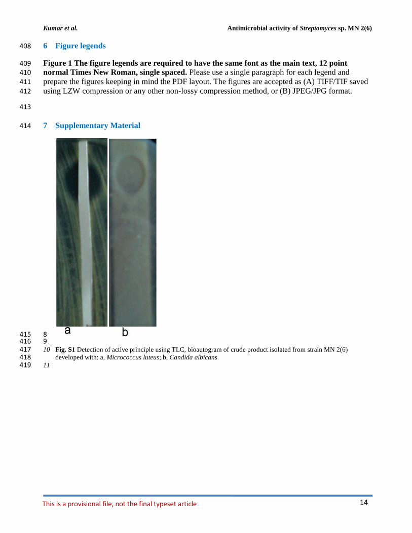
8 415 9 416 10 Fig. S1 Detection of active principle using TLC, bioautogram of crude product isolated from strain MN 2(6) 417
developed with: a, Micrococcus luteus; b, Candida albicans 418 11 419
Kumar et al. Antimicrobial activity of Streptomyces sp. MN 2(6)
15
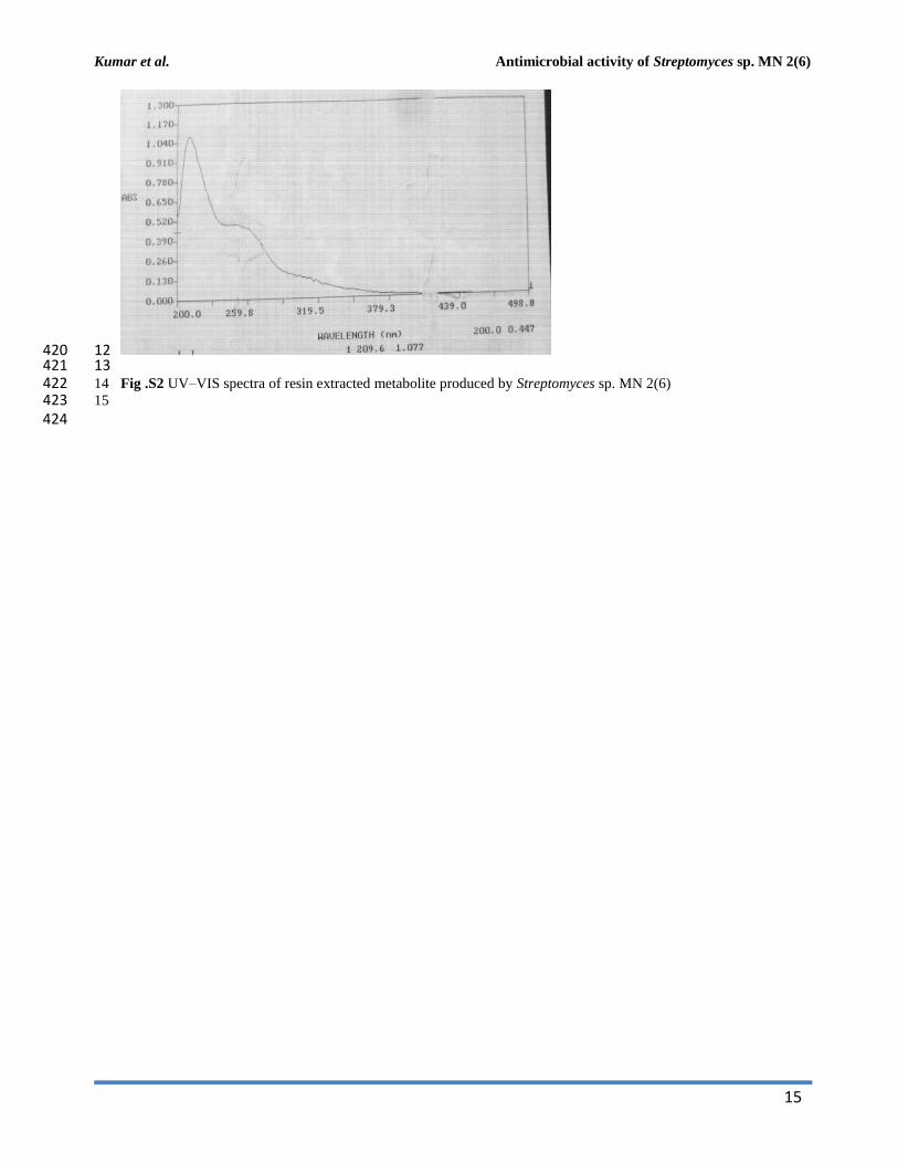
12 420 13 421 14 Fig .S2 UV–VIS spectra of resin extracted metabolite produced by Streptomyces sp. MN 2(6) 422 15 423 424
Copyright © 2022 FDOKUMEN