In A Nutshell: Structure and Assembly of the Vaccinia Virion
94
IN A NUTSHELL: STRUCTURE AND ASSEMBLY OF THE VACCINIA VIRION Richard C. Condit,* Nissin Moussatche,*, † and Paula Traktman ‡ *Department of Molecular Genetics and Microbiology University of Florida, Gainesville, Florida 32610 † Laborato ´rio de Biologia Molecular de Vı ´rus, Instituto de Biofı ´sica Carlos Chagas Filho, CCS, UFRJ, Rio de Janeiro, RJ 21941-590, Brazil ‡ Department of Microbiology and Molecular Genetics Medical College of Wisconsin, Milwaukee, Wisconsin 53226 I. Preface II. Introduction A. Poxvirus Biology and Replication: An Overview B. Genetic Nomenclature C. Poxvirus Genetics: Special Considerations III. Vaccinia Virion Structure A. Imaging Studies of MV Structure B. Chemical Composition of MV C. Controlled Degradation of MV D. Structure of the Genome in the Core E. MV Proteins F. Model for MV Structure IV. Vaccinia Virus Morphogenesis: An Overview V. Formation of Factories VI. Crescent Formation A. One Membrane or More? B. Source of the Virion Membrane C. Regulatory Proteins in Viral Membrane Formation D. Membrane Proteins Essential for Membrane Biogenesis: A14 and A17 E. D13: The Crescent Scaffold Protein VII. Immature Virion Formation: A Complex Process A. Seven-Protein Complex B. A10: p4a/4a VIII. IV!IVN Transition: Genome Encapsidation A. Membrane Proteins B. Core Proteins IX. MV Formation A. Assembly of the Transcription Apparatus Within the Core B. Core Proteins Involved in the IV to MV Transition C. Proteolysis of Virion Protein Precursors; Involvement of Genes I7L and G1L D. Membrane Proteins Involved in the IV to MV Transition E. Restructuring the Particle Surface F. Core Proteins that are not Essential for Virion Morphogenesis but are Required for Transcriptional Competence of the Virion Core 31 Copyright 2006, Elsevier Inc. All rights reserved. 0065-3527/06 $35.00 DOI: 10.1016/S0065-3527(06)66002-8 ADVANCES IN VIRUS RESEARCH, VOL 66
-
Upload
independent -
Category
Documents
-
view
0 -
download
0
Transcript of In A Nutshell: Structure and Assembly of the Vaccinia Virion

ADVANCES IN VIRUS RESEARCH, VOL 66
IN A NUTSHELL: STRUCTURE AND ASSEMBLY OF THEVACCINIA VIRION
Richard C. Condit,* Nissin Moussatche,*, † andPaula Traktman‡
*Department of Molecular Genetics and MicrobiologyUniversity of Florida, Gainesville, Florida 32610
†Laboratorio de Biologia Molecular de Vırus, Instituto de BiofısicaCarlos Chagas Filho, CCS, UFRJ, Rio de Janeiro, RJ 21941-590, Brazil
‡Department of Microbiology and Molecular GeneticsMedical College of Wisconsin, Milwaukee, Wisconsin 53226
I.
P reface II. I ntroductionA.
P oxvirus Biology and Replication: An Overview B. G enetic Nomenclature C. P oxvirus Genetics: Special ConsiderationsIII.
V accinia Virion Structure A. I maging Studies of MV Structure B. C hemical Composition of MV C. C ontrolled Degradation of MV D. S tructure of the Genome in the Core E. M V Proteins F. M odel for MV StructureIV.
V accinia Virus Morphogenesis: An Overview V. F ormation of FactoriesVI.
C rescent Formation A. O ne Membrane or More? B. S ource of the Virion Membrane C. R egulatory Proteins in Viral Membrane Formation D. M embrane Proteins Essential for Membrane Biogenesis: A14 and A17 E. D 13: The Crescent Scaffold ProteinVII.
I mmature Virion Formation: A Complex Process A. S even-Protein Complex B. A 10: p4a/4aVIII.
I V!IVN Transition: Genome Encapsidation A. M embrane Proteins B. C ore ProteinsIX.
M V Formation A. A ssembly of the Transcription Apparatus Within the Core B. C ore Proteins Involved in the IV to MV Transition C. P roteolysis of Virion Protein Precursors; Involvement of Genes I7L and G1L D. M embrane Proteins Involved in the IV to MV Transition E. R estructuring the Particle Surface F. C ore Proteins that are not Essential for Virion Morphogenesisbut are Required for Transcriptional Competence of the Virion Core
31 Copyright 2006, Elsevier Inc.All rights reserved.0065-3527/06 $35.00
DOI: 10.1016/S0065-3527(06)66002-8

32 RICHARD C. CONDIT ET AL.
G.
M embrane Proteins Affecting Virus Binding, Entry, and Fusion X. T ransport, Occlusion, and Secondary Wrapping of MVA.
A 27 B. A 26XI.
V irion Proteins of Unknown Function XII. S ummary and PerspectivesR
eferencesABSTRACT
Poxviruses comprise a large family of viruses characterized by a large,linear dsDNA genome, a cytoplasmic site of replication and a complexvirion morphology. The most notorious member of the poxvirus family isvariola, the causative agent of smallpox. The laboratory prototype virusused for the study of poxviruses is vaccinia, the virus that was used as alive, naturally attenuated vaccine for the eradication of smallpox.
Both the morphogenesis and structure of poxvirus virions are uniqueamong viruses. Poxvirus virions apparently lack any of the symmetryfeatures common to other viruses such as helical or icosahedral capsidsor nucleocapsids. Instead poxvirus virions appear as “brick shaped” or“ovoid” membrane-bound particles with a complex internal structurefeaturing a walled, biconcave core flanked by “lateral bodies.” The virionassembly pathway involves a remarkable fabrication of membrane-containing crescents and immature virions, which evolve into maturevirions in a process that is unparalleled in virology. As a result ofsignificant advances in poxvirus genetics and molecular biology duringthe past 15 years, we can now positively identify over 70 specificgene products contained in poxvirus virions, and we can describe theeffects of mutations in over 50 specific genes on poxvirus assembly. Thisreview summarizes these advances and attempts to assemble them intoa comprehensible and thoughtful picture of poxvirus structure andassembly.
I. PREFACE
Both the morphogenesis and structure of poxvirus virions are uniqueamong viruses. Poxvirus virions apparently lack any of the symmetryfeatures common to other viruses such as helical or icosahedral capsidsor nucleocapsids. Instead poxvirus virions appear as “brick shaped” or

VACCINIA VIRUS: STRUCTURE AND ASSEMBLY 33
“ovoid” membrane-bound particles with a complex internal structurefeaturing awalled, biconcave core flanked by “lateral bodies.” The virionassembly pathway involves a remarkable fabrication of membrane-containing crescents and immature virions, which evolve into maturevirions in a process that is unparalleled in virology.
Poxvirus structure and assembly has been the subject of intensescrutiny since the virus was first subjected to detailed examinationby electron microscopy in 1950s and 1960s, yet the most recent com-prehensive reviews of the subject are at least 15 years old (Dales andPogo, 1981; Fenner et al., 1989; see also Moss, 2001; Sodeik andKrijnse-Locker, 2002). During these 15 years, key advances in poxvir-ology have brought us to a point where a comprehensive review ofvirus structure and assembly is appropriate. First, advances in ge-nome and protein sequencing now readily permit the unambiguousassignment of protein products to specific genes on the poxvirus ge-nome (Chung et al., 2006; Goebel et al., 1990; Jensen et al., 1996;Takahashi et al., 1994). Second, advances in poxvirus genetics haveyielded both well-characterized collections of temperature-sensitivemutants and techniques for targeted construction of conditional lethalmutants with alterations in specific poxvirus genes (Condit and Niles,1990; Hassett and Condit, 1994; Lackner et al., 2003; Rodriguez andSmith, 1990b; Zhang and Moss, 1991a). As a result of these advances,we can now positively identify over 70 specific gene products containedin poxvirus virions, and we can describe the effects of mutations inover 50 specific genes on poxvirus assembly. This review summarizesthese advances and attempts to assemble them into a comprehensibleand thoughtful picture of poxvirus structure and assembly.
II. INTRODUCTION
A. Poxvirus Biology and Replication: An Overview
Poxviruses comprise a large family of viruses that infect a widevariety of vertebrate and invertebrate hosts. Members of the familyPoxviridae are characterized by a large, linear dsDNA genome, acytoplasmic site of replication and a complex virion morphology. Themost notorious member of the poxvirus family is variola, the causativeagent of smallpox. The laboratory prototype virus used for the study ofpoxviruses is vaccinia, the virus that was used as a live, naturallyattenuated vaccine for the eradication of smallpox. Because the vastmajority of genetic and biochemical studies on poxvirus structure and

34 RICHARD C. CONDIT ET AL.
morphogenesis have been performed with vaccinia, this review willconfine itself exclusively to this virus. Members of the poxvirus familyare sufficiently similar such that lessons learned from vaccinia caneasily be applied to other poxviruses.
The vaccinia virus genome is approximately 200 kb in length andencodes approximately 200 genes (there is some strain variation inDNA size and coding capacity) (Goebel et al., 1990; Johnson et al.,1993; Lefkowitz et al., 2005). Because of the cytoplasmic site of virusreplication, vaccinia viral mRNAs are not spliced, and therefore vac-cinia genes do not contain introns, greatly simplifying interpretation ofgenomic sequence. Genes are closely spaced on the genome, and eachgene appears to be controlled by its own transcriptional promoter.Comparison of poxvirus genomes reveals a set of 91 genes that areconserved throughout the Chordopoxvirinae subfamily (vertebratepoxviruses); a subset of 49 of these genes are conserved throughoutthe entire Poxviridae family (including both vertebrate and inver-tebrate poxviruses) (Upton et al., 2003). The remaining nonconservedgenes, many of which are non essential for replication in cellculture, dictate individual virus characteristics of host range andpathogenicity.
Poxvirus virions exist in three infectious forms: mature virions(MV), wrapped virions (WV), and extracellular virions (EV).1, MV,the simplest form of the virus, are membraned particles containing abiconcave, DNA-containing core flanked by lateral bodies, which fillthe concavities of the core. MV are normally found exclusively insidecells and are liberated only by cell lysis. WV consist of MV which aresurrounded by two additional lipid bilayers derived from trans-Golgicisternae. WV, whose outer membranes contain characteristic viralproteins, are precursors of EV and are also found within the cell. EVconsist of WV which have been exocytosed via fusion of the outermostWV membrane with the plasma membrane, leaving an MV wrapped inone additional membrane. A fraction of EV are found attached to thecell surface, while some are found free in the extracellular medium. EVare thought to be important for spread of the virus within an organism,
1 This nomenclature follows a recent proposal by Moss (2005). Previously, MV wascalled intracellular mature virus (IMV), WV was called intracellular enveloped virus(IEV), and EV included both cell associated extracellular enveloped virus (CEV) andextracellular enveloped virus (EEV). In older literature, MV was called “intracellularnaked virus” (INV) (Sodeik et al., 1993), thus MV is identical to both IMV and INV.
VACCINIA VIRUS: STRUCTURE AND ASSEMBLY 35
while MV are thought to be important for long-term stability andtransmission of the virus between hosts in the environment. It shouldbe noted, however, that the EV membrane is disrupted when EVbind to cells, enabling MV to make direct contact with the plasmamembrane and enter cells (Law, M., Carter, G. C., Roberts, K. L.,Hollinshead, M., and Smith, G. L., personal communication). Theextra membrane composition, assembly, and transport of WV and EVhave been reviewed (Smith et al., 2002); therefore, this review willconfine itself exclusively to the structure and assembly of MV.
MV enter cells by fusion of the MV membrane with the plasmamembrane of the host cell, releasing the core (and lateral bodies)into the cytoplasm and activating the virus’ transcriptional program(Armstrong et al., 1973; Carter et al., 2005). The virion cores containthe full complement of virus-coded enzymes required for synthesis andmodification of early mRNA, including a nine subunit RNA polymer-ase, a virus early transcription factor (VETF), a capping enzyme, and apoly(A) polymerase. Early genes encode enzymes required for DNAreplication, and thus as early gene expression peaks, viral DNA repli-cation ensues in cytoplasmic sites termed “factories.” Early genesalso encode intermediate transcription factors, and intermediategenes, in turn, encode late transcription factors, so that intermediateand late genes are expressed in succession after the prerequisite initi-ation of viral DNA replication. Thus, the full complement of viral genesare transcribed in a temporal cascade, with the early, intermediateand late classes being distinguished by class-specific transcriptionalpromoters and virally encoded transcription factors. Furthermore,only replicated genomes are competent templates for intermediateand late transcription. These two classes of genes together encodevirion structural proteins, virion enzymes, and assembly factors re-quired for assembly of new virus particles (for a review of poxvirusreplic ation, see Moss, 2001) .
B. Genetic Nomenclature
The first complete poxvirus genome sequence to be published wasthat of the Copenhagen strain of vaccinia virus (Goebel et al., 1990).With the publication of this sequence, a genetic nomenclature wasadopted in which genes were named according to their position relativeto the left end of individual Hind III restriction fragments of thegenome and also according to their transcriptional orientation. Thus,for example, the virus DNA polymerase is encoded by gene E9L, the

36 RICHARD C. CONDIT ET AL.
ninth gene from the left end of the Hind III E fragment, and the gene istranscribed in a leftward direction relative to the standard orientationof the genome.
While Copenhagen was the first vaccinia strain sequenced, most ofthe genetic and biochemical analysis of vaccinia has been performedusing the WR strain, a strain that is not identical in genetic content toCopenhagen. Initially, the Copenhagen system of nomenclaturewas applied to WR; however, strain variation, sequencing glitches,and the absence of complete genome sequence made this usage occa-sionally confusing. For example, the vaccinia strain WR genes initiallydubbed A5L and A8L are orthologous to the Copenhagen genes A4Land A7L, while further to the right in the Hind III A fragment,WR genes orthologous to Copenhagen genes were given the pre-cise Copenhagen name. Despite this initial confusion, over the yearsthe Copenhagen nomenclature has become the most familiar amongpoxvirologists, and a custom has emerged which is to refer to genes inWR using the orthologous Copenhagen gene identification. According-ly, the WR genes previously called A5L and A8L are now referred to asA4L and A7L.
More recently, faced with an explosion of poxvirus genome sequencesavailable and the impossibility of maintaining the Copenhagen no-menclature across such a diversity of sequences, a new nomenclaturehas emerged in which genes are simply numbered in sequence fromleft to right on the genome, along with an identifier specifying thevirus strain. Thus while the E9L gene in Copenhagen is now officially“VV-Cop-E9L,” in WR it is “VACWR065.”
This review deals almost exclusively with experiments done with theWR strain of vaccinia. For the sake of clarity, we have maintainedthe custom of referring to WR genes using the Hind III designationof the Copenhagen ortholog. Following established convention, genesare referred to using the entire designation, for example, “E9L” for theDNA polymerase gene, while proteins are specified by omitting the “L”or “R,” for example, “E9” for the DNA polymerase enzyme. For the sakeof precision, in Table I we have included a column specifying theofficial WR gene name for each protein.
C. Poxvirus Genetics: Special Considerations
Conditional lethal mutants have been isolated which encode altera-tions in over half of the essential genes in vaccinia virus. These mu-tants are of two different types, inducible and temperature sensitive.The two different types of mutants function by fundamentally different

TABLE IVACCINIA VIRION PROTEINS
Gene* WR designation MW† Location Mutants‡ Comment§
D13L VACWR118 62 Absent ind, ts Rifampicin target, IV scaffold protein
A11R VACWR130 36 Absent ind Hydrophobic, phosphorylated (independentof F10), interacts with A32, itself
A2.5L VACWR121 9 Virion enzyme ind Thiol oxidoreductase
A32L VACWR155 31 Virion ind Putative ATPase, DNA encapsidation
A17L VACWR137 23 Membrane ind TM(2), F10-dependent phosphorylation,cleaved, complex with A14, A27
A14L VACWR133 10 Membrane ind TM(2), F10-dependent phosphorylation,dimerizes, complex with A17, A27
A13L VACWR132 8 Membrane ind, ts TM(1), phosphorylated (independent of F10),DNA encapsidation
A9L VACWR128 12 Membrane ind TM(1)
L1R VACWR088 27 Membrane ind TM(1), myristylated, redox substrate
H3L VACWR101 37 Membrane ind, ko TM(1), heparin binding
A21L VACWR140 14 Membrane ind TM(1), redox substrate, fusion complex
L5R VACWR092 15 Membrane ind TM(1), redox substrate, fusion complex
A28L VACWR151 16 Membrane ind, ts TM(1), redox substrate, fusion complex
H2R VACWR100 22 Membrane ind TM(1), redox substrate, fusion complex
G3L VACWR079 13 Membrane ts TM(1), fusion complex
G9R VACWR087 39 Membrane ind TM(1), myristylated, fusion complex
J5L VACWR097 15 Membrane TM(1), myristylated, fusion complex
A16L VACWR136 43 Membrane ind TM(1), myristylated, fusion complex
I2L VACWR071 8 Membrane ind TM(1)
D8L VACWR113 35 Membrane ko TM(1), chondroitin binding
(continues)
37

A27L VACWR150 13 Membrane ind, ko No TM, complex with A17, A14, heparinbinding
A26L VACWR149 58 Membrane ko p4c, TM(?)
A14.5L VACWR134 6 Membrane ko TM(2)
I5L VACWR074 9 Membrane TM(2)
E10R VACWR066 11 Membraneenzyme
ind Thiol oxidoreductase, no TM
G4L VACWR081 14 Membraneenzyme
ind Thiol oxidoreductase, no TM
H5R VACWR103 22 Core ts F10, B1-dependent phosphorylation, roles inDNA replication, transcription,morphogenesis
G5R VACWR082 50 Core ind
A30L VACWR153 9 Core ind, ts 7 complex, F10-dependentphosphorylation, partially NP40extractable, in viroplasm, IV’s
G7L VACWR085 42 Core ind, ts 7 complex, F10-dependent phosphorylation,cleaved
J1R VACWR093 18 Core ind, ts 7 complex, partially NP40 extractable
A15L VACWR135 11 Core ind 7 complex
D2L VACWR107 17 Core ind, ts 7 complex
D3R VACWR108 28 Core ts 7 complex
A10L VACWR129 102 Core ind, ts p4a/4a, cleaved, complex A4, core wall?
I6L VACWR075 43 Core ts Telomere binding, DNA encapsidation
I1L VACWR070 36 Core ind DNA binding
F17R VACWR056 11 Core ind H1 dependent, partially F10 dependentphosphorylation, DNA binding
TABLE I (continued)
Gene* WR designation MW† Location Mutants‡ Comment§
38

A4L VACWR123 31 Core ind Core wall spike protein(?), membraneassociated(?), F10, H1-dependentphosphorylation, complex with A10
A3L VACWR122 73 Core ts p4b/4b, cleaved, core wall
L4R VACWR091 28 Core ind VP8, cleaved
E8R VACWR064 32 Core ts TM(2), phosphorylated
E11L VACWR067 15 Core ts
L3L VACWR090 41 Core ind
A12L VACWR131 20 Core Cleaved
J6R VACWR098 147 Core enzyme ts RNA polymerase subunit, rpo147
A24R VACWR144 133 Core enzyme ts RNA polymerase subunit, rpo132
H4L VACWR102 94 Core enzyme ind, ts RNA polymerase subunit, rap94, early genespecificity factor, required for earlytranscription termination
A29L VACWR152 35 Core enzyme ts RNA polymerase subunit, rpo35
E4L VACWR060 30 Core enzyme RNA polymerase subunit, rpo30
J4R VACWR096 21 Core enzyme ts RNA polymerase subunit, rpo22
A5R VACWR124 19 Core enzyme RNA polymerase subunit, rpo19
D7R VACWR112 18 Core enzyme ts RNA polymerase subunit, rpo18
G5.5R VACWR083 7 Core enzyme RNA polymerase subunit, rpo7
A7L VACWR126 82 Core enzyme ind Early gene transcription initiationfactor, VETF
D6R VACWR111 74 Core enzyme ind, ts Early gene transcription initiationfactor, VETF
D1R VACWR106 97 Core enzyme ts Capping enzyme, also early genetermination factor, intermediate geneinitiation factor
D12L VACWR117 33 Core enzyme ts Capping enzyme, also early genetermination factor, intermediate geneinitiation factor
(continues)
39

E1L VACWR057 55 Core enzyme Poly(A) polymerase catalytic subunit
J3R VACWR095 39 Core enzyme ko Poly(A) polymerase elongation subunit, also20 0 methyltransferase, transcriptionelongation factor
D11L VACWR116 72 Core enzyme ts Early gene termination factor, NPH I
I8R VACWR077 78 Core enzyme ts RNA helicase
A18R VACWR138 57 Core enzyme ts Postreplicative transcription terminationfactor, RNA release factor
A22R VACWR142 22 Core enzyme ind Holliday resolvase, concatemer resolution,palmitylprotein
H6R VACWR104 37 Core enzyme ko Topoisomerase
K4L VACWR035 49 Core enzyme ko DNA nicking and joining enzyme,nonessential
G1L VACWR078 68 Core enzyme ind Metalloproteinase(?)
I7L VACWR076 49 Core enzyme ind, ts Cysteine proteinase(?), virion proteinprocessing
F10L VACWR049 52 Core enzyme ts, ind Ser/Thr/Tyr kinase, 7 complex
B1R VACWR183 34 Core enzyme ts Ser/Thr kinase, essential for DNAreplication
H1L VACWR099 20 Core enzyme ind Tyr/Ser/Thr phosphatase
O2L VACWR069 12 Core enzyme ko Glutaredoxin, nonessential, not conserved
A45R VACWR171 14 Core enzyme ko Inactive superoxide dismutase, nonessential
* Copenhagen designation.† predicted, apparent MW may differ.‡ ind, inducible; ts, temperature sensitive; ko, knockout.§ TM, transmembrane domain, number of domains in parentheses.
TABLE I (continued)
Gene* WR designation MW† Location Mutants‡ Comment§
40

VACCINIA VIRUS: STRUCTURE AND ASSEMBLY 41
mechanisms; an understanding of these mechanisms is important forproper evaluation of mutant phenotypes.
In inducible mutants, the gene in question is engineered such thatits expression is controlled by the regulatory elements of a bacterialoperon, commonly either lac or tet (Rodriguez and Smith, 1990a;Traktman et al., 2000; Zhang and Moss, 1991a). Thus, in the presenceof inducer, the wild-type gene and protein in question are expressed, apermissive condition exists, and virus can be grown for study. In theabsence of inducer, the gene is not expressed, a nonpermissive condi-tion exists, and the mutant phenotype can be studied. Importantly,under nonpermissive conditions in an inducible mutant, the geneproduct in question is absent or present at only low levels.
In temperature-sensitive mutants, the gene in question contains amissense mutation that renders the protein product either unstable ornonfunctional at high temperature (Condit and Niles, 1990). Thus,virus is grown for study at low temperature, the permissive condition,and the mutant phenotype studied using infections at high tempera-ture, the nonpermissive condition. In mutants where the protein inquestion is unstable at high temperature, the protein is effectivelyabsent from the infection and thus mechanistically the mutant issimilar to an inducible mutant. An important caveat to this statement,however, is that any residual protein is mutant in nature, whereas theresidual protein present in the case of inducible mutants is wild type.For temperature-sensitive mutants in which the target protein is non-functional at the high temperature but nevertheless physically pres-ent, the mechanism of temperature sensitivity is significantly differentthan in an inducible mutant. Most temperature-sensitive mutantsextant have been isolated by classical mutagenesis and random screen-ing methods;2, however, methods exist whereby temperature-sensitivemutants can be engineered into specific genes in a targeted fashion(DeMasi and Traktman, 2000; Grubisha and Traktman, 2003; Hassettand Condit, 1994; Hassett et al., 1997; Ishii and Moss, 2001; Punjabiet al., 2001).
2 Temperature-sensitive mutants are referred to using either a mutant-specific desig-nation or a generic protein-specific designation. The mutant-specific designation consistsof a letter, either C, D, or E, signifying the original mutant collection, (Condit, Dales, orEnsinger) followed by “ts” and a number specific to the mutant, for example, Dts46(Lackner et al., 2003). The generic, protein-specific notation consists of “ts” followed bythe name of the protein, for example, tsA30.

42 RICHARD C. CONDIT ET AL.
Each mutant type has advantages and disadvantages. Advantagesof inducible mutants include the relative ease with which a mutantmay reliably be targeted to a specific gene and the fact that allexperiments can be done at one temperature, thus avoiding thepossibility of temperature-induced abnormalities in the infection.Disadvantages include the inability to use this system effectivelyfor viral genes expressed early in infection and the inability to en-gender a tight phenotype for some late genes, due to some intrinsicleakiness in the system. Advantages of temperature-sensitive mu-tants include the ability sometimes to perform temperature shift-upand shift-down experiments to test for the requirement for a gene atdifferent times during the life cycle and the existence of some mu-tants which synthesize stable but nonfunctional protein at the non-permissive temperature. Disadvantages of temperature-sensitivemutants include the difficulty of isolating mutants in specific genesin a targeted fashion and the necessity of performing experiments athigh temperature, which may induce mutant-independent abnormal-ities in the infection. In the end, the two types of mutants havecomplementary advantages and provide powerful tools for the studyof gene function.
III. VACCINIA VIRION STRUCTURE
A. Imaging Studies of MV Structure
Electron microscopic investigation of vaccinia virus structure hasinvolved analysis of both whole mount preparations of purified virusand thin sections of purified virions or virus-infected cells. Each ap-proach has strengths and limitations that are important to considerin deriving an overall model for the structure of the virus. In overalldimensions, the virion is highly asymmetric, described variously asellipsoidal, brick shaped, or barrel shaped (see the following para-graph). As a result of this asymmetry, in most whole mount pre-parations of virus, the majority of particles appear to be oriented in anonrandom fashion, presenting the broadest surface of the virion per-pendicular to the electron beam, revealing clearly only the two widestdimensions. Whole mount preparations also provide detail relevantto the surface of the particle but usually reveal less informationabout the internal structure. By contrast, thin sections present allpossible orientations and sections of the virion for viewing; however,

VACCINIA VIRUS: STRUCTURE AND ASSEMBLY 43
reconstructing an accurate three-dimensional (3D) view of a relativelycomplex and asymmetric structure can be daunting. While thin sec-tions provide minimal information about the surface of the particle,they reveal significant information about internal structure.
Three studies provide estimates of the external dimensions ofthe virion that are consistent with historical measurements (Dalesand Pogo, 1981; Fenner et al., 1989) and at the same time reflectsome continuing uncertainty regarding the ultimate shape of the viri-on. Measurements of cryo-electron micrographs by Griffiths and co-workers suggest a brick- or pillow-shaped particle, with dimensionsof 310 � 240 � 140 nm (Griffiths et al., 2001b; Roos et al., 1996; Sodeikand Krijnse-Locker, 2002). A study using atomic force microscopysuggests a slightly flattened ellipsoid with dimensions of 350 � 300 �265 nm (Malkin et al., 2003). Lastly, recent cryo-electron tomographyof the virus provides evidence that MV are barrel shaped, with dimen-sions of 360 � 270 � 250 nm (Cyrklaff et al., 2005). Malkin et al. (2003)note that if particles are air dried and examined by atomic forcemicroscopy, they shrink more than twofold along the shortest axisbut only slightly along the longer two axes, yielding final dimens-ions of approximately 330 � 260 � 125 nm, provocatively similar tothe measurements reported by Griffiths and coworkers using cryo-electron microscopy (Griffiths et al., 2001b; Roos et al., 1996). In sum-mary all three of these studies, using different approaches, suggest aflattened ellipsoid or barrel but leave some lingering uncertaintyconcerning the ratio of the length of the shortest axis to the longeraxes.
The surface of purified MV particles, as viewed in whole mountpreparations, assumes two fundamentally different appearances de-pending on the integrity of the particles and the method of prepara-tion. Particles observed using negative staining, metal shadowing,freeze etch or deep etch electron microscopy, or atomic force microscopypossess randomly arranged surface ridges called “surface tubuleelements” (STEs) (Fig. 1A–E) (Dales, 1962; Heuser, 2005; Malkinet al., 2003; Medzon and Bauer, 1970; Noyes, 1962a,b; Stern and Dales,1976; Westwood et al., 1964; Wilton et al., 1995). This form of viruswas named the “M” form by Westwood et al. (1964) for its mulberrylike appearance. Most negatively stained, purified preparations of MValso contain virions which lack obvious STEs but instead possess aclearly delineated boundary surrounding the particle (Fig. 1A). Theselatter forms were dubbed the “C” form by Westwood et al. (1964),because the boundary looks like a capsule surrounding the particle.
44 RICHARD C. CONDIT ET AL.
Visualization of purified MV in whole mount using cryo-electronmicroscopy reveals particles that closely resemble the C form of virus,that is, lacking evidence of STEs and possessing a boundary layeror “surface domain” (Fig. 1F) (Dubochet et al., 1994; Griffiths et al.,2001b). In negatively stained preparations, treatments that arelikely to disturb the integrity of the particle convert the M form ofthe virus to the C form (Dubochet et al., 1994; Westwood et al., 1964).Dubochet et al. (1994) interpreted these results to mean that the STEsare preparation artifacts induced by osmotic stress imposed during
FIG 1. Vaccinia virus surface features. (A) Electron micrograph of purified virus,whole mount, negative stain. “Mulberry” (M) and “capsule” (C) forms are shown(Westwood et al., 1964). (B) Electron micrograph of a purified virion, whole mount,negative stain (Wilton et al., 1995). (C) Freeze etch electron micrograph of a purifiedvirion (Nermut, 1973). (D) Deep etch electron micrograph of a purified virion (Heuser,2005). (E) Atomic force micrograph of a purified virion (Malkin et al., 2003). (F) Cryo-electron micrograph of a whole mount preparation of purified virions (Griffiths et al.,2001b). Arrows denote the limits of the surface domain; arrowhead denotes thepalisade layer. (A) Reprinted from Westwood et al. (1964) with permission. (B) Rep-rinted from Wilton et al. (1995) with permission. (C) Reprinted from Nermut (1973)with permission. (D) Reprinted from Heuser (2005) with permission. (E) Reprintedfrom Malkin et al. (2003) with permission. (F) Reprinted from Griffiths et al. (2001b)with permission.
VACCINIA VIRUS: STRUCTURE AND ASSEMBLY 45
negative staining and that the smooth surfaced C form of virusrevealed by cryo-electron microscopy most accurately represents thesurface of the virus. However, the persistence of STEs in metalshadowing, freeze etch and deep etch electron microscopy, and atomicforce microscopy (Heuser, 2005; Malkin et al., 2003; Medzon andBauer, 1970), plus the biochemical evidence for a structure resem-bling STEs (Wilton et al., 1995) (Sections III.C and IX.E.3) suggeststhat STEs are not artifactual. As originally proposed by Westwoodet al. (1964), the C form of virus present in negatively stained pre-parations most likely results from penetration of stain into damagedvirions, which destroys the contrast that ordinarily highlights STEs.The C form appearance of particles viewed by cryo-electron microsco-py probably results from an insensitivity of cryo-electron microscopyto fine surface detail (Malkin et al., 2003).
Insights into the internal architecture of MV have been gainedprimarily through the use of thin sections, although some informationcan be obtained from whole mount preparations. Thin sections revealtwo distinct boundaries, the membrane,3, which surrounds the entireMV particle, and the core wall, which surrounds an internal core.Cryosections, such as that shown in Fig. 2A, offer the clearest defini-tion of the substructure of the membrane and the core wall, eachof which comprises two layers or domains. Estimates of the thicknessesof each of these domains vary, and the numbers bear significance tothe biochemical makeup of each domain (discussed in Section VI.A.3)(Cyrklaff et al., 2005; Dubochet et al., 1994; Fenner et al., 1989;Hollinshead et al., 1999; Ichihashi et al., 1984). The outer domainof the membrane (outermost dark layer in Fig. 2A) is between 3 and9 nm thick, the inner domain (light layer underneath the outer domainand surrounding the entire particle in Fig. 2A) 5–6 nm thick, andestimates of the thickness of the membrane in aggregate range from10 to 20 nm. The outer core wall (dark layer surrounding the corein Fig. 2A) is clearly thicker than any of the four domains comprising
3 Dales and coworkers proposed the term “envelope” to describe the MV outer bound-ary, based on its lipid content and its function analogous to envelopes of other viruses(Dales and Pogo, 1981). However, the common usage of the term envelope in referring toWV and EV (heretofore called IEV, CEV, and EEV) invites confusion over the origin,structure, and function of the MV outer boundary. We use the term “membrane” todescribe the MV outer boundary; however, it is critical to understand that this two-domain structure is clearly significantly more complex than a simple single lipid mem-brane bilayer. Details of the substructure, lipid and protein composition of the MVmembrane are provided in subsequent sections.

FIG 2. Vaccinia virus internal features. (A) Cryosection (Hollinshead et al., 1999). (B)Whole mount cryo-electron micrograph using uranyl acetate staining (Griffiths et al.,2001b). The authors interpreted images of isolated particles as revealing significantasymmetry in the virion structure, two lipid bilayer membranes, and infoldings of themembranes (Section VI, Fig. 8, Model 4). Hence, the “R” indicates the right hand side ofthe particle. The authors state that “The particle has attached to the grid support in amanner such that a classical side view is evident. The twomembrane layers are indicatedwith large arrows, and the spikes are indicated by an arrowhead. The star indicates theprojection of one of the lateral bodies that we believe is due to the overlapping of theperipheral lobes of the virus (top and bottom). The small arrow indicates an inwardgroove in the membrane.” The image is one of the best available showing the internalstructure of the virion in a whole mount preparation of virus. (C, F, and I) Three differentprojections of a whole mount preparation of virus, phosphotungstic acid stained (Petersand Mueller, 1963). (D, G, and J) Sections of purified virus, uranyl acetate stained, inthree different perpendicular planes (Peters and Mueller, 1963). (E, H, and K) Sections ofMV in three different perpendicular planes (Kato et al., 2004; Condit, unpublished). (E)and (H) are virions in infected cells; (K) was from a preparation of purified virus. (A)Reprinted from Hollinshead et al. (1999) with permission. (B) Reprinted from Griffithset al. (2001b) with permission. (C, D, F, G, I, and J) Reprinted from Peters and Mueller(1963) with permission. (K) Reprinted from Kato et al. (2004) with permission.
46 RICHARD C. CONDIT ET AL.
themembrane and the core wall; estimates of thewidth of the outer corewall range from 8 to 17 nm. The outer core wall has a striated appear-ance and therefore has been called the “palisade layer.” The inner corewall or “smooth layer” (innermost light layer in Fig. 2A), has beenestimated at 5–8 nm in thickness. While biochemical evidence suggests
VACCINIA VIRUS: STRUCTURE AND ASSEMBLY 47
that the layers of the core wall are in fact biochemically distinct, thelayered appearance of the membrane may simply reflect a structuralasymmetry within an otherwise unitary structure. Over much of itssurface, the outer core wall appears to be closely apposed to the innerdomain of the membrane, as is clearly seen at the top and bottom ofthe particle in Fig. 2A. Appropriate sections of the particle revealthe central bilateral concavities in the core, formed by a separationof the core wall from the membrane. Filling these concavities betweenthe core wall and the membrane are two apparently amorphous massescalled “lateral bodies,” as shown clearly in the particles in Fig. 2A andK.
Importantly, different sections through the virion yield very differentimpressions of the relationship between the core and the membrane.Figure 2C–K shows sections in all three possible spatial planes. Sectionsthrough the broadest plane (Fig. 2C–E) closely resemble whole mountcryo-EM images of purified virus or the C form of virus viewed by nega-tive staining. Thus, the capsule of the C form and the surface domain ofthe cryo-EM images most likely represent the four domains of the mem-brane and the core wall, tightly apposed with no separation between thecore wall and the membrane and no evidence of lateral bodies. Whilemost whole mount preparations of virus seem to be nonrandomly orient-ed in this fashion with the broadest plane perpendicular to the electronbeam, particles are occasionally oriented on their sides, revealingthe biconcave shape of the core and hinting at lateral bodies (Fig. 2B)(Griffiths et al., 2001b). Sections through the other two planes perpendic-ular to the broadest plane (Fig. 2F–K) often reveal the lateral bodies butmay not if they are cut close to the ends or the sides of the particle.
The core contains a tube-like structure first detailed by Peters andMueller (1963) (Fig. 2C–K). Images that clearly reveal the length ofthe tube are rare, possibly because this structure is relatively labile(Peters and Mueller, 1963) and also because sections must be cutprecisely parallel to, and must bisect, the broadest plane of the virusin order to clearly visualize the tube. Images of the tube in crosssection are more common, although still relatively rare (Fig. 2F–H)(Heuser, 2005).
B. Chemical Composition of MV
Whole MV particles contain approximately 3.2% (of dry weight)DNA and 5% lipid, the balance of the mass presumably being composedof protein (Zwartouw, 1964). There is general agreement that theparticle does not contain significant amounts of RNA. In addition,the presence of spermine and spermidine in MV has been reported;

48 RICHARD C. CONDIT ET AL.
as described in Section VIII, these polyamines may play a role incondensation of the viral DNA (Lanzer and Holowczak, 1975).
The lipid composition of MV is generally similar to that of the hostcell, with two significant differences. First, the fraction of phosphatidylethanolamine is about one-third less than the host cell and second, MVlipid contains approximately 25% of a biphosphatidic acid analog, acylbis(monoacylglycero)phosphate, three to four times the amount foundin host cells (Hiller et al., 1981; Stern and Dales, 1974). These differ-ences carry implications for the origin of the lipid in MV, discussed in alater section.
C. Controlled Degradation of MV
Additional detail regarding the fine structure ofMVhas been obtainedthrough controlled degradation of the particle, followed by electron mi-croscopic analysis of subparticle fractions. Easterbrook (1966) first de-monstrated that treatment ofMVwith a neutral detergent (NP40) and areducing agent (2-mercaptoethanol or dithiothreitol (DTT)) solubilizesthe virion membrane, leaving behind an intact core with lateral bodiesattached. This observation, coupled with the observation that MV canenter cells by fusion (Armstrong et al., 1973; Carter et al., 2005), demon-strates that the MV membrane contains a lipid bilayer, which probablycorresponds to the innermost of the two visible domains comprising theMV membrane (Section III.A) (Hollinshead et al., 1999). Ichihashi et al.(1984) observed that treatment of MV with SDS alone yielded a “ghost”particle, which remained bounded by a structure that resembles themembrane but is reduced in thickness from 20 to 5–7 nm. This findingimplies that the protein component(s) of the membrane comprise anextensive disulfide bonded network (Section VI.D.2).
Careful titration of NP40 and 2-mercaptoethanol into suspensions ofMVyields cores mixed with an abundance of tubular structures (Fig. 3A)(Wilton et al., 1995). Wilton et al. (1995) equated these structures withSTEs. While the isolated tubules are not identical in dimension andstructure to STEs, the overall similarity in appearance is compelling.After complete solubilization of the MV membrane with saturatingamounts of NP40 and 2-mercaptoethanol, the core, with lateral bodiesattached, can be purified from the solubilized membrane fraction bydifferential centrifugation (Fig. 3B). (It is noteworthy that most investi-gators refer to thisNP40 and 2-mercaptoethanol insoluble fraction as the“core” fraction,when it in fact contains both the virion coreand the lateralbodies.) Isolated coresno longer possess concavities and reveal clearly thedetails of the outer palisade layer of the core wall (Fig. 3C and D)

FIG 3. Vaccinia virion substructures. (A) Negatively stained preparation of MV aftermild treatment with NP40 and 2-mercaptoethanol, so that surface tubule elementsremain intact (Wilton et al., 1995). Bar, 0.2 mm. (B) Isolated core in thin section. Notelateral bodies still attached (Ichihashi et al., 1984). (C) Isolated core visualized by cryo-electron microscopy (Dubochet et al., 1994). (D) Isolated core visualized by negativestaining (Wilton et al., 1995). (A and D) Reprinted from Wilton et al. (1995) withpermission. (B) Reprinted from Ichihashi et al. (1984) with permission. (C) Reprintedfrom Dubochet et al. (1994) with permission.
VACCINIA VIRUS: STRUCTURE AND ASSEMBLY 49
(Dubochet et al., 1994; Easterbrook, 1966; Wilton et al., 1995). Thepalisade is composed of a lattice, sometimes hexagonal, of cylindricalpegs measuring approximately 5 nm in diameter and 10 nm in length(Dubochet et al., 1994; Easterbrook, 1966; Westwood et al., 1964). Thelateral bodies and palisade layer sometimes spontaneously dissociatefrom a fraction of isolated cores, revealing the inner smooth layer andthus substantiating the two-layer character of the corewall. Treatment ofthe NP40, 2-mercaptoethanol insoluble fraction with trypsin activelyremoves the lateral bodies, suggesting that the lateral bodies are protein-aceous in nature (Easterbrook, 1966). Within the core resides the viralDNA genome.
D. Structure of the Genome in the Core
Investigation of the core substructure suggests that the viralgenome exists in the core complexed with viral protein. Rupture ofMV by various methods can release 30–40-nm diameter tubules that

50 RICHARD C. CONDIT ET AL.
possess an apparently helical substructure (Griffiths et al., 2001b;Malkin et al., 2003). The relationship, if any, between these tubulesand the 50-nm diameter tube contained in the core (Peters andMueller, 1963) is unclear. Treatment with protease reduces the30-nm tubes to 16-nm fibers, also helical in substructure, which coatthe viral DNA (Malkin et al., 2003). Holowczak and coworkers, usingvarious treatments with detergents and denaturing agents, isolatedfrom MV DNA-containing substructures of varying complexitythey termed “nucleoids” and “subnucleoids” (Holowczak et al., 1975;Soloski and Holowczak, 1980, 1981; Soloski et al., 1979; reviewed inHolowczak, 1982). Subnucleoids, the simplest of these structures,contained four proteins apparently complexed with supercoiled DNAin 30–60-nm spherical structures interconnected with DNA fibers,roughly reminiscent of a very loose nucleosome structure. In summary,these studies suggest that the vaccinia genome exists within the corecomplexed in an organized fashion with viral proteins. However, thisconclusion is still controversial. As described later in Section VIII.B,repression or inactivation of two different proteins required for genomeencapsidation leads to the production of MV that lack the viral ge-nome. These virions contain what appears to be the full complement ofviral proteins: the absence of the encapsidated genome is not accom-panied by the absence of an abundant DNA-wrapping protein. It maybe that association of the genome with viral proteins to form a nucleo-protein complex occurs after genome encapsidation, within the contextof the nascent virion core.
E. MV Proteins
Historically, the protein composition of MV has been analyzed usingthree fundamentally distinct approaches. First, virion proteins havebeen cataloged by electrophoretic resolution of whole virions or virionsubfractions (Essani and Dales, 1979; Ichihashi et al., 1984; Jensenet al., 1996; Sarov and Joklik, 1972; Takahashi et al., 1994; Wiltonet al., 1995). Second, numerous proteins have been purified from MVby virtue of their enzymatic activities and subsequently characterized(Martin et al., 1975; Morgan et al., 1984; Niles et al., 1989). Finally,antibodies have been generated for individual gene products in a tar-geted fashion, allowing proteins to be subsequently localized to virionsby immunoblot analysis (da Fonseca et al., 2000a, 2004). Early attemptsat cataloging the protein content of virions by gel electrophoresis reliedon apparent molecular weights as a means of identifying proteins.Because of inconsistencies in molecular weight determinations, these

VACCINIA VIRUS: STRUCTURE AND ASSEMBLY 51
studies are often difficult to compare with each other, and an unambig-uous assignment of a specific protein from the older literature to adiscrete vaccinia gene is prone to error. More recently, the combinationof genetics and protein sequencing techniques has made possible theunambiguous correlation of genes with virion proteins. We have con-fined ourselves in this review exclusively to those virion proteins forwhich a positive gene identification has been made.
A complete list of the virion proteins for which a specific gene hasbeen identified is given in Table I. These virion proteins can be sortedinto four groups based on whether or not they are solubilized by NP40and a reducing agent and whether or not they possess enzymaticactivity: (1) proteins that are solubilized by NP40 and a reducing agentbut have not been assigned an enzymatic activity are presumed to bemembrane structural proteins; (2) proteins that are solubilized byNP40 and a reducing agent and also possess an enzymatic activityare classified as membrane enzymes; (3) proteins that are not solubi-lized by NP40 and a reducing agent and have not been assigned anenzymatic activity are presumed to be core structural proteins; and (4)proteins that are not solubilized by NP40 and a reducing agent andalso possess an enzymatic activity are classified as core enzymes. Forcompleteness, Table I also includes two proteins (D13 and A11) thataffect virion morphogenesis but are not encapsidated and two proteins(A32 and A2.5) whose subvirion localization remains to be determined.The list given in Table I is undoubtedly incomplete but is probablyapproaching saturation. Indeed, a comprehensive catalog of specificvaccinia gene products contained in MV has been compiled based ongel-free liquid chromatography and tandem mass spectroscopy ofwhole virions (Chung et al., 2006). These new data correlate remark-ably well with the data in Table I and identify an additional 10 proteins(C6, F8, F9, E6, I3, A6, A25, A31, A42, and A46), whose virion locali-zation is currently either disputed or unconfirmed by other techniques.Because of the possibility of nonspecific association of proteins withvirions (Franke and Hruby, 1987), we have not included in Table Ithese 10 new candidate virion proteins and have instead focused at-tention on proteins whose presence in the virion has been confirmedby additional experiments. A few general features of the catalog ofvirion proteins are highlighted here, and otherwise the details of thecontributions of individual proteins to structure and morphogenesis ofthe virions are detailed in the remainder of the review.
To date, a total of 22 proteins have been localized to the virion mem-brane. Of these, 2 are enzymes associated with a virus coded redoxpathway responsible for maintenance of some of the disulfide bonds in
52 RICHARD C. CONDIT ET AL.
the membrane, while the remaining 20 proteins can be thought of asstructural proteins. Eighteen of the 20 structural proteins are integralmembrane proteins containing at least one transmembrane domain.Interestingly, none of these proteins are glycosylated in the MV mem-brane, reflecting the unusual origin and structure of the membrane.
To date, 47 proteins have been localized to the virion “core,” that is,the NP40, reducing agent insoluble fraction, which includes both thecore and the lateral bodies. Of these 47 proteins, 19 have no knownenzymatic function and are presumed to be structural proteins. Six-teen of the enzyme proteins have well-characterized roles in early viralmRNA synthesis, including initiation, elongation and termination oftranscription, mRNA capping and polyadenylation. The remainingenzymes play less clearly defined roles in the viral life cycle but havein some cases been shown to interact with and/or modify both proteinsand nucleic acids. It is important to note that virion enzymes may havestructural as well as enzymatic roles in the virion. While attemptshave been made to sublocalize proteins to core nucleoprotein, innercore wall, outer core wall, and lateral bodies (Ichihashi et al., 1984;Sarov and Joklik, 1972), these studies date from the pregenomic eraand few if any unambiguous assignments of core proteins to coresubstructures have been made.
Finally, several cellular proteins have been found associated withpurified particles (Castro et al., 2003; Chung et al., 2006; Jensen et al.,1996; Webb et al., 1999). It is possible that some of these proteinsadhere to the MV during purification or are incorporated nonspecifi-cally during virus maturation (Franke and Hruby, 1987). However,cyclophilin A has been clearly demonstrated to be packaged into viruscores (Castro et al., 2003). The role of this protein in virus formation isnot known, although it could participate in the trafficking of virusproteins to the factories or in the proper folding of viral proteins duringthe maturation process.
F. Model for MV Structure
Based on the evidence presented previously, we have constructed aschematic 3Dmodel of the vaccinia virion. The model is not intended tobe definitive in its details, especially given the continuing uncertaintyregarding dimensions of the virion and its substructures. Neverthe-less, we feel that such a schematic will clarify some of the microscopicimages available and the interrelationships of the virion components.For external dimensions we have used the measurements determinedby Cyrklaff et al. (2005) using cryo-electron tomography, and for

VACCINIA VIRUS: STRUCTURE AND ASSEMBLY 53
internal dimensions we have used the measurements determined byHollinshead et al. (1999) from cryosections (Fig. 2A). Thus the virusappears as a slightly flattened barrel (Fig. 4A) with overall dimensionsof approximately 360� 270� 250 nm. The particle is encased in an outermembrane, which itself consists of two component domains. The outer-most membrane domain is 9 nm thick, and the innermost membranedomain is 5 nm thick (Fig. 4C–F). The membrane clearly contains lipid;however, the details of the lipid bilayer content and structure of MVarethe subject of some debate and will be therefore be discussed at lengthlater in the review (Section VI.A). Within the membrane is the core,which is also barrel shaped but contains two indentations, one on each
FIG 4. A model for vaccinia virion structure. (A) The intact MV. No attempthas been made to represent surface tubule elements. (B) The virion core. (C) Cutawayview. The membrane has been removed from the upper half of the virion, the near endhas been removed, and the core wall has been rendered transparent, thus revealingthe multiple layers, the concavities in the core, the lateral bodies, and the tubularinternal structure. (D and E) Sections through the virion in three different perpendi-cular planes. The sections correspond to sections shown in Fig. 2. See text fordetails. (Model courtesy of Michel Moussatche. A dynamic 3D model is available onlineat http://www.vacciniamodel.com/.)

54 RICHARD C. CONDIT ET AL.
of the largest surfaces (Fig. 4B). The core is defined by a core wall, whichis also composed of two layers, an outer, thicker, striated “palisade” layer17 nm thick, and an inner smooth layer 8 nm thick (Fig. 4C–F). Fillingthe spaces between the core wall and the membrane that are createdby the indentations in the core are “lateral bodies.” Within the core is atube-like structure, 50 nm in diameter with a 10 nm diameter core. Thistube is folded upon itself into three continuous segments, with a totallength of approximately 250 nm. Importantly, sections through the cen-ter of the particle in each of the three possible spatial planes yield threesignificantly different impressions of the overall shape and internalstructure of the particle (Fig. 4D–F). For comparison, the electron mi-crographic images in Fig. 2C–E correspond to the model section shownin Fig. 4D; Fig. 2F–H correspond to Fig. 4E; Fig. 2I–K corresponds toFig. 4F. A dynamic 3D rendering of the model can be found online atht tp :/ /w ww.v ac ci ni am od el .c om /.
IV. VACCINIA VIRUS MORPHOGENESIS: AN OVERVIEW
The balance of this review focuses on the details of the assembly ofvaccinia, with particular emphasis on lessons learned from the studyof mutants affected in individual virus genes that influence virusmorphogenesis. To establish a context for this discussion, we offer herea bird’s-eye view of the entire assembly process.
The temporal sequence of events comprising vaccinia assembly wasfirst deciphered experimentally by electron microscopic examination ofcultured cells synchronously infected with virus (Dales, 1963; Dalesand Siminovitch, 1961). At very early times following uptake of virusand dissolution of the core particle, infection-specific cytoplasmic do-mains are observed that are uniform in density, contain few if anycellular organelles, and are sometimes surrounded by ER derivedcisternae (Fig. 5A and B) (Tolonen et al., 2001). These domains repre-sent sites of viral DNA replication, sometimes called “factories,” “viralfactories,” or “DNA factories,” and they increase in size with time. Theearliest evidence of virus assembly is the appearance within factoriesof rigid crescent-shaped structures, cupules in three dimensions,10–15 nm in thickness (Figs. 5C and 6). In most electron micrographscrescents comprise two distinct layers, an inner, smooth layer that athigh resolution in fixed embedded sections has the trilamellar appear-ance of a lipid bilayer and an outer layer composed of regularly spacedprojections termed “spicules” (Dales and Mosbach, 1968; Risco et al.,2002). It is universally accepted that crescents contain at least one

FIG 5. Vaccinia factories. (A) An early factory, 4 hpi. The factory is represented by thecentral cleared area surrounded by cytoplasmic organelles. Bar, 1 mm. (Kato and Condit,unpublished) (B)A factory2 h, 45minpi (Tolonen et al., 2001). The star indicates the factory.G, Golgi stack. M, mitochondria. Bar, 0.2 mm. (C) A factory late during infection (Rodriguezet al., 1998). N, nucleus. IV, immature virions. Arrows indicate IVN. Arrowheads indicateMV.Note that theMVare removed from the factory. (B) Reprinted fromTolonen et al. (2001)with permission. (C) Reprinted from Rodriguez et al. (1998) with permission.
VACCINIA VIRUS: STRUCTURE AND ASSEMBLY 55
lipid bilayer, although the precise membrane makeup of crescentshas been the subject of some debate, discussed in more detail inSection VI.A. Crescents apparently grow in length while maintainingthe same curvature until they become closed circles, spheres in threedimensions, called immature virions (IV) (Figs. 5C and 6). IV areapproximately 350 nm in diameter and are filled with “viroplasm,”material that is uniform in density but discernibly more electron densethan the surrounding factory. Viroplasm appears not only in IV but italso often appears to fill the concavities defined by crescents. Viroplasmis also often present as relatively large sub domains, “virosomes,” withinthe surrounding factory, sometimes unbounded, sometimes surrounded

FIG 6. Vacciniamorphogenesis intermediates. (1) Crescents. (2) IV. (3) IVN. (4) Structuresintermediate between IVand MV. (5) MV. Reprinted from Moss (2001) with permission.
56 RICHARD C. CONDIT ET AL.
by multiple crescents that appear to “bite off” portions of viroplasm.Appearing at approximately the same time as IV are IV which containan electron dense, round or ovoid subdomain called a “nucleoid” (Figs. 5Cand 6).We refer to IVs that contain nucleoids as “IVN.”Nucleoids containDNA (Dales, 1963; Ericsson et al., 1995). Nucleoid material is frequentlyobserved spanning a small gap in a nearly complete IV, so that it liespartially inside and partially outside of the particle (Morgan, 1976a).Under some conditions, larger inclusions of DNA-containing nucleoidmaterial are found within factories, and may appear as either large,spherical, granular inclusions or alternatively paracrystalline arrays.Importantly, serial sections reveal that most if not all IVs containnucleoids, so that in fact in a normal infection, there may be no realdistinction between IVs and IVNs, rather, an IV is merely an IVN thathas been sectioned through a plane that does not include the nucleoid(Morgan et al., 1955). Following the appearance of IVNs, MV appear(Figs. 5C and 6). Proteolytic cleavage of several virion protein precursorsto amature form accompanies, and in fact is required for, morphogenesisfrom IVN to MV (Ansarah-Sobrinho and Moss, 2004b; Katz and Moss,1970b). The majority of MVare found outside factories and may exist inclusters either at the periphery of a factory or apparently separated by a

VACCINIA VIRUS: STRUCTURE AND ASSEMBLY 57
significant distance from the nearest factory. Particles which have astructure that appears to be intermediate between IVN and MV havebeen described, but they are rare, suggesting that the transition from IVto MV is a rapid and concerted process (Fig. 6).
For the purposes of this review, we have artificially divided vacciniamorphogenesis into stages, including formation of factories, crescents,IV, IVN, and MV. A summary of the entire process is shown in Fig. 7. Wenow consider the details of each of these stages separately. Critical to theunderstanding of the assembly process is the phenotypic analysis ofinfections performed with conditional lethal mutants; a complete listof the currently available mutants affected in genes that encode proteinsrequired for virion morphogenesis or infectivity is shown in Table II.
FIG 7. Vaccinia morphogenesis summary. Following the arrows in sequence from theupper left corner: Several integral viral membrane proteins (black Xs) are made in theER (red lines) and transported to viral factories (gray area) along with ER derived lipid tobe assembled into crescents which contain a lipid bilayer (red line) and the membraneproteins, scaffolded on a honeycomb structure composed of the D13 protein (yellowcircles). Crescent formation is controlled by phosphorylation. The crescents mature toIV and IVN, accompanied by encapsidation of the genome. The dotted line surroundingIV, IVN, and encapsidation signifies that the order of these events is uncertain. Meta-morphosis to MV is accompanied by loss of the D13 scaffold, proteolysis, further additionof membrane proteins (blue line) and movement of particles outside of factories. MVacquire Golgi derived membranes (green lines) to become WV and are exocytosedthrough the plasma membrane (black line) to become EV.

TABLE IIVACCINIA MORPHOGENESIS GENETICS
Phenotype*
Notes on phenotypeGene Identity Location Mutants Cres IV MV EV Telo Cleave DNA
J4R rpo22 Core ts � � � Factories only
J6R rpo147 Core ts � � � Factories only
F10L Kinase,7 complex
Core ts, ind � � � � � Factories only
H5R Core ts � � � � þ � Curdled virosomes or DNA
G5R Core ts � � � � þ � Curdled virosomes
A11R Absent ind � � � � Some normal and aberrantvirosomes
A17L A17-A14-A27complex
Membrane ind � � � � � Virosomes, vesicles
A14L A17-A14-A27complex
Membrane ind, ts � � � � � Virosomes, vesicles
D13L IV scaffoldprotein
Absent ind, ts � � � � � Aberrant crescents, rifresistance locus
A30L 7 complex Membrane/core
ind, ts þ � � � � Empty IV
G7L 7 complex Core ind, ts þ � � � Virosomes þ crescents (ts), orempty IV (ind)
J1R 7 complex Membrane/core
ind, ts þ � � � þ � Aberrant, partial, empty IV
A15L 7 complex Core ind þ � � � � Empty IV
D2L 7 complex Core ind, ts þ � � � � Empty IV
D3R 7 complex Core ts þ � � � � Empty IV
58

A10L p4a Core ind, ts þ � � � � Aberrant, empty IV’s, DNAnucleoid aggregates
A13L Membrane ind, ts þ þ � þ � No IVN, membraned DNAcrystalloids
A32L NTP motif Virion ind þ þ � þ þ � � Dense spherical particleslacking DNA
I6L Telomerebinding
Core ts þ þ � � þ � � Dense spherical particleslacking DNA
A22R Hollidayresolvase
Core ind þ þ � � � � Dense spherical particles
A7L VETF Core ind þ þ � � þ IVN, dense spherical particles,viroplasm
D6R VETF Core ind, ts þ þ � � IVN, dense spherical particles,viroplasm
I1L DNA bindingprotein
Core ind þ þ � � þ � IVN, DNA crystalliods
F17R 11k Core ind þ þ � � þ � Some aberrant, unstable IV
A4L Membrane/core
ind þ þ � � þ � þ IVN
A3L p4b Core ts þ þ � þ þ þ þ Aberrant MV
G1L Metallo-proteinase(?)
Core ind þ þ � þ þ þ Aberrant MV
I7L Cysteineproteinase(?)
Core ind, ts þ þ � þ � þ Aberrant MV
A9L Membrane ind þ þ � � � Some aberrant IV
L1R Myristylatedprotein
Membrane ind þ þ � � � Unstable IV, IVN
H3L Heparinbinding
Membrane ind, ko þ þ � � � Some MV, defective, viroplasm
(continues)
59

E10R Thioloxidoreductase
Membrane ind þ þ � � �
G4L Thioloxidoreductase
Membrane ind þ þ � � �
A2.5L Thioloxidoreductase
Virion ind þ þ � �
H4L rap94 Core ind, ts þ þ þ Noninfectious MV, transcriptionenzymes missing
I8R RNA helicase Core ts þ þ þ þ Transcription defective MV
H6R Topoisomerase Core ko þ þ þ þ MV show reduced infectivity,reduced early txn
L4R VP8 Core ind þ þ þ þ þ þ Noninfectious, unstable,transcription defective MV
H1L Phosphatase Core ind þ þ þ Noninfectious,transcription defective MV
E8R Core ts þ þ þ þ Noninfectious,transcription defective MV
L3L Core ind þ þ þ þ þ þ Noninfectious,transcription defective MV
L5R Membrane ind þ þ þ þ Noninfectious MV, fusion/entrydefective
TABLE II (continued)
Phenotype*
Notes on phenotypeGene Identity Location Mutants Cres IV MV EV Telo Cleave DNA
60

A21L Membrane ind þ þ þ þ þ Noninfectious MV, fusion/entrydefective
A28L Membrane ind, ts þ þ þ þ Noninfectious MV, fusion/entrydefective
H2R Membrane ind þ þ þ þ Noninfectious MV, fusion/entrydefective
A16L Membrane ind þ þ þ þ Noninfectious MV, fusion/entrydefective
G3L Membrane ts þ þ þ þ Noninfectious MV
I2L Membrane ind þ þ þ þ Noninfectious MV, fusion/entrydefective
D8L Membrane ko þ þ þ Reduced infectivity MV, cellbinding
A27L A17-A14-A27complex
Membrane ind, ko þ þ þ - Infectious MV, defective inEV formation
A26L p4c Membrane ko þ þ þ þ Infectious MV; empty ATI
A14.5L Membrane ko þ þ þ Infectious MV, attenuated inanimals
E11L Core ts þ þ þA45R Inactive SOD Core ko þ þ þ þ Nonessential
K4L Nicking/joiningenzyme
Core ko þ þ þ þ Nonessential
* Mutant designations: ind, inducible; ts, temperature sensitive; ko, knockout; Cres, crescents; Telo, telomere resolution; Cleave,virion protein proteolyzed; DNA, purified particles contain DNA; þ, normal structure formed; �, normal structure not formed; blank, notreported.
61

62 RICHARD C. CONDIT ET AL.
V. FORMATION OF FACTORIES
Early studies by Cairns (1960) demonstrated that the number of fac-tories observed early during vaccinia infection was proportional to themultiplicity of infection and concluded that each factory observed arosefrom a single infecting particle. Factories represent sites of viral DNAreplication: factories become labeledwithDNAprecursors as assessed byEM autoradiography, factory formation is absent or severely restrictedfollowing infection withmutants defective inDNA replication, and infec-tion with mutants that are DNA replication competent but defective inlate viral protein synthesis results in formation of large factories that arelacking in crescents or any other evidence of virus morphogenesis(Cairns, 1960; Hooda-Dhingra et al., 1989).4, Most reports describedearly factories as unbounded in the cytoplasm (Fig. 5A) (Dales andKajioka, 1964); however, recent reports suggest that at early times,before the appearance of any specific viral structures, factories are tran-siently surrounded by cellular ER-derivedmembrane cisternae (Fig. 5B)(Doglio et al., 2002; Tolonen et al., 2001; Traktman, unpublished).
VI. CRESCENT FORMATION
A. One Membrane or More?
The earliest detailed descriptions of the substructure of vacciniaviral crescents concluded that they consisted of a single lipid bilayercoated on its outer surface with a layer of spicules, which conferred therigid arched shape to the crescents. Furthermore, these studies con-cluded that the lipid bilayer of the crescent formed within factorieswithout any direct connection to other existing cellular membranes(Fig. 8, Model 1) (Dales and Mosbach, 1968). This de novo formationmodel, although unique in biology, nevertheless stood as the acceptedmodel until it was challenged in 1993 by Griffiths and coworkers(Sodeik et al., 1993). Central to this challenge was the commonly held
4 A significant body of literature has made use of inhibitors of DNA replication tostudy morphogenesis. For example, hydroxyurea has been used to segregate early andlate stages of morphogenesis (Morgan, 1976a; Pogo and Dales, 1971). It is now clear thatin the absence of DNA replication, factory formation and therefore morphogenesis isinhibited. It seems probable that in reports where factory formation and some morpho-genesis were observed in the presence of DNA replication inhibitors, the inhibitors wereused under conditions that were not completely inhibitory, and thus some DNA replica-tion and late viral protein synthesis occurred. These studies should be interpreted withthis caveat in mind.

FIG 8. Models for membrane processing during vaccinia morphogenesis. In each case, asingle solid red line represents a single lipid bilayer. DNA/nucleoid material is representedin gray; lateral bodies are represented in blue. Fourmodels are shown in four columns.Eachrow represents and compares a discrete state ofmorphogenesis, with the top row represent-ing crescents, the middle row representing immature virions with nucleoids (IVN) and thebottom row representingmature virions (MV).Model 1: Crescents and IVN contain a singlelipid bilayer, which becomes a single lipid bilayer of the structure surrounding MV (DalesandMosbach, 1968;Hollinshead et al., 1999). In this case the corewall does not contain lipidand is therefore represented in black. Model 2: Crescents are derived from cisternae andcomprise two tightly apposed lipid bilayers. The ends of cisternae fuse or are sealed to formIVN containing two tightly apposed bilayers. During MV formation from IVN, the innerbilayer separates from the outer bilayer so that the inner bilayer becomes a component ofthe core wall while the outer bilayer remains as a component of theMVmembrane (Sodeiket al., 1993). Model 3: Crescent and IVN formation is similar to Model 2; however, thecisternae do not fuse but are “sealed.” During morphogenesis to MV the two bilayers donot separate, and thus theMVmembrane contains two tightly apposed bilayers (Roos et al.,1996). As in Model 1, the core wall does not contain lipid and is therefore represented inblack. Model 4: Crescents are derived from cisternae as in Models 2 and 3; emphasis isplaced on the continuity of the rigid crescent domain of this cisternae with extensive flaccidcisternal domains. DNA in a prenucleoid structure is associated with the flaccid cisternaldomain. IVN is formed by the infolding of the flaccid domain within the rigid crescentdomain. The cisterna of IVNare sealed so that the resultingMVcontains four lipid bilayers,two forming the virion envelope and two forming the corewall (Griffiths et al., 2001a; Sodeikand Krijnse-Locker, 2002).
VACCINIA VIRUS: STRUCTURE AND ASSEMBLY 63
view that all cellular membranes are derived from preexisting orga-nelles (Palade, 1983). Sodeik et al. (1993) proposed that crescents infact were derived from preexisting cisternae of the cellular secretorymachinery, which had collapsed on themselves through the action of

64 RICHARD C. CONDIT ET AL.
virus proteins. Crescents formed in this manner would consist of twotightly apposed bilayers rather than a single bilayer (Fig. 8, Model 2).(Crescents would therefore be analogous to the membranes involved inthe development of spherical autophagosomes, which are delimited bya double bilayer [Reggiori and Klionsky, 2005].) Investigations into theconsequences of this cisternae-based model for crescent formationultimately resulted in three different new models for IV and MVformation, shown in Fig. 8, Models 2–4. In the balance of this section,we describe and evaluate these models.
Model 1. Single membrane bilayer (Dales and Mosbach, 1968). Asdescribed previously, this model states that crescents arise de novo,that is, without apparent continuity with other preexisting membraneorganelles, and that crescents contain a single lipid bilayer coated onits outer surface with a layer of spicules. During morphogenesis fromIVN to MV, the spicules are lost, the surface undergoes some additionalmodifications, and the contents of IVN are reorganized to yield the coreand lateral bodies (Essani et al., 1982; Stern and Dales, 1976). In thismodel, the core is not surrounded by a lipid membrane (Wilton et al.,1995). The primary objections to this model are that it seems to violatea principle which dictates that all membranes in the cell are derivedfrom preexisting organelles and that at the edges of a crescent, thehydrophobic core of the bilayer is left exposed to the cytoplasm, atheoretically unstable condition. The model is supported by the factthat it is consistent with some existing microscopic data and that ityields a single membraned MV that can enter cells by a simple fusionevent at the plasma membrane.
Model 2. Double membrane bilayer; membrane bound core (Sodeiket al., 1993). As described previously, this model states that crescentsactually contain two tightly apposed lipid bilayers, formed by collapseof preexisting cellular membrane cisternae, modified by virus proteins.The model states further that during morphogenesis from IVN to MV,the two membranes separate and the innermost membrane collapsesaround the core, so that both the MV surface and the core are each nowbounded by a single lipid bilayer. The primary theoretical objections tothis model are that it introduces a problem of how the material makingup the lateral bodies is transported across the innermost membraneduring morphogenesis from IV to MV and that it would result indelivery of a membrane-bound core to the cytoplasm if MV were toenter cells by a simple fusion event at the plasma membrane. Themodel is supported by numerous electron micrographs which seem toshow continuity between crescents and cisternae (Ericsson et al., 1997;Sodeik et al., 1993, 1994), and by electron microscopic images of MV

VACCINIA VIRUS: STRUCTURE AND ASSEMBLY 65
which seem to show a membrane surrounding the core (Cyrklaff et al.,2005; Sodeik et al., 1993).
Model 3. Double membrane bilayer; naked core (Roos et al., 1996).This model also proposes that crescents are composed of two tightlyapposed lipid bilayers; however, it differs from Model 2 with respect tothe fate of the innermost bilayer during the morphogenesis from IV toMV. Specifically, Model 3 states that the two bilayers do not separateduring MV morphogenesis, so that the resulting MV contains twotightly apposed bilayers on its surface, and the core is not bound by amembrane. The model also states that the membranes surroundingthe particle are not fused but rather joined in an overlap by a protein-aceous plug. Relative to Model 2, this model resolves the theoreticalproblem of transport of material across the innermost membrane dur-ing formation of lateral bodies but complicates further the problemof virus entry; in this case, a simple fusion event between the virionand the plasma membrane would leave the entire contents of theparticle outside of the cell. In support of this model are the samemicrographs that seem to show continuity between crescents andcisternae (Ericsson et al., 1997; Sodeik et al., 1993, 1994), coupled withexperiments that indicate that core surface antigens are exposed whenMV are disrupted with a reducing agent (Roos et al., 1996).
Mode l 4. Fol ded cistern ae ( Griffiths et al ., 2001a ; Rod riguez et al .,2006). In this model colla psed cist ernae evo lve dist inct viru s-specifi cdomains, including a rigid crescent domain that is continuous with aDNA binding domain. IVandMVare formed by folding of these domainsupon themselves, resulting in a particle which now contains, in effect,four lipid bilayers, two on the surface of MV and two surrounding thecore. Like Model 3, Model 4 proposes that the membranes are not fusedbut rather joined in an overlap by a proteinaceous plug. Like Model 3,this model resolves the theoretical problem of transport of materialacross the innermost membrane during formation of lateral bodies,but leaves the same problem of virus entry described previously forModel 3. The model is supported by electron micrographs whichseem to show the continuity of the cisternal domains, and by electronmicrographs of isolated MV which are interpreted to reveal a complexi-ty consistent with the model (Griffiths et al., 2001a,b).
The distinction among these four models is embodied by four ques-tions relating to crescent, IV and MV structure and assembly: (1) Dothe crescents contain two membranes? (2) Are the crescents continu-ous with cisternae, and is this continuity theoretically necessary? (3)Are nucleoids or cores bounded by membranes? (4) How does the virusenter cells? Each of these questions is addressed separately below.

66 RICHARD C. CONDIT ET AL.
1. Do Crescents Contain Two Membranes?
The structure of viral crescents has been investigated by variousmethods of electron microscopy, with conflicting interpretations of theresults. The overall thickness of crescents including the inner smoothlater and the outer “spicule” layer is 10–15 nm, at least enough toaccommodate two tightly apposed bilayers. Occasional micrographsshow that these layers are physically distinct in that they sometimesappear separated (Dales and Mosbach, 1968; Essani et al., 1982;Hollinshead et al., 1999; Sodeik et al., 1993). Some investigators inter-pret the outermost layer to be devoid of lipid membrane and concludethat crescents contain only one membrane corresponding to the inner-most layer (Dales and Mosbach, 1968; Essani et al., 1982; Hollinsheadet al., 1999). Other investigators conclude that both layers representlipid bilayers and that the spicules can obscure the outermost lipidbilayer in conventional electron micrographs (Risco et al., 2002; Sodeiket al., 1993). Freeze fracture of multiply membraned organelles oftenclearly reveals the individual membranes as steps in the fracture pat-tern. While one freeze fracture study of vaccinia IVs suggests a doublemembraned particle (Risco et al., 2002), most freeze fracture studies failto reveal evidence of more than one membrane in IV (Heuser, 2005).
Electron microscopy of IVs formed after reversal of a rifampicin blockprovides unique insight into the formation of crescents. As detailed inSection VI.E, it is now clearly established that the major component ofthe spicule layer is a lattice of the D13 protein. In the presence of theantibiotic rifampicin, the association of D13 protein with viral mem-branes is prevented, so that flaccid viral membranes accumulate at theperiphery of virosomes while D13 protein accumulates within viroplas-mic inclusions distant from the membranes. While some investigatorsdescribe only single lipid bilayers accumulating in the presence ofrifampicin (Grimley et al., 1970), others describe both single mem-branes and cisternae (Sodeik et al., 1994), or “dense membranes(around 18 nm thick), twisted double membranes with vesicular ends,and tubular elements (30 nm thick), some of them with vesicles at oneend” (Risco et al., 2002). Removal of rifampicin results in formation ofrigid crescents within minutes, presumably via association of accumu-lated D13 with viral membranes. Under conditions of rifampicin rever-sal, multiple crescents can form on one continuous membrane,providing a direct comparison of crescents with spicule-free viral mem-branes. Under these circumstances, clear images have been obtainedwhich suggest that the crescent membrane is a single lipid bilayer andthat the spicule layer is attached in patches along this unit membrane(Fig. 9) (Grimley et al., 1970).

FIG 9. Evidence that crescents contain a single lipid bilayer. Electronmicrograph of aninfected cell 10 min following reversal of a rifampicin block (Grimley et al., 1970). Twocrescents are connected by a continuous membrane, which has the dimensions andappearance of a single lipid bilayer. Reprinted fromGrimley et al. (1970) with permission.
VACCINIA VIRUS: STRUCTURE AND ASSEMBLY 67
In summary, while electron micrographs are subject to differentinterpretations, the bulk of the evidence, in particular from freezefracture microscopy and rifampicin reversal, favors the model thatcrescents contain a single lipid bilayer formed beneath a scaffoldcomposed predominantly of D13 protein.
2. Are the Crescents Continuous with Cisternae, and is this ContinuityTheoretically Necessary?
Much has been made of the use of the term de novo to describevaccinia viral membrane biogenesis. To some the appearance of mem-branes without continuity with other intracellular membranes is aheretical violation of biological principle, while to others it is simplyanother amusing example of the inventiveness of nature. Numerouselectron micrographic studies have drawn conclusions on either side ofthis debate. On the one hand, for example, serial sections of crescentshave been published which show no continuity between crescents andother membranes, and in the same study tilt series electron micro-graphs reveal that crescents which are in fact not continuous withcisternae, may appear to be in continuity when viewed at an appropri-ate angle, raising the possibility of artifact (Hollinshead et al., 1999). Onthe other hand, numerous, highly convincing electron micrographsexist which seem to show crescents in continuity with membranes orcisternae, or crescentswithmembranous extensions or loops at the ends(Ericsson et al., 1997; Griffiths et al., 2001a; Risco et al., 2002; Sodeiket al., 1993, 1994). In fact, the de novo argument may be irrelevant.Based on evidence cited in Section VI.B, a reasonable model for viral

68 RICHARD C. CONDIT ET AL.
membrane biogenesis is that lipid is delivered to factories from the ERas vesicles complexed with viral protein and that crescents may formand grow via addition of lipid and viral protein from these vesicles.Assuming the vesicles are relatively small and short lived, any numberof structures may be visible in viral factories, including vesicles, tu-bules, crescents with no apparent continuity with other membranes, orcrescents with long, short, or looped extensions at their ends. Further-more, all these structures may be subject to variation in sample prepa-ration and interpretation. Formation of a crescent, which contains adouble bilayer, would be the obvious outcome of vesicle fusion.However,if crescents contain a single bilayer, then some mechanism must existfor building a single bilayer from a vesicle, tubule, or cisterna, and some“capping” mechanism must exist to sequester the hydrophobic core ofthe single membrane bilayer from the aqueous environment of thecytoplasm. Heuser (2005) suggests one possible mechanism for forma-tion of a single bilayer from virtually any other membrane, based on acurrent “T junction”model for lipid droplet formation (Fig. 10). Cappingof the crescent ends might be accomplished by a novel, yet to be discov-ered viral gene product. Alternatively, the lateral edges of the crescentmembranemay contain phospholipids which assume a curved, micellarformation rather than a true lipid bilayer, such that only polar headgroups are in contact with the cytoplasm.
In summary, it is possible that growing crescents may appear eithercontinuous or discontinuous with other membranes; however, the pres-ence of absence of such continuities does not necessarily dictate wheth-er the crescent and IV membrane is a single bilayer or two tightlyapposed bilayers.
3. Are Nucleoids or Cores Bounded by Membranes?
As described previously, two of the double membrane models requirethat the core be surrounded by at least one membrane; therefore, thequestion of whether or not the core is bounded by a membrane isrelevant to distinguish between one and two membrane models forcrescent and IV assembly. In common with other aspects of the debate,much of this discussion rests on electron microscopic data, which issubject to varied interpretation. For example, in cryo-electron micro-graphs, the inner layer of the core wall appears electron lucent or“white,” consistent with a lipid bilayer (Fig. 2A). While some authorsinterpret this layer as lipid (Cyrklaff et al., 2005; Sodeik et al., 1993),others conclude that its dimensions are inconsistent with a lipid bi-layer (Hollinshead et al., 1999). Similarly, while some images areinterpreted to reveal a complex folding of membranes in developing

FIG 10. Model for poxvirus membrane growth. (Top) Model for lipid droplet formationfrom ER. (Bottom) Model for vaccinia crescent formation based on the lipid dropletformation model (Heuser, 2005). A crescent membrane, represented by a bilayer studdedwith lollipops, is extruded in a T junction from another bilayer, which could be part of anymembrane structure. The lollipops represent a viral protein that directs the process.Reprinted from Heuser (2005) with permission.
VACCINIA VIRUS: STRUCTURE AND ASSEMBLY 69
ormature particles consistent with the folded cisternaemodel (Model 4)(Griffiths et al., 2001a,b; Sodeik et al., 1993), numerous other imagesshowno evidence ofmembrane boundarieswithin IV, IVN, orMVandnosurface folds or continuity between internal and external boundaries inMV (Dales and Mosbach, 1968; Dales and Siminovitch, 1961; Essaniet al., 1982; Heuser, 2005; Hollinshead et al., 1999; Mohandas andDales, 1995; Morgan, 1976a,b; Risco et al., 2002). Perhaps the mostcompelling data relevant to the lipid content of cores consists of exami-nation of isolated cores. As described previously, cores are typicallyisolated by dissociating the MV membrane with a neutral detergent(NP40) and DTT. This treatment completely dissolves the MV mem-brane and should therefore extract any lipid from the cores, and yetthe structure of the isolated core remains essentially the same asthe core found in intact MV (Dubochet et al., 1994; Easterbrook,1966; Ichihashi et al., 1984; Wilton et al., 1995). Isolated cores lose theconcavities present in mature MV and assume a more regular barrelshape but nevertheless maintain a bilaminar structure with an inner,smooth layer and an outer palisade layer, virtually identical to the corepresent in mature MV. If the core were to be encased in one or two lipidbilayers, then this lipid would have to be resistant to detergent extrac-tion sufficient to dissolve the membrane, or the lipid would have tocontribute very little to the structure of the core, so that its absence

70 RICHARD C. CONDIT ET AL.
would not affect the gross structure of the core. Both of these latterpossibilities seem unlikely.
In summary, the weight of the evidence seems to favor the conclu-sion that the core is not bounded by a lipid bilayer.
4. How Does the Virus Enter Cells?
As noted previously, any of the two membrane models present pro-blems for entry of virus into cells. If Model 2 were correct, a simplefusion of the outermost viral membrane with the plasma membranewould deliver a membraned subparticle to the cell cytoplasm, whichwould then require further uncoating. If Model 3 or 4 were correct, thenfusion of the outermost lipid bilayer of MV with the plasma membranewould leave the entire contents of the particle outside of the cell. Whilenovel models for entry of a multiply membraned particle have beenproposed, backed by some electron microscopy (Griffiths et al., 2001a;Locker et al., 2000; Pedersen et al., 2000), the best evidence to dateshows that MV enters cells by a simple fusion event between the virusmembrane and the plasma membrane, releasing the core and lateralbodies into the cell cytoplasm (Carter et al., 2005). Therefore, the mech-anism of entry of the virus particle favors the single membrane model(Model 1) for vaccinia crescent, IV, and MV formation.
In summary, while no single experiment necessarily provides anincontrovertible distinction among the models proposed for the mem-brane structure of maturing and mature vaccinia virus, taken togetherthe data strongly favor the original one membrane model. This leavesopen several interesting issues to be resolved, foremost being themechanism by which a single bilayer may be fabricated and stabilizedusing virus-modified cellular precursors in the viroplasm. For thebalance of this review, we will use the single membrane model indiscussing vaccinia structure and assembly.
B. Source of the Virion Membrane
Few topics in vaccinia morphogenesis have been as controversial asthe source of the virion membrane (Sodeik and Krijnse-Locker, 2002).As described previously, de novo biogenesis was initially favored, firstbecause no ultrastructural continuity with intracellular membranescould be observed and second, because the lipid content of the virionmembrane was distinct from known cellular membranes (Dales andMosbach, 1968). However, no plausible mechanism for the de novoassembly of a membrane within the cytosol has been proposed. Thesecond model for membrane biogenesis was diversion of membranes
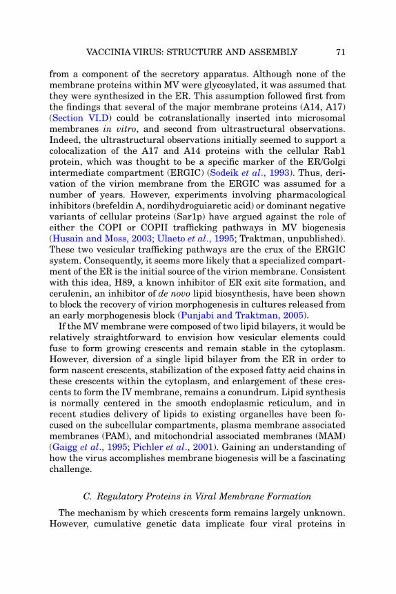
VACCINIA VIRUS: STRUCTURE AND ASSEMBLY 71
from a component of the secretory apparatus. Although none of themembrane proteins within MV were glycosylated, it was assumed thatthey were synthesized in the ER. This assumption followed first fromthe findings that several of the major membrane proteins (A14, A17)(Section VI.D) could be cotranslationally inserted into microsomalmembranes in vitro, and second from ultrastructural observations.Indeed, the ultrastructural observations initially seemed to support acolocalization of the A17 and A14 proteins with the cellular Rab1protein, which was thought to be a specific marker of the ER/Golgiintermediate compartment (ERGIC) (Sodeik et al., 1993). Thus, deri-vation of the virion membrane from the ERGIC was assumed for anumber of years. However, experiments involving pharmacologicalinhibitors (brefeldin A, nordihydroguiaretic acid) or dominant negativevariants of cellular proteins (Sar1p) have argued against the role ofeither the COPI or COPII trafficking pathways in MV biogenesis(Husain and Moss, 2003; Ulaeto et al., 1995; Traktman, unpublished).These two vesicular trafficking pathways are the crux of the ERGICsystem. Consequently, it seems more likely that a specialized compart-ment of the ER is the initial source of the virion membrane. Consistentwith this idea, H89, a known inhibitor of ER exit site formation, andcerulenin, an inhibitor of de novo lipid biosynthesis, have been shownto block the recovery of virion morphogenesis in cultures released froman early morphogenesis block (Punjabi and Traktman, 2005).
If the MVmembrane were composed of two lipid bilayers, it would berelatively straightforward to envision how vesicular elements couldfuse to form growing crescents and remain stable in the cytoplasm.However, diversion of a single lipid bilayer from the ER in order toform nascent crescents, stabilization of the exposed fatty acid chains inthese crescents within the cytoplasm, and enlargement of these cres-cents to form the IV membrane, remains a conundrum. Lipid synthesisis normally centered in the smooth endoplasmic reticulum, and inrecent studies delivery of lipids to existing organelles have been fo-cused on the subcellular compartments, plasma membrane associatedmembranes (PAM), and mitochondrial associated membranes (MAM)(Gaigg et al., 1995; Pichler et al., 2001). Gaining an understanding ofhow the virus accomplishes membrane biogenesis will be a fascinatingchallenge.
C. Regulatory Proteins in Viral Membrane Formation
The mechanism by which crescents form remains largely unknown.However, cumulative genetic data implicate four viral proteins in

72 RICHARD C. CONDIT ET AL.
regulating the earliest steps of this process. These are encoded by theF10L, H5R, G5R, and A11R genes.
1. F10
The F10 protein was first identified as the major protein kinaseencapsidated in the virion core (VPK2) (Lin and Broyles, 1994). Re-combinant F10 has enzymatic activity in vitro, indicating that theprotein possesses intrinsic kinase activity and does not need additionalprotein cofactors (Lin and Broyles, 1994; Traktman et al., 1995; Wangand Shuman, 1995). The predicted amino acid sequence of F10 retainssome of the conserved sequence motifs associated with ATP binding orphosphotransfer, but other motifs cannot be recognized due to thedivergence of the F10 sequence from that of the majority of proteinkinases. F10 has been shown to direct phosphorylation of serine, thre-onine and tyrosine residues, indicating that it is a dual specificitykinase (DSP) (Derrien et al., 1999). The 50-kDa protein is expressedlate during infection and undergoes autophosphorylation but does notappear to be proteolytically processed despite having the canonicalAG#X motif associated with the virally encoded proteolytic processingmachinery (Section IX.C) (Punjabi and Traktman, 2005). F10 is knownto be part of a seven-protein complex implicated in the filling of grow-ing crescents with viroplasmic proteins (Section VII.A), but it has alsobeen shown to be tightly associated with membranes during infectionand to bind to some lipid species (phosphoinositide phosphates) in vitro(Punjabi and Traktman, 2005; Szajner et al., 2004a). F10 is itselfencapsidated in nascent virions, with an average abundance of �300copies/core.
The importance of F10 during infection was foreshadowed bythe inability of investigators to generate a viable virus in which theF10L gene had been deleted. Subsequent studies identified fourtemperature-sensitive mutants as encoding mutant alleles of F10L;cells infected with these viruses show a profound defect in virionmorphogenesis at the nonpermissive temperature (Traktman et al.,1995; Wang and Shuman, 1995). This phenotype has been recapitu-lated by engineering viruses in which F10 expression is dependent oninclusion of IPTG or tetracycline in the culture medium (Punjabi andTraktman, 2005; Szajner et al., 2004b). The latter studies also con-firmed that expression of a catalytically active F10 protein is essentialfor virus morphogenesis. The first arrest observed on inactivation ofF10 occurs prior to the formation of any signs of membrane biogenesis.Although the late viral proteins accumulate normally and factories areformed, virosomes are either absent or malformed, and no crescents,

VACCINIA VIRUS: STRUCTURE AND ASSEMBLY 73
IV, IVN, or MV are observed (Fig. 11A). Under these circumstances,there is also no phosphorylation of the major membrane proteins A14or A17 (Section VI.D). When cells infected with tsF10 mutants undernonpermissive conditions were shifted to the permissive temperatureat 12 hour postinfection (hpi), membrane biogenesis resumed, andcrescents were observed within 45 min. Execution point studies haveshown that the F10 kinase is also required for later steps in morpho-genesis; under certain experimental protocols, tsF10-infected cellsshow a defect in crescent filling that it is nearly indistinguishable fromthat observed when other proteins found within the seven-proteincomplex are repressed or inactivated (Section VII.A). Identification ofviral and cellular proteins whose phosphorylation by F10 is importantin morphogenesis is an area of intense interest. Although the phos-phorylation of a number of viral proteins either fails to occur duringnonpermissive infections with tsF10 mutants (e.g., A14, A17, A30,G7) or is diminished (H5, F17), the phosphorylation of at least someof these proteins has been shown to be blocked when morphogenesis isarrested by other means. Thus, it is not clear which phosphorylationevents are directly mediated by F10, and which may be mediatedby other viral or cellular kinases in the context of productive morpho-genesis. Phosphorylation of at least H5 and A30 has been verifiedin vitro.
In summary, studies with mutants in F10L suggest that virus-mediated phosphorylation plays a critical role throughout MVassembly,starting from the very earliest events in virus membrane formation. It isimportant to reiterate that there is no evidence ofmembrane diversion orremodeling when F10 is repressed or inactivated.
2. H5
The H5 protein has a predicted MW of 22 kDa but has an electro-phoretic mobility consistent with a MWof 35 kDa, due to a proline-richdomain within the N-terminus (Gordon et al., 1991). The protein isexpressed constitutively and is encapsidated at �2500 copies/core(Traktman, unpublished). Some of the early literature on the H5 pro-tein is misleading because an antiserum purported to show specificityfor H5 was in fact quite reactive against the comigrating H3 protein.The bona fide role of H5 during the viral life cycle has been confusingbecause genetic and biochemical studies have implicated H5 in severalprocesses. The protein has an amphipathic helix and is phosphorylatedin vivo; H5 can be phosphorylated by both vaccinia-encoded kinases(B1 and F10) as well as by cellular kinases (Beaud and Beaud, 2000;Beaud et al., 1995; Brown et al., 2000; Traktman, unpublished).

FIG 11. Representative morphogenesis arrests seen during nonpermissive infectionswith mutants affected in genes involved in MV formation. (A–C) Mutants affectingmembrane formation. (D–F) Mutants affecting IV formation and encapsidation. (G–I)Mutants affecting core formation. (A) Formation of factories, but no virosomes and noevidence of morphogenesis: tsF10 mutant (Traktman et al., 1995); (B) accumulation ofvirosomes, membrane vesicles, and a few aberrant crescents: inducible A14 mutant(Traktman et al., 2000); (C) formation of virosomes and accumulation of membranevesicles: inducible A17 mutant (Punjabi, Mercer, and Traktman, unpublished); (D) accu-mulation of empty IV and virosomes: tsG7 mutant (2nd arrest) (Mercer and Traktman,2005); (E) formation of IV and DNA crystalloids due to arrest prior to genome encap-sidation: inducible A13 mutant (Unger and Traktman, 2004); (F) accumulation of IV,DNA crystalloids and aberrant MV due to lack of genome encapsidation: tsI6 mutant(Grubisha and Traktman, 2003); (G) accumulation of aberrant IV due to absence ofnucleoid and/or core formation: inducible F17 mutant (Mercer and Traktman, unpub-lished); (H) accumulation of IV and aberrant particles due to lack of core formation; tsI7
74 RICHARD C. CONDIT ET AL.

VACCINIA VIRUS: STRUCTURE AND ASSEMBLY 75
An interaction between H5 and A20, a stoichiometric component of theprocessive DNA polymerase, has been found by yeast two hybrid anal-ysis, thus implicating H5 in DNA replication (Ishii and Moss, 2002;McCraith et al., 2000). Consistent with this finding, one of thetemperature-sensitive mutants from the Dales collection, which hasbeen the subject of recent systematic analysis, has been shown to havea lesion within the H5 gene, and preliminary analysis of this mutantreveals a defect in DNA replication (D’Costa, Prins, and Condit, un-published; Lackner et al., 2003). Two lines of evidence also implicateH5 in the process of viral transcription: H5 stimulates late genetranscription in vitro, and H5 has been shown to interact with proteinswhich regulate late gene transcription (G2, G8, A2) (Black et al.,1998; Dellis et al., 2004; Kovacs and Moss, 1996). However, a differenttemperature-sensitive mutant, generated by targeted alanine-scanning mutagenesis of the H5R gene, implicates H5 in virus assem-bly. This mutant, which has a dominant phenotype, is unimpaired inits ability to direct viral gene expression but shows a profound defectin virion morphogenesis (DeMasi and Traktman, 2000). The phenotypeis quite similar to that seen in tsF10 mutants: virosomes are eitherabsent or malformed, and no signs of membrane biogenesis are seen.Whether H5 plays a direct role in morphogenesis, or whether themutant H5 protein somehow sequesters F10 and hence arrests mor-phogenesis, is not known. Because H5 is so abundant, it is also possiblethat it serves as a scaffold for transcription, replication, and virionassembly, thus accounting for the apparent diversity of roles for the H5protein during infection.
3. G5
The 50-kDaG5 protein is expressed at early times postinfection and isencapsidated in the virion core, although the abundance of the proteinwithin virions has not been reported (da Fonseca et al., 2004).The essentiality of the G5R gene is suggested by its conservation
mutant (Kane and Shuman, 1993); (I) accumulation of aberrant MV due to defect in corewall formation: tsA3 mutant (insets are wrapped aberrant MV [lower left] and extracel-lular aberrant MV [mid right]) (Kato et al., 2004). (A) Reprinted from Traktman et al.(1995) with permission. (B) Reprinted from Traktman et al. (2000) with permission. (D)Reprinted from Mercer and Traktman (2005) with permission. (E) Reprinted from Ungerand Traktman (2004) with permission. (F) Reprinted from Grubisha and Traktman(2003) with permission. (H) Reprinted from Kane and Shuman (1993) with permission.(I) Reprinted from Kato et al. (2004) with permission.

76 RICHARD C. CONDIT ET AL.
within most if not all poxviral genomes; attempts to isolate a virusdeleted for the G5R ORF have been unsuccessful. Temperature-sensi-tive mutants carrying G5R lesions were isolated by targeted alaninescanning mutagenesis, and phenotypic analysis of two such mutantsrevealed a role for G5R in the earliest stages of virion morphogenesis(da Fonseca et al., 2004). As was seen in tsF10 and tsH5 infections,factories were formed during tsG5 infections; however, electron-densevirosomes were either absent or malformed, and, most importantly, nosigns of membrane biogenesis were seen.
4. A11
The 40-kDa A11 protein is expressed at late times postinfection andis phosphorylated in an F10-independent manner (Resch et al., 2005).An inducible recombinant in which A11 expression is dependent oninclusion of IPTG in the culture medium has been generated; repres-sion of A11R transcription has a profound impact on virus productionand leads to an early arrest in virion morphogenesis. This is a strikingobservation, since A11 itself does not appear to be encapsidated and istherefore the first nonstructural protein to be identified as playing anessential role in the biogenesis of the virion membrane. In the absenceof A11, factories were formed; however, no crescents, IV, IVN, or MVwere seen. Electron-dense virosomes were observed, as were unusualareas of intermediate electron-density that were associated with mem-branous structures containing markers of the endoplasmic reticulum.These areas of intermediate density also contained the D13 crescentscaffold protein (Section VI.E).
D. Membrane Proteins Essential for Membrane Biogenesis:A14 and A17
The proteins encoded by the A14L and A17L genes are abundantcomponents of the MV membrane, and both are essential for itsbiogenesis.
1. A17
The 203-aa A17 protein is expressed at late times of infection. Theprotein has been shown to undergo cotranslational insertion into mi-crosomal membranes in vitro and to associate with ER membranesin vivo (Krijnse-Locker et al., 1996). In the context of its associationwith microsomal membranes, A17 spans the membrane twice, with
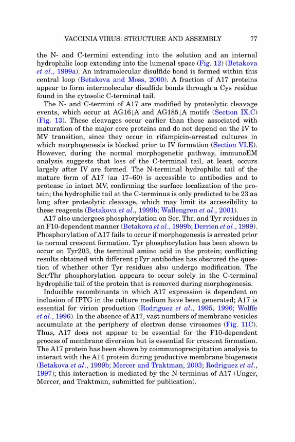
VACCINIA VIRUS: STRUCTURE AND ASSEMBLY 77
the N- and C-termini extending into the solution and an internalhydrophilic loop extending into the lumenal space (Fig. 12) (Betakovaet al., 1999a). An intramolecular disulfide bond is formed within thiscentral loop (Betakova and Moss, 2000). A fraction of A17 proteinsappear to form intermolecular disulfide bonds through a Cys residuefound in the cytosolic C-terminal tail.
The N- and C-termini of A17 are modified by proteolytic cleavageevents, which occur at AG16#A and AG185#A motifs (Section IX.C)(Fig. 13). These cleavages occur earlier than those associated withmaturation of the major core proteins and do not depend on the IV toMV transition, since they occur in rifampicin-arrested cultures inwhich morphogenesis is blocked prior to IV formation (Section VI.E).However, during the normal morphogenetic pathway, immunoEManalysis suggests that loss of the C-terminal tail, at least, occurslargely after IV are formed. The N-terminal hydrophilic tail of themature form of A17 (aa 17–60) is accessible to antibodies and toprotease in intact MV, confirming the surface localization of the pro-tein; the hydrophilic tail at the C-terminus is only predicted to be 23 aalong after proteolytic cleavage, which may limit its accessibility tothese reagents (Betakova et al., 1999b; Wallengren et al., 2001).
A17 also undergoes phosphorylation on Ser, Thr, and Tyr residues inan F10-dependentmanner (Betakova et al., 1999b; Derrien et al., 1999).Phosphorylation of A17 fails to occur if morphogenesis is arrested priorto normal crescent formation. Tyr phosphorylation has been shown tooccur on Tyr203, the terminal amino acid in the protein; conflictingresults obtained with different pTyr antibodies has obscured the ques-tion of whether other Tyr residues also undergo modification. TheSer/Thr phosphorylation appears to occur solely in the C-terminalhydrophilic tail of the protein that is removed during morphogenesis.
Inducible recombinants in which A17 expression is dependent oninclusion of IPTG in the culture medium have been generated; A17 isessential for virion production (Rodriguez et al., 1995, 1996; Wolffeet al., 1996). In the absence of A17, vast numbers of membrane vesiclesaccumulate at the periphery of electron dense virosomes (Fig. 11C).Thus, A17 does not appear to be essential for the F10-dependentprocess of membrane diversion but is essential for crescent formation.The A17 protein has been shown by coimmunoprecipitation analysis tointeract with the A14 protein during productive membrane biogenesis(Betakova et al., 1999b; Mercer and Traktman, 2003; Rodriguez et al.,1997); this interaction is mediated by the N-terminus of A17 (Unger,Mercer, and Traktman, submitted for publication).
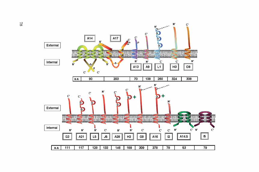
78

VACCINIA VIRUS: STRUCTURE AND ASSEMBLY 79
2. A14
The 90-aa A14 protein, which interacts with the A17 protein, isanother major component of the MVmembrane. A14 undergoes cotran-slational insertion into microsomal membranes in vitro and has beenshown to associate with ER membranes in vivo (Rodriguez et al., 1997;Salmons et al., 1997). The protein appears to span the membrane twice,assuming a topology in which the N- and C-terminal tails are lumenaland the central hydrophilic loop is exposed (Fig. 12). The C-terminal tailcontains an N83HS motif, which is readily glycosylated in vitro butpoorly glycosylated in vivo during wt infections (Mercer and Traktman,2003). Moreover, none of the glycosylated A14 protein appears to beencapsidated, suggesting that interactions of A14 with other viral pro-teins, or further posttranslationalmodifications (see later) may preventglycosylation of A14. Alternatively, the majority of A14 may be synthe-sized in, or rapidly diverted to, a membrane domain that does notsupport glycosylation. This latter hypothesis is supported by observa-tions that the fraction of A14 that undergoes glycosylation is signifi-cantly higher whenmembrane biogenesis is prevented at an early stageby repression or inactivation of F10, H5, A17, or G7.
A14 undergoes F10-dependent phosphorylation on Ser85, which isalso part of the glycosylation motif discussed previously (Mercer andTraktman, 2003). Disruption of the glycosylation motif increases thelevel of A14 phosphorylation, suggesting that the two posttranslation-al modification systems are competing for access to the protein. Final-ly, the vast majority of A14 dimerizes by forming intermoleculardisulfide bonds through Cys71. A mutant allele of A14 in whichCys71 has been altered to Ser is viable, although viral yields arereduced approximately fivefold. Virions containing this altered A14
FIG 12. Schematic view of integral MV membrane proteins. The overall topology ofknown transmembrane proteins contained within the MV membrane is shown; externaland internal refers to the orientation within MV. A14 and A17 are essential for biogene-sis; A13, A9, and L1 are required for virion maturation; H3 stimulates virion maturationand, along with D8, binds to glycosaminoglycans on the surface of target cells; G3, A21,L5, J5, A28, H2, G9, and A16 comprise an eight-member fusion/entry complex; 12 is alsorequired for virion entry; A14.5 affects virulence in mice, and the function of I5 has notbeen determined. The number of amino acid residues is shown beneath each protein.Sites of phosphorylation, P0 myristylation (chemical structure), proteolytic cleavage(dotted line), and disulfide bond formation (-S-) are shown. The symbol shown adjacentto the H2 and A16 proteins✦ indicaes that, although these proteins are known to containintramolecular disulfide bonds, the number and position of these linkages are not known.

80 RICHARD C. CONDIT ET AL.
protein are exquisitely sensitive to nonionic detergents. Under condi-tions in which wt virions remain intact and infectious, NP40 treatmentof these A14-Cys71 virions leads to the extraction of both A14 and A17and causes a 1000-fold decrease in specific infectivity. These findingssupport a model in which dimerized A14, interacting noncovalentlywith A17, forms a lattice within the MVmembrane that sustains virionintegrity, consistent with previous results from controlled degradationof MV described in Section III.C.
Recombinant viruses in which expression of A14 is dependent oninclusion of either IPTG or tetracycline in the culture medium havebeen generated; expression of A14 is essential for virion production(Rodriguez et al., 1998; Traktman et al., 2000). In the absence of A14,vast numbers of vesicles (and/or tubules) accumulate within the cyto-plasm (Fig. 11B). Some aberrant crescents are seen which are adjacentto, but fail to make contact with, electron-dense virosomes that accu-mulate in factories. Importantly, the vesicles formed in the absence ofA14 contain A17 and another membrane protein, D8, while the viro-somes formed contain the core proteins L4 and F17 (Traktman et al.,2000). Thus, the A14 mutant phenotype is similar but not identical tothat seen upon repression of A17. Most importantly, the presence ofboth of A14 and A17 is required to form bona fide crescents, which canprogress to form the IV membrane. Whether the vesicles seen upontheir repression represent stalled intermediates, or aberrant struc-tures formed by the collapse of unstable membranes, has not beendetermined. This is a question of considerable relevance to a fullerappreciation of the mechanism of IV membrane biogenesis.
Transient complementation has been used to probe which features ofthe A14 protein are essential for membrane biogenesis. Sequences thatflank the first transmembrane domain, and those within the external,hydrophilic loop are most sensitive to mutation. The latter region hasbeen found to interact with the N-terminus of A17 in vitro; moreover,mutations that disrupt complementation activity in vivo disruptinteraction in vitro (Unger, Mercer, and Traktman, submitted forpublication).
In summary, the A17 and A14 membrane proteins are criticalplayers in diversion of ER membranes to viral factories and formationof the viral membrane. The proteins are synthesized in the ER andtrafficked to viral factories to form membrane-containing crescents.Within crescents and the mature virion membrane, A14 and A17appear to form a lattice that is stabilized by disulfide bonds, probablycontributing to the sensitivity of the virion membrane to reducingagents. Finally, the A14–A17 lattice serves as an anchor within the

VACCINIA VIRUS: STRUCTURE AND ASSEMBLY 81
viral membrane to which several other proteins important in virionstructure and morphogenesis attach (see in a later section).
E. D13: The Crescent Scaffold Protein
The “spicule” layer that comprises the convex surface of crescents,IV and IVN was first described in detail by Dales and Mosbach (1968).Based on studies with metabolic inhibitors that partially disruptedcrescent formation these authors concluded that the spicule layer con-ferred curvature and rigidity to the crescents. This conclusion wasconfirmed and extended through studies with the antibiotic inhibitorrifampicin. Rifampicin, better known as an inhibitor of prokaryotictranscription, inhibits vaccinia virus morphogenesis (Grimley et al.,1970; Moss et al., 1969a,b; Nagayama et al., 1970). In the presence ofthis drug, irregularly shaped membranes that lack a spicule layeraccumulate around areas of electron-dense viroplasm. These mem-branes can be labeled with anti-A17 antibody using immune electronmicroscopy, confirming a relatedness to virus crescent membranes(Wolffe et al., 1996). Inhibition with rifampicin is rapidly reversible;normal crescents form within minutes after the removal of drug.
Several lines of evidence demonstrate that the spicule protein is theproduct of gene D13L. First, immune electron microscopy shows thatin a normal infection, antibody specific for the D13 protein decoratescrescents and IV (Miner and Hruby, 1989; Mohandas and Dales, 1995;Sodeik et al., 1994; Szajner et al., 2005). In the presence of rifampicin,virus membranes do not label with D13 antibody, and instead D13antigen accumulates in inclusions within factories, removed frommembranes. When the rifampicin block is reversed, newly formedcrescents are now labeled with D13 antibody and the D13 inclusionsdisappear. Second, mutations that confer resistance to rifampicin mapto the D13L gene (Baldick and Moss, 1987; Tartaglia and Paoletti,1985). Third, conditional lethal mutations in the D13L gene display aphenotype identical to the effects of rifampicin (Lackner et al., 2003;Stern et al., 1977; Zhang and Moss, 1992).
Experiments demonstrate that the “spicule” layer is in fact a spheri-cal honeycomb lattice composed of D13 trimers (Heuser, 2005; Szajneret al., 2005). The D13 protein itself can associate with two integralvirus membrane proteins, A17 and H3, thus providing a mechanismfor scaffolding of the virus membrane onto the D13 lattice. As dis-cussed in Section IX.E.1, D13 is lost from virions during maturationfrom IVN to MV (Essani et al., 1982; Mohandas and Dales, 1995;Sodeik et al., 1994); how the completion of the IV membrane sends a

82 RICHARD C. CONDIT ET AL.
signal that activates dissolution of the D13 lattice, and how this latticeis dismantled, are intriguing questions that remain to be answered.
In summary, the “spicule” layer that appears on the convex surface ofcrescents and the outside of IV and IVN is a temporary scaffold forassembly of the virus membrane and is composed primarily of a honey-comb lattice built from D13 protein trimers. It follows that the curva-ture of the IV membrane, and therefore the final size of IV, is largelydetermined by the structure of the D13 lattice. Parallels between theD13 lattice and the clathrin coats of endocytic vesicles are evident.
VII. IMMATURE VIRION FORMATION: A COMPLEX PROCESS
Mutants in seven different vaccinia genes produce a similar pheno-type which suggests a role for the gene products in the association ofviral crescents with viroplasm. One of these seven proteins, A10 or 4a,is one of the most abundant virion core proteins. The other six pro-teins, A15, A30, D2, D3, G7, and J1, are found in a seven-proteincomplex along with the F10 protein kinase, which, as described previ-ously, phosphorylates several membrane and core proteins and playsa critical role throughout virus assembly. The role of each of theseproteins in IV formation is summarized in the following sections.
A. Seven-Protein Complex
As membrane crescents appear, they are often seen at the peripheryof electron-dense regions of viroplasm, which is known to contain viralproteins destined for encapsidation into the virion core. It is not knownwhat protein–protein or protein–lipid interactions are involved inmediating the association of viroplasm with growing crescents: thereare no known interactions of individual membrane proteins with coreproteins. However, a group of seven core proteins, F10, A30, G7, J1,D2, D3, and A15, has been shown to form a multimeric complex, and insome cases the proteins are mutually dependent on one another forstability (Szajner et al., 2004a). Several members of the complex arephosphorylated in vivo in an F10-dependent manner. The stoichiome-try of the individual components within the complex has not beendetermined. In addition to being physically linked, these proteins showa genetic interaction, since comparable phenotypes are observed uponmutation or repression of individual members of the complex. Asdetailed in a later section, the phenotypes suggest a role for theseproteins in the association of viroplasm with crescents.

VACCINIA VIRUS: STRUCTURE AND ASSEMBLY 83
1. F10
F10 has been described previously in significant detail (SectionVI.C.1). With regard to its involvement as a member of the seven-protein complex which mediates the association of viroplasm withgrowing crescents, it is worth recalling the fact that F10 has beenshown to be tightly associated with membranes in vivo (Punjabi andTraktman, 2005). Direct evidence for the functional involvement ofF10 in the association of viroplasm with crescents was obtained fromultrastructural analyses of tsF10 infections that were initiated at thepermissive temperature in the presence of rifampicin, and then shiftedto the nonpermissive in the absence of rifampicin at 12 hpi. Thephenotype seen here was nearly identical to that observed upon dis-ruption of A30 or G7 expression (see later). Indeed, the phosphoryla-tion of A30 and G7 is dependent on F10 in vivo and also appears to bedependent on the assembly of the multimeric complex and the normalprogression of morphogenesis (Mercer and Traktman, 2005; Szajneret al., 2004c). It seems likely that F10 mediates phosphorylation ofthese proteins in a context-dependent manner.
2. A30
Disruption of theA30L genewas shown to lead to a phenotype inwhichthe association of nascent membranes with viroplasm was disrupted.This disruption led to the appearance of large virosomes and “empty”IV and/or pseudo-IV with multiple membrane wrappings. This pheno-type was observed when an inducible recombinant was generatedpermitting the repression of A30, or when a temperature-sensitivemutant, Dts46, was analyzed at the nonpermissive temperature (Szajneret al., 2001a,b). The tsA30 protein is extremely thermolabile: effectively,at the nonpermissive temperature, repression of the A30 protein is mim-icked. A30 is a 9-kDaprotein that is found in viroplasm and encapsidatedwithin virions; fractionation studies have demonstrated that A30 isloosely associated with the virion core. A30 is phosphorylated on Serresidues in vivo in an F10-dependent manner, and it has been shown tobe a direct substrate of F10 in vitro. A30 has been shown to interactdirectly with the G7 protein (discussed in a later section), and theseproteins appear to depend on one another for their mutual stability.
3. G7
G7 was first implicated in virion morphogenesis because of its asso-ciation with the A30 protein (Szajner et al., 2003). The 42-kDa G7protein is expressed at late times postinfection, encapsidated into

84 RICHARD C. CONDIT ET AL.
virion cores, and processed by proteolytic cleavage at two AG#X motifsduring virion maturation (Fig. 13). The G7 protein is phosphorylatedin vivo in an F10-dependent manner, but F10 does not appear tophosphorylate G7 in vitro, either when presented alone or in thepresence of its binding partner A30 (Mercer and Traktman, 2005;Szajner et al., 2004c). The function of G7 in vivo has been addressedboth by the generation of an inducible recombinant in which G7 ex-pression is IPTG-dependent and by characterization of Cts11, whichencodes a defective G7 protein (Mercer and Traktman, 2005; Szajneret al., 2003). These two approaches have yielded somewhat differentanswers regarding G7’s function, as detailed below.
During infections with Cts11, virosomes accumulate as do shortmembrane crescents that are embedded within a distinct matrix ofmedium electron density. Both the G7 and A30 proteins are stableduring these infections, permitting the examination of the impact ofa specific loss of G7 function. When various G7 and A30 proteins weresynthesized in vitro and subjected to coimmunoprecipitation protocols,the tsG7 protein had lost the ability to interact with A30. When Cts11
FIG 13. Proteolysis of vaccinia core and membrane proteins. The proteolysis of fivecore proteins (A10, A3, G7, A12, and L4) and one membrane protein (A17) is shown.Black lines represent the length of the precursor polypeptide, drawn to scale. Red arrow-heads represent known cleavage sites. Information concerning the precursor (MW, anypseudonyms in brackets, and localization if relevant) is given above each line. Informa-tion concerning each product (MW, pseudonyms in brackets, localization if known) isgiven below each line. ? ¼ fate of the product is unknown.

VACCINIA VIRUS: STRUCTURE AND ASSEMBLY 85
infections were performed at permissive temperature in the presenceof rifampicin for 12 h and then removed from the rifampicin block andshifted to the nonpermissive temperature, the phenotype observedmimicked what had been observed on repression of A30 (Fig. 11D).This phenotype, characterized by the accumulation of empty IV, isalso observed when G7 is repressed. Interestingly, tsG7 mutants arenoncomplementing with tsA30 mutants, providing additional geneticevidence for an interaction between G7 and A30 (Lackner et al., 2003).In summary, G7 appears to play crucial roles at two stages of morpho-genesis (Mercer and Traktman, 2005). First, G7 is required for themovement of crescents to the periphery of virosomes, where theyenlarge; later, G7 is required for the filling of the crescents withviroplasmic material. The small amount of wt G7 that is likely toaccumulate when infections with the inducible recombinant are per-formed in the absence of the IPTG inducer may be sufficient for theinitial G7-dependent step, whereas the tsG7 protein must be unable tomediate this effect.
4. J1
The21-kDaJ1protein is expressed at late times and encapsidated; likeA30, although J1 is associated with virion cores, a fraction of the proteincan be extractedwith nonionic detergents. Purified J1 has been shown toform oligomers (Chiu et al., 2005), and interactions between the N- andC-termini of J1 have been observed in yeast two-hybrid analyses. Therole of J1 in vivo has been analyzed using both an inducible recombinantin which J1 expression is IPTG-dependent and through the analysis ofCts45, which encodes a mutant J1 protein (Chiu and Chang, 2002; Chiuet al., 2005). Repression or inactivation of J1 phenocopies the repressionofA30 orG7, and indeedbothA30andG7becomeunstable in the absenceof J1 at 40�C. J1 is part of the seven-protein complex involved in viro-plasm/membrane association, although the proteins with which J1 as-sociates directly have not been identified (Szajner et al., 2004a). The tsJ1protein remains stable at the nonpermissive temperature but does notassociate with the multiprotein complex.
5. A15
The 10-kDa A15 protein was shown to be a component of the seven-protein complex by mass spectroscopy (Szajner et al., 2004a). This pro-tein is expressed at late times postinfection and encapsidated in thevirion core. An inducible recombinant in which A15 expression isIPTG-dependent has been generated; in the absence of A15, the charac-teristic accumulation of empty IV was observed (Szajner et al., 2004a).
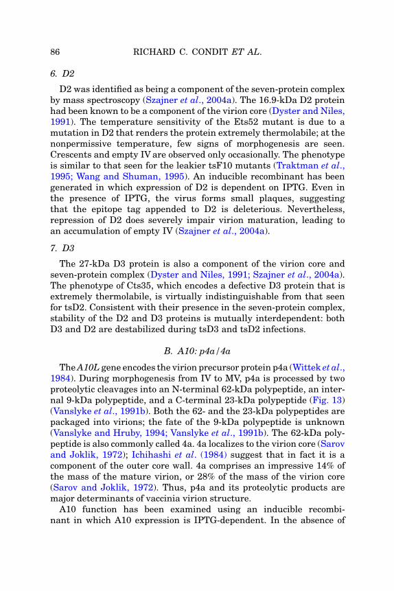
86 RICHARD C. CONDIT ET AL.
6. D2
D2 was identified as being a component of the seven-protein complexby mass spectroscopy (Szajner et al., 2004a). The 16.9-kDa D2 proteinhad been known to be a component of the virion core (Dyster and Niles,1991). The temperature sensitivity of the Ets52 mutant is due to amutation in D2 that renders the protein extremely thermolabile; at thenonpermissive temperature, few signs of morphogenesis are seen.Crescents and empty IV are observed only occasionally. The phenotypeis similar to that seen for the leakier tsF10 mutants (Traktman et al.,1995; Wang and Shuman, 1995). An inducible recombinant has beengenerated in which expression of D2 is dependent on IPTG. Even inthe presence of IPTG, the virus forms small plaques, suggestingthat the epitope tag appended to D2 is deleterious. Nevertheless,repression of D2 does severely impair virion maturation, leading toan accumulation of empty IV (Szajner et al., 2004a).
7. D3
The 27-kDa D3 protein is also a component of the virion core andseven-protein complex (Dyster and Niles, 1991; Szajner et al., 2004a).The phenotype of Cts35, which encodes a defective D3 protein that isextremely thermolabile, is virtually indistinguishable from that seenfor tsD2. Consistent with their presence in the seven-protein complex,stability of the D2 and D3 proteins is mutually interdependent: bothD3 and D2 are destabilized during tsD3 and tsD2 infections.
B. A10: p4a/4a
TheA10L gene encodes the virion precursor protein p4a (Wittek et al.,1984). During morphogenesis from IV to MV, p4a is processed by twoproteolytic cleavages into an N-terminal 62-kDa polypeptide, an inter-nal 9-kDa polypep tide, and a C-term inal 23-kDa polypep tide (Fig. 13)(Vanslyke et al., 1991b). Both the 62- and the 23-kDa polypeptides arepackaged into virions; the fate of the 9-kDa polypeptide is unknown(Vanslyke and Hruby, 1994; Vanslyke et al., 1991b). The 62-kDa poly-peptide is also commonly called 4a. 4a localizes to the virion core (Sarovand Joklik, 1972); Ichihashi et al. (1984) suggest that in fact it is acomponent of the outer core wall. 4a comprises an impressive 14% ofthe mass of the mature virion, or 28% of the mass of the virion core(Sarov and Joklik, 1972). Thus, p4a and its proteolytic products aremajor determinants of vaccinia virion structure.
A10 function has been examined using an inducible recombi-nant in which A10 expression is IPTG-dependent. In the absence of

VACCINIA VIRUS: STRUCTURE AND ASSEMBLY 87
inducer, normal crescents are seen, but the IV that form are aberrant(Heljasvaara et al., 2001). Many of the abnormal IV particles appearempty, but others contain dense patches of DNA-containing material.DNA aggregates also accumulate in the cytoplasm. The membranessurrounding the IV are normal in appearance and contain the A14 andA17 membrane proteins as well as the D13 scaffold protein, as judgedby immunoelectron microscopy. Thus, A10 appears to be dispensablefor the formation of the IV membrane but is required for the properorganization and packaging of the viroplasmic matrix that is normallyincluded in IV. The phenotype observed on repression of A10 mimics toa great degree the phenotype of mutants affected in components of theseven-protein complex described previously.
The A10 precursor and the 60-kDa cleavage product form stablecomplexes with another abundant virion core protein, A4 (Riscoet al., 1999). The implications of this interaction are discussed inSection IX.B.3 along with the role of A4 in MV formation.
VIII. IV!IVN TRANSITION: GENOME ENCAPSIDATION
The process of viral DNA replication produces progeny genomes thatwill be encapsidated into nascent virions. Concatemeric intermediatesare formed during the replication process, and these are processed tomonomeric genomes by a virally encoded resolvase (Garcia et al., 2000)(Section VIII.B.3). Genome maturation occurs normally when virionmorphogenesis is blocked in the very earliest stages of morphogenesis,for example, in H5, G5, and J1 mutant infections (Table II), indicatingthat this maturation is a prerequisite for, but is not coupled to, genomeencapsidation (Chiu and Chang, 2002; da Fonseca et al., 2004; DeMasiand Traktman, 2000). Much remains to be discovered about the en-cpasidation of the viral genome, although it appears to be distinct fromthe process described previously for the filling of maturing crescentswith viroplasmic proteins destined to form the virion core. One of theunresolved questions is whether genome entry precedes closure ofthe IV membrane. Serial section analysis shows that most fully closedIV do indeed have nucleoids (Morgan et al., 1955), suggesting thatgenome encapsidation is likely to occur prior to the closure of the IV(Morgan, 1976a,b). If this model is not correct, then the encapsidationmachinery must either pass through a proteinaceous plug that hasbeen proposed to mark the site of IV membrane closure, or it must passthrough the IV membrane itself. Some investigators have proposedthat genome entry causes membrane invagination, leading to the

88 RICHARD C. CONDIT ET AL.
presence of a membrane around the nucleoid and, consequently, thecore. However, this model is not widely accepted (Section VI.A) (Fig. 8).
It is not known exactly how replicated genomes are distributedthroughout the cytoplasm, although sites of replication have beenvisualized by autoradiography or with antisera directed against BrdU,DNA, or the single-stranded binding protein, I3. When morphogenesisis arrested prior to the appearance of IVN, DNA crystalloids accumu-late in the cytoplasm (Ericsson et al., 1995; Grimley et al., 1970;Grubisha and Traktman, 2003; Unger and Traktman, 2004). Thesecrystalloids are often surrounded by membranes, consistent with theproposal that DNA replication occurs within ER-delimited domainsof the cytoplasm (Schramm and Locker, 2005; Tolonen et al., 2001)(Section V) (Fig. 5).
The literature contains a number of ultrastructural images that havebeen interpreted as representing the ongoing process of DNA encapsi-dation (Morgan, 1976a,b). Although this process is commonly referredto as encapsidation of the viral “nucleoprotein,” there is in fact nothingknown about how the genome is condensed and whether it is indeedbound to proteins. As described later, when either of the two proteinsthat have been specifically associated with genome encapsidation arerepressed or inactivated, no other proteins appear to be missingfrom the virions (Cassetti et al., 1998; Grubisha and Traktman, 2003).These data do not support a model in which the genome is assembledinto a conventional nucleoprotein complex. In fact, it may be thatcondensation of the genome is effected by interactions with polyamines;encapsidation of spermidine has been reported (Lanzer andHolowczak,1975; Saminathan et al., 2002). In any case, the genome has a predicteddimension of 2 nm � 68 mm, which must fit into the viral core. Theapproximate dimensions of the nucleoid, as observed in ultra thin sec-tions, are 0.1 � 0.15 mm, indicating that the genome must undergomassive condensation before encapsidation.
To date, four viral proteins have been implicated in maturation andprocessing of the viral genome. These include the core proteins A22(a Holliday resolvase), I6 and A32, and the membrane protein A13.
A. Membrane Proteins
1. A13
The 70-amino acid A13 protein has a hydrophobic N-terminus,which is predicted to be the membrane-spanning domain; indeedA13 undergoes cotranslational insertion into microsomal membranesin vitro (Salmons et al., 1997) and is a component of the virion

VACCINIA VIRUS: STRUCTURE AND ASSEMBLY 89
mlpbNkamhiaoaeI
1
pe3dtibvasnhb
2
teotpo
embrane (Fig. 12). The hydrophilic tail of A13 undergoes phosphory-ation in vivo on Ser residues; mutation of Ser40!Ala prevents phos-horylation (Unger and Traktman, 2004). This modification appears toe independent of the F10 kinase; Ser40 is found within the motifSPPP, which is predicted to be a high-affinity site for the cellularinase ERK. The function of A13 has been elucidated both by thenalysis of a TET-inducible recombinant and a temperature-sensitiveutant (Cts40) (Unger and Traktman, 2004); the A13 protein in Cts40as a Thr48!Ile substitution and is thermostable but apparentlynactive. When A13 is inactivated, a morphogenesis arrest is seen; IVre formed, but IVN are rare and MV are not seen (Fig. 11E). Numer-us DNA crystalloids, which appear to be surrounded by membranes,ccumulate in the cytoplasm. Thus, A13 appears to be essential for thencapsidation of DNA into IV and the subsequent maturation ofV!IVN!MV.
B. Core Proteins
. A32
Bioinformatic analysis was first used to identify the A32 protein as autative ATPase with limited homology to the encapsidation proteinsncoded by bacteriophages and adenovirus (Koonin et al., 1993). The4-kDa protein is expressed at late times postinfection and is encapsi-ated; to date, there has been no experimental data confirming thathe protein indeed possesses the predicted ATPase activity. An induc-ble recombinant in which expression of A32 is IPTG-dependent haseen generated; in the absence of A32, the production of infectiousirus is severely diminished (Cassetti et al., 1998). Morphogenesisppears normal through the IV stage, but then aberrant forms areeen. Spherical virions that appear dense or half-dense/half-empty areumerous, as are cytoplasmic DNA crystalloids. The aberrant virionsave been purified and shown to contain the full-protein complementut to be devoid of viral DNA.
. I6
The 44-kDa I6 protein was identified as binding with great specifici-y and stability to the telomeric hairpins of the viral genome (Demasit al., 2001b). The telomere–I6 interaction was shown to be dependentn the presence of the extrahelical bases, which are so characteristic ofhe viral telomeres. Very low levels of I6 are made at early timesostinfection; the majority of the protein is synthesized after the onsetf DNA replication. An average of�35 copies of I6 is encapsidated in the
virion core. A series of mutants with lesions in the I6L gene have been

90 RICHARD C. CONDIT ET AL.
generated by clustered charge-to-alaninemutagenesis, and one of thesehas a tight temperature-sensitive phenotype (Grubisha and Traktman,2003). At the nonpermissive temperature, the phenotype observed isalmost indistinguishable from that seen on repression of A32: IVappearnormal, cytoplasmic DNA crystalloids are abundant, and aberrant,spherical virions are produced that contain the full-protein complementbut no viral DNA (Fig. 11F). A32 remains stable during these infectionsand is encapsidated into the aberrant virions, indicating that itsencapsidation is independent of DNA encapsidation and I6 function.Conversely, no I6 is encapsidated into A32-deficient virions.
These cumulative data support a model in which the presence ofA32 within virions enables the encapsidation of a DNA genome onwhich I6 protein is bound to the telomeric hairpins. As describedpreviously, repression of certain membrane proteins, such as A13,leads to the accumulation of IV and DNA crystalloids; it is possiblethat the presence of these proteins in the membrane is also importantfor A32 and/or I6-DNA encapsidation.
3. A22, a Holliday Resolvase
As described previously, concatemeric replication intermediates areprocessed to mature genomes by a virally encoded Holliday junctionresolvase, the A22 protein (Garcia et al., 2000). The product of theA22Rgene is a palmityl protein that is expressed late during virus infectionand packaged into the virion core (Garcia and Moss, 2001; Grosenbachet al., 2000). Analysis of an inducible mutant demonstrates that in theabsence of A22, concatemer resolution is defective, and virion morpho-genesis is arrested at an intermediate stage (Garcia andMoss, 2001). Inthese A22-deficient infections, MV formation is inhibited and IV accu-mulate, a very few of which have nucleoids. In addition, there is anaccumulation of dense spherical particles similar to those seen in otherencapsidation defective mutants. Thus, while genome maturation canproceed in the absence of virionmorphogenesis, the converse is not true.If concatemer resolution is inhibited, virion morphogenesis is arrestedand the nature of the intermediate particles that accumulate suggeststhat the defect lies in DNA packaging.
IX. MV FORMATION
Perhaps the most striking event during morphogenesis of vaccinia isthe transition from IVN to MV. The precise protein composition ofimmature particles is difficult to assess because normal IV and IVN

VACCINIA VIRUS: STRUCTURE AND ASSEMBLY 91
are difficult to isolate in sufficient quantity or purity to permit rigorousbiochemical study (Vanslyke et al., 1993). Nevertheless, as detailedlater, a synthesis of the evidence to date suggests that IVN containthe full complement of proteins contained in MV, with the exceptionof a few membrane proteins that are added during the IV/IVN to MVtransition. Furthermore, since both the IV membrane and theMV membrane contain the major viral membrane proteins A14 andA17, the membrane of IVN is the precursor for the MV membrane(Griffiths et al., 2001a; Rodriguez et al., 1997; Traktman et al., 2000).Thus the IV/IVN to MV transition involves a rearrangement of thecontents of IVN to form a distinct core and lateral bodies, and in theprocess the particle is transformed from a spherical to a barrel shape.Indicative of the complexity of this process, a significant majority ofvirus mutants that affect virion morphogenesis affect the IV/IVN toMV transition. These mutants fall into two general classes: those thatfail to assemble morphologically normal lookingMV particles and thosethat assemble normal lookingMV particles, which are nevertheless notinfectious. Analysis of these mutants informs us that the IV/IVN to MVtransition involves or requires: (1) assembly of the transcription appa-ratus within the core, (2) restructuring of the particle surface, includingloss of the D13 protein, formation of numerous disulfide bond linkagesby the virally encoded redox system and addition of several additionalsurface proteins, (3) proteolysis of several virion protein precursors andformation of an enzymatically active core, in part through the action ofthe viral phosphatase, and (4) movement of the finished MV fromviroplasm to the periphery of the cell. Each of these events is discussedin more detail in later sections.
A. Assembly of the Transcription Apparatus Within the Core
The phenotype of virus mutants affecting two different virus codedtranscription factors, VETF and rap94, reveals an intriguing relation-ship between assembly of the early transcription machinery within thevirion core and formation of MV. Vaccinia RNA polymerase exists intwo different forms, one that contains the subunit rap94 (the productof the H4L gene), and one that lacks rap94 (Broyles, 2003). The rap94-containing enzyme transcribes early viral genes exclusively, whileRNA polymerase lacking rap94 transcribes exclusively intermediateand late genes. VETF, composed of the D6 and A7 proteins, bindsspecifically to early virus promoters and is required for early genetranscription in vitro (Broyles, 2003). Thus, specificity of the RNApolymerase for early promoters is thought to result from a specific

92 RICHARD C. CONDIT ET AL.
interaction between rap94 and VETF, although such an associationhas never been proven directly.
1. D6 and A7: VETF
Genetic analysis of VETF is facilitated by the existence of induciblerecombinants and temperature-sensitive mutants. There exist multi-ple temperature-sensitive mutants with lesions in the D6R gene, andinducible recombinants have been generated which allow IPTG-dependent expression of both subunits of VETF, D6 and A7 (Christenet al., 1992; Hu et al., 1996, 1998). Ultrastructural analysis of cellsinfected with the temperature-sensitive mutants has not been re-ported, but such studies have been published for the inducible recom-binants. When either VETF subunit is repressed, a “normal” geneexpression phenotype is observed; that is, all three classes of viralproteins, early, intermediate, and late, are synthesized in normalamounts and with normal kinetics.5, However, electron microscopicanalysis of the mutant infections reveals a block in morphogenesis atthe IV/IVN stage. MVare either absent (D6) or significantly reduced innumber (A7) in the mutant infections. Instead, an abundance of mor-phologically normal IV and IVN accumulate and occasional abnormal,dense, spherical particles are seen in viral factories. Confocal micros-copy of A7-deficient infections stained for DNA and a virion membraneprotein suggests that at least a fraction of the immature particles
5 A general and seemingly peculiar feature of mutants in vaccinia genes affectingvirion transcription enzymes is that they confer a phenotype that is expressed late ratherthan early during infection. Offhand, one might expect a mutation in a virion transcrip-tion enzyme, in particular a temperature-sensitive mutant, to affect early viral tran-scription and thus disrupt the infection at an early stage. However, all mutants intranscription enzymes reported to date proceed through the early phase of infectionnormally and display a phenotype that impacts the late phase of infection. In the caseof inducible mutants, this phenomenon is not difficult to understand. The induciblemutant must be grown under permissive conditions in order to study the phenotypeunder nonpermissive conditions. During growth under permissive conditions, the wildtype gene product is synthesized and packaged into virions, which are therefore compe-tent for early transcription under nonpermissive conditions. The nonpermissive infectiontherefore only tests for any requirement for the protein during the later phases ofinfection. In the case of temperature-sensitive mutations the same phenomenon isobserved; however, in this case the virions synthesized under permissive conditionspackage mutant enzyme. One must therefore argue that the mutant proteins are notthermolabile in virions, but rather that they are temperature sensitive for synthesis, thatis, newly synthesized mutant protein is either rapidly degraded or cannot form an activeconformation at the nonpermissive temperature, and thus the phenotype observedrepresents the requirement for newly synthesized enzyme late during infection(Hooda-Dhingra et al., 1989).

VACCINIA VIRUS: STRUCTURE AND ASSEMBLY 93
contain DNA. Both mutants also accumulate abnormally largemasses of viroplasm within viral factories. The protein composition ofthe accumulated IV and IVN is unknown. Thus in the absence ofVETF, virus assembly arrests at the IV/INV stage, implying a pivotalrole for VETF in formation of the viral core and transition from IVNto MV.
2. H4: rap94
Genetic analysis of the H4L gene, which encodes rap94, is also en-abled by the existence of both inducible and temperature-sensitivemutants affected in this gene (Kane and Shuman, 1992; Zhang et al.,1994). As was described for the VETF mutants, inactivation or repres-sion of rap94 does not impair the profile of viral gene expression. Unlikethe VETF mutants however, both the temperature sensitive and theinducible mutants in rap94 assemble particles that aremorphologicallyindistinguishable from wt MV as judged by electron microscopy. TheseMV made under nonpermissive conditions are deficient in their abilityto direct in situ transcription in vitro. Moreover, they lack not onlyrap94 as expected but also specifically lack all of the major known orsuspected enzymes involved in early viral transcription, including theviral RNA polymerase, the capping enzyme/termination factor, thepoly(A) polymerase large subunit, DNA-dependent ATPase I, RNA heli-case, and topoisomerase. These abnormal MV contain normal amountsof VETF. These results are interpreted tomean that a presumed rap94–VETF interaction seeds assembly of a transcription complex onto theviral genome and thus coordinates packaging of the transcription ma-chinery during virus assembly. Notably, the result also implies that themorphological transformation from IV toMV, including formation of thecore and lateral bodies, occurs independently of the assembly of thetranscription apparatus within the core.
Together, studies with mutants in VETF and rap94 suggest that theearly viral transcription factor VETF, perhaps in conjunction with orthrough interaction with other DNA binding or structural proteins,plays a critical role in the morphological transition from IVN to MV,and also, perhaps through an interaction with rap94, plays a criticalrole in recruitment and assembly of a transcription complex in thedeveloping virion. The recruitment and assembly of the transcriptioncomplex itself, however, is not a prerequisite for virion maturation, inthat normal appearing MV can be assembled in the absence of thetranscription complex. Finally, it is worth recalling that encapsidationof VETF and the early transcriptional machinery does not appear torequire their association with the viral genome, since DNA-deficient

94 RICHARD C. CONDIT ET AL.
particles assembled in the absence of the A32 protein do contain theearly transcriptional machinery (Cassetti et al., 1998).
B. Core Proteins Involved in the IV to MV Transition
A number of core proteins are required for virion maturation, asrevealed by the phenotypes exhibited by temperature-sensitive mu-tants that encode mutant forms of these proteins, or inducible recom-binants which allow the expression of the proteins of interest to beexperimentally repressed. In some cases, repression or inactivation ofthe protein simply arrests morphogenesis at the IV or IVN stage; inother cases, aberrant particles are seen. Since not all publications areequally thorough in their ultrastructural analysis, implication of agiven protein in a precise step of virion assembly is not always possi-ble. It would appear that when the D13 scaffold remains intact,there is an accumulation of IV and/or IVN. In contrast, when the D13scaffold is removed, then further virion maturation is attemptedunsuccessfully, leading to the production of aberrant particles.
1. I1
The 36-kDa I1 protein is expressed at late times postinfection andencapsidated into virion cores (�700 copies/virion) (Klemperer et al.,1997). The protein has nonspecific DNA binding activity but shows somepreference for binding to the viral telomeres (Demasi et al., 2001a; Klem-perer et al., 1997). An inducible recombinant in which I1 expression isIPTG-dependent has been generated; upon the repression of I1, morpho-genesis arrests with the accumulation of numerous IV. IVN were alsoseen, although some DNA crystalloids were observed in the cytoplasm(Klemperer et al., 1997). The I1 protein is highly conserved amongpoxviral genomes, in keeping with its essentiality for virion maturation.
2. F17
The 11-kDa F17 protein is expressed at late times postinfection andis one of the major components of the core; �27,000 copies of theprotein are encapsidated per virion. This protein was initially referredto as p11 or VP11 (Kao and Bauer, 1987; Kao et al., 1981; Nowakowskiet al., 1978a,b). The protein is phosphorylated in vivo on Ser residues;phosphorylation is only minimally dependent on the F10 kinase but ismodulated by expression of the viral H1 phosphatase. It is likely thatthe protein is phosphorylated on two PSSP motifs which are targetsites for cellular proline-directed kinases such as ERK1 and CDK1(Reddy, unpublished; Traktman, unpublished). The F17 protein is

VACCINIA VIRUS: STRUCTURE AND ASSEMBLY 95
quite basic and was originally described as a DNA-binding protein thatcould coat DNA stoichiometrically. However, normal amounts of F17are encapsidated even when the genomic DNA is not, suggesting thatF17 is certainly not a classical “nucleoprotein” in the sense that wasoriginally proposed (Cassetti et al., 1998; Grubisha and Traktman,2003). An inducible recombinant has been generated in which ex-pression of the F17R gene is IPTG-dependent; in the absence of F17,morphogenesis arrests with the appearance of aberrant IV (Fig. 11G)(Zhang and Moss, 1991a,b). In some of these particles, there is aninternal nucleoid that resembles the DNA crystalloids describedearlier. In other cases, the IV appear almost empty or contain smallpatches of condensed material and/or membrane. Because DNA crys-talloids do not accumulate in the cytoplasm, however (Traktman, un-published), it is likely that the absence of F17 does not impair genomeencapsidation per se but impairs nucleoid formation and disrupts theinternal organization of the IV. These aberrant IV do not form a lightscattering band upon sucrose gradient ultracentrifugation, indicatingthat they may be unstable.
3. A4
The A4L gene encodes a 39-kDa protein that is expressed late andencapsidated into the virion. This protein was first identified as animmunodominant antigen and was referred to as p39 (Cudmore et al.,1996; Demkowicz et al., 1992; Maa and Esteban, 1987; Roos et al.,1996). A4 appears to be phosphorylated in vivo and is a substrate forthe F10 kinase in vitro; A4 phosphorylation appears to be modulatedby the expression of the viral H1 phosphatase (Traktman, unpub-lished). Immunoelectron microscopy has revealed that antibodies toA4 decorate the interior of IV but not the nucleoid, and within MV,appear to localize to a region between the core and membrane. Onextraction of purified MV with neutral detergent and DTT, A4 parti-tions into both the core (insoluble) and the membrane (soluble) frac-tions. Consistent with these results, anti-A4 stains the surface ofisolated cores, leading Roos et al. (1996) and Cudmore et al. (1996) toconclude that A4 comprises, at least in part, the “spike” or palisadelayer of the outer core wall. The A4 protein forms a complex with boththe A10 precursor protein, p4a, and the 62-kDa proteolysis product, 4a(Risco et al., 1999). Nevertheless, inducible A10 mutants present asignificantly different phenotype than A4 mutants. A10 mutants aredefective in the association of viroplasm with crescents that is requiredto form IV (Section VII.B). In contrast, A4 mutants are defective information of MV but form both normal IV and IVN; some abnormal

96 RICHARD C. CONDIT ET AL.
structures consisting of IV which appear to be surrounded by an addi-tional membrane are also seen (Williams et al., 1999). In this case theauthors were able to purify virions from the A4-deficient infections. Themutant particles were seemingly normal in protein content, althoughthey contained slightly more unprocessed A10 protein, consistent withan assembly block at the IV/IVN to MV transition. Thus, A4 appears tobe necessary for core assembly during the IV to MV transition.
4. A3
The A3L gene encodes the p4b/4b protein, a major component of thevirion core that undergoes proteolytic processing during the IV to MVtransition. The 73-kDa precursor is cleaved to a mature protein of 60kDa, called 4b (Fig. 13), which accounts for 11% of the dry mass of thevirion (Sarov and Joklik, 1972). Several independent studies localize4b to the core wall (Ichihashi et al., 1984; Sarov and Joklik, 1972;Wilton et al., 1995). Analysis of temperature-sensitive mutants thatencode defective A3 proteins (Cts8, Cts26) has revealed that A3 isessential for the formation of a structurally normal core (Kato et al.,2004). Under nonpermissive conditions, tsA3 mutants form normal IVand IVN but do not form normal MV. Instead, spherical or irregularlyshaped particles are formed which accumulate at a distance from viralfactories and can be wrapped in Golgi-derived membranes and releasedby exocytosis, as if they were normal MV (Fig. 11I). The aberrant tsA3particles can be purified for analysis; they contain the normal comple-ment of virion proteins andDNA; however, their internal structures aremalformed relative to wild-type MV. In the tsA3 mutants, mutant 4bprotein is made, processed, and packaged in normal amounts in aber-rant virions, suggesting that aberrant particles observed in tsA3 mu-tants result from the inability of the mutant 4b protein to function information of a normal core wall. Importantly, the aberrant A3 mutantparticles are defective in virion transcription despite containing viralDNA and an active transcriptional apparatus, demonstrating thatproper core formation is required for transcription in situ.
C. Proteolysis of Virion Protein Precursors; Involvement of GenesI7L and G1L
An important role for proteolytic processing in virion maturation is acommon theme in numerous systems, and vaccinia is no exception(Hellen and Wimmer, 1992). Early studies revealed that the majorstructural protein 4a, the product of the A10L gene, is derived fromthe p4a precursor by proteolytic processing during the late stages of

VACCINIA VIRUS: STRUCTURE AND ASSEMBLY 97
virion asse mbly (Katz and Moss, 1970a, b). Subsequent studie s haveshown that in additi on to A10 (p4a/4a), the virion core protein s A3(p4b/4 b), L4 (VP8), A12, and G7, and the membr ane prot ein A17are also deriv ed via proteoly tic process ing of prec ursor polypep tides( Betakova et al ., 1999b; Merc er and Traktman, 2005; Rodrig uez et al .,1997; Rosel and Moss, 1985; Sarov and Joklik, 1972; Szajn eret al ., 2003; Takahas hi et al ., 1994; Vanslyke et al., 1991a ,b; We ir andMos s, 1985; Whi tehead and Hruby, 1994b; Yang et al., 1988 ). Theproc essing of each of the prot eolyzed virion prot eins is unders tood insignifica nt detail, as summ arized in Fig. 13.
Alignm ent of all of the known clea vage sites for proteoly tically pro-cessed vaccinia prot eins yields th e conse nsus sequence AG #X, wher ecleava ge occurs between the G and the X ( Vans lyke et al., 1991b ).A survey of the vaccinia geno me reveals 82 potent ial AG #X clea vagesites, altho ugh it has been show n experim entally that no t all of thesesites ar e cleaved ( Whi tehead and Hruby, 1994b ). Neverthel ess , giventhe abund ance of potent ial cleavag e sites, it seem s likel y that additi on-al targets for morp hogen esis-associat ed proteoly sis will be disc overed.
Vaccinia contains two gene s, I7L and G1L , whic h encode prot einswith significant homology to known proteases. The G1 protein, a com-ponent of the virion core, contains the motif HXXEH that is common toa subset of metalloproteases (Ansarah-Sobrinho and Moss, 2004a;Hedengren-Olcott et al., 2004; Whitehead and Hruby, 1994a). In tran-sient infection–transfection assays, the G1 gene product can cleave theL4 precursor protein in a manner that depends on an intact HXXEHmotif. G1-dependent cleavage of L4 does occur at an AG#X motif,although cleavage occurs at a cryptic AG#S site rather that the naturalAG#A site (Whitehead and Hruby, 1994a). An inducible recombinant inwhich expression of G1 can be experimentally regulated has beenconstructed; repression of G1 blocks the production of infectious virusat the IV to MV transition (see later) (Ansarah-Sobrinho and Moss,2004a). This phenotype cannot be complemented by expression of aG1L allele containing mutations in the HXXEH motif, implying thatthe predicted metalloprotease activity of G1 is essential for virusgrowth. However, although normal MV do not form when G1 is re-pressed, cleavage of the A3, A10, L4, G7, and A17 precursors occursnormally, showing that the G1 protein is not required for their proteo-lytic maturation. Thus, although G1 is clearly required for virionmaturation, its role as a protease remains unresolved.
The I7 protein, which is also a component of the virion core, hashomology to known cysteine proteinases encoded by African swinefever virus and yeast (Andres et al., 2001; Kane and Shuman, 1993;

98 RICHARD C. CONDIT ET AL.
Li and Hochstrasser, 1999). Nonpermissive infections performed withtemperature-sensitive mutants or an inducible recombinant affectedin the I7L gene are characterized by defective processing of the A3,A10, L4, and A17 precursors. Neither this proteolysis defect nor theaccompanying morphogenesis arrest at the IV to MV transition can becomplemented by mutants containing alterations in residues predictedto comprise the active site of the I7 protease (Ansarah-Sobrinho andMoss, 2004b; Byrd et al., 2002, 2003; Ericsson et al., 1995). Directbiochemical evidence that purified I7 protein can cleave appropriatesubstrates in vitro is still lacking, and it is therefore formally possiblethat the impact of repression or inactivation of I7 on proteolysis in vivois indirect. Nevertheless, it seems highly probable that the I7 cysteineprotease is directly responsible for processing most or all of the viralproteins known to undergo morphogenesis-associated proteolysis.
Proteolysis of the virion core proteins, but not the A17 membraneprotein, is coupled to and probably required for maturation of IV/IVN toMV. Rifampicin treatment prevents proteolysis of core proteins but notthe A17 membrane protein, suggesting that membrane protein matu-ration is an early event in virus assembly while core protein proteolysisis a late event (Betakova et al., 1999b; Katz andMoss, 1970a; Rodriguezet al., 1993). Inspection of Table II reveals that where tested, all mu-tants that are blocked in the IV/IVN toMV transition are also blocked incore protein proteolysis, with two notable exceptions, namely G1 mu-tants and A3 mutants, discussed later. Conversely, all mutants thataccumulate morphologically normal MV show normal proteolysis ofcore proteins. Thus, there exists a correlation between proteolysis andthe IV/IVN to MV transition. The core protein precursors are almostcertainly containedwithin completed IVNprior to the conversion toMV.Because IV and IVN appear to be unstable in the standard hypotonicbuffers used for virus purifications, the precise protein composition ofimmature virions is difficult to assess (Hu et al., 1996; Klemperer et al.,1997; Ravanello andHruby, 1994b; Vanslyke et al., 1993). Nevertheless,the presence of the A3, A10, L4, and G7 proteins, or more likely theirprecursors, within IV/IVN has been demonstrated by immunoelectronmicroscopy (Szajner et al., 2003; Vanslyke andHruby, 1994). Important-ly, mutation of the I7 protease not only prevents proteolysis of coreproteins but also prevents formation of MV (Ansarah-Sobrinho andMoss, 2004b; Byrd and Hruby, 2005; Ericsson et al., 1995; Kane andShuman, 1993) (Fig. 11H). Instead, IV and IVN accumulate, and inaddition dense, spherical aberrant particles (not visible in Fig. 11H)are formed (Ansarah-Sobrinho and Moss, 2004b; Ericsson et al., 1995).

VACCINIA VIRUS: STRUCTURE AND ASSEMBLY 99
The aberrant particles contain viral DNA and are identical to MV inprotein composition, with the exception that the normally processedcore proteins remain in their precursor form. The aberrant I7 particlesalso become wrapped to form aberrant WVand EV, suggesting that themembrane has been properly restructured (Section IX.E). In sum, thedata indicate that proteolysis of one or more core proteins is requiredfor morphogenesis from IVN to MV, although the presence of the I7protein itself may also be required for the internal restructuring of thevirion. The phenotype of I7 mutants also suggests that core formationand restructuring of the particle surface can occur independently ofeach other.
The phenotypes of A3 mutants (described previously in SectionIX.B.4) and G1 mutants provide interesting exceptions to the corre-lation between proteolysis of core proteins and the IV/IVN to MV tran-sition. These mutants display a phenotype that is virtually identical tothe phenotype of mutants affected in the I7 cysteine proteinase: accu-mulation of IV and IVN, inhibition of MV formation, and accumulationof dense, spherical, or abnormally shaped particles that are indistin-guishable from MV in protein and DNA content and are wrappedto form aberrant WV and released by exocytosis to form aberrant EV(Fig. 11I) (Ansarah-Sobrinho andMoss, 2004a; Hedengren-Olcott et al.,2004; Kato et al., 2004). However, the core proteins contained in theaberrant virions that form when A3 or G1 are mutated have beenproteolytically processed to the mature forms. In the case of the A3mutant, the mutant phenotype likely results directly from an inabilityof the mutant A3 protein to form a good core wall (Section IX.B.4). Thesimilarity between the I7 andA3mutant phenotypes therefore suggeststhat failure to process the core proteins has a significant impact onformation of the core wall. The similarity of the G1 metalloproteasemutant phenotype to the I7 cysteine protease mutant phenotype isprovocative, in that it suggests that the G1 protein does indeed act asa protease during infection, targeting substrates whose processing isalso critical for core formation and the IV/IVN to MV transition.
D. Membrane Proteins Involved in the IV to MV Transition
There are a number of additional membrane proteins that aredispensable for formation of the IV membrane but play key roles inthe IV!MV transition. How and when these proteins are added to themembrane, and how they assist in genome encapsidation and/or coreformation, is a question of significant interest.

100 RICHARD C. CONDIT ET AL.
1. A9
The 12-kDa A9 protein is expressed at late times of infection andincorporated into the virion membrane (Yeh et al., 2000). It is orientedwith its C-terminus exposed on the surface of the virion and it appearsto be present in both IV and MV (Fig. 12). The predicted trans-membrane domain is located in the middle of the protein; althoughthe N-terminus of the protein contains a predicted signal sequence,cotranslational insertion of A9 into membranes has not been docu-mented. An inducible recombinant in which A9 expression is IPTG-dependent has been generated, and A9 has been shown to be essentialfor virion production. In the absence of A9, IV are formed, as well asaberrant IV that appear to be “empty” or only partially filled withviroplasmic material (Yeh et al., 2000).
2. L1
The L1 protein is a myristylated component of the virion membrane;a C-terminal hydrophobic domain is responsible for anchoring theprotein in the membrane, although the timing and mode of insertionare not known (Fig. 12) (Franke et al., 1990; Ravanello and Hruby,1994a). L1 was recognized early on as containing immunodominantepitopes, and antisera to the L1 protein have potent neutralizingactivity (Wolffe et al., 1995). The ectodomain of the L1 protein containsthree intramolecular disulfide bonds that are formed by the vaccinia-encoded cytoplasmic thiol oxyreductase (Section IX.E.2) (Senkevichet al., 2002a); the crystal structure of the ectodomain of L1 has beensolved (Su et al., 2005). An inducible recombinant in which expressionof the L1R gene is IPTG-dependent has been generated (Ravanello andHruby, 1994b). L1 is essential for virion production; in the absence ofL1, crescents are numerous and IV do form, but maturation of IV toMV does not occur. This defect can be complemented in trans byexpression of wt L1, but expression of a derivative lacking the targetsite for myristylation, Gly2, fails to complement. Likewise, comple-mentation is lost when the Cys residues implicated in disulfide bondformation are altered (Blouch et al., 2005).
3. H3
The H3L gene encodes an immunodominant protein which is ex-posed on the surface of MV (Chertov et al., 1991; Zinoviev et al.,1994). Expressed at late times postinfection, the H3 protein has aC-terminal hydrophobic tail which mediates posttranslational inser-tion of H3 into membranes; immunoelectron microscopy suggests that

VACCINIA VIRUS: STRUCTURE AND ASSEMBLY 101
H3 is not present in membrane crescents but associates with virionmembranes during IV maturation (Fig. 12) (Section IX.E) (da Fonsecaet al., 2000a). Antisera generated against H3 have been shown toneutralize viral infectivity (Lin et al., 2000). A soluble form of H3 bindsto heparin sulfate on the cell surface, and indeed this protein competeswith virions when present during adsorption (Lin et al., 2000). Togeth-er, these data support the conclusion that H3 may mediate initial,low-affinity interactions between MV and the cell surface.
The H3L gene is not essential in tissue culture, since deletion mu-tants in which H3L has been insertionally inactivated are viable (daFonseca et al., 2000b; Lin et al., 2000). To aid in the analysis of thefunction of H3, an inducible recombinant in which H3 expression isIPTG-dependent has also been constructed (da Fonseca et al., 2000b).Deletion or repression of H3 causes a distinct phenotype: plaque size isdiminished, and viral yields are reduced by approximately 10-fold.Electron microscopic analysis revealed that, in the absence of H3,production of MV is reduced, and virosomes, crescents, and IV accu-mulate to higher than normal levels. The absence of H3 does notprevent the incorporation of L1 (see in an earlier section) or D8 (an-other membrane protein associated with binding to chondroitin sul-fate) into virion membranes. In sum, H3 appears to enhance theefficiency of the IV!MV transition, as well as mediating interactionsbetween virions and heparin sulfate moieties found on the surface oftarget cells.
E. Restructuring the Particle Surface
1. Loss of D13
As described previously, IVand IVN are coated on their outer surfacewith a “spicule layer” composed of a honeycomb lattice of the D13protein (Section VI.E) (Dales and Mosbach, 1968; Szajner et al.,2005). Electron microscopic observations led Dales and coworkers toconclude that during the transition from IVN to MV, the spicule layerwas lost and replaced by a morphologically distinct structure, “surfacetubule elements” (Dales and Pogo, 1981). Although low levels ofthe D13 protein have been found within purified MV by immunoblotanalysis and mass spectroscopy (Chung et al., 2006; Miner and Hruby,1989), this association is probably fortuitous (Franke and Hruby, 1987);immunoelectron microscopy reveals that anti-D13 antibodies decorateIV but not MV within infected cells (Mohandas and Dales, 1995;Sodeik et al., 1994). It is now accepted that, during the transition from
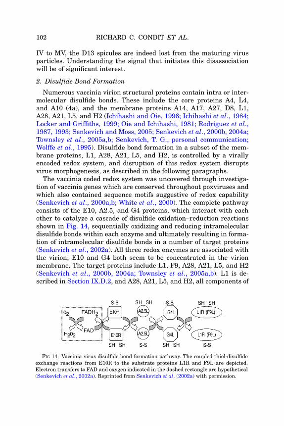
102 RICHARD C. CONDIT ET AL.
IV to MV, the D13 spicules are indeed lost from the maturing virusparticles. Understanding the signal that initiates this disassociationwill be of significant interest.
2. Disulfide Bond Formation
Numerous vaccinia virion structural proteins contain intra or inter-molecular disulfide bonds. These include the core proteins A4, L4,and A10 (4a), and the membrane proteins A14, A17, A27, D8, L1,A28, A21, L5, and H2 (Ichihashi and Oie, 1996; Ichihashi et al., 1984;Locker and Griffiths, 1999; Oie and Ichihashi, 1981; Rodriguez et al.,1987, 1993; Senkevich and Moss, 2005; Senkevich et al., 2000b, 2004a;Townsley et al., 2005a,b; Senkevich, T. G., personal communication;Wolffe et al., 1995). Disulfide bond formation in a subset of the mem-brane proteins, L1, A28, A21, L5, and H2, is controlled by a virallyencoded redox system, and disruption of this redox system disruptsvirus morphogenesis, as described in the following paragraphs.
The vaccinia coded redox system was uncovered through investiga-tion of vaccinia genes which are conserved throughout poxviruses andwhich also contained sequence motifs suggestive of redox capability(Senkevich et al., 2000a,b; White et al., 2000). The complete pathwayconsists of the E10, A2.5, and G4 proteins, which interact with eachother to catalyze a cascade of disulfide oxidation–reduction reactionsshown in Fig. 14, sequentially oxidizing and reducing intramoleculardisulfide bonds within each enzyme and ultimately resulting in forma-tion of intramolecular disulfide bonds in a number of target proteins(Senkevich et al., 2002a). All three redox enzymes are associated withthe virion; E10 and G4 both seem to be concentrated in the virionmembrane. The target proteins include L1, F9, A28, A21, L5, and H2(Senkevich et al., 2000b, 2004a; Townsley et al., 2005a,b). L1 is de-scribed in Section IX.D.2, and A28, A21, L5, and H2, all components of
FIG 14. Vaccinia virus disulfide bond formation pathway. The coupled thiol-disulfideexchange reactions from E10R to the substrate proteins L1R and F9L are depicted.Electron transfers to FAD and oxygen indicated in the dashed rectangle are hypothetical(Senkevich et al., 2002a). Reprinted from Senkevich et al. (2002a) with permission.

VACCINIA VIRUS: STRUCTURE AND ASSEMBLY 103
a membrane-anchored complex controlling virus entry, are discussedin Section IX.G.3. The F9 protein shows significant homology to L1,containing the same six conserved cysteines, although the role of F9 ininfection is currently unknown (Senkevich et al., 2000b).
Repression of any of the three enzymes of the redox pathway, E10,A2.5, or G4, interrupts virion morphogenesis at the IVN to MV transi-tion (Ravanello and Hruby, 1994b; Senkevich et al., 2000a, 2002b;White et al., 2000). Specifically, in these mutants, MV are significantlydecreased in number or absent from infection while IV accumulate.This does not imply that the redox pathway is active specifically duringthe IVN to MV transition; on the contrary, the redox substrates L1 andF9 are oxidized when morphogenesis is blocked at earlier stages byrepression of the A17 membrane protein or treatment with rifampicin(Senkevich et al., 2000b). However, the fact that redox pathway mu-tants and L1 mutants (Section IX.D.2) produce the same phenotypesuggests that the phenotype of the redox pathway mutants is, atleast in part, a manifestation of their inability to oxidize the L1 disul-fide bonds, thereby presumably rendering L1 nonfunctional (Blouchet al., 2005).
3. Addition of New Surface Proteins
In 1962, in a brief, prescient, and elegant report, Morgan and cow-orkers showed that when ferritin-labeled antibodies prepared fromrabbits immunized with vaccinia virus were used in immunoelectronmicroscopic examination of vaccinia-infected cells, MV were stainedbut IV were not stained. The authors concluded “that maturation ofvaccinia virus into the stable, infectious form is accompanied not onlyby an alteration in structure but also by the acquisition of a surfacecoating of antigen not present on the immature particles” (Morganet al., 1962). Forty-four years later, we understand with some precisionthe nature of a few of these modifications. Specifically, immunoelectronmicroscopy of infected cells demonstrates that antibody against eitherof the surface proteins A27 (Section X) or H3 shows significantly morestaining of MV than IV, suggesting that both proteins are added toparticles during the IV to MV transition (da Fonseca et al., 2000a;Sodeik et al., 1995). As described previously (Section IX.D.3), H3 is atransmembrane protein that undergoes posttranslational insertioninto virion membranes. In contrast, the peripheral protein A27 isanchored to the viral membrane via its interactions with the A17transmembrane protein. A27 is not required for MV formation butrather for wrapping of MV with Golgi membranes to form WV. Thus,surface proteins can be incorporated during the later stages of virion

104 RICHARD C. CONDIT ET AL.
maturation by distinct mechanisms. We speculate that these additionsoccur after the departure of the D13 lattice from the IV membrane.Other candidates for surface proteins added during the IV to MVtransition include the L1 protein (Section IX.D.2). Immunoelectronmicroscopy of virus infected cells using anti-L1 antibody shows a high-er density of L1 staining in regions containing MV than regions con-taining IV (Wolffe et al., 1995). As described previously, L1 is requiredfor the IV to MV transition, a process that is also stimulated by H3. Itseems likely that additional surface modifications during the IV to MVtransition will be revealed in the future.
Dales and coworkers have suggested that the spicule layer surround-ing IV, now known to be composed of D13 protein, is replaced by theSTEs appearing onMV, raising the question of the relationship betweenSTE and the proteins added late during MV maturation (Essani et al.,1982; Stern and Dales, 1976). Wilton et al. (1995) examined the proteincomposition of isolated STE, and while they did not unambiguouslyidentify each of the STE proteins, the results suggest that STE containa mixture of proteins that appear in the crescents and MV membraneboth early and late during morphogenesis, such as A17 and H3. Thuswhile the removal of D13 from IVand addition of several immunodomi-nantMVmembrane antigens to the developingMVmay restructure theparticle surface, it is likely that STE represent a complex of several ofthe full complement of MVmembrane proteins, rather than exclusivelyproteins added after the removal of D13.
F. Core Proteins that are not Essential for Virion Morphogenesisbut are Required for Transcriptional Competence of the Virion Core
Mutation of the genes encoding certain core proteins results, undernonpermissive conditions, in the formation of MV that are normal inappearance but not infectious. Two of these proteins, the I8 RNA heli-case and the H6 topoisomerase, are virion enzymes. The phenotypes ofI8 and H6 mutants are similar: permeabilized virions purified fromnonpermissive infections display defects in early transcription. Thesedata imply that the RNA helicase and DNA topoisomerase are bothrequired to deal with the topological constraints associated with thevirion transcription reaction (da Fonseca and Moss, 2003; Gross andShuman, 1996). There also exist at least four virion proteins that arenot thought to function in transcription per se but rather seem to affectthe assembly of a core that is competent for in situ transcription. Theseinclude L4, L3, H1, and E8, described in more detail in the followingsections.

VACCINIA VIRUS: STRUCTURE AND ASSEMBLY 105
1. L4
The L4R gene encodes a 29-kDa protein that is proteolyticallyprocessed to the mature 25-kDa form during the IV to MV transition(Fig. 13). L4 was originally referred to as VP8 and has been shown tohave both DNA and RNA binding activity (Bayliss and Smith, 1997;Yang and Bauer, 1988; Yang et al., 1988). An inducible recombinant hasbeen generated inwhich L4 expression is IPTG-dependent (Wilcock andSmith, 1994, 1996). When L4 is repressed, formation of both IVandMVis observed by electron microscopy. The IV, however, appear slightlyabnormal: they possess a space between the viroplasm and the innersurface of the membrane, as if bonding of the viroplasm to the mem-brane is weaker than normal. L4-deficient MV contain an otherwisenormal complement of virion proteins, but they are defective in tran-scription as judged by both in vivo and in vitro assays, and hence theyare not infectious. Furthermore, when these virions are extracted withdeoxycholate to solubilize virion enzymes, more proteins appear to bemore easily released than when L4-containing virions are examined inparallel. Thus, the internal structure ofMVassembled in the absence ofL4 appears to be somewhat fragile. These findings suggest that L4 playsa structural role in the assembly of a transcriptionally-competent core;in this regard, it is worth noting that L4 was originally reported to beencapsidated at �7000 copies/virion.
2. L3
The L3L gene encodes a highly conserved 41-kDa protein that ispackaged into virion cores. Repression of L3 expression results in theassembly of MV that are ultrastructurally normal in appearance butare reduced in infectivity (Resch and Moss, 2005). L3-deficient virionscontain the normal complement of active transcription enzymes, butthey are nevertheless defective for transcription in situ. Consistentwith these observations, the L3-deficient MV can bind and enter cells,however, only very small amounts of early viral mRNA are synthe-sized. Thus, similar to L4 (above) and E8 (below), L3 deficiency mayproduce a subtle defect in MV structure that ultimately impacts on thetranscriptional competency of the mutant virions.
3. H1
The H1L gene encodes a dual specificity protein phosphatase. This20-kDa protein is encapsidated at �200 copies/virion (Guan et al.,1991; Liu et al., 1995). It is known to regulate the phosphoryla-tion status of a number of viral and cellular proteins in vivo and has
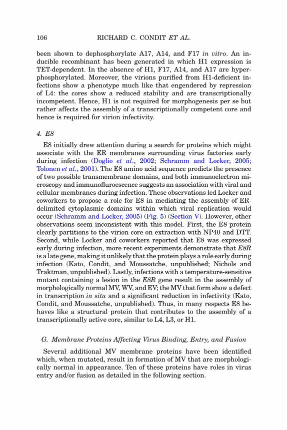
106 RICHARD C. CONDIT ET AL.
been shown to dephosphorylate A17, A14, and F17 in vitro. An in-ducible recombinant has been generated in which H1 expression isTET-dependent. In the absence of H1, F17, A14, and A17 are hyper-phosphorylated. Moreover, the virions purified from H1-deficient in-fections show a phenotype much like that engendered by repressionof L4: the cores show a reduced stability and are transcriptionallyincompetent. Hence, H1 is not required for morphogenesis per se butrather affects the assembly of a transcriptionally competent core andhence is required for virion infectivity.
4. E8
E8 initially drew attention during a search for proteins which mightassociate with the ER membranes surrounding virus factories earlyduring infection (Doglio et al., 2002; Schramm and Locker, 2005;Tolonen et al., 2001). The E8 amino acid sequence predicts the presenceof two possible transmembrane domains, and both immunoelectron mi-croscopy and immunofluroescence suggests an associationwith viral andcellular membranes during infection. These observations led Locker andcoworkers to propose a role for E8 in mediating the assembly of ER-delimited cytoplasmic domains within which viral replication wouldoccur (Schramm and Locker, 2005) (Fig. 5) (Section V). However, otherobservations seem inconsistent with this model. First, the E8 proteinclearly partitions to the virion core on extraction with NP40 and DTT.Second, while Locker and coworkers reported that E8 was expressedearly during infection, more recent experiments demonstrate that E8Ris a late gene,making it unlikely that the protein plays a role early duringinfection (Kato, Condit, and Moussatche, unpublished; Nichols andTraktman, unpublished). Lastly, infections with a temperature-sensitivemutant containing a lesion in the E8R gene result in the assembly ofmorphologically normalMV,WV, andEV; theMV that form showadefectin transcription in situ and a significant reduction in infectivity (Kato,Condit, and Moussatche, unpublished). Thus, in many respects E8 be-haves like a structural protein that contributes to the assembly of atranscriptionally active core, similar to L4, L3, or H1.
G. Membrane Proteins Affecting Virus Binding, Entry, and Fusion
Several additional MV membrane proteins have been identifiedwhich, when mutated, result in formation of MV that are morphologi-cally normal in appearance. Ten of these proteins have roles in virusentry and/or fusion as detailed in the following section.
VACCINIA VIRUS: STRUCTURE AND ASSEMBLY 107
1. Entry/Fusion Complex
A complex of eight interacting proteins has been described, which isanchored in the virion membrane and whose components appear tomediate fusion of the virion membrane with the plasma membraneduring virus entry (Ojeda et al., 2006; Senkevich and Moss, 2005;Senkevich et al., 2004b, 2005; Townsley et al., 2005a,b). The complexcontains the proteins A21, L5, A28, H2, G3, G9, A16, and J5. At leastfour of the proteins contain intramolecular disulfide bonds catalyzedby the virus coded redox system (Section IX.E.2), and three are myr-istylated. Inducible mutants have been constructed for five of thegenes encoding fusion complex proteins, and in all cases the mutantsproduce MV that are morphologically normal and transcriptionallyactive and which can undergo wrapping and export from cells as EV.These mutant MV can bind to cells but are defective in penetration.Repression of individual components of the complex does not appearto block the insertion of the remaining proteins into the virion mem-brane, although it does disrupt their ability to mediate virion entry.In sum, the membrane fusion complex is dispensable for virionmorphogenesis but essential for virion entry.
2. I2
The I2L gene encodes a 78-amino acid protein with a hydrophobicC-terminus predicted to serve as a transmembrane anchor. Indeed(Nichols, Stanitsa, Traktman, submitted for publication), I2L is ex-pressed late, behaves as an integral membrane protein, and is encap-sidated into the virion membrane. I2 appears to be an essentialprotein, and an inducible recombinant in which I2 expression is TETdependent has been generated. In the absence of I2, the viral life cycleprogresses normally, but the MV that are produced show an �400-foldreduction in specific infectivity. These virions are transcriptionallycompetent and can bind to target cells but are defective in fusion-mediated entry. Interactions between I2 and the entry complexdescribed previously have not been investigated.
3. D8
The D8 protein is expressed late and incorporated into the virionmembrane; the transmembrane domain is found at the N-terminus ofthe protein, and the C-terminus is exposed on the surface of the virions(Niles and Seto, 1988). D8 is one of three proteins, including A27 andH3, which bind cell surface glycosaminoglycans and are therefore

108 RICHARD C. CONDIT ET AL.
implicated in initial events that facilitate the binding of virus to cells(Chung et al., 1998; Hsiao et al., 1998, 1999; Lin et al., 2000). A solubleform of D8 has been shown to bind to chondroitin sulfate on the cellsurface and to compete with virions for binding to the cell (Hsiao et al.,1999). Viruses in which D8 has been insertionally inactivated areviable and direct normal morphogenesis; however, the virions thatare produced show a 10-fold reduction in specific infectivity due to animpaired ability to bind to target cells.
X. TRANSPORT, OCCLUSION, AND SECONDARY WRAPPING OF MV
As noted previously (Section III), most MV localize in clusters withinthe cytoplasm that are distal from the virus factories in which IV andIVN are assembled. Treatment of infected cells with drugs that in-hibit microtubule formation prevents this characteristic trafficking,implying that MV dissemination is a microtubule-mediated process(Sanderson et al., 2000). The mechanism by which MV interactswith microtubules has been investigated but remains unresolved(Sanderson et al., 2000; Ward, 2005).
While the majority of MV remain intracellular and represent afunctional end stage to virus assembly, a fraction of MV may undergofurther processing to be either exported as EV or embedded withinA-type inclusions (ATIs). Two MV membrane proteins are implicatedin these processes, A27 and A26.
A. A27
A27 is unusual among MV membrane proteins in that it does notcontain a transmembrane domain. Instead, the A27 protein forms adisulfide-bonded trimer, which is anchored to the MV membrane via astrong interaction with the integral A17 membrane protein (Lai et al.,1990; Rodriguez et al., 1987, 1993). A27 is an immunodominant anti-gen, and anti-A27 antibodies possess virus neutralizing activity(Rodriguez et al., 1985). As described previously, A27 is added to virusparticles late during maturation; it appears on MV but not IV (SectionIX.E.3). When A27 is repressed, morphologically normal MV form thatare fully infectious, although they are unable to undergo secondarywrapping by Golgi-derived membranes to form WV and, ultimately, tobe released by exocytosis as EV (Rodriguez and Smith, 1990b; Ward,2005). The A27 protein has also been implicated in attachment of virusto cells and in intracellular transport of MV (Chung et al., 1998;Sanderson et al., 2000).

VACCINIA VIRUS: STRUCTURE AND ASSEMBLY 109
B. A26
ATIs are large, cytoplasmic, electron-dense regions within whichnumerous MV are embedded (McKelvey et al., 2002). Formation ofATIs requires two viral genes, the ATI gene and the A26L gene. TheATI gene encodes the protein that forms the inclusion; the ATI gene isnot essential, and indeed the WR strain of vaccinia does not express anintact ATI protein and hence does not form inclusions. The A26L gene,on the other hand, encodes an MV membrane protein, initially namedp4c, which controls whether or not MV become embedded in inclu-sions. A26 is nonessential: if the p4c gene is inactivated, infectious MV,WV, and EV are produced. However, any inclusions that are formed donot contain embedded virus (McKelvey et al., 2002). The A26 protein ispresent on the surface of MV but not EEV, and it is therefore possiblethat the presence or absence of p4c in the virion membrane mayinfluence whether MV undergo secondary wrapping to become WVand EV, or whether they remain unwrapped in the cytoplasm (Ulaetoet al., 1996). It is not yet known how A26 associates with the MVmembrane; although some computer algorithms have predicted thepresence of a transmembrane region, others do not, and the proteindoes not contain any regions of significant hydrophobicity. However,the A26 protein does contain a C-terminal region that is 44% identicalto the region of the A27 protein that interacts with the membrane-bound A17 protein (see earlier). This observation raises the interestingpossibility that A26 and A27 may compete for binding to A17 and thusdetermine the balance between MV that remain unwrapped and thosethat continue on to form WV and EV.
XI. VIRION PROTEINS OF UNKNOWN FUNCTION
Four additional virion proteins of unknown function have been dis-cussed in the literature: the E11 and A12 core proteins and the I5 andA14.5 membrane proteins (Fig. 12). Little is known about E11 exceptthat temperature-sensitive mutants with lesions in this gene producenoninfectious virions at the nonpermissive temperature (Wang andShuman, 1996). An inducible recombinant in which expression ofA14.5 is IPTG-dependent has been generated; repression of this proteinhas no impact on the viral life cycle in culture but leads to an attenuatedphenotype in mice (Betakova et al., 2000). I5 (VP13) and A12 wereidentified during proteomic analyses of the vaccinia virion (Chunget al., 2006; Takahashi et al., 1994). No function has been ascribed to

110 RICHARD C. CONDIT ET AL.
either protein as neither gene has been subjected to genetic analysis,although both genes are highly conserved among poxviral genomes andas such the proteins are likely to play significant roles(s) during somefacet of the viral life cycle. Encapsidation of I5 into the virionmembranehas been verified by analysis of a virus encoding an epitope-taggedI5 allele at the endogenous locus (Nichols, Stanitsa, and Traktman,unpublished). The A12 core protein undergoes proteolytic maturationduring virion morphogenesis (Whitehead and Hruby, 1994b).
XII. SUMMARY AND PERSPECTIVES
Investigation into poxvirus structure and assembly is now at a tech-nical and conceptual crossroads. Identification of proteins in the virionand basic characterization of mutants affecting assembly is approach-ing saturation. A detailed picture of the contribution of individual genesto the structure and stepwise assembly of the virion is therefore begin-ning to emerge. The genetic, biochemical and cell biological tools arein place to bring this task to completion. Fascinating and significantquestions remain to be answered, including:
A. What are the precise localizations within the virion of each of theencapsidated proteins? What is the composition and structure of thetube-like structure within the core? What proteins make up the indi-vidual layers of the core wall? What proteins are contained inthe lateral bodies and what are their functions? What are the struc-tural relationships among the proteins in the membrane and how doesthis contribute to the bilaminar appearance of this structure?
B. What is the initial source of the membrane used to form crescents,how is the single bilayer of the crescents and IV assembled and stabi-lized in the virus factories, and what are the contributions of cellularcomponents and individual viral proteins to this process?
C. How are viral factories organized so that assembly componentsare delivered to maturing virions in an organized fashion?
D. What are the details of the molecular interactions that bind cres-cents to viroplasm, andhowdoes this relate to the ultimatematuration ofthe virion core?
E. What is the precise mechanism of DNA encapsidation intovirions?
F. What triggers the transition from IVN to MV, and how are thevarious events in this transition co-coordinated, including loss of D13,restructuring of the virion surface, proteolysis of structural proteins,
VACCINIA VIRUS: STRUCTURE AND ASSEMBLY 111
maturation of the core, and migration of maturing virions fromfactories to the periphery of the cell?
A thorough understanding of poxvirus morphogenesis will provideinvaluable insights into this complex and biomedically significant virus.Moreover, these studies should lead to the elucidation and discovery ofcell biological strategies that have deep relevance to such diverse pro-cesses as organelle biogenesis, intracellular protein trafficking, andmembrane remodeling.
ACKNOWLEDGMENTS
We thank Bernie Moss and John Heuser for helpful discussions while the manuscriptwas being written. We thank Bernie Moss and Wen Chang for communicating datain advance of publication. We thank Jason Mercer for help in figure preparation. Wethank Michel Moussatche for his expert rendering of the vaccinia virion model.Work described herein was supported by a grant to RCC from the NIH (5R01-AI055560) and by grants to PT from the NIH (9R01 AI 063620-11) and the GLRCE(sub-contract, 1-U54-AI057153).
REFERENCES
Andres, G., Alejo, A., Simon-Mateo, C., and Salas, M. L. (2001). African swine fever virusprotease, a new viral member of the SUMO-1-specific protease family. J. Biol. Chem.276:780–787.
Ansarah-Sobrinho, C., and Moss, B. (2004a). Vaccinia virus G1 protein, a predictedmetalloprotease, is essential for morphogenesis of infectious virions but not for cleav-age of major core proteins. J. Virol. 78:6855–6863.
Ansarah-Sobrinho, C., and Moss, B. (2004b). Role of the I7 protein in proteolytic proces-sing of vaccinia virus membrane and core components. J. Virol. 78:6335–6343.
Armstrong, J. A., Metz, D. H., and Young, M. R. (1973). The mode of entry of vacciniavirus into L cells. J. Gen. Virol. 21:533–537.
Baldick, C. J., Jr., and Moss, B. (1987). Resistance of vaccinia virus to rifampicinconferred by a single nucleotide substitution near the predicted NH2 terminus of agene encoding an Mr 62,000 polypeptide. Virology 156:138–145.
Bayliss, C. D., and Smith, G. L. (1997). Vaccinia virion protein VP8, the 25 kDa product ofthe L4R gene, binds single-stranded DNA and RNAwith similar affinity. Nucleic AcidsRes. 25:3984–3990.
Beaud, G., and Beaud, R. (2000). Temperature-dependent phosphorylation state of theH5R protein synthesised at the early stage of infection in cells infected with vacciniavirus ts mutants of the B1R and F10L protein kinases. Intervirology 43:67–70.
Beaud, G., Beaud, R., and Leader, D. P. (1995). Vaccinia virus gene H5R encodes aprotein that is phosphorylated by the multisubstrate vaccinia virus B1R proteinkinase. J. Virol. 69:1819–1826.

112 RICHARD C. CONDIT ET AL.
Betakova, T., and Moss, B. (2000). Disulfide bonds and membrane topology of thevaccinia virus A17L envelope protein. J. Virol. 74:2438–2442.
Betakova, T., Wolffe, E. J., andMoss, B. (1999a). Membrane topology of the vaccinia virusA17L envelope protein. Virology 261:347–356.
Betakova, T., Wolffe, E. J., and Moss, B. (1999b). Regulation of vaccinia virus morpho-genesis: Phosphorylation of the A14L and A17L membrane proteins and C-terminaltruncation of the A17L protein are dependent on the F10L kinase. J. Virol.73:3534–3543.
Betakova, T., Wolffe, E. J., and Moss, B. (2000). The vaccinia virus A14.5L gene encodes ahydrophobic 53-amino-acid virion membrane protein that enhances virulence in miceand is conserved among vertebrate poxviruses. J. Virol. 74:4085–4092.
Black, E. P., Moussatche, N., and Condit, R. C. (1998). Characterization of the interac-tions among vaccinia virus transcription factors G2R, A18R, and H5R. Virology245:313–322.
Blouch, R. E., Byrd, C. M., and Hruby, D. E. (2005). Importance of disulphide bonds forvaccinia virus L1R protein function. Virology J. 2:91.
Brown, N. G., Nick, M. D., Beaud, G., Hardie, G., and Leader, D. P. (2000). Identificationof sites phosphorylated by the vaccinia virus B1R kinase in viral protein H5R. BMCBiochem. 1:2.
Broyles, S. S. (2003). Vaccinia virus transcription. J. Gen. Virol.(on line. 2003).Byrd, C. M., Bolken, T. C., and Hruby, D. E. (2002). The vaccinia virus I7L gene productis the core protein proteinase. J. Virol. 76:8973–8976.
Byrd, C. M., Bolken, T. C., and Hruby, D. E. (2003). Molecular dissection of the vacciniavirus I7L core protein proteinase. J. Virol. 77:11279–11283.
Byrd, C. M., and Hruby, D. E. (2005). A conditional-lethal vaccinia virus mutant demon-strates that the I7L gene product is required for virion morphogenesis. Virology J. 2:4.
Cairns, J. (1960). The initiation of vaccinia infection. Virology 11:603–623.Carter, G. C., Law, M., Hollinshead, M., and Smith, G. L. (2005). Entry of the vacciniavirus intracellular mature virion and its interactions with glycosaminoglycans. J. Gen.Virol. 86:1279–1290.
Cassetti, M. C., Merchlinsky, M., Wolffe, E. J., Weisberg, A. S., and Moss, B. (1998). DNApackaging mutant: Repression of the vaccinia virus A32 gene results in noninfectious,DNA-deficient, spherical, enveloped particles. J. Virol. 72:5769–5780.
Castro, A. P., Carvalho, T. M., Moussatche, N., and Damaso, C. R. (2003). Redistributionof cyclophilin A to viral factories during vaccinia virus infection and its incorporationinto mature particles. J. Virol. 77:9052–9068.
Chertov, O. Y., Telezhinskaya, I. N., Zaitseva, E. V., Golubeva, T. B., Zinov’ev, V. V.,Ovechkina, L. G., Mazkova, L. B., and Malygin, E. G. (1991). Amino acid sequencedetermination of vaccinia virus immunodominant protein p35 and identification of thegene. Biomed. Sci. 2:151–154.
Chiu, W. L., and Chang, W. (2002). Vaccinia virus J1R protein: A viral membrane proteinthat is essential for virion morphogenesis. J. Virol. 76:9575–9587.
Chiu, W. L., Szajner, P., Moss, B., and Chang, W. (2005). Effects of a temperaturesensitivity mutation in the J1R protein component of a complex required for vacciniavirus assembly. J. Virol. 79:8046–8056.
Christen, L., Higman, M. A., and Niles, E. G. (1992). Phenotypic characterization of threetemperature-sensitive mutations in the vaccinia virus early gene transcription initia-tion factor. J. Gen. Virol. 73(Pt. 12):3155–3167.
Chung, C. S., Hsiao, J. C., Chang, Y. S., and Chang, W. (1998). A27L protein mediatesvaccinia virus interaction with cell surface heparan sulfate. J. Virol. 72:1577–1585.

VACCINIA VIRUS: STRUCTURE AND ASSEMBLY 113
Chung, C. S., Chen, C. H., Ho, M. Y., Hwang, C. Y., Liao, C. L., and Chang, W. (2006).Vaccinia virus proteome: Identification of proteins in vaccinia virus intracellularmature virion particles. J. Virol. 80: 2127–2140.
Condit, R. C., and Niles, E. G. (1990). Orthopoxvirus genetics. Curr. Top. Microbiol.Immunol. 163:1–39.
Cudmore, S., Blasco, R., Vincentelli, R., Esteban, M., Sodeik, B., Griffiths, G., andKrijnse, L. J. (1996). A vaccinia virus core protein, p39, is membrane associated.J. Virol. 70:6909–6921.
Cyrklaff, M., Risco, C., Fernandez, J. J., Jimenez, M. V., Esteban, M., Baumeister, W.,and Carrascosa, J. L. (2005). Cryo-electron tomography of vaccinia virus. Proc. Natl.Acad. Sci. USA 102:2772–2777.
da Fonseca, F., and Moss, B. (2003). Poxvirus DNA topoisomerase knockout mutantexhibits decreased infectivity associated with reduced early transcription. Proc. Natl.Acad. Sci. USA 100:11291–11296.
da Fonseca, F. G., Wolffe, E. J., Weisberg, A., and Moss, B. (2000a). Characterization ofthe vaccinia virus H3L envelope protein: Topology and posttranslational membraneinsertion via the C-terminal hydrophobic tail. J. Virol. 74:7508–7517.
da Fonseca, F. G., Wolffe, E. J., Weisberg, A., and Moss, B. (2000b). Effects of deletion orstringent repression of the H3L envelope gene on vaccinia virus replication. J. Virol.74:7518–7528.
da Fonseca, F. G., Weisberg, A. S., Caeiro, M. F., and Moss, B. (2004). Vaccinia virusmutants with alanine substitutions in the conserved G5R gene fail to initiate morpho-genesis at the nonpermissive temperature. J. Virol. 78:10238–10248.
Dales, S. (1962). An electron microscope study of the early association between twomammalian viruses and their hosts. J. Cell Biol. 13:303–322.
Dales, S. (1963). The uptake and development of vaccinia virus in strain L cells followedwith labeled viral deoxyribonucleic acid. J. Cell Biol. 18:51–72.
Dales, S., and Kajioka, R. (1964). The cycle of multiplication of vaccinia virus in Earle’sstrain L cells. I. Uptake and penetration. Virology 24:278–294.
Dales, S., and Mosbach, E. H. (1968). Vaccinia as a model for membrane biogenesis.Virology 35:564–583.
Dales, S., and Pogo, B. G. (1981). Biology of poxviruses. Virol. Monogr. 18:1–109.Dales, S., and Siminovitch, L. (1961). The development of vaccinia virus in Earle’s L strain
cells as examined by electron microscopy. J. Biophys. Biochem. Cytol. 10:475–503.Dellis, S., Strickland, K. C., McCrary, W. J., Patel, A., Stocum, E., and Wright, C. F.
(2004). Protein interactions among the vaccinia virus late transcription factors.Virology 329:328–336.
Demasi, J., and Traktman, P. (2000). Clustered charge-to-alanine mutagenesis of thevaccinia virus H5 gene: Isolation of a dominant, temperature-sensitive mutant with aprofound defect in morphogenesis. J. Virol. 74:2393–2405.
Demasi, J., Du, S., Lennon, D., and Traktman, P. (2001a). Vaccinia virus telomeres:Interaction with the viral I1, I6, and K4 proteins. J. Virol. 75:10090–10105.
Demasi, J., Du, S., Lennon, D., and Traktman, P. (2001b). Vaccinia virus telomeres:Interaction with the viral I1, I6, and K4 proteins. J. Virol. 75:10090–10105.
Demkowicz, W. E., Maa, J. S., and Esteban, M. (1992). Identification and characteriza-tion of vaccinia virus genes encoding proteins that are highly antigenic in animals andare immunodominant in vaccinated humans. J. Virol. 66:386–398.
Derrien, M., Punjabi, A., Khanna, M., Grubisha, O., and Traktman, P. (1999). Tyrosinephosphorylation of A17 during vaccinia virus infection: Involvement of the H1 phos-phatase and the F10 kinase. J. Virol. 73:7287–7296.

114 RICHARD C. CONDIT ET AL.
Doglio, L., De, M. A., Schleich, S., Roos, N., and Krijnse, L. J. (2002). The vaccinia virusE8R gene product: A viral membrane protein that is made early in infection andpackaged into the virions’ core. J. Virol. 76:9773–9786.
Dubochet, J., Adrian, M., Richter, K., Garces, J., and Wittek, R. (1994). Structure ofintracellular mature vaccinia virus observed by cryoelectron microscopy. J. Virol.68:1935–1941.
Dyster, L. M., and Niles, E. G. (1991). Genetic and biochemical characterization ofvaccinia virus genes D2L and D3R which encode virion structural proteins. Virology182:455–467.
Easterbrook, K. B. (1966). Controlled degradation of vaccinia virions in vitro: An electronmicroscopic study. J. Ultrastruct. Res. 14:484–496.
Ericsson, M., Cudmore, S., Shuman, S., Condit, R. C., Griffiths, G., and Locker, J. K.(1995). Characterization of ts 16, a temperature-sensitive mutant of vaccinia virus.J. Virol. 69:7072–7086.
Ericsson, M., Sodeik, B., Locker, J. K., and Griffiths, G. (1997). In vitro reconstitution ofan intermediate assembly stage of vaccinia virus. Virology 235:218–227.
Essani, K., and Dales, S. (1979). Biogenesis of vaccinia: Evidence for more than 100polypeptides in the virion. Virology 95:385–394.
Essani, K., Dugre, R., and Dales, S. (1982). Biogenesis of vaccinia: Involvement ofspicules of the envelope during virion assembly examined by means of conditionallethal mutants and serology. Virology 118:279–292.
Fenner, F., Wittek, R., and Dumbell, K. R. (1989). “The orthopoxviruses.” AcademicPress, San Diego.
Franke, C. A., and Hruby, D. E. (1987). Association of non-viral proteins with recombi-nant vaccinia virus virions. Arch. Virol. 94:347–351.
Franke, C. A., Wilson, E. M., and Hruby, D. E. (1990). Use of a cell-free system to identifythe vaccinia virus L1R gene product as the major late myristylated virion protein M25.J. Virol. 64:5988–5996.
Gaigg, B., Simbeni, R., Hrastnik, C., Paltauf, F., and Daum, G. (1995). Characterizationof a microsomal subfraction associated with mitochondria of the yeast, Saccharomycescerevisiae. Involvement in synthesis and import of phospholipids into mitochondria.Biochim. Biophys. Acta 1234:214–220.
Garcia, A. D., andMoss, B. (2001). Repression of vaccinia virus holliday junction resolvaseinhibits processing of viral DNA into unit-length genomes. J. Virol. 75:6460–6471.
Garcia, A. D., Aravind, L., Koonin, E. V., and Moss, B. (2000). Bacterial-type DNAholliday junction resolvases in eukaryotic viruses. Proc. Natl. Acad. Sci. USA97:8926–8931.
Goebel, S. J., Johnson, G. P., Perkus, M. E., Davis, S. W., Winslow, J. P., and Paoletti, E.(1990). The complete DNA sequence of vaccinia virus. Virology 179:247–263.
Gordon, J., Mohandas, A., Wilton, S., and Dales, S. (1991). A prominent antigenic surfacepolypeptide involved in the biogenesis and function of the vaccinia virus envelope.Virology 181:671–686.
Griffiths, G., Roos, N., Schleich, S., and Locker, J. K. (2001a). Structure and assemblyof intracellular mature vaccinia virus: Thin-section analyses. J. Virol. 75:11056–11070.
Griffiths, G., Wepf, R., Wendt, T., Locker, J. K., Cyrklaff, M., and Roos, N. (2001b).Structure and assembly of intracellular mature vaccinia virus: Isolated-particle anal-ysis. J. Virol. 75:11034–11055.
Grimley, P. M., Rosenblum, E. N., Mims, S. J., and Moss, B. (1970). Interruption byRifampin of an early stage in vaccinia virus morphogenesis: Accumulation of mem-branes which are precursors of virus envelopes. J. Virol. 6:519–533.

VACCINIA VIRUS: STRUCTURE AND ASSEMBLY 115
Grosenbach, D. W., Hansen, S. G., and Hruby, D. E. (2000). Identification and analysis ofvaccinia virus palmitylproteins. Virology 275:193–206.
Gross, C. H., and Shuman, S. (1996). Vaccinia virions lacking the RNA helicase nucleo-side triphosphate phosphohydrolase II are defective in early transcription. J. Virol.70:8549–8557.
Grubisha, O., and Traktman, P. (2003). Genetic analysis of the vaccinia virus I6telomere-binding protein uncovers a key role in genome encapsidation. J. Virol.77:10929–10942.
Guan, K. L., Broyles, S. S., and Dixon, J. E. (1991). A Tyr/Ser protein phosphataseencoded by vaccinia virus. Nature 350:359–362.
Hassett, D. E., and Condit, R. C. (1994). Targeted construction of temperature-sensitivemutations in vaccinia virus by replacing clustered charged residues with alanine. Proc.Natl. Acad. Sci. USA 91:4554–4558.
Hassett, D. E., Lewis, J. I., Xing, X., DeLange, L., and Condit, R. C. (1997). Analysis of atemperature-sensitive vaccinia virus mutant in the viral mRNA capping enzymeisolated by clustered charge-to-alanine mutagenesis and transient dominant selection.Virology 238:391–409.
Hedengren-Olcott, M., Byrd, C. M., Watson, J., and Hruby, D. E. (2004). The vacciniavirus G1L putative metalloproteinase is essential for viral replication in vivo. J. Virol.78:9947–9953.
Heljasvaara, R., Rodriguez, D., Risco, C., Carrascosa, J. L., Esteban, M., and Rodriguez,J. R. (2001). The major core protein P4a (A10L gene) of vaccinia virus is essential forcorrect assembly of viral DNA into the nucleoprotein complex to form immature viralparticles. J. Virol. 75:5778–5795.
Hellen, C. U., and Wimmer, E. (1992). The role of proteolytic processing in the morpho-genesis of virus particles. Experientia 48:201–215.
Heuser, J. (2005). Deep-etch EM reveals that the early poxvirus envelope is a singlemembrane bilayer stabilized by a geodetic “honeycomb” surface coat. J. Cell Biol.169:269–283.
Hiller, G., Eibl, H., and Weber, K. (1981). Acyl bis(monoacylglycero)phosphate, assumedto be a marker for lysosomes, is a major phospholipid of vaccinia virions. Virology113:761–764.
Hollinshead, M., Vanderplasschen, A., Smith, G. L., and Vaux, D. J. (1999). Vacciniavirus intracellular mature virions contain only one lipid membrane. J. Virol.73:1503–1517.
Holowczak, J. A. (1982). Poxvirus DNA. Curr. Top. Microbiol. Immunol. 97:27–79.Holowczak, J. A., Thomas, V. I., and Flores, L. (1975). Isolation and characterization of
vaccinia virus “nucleoids.” Virology 67:506–519.Hooda-Dhingra, U., Thompson, C. L., and Condit, R. C. (1989). Detailed phenotypic charac-
terization of five temperature-sensitivemutants in the 22- and 147-kilodalton subunits ofvaccinia virus DNA-dependent RNA polymerase. J. Virol. 63:714–729.
Hsiao, J. C., Chung, C. S., and Chang, W. (1998). Cell surface proteoglycans are neces-sary for A27L protein-mediated cell fusion: Identification of the N-terminal region ofA27L protein as the glycosaminoglycan-binding domain. J. Virol. 72:8374–8379.
Hsiao, J. C., Chung, C. S., and Chang, W. (1999). Vaccinia virus envelope D8L proteinbinds to cell surface chondroitin sulfate and mediates the adsorption of intracellularmature virions to cells. J. Virol. 73:8750–8761.
Hu, X., Carroll, L. J., Wolffe, E. J., and Moss, B. (1996). De novo synthesis of the earlytranscription factor 70-kilodalton subunit is required for morphogenesis of vacciniavirions. J. Virol. 70:7669–7677.
116 RICHARD C. CONDIT ET AL.
Hu, X., Wolffe, E. J., Weisberg, A. S., Carroll, L. J., and Moss, B. (1998). Repression of theA8L gene, encoding the early transcription factor 82-kilodalton subunit, inhibits mor-phogenesis of vaccinia virions. J. Virol. 72:104–112.
Husain, M., and Moss, B. (2003). Evidence against an essential role of COPII-mediatedcargo transport to the endoplasmic reticulum-Golgi intermediate compartment in theformation of the primary membrane of vaccinia virus. J. Virol. 77:11754–11766.
Ichihashi, Y., and Oie, M. (1996). Neutralizing epitope on penetration protein of vacciniavirus. Virology 220:491–494.
Ichihashi, Y., Oie, M., and Tsuruhara, T. (1984). Location of DNA-binding proteins anddisulfide-linked proteins in vaccinia virus structural elements. J. Virol. 50:929–938.
Ishii, K., and Moss, B. (2001). Role of vaccinia virus A20R protein in DNA replication:Construction and characterization of temperature-sensitive mutants. J. Virol.75:1656–1663.
Ishii, K., and Moss, B. (2002). Mapping interaction sites of the A20R protein componentof the vaccinia virus DNA replication complex. Virology 303:232–239.
Jensen, O. N., Houthaeve, T., Shevchenko, A., Cudmore, S., Ashford, T., Mann, M.,Griffiths, G., and Krijnse, L. J. (1996). Identification of the major membrane and coreproteins of vaccinia virus by two-dimensional electrophoresis. J. Virol. 70:7485–7497.
Johnson, G. P., Goebel, S. J., and Paoletti, E. (1993). An update on the vaccinia virusgenome. Virology 196:381–401.
Kane, E. M., and Shuman, S. (1992). Temperature-sensitive mutations in the vacciniavirus H4 gene encoding a component of the virion RNA polymerase. J. Virol.66:5752–5762.
Kane, E. M., and Shuman, S. (1993). Vaccinia virus morphogenesis is blocked by atemperature-sensitive mutation in the I7 gene that encodes a virion component.J. Virol. 67:2689–2698.
Kao, S. Y., and Bauer, W. R. (1987). Biosynthesis and phosphorylation of vaccinia virusstructural protein VP11. Virology 159:399–407.
Kao, S. Y., Ressner, E., Kates, J., and Bauer, W. R. (1981). Purification and characteriza-tion of a superhelix binding protein from vaccinia virus. Virology 111:500–508.
Kato, S. E., Strahl, A. L., Moussatche, N., and Condit, R. C. (2004). Temperature-sensitive mutants in the vaccinia virus 4b virion structural protein assemble mal-formed, transcriptionally inactive intracellular mature virions. Virology 330:127–146.
Katz, E., and Moss, B. (1970a). Formation of a vaccinia virus structural polypeptide froma higher molecular weight precursor: Inhibition by rifampicin. Proc. Natl. Acad. Sci.USA 66:677–684.
Katz, E., andMoss, B. (1970b). Vaccinia virus structural polypeptide derived from a high-molecular-weight precursor: Formation and integration into virus particles. J. Virol.6:717–726.
Klemperer, N., Ward, J., Evans, E., and Traktman, P. (1997). The vaccinia virus I1protein is essential for the assembly of mature virions. J. Virol. 71:9285–9294.
Koonin, E. V., Senkevich, T. G., and Chernos, V. I. (1993). Gene A32 product of vacciniavirus may be an ATPase involved in viral DNA packaging as indicated by sequencecomparisons with other putative viral ATPases. Virus Genes 7:89–94.
Kovacs, G. R., and Moss, B. (1996). The vaccinia virus H5R gene encodes late genetranscription factor 4: Purification, cloning, and overexpression.J. Virol. 70:6796–6802.
Krijnse-Locker, J., Schleich, S., Rodriguez, D., Goud, B., Snijder, E. J., and Griffiths, G.(1996). The role of a 21-kDa viral membrane protein in the assembly of vaccinia virusfrom the intermediate compartment. J. Biol. Chem. 271:14950–14958.
VACCINIA VIRUS: STRUCTURE AND ASSEMBLY 117
Lackner, C. A., D’Costa, S. M., Buck, C., and Condit, R. C. (2003). Complementa-tion analysis of the Dales collection of vaccinia virus temperature-sensitive mutants.Virology 305:240–259.
Lai, C. F., Gong, S. C., and Esteban, M. (1990). Structural and functional properties ofthe 14-kDa envelope protein of vaccinia virus synthesized in Escherichia coli. J. Biol.Chem. 265:22174–22180.
Lanzer, W., and Holowczak, J. A. (1975). Polyamines in vaccinia virions and polypeptidesreleased from viral cores by acid extraction. J. Virol. 16:1254–1264.
Law, M., Carter, G. C., Roberts, K. L., Hollinshead, M., and Smith, G. L. (2006). Ligand-induced and nonfusogenic dissolution of a viral membrane. Proc. Natl. Acad. Sci.103:5989–5994.
Lefkowitz, E. J., Upton, C., Changayil, S. S., Buck, C., Traktman, P., and Buller, R. M.(2005). Poxvirus bioinformatics resource center: A comprehensive poxviridae informa-tional and analytical resource. Nucleic Acids Res. 33:D311–D316.
Li, S. J., and Hochstrasser, M. (1999). A new protease required for cell-cycle progressionin yeast. Nature 398:246–251.
Lin, C. L., Chung, C. S., Heine, H. G., and Chang, W. (2000). Vaccinia virus envelope H3Lprotein binds to cell surface heparan sulfate and is important for intracellular maturevirion morphogenesis and virus infection in vitro and in vivo. J. Virol. 74:3353–3365.
Lin, S., and Broyles, S. S. (1994). Vaccinia protein kinase 2: A second essential serine/threonine protein kinase encoded by vaccinia virus. Proc. Natl. Acad. Sci. USA91:7653–7657.
Liu, K., Lemon, B., and Traktman, P. (1995). The dual-specificity phosphatase encoded byvaccinia virus, VH1, is essential for viral transcription in vivo and in vitro. J. Virol.69:7823–7834.
Locker, J. K., and Griffiths, G. (1999). An unconventional role for cytoplasmic disulfidebonds in vaccinia virus proteins. J. Cell Biol. 144:267–279.
Locker, J. K., Kuehn, A., Schleich, S., Rutter, G., Hohenberg,H.,Wepf, R., andGriffiths, G.(2000). Entry of the two infectious forms of vaccinia virus at the plasma membane issignaling-dependent for the IMV but not the EEV. Mol. Biol. Cell 11:2497–2511.
Maa, J. S., and Esteban, M. (1987). Structural and functional studies of a 39,000-Mrimmunodominant protein of vaccinia virus. J. Virol. 61:3910–3919.
Malkin, A. J., McPherson, A., and Gershon, P. D. (2003). Structure of intracellularmature vaccinia virus visualized by in situ atomic force microscopy. J. Virol.77:6332–6340.
Martin, S. A., Paoletti, E., and Moss, B. (1975). Purification of mRNA guanylyltransfer-ase and mRNA (guanine-7-) methyltransferase from vaccinia virions. J. Biol. Chem.250:9322–9329.
McCraith, S., Holtzman, T., Moss, B., and Fields, S. (2000). Genome-wide analysis ofvaccinia virus protein-protein interactions. Proc. Natl. Acad. Sci. USA 97:4879–4884.
McKelvey, T. A., Andrews, S. C., Miller, S. E., Ray, C. A., and Pickup, D. J. (2002).Identification of the orthopoxvirus p4c gene, which encodes a structural protein thatdirects intracellular mature virus particles into A-type inclusions. J. Virol.76:11216–11225.
Medzon, E. L., and Bauer, H. (1970). Structural features of vaccinia virus revealed bynegative staining, sectioning, and freeze-etching. Virology 40:860–867.
Mercer, J., and Traktman, P. (2003). Investigation of structural and functional motifswithin the vaccinia virus A14 phosphoprotein, an essential component of the virionmembrane. J. Virol. 77:8857–8871.

118 RICHARD C. CONDIT ET AL.
Mercer, J., and Traktman, P. (2005). Genetic and cell biological characterization of thevaccinia virus A30 and G7 phosphoproteins. J. Virol. 79:7146–7161.
Miner, J. N., and Hruby, D. E. (1989). Rifampicin prevents virosome localization of L65,an essential vaccinia virus polypeptide. Virology 170:227–237.
Mohandas, A. R., and Dales, S. (1995). Involvement of spicules in the formation ofvaccinia virus envelopes elucidated by a conditional lethal mutant. Virology214:494–502.
Morgan, C. (1976a). The insertion of DNA into vaccinia virus. Science 193:591–592.Morgan, C. (1976b). Vaccinia virus reexamined: Development and release. Virology73:43–58.
Morgan, C., Ellison, S. A., Rose, H. M., and Moore, D. H. (1955). Serial sections ofvaccinia virus examined at one stage of development in the electron microscope. Exp.Cell Res. 9:572–578.
Morgan, C., Rose, H. M., and Rifkind, R. A. (1962). Use of ferritin-conjugated antibodiesin electron microscopic studies of influenza and vaccinia viruses. Cold Spring Harb.Symp. Quant. Biol. 27:57–65.
Morgan, J. R., Cohen, L. K., and Roberts, B. E. (1984). Identification of the DNAsequences encoding the large subunit of the mRNA-capping enzyme of vaccinia virus.J. Virol. 52:206–214.
Moss, B. (2001). Poxviridae: The viruses and their replication. In “Fields Virology”(D. M. Knipe and P. M. Howley, eds.), pp. 2849–2884. Lippincott Williams &Wilkins, Philadelphia.
Moss, B. (2006). Poxvirus entry and membrane fusion. Virology 344:48–54.Moss, B., Katz, E., andRosenblum,E. N. (1969a). Vaccinia virus directedRNAand proteinsynthesis in the presence of rifampicin. Biochem. Biophys. Res. Commun. 36:858–865.
Moss, B., Rosenblum, E. N., Katz, E., and Grimley, P. M. (1969b). Rifampicin: A specificinhibitor of vaccinia virus assembly. Nature 224:1280–1284.
Nagayama, A., Pogo, B. G., and Dales, S. (1970). Biogenesis of vaccinia: Separation ofearly stages from maturation by means of rifampicin. Virology 40:1039–1051.
Nermut, M. V. (1973). Freeze-drying and freeze-etching of viruses. In “Freeze-EtchingTechniques and Applications” (E. L. Benedetti and P. Favard, eds.), pp. 135–150.Societe Francaise de Microscopie Electronique, Paris.
Niles, E. G., and Seto, J. (1988). Vaccinia virus gene D8 encodes a virion transmembraneprotein. J. Virol. 62:3772–3778.
Niles, E. G., Lee-Chen, G. J., Shuman, S., Moss, B., and Broyles, S. S. (1989). Vacciniavirus gene D12L encodes the small subunit of the viral mRNA capping enzyme.Virology 172:513–522.
Nowakowski, M., Bauer, W., and Kates, J. (1978a). Characterization of a DNA-bindingphosphoprotein from vaccinia virus replication complex. Virology 86:217–225.
Nowakowski, M., Kates, J., and Bauer, W. (1978b). Isolation of two DNA-binding proteinsfrom the intracellular replication complex of vaccinia virus. Virology 84:260–267.
Noyes, W. F. (1962a). Further studies on the structure of vaccinia virus. Virology18:511–516.
Noyes, W. F. (1962b). The surface fine structure of vaccinia virus. Virology 17:282–287.Oie, M., and Ichihashi, Y. (1981). Characterization of vaccinia polypeptides. Virology113:263–276.
Ojeda, S., Senkevich, T. G., and Moss, B. (2006). Entry of vaccinia virus and cell-cellfusion require a highly conserved cysteine-rich membrane protein encoded by theA16L gene. J. Virol. 80:51–61.
Palade, G. E. (1983). Membrane biogenesis: An overview.Methods Enzymol. 96:xxix–xxLv.

VACCINIA VIRUS: STRUCTURE AND ASSEMBLY 119
Pedersen, K., Snijder, E. J., Schleich, S., Roos, N., Griffiths, G., and Locker, J. K. (2000).Characterization of vaccinia virus intracellular cores: Implications for viral uncoatingand core structure. J. Virol. 74:3525–3536.
Peters, D., and Mueller, G. (1963). The fine structure of the DNA-containing core ofvaccinia virus. Virology 21:267–269.
Pichler, H., Gaigg, B., Hrastnik, C., Achleitner, G., Kohlwein, S.D., Zellnig, G., Perktold, A.,and Daum, G. (2001). A subfraction of the yeast endoplasmic reticulum associates withthe plasma membrane and has a high capacity to synthesize lipids. Eur. J. Biochem.268:2351–2361.
Pogo, B. G., and Dales, S. (1971). Biogenesis of vaccinia: Separation of early stages frommaturation by means of hydroxyurea. Virology 43:144–151.
Punjabi, A., and Traktman, P. (2005). Cell biological and functional characterization ofthe vaccinia virus f10 kinase: Implications for the mechanism of virion morphogenesis.J. Virol. 79:2171–2190.
Punjabi, A., Boyle, K., DeMasi, J., Grubisha, O., Unger, B., Khanna,M., and Traktman, P.(2001). Clustered charge-to-alanine mutagenesis of the vaccinia virus A20 gene:Temperature-sensitive mutants have a DNA-minus phenotype and are defective inthe production of processive DNA polymerase activity. J. Virol. 75:12308–12318.
Ravanello, M. P., and Hruby, D. E. (1994a). Characterization of the vaccinia virus L1Rmyristylprotein as a component of the intracellular virion envelope. J. Gen. Virol. 75(Pt. 6):1479–1483.
Ravanello, M. P., and Hruby, D. E. (1994b). Conditional lethal expression of the vacciniavirus L1R myristylated protein reveals a role in virion assembly. J. Virol.68:6401–6410.
Reggiori, F., and Klionsky, D. J. (2005). Autophagosomes: Biogenesis from scratch? Curr.Opin. Cell Biol. 17:415–422.
Resch, W., and Moss, B. (2005). The conserved poxvirus L3 virion protein is required fortranscription of vaccinia virus early genes. J. Virol. 79:14719–14729.
Resch, W., Weisberg, A. S., and Moss, B. (2005). Vaccinia virus nonstructural proteinencoded by the A11R gene is required for formation of the virion membrane. J. Virol.79:6598–6609.
Risco, C., Rodriguez, J. R., Demkowicz,W.,Heljasvaara,R., Carrascosa, J. L., Esteban,M.,andRodriguez,D. (1999). The vaccinia virus 39-kDaprotein forms a stable complexwiththe p4a/4a major core protein early in morphogenesis. Virology 265:375–386.
Risco, C., Rodriguez, J. R., Lopez-Iglesias, C., Carrascosa, J. L., Esteban, M., andRodriguez, D. (2002). Endoplasmic reticulum-Golgi intermediate compartment mem-branes and vimentin filaments participate in vaccinia virus assembly. J. Virol.76:1839–1855.
Rodriguez, D., Rodriguez, J. R., and Esteban, M. (1993). The vaccinia virus 14-kilodaltonfusion protein forms a stable complex with the processed protein encoded by thevaccinia virus A17L gene. J. Virol. 67:3435–3440.
Rodriguez, D., Esteban, M., and Rodriguez, J. R. (1995). Vaccinia virus A17L geneproduct is essential for an early step in virion morphogenesis. J. Virol. 69:4640–4648.
Rodriguez, D., Risco, C., Rodriguez, J. R., Carrascosa, J. L., and Esteban, M. (1996).Inducible expression of the vaccinia virus A17L gene provides a synchronized systemto monitor sorting of viral proteins during morphogenesis. J. Virol. 70:7641–7653.
Rodriguez, D., Barcena, M., Mobius, W., Schleich, S., Esteban, M., Geerts, W. J. C.,Koster, A. J., Griffiths, G., and Locker, J. K. (2006). A vaccinia virus lacking A10L:Viral core proteins accumulate on structures derived from the endoplasmic reticulum.Cellular Microbiology 8:427–437.

120 RICHARD C. CONDIT ET AL.
Rodriguez, J. F., and Smith, G. L. (1990a). Inducible gene expression from vaccinia virusvectors. Virology 177:239–250.
Rodriguez, J. F., and Smith, G. L. (1990b). IPTG-dependent vaccinia virus: Identificationof a virus protein enabling virion envelopment by Golgi membrane and egress. NucleicAcids Res. 18:5347–5351.
Rodriguez, J. F., Janeczko, R., and Esteban, M. (1985). Isolation and characterization ofneutralizing monoclonal antibodies to vaccinia virus. J. Virol. 56:482–488.
Rodriguez, J. F., Paez, E., and Esteban, M. (1987). A 14,000-Mr envelope protein ofvaccinia virus is involved in cell fusion and forms covalently linked trimers. J. Virol.61:395–404.
Rodriguez, J. R., Risco, C., Carrascosa, J. L., Esteban, M., and Rodriguez, D. (1997).Characterization of early stages in vaccinia virus membrane biogenesis: Implicationsof the 21-kilodalton protein and a newly identified 15-kilodalton envelope protein.J. Virol. 71:1821–1833.
Rodriguez, J. R., Risco, C., Carrascosa, J. L., Esteban, M., and Rodriguez, D. (1998).Vaccinia virus 15-kilodalton (A14L) protein is essential for assembly and attachment ofviral crescents to virosomes. J. Virol. 72:1287–1296.
Roos, N., Cyrklaff, M., Cudmore, S., Blasco, R., Krijnse-Locker, J., and Griffiths, G.(1996). A novel immunogold cryoelectron microscopic approach to investigate thestructure of the intracellular and extracellular forms of vaccinia virus. EMBO J.15:2343–2355.
Rosel, J., and Moss, B. (1985). Transcriptional and translational mapping and nucleotidesequence analysis of a vaccinia virus gene encoding the precursor of the major corepolypeptide 4b. J. Virol. 56:830–838.
Salmons, T., Kuhn, A., Wylie, F., Schleich, S., Rodriguez, J. R., Rodriguez, D., Esteban,M., Griffiths, G., and Locker, J. K. (1997). Vaccinia virus membrane proteins p8 andp16 are cotranslationally inserted into the rough endoplasmic reticulum and retainedin the intermediate compartment. J. Virol. 71:7404–7420.
Saminathan, M., Thomas, T., Shirahata, A., Pillai, C. K., and Thomas, T. J. (2002).Polyamine structural effects on the induction and stabilization of liquid crystallineDNA: Potential applications to DNA packaging, gene therapy and polyamine thera-peutics. Nucleic Acids Res. 30:3722–3731.
Sanderson, C. M., Hollinshead, M., and Smith, G. L. (2000). The vaccinia virus A27Lprotein is needed for the microtubule-dependent transport of intracellular maturevirus particles. J. Gen. Virol. 81(Pt. 1):47–58.
Sarov, I., and Joklik, W. K. (1972). Studies on the nature and location of the capsidpolypeptides of vaccinia virions. Virology 50:579–592.
Schramm, B., and Locker, J. K. (2005). Cytoplasmic organization of POXvirus DNAreplication. Traffic 6:839–846.
Senkevich, T. G., and Moss, B. (2005). Vaccinia virus H2 protein is an essential compo-nent of a complex involved in virus entry and cell-cell fusion. J. Virol. 79:4744–4754.
Senkevich, T. G., Weisberg, A. S., and Moss, B. (2000a). Vaccinia virus E10R protein isassociated with the membranes of intracellular mature virions and has a role inmorphogenesis. Virology 278:244–252.
Senkevich, T. G., White, C. L., Koonin, E. V., and Moss, B. (2000b). A viral member of theERV1/ALR protein family participates in a cytoplasmic pathway of disulfide bondformation. Proc. Natl. Acad. Sci. USA 97:12068–12073.
Senkevich, T. G., White, C. L., Koonin, E. V., and Moss, B. (2002a). Complete pathway forprotein disulfide bond formation encoded by poxviruses. Proc. Natl. Acad. Sci. USA99:6667–6672.

VACCINIA VIRUS: STRUCTURE AND ASSEMBLY 121
Senkevich, T. G., White, C. L., Weisberg, A., Granek, J. A., Wolffe, E. J., Koonin, E. V.,and Moss, B. (2002b). Expression of the vaccinia virus A2.5L redox protein is requiredfor virion morphogenesis. Virology 300:296–303.
Senkevich, T. G., Ward, B. M., andMoss, B. (2004a). Vaccinia virus A28L gene encodes anessential protein component of the virion membrane with intramolecular disulfidebonds formed by the viral cytoplasmic redox pathway. J. Virol. 78:2348–2356.
Senkevich, T. G., Ward, B. M., and Moss, B. (2004b). Vaccinia virus entry into cells isdependent on a virion surface protein encoded by the A28L gene. J. Virol.78:2357–2366.
Senkevich, T. G., Ojeda, S., Townsley, A. C., Nelson, G. E., and Moss, B. (2005). Poxvirusmultiprotein entry-fusion complex. Proc. Natl. Acad. Sci. USA 102:18572–18577.
Smith, G. L., Vanderplasschen, A., and Law, M. (2002). The formation and function ofextracellular enveloped vaccinia virus. J. Gen. Virol. 83:2915–2931.
Sodeik, B., and Krijnse-Locker, J. (2002). Assembly of vaccinia virus revisited: De novomembrane synthesis or acquisition from the host? Trends Microbiol. 10:15–24.
Sodeik, B., Doms, R. W., Ericsson, M., Hiller, G., Machamer, C. E., van’t, G., vanMeer,H. W., Moss, B., and Griffiths, G. (1993). Assembly of vaccinia virus: Role of theintermediate compartment between the endoplasmic reticulum and the Golgi stacks.J. Cell Biol. 121:521–541.
Sodeik, B., Griffiths, G., Ericsson, M., Moss, B., and Doms, R. W. (1994). Assembly ofvaccinia virus: Effects of rifampin on the intracellular distribution of viral protein p65.J. Virol. 68:1103–1114.
Sodeik, B., Cudmore, S., Ericsson, M., Esteban, M., Niles, E. G., and Griffiths, G. (1995).Assembly of vaccinia virus: Incorporation of p14 and p32 into the membrane of theintracellular mature virus. J. Virol. 69:3560–3574.
Soloski, M., and Holowczak, J. A. (1980). Preparation of subviral particles from vacciniavirions irradiated with ultraviolet light. J. Virol. Methods 1:185–195.
Soloski, M. J., and Holowczak, J. A. (1981). Characterization of supercoiled nucleoproteincomplexes released from detergent-treated vaccinia virions. J. Virol. 37:770–783.
Soloski, M. J., Cabrera, C. V., Esteban, M., and Holowczak, J. A. (1979). Studiesconcerning the structure and organization of the vaccinia virus nucleoid. I. Isolationand characterization of subviral particles prepared by treating virions with guanidine-HCL, nonidet-P40, and 2-mercaptoethanol. Virology 99:209–217.
Stern, W., and Dales, S. (1974). Biogenesis of vaccinia: Concerning the origin of theenvelope phospholipids. Virology 62:293–306.
Stern, W., and Dales, S. (1976). Biogenesis of vaccinia: Isolation and characterization of asurface component that elicits antibody suppressing infectivity and cell-cell fusion.Virology 75:232–241.
Stern, W., Pogo, B. G., and Dales, S. (1977). Biogenesis of poxviruses: Analysis of themorphogenetic sequence using a conditional lethal mutant defective in envelope self-assembly. Proc. Natl. Acad. Sci. USA 74:2162–2166.
Su, H. P., Garman, S. C., Allison, T. J., Fogg, C., Moss, B., and Garboczi, D. N. (2005). The1.51-angstrom structure of the poxvirus L1 protein, a target of potent neutralizingantibodies. Proc. Natl. Acad. Sci. USA 102:4240–4245.
Szajner, P., Weisberg, A. S., andMoss, B. (2001a). Unique temperature-sensitive defect invaccinia virus morphogenesis maps to a single nucleotide substitution in the A30Lgene. J. Virol. 75:11222–11226.
Szajner, P., Weisberg, A. S., Wolffe, E. J., and Moss, B. (2001b). Vaccinia virus a30lprotein is required for association of viral membranes with dense viroplasm to formimmature virions. J. Virol. 75:5752–5761.

122 RICHARD C. CONDIT ET AL.
Szajner, P., Jaffe, H., Weisberg, A. S., and Moss, B. (2003). Vaccinia virus G7L proteinInteracts with the A30L protein and is required for association of viral membraneswith dense viroplasm to form immature virions. J. Virol. 77:3418–3429.
Szajner, P., Jaffe, H., Weisberg, A. S., and Moss, B. (2004a). A complex of seven vacciniavirus proteins conserved in all chordopoxviruses is required for the association ofmembranes and viroplasm to form immature virions. Virology 330:447–459.
Szajner, P., Weisberg, A. S., and Moss, B. (2004b). Evidence for an essential catalytic roleof the F10 protein kinase in vaccinia virus morphogenesis. J. Virol. 78:257–265.
Szajner, P., Weisberg, A. S., and Moss, B. (2004c). Physical and functional interactionsbetween vaccinia virus F10 protein kinase and virion assembly proteins A30 and G7.J. Virol. 78:266–274.
Szajner, P., Weisberg, A. S., Lebowitz, J., Heuser, J., and Moss, B. (2005). Externalscaffold of spherical immature poxvirus particles is made of protein trimers forminga honeycomb lattice. J. Cell Biol. 170:971–981.
Takahashi, T., Oie, M., and Ichihashi, Y. (1994). N-terminal amino acid sequences ofvaccinia virus structural proteins. Virology 202:844–852.
Tartaglia, J., and Paoletti, E. (1985). Physical mapping and DNA sequence analysis ofthe rifampicin resistance locus in vaccinia virus. Virology 147:394–404.
Tolonen, N., Doglio, L., Schleich, S., and Krijnse, L. J. (2001). Vaccinia virus DNAreplication occurs in endoplasmic reticulum-enclosed cytoplasmic mini-nuclei. Mol.Biol. Cell 12:2031–2046.
Townsley, A. C., Senkevich, T. G., and Moss, B. (2005a). The product of the vaccinia virusL5R gene is a fourth membrane protein encoded by all poxviruses that is required forcell entry and cell-cell fusion. J. Virol. 79:10988–10998.
Townsley, A. C., Senkevich, T. G., and Moss, B. (2005b). Vaccinia virus a21 virionmembrane protein is required for cell entry and fusion. J. Virol. 79:9458–9469.
Traktman, P., Caligiuri, A., Jesty, S. A., Liu, K., and Sankar, U. (1995). Temperature-sensitive mutants with lesions in the vaccinia virus F10 kinase undergo arrest at theearliest stage of virion morphogenesis. J. Virol. 69:6581–6587.
Traktman, P., Liu, K., DeMasi, J., Rollins, R., Jesty, S., and Unger, B. (2000). Elucidatingthe essential role of the A14 phosphoprotein in vaccinia virus morphogenesis: Con-struction and characterization of a tetracycline-inducible recombinant. J. Virol.74:3682–3695.
Ulaeto, D., Grosenbach, D., and Hruby, D. E. (1995). Brefeldin A inhibits vaccinia virusenvelopment but does not prevent normal processing and localization of the putativeenvelopment receptor P37. J. Gen. Virol. 76(Pt. 1):103–111.
Ulaeto, D., Grosenbach, D., and Hruby, D. E. (1996). The vaccinia virus 4c and A-typeinclusion proteins are specific markers for the intracellular mature virus particle.J. Virol. 70:3372–3377.
Unger, B., and Traktman, P. (2004). Vaccinia virus morphogenesis: A13 phosphoproteinis required for assembly of mature virions. J. Virol. 78:8885–8901.
Upton, C., Slack, S., Hunter, A. L., Ehlers, A., and Roper, R. L. (2003). Poxvirus ortho-logous clusters: Toward defining the minimum essential poxvirus genome. J. Virol.77:7590–7600.
Vanslyke, J. K., and Hruby, D. E. (1994). Immunolocalization of vaccinia virus structuralproteins during virion formation. Virology 198:624–635.
Vanslyke, J. K., Franke, C. A., and Hruby, D. E. (1991a). Proteolytic maturation ofvaccinia virus core proteins: Identification of a conserved motif at the N termini ofthe 4b and 25K virion proteins. J. Gen. Virol. 72(Pt. 2):411–416.

VACCINIA VIRUS: STRUCTURE AND ASSEMBLY 123
Vanslyke, J. K., Whitehead, S. S., Wilson, E. M., and Hruby, D. E. (1991b). The multistepproteolytic maturation pathway utilized by vaccinia virus P4a protein: A degenerateconserved cleavage motif within core proteins. Virology 183:467–478.
Vanslyke, J. K., Lee, P., Wilson, E. M., and Hruby, D. E. (1993). Isolation and analysis ofvaccinia virus previrions. Virus Genes 7:311–324.
Wallengren, K., Risco, C., Krijnse-Locker, J., Esteban, M., and Rodriguez, D. (2001). TheA17L gene product of vaccinia virus is exposed on the surface of IMV. Virology290:143–152.
Wang, S., and Shuman, S. (1995). Vaccinia virus morphogenesis is blocked bytemperature-sensitive mutations in the F10 gene, which encodes protein kinase 2.J. Virol. 69:6376–6388.
Wang, S. P., and Shuman, S. (1996). A temperature-sensitive mutation of the vacciniavirus E11 gene encoding a 15-kDa virion component. Virology 216:252–257.
Ward, B. M. (2005). Visualization and characterization of the intracellular movement ofvaccinia virus intracellular mature virions. J. Virol. 79:4755–4763.
Webb, J. H., Mayer, R. J., and Dixon, L. K. (1999). A lipid modified ubiquitin is packagedinto particles of several enveloped viruses. FEBS Lett. 444:136–139.
Weir, J. P., and Moss, B. (1985). Use of a bacterial expression vector to identify the geneencoding a major core protein of vaccinia virus. J. Virol. 56:534–540.
Westwood, J. C., Harris, W. J., Zwartouw, H. T., Titmuss, D. H., and Appleyard, G. (1964).Studies on the structure of vaccinia virus. J. Gen. Microbiol. 34:67–78.
White, C. L., Weisberg, A. S., and Moss, B. (2000). A glutaredoxin, encoded by the G4Lgene of vaccinia virus, is essential for virion morphogenesis. J. Virol. 74:9175–9183.
Whitehead, S. S., and Hruby, D. E. (1994a). A transcriptionally controlled trans-processing assay: Putative identification of a vaccinia virus-encoded proteinase whichcleaves precursor protein P25K. J. Virol. 68:7603–7608.
Whitehead, S. S., and Hruby, D. E. (1994b). Differential utilization of a conserved motiffor the proteolytic maturation of vaccinia virus proteins. Virology 200:154–161.
Wilcock, D., and Smith, G. L. (1994). Vaccinia virus core protein VP8 is required for virusinfectivity, but not for core protein processing or for INV and EEV formation. Virology202:294–304.
Wilcock, D., and Smith, G. L. (1996). Vaccinia virions lacking core protein VP8 aredeficient in early transcription. J. Virol. 70:934–943.
Williams, O., Wolffe, E. J., Weisberg, A. S., and Merchlinsky, M. (1999). Vacciniavirus WR gene A5L is required for morphogenesis of mature virions. J. Virol.73:4590–4599.
Wilton, S., Mohandas, A. R., and Dales, S. (1995). Organization of the vaccinia envelopeand relationship to the structure of intracellular mature virions. Virology214:503–511.
Wittek, R., Richner, B., and Hiller, G. (1984). Mapping of the genes coding for the twomajor vaccinia virus core polypeptides. Nucleic Acids Res. 12:4835–4848.
Wolffe, E. J., Vijaya, S., and Moss, B. (1995). A myristylated membrane protein encodedby the vaccinia virus L1R open reading frame is the target of potent neutralizingmonoclonal antibodies. Virology 211:53–63.
Wolffe, E. J., Moore, D. M., Peters, P. J., and Moss, B. (1996). Vaccinia virus A17L openreading frame encodes an essential component of nascent viral membranes that isrequired to initiate morphogenesis. J. Virol. 70:2797–2808.
Yang, W. P., and Bauer, W. R. (1988). Purification and characterization of vaccinia virusstructural protein VP8. Virology 167:578–584.

124 RICHARD C. CONDIT ET AL.
Yang, W. P., Kao, S. Y., and Bauer, W. R. (1988). Biosynthesis and post-translationalcleavage of vaccinia virus structural protein VP8. Virology 167:585–590.
Yeh, W. W., Moss, B., and Wolffe, E. J. (2000). The vaccinia virus A9L gene encodes amembrane protein required for an early step in virion morphogenesis. J. Virol.74:9701–9711.
Zhang, Y., and Moss, B. (1992). Immature viral envelope formation is interrupted at thesame stage by lac operator-mediated repression of the vaccinia virus D13L gene and bythe drug rifampicin. Virology 187:643–653.
Zhang, Y. F., and Moss, B. (1991a). Inducer-dependent conditional-lethal mutant animalviruses. Proc. Natl. Acad. Sci. USA 88:1511–1515.
Zhang, Y. F., and Moss, B. (1991b). Vaccinia virus morphogenesis is interrupted whenexpression of the gene encoding an 11-kilodalton phosphorylated protein is preventedby the Escherichia coli lac repressor. J. Virol. 65:6101–6110.
Zhang, Y., Ahn, B. Y., and Moss, B. (1994). Targeting of a multicomponent transcriptionapparatus into assembling vaccinia virus particles requires RAP94, an RNApolymerase-associated protein. J. Virol. 68:1360–1370.
Zinoviev, V. V., Tchikaev, N. A., Chertov, O. Y., and Malygin, E. G. (1994). Identification ofthe gene encoding vaccinia virus immunodominant protein p35. Gene 147:209–214.
Zwartouw, H. T. (1964). The chemical composition of vaccinia virus. J. Gen. Microbiol.34:115–123.