Vaccinia virus: a model system for actin-membrane interactions
Upload
independentCategory
view
2download
0
REPORTS
Analysis of cases reported as generalized vacciniaduring the US military smallpox vaccinationprogram, December 2002 to December 2004
Felisa S. Lewis, MD,a Scott A. Norton, MD, MPH,a R. Dana Bradshaw, MD, MPH,b
Joyce Lapa, MD, MPH,c and John D. Grabenstein, RPh, PhDd
Washington, District of Columbia; Bethesda and Silver Spring, Maryland;
and Falls Church, Virginia
Background: We evaluated military personnel who developed dermatologic reactions suggestive ofgeneralized vaccinia (GV) after smallpox vaccination.
Methods: We conducted surveillance and retrospective analysis of cases from the Vaccine Adverse EventReporting System (a passive reporting system managed by the Centers for Disease Control and Prevention),and the military’s preventive medicine channels, vaccine healthcare centers, clinical laboratory network,dermatology clinics, and pathology departments from December 2002 to December 2004.
Results: Of 74 cases investigated in 753,226 vaccinations, 50 (67.6%) met the case definition of possible GV(rate 66/million), 95% confidence interval (49-88/million), consistent with historically reported rates. Casesof possible GV occurred more frequently in primary vaccinees (81/million) than in those revaccinated(32/million) (relative risk 2.6, 95% confidence interval 1.2-5.9, P = .013). None met the case definition ofprobable or confirmed GV, including 15 with virologically negative laboratory evaluations (eg, culture, skinbiopsy, or polymerase chain reaction).
Limitations: The methods of case collection and retrospective nature of this study are its limitations. Theclinical diagnosis of possible GV was made on the basis of the authors’ interpretation of clinical notes andadverse events submitted by more than 100 different providers. Only 15 of the 74 cases of possible GV hadlaboratory attempts for virological confirmation.
Conclusion: GV is still a rarely reported complication of smallpox vaccination. True GV, strictly defined,may be even less common than previously reported. We named one self-limited dermatologic manifes-tation confused with GV ‘‘postvaccinial nonviral pustulosis.’’ Properly screened individuals consideringsmallpox vaccination may be assured most exanthemata after vaccination are benign. ( J Am Acad Dermatol2006;55:23-31.)
Generalized vaccinia (GV) is a noteworthyadverse event associated with smallpoxvaccination using live virus. Although GV
is often dramatic in appearance, it typically resolvesspontaneously without serious consequences.1,2
Historically, this condition is said to present with
From the Dermatology Service, Walter Reed Army Medical Center,
Washingtona; Department of Preventive Medicine, Uniformed
Services University of the Health Sciences, Bethesdab; Naval
Medical Research Center, Silver Springc; and Military Vaccine
Agency, US Army Medical Command, Falls Church.d
Funding sources: None.
Conflicts of interest: None identified.
The opinions contained herein are the private views of the authors
and are not to be construed as reflecting the views of the US
Army, US Navy, US Air Force, or the US Department of Defense.
widespread vesicular lesions and is believed to becaused by hematogenous spread of vaccinia virus asa result of vaccination. In the large epidemiologicsurveys conducted in this country and elsewhereduring the era of smallpox eradication (ie, the 1960sand 1970s), GV was identified as an infrequent,
Accepted for publication April 12, 2006.
Reprints not available from the authors.
Correspondence to: Felisa S. Lewis, MD, 231 Little Quarry Rd,
Gaithersburg, MD 20878. E-mail: [email protected].
mil.
0190-9622/$32.00
ª 2006 by the American Academy of Dermatology, Inc.
doi:10.1016/j.jaad.2006.04.017
23

J AM ACAD DERMATOL
JULY 2006
24 Lewis et al
usually benign, adverse event, occurring in a rangebetween 23.4 to 238.2 cases per million vaccina-tions.3-6 After the attacks of September 11, 2001, acampaign of smallpox vaccinations for biologicaldefense preparedness among US military personneland civilian healthcare workers began in December2002. Adverse events, including suspected GV, havebeen closely monitored during the course of thesevaccinations.
It is important to evaluate suspected cases ofGV forseveral reasons. First, it is essential to recognize anddifferentiate GV from the more serious and potentiallyfatal dermatologic adverse events of eczema vaccina-tum (EV) and progressive vaccinia (PV) with which itcan sometimes be confused.7-9 These latter conditionshave stronger indications for intervention and treat-ment with the currently limited supply of vacciniaimmune globulin (VIG), which is seldom necessary incases of GV. However, some patients with GV, suchas those with mild underlying immunodeficiencies,may become systemically ill or have recurrences ofskin lesions over several months.8-10 Such cases maywarrant treatment with VIG or with nonapprovedantivaccinial therapies, such as intravenous cidofovir.In addition, these patients theoretically present withlive and, hence, potentially transmissible virus at sitesdistant from the vaccination.
The diagnosis of GV, although seemingly basedon a simple clinical description, has been confusingto clinicians and difficult to apply consistently. Thishas been true since the earliest use of the term in theliterature of the late 19th century.7 As our under-standing of the immunology of the virus used insmallpox vaccination has advanced, we are betterable to differentiate among the dermatologic adverseevents associated with this vaccine. These eventsinclude GV, EV, PV, inadvertent autoinoculation andcontact inoculation (also termed accidental infectionor implantation), and erythema multiforme. Itappears that these events were often mistakenlydiagnosed as GV because there were no generallyaccepted diagnostic criteria in the past. Furthermore,coincident exanthemata such as chickenpox or pus-tular impetigo must also be differentiated from GV.3,9
Abbreviations used:
CDC: Centers for Disease Control andPrevention
DoD: Department of DefenseEV: eczema vaccinatumGV: generalized vacciniaPCR: polymerase chain reactionPV: progressive vacciniaVAERS: Vaccine Adverse Event Reporting SystemVIG: vaccinia immune globulin
Today, the wider availability of confirmatory labora-tory techniques, including viral culture, immuno-histology, and polymerase chain reaction (PCR)facilitates our ability to distinguish between post-vaccination rashes, although clinical features are stillimportant.
The current case definition of the Centers forDisease Control and Prevention (CDC) of GV is‘‘the spread of lesions to other parts of the body thatare benign in appearance and occur as a result ofviremia.’’11 Table I illustrates some of the definitionsand criteria used in the differential diagnosis of GVover time by various experts.
METHODSThe US Department of Defense (DoD) smallpox
vaccination program used full-strength Dryvax(Wyeth Laboratories, Inc, Marietta, Pa), which con-tains the New York City Board of Health strain ofvaccinia virus.14 From December 2002 to December2004, 753,226 DoD personnel were vaccinated withthis virus. This study is a retrospective analysis ofcases gathered through the Vaccine Adverse EventReporting System (VAERS) (a passive reporting sys-tem managed by the CDC), the military’s preventivemedicine channels, the DoD vaccine healthcarecenters, the military’s clinical laboratory network(including the US Armed Forces Institute of Pathol-ogy), and military dermatology clinics and pathologydepartments from December 2002 to December2004. We identified all cases that were reported asGV or in which the differential diagnosis includedGV. In addition, we identified all cases that weredescribed as a generalized, systemic, disseminated,or otherwise widespread cutaneous eruption com-posed of vesicles or pustules distant from the vacci-nation site, occurring within 1 month after thesmallpox vaccination. In addition, we comparedthese cases with those recorded in the DefenseMedical Surveillance System by the InternationalClassification of Disease, Ninth Revision codes forGV(999.00) through both the inpatient and ambulatorydata records.
RESULTSIn all, 74 putative cases of GV were identified.
The mean age of the group of vaccinees suggestedto have GV was 27.2 years, 5 (6.8%) were female,and 62 (83.8%) were vaccine naive (Table II). Incomparison, the mean age of all military membersvaccinated with smallpox vaccine to date was 27.9years, 12% were women, and 70.5% were primaryvaccinees. To analyze the presumed cases further,we adapted the case definitions distributed withinDoD early in 2003 (Table III). Of the 74 cases that we

J AM ACAD DERMATOL
VOLUME 55, NUMBER 1
Lewis et al 25
Table I. Historic definitions of generalized vaccinia
Jubb, 194312 ‘‘It is only a papular rash that should raise suspicion of generalized vaccinia, andeven then, the diagnosis should. . .be withheld unless the papules proceed tovesiculation. . .In diagnosis, the following considerations will be helpful:
1. Autoinoculation should be excluded.2. The eruption does not appear earlier than the fourth, and seldom earlier
than the ninth day after vaccination.3. The eruption must be elsewhere than in the neighborhood of the vaccina-
tion site.4. There must be a vesicular stage.’’
Barbero et al, 19557 ‘‘There appear to be 3 conditions in which vaccinia virus gives clinical evidenceof blood stream dissemination. . .it seems reasonable that a clinicallyconvenient term be applied to each:
GENERALIZED VACCINIA: Although applicable to all 3 clinical conditions [GV,eczema vaccinatum, and vaccinia necrosum/progressive vaccinia], it issuggested that this termbe confined to those cases in which the primary site heals normally but inwhich vaccinal lesions erupt on the body between the sixth and thefourteenth day after vaccination. These lesions follow the same evolution asthe primary site. . .and heal at the same time as the primary lesion withoutscarring.’’
Neff et al, national survey, 19674 ‘‘Generalized vaccinial lesions that occur in the absence of eczema or otherpre-existing skin lesions. . .. Although the spectrum of generalizedvaccinia was variable the most common manifestation was satellitevesiculation around the vaccination site.’’
Lane et al, national surveillance, 19695 ‘‘The clinical spectrum of generalized vaccinia was broad. . ..In most cases theclinical descriptions obtained were insufficient to distinguish patientswith vesicular or pustular rashes from those with maculopapular orerythema-multiforme-like rashes. . .. We have preferred to call all suchrashes except erythema multiforme ‘generalized vaccinia.’ It should beunderstood that ‘generalized vaccinia’ is a heterogeneous group.’’
Lane et al, 10 states, 19706 ‘‘The diagnoses of the local physician was accepted in most instances of minorcomplications. . ..Generalized vaccinia comprised 19% of the primary-vaccination complications. Only patients with vesicular lesions away from thesite of vaccination were included in this category. In the descriptions of somepatients, it was difficult to distinguish generalized vaccinia from erythemamultiforme, because of inadequate descriptions of the rash. The diagnosis bythe reporting physician was accepted in such instances. . . .Thetechniques applied in these state surveys allowed many of the clinical detailsof cases to be lost. The surveys should be regarded as reports of diagnoses ofthe physicians, rather than as reports of proven disease entities.’’
Goldstein et al, 197513 ‘‘Generalized Vaccinia. DiagnosiseThis is a generalized erythematousmaculopapular rash occurring in primary vaccinees on otherwise normal skin.The lesions often vesiculate and then umbilicate as would any vacciniallesion. Generalized vaccinia probably results from the rare bloodbornedissemination of virus in normal individuals. Allergic rashes are commonlyconfused with this rare complication.’’
Fenner et al, 19883 ‘‘. . .[A] generalized vaccinial rash, sometimes covering the whole body,occurred 6-9 days after vaccination. The course of the individual skin lesionsresembled that of the lesion at the vaccination site, but if the rash wasprofuse the lesions sometimes varied greatly in size. The generalizederuption usually did not have the ‘centrifugal’ distribution which wascharacteristic of the rash of smallpox. Generalized vaccinia was not associ-ated with severe immunodeficiency, and the prognosis was good.’’
Continued
J AM ACAD DERMATOL
JULY 2006
26 Lewis et al
Table I. Cont’d
Fulginiti et al, 20039 ‘‘Generalized vaccinia is a specific syndrome resulting from viremic spread of virus from thevaccination site in presumably healthy individuals. . .it is almost always a benign complicationof primary vaccination. Previous reports have mistaken EV, progressive vaccinia, andinadvertent inoculation syndromes for generalized vaccinia. . .Within a week after vaccina-tion, lesions appear on unimmunized skin that appear to derive from viremia. Lesions aresimilar in appearance to those associated with primary vaccination. . .may occur on any partof the body and are seen most often on the trunk and abdomen. . .even more rarely lesionsmay recur for 4-6 week intervals. . .[T]his disorder must be differentiated from. . .erythemamultiforme. . .EV. . .progressive vaccinia. . .chickenpox. . .and pustular impetigo. . .[V]irologicdifferentiation is essential [if patient is exposed to smallpox]. . .otherwise is seldom needed.’’
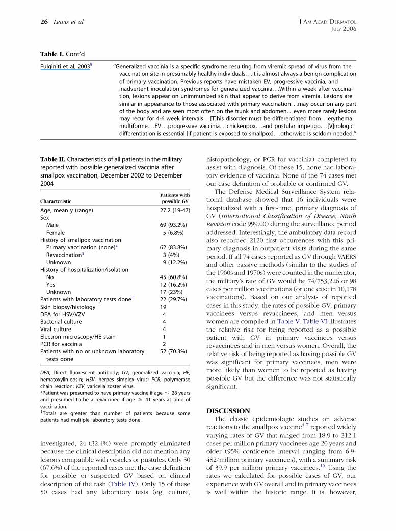
investigated, 24 (32.4%) were promptly eliminatedbecause the clinical description did not mention anylesions compatible with vesicles or pustules. Only 50(67.6%) of the reported cases met the case definitionfor possible or suspected GV based on clinicaldescription of the rash (Table IV). Only 15 of these50 cases had any laboratory tests (eg, culture,
Table II. Characteristics of all patients in the militaryreported with possible generalized vaccinia aftersmallpox vaccination, December 2002 to December2004
Characteristic
Patients with
possible GV
Age, mean y (range) 27.2 (19-47)Sex
Male 69 (93.2%)Female 5 (6.8%)
History of smallpox vaccinationPrimary vaccination (none)* 62 (83.8%)Revaccination* 3 (4%)Unknown 9 (12.2%)
History of hospitalization/isolationNo 45 (60.8%)Yes 12 (16.2%)Unknown 17 (23%)
Patients with laboratory tests doney 22 (29.7%)Skin biopsy/histology 19DFA for HSV/VZV 4Bacterial culture 4Viral culture 4Electron microscopy/HE stain 1PCR for vaccinia 2Patients with no or unknown laboratory
tests done52 (70.3%)
DFA, Direct fluorescent antibody; GV, generalized vaccinia; HE,
hematoxylin-eosin; HSV, herpes simplex virus; PCR, polymerase
chain reaction; VZV, varicella zoster virus.
*Patient was presumed to have primary vaccine if age # 28 years
and presumed to be a revaccinee if age $ 41 years at time of
vaccination.yTotals are greater than number of patients because some
patients had multiple laboratory tests done.
histopathology, or PCR for vaccinia) completed toassist with diagnosis. Of these 15, none had labora-tory evidence of vaccinia. None of the 74 cases metour case definition of probable or confirmed GV.
The Defense Medical Surveillance System rela-tional database showed that 16 individuals werehospitalized with a first-time, primary diagnosis ofGV (International Classification of Disease, NinthRevision code 999.00) during the surveillance periodaddressed. Interestingly, the ambulatory data recordalso recorded 2120 first occurrences with this pri-mary diagnosis in outpatient visits during the sameperiod. If all 74 cases reported as GV through VAERSand other passive methods (similar to the studies ofthe 1960s and 1970s) were counted in the numerator,the military’s rate of GV would be 74/753,226 or 98cases per million vaccinations (or one case in 10,178vaccinations). Based on our analysis of reportedcases in this study, the rates of possible GV, primaryvaccinees versus revaccinees, and men versuswomen are compiled in Table V. Table VI illustratesthe relative risk for being reported as a possiblepatient with GV in primary vaccinees versusrevaccinees and in men versus women. Overall, therelative risk of being reported as having possible GVwas significant for primary vaccinees; men weremore likely than women to be reported as havingpossible GV but the difference was not statisticallysignificant.
DISCUSSIONThe classic epidemiologic studies on adverse
reactions to the smallpox vaccine4-7 reported widelyvarying rates of GV that ranged from 18.9 to 212.1cases per million primary vaccinees age 20 years andolder (95% confidence interval ranging from 6.9-482/million primary vaccinees), with a summary riskof 39.9 per million primary vaccinees.15 Using therates we calculated for possible cases of GV, ourexperience with GVoverall and in primary vaccineesis well within the historic range. It is, however,

J AM ACAD DERMATOL
VOLUME 55, NUMBER 1
Lewis et al 27
Table III. Case definitions for generalized vaccinia after smallpox vaccination applied to assess reported casesof generalized vaccinia (Military Vaccine Agency, US Department of Defense, 2003)
Possible or suggested generalized vacciniaAll of the following must be present:1. A generalized eruption occurring beyond the local vaccination site consisting of lesions with a vesicular or pustular
presentation or having a mixture of papules and vesicles or pustulesAND2. Eruption occurred within 1 month of vaccinationAND3. No plausible explanation for an adverse reaction
Probable generalized vacciniaAt least 1, 2, and 3 must be present:1. A generalized eruption distant from the vaccination site
A. Vesicles or pustules appear on normal skin, with morphology consistent with vacciniaB. Lesions occur on at least 3 regions of the body (each extremity and both anterior and posterior torso count as
separate regions)AND2. Eruption occurred within 6-9 days after vaccinationAND3. At least 12 individual lesions in the same stage of development and evolving through normal vaccination stages,
typically over several days or a week4. Fever, myalgias, and related symptoms may be present
Confirmed generalized vacciniaAll of the clinical criteria for probable generalized vaccinia, aboveANDLaboratory confirmation of vaccinia or orthopoxvirus by at least one of the following methods:1. Conventional histology2. Electron microscopy3. Viral culture4. Polymerase chain reaction
slightly higher than the 9 cases per million of GVreported in the 1990s by the Israeli Defense Forces,who used the Lister vaccine in their military re-cruits.16 The rate of GV in DoD revaccinees inferredbased on age was significantly lower in comparisonwith the reported rate of GV in those naive tovaccinia, also consistent with past experience.However, our case rate of 32 per million vaccinationsis somewhat higher than the rates historically re-ported in the 1960s and 1970s.4-6 This may be a resultof intense surveillance with the current campaign,overreporting, misclassification, or a combination ofthese because we had no probable or confirmedcases identified. In addition, given that it has beendecades since most individuals were originally vac-cinated, our group of revaccinees may have beensomewhat more likely to immunologically respondsimilarly to a vaccine-naive person. The actual rate ofGV today, determined from our experience, is prob-ably much lower than previously reported if weapply the strictest criteria for GV, in which livevaccinia virus is detected in lesions distant from thevaccination site (although this confirmatory test wasseldom used).
We examined the consistency of the case defini-tion of GV in place during the 1960s and 1970s versusthe definition in use today. The current case defini-tion used by the DoD is more stringent than theprevious case definition of 40 years ago, because ofthe additional requirement for confirmation of vac-cinia. During the eradication era, reports of GVreliedprimarily on clinical impression. At that time, themain effort of surveillance was to identify cases ofsevere vaccination reactions, especially those thatwere potentially life-threatening or that required theadministration of VIG. Exanthemata that were clin-ically benign or inconsequential were not scrutinizedto the degree that they may be currently.
We queried the authors of the classic epidemio-logic studies (J. M. Lane, J. M. Neff, and V.A. Fulginiti)about these discrepancies, and with their permis-sion, we list their comments and reflections on theearlier surveillance definitions of GV (Table VII).They acknowledge that many of the cases oncecategorized as GV were probably not caused byviremia and lacked virus in the distant skin lesions.Hence, they would not be characterized as GV todayby the case definition we used.

J AM ACAD DERMATOL
JULY 2006
28 Lewis et al
The strategy behind current smallpox vaccinationprograms is to perform a thorough prevaccinationscreening to exclude all potential vaccinees who areat identifiable risk for clinically significant adversereactions, especially EV and PV.8,11 As a result,relatively few adverse reactions seen today are clin-ically significant. Most dermatologic reactions todaywould have been regarded 50 years ago as benignand inconsequential. Still, more recent guidancedistributed to clinicians regarding the managementof postvaccination adverse reactions can still leaveroom for confusion. Thus, under the current vacci-nation program, healthcare workers and publichealth officials conscientiously detect and reportadverse events, to the extent that a generalized
Table IV. Analysis of 74 cases reported asgeneralized vaccinia between December2002 and December 2004
Cases reported as GV or possibleGV* (No. presumed to be revaccinees,
ie, born # 1971)
74 (12)
Cases with vesicular or pustular lesions 50 (7)Possible or suggested GV 50 (7)Location
Trunk/back/chest 36Upper extremities only 6Diffuse 2Unspecified 5
Laboratory confirmation soughty 15Viral culture performed 4z
Bacterial culture performed 4§
Skin biopsy performed 12k
EM/HE stain performed 1{
DFA for HSV/VZV performed 3#
PCR for vaccinia performed 2**Probable GV 0 (0)$ 3 Regions involved 34 (2)Above 1 within 6-9 d of vaccination 8 (1)Above 1 [12 lesions
morphologically like smallpox0 (0)
DFA, Direct fluorescent antibody; EM, electron microscopy; GV,
generalized vaccinia; HE, hematoxylin-eosin; HSV, herpes simplex
virus; PCR, polymerase chain reaction; VZV, varicella zoster virus.
*Classification of rashes by description and location and other
symptoms were based on clinician description in written notes or
reports.yTotal number of laboratory tests performed is greater than the
number of patients because in some cases multiple tests were
done for a single patient.zOne viral culture grew HSV-1, the rest were negative.§One bacterial culture grew coagulase-negative staphylococci, the
other 3 were negative.kSpongiosis = 3; superficial/mild epidermal perivascular/chronic
dermal inflammation = 7; ruptured milia = 1; folliculitis = 2.{Negative for vaccinia.#All were negative.
**Both were negative.
rash occurring soon after smallpox vaccination maybe hastily identified as GV, based only on a cursoryclinical diagnosis. The fact that 2120 ambulatoryreports in the Standard Ambulatory Data Recordwere coded as GV but not otherwise reportedthrough VAERS suggests that this was used as ageneral-purpose code for any dermatologic adversereaction after smallpox vaccination. We are review-ing a sample of these records to characterize andidentify potential misclassification further.
Jubb,12 reviewing the literature and experiencefrom the early periods of widespread vaccination,noted that rashes postvaccination were frequent, andcould appear as ‘‘papular, pustular, punctuate, ery-thematous, morbilliform, urticarial, roseolar, ecze-matous, and macular,’’ and he considered erythemamultiforme one of the more frequent manifestations(although few of these would today be consideredtrue erythema multiforme). In the early stages of thesmallpox preparedness program after September 11,2001, Frey et al17 noted, when testing clinical re-sponses to diluted and undiluted vaccine, that 14.3%of their 665 participants had rashes in a part of thebody other than the vaccination site, with pustular orvesicular rashes on the chest and back being mostcommon. None were considered to be GVor yieldedlive virus and all resolved spontaneously. Based onour observations, many of the vesicopustular rashesseen since 2002 appear to be an entity that probablyexisted earlier but was not definitively described
Table VI. Analysis of relative risk of being reportedas having possible generalized vaccinia in thisstudy
Relative risk of
possible GV 95% CI x2 P
Primary vaccineevs revaccinee
2.6 1.2-5.9 6.1 .013
Male vs female 2.0 0.63-6.48 0.96 .32
CI, Confidence interval; GV, generalized vaccinia.
Table V. Analysis of rates of generalized vacciniain specific populations
No. of 74
reported
cases
Rate
(per million
vaccinees)
95% CI
(per million
vaccinees)
Possible GV 50 66 49-88Primary
vaccinees43 81 59-109
Revaccinees 7 32 13-65Male 47 70 52-94Female 3 35 7-10
CI, Confidence interval; GV, generalized vaccinia.

J AM ACAD DERMATOL
VOLUME 55, NUMBER 1
Lewis et al 29
Table VII. Recent opinions from authors of classic smallpox studies
V. A. Fulginiti, MD (writtencommunication,December 10, 2004)
‘‘GV is [the] viremic spread of lesions which have the very characteristic appearance of aprimary vaccination at the secondary sites and which usually, but not always heal rapidlywhether we treat them or not. Rarely, crops occur at intervals for up to one year. . . [W]erecovered virus from the site(s). . .and in a few instances from the blood. Our belief thenand now is that this is a very rare complication, and is probably a mild immunodeficiencywhich we could not ascertain in those days. . .Rashes which do not have the typicalpustular, primary-like appearance, are not GV to me.’’
J. M. Lane, MD, MPH(written communication,December 10, 2004)
‘‘[T]he vast majority of the cases [of GV] in our tables in the 1968 data were [non-descriptbenign] rashes. . . [A] modest macular rash with minimal constitutional symptoms issimply part of the normal spectrum of primary vaccinia, and [should] not [be] label[ed]. . .as ‘adverse events’.’’
J. M. Neff, MD (writtencommunication,December 9, 2004)
‘‘[C]ases that were reported in the 1960s represented a hodge podge of conditions.Physicians reported these cases using the definition of the appearance of a post vaccinialvesicular rash in the absence of eczema or immune deficiency. . ..By the late 1960s webegan to question the frequency of this condition and think that most of these casesreported as GV were hypersensitivity reactions or a hodge podge of vesicular conditions,some auto inoculations or folliculitis.’’
40 years ago. We call this entity postvaccinial nonvi-ral pustulosis. This phenomenon was readily evidentto military dermatologists by early 2003, but was firstdescribed in print as focal and generalized folliculitisafter smallpox vaccination.18 Two other compatiblecases from the recent military campaign have sincebeen described in the literature.19 In brief, this entityis primarily a truncal eruption composed of follicularand perifollicular papules and pustules, each sur-rounded by a small edematous, red areola approx-imately 3 to 4 mm in diameter (Fig 1). Lesions areusually discrete, clustered on the upper aspect of theback and chest, and occur principally in young adultsapproximately 1 to 2 weeks (range 5-30 days, mean11.2 days) after primary vaccination (Fig 2). Somepatients report mild pruritus, but patients withpostvaccinial nonviral pustulosis are not toxic. Thecondition is self-limiting and requires no further care
Fig 1. Postvaccinial nonviral pustulosis occurring on neck,shoulders, and upper aspect of chest of young man. Photo-graph courtesy of Melinda A. Cavicchia, LTC, MC, USA.
other than symptomatic relief. In all 15 cases thatwe examined where laboratory confirmation wassought, the lesions were virologically negative byhistology, culture, and PCR for vaccinia. The differ-ential diagnosis for this eruption includes GV,bacterial folliculitis, pityrosporum folliculitis, andvaricella zoster (eg, varicella or disseminated zoster)or herpes simplex virus infection.
Even with the likelihood of overdiagnosis andmisclassification, GV is a rare event, and true GV isprobably rarer still. This is supported by a 2004 studythat showed that viremia is an uncommon occur-rence with the New York City Board of Health strainof vaccinia. Investigators took periodic blood sam-ples from 28 recently vaccinated primary vaccineesfor viral analysis by culture, rapid PCR, and electro-luminescence antigen detection assay. In a total of220 samples obtained over 3 weeks after vaccination,
Fig 2. Typical lesions are perifollicular papules and pus-tules with surrounding erythema. Photograph courtesy ofMelinda A. Cavicchia, LTC, MC, USA.

J AM ACAD DERMATOL
JULY 2006
30 Lewis et al
viremia was not detected in any vacinee.20 In addi-tion, a 2005 parallel study of 38,440 civilian smallpoxvaccinees reported two cases of exanthemata meet-ing the case definition of GV (an incidence rate of52/1,000,000 vaccinations), and one case of GVconfirmed by PCR.21 In 1960, Kempe22 reportedthat he and another individual were separatelyunable to confirm viremia after routine vaccinationin a total of 131 otherwise healthy people. Blattneret al23 noted failure to confirm virus in the blood of7 vaccinated people, although he did find ‘‘bound’’virus in the blood of two patients admitted for theapparent complication of inadvertent inoculation.Viremia has been documented in fatal cases ofcutaneous complications after vaccination in indi-viduals who are immunocompromised24 and is im-plied by the existence of vaccinia osteomyelitis andfetal vaccinia, but these examples are also rare.2,3,7-9
European authors during the first part of the 20thcentury have been cited as finding vaccinia in theblood in the first week after routine vaccination withvirus strains other than New York City Board ofHealth.23,24
The recent medical literature includes 4 articlesthat report GVafter smallpox vaccination. We believethat these cases warrant further scrutiny. Two ofthe reports describe virologic confirmation of GV;however, the same patient is described in bothreports.25,26 Furthermore, in this patient, the ectopicvaccinial lesions appeared less than 48 hours aftervaccination, whereas several earlier definitions ofGV suggested that it occurred not sooner than 4 to6 days after inoculation.3,7,12 Based on the consensusof the Bioterrorism Taskforce of the AmericanAcademy of Dermatology, and after conferring withthe author of the report, the patient described in a2004 Journal of Emergency Medicine article27 is morecorrectly given the diagnosis of classic erythemamultiforme sensu stricto. Meanwhile, the imagesused to illustrate a purported case of GV in another2004 article fail to demonstrate a definitive mor-phology or distribution of the patient’s lesions.28 Onfurther review of these cases and discussions withtheir authors, we believe that these reports wouldmislead one to believe that GV is more prevalentthan it actually is. More importantly, readers of thesereports might infer that generalized eruptions aftersmallpox vaccination that appear similar to thearticles’ photographs should be diagnosed as GV,without the need for laboratory confirmation.
Our study was primarily limited by our methodsof case collection, especially with respect to casesidentified through VAERS, which relies on sponta-neous reporting of adverse events. The retrospectivenature of our study was also a limitation, particularly
as it related to the inability to quickly follow up andrequest a more exhaustive laboratory workup for thepatient. It was challenging to definitively interpret orcorroborate the clinical description of the primarycare providers (the majority of whom were notdermatologists, immunologists, or infectious diseasephysicians), as only a few patients had photographstaken of the acute rash; thus, the written descriptionof the rash was largely the basis for our classificationof the rashes. In some cases, the description on theVAERS report was simply ‘‘generalized vaccinia’’ withno further elaboration. Nonetheless, we believe thisstudy contributes to the smallpox literature as afurther refinement of the postvaccinial reactionsthat fall in the benign spectrum. We further recom-mend that future diagnosis of GV, particularly whenthe use of VIG is being considered as a therapy, beconfirmed with laboratory studies and not be estab-lished solely on the clinical picture. Furthermore, inall cases where GV is in the differential diagnosis, theclinician should seek to rule out the other possiblediagnoses, some of which may be of more concernthan GV, such as PV, EV, and others with lesserimport, including inadvertent inoculation, the sterilepostvaccination pustular eruption described hereand by Talbot et al,18 and diseases unrelated tovaccinia (eg, bacterial folliculitis, pityrosporum fol-liculitis, and varicella zoster or herpes simplex virusinfection). Consultation with a dermatologist, immu-nologist, or infectious disease physician should bepursued, as should laboratory assays such as bacte-rial and viral cultures, direct fluorescent antibodyagainst vaccinia and herpesviruses, or skin biopsy.PCR confirmation of vaccinia should be sought onlyafter these avenues have been exhausted, becauseof the increased likelihood of false-positive rates inpatients at low risk for GV.26
Significance of our findingsThe very low incidence of GV helps us marshal
our limited supplies of VIG. Properly screened indi-viduals who are considering smallpox vaccinationcan be further assured that most exanthemata aftervaccination are benign entities.
We would like to thank J. Michael Lane, John Neff, andVincent Fulginiti for valuable discussions and permissionto quote their remarks.
REFERENCES
1. Centers for Disease Control. Smallpox vaccination and ad-
verse events training module, 2002. Available from: URL:www.
bt.cdc.gov/training/smallpoxvaccine/reactions/gen_vac.html.
Accessed February 18, 2005.
2. Henderson DA, Borio LL, Lane MJ. Smallpox and vaccinia. In:
Plotkin SA, Orenstein WA, Offitt PA, editors. Vaccines. 4th ed.
Philadelphia: Elsevier; 2004. pp. 123-53.

J AM ACAD DERMATOL
VOLUME 55, NUMBER 1
Lewis et al 31
3. Fenner F, Henderson DA, Arita I, Jezek Z, Ladnyi ID. Smallpox
and its eradication. Geneva: World Health Organization; 1988.
4. Neff JM, Lane JM, Pert JH, Moore R, Millar JD, Henderson DA.
Complications of smallpox vaccination, I: national survey in
the United States, 1963. N Engl J Med 1967;276:125-32.
5. Lane JM, Ruben FL, Neff JM, Millar JD. Complications of
smallpox vaccination: national surveillance in the United
States, 1968. N Engl J Med 1969;281:1201-7.
6. Lane JM, Ruben FL, Neff JM, Millar JD. Complications of
smallpox vaccination 1968: results of ten statewide surveys.
J Infect Dis 1970;122:303-9.
7. Barbero GJ, Gray A, Scott TFM, Kempe CH. Vaccinia gangre-
nosa treated with hyperimmune vaccinal gamma globulin.
Pediatrics 1955;16:609-18.
8. Cono J, Casey CG, Bell DM. Smallpox vaccination and adverse
reactions: guidance for clinicians. MMWR Morb Mortal Wkly
Rep 2003;52:1-28.
9. Fulginiti VA, Papier A, Lane JM, Henderson DA. Smallpox
vaccination, a review, part II: adverse events. Clin Infect Dis
2003;37:251-71.
10. Redfield RR, Wright DC, James WD, Jones TS, Brown C, Burke
DS. Disseminated vaccinia in a military recruit with human
immunodeficiency virus (HIV) disease. N Engl J Med 1987;
316:673-6.
11. Centers for Disease Control and Prevention. Smallpox vaccina-
tion: vaccination method and reactions, 2002. Available from:
URL:http://www.bt.cdc.gov/training/smallpoxvaccine/reactions/
SmallpoxVaccinationGuide.pdf. Accessed February 18, 2005.
12. Jubb AA. Generalized vaccinia. Br Med J 1943;1:91-4.
13. Goldstein JA, Neff JM, Lane JM, Koplan JP. Smallpox vaccina-
tion reactions, prophylaxis, and therapy of complications.
Pediatrics 1975;55:342-7.
14. Grabenstein JD, Winkenwerder W Jr. US military smallpox
vaccination program experience. JAMA 2003;289:3278-82.
15. Aragon TJ, Ulrich S, Fernyak S, Rutherford GW. Risks of serious
complications and death from smallpox vaccination: a sys-
tematic review of the United States experience, 1963-1968.
BMC Public Health 2003;3:26.
16. Haim M, Gdalevich M, Mimouni D, Ashkenazi I, Shemer J.
Adverse reactions to smallpox vaccine: the Israel Defense
Force experience, 1991 to 1996, a comparison with previous
surveys. Mil Med 2000;165:287-9.
17. Frey SE, Couch RB, Tacket CO, Treanor JJ, Wolff M, Newman
FK, et al. Clinical responses to undiluted and diluted smallpox
vaccine. N Engl J Med 2002;346:1265-74.
18. Talbot TR, Bredenberg HK, Smith M, LaFleur BJ, Boyd A,
Edwards KM. Focal and generalized folliculitis following small-
pox vaccination. Clin Infect Dis 2004;38:456-8.
19. Oh RC. Folliculitis after smallpox vaccination: a report of two
cases. Mil Med 2005;170:133-6.
20. Cummings JF, Polhemus ME, Hawkes C, Klote M, Ludwig GV,
Wortmann G. Lack of vaccinia viremia after smallpox vaccina-
tion. Clin Infect Dis 2004;38:456-8.
21. Vellozzi C, Lane JM, Averhoff F, Maurer T, Norton S, Damon I,
et al. Generalized vaccinia, progressive vaccinia, and eczema
vaccinatum are rare following smallpox (vaccinia) vaccination:
United States surveillance, 2003. Clin Infect Dis 2005;41:
689-97.
22. Kempe CH. Studies on smallpox and complications of small-
pox vaccination. Pediatrics 1960;26:176-89.
23. Blattner RJ, Norman JO, Heys FM, Aksu I. Antibody response to
cutaneous inoculation with vaccinia virus: viremia and viruria
in vaccinated children. J Pediatr 1964;64:839-52.
24. Keidan SE, McCarthy K, Haworth JC. Fatal generalized vaccinia
with failure of antibody production and absence of serum
gamma globulin. Arch Dis Child 1953;28:110-6.
25. Miller JR, Cirino NM, Philbin EF. Generalized vaccinia 2 days
after smallpox revaccination. Emerg Infect Dis 2003;9:1649-50.
26. Kelly CD, Egan C, Davis SW, Samsonoff WA, Musser KA,
Drabkin P, et al. Laboratory confirmation of generalized
vaccinia following smallpox vaccination. J Clin Microbiol 2004;
42:1373-5.
27. Gibson W, Langsten RE. Generalized vaccinia in a deployed
military member. J Emerg Med 2004;27:127-31.
28. Lemery J. Images in emergency medicine. Ann Emerg Med
2004;43:783-91.
Copyright © 2022 FDOKUMEN