Antibody Profiling by Proteome Microarray Reveals the Immunogenicity of the Attenuated Smallpox...
13
Published Ahead of Print 31 October 2007. 2008, 82(2):652. DOI: 10.1128/JVI.01706-07. J. Virol. E. Frey and Philip L. Felgner Douglas M. Molina, Siddiqua Hirst, Bernard Moss, Sharon Patricia L. Earl, Sookhee Chun, Jenny E. Hernandez, D. Huw Davies, Linda S. Wyatt, Frances K. Newman, That of Dryvax Vaccinia Virus Ankara Is Comparable to Attenuated Smallpox Vaccine Modified Reveals the Immunogenicity of the Antibody Profiling by Proteome Microarray http://jvi.asm.org/content/82/2/652 Updated information and services can be found at: These include: SUPPLEMENTAL MATERIAL Supplemental material REFERENCES http://jvi.asm.org/content/82/2/652#ref-list-1 at: This article cites 37 articles, 13 of which can be accessed free CONTENT ALERTS more» articles cite this article), Receive: RSS Feeds, eTOCs, free email alerts (when new http://journals.asm.org/site/misc/reprints.xhtml Information about commercial reprint orders: http://journals.asm.org/site/subscriptions/ To subscribe to to another ASM Journal go to: on November 6, 2013 by guest http://jvi.asm.org/ Downloaded from on November 6, 2013 by guest http://jvi.asm.org/ Downloaded from on November 6, 2013 by guest http://jvi.asm.org/ Downloaded from on November 6, 2013 by guest http://jvi.asm.org/ Downloaded from on November 6, 2013 by guest http://jvi.asm.org/ Downloaded from on November 6, 2013 by guest http://jvi.asm.org/ Downloaded from on November 6, 2013 by guest http://jvi.asm.org/ Downloaded from on November 6, 2013 by guest http://jvi.asm.org/ Downloaded from on November 6, 2013 by guest http://jvi.asm.org/ Downloaded from on November 6, 2013 by guest http://jvi.asm.org/ Downloaded from on November 6, 2013 by guest http://jvi.asm.org/ Downloaded from on November 6, 2013 by guest http://jvi.asm.org/ Downloaded from on November 6, 2013 by guest http://jvi.asm.org/ Downloaded from
Transcript of Antibody Profiling by Proteome Microarray Reveals the Immunogenicity of the Attenuated Smallpox...
Published Ahead of Print 31 October 2007. 2008, 82(2):652. DOI: 10.1128/JVI.01706-07. J. Virol.
E. Frey and Philip L. FelgnerDouglas M. Molina, Siddiqua Hirst, Bernard Moss, SharonPatricia L. Earl, Sookhee Chun, Jenny E. Hernandez, D. Huw Davies, Linda S. Wyatt, Frances K. Newman, That of DryvaxVaccinia Virus Ankara Is Comparable to Attenuated Smallpox Vaccine ModifiedReveals the Immunogenicity of the Antibody Profiling by Proteome Microarray
http://jvi.asm.org/content/82/2/652Updated information and services can be found at:
These include:
SUPPLEMENTAL MATERIAL Supplemental material
REFERENCEShttp://jvi.asm.org/content/82/2/652#ref-list-1at:
This article cites 37 articles, 13 of which can be accessed free
CONTENT ALERTS more»articles cite this article),
Receive: RSS Feeds, eTOCs, free email alerts (when new
http://journals.asm.org/site/misc/reprints.xhtmlInformation about commercial reprint orders: http://journals.asm.org/site/subscriptions/To subscribe to to another ASM Journal go to:
on Novem
ber 6, 2013 by guesthttp://jvi.asm
.org/D
ownloaded from
on N
ovember 6, 2013 by guest
http://jvi.asm.org/
Dow
nloaded from
on Novem
ber 6, 2013 by guesthttp://jvi.asm
.org/D
ownloaded from
on N
ovember 6, 2013 by guest
http://jvi.asm.org/
Dow
nloaded from
on Novem
ber 6, 2013 by guesthttp://jvi.asm
.org/D
ownloaded from
on N
ovember 6, 2013 by guest
http://jvi.asm.org/
Dow
nloaded from
on Novem
ber 6, 2013 by guesthttp://jvi.asm
.org/D
ownloaded from
on N
ovember 6, 2013 by guest
http://jvi.asm.org/
Dow
nloaded from
on Novem
ber 6, 2013 by guesthttp://jvi.asm
.org/D
ownloaded from
on N
ovember 6, 2013 by guest
http://jvi.asm.org/
Dow
nloaded from
on Novem
ber 6, 2013 by guesthttp://jvi.asm
.org/D
ownloaded from
on N
ovember 6, 2013 by guest
http://jvi.asm.org/
Dow
nloaded from
on Novem
ber 6, 2013 by guesthttp://jvi.asm
.org/D
ownloaded from
JOURNAL OF VIROLOGY, Jan. 2008, p. 652–663 Vol. 82, No. 20022-538X/08/$08.00�0 doi:10.1128/JVI.01706-07Copyright © 2008, American Society for Microbiology. All Rights Reserved.
Antibody Profiling by Proteome Microarray Reveals the Immunogenicity ofthe Attenuated Smallpox Vaccine Modified Vaccinia Virus Ankara Is
Comparable to That of Dryvax�†D. Huw Davies,1* Linda S. Wyatt,2 Frances K. Newman,3 Patricia L. Earl,2 Sookhee Chun,1
Jenny E. Hernandez,1‡ Douglas M. Molina,4 Siddiqua Hirst,1 Bernard Moss,2Sharon E. Frey,3 and Philip L. Felgner1
Division of Infectious Diseases, Department of Medicine, Hewitt Hall, University of California, Irvine, California 926971;Laboratory of Viral Diseases, NIAID, National Institutes of Health, Bethesda, Maryland 208922; Division ofInfectious Diseases and Immunology, Saint Louis University School of Medicine, St. Louis, Missouri 631103;
and ImmPORT Therapeutics Inc., Irvine, California 926184
Received 6 August 2007/Accepted 15 October 2007
Modified vaccinia virus Ankara (MVA) is a highly attenuated vaccinia virus that is under consideration asan alternative to the conventional smallpox vaccine Dryvax. MVA was attenuated by extensive passage ofvaccinia virus Ankara in chicken embryo fibroblasts. Several immunomodulatory genes and genes that influ-ence host range are deleted or mutated, and replication is aborted in the late stage of infection in mostnonavian cells. The effect of these mutations on immunogenicity is not well understood. Since the structuralgenes appear to be intact in MVA, it is hypothesized that critical targets for antibody neutralization have beenretained. To test this, we probed microarrays of the Western Reserve (WR) proteome with sera from humansand macaques after MVA and Dryvax vaccination. As most protein sequences of MVA are 97 to 99% identicalto those of other vaccinia virus strains, extensive binding cross-reactivity is expected, except for those deletedor truncated. Despite different hosts and immunization regimens, the MVA and Dryvax antibody profiles werebroadly similar, with antibodies against membrane and core proteins being the best conserved. The responsesto nonstructural proteins were less well conserved, although these are not expected to influence virus neutral-ization. The broadest antibody response was obtained for hyperimmune rabbits with WR, which is pathogenicin rabbits. These data indicate that, despite the mutations and deletions in MVA, its overall immunogenicityis broadly comparable to that of Dryvax, particularly at the level of antibodies to membrane proteins. The worksupports other information suggesting that MVA may be a useful alternative to Dryvax.
The eradiation of smallpox by use of vaccinia virus was oneof the major accomplishments of vaccination. However, thepotential threat of smallpox (variola virus) or monkeypox vi-ruses being used as a biological weapon may again requiremass vaccination of the general public, which is largely vacciniavirus naı̈ve. Many of the laboratory and vaccine strains avail-able today are derived from the prototype vaccinia virus straindeposited at the New York City Board of Health in 1874 andinclude the Dryvax (Wyeth) strain, which was widely used inthe Americas and West Africa during the smallpox eradicationcampaign. The production of Dryvax was discontinued in 1982,and current stocks are over 25 years old. Production methodsin use then (i.e., propagation on calf skin) are less acceptabletoday, owing to the potential for contamination with adventi-tious agents. Moreover the vaccine is associated with a signif-icant risk of adverse reactions. For example, data collected
during the eradication campaign revealed the risk of compli-cations to be 188 per million vaccinations, with death occurringat a rate of 1 to 5 per million (15). Generalized vaccinia was themost commonly observed side effect, with more-serious reac-tions (eczema vaccinatum, progressive vaccinia, and neurolog-ical/cardiac complications) responsible for 4 to 7% of all ad-verse reactions. Conventional nonattenuated vaccines are nowconsidered unsuitable for a significant proportion of the pop-ulation, including those individuals and their families that areimmunocompromised and those individuals who have atopicdermatitis (eczema) or other skin conditions or are pregnant.There is therefore considerable interest in developing saferalternatives to Dryvax which are equally immunogenic but lackthe pathogenicity.
The highly attenuated vaccine strain modified vaccinia virusAnkara (MVA) is under consideration as an alternative toDryvax. MVA was developed towards the end of the eradica-tion campaign and so has not been evaluated in areas of small-pox endemicity. Since it is no longer possible to evaluate theefficacy of new-generation smallpox vaccines in humans, esti-mations are being made from animal models using relatedorthopoxviruses. The MVA prototype was developed by AntonMayr in Germany through a process of 516 serial passages ofthe chorioallantois vaccinia virus Ankara strain of the vacciniavirus on chicken embryo fibroblasts (CEF) (18). As a result ofadapting to avian cells in vitro, several genes required for
* Corresponding author. Mailing address: University of CaliforniaIrvine, Department of Medicine/Division of Infectious Diseases, 3501Hewitt Hall, Irvine, CA 92697. Phone: (949) 824-0375. Fax: (949)824-9675. E-mail: [email protected].
† Supplemental material for this article may be found at http://jvi.asm.org/.
‡ Present address: Department of Pathology and Laboratory Med-icine, David Geffen School of Medicine at UCLA, 10833 LeConteAvenue, Los Angeles, CA 90095.
� Published ahead of print on 31 October 2007.
652

immune escape and host range were mutated or deleted (sixregions totaling �31 kb) near the termini of the genome (3,22). This causes a block in MVA morphogenesis in most nona-vian cells, resulting in reduced cytopathic effect or plaque for-mation (5) and causing replication to be aborted at the latestage of infection (5, 7). The consequence is severe attenuationof MVA in mammalian hosts in vivo. Despite these gene mu-tations and deletions, MVA has retained its ability to protectanimals against orthopoxvirus challenge nearly as effectively asnonattenuated strains (4, 11, 13, 20, 21, 26, 27, 32, 37). More-over, the immunogenicity of MVA is thought to be equivalentto that of conventional smallpox vaccines (13, 21, 27). MVAalso displays reduced virulence in animals (1, 31, 37), andclinical trials in West Germany in the 1970s demonstrated ithas an excellent safety profile in humans (19). MVA is there-fore considered more suitable for immunocompromised indi-viduals, children, or those with skin conditions such as atopicdermatitis, for whom conventional vaccines are contraindi-cated. MVA is also under consideration as a delivery vehiclefor other vaccines (6, 23, 33, 34) and for immunization prior toDryvax administration to reduce its reactogenicity (13, 25).Molecular studies have shown that the structural genes areintact in MVA (3, 22), suggesting that critical targets for anti-body-mediated neutralization have been retained. However,the effect of the mutations and deletions in MVA on theantibody response is not well understood. To address this, wehave used vaccinia virus proteome microarrays (9) to profileantibody specificities in sera from rabbits, macaques, and hu-mans inoculated with MVA and compared these with profilesinduced by the conventional vaccine Dryvax in humans andmacaques and the pathogenic strain WR in rabbits.
MATERIALS AND METHODS
Viruses. MVA is a clonal, host-restricted vaccinia virus that was developed by�500 serial passages of the Ankara strain of vaccinia virus on CEF. Two MVApreparations were used in this study. Each was derived by plaque purification ofseed stocks from A. Mayr (University of Munich) and propagated in CEF.MVA-BN (Imvamune from Bavarian Nordic A/S, Kvistgård, Denmark) (36) wasused for clinical studies, and sucrose gradient-purified MVA 1974/NIH clone 1(37) was used to immunize macaques and rabbits. Dryvax (Wyeth) was derivedfrom the New York City Board of Health (NYCBOH) strain and is a replicative,nonclonal heterogeneous population of vaccinia virus. Lyophilized Dryvax wasobtained from the CDC and reconstituted according to the manufacturer’s in-structions. Vaccinia virus strain Western Reserve (WR) (ATCC VR-1354) is aclonal and fully replicative laboratory strain derived from Wyeth NYCBOH byrepeated passage on cells including mouse brain and cultured cells. WR isneurotropic in mice and more virulent than the Wyeth strain. Stocks of WR wereprepared in serum-free medium in rabbit kidney fibroblasts and purified bysedimentation through a sucrose cushion (14).
Sera. Hyperimmune rabbit sera (Fig. 1A) were generated in two groups ofthree animals inoculated with MVA and WR by the intramuscular (i.m.) andintradermal routes, respectively, followed by intravenous boosting. New ZealandWhite rabbits (Harlan) (13 weeks old and weighing between 2.75 and 3.0 kg)were inoculated with 108 PFU of vaccinia virus strain WR intradermally at eachof four sites on the shaved back (100 �l/site) or inoculated with 5 � 108 PFU ofMVA i.m. at two sites in each hind leg (100 �l/site). Booster intravenous inoc-ulations of 109 PFU of each virus were given at weeks 9 and 17. Sera werecollected before inoculation (preimmune sera) and at weeks 4, 9, 11, 15, and 19.Cynomolgus macaque sera (Fig. 1B) were obtained from four groups of sixanimals immunized and boosted on week 8 as previously described (13): group1 (“MVA/MVA”) animals were immunized and boosted i.m. with MVA, group2 (“MVA/DVX”) animals were immunized i.m. with MVA followed by a per-cutaneous boost with Dryvax, group 3 (“�/DVX”) animals were immunized bypercutaneous immunization with Dryvax alone at week 8, and group 4 animalswere unimmunized. Sera from prebleed, week 2, and week 14 animals were
analyzed for this study. All groups were challenged with monkeypox virus onweek 16. Unimmunized animals became gravely ill or died, whereas vaccinatedanimals were healthy and asymptomatic, except for transient skin lesions in theMVA/MVA group (13). Human sera (Fig. 1C) were obtained from a random-ized, partially blinded placebo controlled trial of Imvamune conducted at theNIAID-sponsored Saint Louis University Vaccine and Treatment EvaluationUnit (15a). Ninety subjects were equally randomized into six arms as follows.Seventy-five subjects received two doses of Imvamune (2 � 107, 5 � 107, or 1 �108 50% tissue culture infective dose [TCID50]) or saline placebo at weeks 0 and4, followed by 1 � 108 PFU/ml of Dryvax or saline placebo by scarification atmonth 4 after the first vaccination. Fifteen subjects also received two doses ofsaline administered subcutaneously followed by Dryvax. Blood was drawn forplaque reduction neutralizing antibody response at 2 weeks after the first andsecond vaccinations and 4 weeks after the third vaccination. The mean (�standard deviation [SD]) age was 24.8 (� 3.8) years. The sera used in this studywere from 10 vaccinia virus naı̈ve-individuals that were inoculated and boostedwith 1 � 108 TCID50 of MVA; sera were collected prevaccination and 2 weeksafter the second dose of vaccine. Human Dryvax sera were collected from 25laboratory staff members before inoculation and 4 weeks after a single dose of
FIG. 1. Immunization and bleeding schedules. Vaccinia virus inoc-ulations are designated by open arrowheads, and blood draws forserum are designated by small arrows. (A) Rabbits. Group 1 wasprimed i.m. and then boosted twice intravenously with MVA, andgroup 2 was primed intradermally with WR and boosted twice intra-venously. (B) Macaques (13). Group 1 was immunized and boostedi.m. with MVA, group 2 was immunized i.m. with MVA followed by apercutaneous boost with Dryvax (DVX), group 3 was immunized bypercutaneous immunization with Dryvax alone, and group 4 was un-immunized. All groups were challenged with monkeypox (MPX) onweek 16. (C) Humans. Group 1 was inoculated and boosted with 1 �108 TCID50 of MVA (Imvamune), and group 2 was immunized bysingle intradermal inoculation with Dryvax (10). pre, preimmunization.
VOL. 82, 2008 ANTIBODY PROFILING OF SMALLPOX VACCINES Dryvax AND MVA 653

Dryvax as described previously (10). The mean (� SD) age of the subjects was33.3 (� 8.8) years. Thirteen subjects were vaccinia virus naı̈ve at the time ofvaccination, and 12 were receiving a boost.
Virus neutralization assays. Neutralization titers of rabbit and monkey serawere determined by incubation of twofold serial dilutions of sera with recombi-nant vaccinia virus intracellular mature virions (MVs) that expresses green flu-orescent protein (GFP) and then quantifying infected cells by flow cytometry, asdescribed previously (12). Dilutions required for 50% neutralization relative tono-serum neutralization (50% inhibitory concentration values) were calculatedwith PRISM software (GraphPad, San Diego, CA). Titers of MVA-neutralizingactivity in human Imvamune sera were determined by overnight incubation oftwofold serial dilutions of heat-inactivated sera with MVA (30 to 50 PFU),followed by incubation with BSC-40 cell monolayers for 2 days, as describedpreviously (24). Plaques were counted using a dissecting microscope and the 60%plaque reduction neutralization test titer was determined by interpolation by useof a linear regression function in Microsoft Excel.
Measurement of serum antibody by enzyme-linked immunosorbent assay(ELISA). Procedures for measurement of serum antibody responses by use ofpurified WR virus-coated plates and specific vaccinia virus protein-coated plateshave been previously reported (13).
Proteome microarrays. Arrays displaying the prototype vaccinia virus strainWR proteome were produced as described previously (10). Briefly, vaccinia virusWR genomic DNA was used as a template for PCRs to amplify individual openreading frames (ORFs), which were then cloned into a T7 expression vector byuse of homologous recombination. All proteins were expressed from purifiedplasmids in Escherichia coli-based coupled in vitro transcription/translation re-action (RTS 100 kits; Roche), except for L1, which was expressed in RTSdisulfide kits (Roche) according to the manufacturer’s instructions and desig-nated “L1ss” in the figures. Detection of antibodies to L1 by microarray wasfound to be dependent on expression of the L1 protein in conditions thatpermitted disulfide bond formation (see Fig. S1 in the supplemental material).Control reactions that lacked template DNA or empty expression vector werealso set up. Reactions were printed without further purification onto nitrocellu-lose-coated FAST slides (Whatman) by use of an Omni Grid 100 microarray
printer (Gene Machines). For probing, sera were used at 1/50 or 1/100 dilutionin protein array blocking buffer (Whatman) plus 10% E. coli lysate to blockantibodies to E. coli. Bound hyperimmune rabbit antibodies were visualized withCy3-conjugated goat anti-rabbit immunoglobulin G (IgG) (Jackson Immuno-Research) diluted 1/200 in blocking buffer. For human and macaque sera, thedirectly conjugated antibody was found to give low signals, and experiments wererepeated using a biotinylated anti-human IgG (Jackson) followed by streptavi-din-PBXL3 conjugate (Martek Biosciences), both used at 1/200 in blockingbuffer. After being washed five times, slides were air dried under brief centrif-ugation and examined in a ScanArray ExpressHT microarray scanner(PerkinElmer). Fluorescence intensities were quantified by using ProScanArrayExpress software (PerkinElmer). Data handling and statistical analyses wereperformed as described in the figure legends and table footnotes.
RESULTS
Production of hyperimmune MVA and WR sera in rabbits.Hyperimmune sera were generated in rabbits against MVAand the replication-competent strain WR as laboratory re-agents and used in this study to partially verify the arraysagainst standard ELISAs. In rabbits, MVA infections wereasymptomatic, whereas WR was pathogenic and caused a tran-sient loss of appetite (days 4 to 7), lethargy (days 5 to 6), andlesions at the injection site. Virus-specific antibody titers were
FIG. 2. Development of vaccinia virus antibodies in hyperimmu-nized rabbits. Two groups of rabbits were inoculated with MVA or WRto generate hyperimmune sera, as shown in Fig. 1A. (A) Serum anti-body titers of individual rabbits inoculated with MVA or WR weredetermined by whole-virus ELISA. Shown are average titers (� SD) ofthree rabbits in each group. (B) Neutralization titers were determinedby incubation of twofold serial dilutions of sera with a recombinantvaccinia virus that expresses enhanced GFP and then quantifying in-fected cells by flow cytometry. Shown are average (Avg) neutralizationtiters (� SD) of three rabbits in each group. IC50, 50% inhibitoryconcentration. FIG. 3. Comparable data obtained by ELISA and by protein mi-
croarray for four signature membrane proteins. (A) ELISAs of rabbithyperimmune WR and MVA sera by use of plates coated with bacu-lovirus-expressed vaccinia virus proteins. MV membrane proteins wererepresented by L1 and A27 and EV proteins by B5 and A33. (B) Cor-responding SIs revealed by proteome microarrays probed with thesame hyperimmune rabbit sera as used for the ELISAs in panel A.L1ss, L1 expressed in RTS disulfide kits (see text for details); avg,average.
654 DAVIES ET AL. J. VIROL.
monitored by ELISA of whole vaccinia virus and by virusneutralization measured by flow cytometry with vaccinia virusexpressing GFP. Robust immune responses occurred to bothMVA and WR inoculation, although WR induced higher an-tibody titers and �10-fold-higher virus-neutralizing titers (Fig.2A and B, respectively). ELISAs were also performed with thefollowing purified membrane proteins expressed in insect cells:L1 and A27, as representatives of MVs, and B5 and A33, asrepresentatives of extracellular mature virions (EVs). Bothhyperimmune MVA and WR sera showed strong responses toall four proteins (Fig. 3A), with the data essentially mirroringthat obtained by ELISAs against whole vaccinia virus. Re-sponses to L1, B5, and A33 reached similar maximal titers inMVA- and WR-inoculated rabbits, although the rise to plateau
was more rapid in the latter. The titers to A27 were lower afterMVA inoculation, which may contribute to the lower neutral-izing activity of MVA hyperimmue sera (Fig. 2B). The differ-ences in immunogenicity between MVA and WR viruses areprobably due to the fact that while WR is replication compe-tent and pathogenic in mammalian hosts, MVA is not.
We then used the hyperimmune rabbit sera to probe pro-teome microarrays displaying 210 different vaccinia virus WRproteins. Shown in Fig. 3B are the array signal intensities (SIs)for A27, L1, B5, and A33 plotted alongside the correspondingELISA data for comparison. Array data for A27, L1, and B5corresponded well to that obtained by ELISA, providing ver-ification of the array platform. The response to A27 matchedparticularly closely, with both assays reporting a low titer in
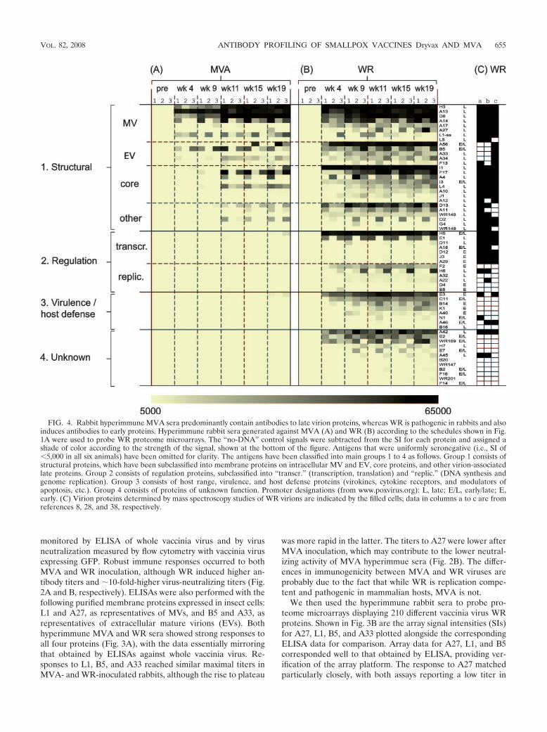
FIG. 4. Rabbit hyperimmune MVA sera predominantly contain antibodies to late virion proteins, whereas WR is pathogenic in rabbits and alsoinduces antibodies to early proteins. Hyperimmune rabbit sera generated against MVA (A) and WR (B) according to the schedules shown in Fig.1A were used to probe WR proteome microarrays. The “no-DNA” control signals were subtracted from the SI for each protein and assigned ashade of color according to the strength of the signal, shown at the bottom of the figure. Antigens that were uniformly seronegative (i.e., SI of�5,000 in all six animals) have been omitted for clarity. The antigens have been classified into main groups 1 to 4 as follows. Group 1 consists ofstructural proteins, which have been subclassified into membrane proteins on intracellular MV and EV, core proteins, and other virion-associatedlate proteins. Group 2 consists of regulation proteins, subclassified into “transcr.” (transcription, translation) and “replic.” (DNA synthesis andgenome replication). Group 3 consists of host range, virulence, and host defense proteins (virokines, cytokine receptors, and modulators ofapoptosis, etc.). Group 4 consists of proteins of unknown function. Promoter designations (from www.poxvirus.org): L, late; E/L, early/late; E,early. (C) Virion proteins determined by mass spectroscopy studies of WR virions are indicated by the filled cells; data in columns a to c are fromreferences 8, 28, and 38, respectively.
VOL. 82, 2008 ANTIBODY PROFILING OF SMALLPOX VACCINES Dryvax AND MVA 655
MVA sera relative to that in WR sera. The array format alsoreadily detected antibodies to A33, but only in WR-hyperim-mune serum. Since antibodies from MVA-inoculated rabbitswere detected by ELISA using A33 secreted from insect cells,we assume that these sera recognized native protein predom-inantly, whereas WR sera recognized both native insect celland nonnative E. coli-expressed protein.
Hyperimmune rabbit MVA sera recognize mainly late virionproteins, whereas WR sera also recognize early proteins. Wenext analyzed the antibody responses at the level of the vac-cinia virus proteome (Fig. 4). Responses to individual proteinswere very consistent between individual rabbits, particularlyfor the strongly immunoreactive antigens. In hyperimmuneMVA sera (Fig. 4A), 18 different antigens were recognized intotal, of which 13 were recognized by at least two of the threeanimals in each group. The MVA antibody profile was focusedoverwhelmingly on virion proteins (summarized in Table 1). Ofthe 13 commonly recognized antigens, 7 were membrane pro-teins and 4 were core proteins. Longitudinal sampling revealedthe responses to envelope proteins appeared first and prior tothe first boost, whereas the appearance of antibodies to thecore proteins was not seen until after the first boost.
By comparison, the responses to WR evolved more rapidly,and near-maximal signals were attained prior to the first boost(Fig. 4B). WR also engendered a much broader antibody pro-file than MVA. Fifty antigens were recognized overall (�22%of the proteome), of which 40 were recognized by two or moreanimals. Moreover, all of the antigens in the MVA profile werealso present in the WR profile. The additional antigens in thelatter were mostly nonstructural/early proteins. Several ofthese are present in virions (8, 28, 38), while others, particu-larly those involved in host defense, are absent from virions(Fig. 4C).
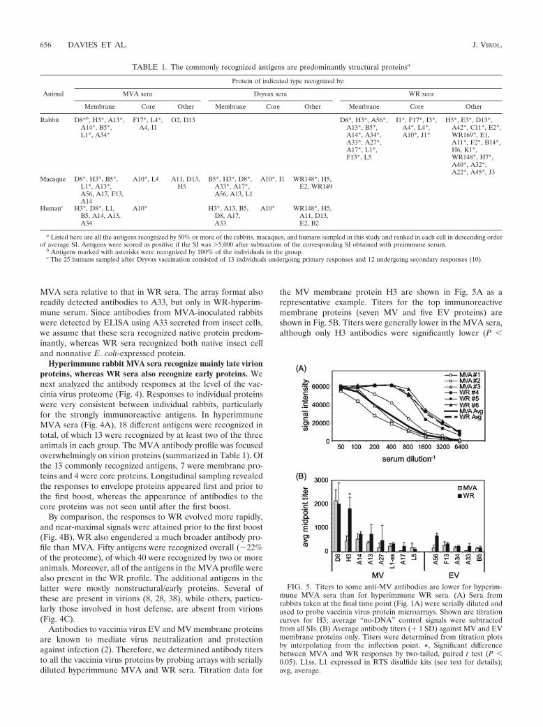
Antibodies to vaccinia virus EV and MV membrane proteinsare known to mediate virus neutralization and protectionagainst infection (2). Therefore, we determined antibody titersto all the vaccinia virus proteins by probing arrays with seriallydiluted hyperimmune MVA and WR sera. Titration data for
the MV membrane protein H3 are shown in Fig. 5A as arepresentative example. Titers for the top immunoreactivemembrane proteins (seven MV and five EV proteins) areshown in Fig. 5B. Titers were generally lower in the MVA sera,although only H3 antibodies were significantly lower (P �
FIG. 5. Titers to some anti-MV antibodies are lower for hyperim-mune MVA sera than for hyperimmune WR sera. (A) Sera fromrabbits taken at the final time point (Fig. 1A) were serially diluted andused to probe vaccinia virus protein microarrays. Shown are titrationcurves for H3; average “no-DNA” control signals were subtractedfrom all SIs. (B) Average antibody titers (� 1 SD) against MV and EVmembrane proteins only. Titers were determined from titration plotsby interpolating from the inflection point. *, Significant differencebetween MVA and WR responses by two-tailed, paired t test (P �0.05). L1ss, L1 expressed in RTS disulfide kits (see text for details);avg, average.
TABLE 1. The commonly recognized antigens are predominantly structural proteinsa
Animal
Protein of indicated type recognized by:
MVA sera Dryvax sera WR sera
Membrane Core Other Membrane Core Other Membrane Core Other
Rabbit D8*b, H3*, A13*,A14*, B5*,L1*, A34*
F17*, L4*,A4, I1
O2, D13 D8*, H3*, A56*,A13*, B5*,A14*, A34*,A33*, A27*,A17*, L1*,F13*, L5
I1*, F17*, I3*,A4*, L4*,A10*, J1*
H5*, E3*, D13*,A42*, C11*, E2*,WR169*, E1,A11*, F2*, B14*,H6, K1*,WR148*, H7*,A40*, A32*,A22*, A45*, J3
Macaque D8*, H3*, B5*,L1*, A13*,A56, A17, F13,A14
A10*, L4 A11, D13,H5
B5*, H3*, D8*,A33*, A17*,A56, A13, L1
A10*, I1 WR148*, H5,E2, WR149
Humanc H3*, D8*, L1,B5, A14, A13,A34
A10* H3*, A13, B5,D8, A17,A33
A10* WR148*, H5,A11, D13,E2, B2
a Listed here are all the antigens recognized by 50% or more of the rabbits, macaques, and humans sampled in this study and ranked in each cell in descending orderof average SI. Antigens were scored as positive if the SI was �5,000 after subtraction of the corresponding SI obtained with preimmune serum.
b Antigens marked with asterisks were recognized by 100% of the individuals in the group.c The 25 humans sampled after Dryvax vaccination consisted of 13 individuals undergoing primary responses and 12 undergoing secondary responses (10).
656 DAVIES ET AL. J. VIROL.

0.05). Overall, the reduced titers of antibodies to MV mem-brane proteins in MVA sera, as determined both by ELISA(Fig. 2A) and array (Fig. 5B), are consistent with reducedMV-neutralizing activity (Fig. 2B).
The data set of immunoreactive viral proteins also allowedus to identify properties of viral proteins that were associatedwith immunogenicity (Table 2). We found that membrane andcore proteins, proteins with late or early/late temporal expres-sion, and proteins with transmembrane domains were overrep-resented in the immunoreactive antigen set relative to thewhole proteome. These predictors are strongest in MVA pro-files, since the antibody profile to MVA is more heavily skewedtoward structural proteins. In contrast, early proteins wereunderrepresented relative to the whole proteome, and therewas negligible influence of molecular weight, isoelectric point,or the presence of a signal sequence on immunogenicity.
MVA and Dryvax antibody profiles in macaques are broadlysimilar to each other. Hyperimmune rabbit sera have beenparticularly useful here for the verification of arrays againstexisting immunoassay platforms. However, from a vaccine per-spective, the responses by humans and nonhuman primates aremore informative. The macaque is of particular importancesince it is a close model of the human immune response andbecause smallpox vaccines can no longer be evaluated in hu-man populations with endemic infections. Therefore, weprobed arrays with sera from macaques inoculated with MVAand boosted with either MVA or Dryvax or else given Dryvaxalone (13). Each of these regimens was shown to be equallyprotective in macaques against a lethal monkeypox challenge.Macaque profiles, shown in Fig. 6, showed interindividual het-erogeneity greater than that seen for rabbits. In the MVA/MVA group (Fig. 6A), the strongest and most frequent posi-tive signals were against structural proteins, particularly to themembrane proteins H3, D8, L1, and B5 and to core proteinA10. The total number of commonly recognized antigens was14, of which 8 were also commonly recognized by rabbits (Ta-ble 1). Robust responses were also seen to virion proteins A11and D13, the latter being a highly expressed protein detectablein MVs (8, 28).
The response to Dryvax alone (Fig. 6C) was broadly similarto that seen for the MVA/MVA group, with the correlationcoefficients being slightly higher for membrane proteins, ormembrane and core proteins, than for other proteins (Table3). The most striking difference between macaque MVA/MVAand Dryvax antibody profiles was the response to the WR148A-type inclusion protein homolog. This protein is immuno-dominant in the Dryvax profile but absent from the MVAprofile owing to a deletion of this region of the MVA genome.Other differences were antibodies to A33, I1, E2, and WR149,which were found only in the response to Dryvax (the lack ofA33 response in MVA sera being a possible conformationissue, as discussed earlier). As seen previously, strong corre-lates of antigenicity in responses to both MVA and Dryvaxwere membrane/core proteins, late temporal expression,and/or the presence of transmembrane domain(s) (Table 2).
Macaques inoculated with MVA followed by Dryvax showsignature WR148 antibody. It has been proposed that MVAcould be used to prevaccinate individuals prior to receivingDryvax to reduce its reactogenicity (13, 25). However, thereare concerns that preexisting immunity to MVA may block the
TA
BL
E2.
Propertiesof
comm
onlyrecognized
vacciniavirus
antigens
Category
d
No.of
vacciniavirus
OR
Fs
(%) e
No.of
antigensrecognized
byindicated
group(%
)and
foldenrichm
ent a
MV
Ab
WR
/DV
Xc
Rabbit
(n
3)M
acaque(n
6)
Hum
an(n
10)
Rabbit
(WR
)(n
3)
Macaque
(�/D
VX
)(n
6)
Hum
an(D
VX
)(n
25)
Antigen
Fold
enrichment
Antigen
Fold
enrichment
Antigen
Fold
enrichment
Antigen
Fold
enrichment
Antigen
Fold
enrichment
Antigen
Fold
enrichment
Total
17913
148
4014
13
Late
promoter
63(35)
9(69)
2.09
(64)1.8
5(63)
1.819
(48)1.4
7(50)
1.46
(46)1.3
Early/lateprom
oter63
(35)4
(31)0.9
5(36)
1.23
(38)1.1
15(38)
1.17
(50)1.4
6(46)
1.3
Early
promoter
53(30)
0(0)
00
(0)0
0(0)
06
(15)0.5
0(0)
01
(8)0.3
Mem
brane/core45
(25)11
(85)3.4
11(79)
3.28
(100)4.0
20(50)
2.010
(71)2.8
7(54)
2.2T
M40
(22)7
(54)2.2
9(64)
2.97
(88)4.0
15(38)
1.78
(57)2.6
7(54)
2.5SignalS
28(16)
3(23)
1.45
(36)2.3
3(38)
2.37
(18)1.1
5(36)
2.34
(31)1.9
Avg
pI6.99
7.246.32
7.466.96
6.255.96
Avg
Mr
33,86027,181
35,96334,553
31,02243,564
44,363
aV
aluesare
thenum
bersof
antigensrecognized
by50%
orm
oreof
thevaccinated
animal
orhum
ansera
ineach
group,with
eachexpressed
asa
percentageof
thetotal
number
ofantigens
inparentheses.“F
oldenrichm
ent”is
aratio
obtainedby
dividingthis
percentageby
thepercentage
ofeach
categoryof
thew
holegenom
e.Inthis
analysis,anantigen
was
scoredpositive
forrecognition
ifthe
SIw
as�
5,000after
subtractionof
thecorresponding
SIobtained
with
preimm
uneserum
.b
Rabbit
serafrom
week
12(group
1in
Fig.1A
);macaque
serafrom
week
14(group
1in
Fig.1B
);human
serafrom
week
16(group
1in
Fig.1C
).cD
VX
,Dryvax.R
abbitsera
fromw
eek12
(group2
inFig.1A
);macaque
serafrom
week
14(group
3in
Fig.1B);hum
anprim
aryplus
secondaryresponses
fromw
eek4
post-Dryvax
(group2
inFig.1C
).d
Promoter
designationsand
proteinfunctions
obtainedfrom
thePoxvirus
Bioinform
aticsR
esourceC
enter(w
ww
.poxvirus.org).TM
,transmem
branedom
ain;SignalS,signalsequence.eA
rraysfabricated
with
proteinsfrom
WR
;pseudogenesand
noncodingO
RF
sare
excludedfrom
thislist.
VOL. 82, 2008 ANTIBODY PROFILING OF SMALLPOX VACCINES Dryvax AND MVA 657
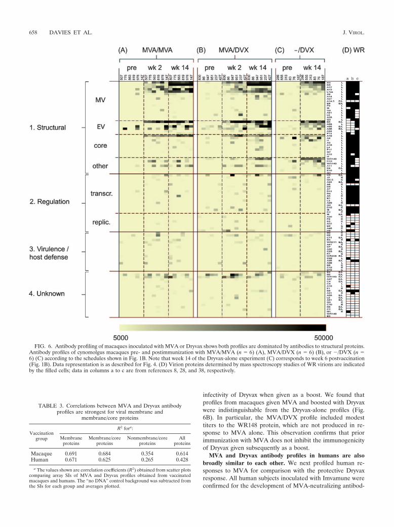
infectivity of Dryvax when given as a boost. We found thatprofiles from macaques given MVA and boosted with Dryvaxwere indistinguishable from the Dryvax-alone profiles (Fig.6B). In particular, the MVA/DVX profile included modesttiters to the WR148 protein, which are not produced in re-sponse to MVA alone. This observation confirms that priorimmunization with MVA does not inhibit the immunogenicityof Dryvax given subsequently as a boost.
MVA and Dryvax antibody profiles in humans are alsobroadly similar to each other. We next profiled human re-sponses to MVA for comparison with the protective Dryvaxresponse. All human subjects inoculated with Imvamune wereconfirmed for the development of MVA-neutralizing antibod-
FIG. 6. Antibody profiling of macaques inoculated with MVA or Dryvax shows both profiles are dominated by antibodies to structural proteins.Antibody profiles of cynomolgus macaques pre- and postimmunization with MVA/MVA (n 6) (A), MVA/DVX (n 6) (B), or �/DVX (n 6) (C) according to the schedules shown in Fig. 1B. Note that week 14 of the Dryvax-alone experiment (C) corresponds to week 6 postvaccination(Fig. 1B). Data representation is as described for Fig. 4. (D) Virion proteins determined by mass spectroscopy studies of WR virions are indicatedby the filled cells; data in columns a to c are from references 8, 28, and 38, respectively.
TABLE 3. Correlations between MVA and Dryvax antibodyprofiles are strongest for viral membrane and
membrane/core proteins
Vaccinationgroup
R2 fora:
Membraneproteins
Membrane/coreproteins
Nonmembrane/coreproteins
Allproteins
Macaque 0.691 0.684 0.354 0.614Human 0.671 0.625 0.265 0.428
a The values shown are correlation coefficients (R2) obtained from scatter plotscomparing array SIs of MVA and Dryvax profiles obtained from vaccinatedmacaques and humans. The “no DNA” control background was subtracted fromthe SIs for each group and averages plotted.
658 DAVIES ET AL. J. VIROL.
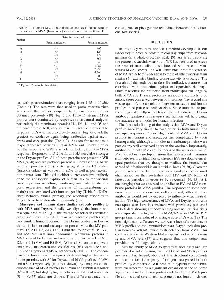
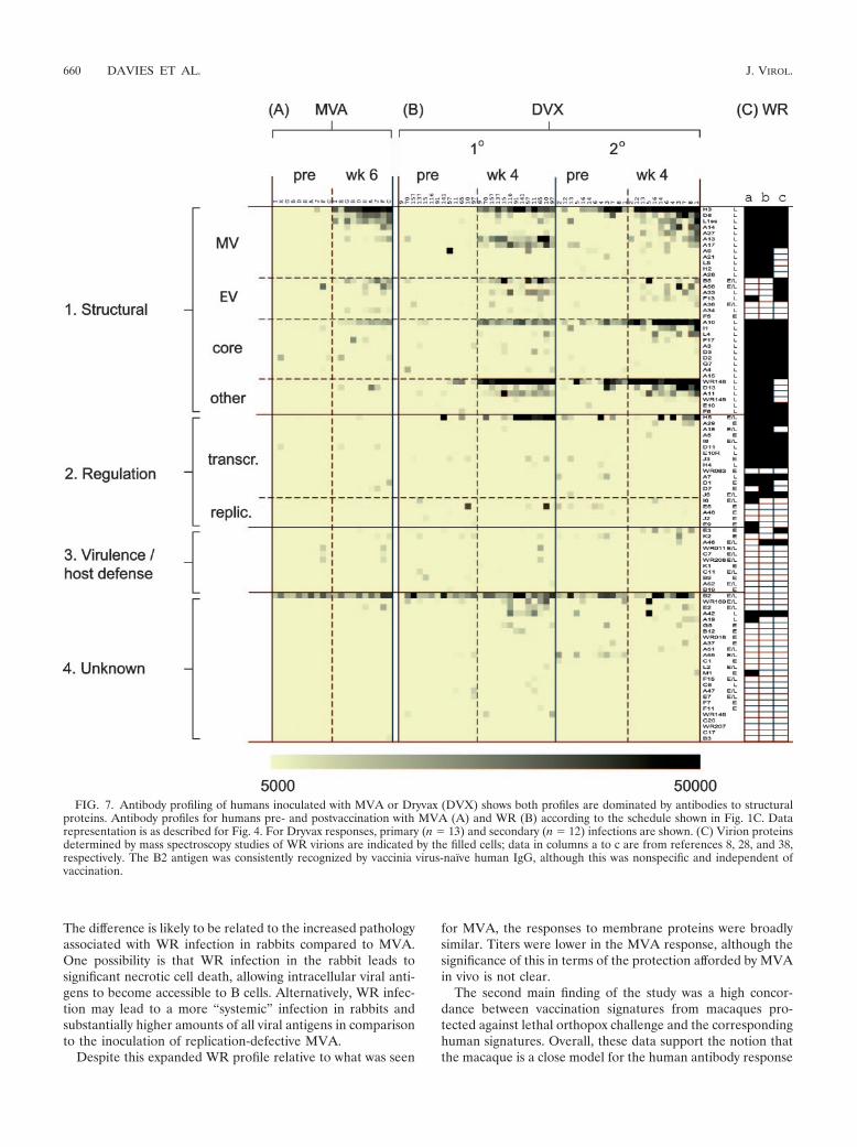
ies, with postvaccination titers ranging from 1/45 to 1/4,569(Table 4). The sera were then used to probe vaccinia virusarrays and the profiles compared to human Dryvax profilesobtained previously (10) (Fig. 7 and Table 1). Human MVAprofiles were dominated by responses to structural antigens,particularly the membrane proteins H3, D8, L1, and B5 andthe core protein A10, consistent with macaque profiles. Theresponse to Dryvax was also broadly similar (Fig. 7B), with thegreatest concordance again being antibodies against mem-brane and core proteins (Table 3). As seen for macaques, amajor difference between human MVA and Dryvax profileswas the response to WR148, which was lacking from the MVAresponse. Responses to D13, A11, and H5 were also strongerin the Dryvax profiles. All of these proteins are present in WRMVs (8, 28) and are probably present in Dryvax virions. As wereported previously (10), a strong signal to the B2 protein(function unknown) was seen in naı̈ve as well as postvaccina-tion human sera. This is due either to cross-reactive antibodyor to the nonspecific capture of human IgG by the vacciniavirus B2 protein. As before, membrane/core proteins, late tem-poral expression, and the presence of transmembrane do-main(s) are correlated with immunogenicity (Table 2). Differ-ences between human primary and secondary responses toDryvax have been described previously (10).
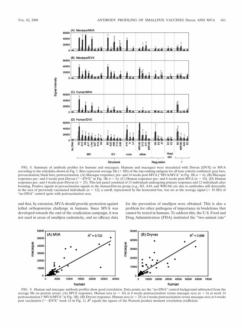
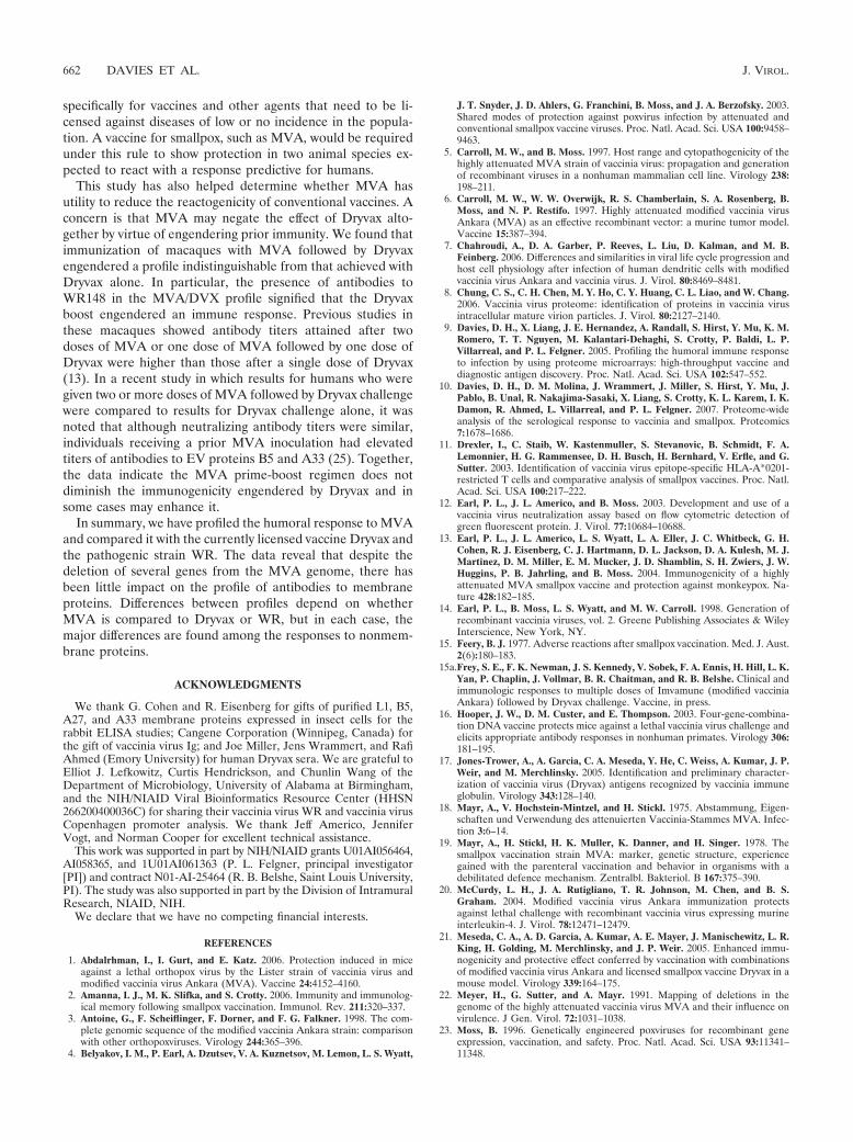
Macaques and humans share similar antibody profiles toboth MVA and Dryvax. Finally, we aligned the human andmacaque profiles. In Fig. 8, the average SIs for each vaccinatedgroup are shown. Overall, human and macaque profiles werevery similar. Immunodominant membrane proteins in Dryvaxshared in human and macaque responses were the MV pro-teins H3, A13, D8, A17, and L1 and the EV proteins B5, A33,and A56. Similarly, immunodominant membrane proteins inMVA shared by human and macaque profiles were H3, A13,D8, and L1 (MV) and B5 (EV). When all SIs on the chip werecompared, the correlation coefficients (R2) were 0.656 and0.722 for Dryvax and MVA, respectively (Fig. 9). The concor-dance of human and macaque signals was highest for mem-brane proteins, with R2 for Dryvax and MVA profiles of 0.696and 0.827, respectively (data not shown). By comparison, theconcordance of MVA profiles in humans and rabbits was lower(R2 0.357) but slightly higher between rabbits and macaques(R2 0.437) (data not shown). These differences may be a
consequence of phylogenetic relatedness between these differ-ent host species.
DISCUSSION
In this study we have applied a method developed in ourlaboratory to produce protein microarray chips from microor-ganisms on a whole-proteome scale (9). An array displayingthe prototypic vaccinia virus strain WR has been used to screenthe sera of mammalian hosts infected with vaccinia virusstrains MVA, Dryvax, and WR. Since most protein sequencesof MVA are 97 to 99% identical to those of other vaccinia virusstrains (3), extensive binding cross-reactivity is expected. Thefirst aim of the study was to describe antibody signatures thatcorrelated with protection against orthopoxvirus challenge.Since macaques are protected from monkeypox challenge byboth MVA and Dryvax, protective antibodies are likely to beamong those conserved between both profiles. The second aimwas to quantify the correlation between macaque and humanprofiles in response to both vaccines. Since humans are pro-tected against smallpox by Dryvax, the relatedness of Dryvaxantibody signatures in macaques and humans will help gaugethe macaque as a model for human infection.
The first main finding of the study is that MVA and Dryvaxprofiles were very similar to each other, in both human andmacaque responses. Precise alignments of MVA and Dryvaxprofiles in humans and macaques are complicated by theirheterogeneity, although antibodies to membrane proteins wereparticularly well conserved between the vaccines. Importantly,antibodies to both MV and EV forms of the virus were found.MVs are robust, enveloped virions that may mediate transmis-sion between individual hosts, whereas EVs are double-envel-oped particles that are thought to mediate the intercellularspread of infection within an individual host (29, 30). Given thegeneral acceptance that a replacement smallpox vaccine mustelicit antibodies that neutralize both MV and EV forms ofinfectious particles in order to be successful (16, 35), it isencouraging that we observed antibodies to EV and MV mem-brane proteins in MVA profiles. The responses to some non-membrane proteins were less well conserved, although theseantibodies would not be expected to influence virus neutral-ization. The high concordance of MVA and Dryvax profiles inmacaques seen here is consistent with previously publishedELISA data showing antibody binding and neutralizing titerswere equivalent or higher in the MVA/MVA and MVA/DVXgroups than those induced by a single dose of Dryvax (13). Themost significant difference we saw was a lack of a response inMVA profiles to the immunodominant A-type inclusion pro-tein homolog WR148, owing to its deletion from MVA. Thisconfirms an earlier Western blot comparison of vaccinia virusIg and MVA sera (17) and suggests that this antigen mayprovide a useful diagnostic tool.
Given the ability of MVA to synthesize both early and lateproteins, it is not surprising that the Dryvax and MVA profilesare so similar. Indeed, abundant late structural componentscan account for the majority of antigens recognized in bothprofiles. By comparison, WR profiles in hyperimmune rabbitswere characterized by a significant expansion in the responseagainst nonstructural/early proteins relative to the MVA pro-file and included several against proteins not found in virions.
TABLE 4. Titers of MVA-neutralizing antibodies in human sera onweek 6 after MVA (Imvamune) vaccination on weeks 0 and 4a
SubjectTiter for indicated serum
Preimmunization Postimmunization
A �4 963B �4 128C �4 472D �4 553E �4 45F �4 1,089G �4 2,253H �4 4,569I �4 208J �4 768
a Figure 1C shows further detail.
VOL. 82, 2008 ANTIBODY PROFILING OF SMALLPOX VACCINES Dryvax AND MVA 659
The difference is likely to be related to the increased pathologyassociated with WR infection in rabbits compared to MVA.One possibility is that WR infection in the rabbit leads tosignificant necrotic cell death, allowing intracellular viral anti-gens to become accessible to B cells. Alternatively, WR infec-tion may lead to a more “systemic” infection in rabbits andsubstantially higher amounts of all viral antigens in comparisonto the inoculation of replication-defective MVA.
Despite this expanded WR profile relative to what was seen
for MVA, the responses to membrane proteins were broadlysimilar. Titers were lower in the MVA response, although thesignificance of this in terms of the protection afforded by MVAin vivo is not clear.
The second main finding of the study was a high concor-dance between vaccination signatures from macaques pro-tected against lethal orthopox challenge and the correspondinghuman signatures. Overall, these data support the notion thatthe macaque is a close model for the human antibody response
FIG. 7. Antibody profiling of humans inoculated with MVA or Dryvax (DVX) shows both profiles are dominated by antibodies to structuralproteins. Antibody profiles for humans pre- and postvaccination with MVA (A) and WR (B) according to the schedule shown in Fig. 1C. Datarepresentation is as described for Fig. 4. For Dryvax responses, primary (n 13) and secondary (n 12) infections are shown. (C) Virion proteinsdetermined by mass spectroscopy studies of WR virions are indicated by the filled cells; data in columns a to c are from references 8, 28, and 38,respectively. The B2 antigen was consistently recognized by vaccinia virus-naı̈ve human IgG, although this was nonspecific and independent ofvaccination.
660 DAVIES ET AL. J. VIROL.
and that, by extension, MVA should provide protection againstlethal orthopoxvirus challenge in humans. Since MVA wasdeveloped towards the end of the eradication campaign, it wasnot used in areas of smallpox endemicity, and no efficacy data
for the prevention of smallpox were obtained. This is also aproblem for other pathogens of importance in biodefense thatcannot be tested in humans. To address this, the U.S. Food andDrug Administration (FDA) instituted the “two-animal rule”
FIG. 8. Summary of antibody profiles for humans and macaques. Humans and macaques were inoculated with Dryvax (DVX) or MVAaccording to the schedules shown in Fig. 1. Bars represent average SIs (� SD) of the top-ranking antigens for all four cohorts combined: gray bars,prevaccination; black bars, postvaccination. (A) Macaque responses, pre- and 14 weeks post-MVA (“MVA/MVA” in Fig. 1B; n 6). (B) Macaqueresponses pre- and 6 weeks post-Dryvax (“�/DVX” in Fig. 1B; n 6). (C) Human responses pre- and 6 weeks post-MVA (n 10). (D) Humanresponses pre- and 4 weeks post-Dryvax (n 25). This last panel consisted of 13 individuals undergoing primary responses and 12 individuals afterboosting. Positive signals in prevaccination signals in the human/Dryvax group (e.g., H3, A10, and WR148) are due to antibodies still detectablein the sera of previously vaccinated individuals (n 12); a cutoff, represented by the horizontal bar, was set as the average signal (� 10 SD) of“no-DNA” control spots with postvaccination sera.
FIG. 9. Human and macaque antibody profiles show good correlation. Data points are the “no-DNA” control background subtracted from theaverage SIs on protein arrays. (A) MVA responses. Human sera (n 10) at 6 weeks postvaccination versus macaque sera (n 6) at week 14postvaccination (“MVA/MVA” in Fig. 1B). (B) Dryvax responses. Human sera (n 25) at 4 weeks postvaccination versus macaque sera at 6 weekspost vaccination (“�/DVX” week 14 in Fig. 1). R2 equals the square of the Pearson product moment correlation coefficient.
VOL. 82, 2008 ANTIBODY PROFILING OF SMALLPOX VACCINES Dryvax AND MVA 661
specifically for vaccines and other agents that need to be li-censed against diseases of low or no incidence in the popula-tion. A vaccine for smallpox, such as MVA, would be requiredunder this rule to show protection in two animal species ex-pected to react with a response predictive for humans.
This study has also helped determine whether MVA hasutility to reduce the reactogenicity of conventional vaccines. Aconcern is that MVA may negate the effect of Dryvax alto-gether by virtue of engendering prior immunity. We found thatimmunization of macaques with MVA followed by Dryvaxengendered a profile indistinguishable from that achieved withDryvax alone. In particular, the presence of antibodies toWR148 in the MVA/DVX profile signified that the Dryvaxboost engendered an immune response. Previous studies inthese macaques showed antibody titers attained after twodoses of MVA or one dose of MVA followed by one dose ofDryvax were higher than those after a single dose of Dryvax(13). In a recent study in which results for humans who weregiven two or more doses of MVA followed by Dryvax challengewere compared to results for Dryvax challenge alone, it wasnoted that although neutralizing antibody titers were similar,individuals receiving a prior MVA inoculation had elevatedtiters of antibodies to EV proteins B5 and A33 (25). Together,the data indicate the MVA prime-boost regimen does notdiminish the immunogenicity engendered by Dryvax and insome cases may enhance it.
In summary, we have profiled the humoral response to MVAand compared it with the currently licensed vaccine Dryvax andthe pathogenic strain WR. The data reveal that despite thedeletion of several genes from the MVA genome, there hasbeen little impact on the profile of antibodies to membraneproteins. Differences between profiles depend on whetherMVA is compared to Dryvax or WR, but in each case, themajor differences are found among the responses to nonmem-brane proteins.
ACKNOWLEDGMENTS
We thank G. Cohen and R. Eisenberg for gifts of purified L1, B5,A27, and A33 membrane proteins expressed in insect cells for therabbit ELISA studies; Cangene Corporation (Winnipeg, Canada) forthe gift of vaccinia virus Ig; and Joe Miller, Jens Wrammert, and RafiAhmed (Emory University) for human Dryvax sera. We are grateful toElliot J. Lefkowitz, Curtis Hendrickson, and Chunlin Wang of theDepartment of Microbiology, University of Alabama at Birmingham,and the NIH/NIAID Viral Bioinformatics Resource Center (HHSN266200400036C) for sharing their vaccinia virus WR and vaccinia virusCopenhagen promoter analysis. We thank Jeff Americo, JenniferVogt, and Norman Cooper for excellent technical assistance.
This work was supported in part by NIH/NIAID grants U01AI056464,AI058365, and 1U01AI061363 (P. L. Felgner, principal investigator[PI]) and contract N01-AI-25464 (R. B. Belshe, Saint Louis University,PI). The study was also supported in part by the Division of IntramuralResearch, NIAID, NIH.
We declare that we have no competing financial interests.
REFERENCES
1. Abdalrhman, I., I. Gurt, and E. Katz. 2006. Protection induced in miceagainst a lethal orthopox virus by the Lister strain of vaccinia virus andmodified vaccinia virus Ankara (MVA). Vaccine 24:4152–4160.
2. Amanna, I. J., M. K. Slifka, and S. Crotty. 2006. Immunity and immunolog-ical memory following smallpox vaccination. Immunol. Rev. 211:320–337.
3. Antoine, G., F. Scheiflinger, F. Dorner, and F. G. Falkner. 1998. The com-plete genomic sequence of the modified vaccinia Ankara strain: comparisonwith other orthopoxviruses. Virology 244:365–396.
4. Belyakov, I. M., P. Earl, A. Dzutsev, V. A. Kuznetsov, M. Lemon, L. S. Wyatt,
J. T. Snyder, J. D. Ahlers, G. Franchini, B. Moss, and J. A. Berzofsky. 2003.Shared modes of protection against poxvirus infection by attenuated andconventional smallpox vaccine viruses. Proc. Natl. Acad. Sci. USA 100:9458–9463.
5. Carroll, M. W., and B. Moss. 1997. Host range and cytopathogenicity of thehighly attenuated MVA strain of vaccinia virus: propagation and generationof recombinant viruses in a nonhuman mammalian cell line. Virology 238:198–211.
6. Carroll, M. W., W. W. Overwijk, R. S. Chamberlain, S. A. Rosenberg, B.Moss, and N. P. Restifo. 1997. Highly attenuated modified vaccinia virusAnkara (MVA) as an effective recombinant vector: a murine tumor model.Vaccine 15:387–394.
7. Chahroudi, A., D. A. Garber, P. Reeves, L. Liu, D. Kalman, and M. B.Feinberg. 2006. Differences and similarities in viral life cycle progression andhost cell physiology after infection of human dendritic cells with modifiedvaccinia virus Ankara and vaccinia virus. J. Virol. 80:8469–8481.
8. Chung, C. S., C. H. Chen, M. Y. Ho, C. Y. Huang, C. L. Liao, and W. Chang.2006. Vaccinia virus proteome: identification of proteins in vaccinia virusintracellular mature virion particles. J. Virol. 80:2127–2140.
9. Davies, D. H., X. Liang, J. E. Hernandez, A. Randall, S. Hirst, Y. Mu, K. M.Romero, T. T. Nguyen, M. Kalantari-Dehaghi, S. Crotty, P. Baldi, L. P.Villarreal, and P. L. Felgner. 2005. Profiling the humoral immune responseto infection by using proteome microarrays: high-throughput vaccine anddiagnostic antigen discovery. Proc. Natl. Acad. Sci. USA 102:547–552.
10. Davies, D. H., D. M. Molina, J. Wrammert, J. Miller, S. Hirst, Y. Mu, J.Pablo, B. Unal, R. Nakajima-Sasaki, X. Liang, S. Crotty, K. L. Karem, I. K.Damon, R. Ahmed, L. Villarreal, and P. L. Felgner. 2007. Proteome-wideanalysis of the serological response to vaccinia and smallpox. Proteomics7:1678–1686.
11. Drexler, I., C. Staib, W. Kastenmuller, S. Stevanovic, B. Schmidt, F. A.Lemonnier, H. G. Rammensee, D. H. Busch, H. Bernhard, V. Erfle, and G.Sutter. 2003. Identification of vaccinia virus epitope-specific HLA-A*0201-restricted T cells and comparative analysis of smallpox vaccines. Proc. Natl.Acad. Sci. USA 100:217–222.
12. Earl, P. L., J. L. Americo, and B. Moss. 2003. Development and use of avaccinia virus neutralization assay based on flow cytometric detection ofgreen fluorescent protein. J. Virol. 77:10684–10688.
13. Earl, P. L., J. L. Americo, L. S. Wyatt, L. A. Eller, J. C. Whitbeck, G. H.Cohen, R. J. Eisenberg, C. J. Hartmann, D. L. Jackson, D. A. Kulesh, M. J.Martinez, D. M. Miller, E. M. Mucker, J. D. Shamblin, S. H. Zwiers, J. W.Huggins, P. B. Jahrling, and B. Moss. 2004. Immunogenicity of a highlyattenuated MVA smallpox vaccine and protection against monkeypox. Na-ture 428:182–185.
14. Earl, P. L., B. Moss, L. S. Wyatt, and M. W. Carroll. 1998. Generation ofrecombinant vaccinia viruses, vol. 2. Greene Publishing Associates & WileyInterscience, New York, NY.
15. Feery, B. J. 1977. Adverse reactions after smallpox vaccination. Med. J. Aust.2(6):180–183.
15a.Frey, S. E., F. K. Newman, J. S. Kennedy, V. Sobek, F. A. Ennis, H. Hill, L. K.Yan, P. Chaplin, J. Vollmar, B. R. Chaitman, and R. B. Belshe. Clinical andimmunologic responses to multiple doses of Imvamune (modified vacciniaAnkara) followed by Dryvax challenge. Vaccine, in press.
16. Hooper, J. W., D. M. Custer, and E. Thompson. 2003. Four-gene-combina-tion DNA vaccine protects mice against a lethal vaccinia virus challenge andelicits appropriate antibody responses in nonhuman primates. Virology 306:181–195.
17. Jones-Trower, A., A. Garcia, C. A. Meseda, Y. He, C. Weiss, A. Kumar, J. P.Weir, and M. Merchlinsky. 2005. Identification and preliminary character-ization of vaccinia virus (Dryvax) antigens recognized by vaccinia immuneglobulin. Virology 343:128–140.
18. Mayr, A., V. Hochstein-Mintzel, and H. Stickl. 1975. Abstammung, Eigen-schaften und Verwendung des attenuierten Vaccinia-Stammes MVA. Infec-tion 3:6–14.
19. Mayr, A., H. Stickl, H. K. Muller, K. Danner, and H. Singer. 1978. Thesmallpox vaccination strain MVA: marker, genetic structure, experiencegained with the parenteral vaccination and behavior in organisms with adebilitated defence mechanism. Zentralbl. Bakteriol. B 167:375–390.
20. McCurdy, L. H., J. A. Rutigliano, T. R. Johnson, M. Chen, and B. S.Graham. 2004. Modified vaccinia virus Ankara immunization protectsagainst lethal challenge with recombinant vaccinia virus expressing murineinterleukin-4. J. Virol. 78:12471–12479.
21. Meseda, C. A., A. D. Garcia, A. Kumar, A. E. Mayer, J. Manischewitz, L. R.King, H. Golding, M. Merchlinsky, and J. P. Weir. 2005. Enhanced immu-nogenicity and protective effect conferred by vaccination with combinationsof modified vaccinia virus Ankara and licensed smallpox vaccine Dryvax in amouse model. Virology 339:164–175.
22. Meyer, H., G. Sutter, and A. Mayr. 1991. Mapping of deletions in thegenome of the highly attenuated vaccinia virus MVA and their influence onvirulence. J Gen. Virol. 72:1031–1038.
23. Moss, B. 1996. Genetically engineered poxviruses for recombinant geneexpression, vaccination, and safety. Proc. Natl. Acad. Sci. USA 93:11341–11348.
662 DAVIES ET AL. J. VIROL.
24. Newman, F. K., S. E. Frey, T. P. Blevins, M. Mandava, A. Bonifacio, Jr., L.Yan, and R. B. Belshe. 2003. Improved assay to detect neutralizing antibodyfollowing vaccination with diluted or undiluted vaccinia (Dryvax) vaccine.J. Clin. Microbiol. 41:3154–3157.
25. Parrino, J., L. H. McCurdy, B. D. Larkin, I. J. Gordon, S. E. Rucker, M. E.Enama, R. A. Koup, M. Roederer, R. T. Bailer, Z. Moodie, L. Gu, L. Yan, andB. S. Graham. 2007. Safety, immunogenicity and efficacy of modified vacciniaAnkara (MVA) against Dryvax challenge in vaccinia-naive and vaccinia-immune individuals. Vaccine 25:1513–1525.
26. Phelps, A., A. J. Gates, M. Hillier, L. Eastaugh, and D. O. Ulaeto. 2005.Comparative efficacy of replicating smallpox vaccine strains in a murinechallenge model. Vaccine 23:3500–3507.
27. Phelps, A. L., A. J. Gates, M. Hillier, L. Eastaugh, and D. O. Ulaeto. 2007.Comparative efficacy of modified vaccinia Ankara (MVA) as a potentialreplacement smallpox vaccine. Vaccine 25:34–42.
28. Resch, W., K. K. Hixson, R. J. Moore, M. S. Lipton, and B. Moss. 2007.Protein composition of the vaccinia virus mature virion. Virology 358:233–247.
29. Smith, G. L., and M. Law. 2004. The exit of vaccinia virus from infected cells.Virus Res. 106:189–197.
30. Smith, G. L., and A. Vanderplasschen. 1998. Extracellular enveloped vac-cinia virus. Entry, egress, and evasion. Adv. Exp. Med. Biol. 440:395–414.
31. Stittelaar, K. J., T. Kuiken, R. L. de Swart, G. van Amerongen, H. W. Vos,H. G. Niesters, P. van Schalkwijk, T. van der Kwast, L. S. Wyatt, B. Moss,and A. D. Osterhaus. 2001. Safety of modified vaccinia virus Ankara (MVA)in immune-suppressed macaques. Vaccine 19:3700–3709.
32. Stittelaar, K. J., G. van Amerongen, I. Kondova, T. Kuiken, R. F. vanLavieren, F. H. Pistoor, H. G. Niesters, G. van Doornum, B. A. van derZeijst, L. Mateo, P. J. Chaplin, and A. D. Osterhaus. 2005. Modified vacciniavirus Ankara protects macaques against respiratory challenge with monkey-pox virus. J. Virol. 79:7845–7851.
33. Sutter, G., and B. Moss. 1992. Nonreplicating vaccinia vector efficientlyexpresses recombinant genes. Proc. Natl. Acad. Sci. USA 89:10847–10851.
34. Sutter, G., L. S. Wyatt, P. L. Foley, J. R. Bennink, and B. Moss. 1994. Arecombinant vector derived from the host range-restricted and highly atten-uated MVA strain of vaccinia virus stimulates protective immunity in mice toinfluenza virus. Vaccine 12:1032–1040.
35. Viner, K. M., and S. N. Isaacs. 2005. Activity of vaccinia virus-neutralizingantibody in the sera of smallpox vaccinees. Microbes Infect. 7:579–583.
36. Vollmar, J., N. Arndtz, K. M. Eckl, T. Thomsen, B. Petzold, L. Mateo, B.Schlereth, A. Handley, L. King, V. Hulsemann, M. Tzatzaris, K. Merkl, N.Wulff, and P. Chaplin. 2006. Safety and immunogenicity of IMVAMUNE, apromising candidate as a third generation smallpox vaccine. Vaccine 24:2065–2070.
37. Wyatt, L. S., P. L. Earl, L. A. Eller, and B. Moss. 2004. Highly attenuatedsmallpox vaccine protects mice with and without immune deficiencies againstpathogenic vaccinia virus challenge. Proc. Natl. Acad. Sci. USA 101:4590–4595.
38. Yoder, J. D., T. S. Chen, C. R. Gagnier, S. Vemulapalli, C. S. Maier, andD. E. Hruby. 2006. Pox proteomics: mass spectrometry analysis and identi-fication of Vaccinia virion proteins. Virol. J. 3:10.
VOL. 82, 2008 ANTIBODY PROFILING OF SMALLPOX VACCINES Dryvax AND MVA 663