Direct TLR2 Signaling Is Critical for NK Cell Activation and Function in Response to Vaccinia Viral...
13
Direct TLR2 Signaling Is Critical for NK Cell Activation and Function in Response to Vaccinia Viral Infection Jennifer Martinez 1 , Xiaopei Huang 2 , Yiping Yang 1,2 * 1 Department of Immunology, Duke University Medical Center, Durham, North Carolina, United States of America, 2 Department of Medicine, Duke University Medical Center, Durham, North Carolina, United States of America Abstract Natural killer (NK) cells play an essential role in innate immune control of poxviral infections in vivo. However, the mechanism(s) underlying NK cell activation and function in response to poxviruses remains poorly understood. In a mouse model of infection with vaccinia virus (VV), the most studied member of the poxvirus family, we identified that the Toll-like receptor (TLR) 2-myeloid differentiating factor 88 (MyD88) pathway was critical for the activation of NK cells and the control of VV infection in vivo. We further showed that TLR2 signaling on NK cells, but not on accessory cells such as dendritic cells (DCs), was necessary for NK cell activation and that this intrinsic TLR2-MyD88 signaling pathway was required for NK cell activation and played a critical role in the control of VV infection in vivo. In addition, we showed that the activating receptor NKG2D was also important for efficient NK activation and function, as well as recognition of VV-infected targets. We further demonstrated that VV could directly activate NK cells via TLR2 in the presence of cytokines in vitro and TLR2-MyD88- dependent activation of NK cells by VV was mediated through the phosphatidylinositol 3-kinase (PI3K)-extracellular signal- regulated kinase (ERK) pathway. Taken together, these results represent the first evidence that intrinsic TLR signaling is critical for NK cell activation and function in the control of a viral infection in vivo, indicate that multiple pathways are required for efficient NK cell activation and function in response to VV infection, and may provide important insights into the design of effective strategies to combat poxviral infections. Citation: Martinez J, Huang X, Yang Y (2010) Direct TLR2 Signaling Is Critical for NK Cell Activation and Function in Response to Vaccinia Viral Infection. PLoS Pathog 6(3): e1000811. doi:10.1371/journal.ppat.1000811 Editor: Luis J. Sigal, Fox Chase Cancer Center, United States of America Received August 17, 2009; Accepted February 5, 2010; Published March 12, 2010 Copyright: ß 2010 Martinez et al. This is an open-access article distributed under the terms of the Creative Commons Attribution License, which permits unrestricted use, distribution, and reproduction in any medium, provided the original author and source are credited. Funding: This work was supported by grants AI083000, CA111807, CA136934, AI079336 and CA047741 from the National Institutes of Health, and a grant from the Alliance for Cancer Gene Therapy. The funders had no role in study design, data collection and analysis, decision to publish, or preparation of the manuscript. Competing Interests: The authors have declared that no competing interests exist. * E-mail: [email protected] Introduction Vaccinia virus (VV) is a member of the Orthopoxvirus genus of the Poxviridae family, including smallpox (variola) virus, monkeypox virus, cowpox virus, and mousepox (ectromelia) virus. It has a large and complex, double-stranded DNA genome, measuring about 200 Kb, that encodes most of the genes required for cytoplasmic replication of the virus [1]. VV is the most studied member of the poxvirus family and is the live vaccine responsible for successful elimination of smallpox in the late 1970s [2]. This triumph is now being threatened by bioterrorists deliberately reintroducing smallpox, against which vaccination is no longer routine [3–5]. Thus, widespread public vaccination is being considered to counter this potential threat. However, the currently used live VV vaccine is associated with a relatively high incidence of severe adverse events, particularly in individuals with eczema and immunodeficiency [6–9]. Therefore, there is an imminent need to explore new and safe approaches to control, not only the actual smallpox infection, but also the potential complications from smallpox vaccination with the live VV. Critical for the development of novel strategies is a better understanding of the host’s defense mechanism(s) against poxvi- ruses in vivo. Recent advances have shown that recovery from viral infections depends on the host’s ability to mount effective innate immune responses. NK cells represent an important component of the innate immune system and play a critical role in innate immune defense against various viral infections in vivo [10,11]. Clinically, individuals who are NK cell-deficient suffer from severe, recurrent viral infections [12]. NK cells are also crucial in the control of poxviruses. Upon poxviral infection, NK cells are activated, expand and accumulate at the site of infection, and these activated NK cells are important for the clearance of the infection [13–16]. Activation of NK cells is tightly controlled by both inhibitory and activating receptors [17]. Previous studies have shown that upon murine CMV (MCMV) infection, NK cell activation is mediated by the NK cell activating receptor, Ly49H, which specifically recognizes the m157 gene product of MCMV expressed on the surface of infected cells [18,19]. Similarly, recognition of influenza virus hemagglutinin on virus-infected cells by another activating receptor, NKp46, activates lysis by human NK cells [20], and the murine NKp46 equivalent, NCR1, is required for protection against lethal influenza infection [21]. In addition, the NKG2D activating receptor has been shown to recognize host stress proteins induced upon viral infections including human CMV and MCMV infections [22,23]. How NK cells are activated upon poxviral infection remains poorly understood. It is known that Ly49H is not involved in the control of VV in mice [24,25]. Studies in vitro have shown that recognition of VV-infected cells by human NK cells is, in part, mediated by NKp30, NKp44 and NKp46, but not NKG2D [26]. On the other hand, recent studies have suggested that NKG2D is partially involved in NK cell-mediated control of mousepox virus PLoS Pathogens | www.plospathogens.org 1 March 2010 | Volume 6 | Issue 3 | e1000811
-
Upload
independent -
Category
Documents
-
view
3 -
download
0
Transcript of Direct TLR2 Signaling Is Critical for NK Cell Activation and Function in Response to Vaccinia Viral...
Direct TLR2 Signaling Is Critical for NK Cell Activationand Function in Response to Vaccinia Viral InfectionJennifer Martinez1, Xiaopei Huang2, Yiping Yang1,2*
1 Department of Immunology, Duke University Medical Center, Durham, North Carolina, United States of America, 2 Department of Medicine, Duke University Medical
Center, Durham, North Carolina, United States of America
Abstract
Natural killer (NK) cells play an essential role in innate immune control of poxviral infections in vivo. However, themechanism(s) underlying NK cell activation and function in response to poxviruses remains poorly understood. In a mousemodel of infection with vaccinia virus (VV), the most studied member of the poxvirus family, we identified that the Toll-likereceptor (TLR) 2-myeloid differentiating factor 88 (MyD88) pathway was critical for the activation of NK cells and the controlof VV infection in vivo. We further showed that TLR2 signaling on NK cells, but not on accessory cells such as dendritic cells(DCs), was necessary for NK cell activation and that this intrinsic TLR2-MyD88 signaling pathway was required for NK cellactivation and played a critical role in the control of VV infection in vivo. In addition, we showed that the activating receptorNKG2D was also important for efficient NK activation and function, as well as recognition of VV-infected targets. We furtherdemonstrated that VV could directly activate NK cells via TLR2 in the presence of cytokines in vitro and TLR2-MyD88-dependent activation of NK cells by VV was mediated through the phosphatidylinositol 3-kinase (PI3K)-extracellular signal-regulated kinase (ERK) pathway. Taken together, these results represent the first evidence that intrinsic TLR signaling iscritical for NK cell activation and function in the control of a viral infection in vivo, indicate that multiple pathways arerequired for efficient NK cell activation and function in response to VV infection, and may provide important insights intothe design of effective strategies to combat poxviral infections.
Citation: Martinez J, Huang X, Yang Y (2010) Direct TLR2 Signaling Is Critical for NK Cell Activation and Function in Response to Vaccinia Viral Infection. PLoSPathog 6(3): e1000811. doi:10.1371/journal.ppat.1000811
Editor: Luis J. Sigal, Fox Chase Cancer Center, United States of America
Received August 17, 2009; Accepted February 5, 2010; Published March 12, 2010
Copyright: � 2010 Martinez et al. This is an open-access article distributed under the terms of the Creative Commons Attribution License, which permitsunrestricted use, distribution, and reproduction in any medium, provided the original author and source are credited.
Funding: This work was supported by grants AI083000, CA111807, CA136934, AI079336 and CA047741 from the National Institutes of Health, and a grant fromthe Alliance for Cancer Gene Therapy. The funders had no role in study design, data collection and analysis, decision to publish, or preparation of the manuscript.
Competing Interests: The authors have declared that no competing interests exist.
* E-mail: [email protected]
Introduction
Vaccinia virus (VV) is a member of the Orthopoxvirus genus of the
Poxviridae family, including smallpox (variola) virus, monkeypox
virus, cowpox virus, and mousepox (ectromelia) virus. It has a
large and complex, double-stranded DNA genome, measuring
about 200 Kb, that encodes most of the genes required for
cytoplasmic replication of the virus [1]. VV is the most studied
member of the poxvirus family and is the live vaccine responsible
for successful elimination of smallpox in the late 1970s [2]. This
triumph is now being threatened by bioterrorists deliberately
reintroducing smallpox, against which vaccination is no longer
routine [3–5]. Thus, widespread public vaccination is being
considered to counter this potential threat. However, the currently
used live VV vaccine is associated with a relatively high incidence
of severe adverse events, particularly in individuals with eczema
and immunodeficiency [6–9]. Therefore, there is an imminent
need to explore new and safe approaches to control, not only the
actual smallpox infection, but also the potential complications
from smallpox vaccination with the live VV.
Critical for the development of novel strategies is a better
understanding of the host’s defense mechanism(s) against poxvi-
ruses in vivo. Recent advances have shown that recovery from
viral infections depends on the host’s ability to mount effective
innate immune responses. NK cells represent an important
component of the innate immune system and play a critical role
in innate immune defense against various viral infections in vivo
[10,11]. Clinically, individuals who are NK cell-deficient suffer
from severe, recurrent viral infections [12]. NK cells are also
crucial in the control of poxviruses. Upon poxviral infection, NK
cells are activated, expand and accumulate at the site of infection,
and these activated NK cells are important for the clearance of the
infection [13–16]. Activation of NK cells is tightly controlled by
both inhibitory and activating receptors [17]. Previous studies
have shown that upon murine CMV (MCMV) infection, NK cell
activation is mediated by the NK cell activating receptor, Ly49H,
which specifically recognizes the m157 gene product of MCMV
expressed on the surface of infected cells [18,19]. Similarly,
recognition of influenza virus hemagglutinin on virus-infected cells
by another activating receptor, NKp46, activates lysis by human
NK cells [20], and the murine NKp46 equivalent, NCR1, is
required for protection against lethal influenza infection [21]. In
addition, the NKG2D activating receptor has been shown to
recognize host stress proteins induced upon viral infections
including human CMV and MCMV infections [22,23].
How NK cells are activated upon poxviral infection remains
poorly understood. It is known that Ly49H is not involved in the
control of VV in mice [24,25]. Studies in vitro have shown that
recognition of VV-infected cells by human NK cells is, in part,
mediated by NKp30, NKp44 and NKp46, but not NKG2D [26].
On the other hand, recent studies have suggested that NKG2D is
partially involved in NK cell-mediated control of mousepox virus
PLoS Pathogens | www.plospathogens.org 1 March 2010 | Volume 6 | Issue 3 | e1000811

[27]. Thus, mechanisms underlying NK cell responses upon
poxviral infection remain largely undefined. In a murine model of
VV infection, we have previously shown that VV activates the
innate immune system through both the Toll-like receptor (TLR)-
dependent and -independent pathways [28]. The TLR pathway is
mediated by TLR2 and dependent on MyD88, leading to
production of pro-inflammatory cytokines, IL-6, IL-1, and IL-
12, whereas activation of the TLR-independent pathway results in
the secretion of type I IFN. More importantly, both TLR2 and
TLR-independent pathways are required for innate immune
control of VV infection [28]. We have shown that the critical role
of the TLR-independent pathway in innate immune control of VV
is mediated by type I IFN, which acts directly on NK cells to
regulate their activation and function [16]. However, how the
TLR2-MyD88 pathway contributes to innate immune control of
VV remains unclear.
In this report, we provided evidence that the TLR2-MyD88
pathway is also critical for NK cell activation and function in
response to VV infection in vivo. This was independent of TLR2-
induced production of pro-inflammatory cytokines. We showed
that direct TLR2 signaling on NK cells was necessary for efficient
NK cell activation upon stimulation with VV and played a critical
role in VV control in vivo. In addition, we showed that the
NKG2D pathway was also required for efficient NK activation
and function, as well as recognition of VV-infected targets.
Furthermore, we demonstrated that that VV could directly
activate NK cells via TLR2 in the presence of cytokines in vitro
and TLR2-dependent activation of NK cells by VV was mediated
through the MyD88-PI3K-ERK pathway. Collectively, these
results suggest that efficient NK cell activation depends on multiple
pathways in response to VV infection and that direct TLR2
activation on NK cells is critical for their function and could lead
to the development of novel strategies to combat poxviral infection.
Results
NK cell activation and function upon VV infectiondepends on the TLR2-MyD88 pathway
Previous studies have shown that NK cells are critical for the
control of VV infection in vivo using antibodies to asialo GM1, but
not NK1.1 [13–16]. Anti-asialo GM1 depletes NK cells and some
T cells, but not NKT cells, whereas anti-NK1.1 depletes NK and
NKT cells, but not T cells. Thus, to further confirm the role of NK
cells in VV control in vivo, we depleted mice of NK cells with anti-
NK1.1 antibodies (PK136), followed by infection with VV
intraperitoneally. Here we showed that mice depleted of NK cells
with anti-NK1.1 also had a defect in NK cell lytic activity
(Figure 1A) and a significantly (p , 0.001) higher viral titer than
the control mice (Figure 1B).
We next investigated how the TLR2-MyD88 pathway
contributed to innate immune control of VV. We hypothesized
that TLR2-dependent control of VV infection was also mediated
through the regulation of NK cell activation. To test this
hypothesis, WT, TLR2-deficient (TLR22/2), or MyD882/2
mice were infected with VV intraperitoneally, and splenic NK
cells were analyzed for their activation and function. We first
showed that splenic NK cell numbers (Figure 2A) and phenotypic
markers (Figure S1) from naı̈ve TLR22/2 or MyD882/2 mice
were similar to those from WT mice. 48 h after infection, at
which time splenic NK cell activation peaked upon VV infection
as shown previously [16], splenic NK cells from WT mice
produced significantly (p ,0.001) higher amounts of effector
molecules such as IFN-c and granzyme B, compared to the
uninfected naı̈ve control (Figure 2B, C), indicating that these NK
cells are activated upon VV infection in vivo. In addition, these
NK cells were functionally active, as they demonstrated lytic
activities on NK-sensitive YAC-1 cells or VV-infected L929 cells
(Figure 2D). In contrast, the production of IFN-c and granzyme
B by splenic NK cells from TLR22/2 or MyD882/2 mice was
significantly (p ,0.001) reduced, compared to the WT controls
(Figure 2B, C). Furthermore, splenic NK cells from VV-infected
TLR22/2 or MyD882/2 mice displayed drastically diminished
lytic activities (Figure 2D), leading to a significant (p ,0.001)
increase in viral load in the ovary (Figure 2E). Collectively, these
results indicate that NK cell activation and their effector function
in response to VV infection is critically dependent on the TLR2-
MyD88 pathway in vivo. However, the expansion of NK cells
upon VV infection appeared unchanged in the absence of TLR2
signaling (Figure 2A).
Figure 1. NK cells are required for the control of VV infection invivo. Female C57BL/6 mice were depleted of NK cells with anti-NK1.1antibodies (25 mg) on days 23 and 0 (+anti-NK1.1) or left untreated(Control), followed by infection with VV on day 0. (A) 48 h afterinfection, splenocytes were assayed for NK lytic activity on YAC-1 cellsfor 4 hr at different effector:target ratios. Naı̈ve splenocytes (Naı̈ve)were used for comparison. The percentage of specific lysis is shown. (B)The ovaries were assayed for viral load. Data represents viral titer 6 SDas pfu per ovary.doi:10.1371/journal.ppat.1000811.g001
Author Summary
NK cells are an important component of innate immunityin fighting against poxviral infections in vivo. However,how NK cells are activated and exert their function incontrolling poxviruses remains poorly understood. In thispaper, we found that VV, the most studied member of thepoxvirus family, could directly activate TLR2 on NK cellsand that the direct TLR2 stimulation was critical for NK cellactivation and function in the control of VV infection invivo. We further showed that TLR2-dependent NK cellactivation by VV was mediated through the PI3K-ERKpathway. In addition, we demonstrated that the activatingreceptor NKG2D was also required for efficient NK cellactivation and function. Collectively, these results repre-sent the first evidence that direct TLR signaling is crucial toNK cell activation and function in the control of a viralinfection in vivo, indicate that multiple pathways arerequired for efficient NK cell activation, and may provideimportant insights into the design of effective strategies tocombat poxviral infections.
Direct TLR2 Signaling in NK Cell Activation
PLoS Pathogens | www.plospathogens.org 2 March 2010 | Volume 6 | Issue 3 | e1000811

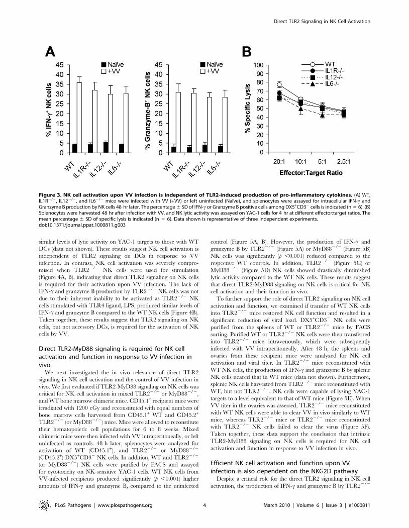
NK cell activation is independent of TLR2-inducedpro-inflammatory cytokines
What then is responsible for TLR2-dependent NK cell
activation and function in response to VV infection in vivo? We
have previously shown that activation of TLR2 on DCs and
macrophages by VV leads to the production of pro-inflammatory
cytokines including IL-6, IL-1, and IL-12 [28]. Since IL-12 and
IL-1 have been implicated in regulating NK cell activation and
function in other models of viral infection [10,11], we investigated
whether the TLR2-dependent NK cell response to VV infection
was due to TLR2-induced secretion of pro-inflammatory cyto-
kines. WT, IL-1 receptor-deficient (IL-1R2/2), IL-122/2, or
IL-62/2 mice were infected with VV intraperitoneally, and NK
cells were analyzed 48 h later. No significant differences (p .0.05)
were observed in the production of IFN-c or granzyme B by
splenic NK cells from IL-1R2/2, IL-122/2, or IL-62/2 mice,
compared to the WT controls (Figure 3A). In addition, splenic NK
cells from IL-1R2/2, IL-122/2, or IL-62/2 mice displayed
similar levels of lytic activities on YAC-1 targets compared to WT
mice (Figure 3B), and no differences in viral load were observed
among all mice (data not shown). These results suggest that TLR2-
induced IL-12, IL-1 and IL-6 are not critical for NK cell activation
and effector function in response to VV infection in vivo.
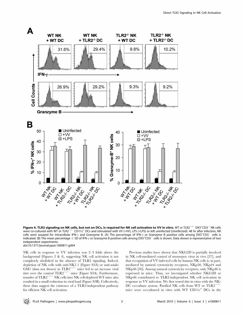
TLR2 signaling on NK cells, but not on DCs, is required forNK cell activation
The observation that NK cell activation and function upon VV
infection in vivo is independent of TLR2-induced IL-1, IL-6, and
IL-12 production may not completely rule out the role of TLR2
signaling on accessory cells, such as DCs, in NK cell activation as
VV could stimulate TLR2 on DCs to secrete other cytokines or
activate other pathways that may be critical for NK cell activation.
To address this question, we utilized an in vitro DC-NK cell co-
culture system as described [16]. DX5+CD32 splenic NK cells
were isolated by FACS sorting with a purity of .98% (Figure S2).
Purified WT or TLR22/2 NK cells were co-cultured in vitro with
WT or TLR22/2 CD11c+ DCs, followed by infection with VV.
48 h later, NK cells were analyzed for the production of IFN-cand granzyme B. Our data showed that similar amounts of IFN-cand granzyme B were produced by WT NK cells when cultured
with TLR22/2 DCs to those with WT DCs (Figure 4A, B). In
addition, WT NK cells co-cultured with TLR22/2 DCs displayed
Figure 2. NK cell activation and function in response to VV in vivo requires an intact TLR2-MyD88 pathway. (A–C) Wild-type (WT),TLR22/2, or MyD882/2 mice were infected with VV (+VV) or left uninfected (Naı̈ve). 48 h later, splenocytes were assayed for total NK cell numbers,intracellular IFN-c and Granzyme B production by NK cells, as well as NK cell lytic assay. (A) The mean numbers 6 SD of total DX5+CD32 NK cells perspleen are indicated (n = 6 per group). (B) FACS plots of intracellular IFN-c and Granzyme B production by NK cells with the percentage of IFN-c orGranzyme B positive cells among DX5+CD32 NK cells indicated. (C) The mean percentage 6 SD of IFN-c or Granzyme B positive cells amongDX5+CD32 cells is indicated (n = 6 per group). (D) 48 h after infection, splenocytes were harvested and NK cell lytic activity was assayed on YAC-1cells or VV-infected L929 cells (L929-VV) for 4 hr at different effector:target ratios. Naı̈ve splenocytes (Naı̈ve) were used as a control. The meanpercentage 6 SD of specific lysis is indicated (n = 6 per group). (E) The ovaries of female mice were harvested for measurement of viral load. Datarepresents the mean viral titer 6 SD as plaque-forming units (pfu) per ovary (n = 6 per group). Data shown is representative of three independentexperiments.doi:10.1371/journal.ppat.1000811.g002
Direct TLR2 Signaling in NK Cell Activation
PLoS Pathogens | www.plospathogens.org 3 March 2010 | Volume 6 | Issue 3 | e1000811
similar levels of lytic activity on YAC-1 targets to those with WT
DCs (data not shown). These results suggest NK cell activation is
independent of TLR2 signaling on DCs in response to VV
infection. In contrast, NK cell activation was severely compro-
mised when TLR22/2 NK cells were used for stimulation
(Figure 4A, B), indicating that direct TLR2 signaling on NK cells
is required for their activation upon VV infection. The lack of
IFN-c and granzyme B production by TLR22/2 NK cells was not
due to their inherent inability to be activated as TLR22/2 NK
cells stimulated with TLR4 ligand, LPS, produced similar levels of
IFN-c and granzyme B compared to the WT NK cells (Figure 4B).
Taken together, these results suggest that TLR2 signaling on NK
cells, but not accessory DCs, is required for the activation of NK
cells by VV.
Direct TLR2-MyD88 signaling is required for NK cellactivation and function in response to VV infection invivo
We next investigated the in vivo relevance of direct TLR2
signaling in NK cell activation and the control of VV infection in
vivo. We first evaluated if TLR2-MyD88 signaling on NK cells was
critical for NK cell activation in mixed TLR22/2 or MyD882/2,
and WT bone marrow chimeric mice. CD45.1+ recipient mice were
irradiated with 1200 cGy and reconstituted with equal numbers of
bone marrow cells harvested from CD45.1+ WT and CD45.2+
TLR22/2 (or MyD882/2) mice. Mice were allowed to reconstitute
their hematopoietic cell populations for 6 to 8 weeks. Mixed
chimeric mice were then infected with VV intraperitoneally, or left
uninfected as controls. 48 h later, splenocytes were analyzed for
activation of WT (CD45.1+), and TLR22/2 or MyD882/2
(CD45.2+) DX5+CD32 NK cells. In addition, WT and TLR22/2
(or MyD882/2) NK cells were purified by FACS and assayed
for cytotoxicity on NK-sensitive YAC-1 cells. WT NK cells from
VV-infected recipients produced significantly (p ,0.001) higher
amounts of IFN-c and granzyme B, compared to the uninfected
control (Figure 5A, B). However, the production of IFN-c and
granzyme B by TLR22/2 (Figure 5A) or MyD882/2 (Figure 5B)
NK cells was significantly (p ,0.001) reduced compared to the
respective WT controls. In addition, TLR22/2 (Figure 5C) or
MyD882/2 (Figure 5D) NK cells showed drastically diminished
lytic activity compared to the WT NK cells. These results suggest
that direct TLR2-MyD88 signaling on NK cells is critical for NK
cell activation and their function in vivo.
To further support the role of direct TLR2 signaling on NK cell
activation and function, we examined if transfer of WT NK cells
into TLR22/2 mice restored NK cell function and resulted in a
significant reduction of viral load. DX5+CD32 NK cells were
purified from the spleens of WT or TLR22/2 mice by FACS
sorting. Purified WT or TLR22/2 NK cells were then transferred
into TLR22/2 mice intravenously, which were subsequently
infected with VV intraperitoneally. After 48 h, the spleens and
ovaries from these recipient mice were analyzed for NK cell
activation and viral titer. In TLR22/2 mice reconstituted with
WT NK cells, the production of IFN-c and granzyme B by splenic
NK cells neared that in WT mice (data not shown). Furthermore,
splenic NK cells harvested from TLR22/2 mice reconstituted with
WT, but not TLR22/2, NK cells were capable of lysing YAC-1
targets to a level equivalent to that of WT mice (Figure 5E). When
VV titer in the ovaries was assessed, TLR22/2 mice reconstituted
with WT NK cells were able to clear VV in vivo similarly to WT
mice, whereas TLR22/2 mice or TLR22/2 mice reconstituted
with TLR22/2 NK cells failed to clear the virus (Figure 5F).
Taken together, these data support the conclusion that intrinsic
TLR2-MyD88 signaling on NK cells is required for NK cell
activation and function in response to VV infection in vivo.
Efficient NK cell activation and function upon VVinfection is also dependent on the NKG2D pathway
Despite a critical role for the direct TLR2 signaling in NK cell
activation, the production of IFN-c and granzyme B by TLR22/2
Figure 3. NK cell activation upon VV infection is independent of TLR2-induced production of pro-inflammatory cytokines. (A) WT,IL1R2/2, IL122/2, and IL62/2 mice were infected with VV (+VV) or left uninfected (Naı̈ve), and splenocytes were assayed for intracellular IFN-c andGranzyme B production by NK cells 48 hr later. The percentage 6 SD of IFN-c or Granzyme B positive cells among DX5+CD32 cells is indicated (n = 6). (B)Splenocytes were harvested 48 hr after infection with VV, and NK lytic activity was assayed on YAC-1 cells for 4 hr at different effector:target ratios. Themean percentage 6 SD of specific lysis is indicated (n = 6). Data shown is representative of three independent experiments.doi:10.1371/journal.ppat.1000811.g003
Direct TLR2 Signaling in NK Cell Activation
PLoS Pathogens | www.plospathogens.org 4 March 2010 | Volume 6 | Issue 3 | e1000811
NK cells in response to VV infection was 2–3 folds above the
background (Figures 2 & 4), suggesting NK cell activation is not
completely abolished in the absence of TLR2 signaling. Indeed,
depletion of NK cells with anti-NK1.1 (Figure S3A) or anti-asialo
GM1 (data not shown) in TLR22/2 mice led to an increase viral
titer over the control TLR22/2 mice (Figure S3A). Furthermore,
transfer of TLR22/2 NK cells into NK cell-depleted WT mice also
resulted in a small reduction in viral load (Figure S3B). Collectively,
these data suggest the existence of a TLR2-independent pathway
for efficient NK cell activation.
Previous studies have shown that NKG2D is partially involved
in NK cell-mediated control of mousepox virus in vivo [27], and
that recognition of VV-infected cells by human NK cells is, in part,
mediated by natural cytotoxicity receptors, NKp30, NKp44 and
NKp46 [26]. Among natural cytotoxicity receptors, only NKp46 is
expressed in mice. Thus, we investigated whether NKG2D or
NKp46 contributed to TLR2-independent NK cell activation in
response to VV infection. We first tested this in vitro with the NK-
DC co-culture system. Purified NK cells from WT or TLR22/2
mice were co-cultured in vitro with WT CD11c+ DCs in the
Figure 4. TLR2 signaling on NK cells, but not on DCs, is required for NK cell activation to VV in vitro. WT or TLR22/2 DX5+CD32 NK cellswere co-cultured with WT or TLR22/2 CD11c+ DCs and stimulated with VV (+VV), LPS (+LPS) or left uninfected (Uninfected). 48 hr after infection, NKcells were assayed for intracellular IFN-c and Granzyme B. (A) The percentage of IFN-c or Granzyme B positive cells among DX5+CD32 cells isindicated. (B) The mean percentage 6 SD of IFN-c or Granzyme B positive cells among DX5+CD32 cells is shown. Data shown is representative of twoindependent experiments.doi:10.1371/journal.ppat.1000811.g004
Direct TLR2 Signaling in NK Cell Activation
PLoS Pathogens | www.plospathogens.org 5 March 2010 | Volume 6 | Issue 3 | e1000811
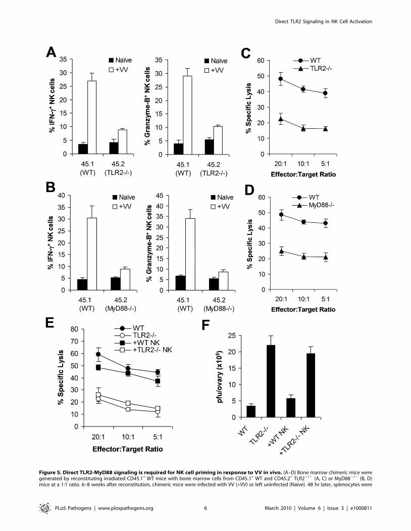
Figure 5. Direct TLR2-MyD88 signaling is required for NK cell priming in response to VV in vivo. (A–D) Bone marrow chimeric mice weregenerated by reconstituting irradiated CD45.1+ WT mice with bone marrow cells from CD45.1+ WT and CD45.2+ TLR22/2 (A, C) or MyD882/2 (B, D)mice at a 1:1 ratio. 6–8 weeks after reconstitution, chimeric mice were infected with VV (+VV) or left uninfected (Naı̈ve). 48 hr later, splenocytes were
Direct TLR2 Signaling in NK Cell Activation
PLoS Pathogens | www.plospathogens.org 6 March 2010 | Volume 6 | Issue 3 | e1000811

presence of a blocking anti-NKG2D antibody or a blocking
NKp46-Fc fusion protein, followed by infection with VV, the
activation of NK cells was analyzed 24 h later. The production of
IFN-c and granzyme B by WT NK cells was significantly (p
,0.01) decreased in the presence of anti-NKG2D compared to
the control without anti-NKG2D (Figure 6A). In addition, the
production of IFN-c and granzyme B by TLR22/2 NK cells was
completely abolished with the NKG2D blocking (Figure 6A).
However, blocking with NKp46-Fc had no effect on activation of
WT or TLR22/2 NK cells (Figure 6A). These results indicate that
NKG2D, but not NKp46, is also involved in NK cell activation
upon VV infection.
We next examined the role of NKG2D in NK cell activation
and function in vivo. WT or TLR22/2 mice were injected with
the blocking anti-NKG2D antibody intravenously 24 h and 6 h
prior to infection with VV intraperitoneally, and splenic NK cells
were analyzed for their activation and function 48 h after
infection. In WT mice, NK cells produced significantly (p
Figure 6. The NKG2D pathway is required for efficient NK activation and function in response to VV infection. (A) WT or TLR22/2
DX5+CD32 NK cells were co-cultured with WT CD11c+ DCs and stimulated with VV alone (+VV), VV in the presence of anti-NKG2D (VV+anti-NKG2D) orNKp46-Fc chimera (VV+NKp46-Fc), or left uninfected (Uninfected). 24 hr later, NK cells were assayed for intracellular IFN-c and Granzyme B. The meanpercentage 6 SD of IFN-c or Granzyme B positive cells among DX5+CD32 cells is shown. (B–C) WT or TLR22/2 mice were infected with VV (+VV) orleft uninfected (Naı̈ve). Some mice were pre-treated with anti-NKG2D antibodies 24 and 6 h prior to infection with VV (VV+anti-NKG2D). 48 h afterinfection, splenic NK cells were analyzed for IFN-c and Granzyme B production. The mean percentage 6 SD of IFN-c or Granzyme B positive cellsamong DX5+CD32 cells is indicated (n = 4 per group) (B). The ovaries of female mice were harvested for measurement of viral load. Data representsthe mean viral titer 6 SD as pfu per ovary (n = 4 per group) (C). (D) 48 h after infection, splenocytes from WT mice were assayed for NK cell lyticactivity on VV-infected L929 cells in the presence of anti-NKG2D antibodies (+anti-NKG2D) or NKp46-Fc chimera (+NKp46-Fc), for 4 hr at differenteffector:target ratios. The mean percentage 6 SD of specific lysis is indicated (n = 4 per group). Data shown is representative of two independentexperiments.doi:10.1371/journal.ppat.1000811.g006
assayed for intracellular IFN-c and Granzyme B production by NK cells. The mean percentage 6 SD of IFN-c or Granzyme B positive cells amongCD45.1+ or CD45.2+ DX5+CD32 cells is indicated (n = 4 per group) (A, B). Splenocytes were sorted into CD45.1+ or CD45.2+ DX5+CD32 populations48 h after VV infection, and NK lytic activity was assayed on YAC-1 cells for 4 hr at different effector:target ratios. The mean percentages of specificlysis are indicated (n = 4 per group) (C, D). Data shown is representative of two independent experiments. (E–F) TLR22/2 mice were reconstitutedwith WT NK cells (+ WT NK) or TLR22/2 NK cells (+ TLR22/2 NK) followed by infection with VV. WT and TLR22/2 mice infected with VV were used ascontrols. 48 hr later, splenocytes were assayed for NK lytic activity on YAC-1 cells at different effector:target ratios. The mean percentage of specificlysis is indicated (n = 4) (E). The ovaries of female mice were harvested for measurement of viral load. Data represents the mean viral titer 6 SD as pfuper ovary (n = 4) (F). Data is representative of two independent experiments.doi:10.1371/journal.ppat.1000811.g005
Direct TLR2 Signaling in NK Cell Activation
PLoS Pathogens | www.plospathogens.org 7 March 2010 | Volume 6 | Issue 3 | e1000811
,0.01) less amounts of IFN-c and granzyme B in the presence of
NKG2D blocking, compared to the control without NKG2D
blocking (Figure 6B), leading to a significant (p ,0.001) increase in
viral load in the ovaries (Figure 6C). Furthermore, in TLR22/2
mice, NK cell activation was completely abolished with NKG2D
blocking (Figure 6B), leading to a further increase in viral load in
the ovaries (Figure 6C). These data indicate that the NKG2D
pathway is also important in the activation of NK cells and the
control of VV infection in vivo.
To address whether NKG2D or NKp46 is involved in the NK
cell recognition of VV-infected targets, NK cells harvested from
VV-infected WT mice were assayed for their lytic activities on
VV-infected L929 targets in vitro in the presence of NKG2D or
NKp46 blocking. A significant reduction in cytolytic activities was
observed with the NKG2D, but not NKp46, blocking (Figure 6D),
suggesting NKG2D is also critical for the recognition of VV-
infected cells.
Collectively, these results suggest that the TLR2-independent
NKG2D pathway is also required for efficient NK cell activation,
recognition of VV-infected targets and VV control in vivo.
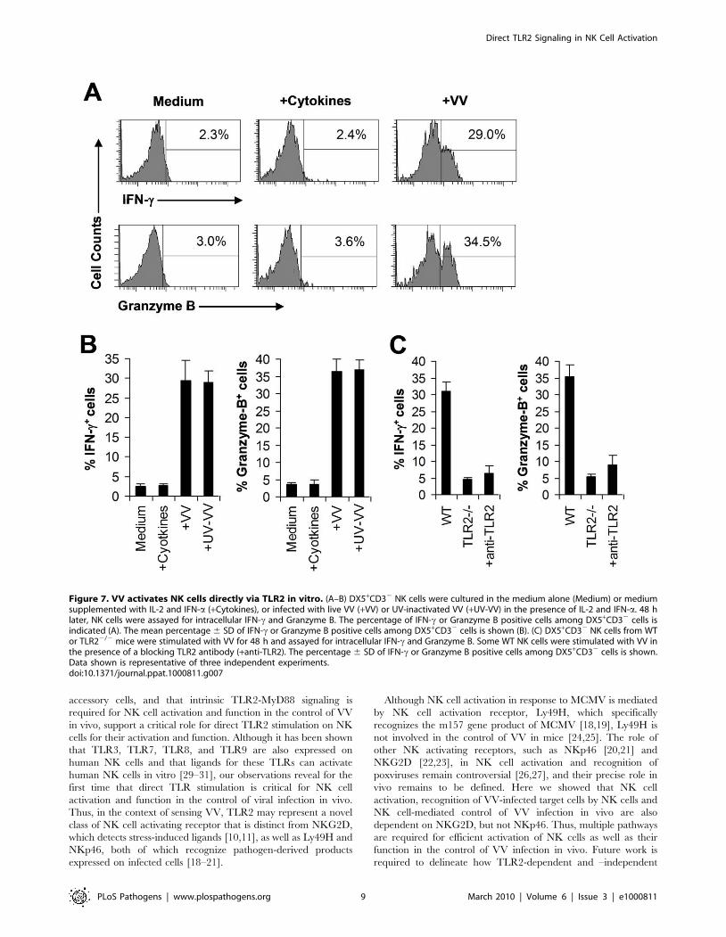
VV can directly activate NK cells via TLR2 in the presenceof cytokines in vitro
The observation that direct TLR2 signaling on NK cells is
required for NK cell activation and function in response to VV
infection suggested that VV might directly activate NK cells via
TLR2. To address this question, an accessory cell-free NK culture
system is required. However, an accessory cell-free system would
lack NKG2D stimulation as NKG2D ligands are usually expressed
by accessory cells upon viral infection. To compensate for lack of
NKG2D stimulation, we established an accessory cell-free NK
culture system in the presence of low dose IL-2 and IFN-a as they
have been used for the activation of NK cells in vitro. FACS-
purified splenic DX5+CD32 NK cells were stimulated with VV
and assayed for NK cell activation 48 h later. No significant IFN-cor granzyme B production was detected in the presence of IL-2
and IFN-a compared to the medium control (Figure 7A, B),
suggesting that IL-2 and IFN-a alone do not activate NK cells.
However, addition of VV stimulated NK cells to produce
significantly (p ,0.001) higher levels of IFN-c and granzyme B
(Figure 7A, B). These results indicate that VV can directly activate
NK cells in the presence of cytokines. Similar results were obtained
when UV-inactivated VV was used for stimulation (Figure 7B),
suggesting that NK cell stimulation by VV is independent of newly
synthesized viral products after infection.
We next investigated whether VV activated NK cells via TLR2.
It has been shown NK cells express multiple TLRs [29–31].
Indeed, we showed here that TLR2, TLR4, TLR8, and TLR9
were expressed at the RNA level in NK cells (Figure S4A).
Furthermore, we found that TLR2 protein is expressed on the
surface of freshly isolated NK cells by FACS and that the level of
TLR2 expression remained constant after incubation with
cytokines or stimulation with VV (Figure S4B). When purified
NK cell from TLR22/2 or TLR92/2 mice were stimulated with
VV, the production of IFN-c and granzyme B by TLR22/2
(Figure 7C), but not TLR92/2 (data not shown), NK cells was
significantly (p ,0.001) reduced, compared to WT controls. To
more directly confirm that VV activated NK cells via TLR2, WT
NK cells were pre-treated with a blocking TLR2 antibody and
stimulated with VV. Our result showed that TLR2 blocking led to
a significant reduction in IFN-c and granzyme B production by
WT NK cells (Figure 7C). Taken together, these data indicate that
VV can directly activate NK cells via TLR2.
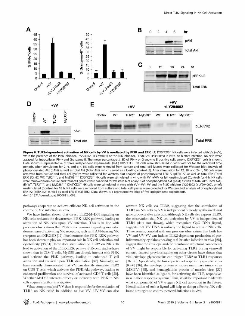
TLR2-MyD88-dependent NK cell activation by VV ismediated by the PI3K-ERK pathway
How does stimulation of TLR2-Myd88 pathway on NK cells by
VV lead to NK cell activation? Recent studies have shown that in
T cells, MyD88 can interact with PI3K and activate the PI3K
pathway, leading to enhanced T cell activation and survival upon
TLR stimulation [32], and PI3K is the common signaling
mediator downstream of activating NK receptors, such as
ITAM-bearing NK receptors and NKG2D [17]. Furthermore,
the PI3K-ERK pathway has been shown to play an important role
in NK cell activation and cytotoxicity [33,34]. Thus, we
hypothesized that TLR2-MyD88 signaling on NK cells activated
the downstream PI3K-ERK pathway, leading to activation of NK
cells upon VV infection. To test this hypothesis, we first examined
if activation of NK cells by VV was mediated by PI3K and ERK.
FACS-purified NK cells were stimulated with VV in the presence
of the PI3K inhibitor, LY294002 or the ERK1/2 inhibitor,
PD98059 and analyzed for their activation 48 h later. Indeed, the
production of IFN-c and granzyme B by NK cells was significantly
(p ,0.001) reduced in the presence of LY294002 or PD98059,
compared to the control without inhibitors (Figure 8A), suggesting
that both PI3K and ERK are involved in NK cell activation
by VV.
We next tested whether stimulation of NK cells with VV led to
the activation of both PI3K and ERK in a TLR2-MyD88-
dependent manner. Stimulation of NK cells with VV resulted in
the activation of PI3K, evidenced by phosphorylation of Akt, an
immediate downstream target of PI3K (indicative of PI3K
activation), peaked at 4 h after stimulation (Figure 8B). Similarly,
ERK was also activated upon stimulation with VV, peaked at 18 h
after the infection (Figure 8C). The phosphorylation of both PI3K
and ERK was critically dependent on an intact TLR2-MyD88
pathway as the activation of Akt (Figure 8D) and ERK (Figure 8E)
was greatly diminished in TLR22/2 or MyD882/2 NK cells
stimulated with VV, compared to that in the WT controls.
Furthermore, ERK phosphorylation was severely compromised in
the presence of LY294002 (Figure 8E), confirming that ERK is
downstream of PI3K. Taken together, these data indicate that
TLR2-MyD88-dependent NK cell activation by VV is mediated
by the PI3K-ERK pathway.
Discussion
NK cells play a critical role in the control of poxviral infection in
vivo [13–16]. We have previously shown that type I IFN, induced
upon VV infection, acts directly on NK cells to regulate their
activation and function [16]. However, the mechanism(s) by which
NK cells are activated and function in response to poxviral
infection remains poorly understood. In this study, we provided
evidence that activation of the TLR2-MyD88 innate immune
pathway is critical for NK cell activation and function in response
to VV infection in vivo. Since VV activates TLR2 on accessory
cells, such as DCs and macrophages, to secrete pro-inflammatory
cytokines IL-6, IL-1, and IL-12 [28], this dependence on TLR2
pathway could be mediated indirectly through IL-1 and IL-12, as
these cytokines have been shown to play a role in NK cell
activation [10,11]. However, our data demonstrates that NK cell
activation and function is independent of TLR2-induced IL-1 and
IL-12 in vivo and that TLR2 signaling on NK cells, but not on
DCs, is necessary for NK cell activation upon stimulation with VV
in vitro. This data suggest a role for direct TLR2 stimulation on
NK cells by VV in NK cell activation and function.
Indeed, the observations that TLR2 is expressed on NK cells,
that VV can directly activate NK cells via TLR2 in the absence of
Direct TLR2 Signaling in NK Cell Activation
PLoS Pathogens | www.plospathogens.org 8 March 2010 | Volume 6 | Issue 3 | e1000811
accessory cells, and that intrinsic TLR2-MyD88 signaling is
required for NK cell activation and function in the control of VV
in vivo, support a critical role for direct TLR2 stimulation on NK
cells for their activation and function. Although it has been shown
that TLR3, TLR7, TLR8, and TLR9 are also expressed on
human NK cells and that ligands for these TLRs can activate
human NK cells in vitro [29–31], our observations reveal for the
first time that direct TLR stimulation is critical for NK cell
activation and function in the control of viral infection in vivo.
Thus, in the context of sensing VV, TLR2 may represent a novel
class of NK cell activating receptor that is distinct from NKG2D,
which detects stress-induced ligands [10,11], as well as Ly49H and
NKp46, both of which recognize pathogen-derived products
expressed on infected cells [18–21].
Although NK cell activation in response to MCMV is mediated
by NK cell activation receptor, Ly49H, which specifically
recognizes the m157 gene product of MCMV [18,19], Ly49H is
not involved in the control of VV in mice [24,25]. The role of
other NK activating receptors, such as NKp46 [20,21] and
NKG2D [22,23], in NK cell activation and recognition of
poxviruses remain controversial [26,27], and their precise role in
vivo remains to be defined. Here we showed that NK cell
activation, recognition of VV-infected target cells by NK cells and
NK cell-mediated control of VV infection in vivo are also
dependent on NKG2D, but not NKp46. Thus, multiple pathways
are required for efficient activation of NK cells as well as their
function in the control of VV infection in vivo. Future work is
required to delineate how TLR2-dependent and –independent
Figure 7. VV activates NK cells directly via TLR2 in vitro. (A–B) DX5+CD32 NK cells were cultured in the medium alone (Medium) or mediumsupplemented with IL-2 and IFN-a (+Cytokines), or infected with live VV (+VV) or UV-inactivated VV (+UV-VV) in the presence of IL-2 and IFN-a. 48 hlater, NK cells were assayed for intracellular IFN-c and Granzyme B. The percentage of IFN-c or Granzyme B positive cells among DX5+CD32 cells isindicated (A). The mean percentage 6 SD of IFN-c or Granzyme B positive cells among DX5+CD32 cells is shown (B). (C) DX5+CD32 NK cells from WTor TLR22/2 mice were stimulated with VV for 48 h and assayed for intracellular IFN-c and Granzyme B. Some WT NK cells were stimulated with VV inthe presence of a blocking TLR2 antibody (+anti-TLR2). The percentage 6 SD of IFN-c or Granzyme B positive cells among DX5+CD32 cells is shown.Data shown is representative of three independent experiments.doi:10.1371/journal.ppat.1000811.g007
Direct TLR2 Signaling in NK Cell Activation
PLoS Pathogens | www.plospathogens.org 9 March 2010 | Volume 6 | Issue 3 | e1000811
pathways cooperate to achieve efficient NK cell activation in the
control of VV infection in vivo.
We have further shown that direct TLR2-MyD88 signaling on
NK cells activates the downstream PI3K-ERK pathway, leading to
activation of NK cells upon VV infection. This is in line with
previous observations that PI3K is the common signaling mediator
downstream of activating NK receptors, such as ITAM-bearing NK
receptors and NKG2D [17]. Furthermore, the PI3K-ERK pathway
has been shown to play an important role in NK cell activation and
cytotoxicity [33,34]. How does stimulation of TLR2 on NK cells
lead to activation of the PI3K-ERK pathway? Recent studies have
shown that in CD4 T cells, MyD88 can directly interact with PI3K
and activate the PI3K pathway, leading to enhanced T cell
activation and survival upon TLR stimulation [32]. Similarly, we
have recently demonstrated that VV can directly stimulate TLR2
on CD8 T cells, which activates the PI3K-Akt pathway, leading to
enhanced proliferation and survival of activated CD8 T cells [35].
Whether MyD88 interacts directly or indirectly with PI3K in NK
cells requires further investigation.
What component(s) of VV then is responsible for the activation of
TLR2 on NK cells? In addition to live VV, UV-VV can also
activate NK cells via TLR2, suggesting that the stimulation of
TLR2 on NK cells by VV is independent of newly synthesized viral
gene products after infection. Although NK cells also express TLR9,
the observation that NK cell activation by VV is independent of
TLR9 (data not shown), which recognizes CpG DNA ligand,
suggests that VV DNA is unlikely the ligand to activate NK cells.
These results, coupled with our previous observation that both live
VV and UV-VV can induce TLR2-dependent production of pro-
inflammatory cytokines peaking at 6 hr after infection in vivo [28],
suggest that the envelope and/or membrane structural components
of VV might be responsible for activating TLR2 during virus-cell
contact. Indeed, previous studies on other viruses have shown that
viral envelope glycoproteins can trigger TLR2 or TLR4 responses
[36–38]. Specifically, the fusion protein of respiratory syncytial virus
(RSV) [36], the envelope protein of mouse mammary tumor virus
(MMTV) [38], and hemagglutinin protein of measles virus [37]
have been identified as ligands for activating the TLR responsive-
ness in their respective systems. Thus, it will be important to identify
what component(s) of VV triggers NK cell activation in the future.
Identification of such a ligand will help us design effective NK cell-
based strategies to control poxviral infections in vivo.
Figure 8. TLR2-dependent activation of NK cells by VV is mediated by PI3K and ERK. (A) DX5+CD32 NK cells were infected with VV (+VV),VV in the presence of the PI3K inhibitor, LY294002 (+LY294002) or the ERK inhibitor, PD98059 (+PD98059) in vitro. 48 h after infection, NK cells wereassayed for intracellular IFN-c and Granzyme B. The mean percentage 6 SD of IFN-c or Granzyme B positive cells among DX5+CD32 cells is shown.Data shown is representative of three independent experiments. (B–C) DX5+CD32 NK cells were stimulated in vitro with VV for the indicated timeperiods. After stimulation for 2, 4, and 6 h, NK cells were removed from culture and total cell lysates were collected for Western blot analysis ofphosphorylated Akt (pAkt) as well as total Akt (Total Akt), which served as a loading control (B). After stimulation for 12, 18, and 24 h, NK cells wereremoved from culture and total cell lysates were collected for Western blot analysis of phosphorylated ERK1/2 (pERK1/2) as well as total ERK (TotalERK) (C). (D) WT, TLR22/2, and MyD882/2 DX5+CD32 NK cells were stimulated in vitro with VV (+VV), or left unstimulated (Control) for 4 h. NK cellswere removed from culture and total cell lysates were collected for Western blot analysis of phosphorylated Akt (pAkt) as well as total Akt (Total Akt).(E) WT, TLR22/2, and MyD882/2 DX5+CD32 NK cells were stimulated in vitro with VV (+VV), VV and the PI3K inhibitor LY294002 (+LY294002), or leftunstimulated (Control) for 18 h. NK cells were removed from culture and total cell lysates were collected for Western blot analysis of phosphorylatedERK1/2 (pERK1/2) as well as total ERK (Total ERK). Data shown is a representative blot of five independent experiments.doi:10.1371/journal.ppat.1000811.g008
Direct TLR2 Signaling in NK Cell Activation
PLoS Pathogens | www.plospathogens.org 10 March 2010 | Volume 6 | Issue 3 | e1000811
In conclusion, we have shown that VV can directly activate NK
cells via TLR2 and this intrinsic TLR2-MyD88 signaling pathway
was critical for NK cell activation and function in the control of
VV infection in vivo. This TLR2-MyD88-dependent NK cell
activation by VV was mediated by the PI3K-ERK pathway.
Furthermore, there is a TLR2-independent NK cell activation
pathway mediated by NKG2D, which is also important for
efficient NK cell activation and function in response to VV
infection. These results identify for the first time that direct
stimulation of TLR on NK cells are critical for their activation and
function following a viral infection in vivo and may shed light on
the design of effective strategies to combat poxviral infections.
Materials and Methods
MiceCD45.1+ and CD45.2+ C57BL/6 mice were obtained from The
Jackson Laboratory (Bar Harbor, ME). TLR22/2, TLR92/2, and
MyD882/2 mice were kindly provided by Shizuo Akira (Osaka
University, Osaka, Japan). These mice have been backcrossed onto
C57BL/6 background for .9 generations. IL-1R2/2, IL-122/2,
and IL-62/2 mice on C57BL/6 background were obtained from
The Jackson Laboratory. Groups of 6- to 10-wk-old mice were
selected for this study. All experiments involving the use of mice
were done in accordance with the A-052-09-02 protocol, and were
approved by the Animal Care and Use Committee of Duke
University.
Vaccinia virusThe Western Reserve (WR) strain of VV was purchased from
American Type Culture Collection (ATCC). VV was grown in TK-
143B cells (ATCC) and purified by a 35% sucrose cushion, and the
titer was determined by plaque assay on TK-143B cells and stored
at 280uC until use as described [28]. UV-inactivation of VV was
performed as described with some modifications [28]. Briefly,
purified virus was resuspended in 1 mg/ml 8-methoxypsoralen
(Sigma) and then exposed to a 365 nm UV light source (UVP model
UVGL-25) on ice for 3 min in a 24-well plate. For in vitro NK
stimulations, both live VV and UV-inactivated VV were used at
MOI of 1. The dose of UV-VV was based on pre-inactivation titer.
For in vivo studies, 16107 pfu of live VV in 0.1 ml of PBS was
injected into mice intraperitoneally as described [16].
Antibodies and flow cytometryFITC-conjugated anti-IFN-c (clone XMG1.2), PE-conjugated
anti-CD49b/Pan-NK Cells (clone DX5), PE-Cy5-conjugated anti-
CD3e (clone 145-2C11), Biotin-conjugated CD45.2 (clone 104),
FITC-conjugated anti-CD69 (clone H1.2F3), FITC-conjugated
anti-CD62L (clone MEL-14), and Streptavidin-conjugated APC
were purchased from BD Biosciences. FITC-conjugated anti-
Granzyme B (clone 16G6) and APC-conjugated anti-KLRG1
(clone 2F1) were purchased from eBioscience. To assess produc-
tion of IFN-c and Granzyme B intracellularly, splenocytes were
incubated with 100 ng/ml PMA and 250 ng/ml ionomycin and
5 mg/ml Brefeldin A containing Golgi-Plug (BD Biosciences) for
4 hours at 37uC. Intracellular staining was performed as
previously described [16]. FACS Canto (BD Biosciences) was
used for flow cytometry event collection, which was analyzed using
FACS DiVA software (BD Biosciences).
NK cell cytotoxicity assayNK cell cytotoxicity assay was performed as previously
described [16]. In brief, splenocytes were enriched for DX5+
NK cells by positive selection with PE-conjugated anti-DX5 and
anti-PE microbeads (Miltenyi Biotec). DX5+ cells were then
incubated with 51Cr-labeled NK sensitive targets, YAC-1 cells
(ATCC), at different effector to target cell ratios for 4 hours at 37uC.
The specific 51Cr release was calculated as (experimentalcpm2
spontaneouscpm)/(maximumcpm2spontaneouscpm)6100. In some
experiments, L929 cells (ATCC) infected with VV at an MOI of
20 for 2 hours, were used for targets as described [39].
Measurement of VV titerViral load in the ovaries was measured by plaque-forming assay
as described [16,28]. Briefly, female mice were sacrificed 2 days
after infection, and ovaries were harvested and stored at 280uC.
Ovaries from individual mice were homogenized and freeze-
thawed 3 times. Serial dilutions were performed on confluent TK-
143B cells, and viral titers were then determined 2 days later by
crystal violet staining.
In vivo depletion of NK cellsFor depletion of NK cells in vivo, mice received 250 mg of anti-
asialo GM1 antiserum (Wako Chemicals) or 25 mg of functional
grade purified anti-mouse NK1.1 (eBioscience, clone PK136)
injected intravenously on day 23 and day 0 of the infection with
VV. Before infections, peripheral blood and splenic cells were
analyzed to confirm elimination of DX5+CD32 NK cells.
For the reconstitution of NK cells after NK cell depletion, mice
received only a single dose of anti-NK1.1 or anti-asialo GM1 on
day 22, and were reconstituted with purified NK cells (56105) on
day 0 as described [40].
NK cell-DC co-culture systemDCs were generated from the bone marrow cells as described
[28]. Briefly, bone marrow cells were harvested from femurs and
tibiae of mice and cultured in the presence of mouse GM-CSF
(1,000 U/ml) and IL-4 (500 U/ml) (R & D Systems) for 5 days.
GM-CSF and IL-4 were replenished on days 2 and 4. On day 5,
CD11c+ DCs were harvested for NK cell stimulation. NK cell-DC
co-culture was performed as described [16,23] with some
modifications. Briefly, DX5+ NK cells were enriched from
splenocytes by positive selection with PE-conjugated anti-DX5
and anti-PE microbeads (Miltenyi Biotec). DX5+CD32 NK cells
were purified from the enriched DX5+ cells via flow cytometry
sorting on a FACS DiVA. NK cells (56105) were co-cultured with
CD11c+ DC (2.56105) at an NK cell:DC ratio of 2:1. The co-
culture was subsequently stimulated with VV with multiplicity of
infection (MOI) of 1, or LPS (100 ng/ml), for 24–48 hours at
37uC.
In some experiments, NK cell cultures were inhibited with
10 mg/ml of functional grade purified anti-mouse NKG2D
(eBioscience, clone CX5) or 30 mg/ml of recombinant mouse
NKp46/NCR1/Fc Chimera (R & D Systems) as described
[27,41].
In vivo inhibition of NK cell activity with anti-NKG2DFor inhibition of NKG2D activity in vivo, mice received 100 mg
of functional grade purified anti-mouse NKG2D (eBioscience,
clone CX5) intravenously 24 and 6 h prior to infection with VV.
Bone marrow chimerasCD45.1+ C57BL/6 recipient mice were irradiated with
1200 cGy of gamma irradiation and reconstituted with 16106 of
CD45.1+ WT and 16106 of CD45.2+ MyD882/2 or TLR22/2
bone marrow cells. Mice were allowed to reconstitute their
hematopoietic cell population for 6 to 8 weeks. Chimerism was
Direct TLR2 Signaling in NK Cell Activation
PLoS Pathogens | www.plospathogens.org 11 March 2010 | Volume 6 | Issue 3 | e1000811
confirmed prior to experimental use. Mixed chimeric mice were
injected with 16107 pfu VV intraperitoneally, and their spleens
were harvested and assayed for NK cell function 48 h later. NK
cell cytotoxicity assay was performed after FACS sorting for WT
(CD45.1+DX5+CD32) and MyD882/2 or TLR22/2 (CD45.2+
DX5+CD32) NK cells.
Reconstitution of NK cells in vivoDX5+CD32 NK cells were purified from splenocytes of naı̈ve
mice via FACS sorting. 26105 DX5+CD32 NK cells were
administered intravenously to TLR2/2 recipients, which were
subsequently injected intraperitoneally with 16107 pfu VV.
Analysis was performed 48 h after infection.
Accessory cell-free NK cell cultureNK cell alone culture was performed as described with some
modifications [23]. Briefly, DX5+CD32 NK cells were purified
from splenocytes of naı̈ve mice via flow cytometry sorting on a
FACS DiVA with a purity of .98%. NK cells (16106) were
cultured in the presence of recombinant murine IL-2 (50U/ml)
and IFN-a (2000U/ml), and infected with VV with MOI of 2 for
48 h at 37uC. In some experiments, the PI3-K inhibitor,
LY294002 (10 mM, Calbiochem), the ERK1/2 inhibitor,
PD98059 (50 mM, Calbiochem) or anti-mouse TLR2 antibody
(eBioscience, clone T2.5).
Reverse-transcriptase PCRTotal RNA was isolated from flow cytometry sorted NK cells
using Trizol reagent according to manufacturer’s instructions.
PCR was performed in the presence or absence of reverse
transcriptase using template RNA and primers specific for TLR2
(forward 59TTGCTCCTGCGAACTCCTAT-39, reverse 59-CA-
ATGGGAATCCTGCTCACT-39), TLR3 (forward 59-CCCC-
CTTTGAACTCCTCTTC-39, reverse 59-TTTCGGCTTCTT-
TTGATGCT-39), TLR4 (forward 59-GCTTTCACCTCTGC-
CTTCAC-39, reverse 59-CGAGGCTTTTCCATCCAATA-39),
TLR7 (forward 59-GGTATGCCGCCAAATCTAAA-39, reverse
59-TTGCAAAGAAAGCGATTGTG-39), TLR8 (forward 59-CA-
AACAACAGCACCCAAATG-39, reverse 59-GGGGGCACAT-
AGAAAAGGTT-39), TLR9 (forward 59-GCAAGCTCAACC-
TGTCCTTC-39, reverse 59-TAGAAGCAGGGGTGCTCAGT-
39) and b-actin (forward 59-AGCCATGTACGTAGCCATCC-39,
reverse 59CTCTCAGCTGTGGTGGTGAA-39).
Western blot analysisNK cells (16106) were infected with VV with MOI of 1 for the
indicated time periods at 37uC. Western blot analysis was conducted
as previously described [35]. Briefly, samples were transferred to a
nitrocellulose membrane following separation on SDS-PAGE gels
(Bio-rad). Membranes were blotted overnight at 4uC with anti-
phospho-p44/42 MAPK (ERK1/2) (Thr202/Tyr204) (E10) Mouse
mAb or Phospho-Akt (Thr308) Rabbit pAb (Cell Signaling
Technologies), washed three times, probed with an Alexa-Fluor
680-conjugated anti-mouse Ig secondary antibody (for pERK1/2)
or an Alexa-Fluor 680-conjugated anti-rabbit Ig secondary antibody
(for pAkt) (Molecular Probes), followed by visualizing the Odyssey
infrared imaging system (LI-COR). Membranes were then stripped
and probed with an anti-total Erk1/2 or anti-total Akt antibody
(Cell Signaling Technologies) to serve as a loading control.
Statistical analysisA two-sided, two sample student t-test with 95% confidence
bounds was used for statistical analysis. Data are presented as
mean 6 sd. All statistical analyses were performed using the SAS/
STAT software (SAS Institute, Cary, NC) as we previously
described [42].
Supporting Information
Figure S1 Phenotypic analysis of WT and TLR22/2 NK cells.
DX5+CD32 NK cells from WT or TLR22/2 mice were stained
with anti-CD69, anti-CD62L or anti-KLRG1, as well as their
corresponding isotype controls, and subjected to FACS analysis.
The percentages of CD69-, CD62L- and KLRG1-postive NK
cells among DX5+CD32 NK cells are indicated.
Found at: doi:10.1371/journal.ppat.1000811.s001 (0.14 MB TIF)
Figure S2 Purity of sorted NK cells. Splenic NK cells were first
enriched using anti-DX5-PE and PE-microbeads. The DX5+ cells
were then stained with anti-CD3-FITC and subjected to FACS
sorting gated on the DX5+CD32 population. The percentages of
DX5+CD32 vs. DX5+CD3+ populations before (Pre-sort) and
after (Post-sort) sorting are indicated.
Found at: doi:10.1371/journal.ppat.1000811.s002 (0.11 MB TIF)
Figure S3 The role of TLR2-independent NK cell activation in
VV control. (A) Female WT or TLR22/2 mice were depleted of
NK cells with anti-NK1.1 antibodies on days 23 and 0 (+anti-
NK1.1) or left untreated (Control), followed by infection with VV.
48 h after infection, the ovaries were assayed for viral load. Data
represents viral titer 6 SD as pfu per ovary. (B) Female WT mice
were depleted of NK cells on days 22 with anti-NK1.1 antibodies.
On day 0, NK cell-depleted mice were reconstituted with highly
purified NK cells, followed by infection with VV. 48 hr after
infection, the ovaries were assayed for viral load. Data represents
viral titer 6 SD as pfu per ovary.
Found at: doi:10.1371/journal.ppat.1000811.s003 (0.11 MB TIF)
Figure S4 TLR2 expression on NK cells. (A) RNA was isolated
from purified DX5+CD32 NK cells and subjected to RT-PCR for
expression of TLR2, 3, 4, 7, 8, and 9. (B) Purified NK cells were
infected with VV (+VV) or left uninfected in the presence of IL2
and IFN-a. 48 h later, cells were stained with anti-TLR2 and
subjected FACS. Untreated freshly isolated NK cells (Fresh) were
stained with anti-TLR2 or an isotype antibody as controls. The
percentages of TLR2-expressing cells are indicated.
Found at: doi:10.1371/journal.ppat.1000811.s004 (0.33 MB TIF)
Acknowledgments
We thank Shizuo Akira at Osaka University, Japan for providing TLR22/2
and MyD882/2 mice.
Author Contributions
Conceived and designed the experiments: JM XH YY. Performed the
experiments: JM XH. Analyzed the data: JM XH YY. Wrote the paper:
XH YY.
References
1. Moss B (2007) Poxviridae: the viruses and their replication. In: Knipe DM,
Howley PM, eds. Fields Virology, 5th ed. Philadelphia: Lippincott Williams &
Wilkins. pp 2905–2945.
2. Fenner F, Henderson D, Arita I, Jezek Z, Ladnyi I (1988) Small-pox and Its
Eradication. Geneva: World Health Organization. 1460 p.
3. Henderson DA (1999) The looming threat of bioterrorism. Science 283: 1279–1282.
4. Lane HC, Montagne JL, Fauci AS (2001) Bioterrorism: a clear and present
danger. Nat Med 7: 1271–1273.
5. Cohen J (2001) Bioterrorism. Smallpox vaccinations: how much protection
remains? Science 294: 985.
Direct TLR2 Signaling in NK Cell Activation
PLoS Pathogens | www.plospathogens.org 12 March 2010 | Volume 6 | Issue 3 | e1000811
6. Baxby D (1991) Safety of recombinant vaccinia vaccines. Lancet 337: 913.
7. Bray M, Wright ME (2003) Progressive vaccinia. Clin Infect Dis 36: 766–774.8. Booss J, Davis LE (2003) Smallpox and smallpox vaccination: neurological
implications. Neurology 60: 1241–1245.
9. Lane JM, Goldstein J (2003) Adverse events occurring after smallpoxvaccination. Semin Pediatr Infect Dis 14: 189–195.
10. French AR, Yokoyama WM (2003) Natural killer cells and viral infections. CurrOpin Immunol 15: 45–51.
11. Lee SH, Miyagi T, Biron CA (2007) Keeping NK cells in highly regulated
antiviral warfare. Trends Immunol 28: 252–259.12. Biron CA, Byron KS, Sullivan JL (1989) Severe herpesvirus infections in an
adolescent without natural killer cells. N Engl J Med 320: 1731–1735.13. Bukowski JF, Woda BA, Habu S, Okumura K, Welsh RM (1983) Natural killer
cell depletion enhances virus synthesis and virus-induced hepatitis in vivo.J Immunol 131: 1531–1538.
14. Natuk RJ, Welsh RM (1987) Accumulation and chemotaxis of natural killer/
large granular lymphocytes at sites of virus replication. J Immunol 138: 877–883.15. Parker AK, Parker S, Yokoyama WM, Corbett JA, Buller RM (2007) Induction
of natural killer cell responses by ectromelia virus controls infection. J Virol 81:4070–4079.
16. Martinez J, Huang X, Yang Y (2008) Direct action of type I IFN on NK cells is
required for their activation in response to vaccinia viral infection in vivo.J Immunol 180: 1592–1597.
17. Lanier LL (2008) Up on the tightrope: natural killer cell activation andinhibition. Nat Immunol 9: 495–502.
18. Brown MG, Dokun AO, Heusel JW, Smith HR, Beckman DL, et al. (2001) Vitalinvolvement of a natural killer cell activation receptor in resistance to viral
infection. Science 292: 934–937.
19. Arase H, Mocarski ES, Campbell AE, Hill AB, Lanier LL (2002) Directrecognition of cytomegalovirus by activating and inhibitory NK cell receptors.
Science 296: 1323–1326.20. Mandelboim O, Lieberman N, Lev M, Paul L, Arnon TI, et al. (2001)
Recognition of haemagglutinins on virus-infected cells by NKp46 activates lysis
by human NK cells. Nature 409: 1055–1060.21. Gazit R, Gruda R, Elboim M, Arnon TI, Katz G, et al. (2006) Lethal influenza
infection in the absence of the natural killer cell receptor gene Ncr1. NatImmunol 7: 517–523.
22. Guma M, Angulo A, Lopez-Botet M (2006) NK cell receptors involved in theresponse to human cytomegalovirus infection. Curr Top Microbiol Immunol
298: 207–223.
23. Andoniou CE, van Dommelen SL, Voigt V, Andrews DM, Brizard G, et al.(2005) Interaction between conventional dendritic cells and natural killer cells is
integral to the activation of effective antiviral immunity. Nat Immunol 6:1011–1019.
24. Dokun AO, Kim S, Smith HR, Kang HS, Chu DT, et al. (2001) Specific and
nonspecific NK cell activation during virus infection. Nat Immunol 2: 951–956.25. Daniels KA, Devora G, Lai WC, O’Donnell CL, Bennett M, et al. (2001)
Murine cytomegalovirus is regulated by a discrete subset of natural killer cellsreactive with monoclonal antibody to Ly49H. J Exp Med 194: 29–44.
26. Chisholm SE, Reyburn HT (2006) Recognition of vaccinia virus-infected cells byhuman natural killer cells depends on natural cytotoxicity receptors. J Virol 80:
2225–2233.
27. Fang M, Lanier LL, Sigal LJ (2008) A role for NKG2D in NK cell-mediated
resistance to poxvirus disease. PLoS Pathog 4: e30. doi:10.1371/journal.ppat.
0040030.
28. Zhu J, Martinez J, Huang X, Yang Y (2007) Innate immunity against vaccinia
virus is mediated by TLR2 and requires TLR-independent production of IFN-
{beta}. Blood 109: 619–625.
29. Sivori S, Falco M, Della Chiesa M, Carlomagno S, Vitale M, et al. (2004) CpG
and double-stranded RNA trigger human NK cells by Toll-like receptors:
induction of cytokine release and cytotoxicity against tumors and dendritic cells.
Proc Natl Acad Sci U S A 101: 10116–10121.
30. Schmidt KN, Leung B, Kwong M, Zarember KA, Satyal S, et al. (2004) APC-
independent activation of NK cells by the Toll-like receptor 3 agonist double-
stranded RNA. J Immunol 172: 138–143.
31. Hart OM, Athie-Morales V, O’Connor GM, Gardiner CM (2005) TLR7/8-
mediated activation of human NK cells results in accessory cell-dependent IFN-
gamma production. J Immunol 175: 1636–1642.
32. Gelman AE, LaRosa DF, Zhang J, Walsh PT, Choi Y, et al. (2006) The adaptor
molecule MyD88 activates PI-3 kinase signaling in CD4+ T cells and enables
CpG oligodeoxynucleotide-mediated costimulation. Immunity 25: 783–793.
33. Jiang K, Zhong B, Gilvary DL, Corliss BC, Hong-Geller E, et al. (2000) Pivotal
role of phosphoinositide-3 kinase in regulation of cytotoxicity in natural killer
cells. Nat Immunol 1: 419–425.
34. Tassi I, Cella M, Gilfillan S, Turnbull I, Diacovo TG, et al. (2007) p110gamma
and p110delta phosphoinositide 3-kinase signaling pathways synergize to control
development and functions of murine NK cells. Immunity 27: 214–227.
35. Quigley M, Martinez J, Huang X, Yang Y (2009) A critical role for direct TLR2-
MyD88 signaling in CD8 T-cell clonal expansion and memory formation
following vaccinia viral infection. Blood 113: 2256–2264.
36. Kurt-Jones EA, Popova L, Kwinn L, Haynes LM, Jones LP, et al. (2000) Pattern
recognition receptors TLR4 and CD14 mediate response to respiratory syncytial
virus. Nat Immunol 1: 398–401.
37. Bieback K, Lien E, Klagge IM, Avota E, Schneider-Schaulies J, et al. (2002)
Hemagglutinin protein of wild-type measles virus activates toll-like receptor 2
signaling. J Virol 76: 8729–8736.
38. Rassa JC, Meyers JL, Zhang Y, Kudaravalli R, Ross SR (2002) Murine
retroviruses activate B cells via interaction with toll-like receptor 4. Proc Natl
Acad Sci U S A 99: 2281–2286.
39. Karupiah G, Coupar BE, Andrew ME, Boyle DB, Phillips SM, et al. (1990)
Elevated natural killer cell responses in mice infected with recombinant vaccinia
virus encoding murine IL-2. J Immunol 144: 290–298.
40. Martin-Fontecha A, Thomsen LL, Brett S, Gerard C, Lipp M, et al. (2004)
Induced recruitment of NK cells to lymph nodes provides IFN-gamma for T(H)1
priming. Nat Immunol 5: 1260–1265.
41. Arnon TI, Achdout H, Lieberman N, Gazit R, Gonen-Gross T, et al. (2004) The
mechanisms controlling the recognition of tumor- and virus-infected cells by
NKp46. Blood 103: 664–672.
42. Yang Y, Huang CT, Huang X, Pardoll DM (2004) Persistent Toll-like receptor
signals are required for reversal of regulatory T cell-mediated CD8 tolerance.
Nat Immunol 5: 508–515.
Direct TLR2 Signaling in NK Cell Activation
PLoS Pathogens | www.plospathogens.org 13 March 2010 | Volume 6 | Issue 3 | e1000811