MyD88 Mediates Neutrophil Recruitment Initiated by IL1R but Not TLR2 Activation in Immunity against...
13
Immunity 24, 79–91, January 2006 ª2006 Elsevier Inc. DOI 10.1016/j.immuni.2005.11.011 MyD88 Mediates Neutrophil Recruitment Initiated by IL-1R but Not TLR2 Activation in Immunity against Staphylococcus aureus Lloyd S. Miller, 1,2,4 Ryan M. O’Connell, 2,4 Miguel A. Gutierrez, 1,2 Eric M. Pietras, 2 Arash Shahangian, 2 Catherine E. Gross, 1,2 Ajaykumar Thirumala, 3 Ambrose L. Cheung, 3 Genhong Cheng, 2, * and Robert L. Modlin 1,2, * 1 Division of Dermatology 2 Department of Microbiology, Immunology, and Molecular Genetics David Geffen School of Medicine at UCLA 52-121 CHS 10833 Le Conte Avenue Los Angeles, California 90095 3 Department of Microbiology and Immunology Dartmouth Medical School Hanover, New Hampshire 03755 Summary MyD88 is an important signaling adaptor for both TLR and IL-1R family members. Here, we evaluated the role of TLR2/MyD88 and IL-1R/MyD88 signaling in host defense against S. aureus by using a cutaneous infection model in conjunction with bioluminescent bacteria. We found that lesions of S. aureus-infected MyD88- and IL-1R-deficient mice were substantially larger with higher bacterial counts compared with wild- type mice. In contrast, TLR2-deficient mice had lesions that were only moderately larger with minimally higher bacterial counts. In addition, MyD88- and IL-1R- but not TLR2-deficient mice had severely decreased recruit- ment of neutrophils to the site of infection. This neutro- phil recruitment was not dependent upon IL-1R/MyD88 signaling by recruited bone marrow-derived cells, sug- gesting that resident skin cells utilize IL-1R/MyD88 sig- naling to promote neutrophil recruitment. Introduction MyD88 (myeloid differentiation factor 88) is a signaling adaptor molecule that is critical for an effective immune response against a wide range of microbial pathogens (Akira and Takeda, 2004; Martin and Wesche, 2002). Much of the recent scientific literature has focused on the role of MyD88 in relationship to Toll-like receptor (TLR) signaling, since TLRs have a cytoplasmic TIR (Toll-IL-1 receptor) domain that interacts with another TIR domain found on MyD88 and its related family members TIRAP/ Mal, TRIF, and TRAM (Akira and Takeda, 2004; Martin and Wesche, 2002). This interaction leads to activation of several signaling cascades including the NF-kB path- way, resulting in production of proinflammatory cyto- kines, chemokines, and upregulation of costimulatory and adhesion molecules involved in innate and acquired immune responses (Akira and Takeda, 2004; Martin and Wesche, 2002). In addition, MyD88 is essential for trans- ducing signals from IL-1R family members, including IL-1R, IL-18R, and several orphan receptors (Adachi et al., 1998; Fitzgerald and O’Neill, 2000). We chose to investigate the role of MyD88-mediated immune responses in host defense by using Staphylo- coccus aureus cutaneous infection as a model. This gram-positive extracellular bacterium is an important human pathogen, becoming increasingly resistant to antibiotic therapy (Lowy, 1998; Melles et al., 2004; Frid- kin et al., 2005). Although S. aureus infections usually originate locally in the skin, invasive and life-threatening infections are common sequelae, including abscesses of various organs, cellulitis, lymphangitis, osteomyelitis, arthritis, pneumonia, meningitis, endocarditis, and sep- sis (Lowy, 1998). Nevertheless, many of the studies of the innate immune response to S. aureus have been carried out in vitro or in systemic infection models. For example, in vitro experiments have demonstrated that TLR2 can recognize lipopeptides and lipoteichoic acid of S. aureus (Takeuchi et al., 2001, 2002). In addition, TLR2- and MyD88-deficient mice have been shown to be more sus- ceptible than wild-type (wt) mice to a systemic S. aureus infection (Hoebe et al., 2005; Takeuchi et al., 2000). How- ever, the severity of infection was far greater in MyD88- deficient mice compared with TLR2-deficient mice (Takeuchi et al., 2000). These data suggest that recep- tors other than TLR2 that signal via MyD88 may contrib- ute to the immune response against this bacterium. Therefore, we chose to compare the role of TLR2 versus IL-1R in host defense against S. aureus. Results MyD88-Deficient Mice Have Markedly Larger Skin Lesions with Higher Bacterial Counts Compared with Wild-Type Mice after Skin Inoculation with S. aureus Mice deficient in MyD88, an important signaling mole- cule for members of the TLR/IL-1R family, have been shown to be highly susceptible to a systemic S. aureus infection (Takeuchi et al., 2000). To evaluate the role of MyD88 signaling in cutaneous host defense against a lo- calized S. aureus infection, we utilized an in vivo biolumi- nescence cutaneous infection model to track bacterial growth. Wild-type and MyD88-deficient mice were inoc- ulated with a bioluminescent strain of S. aureus (2.5 3 10 6 CFUs/100 ml) (SH1000 strain), and lesion size and in vivo bioluminescence of live, actively metabolizing bac- teria within the lesions over time were evaluated (Figures 1A–1E). Wild-type mice developed visible skin lesions on day 2, which expanded in size until day 5 (0.52 6 0.1 cm 2 ) and healed by day 14 (Figures 1A and 1B). In contrast, MyD88-deficient mice developed 3-fold larger lesions by day 3, failed to show any signs of healing by day 10, and were consequently euthanized. In separate experi- ments, we used the common laboratory S. aureus strain 8325-4. Unlike the bioluminescent strain SH1000 in Fig- ure 1, strain 8325-4 has a known defect in the rsbU gene, which codes for a positive regulator of the s B (sigB) virulence pathway (Horsburgh et al., 2002). This *Correspondence: [email protected] (G.C.); rmodlin@ mednet.ucla.edu (R.L.M.) 4 These authors contributed equally to this work.
-
Upload
independent -
Category
Documents
-
view
3 -
download
0
Transcript of MyD88 Mediates Neutrophil Recruitment Initiated by IL1R but Not TLR2 Activation in Immunity against...

Immunity 24, 79–91, January 2006 ª2006 Elsevier Inc. DOI 10.1016/j.immuni.2005.11.011
MyD88 Mediates Neutrophil RecruitmentInitiated by IL-1R but Not TLR2 Activationin Immunity against Staphylococcus aureus
Lloyd S. Miller,1,2,4 Ryan M. O’Connell,2,4
Miguel A. Gutierrez,1,2 Eric M. Pietras,2
Arash Shahangian,2 Catherine E. Gross,1,2
Ajaykumar Thirumala,3 Ambrose L. Cheung,3
Genhong Cheng,2,* and Robert L. Modlin1,2,*1Division of Dermatology2Department of Microbiology, Immunology,
and Molecular GeneticsDavid Geffen School of Medicine at UCLA52-121 CHS10833 Le Conte AvenueLos Angeles, California 900953Department of Microbiology and ImmunologyDartmouth Medical SchoolHanover, New Hampshire 03755
Summary
MyD88 is an important signaling adaptor for both TLRand IL-1R family members. Here, we evaluated the
role of TLR2/MyD88 and IL-1R/MyD88 signaling inhost defense against S. aureus by using a cutaneous
infection model in conjunction with bioluminescentbacteria. We found that lesions of S. aureus-infected
MyD88- and IL-1R-deficient mice were substantiallylarger with higher bacterial counts compared with wild-
type mice. In contrast, TLR2-deficient mice had lesionsthat were only moderately larger with minimally higher
bacterial counts. In addition, MyD88- and IL-1R- but notTLR2-deficient mice had severely decreased recruit-
ment of neutrophils to the site of infection. This neutro-phil recruitment was not dependent upon IL-1R/MyD88
signaling by recruited bone marrow-derived cells, sug-gesting that resident skin cells utilize IL-1R/MyD88 sig-
naling to promote neutrophil recruitment.
Introduction
MyD88 (myeloid differentiation factor 88) is a signalingadaptor molecule that is critical for an effective immuneresponse against a wide range of microbial pathogens(Akira and Takeda, 2004; Martin and Wesche, 2002).Much of the recent scientific literature has focused on therole of MyD88 in relationship to Toll-like receptor (TLR)signaling, since TLRs have a cytoplasmic TIR (Toll-IL-1receptor) domain that interacts with another TIR domainfound on MyD88 and its related family members TIRAP/Mal, TRIF, and TRAM (Akira and Takeda, 2004; Martinand Wesche, 2002). This interaction leads to activationof several signaling cascades including the NF-kB path-way, resulting in production of proinflammatory cyto-kines, chemokines, and upregulation of costimulatoryand adhesion molecules involved in innate and acquiredimmune responses (Akira and Takeda, 2004; Martin andWesche, 2002). In addition, MyD88 is essential for trans-
*Correspondence: [email protected] (G.C.); rmodlin@
mednet.ucla.edu (R.L.M.)4 These authors contributed equally to this work.
ducing signals from IL-1R family members, includingIL-1R, IL-18R, and several orphan receptors (Adachiet al., 1998; Fitzgerald and O’Neill, 2000).
We chose to investigate the role of MyD88-mediatedimmune responses in host defense by using Staphylo-coccus aureus cutaneous infection as a model. Thisgram-positive extracellular bacterium is an importanthuman pathogen, becoming increasingly resistant toantibiotic therapy (Lowy, 1998; Melles et al., 2004; Frid-kin et al., 2005). Although S. aureus infections usuallyoriginate locally in the skin, invasive and life-threateninginfections are common sequelae, including abscessesof various organs, cellulitis, lymphangitis, osteomyelitis,arthritis, pneumonia, meningitis, endocarditis, and sep-sis (Lowy, 1998). Nevertheless, many of the studies of theinnate immune response to S. aureus have been carriedout in vitro or in systemic infection models. For example,in vitro experiments have demonstrated that TLR2 canrecognize lipopeptides and lipoteichoic acid of S. aureus(Takeuchi et al., 2001, 2002). In addition, TLR2- andMyD88-deficient mice have been shown to be more sus-ceptible than wild-type (wt) mice to a systemic S. aureusinfection (Hoebe et al., 2005; Takeuchi et al., 2000). How-ever, the severity of infection was far greater in MyD88-deficient mice compared with TLR2-deficient mice(Takeuchi et al., 2000). These data suggest that recep-tors other than TLR2 that signal via MyD88 may contrib-ute to the immune response against this bacterium.Therefore, we chose to compare the role of TLR2 versusIL-1R in host defense against S. aureus.
Results
MyD88-Deficient Mice Have Markedly LargerSkin Lesions with Higher Bacterial Counts
Compared with Wild-Type Mice after SkinInoculation with S. aureus
Mice deficient in MyD88, an important signaling mole-cule for members of the TLR/IL-1R family, have beenshown to be highly susceptible to a systemic S. aureusinfection (Takeuchi et al., 2000). To evaluate the role ofMyD88 signaling in cutaneous host defense against a lo-calized S. aureus infection, we utilized an in vivo biolumi-nescence cutaneous infection model to track bacterialgrowth. Wild-type and MyD88-deficient mice were inoc-ulated with a bioluminescent strain of S. aureus (2.5 3106 CFUs/100 ml) (SH1000 strain), and lesion size and invivo bioluminescence of live, actively metabolizing bac-teria within the lesions over time were evaluated (Figures1A–1E). Wild-type mice developed visible skin lesions onday 2, which expanded in size until day 5 (0.52 6 0.1 cm2)and healed by day 14 (Figures 1A and 1B). In contrast,MyD88-deficient mice developed 3-fold larger lesionsby day 3, failed to show any signs of healing by day 10,and were consequently euthanized. In separate experi-ments, we used the common laboratory S. aureus strain8325-4. Unlike the bioluminescent strain SH1000 in Fig-ure 1, strain 8325-4 has a known defect in the rsbUgene, which codes for a positive regulator of the sB
(sigB) virulence pathway (Horsburgh et al., 2002). This

Immunity80
Figure 1. MyD88-Deficient Mice Have Markedly Larger Skin Lesions and Higher Bacterial Counts Compared with Wild-Type Mice
MyD882/2 and wt mice were inoculated subcutaneously with S. aureus (2.5 3 106 CFUs in 100 ml PBS) (bioluminescent strain SH1000).
(A) Representative photographs.
(B) Mean total lesion size (cm2) 6 SEM.
(C) Representative photographs of in vivo bioluminescence (Xenogen IVIS).
(D) Mean total flux (photons/s) 6 SEM.
(E) Representative photographs of bacterial culture plates after overnight culture 6 bioluminescence and colony-forming units (CFUs) of
S. aureus recovered from 8 mm lesional punch biopsies on days 3 and 7.
*p < 0.05, yp < 0.01, zp < 0.001 MyD882/2 versus wt mice (Student’s t test).

IL-1R/MyD88 Mediates Neutrophil Recruitment81
less virulent S. aureus strain required a 20-fold higher in-oculum (5 3 107) to induce a similar pattern of skin le-sions in wt mice as seen in Figure 1, namely a peak sizeof w0.47 6 0.1 cm2 with complete healing by day 14(see Figure S1 in the Supplemental Data available withthis article online). In MyD88-deficient mice, the 8325-4strain produced larger lesions than wt mice, but toa lesser degree than the bioluminescent SH1000 strain.This allowed us to follow these lesions in MyD88-defi-cient mice over a longer period of time. We found thatthe lesions in MyD88-deficient mice persisted and pro-duced purulent drainage for up to the termination ofthe experiment at 5 weeks.
To determine whether the larger lesions of MyD88-de-ficient mice were due to a defect in bacterial clearance,we used in vivo bioluminescence (Xenogen IVIS) to esti-mate bacterial counts within the lesions of live anes-thetized mice (Figures 1C and 1D). After infection withS. aureus, wt mice had bioluminescent signals that de-creased over 14 days. In contrast, at all time points afterthe initial inoculation, MyD88-deficient mice had in-creased bioluminescent signals (color scale) that were10-fold higher (logarithmic scale) compared with wtmice. Similar to in vivo bioluminescence, numbers ofS. aureus colony-forming units (CFUs) recovered from8 mm lesional punch biopsies were almost 10-fold higherin MyD88-deficient mice compared with wt mice on days3 and 7 (Figure 1E). Thus, MyD88-deficient mice havea defect in bacterial clearance, which may help explainthe persistence of skin lesions. Furthermore, to testwhether MyD88-deficient mice also have an inherent de-fect in wound healing, a 5 mm punch biopsy was per-formed on MyD88-deficient and wt mice in the absenceof any bacterial inoculation (Figure S2). We found no dif-ference between MyD88-deficient and wt mice in theirability to heal these lesions. Therefore, the defect inMyD88-deficient mice is likely related to bacterial clear-ance and not wound healing.
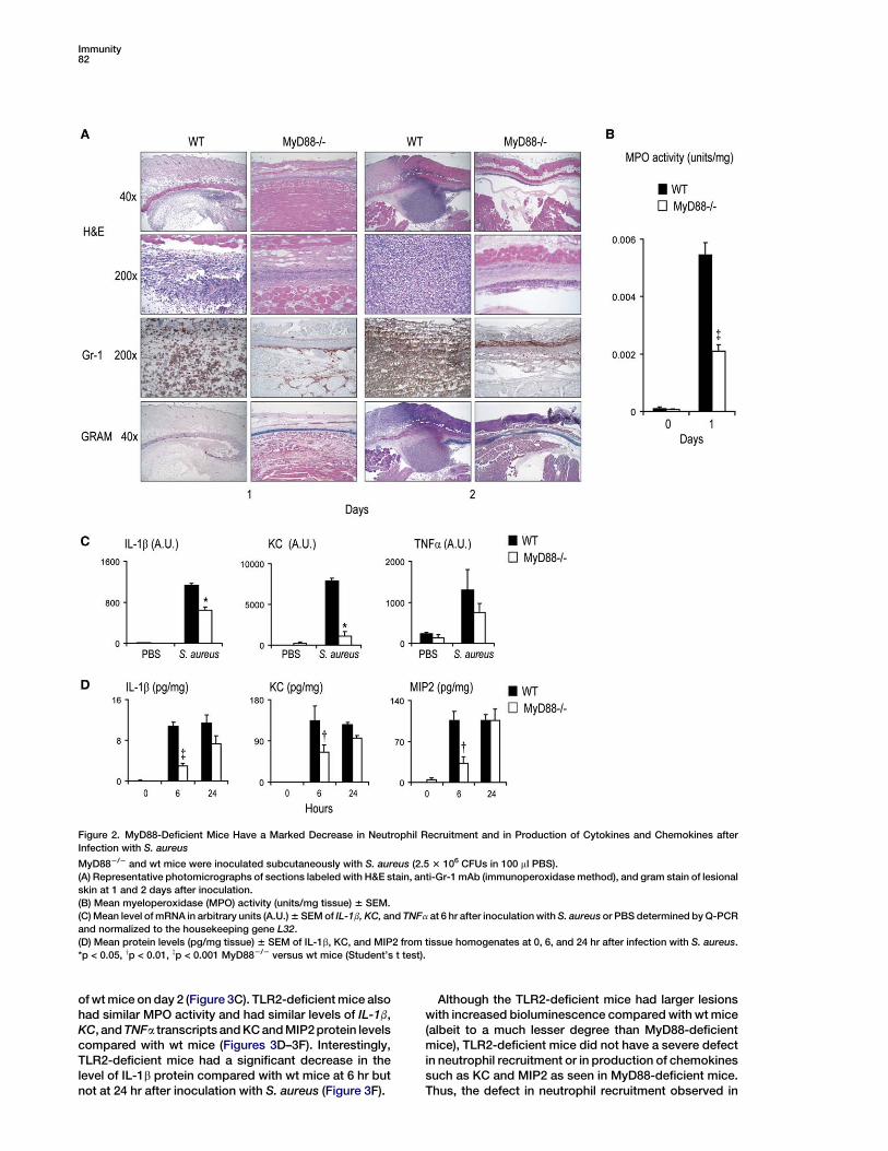
MyD88-Deficient Mice Have a Defect in NeutrophilRecruitment Compared with Wild-Type Mice after
Skin Inoculation with S. aureusSince the increased lesion size and bioluminescent sig-nals in MyD88-deficient mice were observed shortly afterthe infection, histology of lesional skin was evaluated atdays 1 and 2 after inoculation with S. aureus (Figure 2A).We found that lesions of wt mice had large neutrophilicabscesses in both H&E and anti-Gr-1 (neutrophil marker)mAb-labeled sections. In fact, almost all of the cells ofthe infiltrate were Gr-1 positive with only a few scatteredMac-3 (macrophage marker)-positive cells (Figure S3). Inaddition, in wt mice, S. aureus bacteria were barely de-tectable by gram stain, perhaps due to phagocytosis ofthe bacteria by the neutrophils within the abscess (Fig-ure 2A). In contrast, lesions of MyD88-deficient micehad severely decreased numbers of neutrophils, withno observable abscess formation, and contained ablue-staining band of gram-positive bacteria spanningthe entire horizontal length of the section. Thus, MyD88-deficient mice have a severe defect in the recruitment ofneutrophils to the site of infection with S. aureus. Further-more, the readily detectable gram-positive bacteria in le-sions of MyD88-deficient mice corroborate the resultsobtained with in vivo bioluminescence and bacterial
counts, demonstrating that MyD88-deficient mice havea severe defect in bacterial clearance. There was alsosignificantly less myeloperoxidase (MPO) activity, a mar-ker of neutrophil function, in lesions of MyD88-deficientmice compared with wt mice at 1 day after inoculationwith S. aureus (Figure 2B).
A previous study with a similar S. aureus cutaneous in-fection model demonstrated that neutrophil-depletedmice (with mAb RB6-8C5) developed large nonhealingskin lesions and failed to clear S. aureus from these le-sions (Molne et al., 2000). Thus, both MyD88-deficientmice and neutrophil-depleted mice share a similar phe-notype and provide evidence that neutrophils play a keyrole in immunity against cutaneous S. aureus infections.Moreover, our results suggest that MyD88 signaling isessential for this neutrophil recruitment to take place.
MyD88-Deficient Mice Have a Defect in Production of
Cytokines and Chemokines Involved in NeutrophilRecruitment In Vivo after Skin Inoculation
with S. aureusThe induction of cytokines and chemokines involved inneutrophil recruitment was evaluated in lesions ofMyD88-deficient mice and wt mice. mRNA was har-vested from lesions at 6 hr after inoculation and levelsof mRNA transcripts were compared, including IL-1b,KC/IL-8, and TNFa (Figure 2C). We found that MyD88-deficient mice have decreased IL-1b and KC mRNAcompared with wt mice (p < 0.05). The levels of TNFa
mRNA were not significantly decreased in lesions ofMyD88-deficient mice compared with wt mice.
Protein levels of IL-1b, KC, and MIP2 from lesions weredetermined by performing ELISAs on homogenized8 mm punch biopsies of lesional skin at 0, 6, and 24 hrafter inoculation with S. aureus (Figure 2D). MyD88-deficient mice had significantly decreased levels ofIL-1b, KC, and MIP2 compared with wt mice at 6 hr butnot at 24 hr after inoculation. Thus, the decreased neu-trophil recruitment in MyD88-deficient mice may in partbe due to an early decreased production of cytokinesand chemokines involved in neutrophil recruitment.
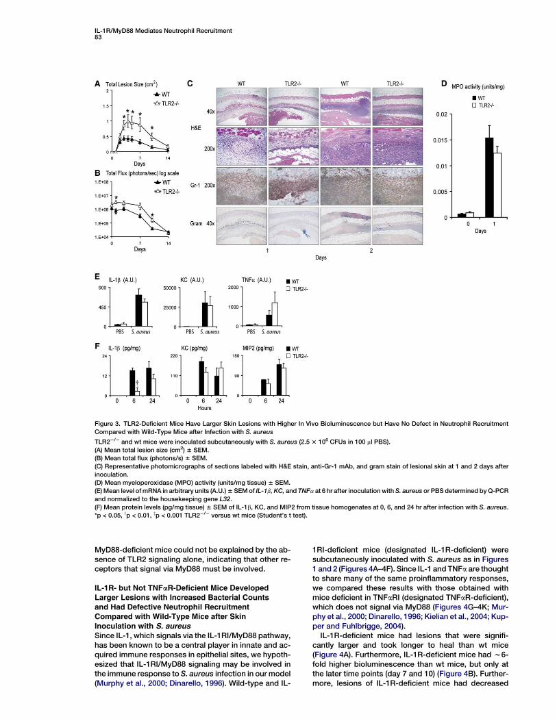
TLR2-Deficient Mice Developed Larger
Lesions Compared with Wild-Type Mice butHad No Defect in Neutrophil Recruitment
after Skin Inoculation with S. aureusPrevious studies have demonstrated that TLR2 can rec-ognize lipopeptides and lipoteichoic acid of S. aureus(Takeuchi et al., 2001, 2002). In addition, TLR2-deficientmice are more susceptible to a S. aureus systemic infec-tion than wt mice (Hoebe et al., 2005; Takeuchi et al.,2000). To investigate whether TLR2/MyD88 signaling isinvolved in our localized S. aureus infection model, wesubcutaneously inoculated wt and TLR2-deficient micewith S. aureus as in Figures 1 and 2 (Figures 3A–3F). Wefound that TLR2-deficient mice developed larger lesions(w2-fold) with higher bioluminescent signals than wtmice (4- to 5-fold) (Figures 3A and 3B), which was consis-tent with previous reports (Hoebe et al., 2005; Takeuchiet al., 2000). However, TLR2-deficient mice healed theirskin lesions at approximately the same time as wt mice(day 14). Furthermore, lesions of TLR2-deficient micehad slightly smaller neutrophilic abscesses than thoseof wt mice on day 1 but were virtually identical to those
Immunity82
Figure 2. MyD88-Deficient Mice Have a Marked Decrease in Neutrophil Recruitment and in Production of Cytokines and Chemokines after
Infection with S. aureus
MyD882/2 and wt mice were inoculated subcutaneously with S. aureus (2.5 3 106 CFUs in 100 ml PBS).
(A) Representative photomicrographs of sections labeled with H&E stain, anti-Gr-1 mAb (immunoperoxidase method), and gram stain of lesional
skin at 1 and 2 days after inoculation.
(B) Mean myeloperoxidase (MPO) activity (units/mg tissue) 6 SEM.
(C) Mean level of mRNA in arbitrary units (A.U.) 6 SEM of IL-1b, KC, and TNFa at 6 hr after inoculation with S. aureus or PBS determined by Q-PCR
and normalized to the housekeeping gene L32.
(D) Mean protein levels (pg/mg tissue) 6 SEM of IL-1b, KC, and MIP2 from tissue homogenates at 0, 6, and 24 hr after infection with S. aureus.
*p < 0.05, yp < 0.01, zp < 0.001 MyD882/2 versus wt mice (Student’s t test).
of wt mice on day 2 (Figure 3C). TLR2-deficient mice alsohad similar MPO activity and had similar levels of IL-1b,KC, and TNFa transcripts and KC and MIP2 protein levelscompared with wt mice (Figures 3D–3F). Interestingly,TLR2-deficient mice had a significant decrease in thelevel of IL-1b protein compared with wt mice at 6 hr butnot at 24 hr after inoculation with S. aureus (Figure 3F).
Although the TLR2-deficient mice had larger lesionswith increased bioluminescence compared with wt mice(albeit to a much lesser degree than MyD88-deficientmice), TLR2-deficient mice did not have a severe defectin neutrophil recruitment or in production of chemokinessuch as KC and MIP2 as seen in MyD88-deficient mice.Thus, the defect in neutrophil recruitment observed in
IL-1R/MyD88 Mediates Neutrophil Recruitment83
Figure 3. TLR2-Deficient Mice Have Larger Skin Lesions with Higher In Vivo Bioluminescence but Have No Defect in Neutrophil Recruitment
Compared with Wild-Type Mice after Infection with S. aureus
TLR22/2 and wt mice were inoculated subcutaneously with S. aureus (2.5 3 106 CFUs in 100 ml PBS).
(A) Mean total lesion size (cm2) 6 SEM.
(B) Mean total flux (photons/s) 6 SEM.
(C) Representative photomicrographs of sections labeled with H&E stain, anti-Gr-1 mAb, and gram stain of lesional skin at 1 and 2 days after
inoculation.
(D) Mean myeloperoxidase (MPO) activity (units/mg tissue) 6 SEM.
(E) Mean level of mRNA in arbitrary units (A.U.) 6 SEM of IL-1b, KC, and TNFa at 6 hr after inoculation with S. aureus or PBS determined by Q-PCR
and normalized to the housekeeping gene L32.
(F) Mean protein levels (pg/mg tissue) 6 SEM of IL-1b, KC, and MIP2 from tissue homogenates at 0, 6, and 24 hr after infection with S. aureus.
*p < 0.05, yp < 0.01, zp < 0.001 TLR22/2 versus wt mice (Student’s t test).
MyD88-deficient mice could not be explained by the ab-sence of TLR2 signaling alone, indicating that other re-ceptors that signal via MyD88 must be involved.
IL-1R- but Not TNFaR-Deficient Mice DevelopedLarger Lesions with Increased Bacterial Counts
and Had Defective Neutrophil RecruitmentCompared with Wild-Type Mice after Skin
Inoculation with S. aureusSince IL-1, which signals via the IL-1RI/MyD88 pathway,has been known to be a central player in innate and ac-quired immune responses in epithelial sites, we hypoth-esized that IL-1RI/MyD88 signaling may be involved inthe immune response to S. aureus infection in our model(Murphy et al., 2000; Dinarello, 1996). Wild-type and IL-
1RI-deficient mice (designated IL-1R-deficient) weresubcutaneously inoculated with S. aureus as in Figures1 and 2 (Figures 4A–4F). Since IL-1 and TNFa are thoughtto share many of the same proinflammatory responses,we compared these results with those obtained withmice deficient in TNFaRI (designated TNFaR-deficient),which does not signal via MyD88 (Figures 4G–4K; Mur-phy et al., 2000; Dinarello, 1996; Kielian et al., 2004; Kup-per and Fuhlbrigge, 2004).
IL-1R-deficient mice had lesions that were signifi-cantly larger and took longer to heal than wt mice(Figure 4A). Furthermore, IL-1R-deficient mice had w6-fold higher bioluminescence than wt mice, but only atthe later time points (day 7 and 10) (Figure 4B). Further-more, lesions of IL-1R-deficient mice had decreased

Immunity84
Figure 4. IL-1R- but Not TNFaR-Deficient Mice Have Markedly Larger Skin Lesions with Higher Bacterial Counts and Decreased Neutrophil Re-
cruitment Compared with Wild-Type Mice after Subcutaneous Inoculation with S. aureus
IL-1R2/2, TNFaR2/2, and wt control mice were inoculated subcutaneously with S. aureus (2.5 3 106 CFUs in 100 ml PBS).
(A and G) Mean total lesion size (cm2) 6 SEM.
(B and H) Mean total flux (photons/s) 6 SEM.
(C and I) Representative photomicrographs of sections labeled with H&E stain, anti-Gr-1 mAb, and gram stain of lesional skin at 1 and 2 days after
inoculation.
(D and J) Mean myeloperoxidase (MPO) activity (units/mg tissue) 6 SEM.
(E) Mean level of mRNA in arbitrary units (A.U.) 6 SEM of IL-1b, KC, and TNFa at 6 hr after inoculation with S. aureus or PBS determined by Q-PCR
and normalized to the housekeeping gene L32.
(F and K) Mean protein levels (pg/mg tissue) 6 SEM of IL-1b, KC, and MIP2 from tissue homogenates at 0, 6, and 24 hr after inoculation.
*p < 0.05, yp < 0.01, zp < 0.001 IL-1R2/2 mice or TNFaR2/2 mice versus wt mice (Student’s t test).

IL-1R/MyD88 Mediates Neutrophil Recruitment85
neutrophilic abscess formation, decreased MPO activ-ity, and decreased levels of KC mRNA and KC andMIP2 protein levels at 6 hr after inoculation withS. aureus compared with wt mice (Figures 4C–4F). Al-though lesions of both IL-1R-deficient mice and wtmice had similar levels of IL-1b mRNA and protein levels(Figures 4E and 4F), IL-1R-deficient mice cannot re-spond to IL-1b. Taken together, IL-1R-deficient mice re-sembled MyD88-deficient mice in that both had sub-stantially larger lesions with higher bacterial countsthan wt mice after skin inoculation with S. aureus. In ad-dition, both IL-1R- and MyD88-deficient mice had a se-vere defect in the ability to recruit neutrophils to thesite of infection, suggesting that IL-1R/MyD88 signalingis essential for adequate neutrophil recruitment.
Interestingly, lesions of IL-1R-deficient mice had sig-nificantly increased levels of TNFa mRNA, which mayhave been a compensatory increase since IL-1R-defi-cient mice cannot respond to IL-1 (Figure 4E). However,the higher levels of TNFa are less likely to be important inimmunity against S. aureus in our model, since we foundno significant differences in lesion size, in vivo biolumi-nescence, neutrophilic abscess formation, MPO activ-ity, or induction of KC and MIP2 in lesions of TNFaR-deficient mice compared with wt mice (Figures 4G–4K).
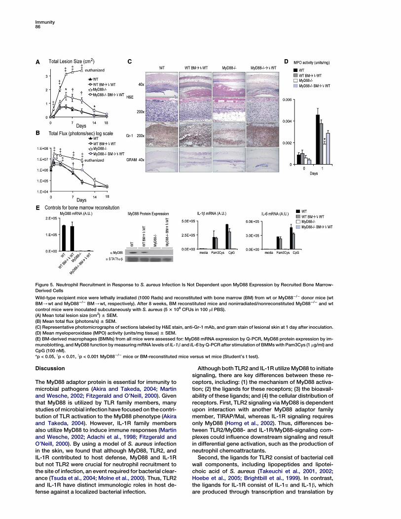
Neutrophil Recruitment in Response to S. aureusInfection Is Not Dependent upon MyD88 Expression
by Recruited Bone Marrow-Derived CellsSince the lesions of S. aureus-infected mice are com-prised of both resident skin cells and recruited bone mar-row (BM)-derived cells, we wanted to determine whichpopulation of cells utilized IL-1R/MyD88 signaling to pro-mote neutrophil recruitment. Lethally irradiated recipientwt mice were reconstituted with BM from donor wt miceor MyD88-deficient mice to generate two groups: (1) wtmice reconstituted with wt BM (wt BM/wt mice) and(2) wt mice reconstituted with MyD88-deficient BM(MyD882/2 BM/wt mice) (Figure 5). At 8 weeks postre-constitution, these BM-reconstituted mice and normalnonirradiated/nonreconstituted wt and MyD88-deficientmice were subcutaneously inoculated with S. aureus asin Figures 1 and 2. We found that MyD882/2 BM/wtmice had lesions that were only moderately larger thanthose of wt BM/wt mice (Figure 5A). Furthermore, le-sions in both groups healed at approximately the sametime (day 18). These results were in contrast to lesionsof nonirradiated/nonreconstituted MyD88-deficient mice,which had 3-fold larger lesion sizes with no signs of heal-ing and were thus euthanized on day 10. Similarly, in vivobioluminescence signals of MyD882/2 BM/wt micewere moderately higher than that of wt BM/wt mice,but to levels much less than that of nonirradiated/nonre-constituted MyD88-deficient mice (Figure 5B).
MyD882/2 BM/wt mice, wt BM/wt mice, and nonir-radiated/nonreconstituted wt mice all had large neutro-philic abscesses at day 1, whereas nonirradiated/nonre-constituted MyD88-deficient mice had markedlydecreased neutrophil recruitment (Figure 5C). Further-more, there was no significant difference between MPOactivity in lesions of nonirradiated/nonreconstituted wtmice compared with MyD882/2 BM/wt mice or wtBM/wt (Figure 5D). To control for BM reconstitution,BM-derived macrophages (BMMs) from all mice were
evaluated for the presence of MyD88 mRNA and proteinexpression (Figure 5E). Functional activity of MyD88-dependent signaling was assayed by measuring IL-1b
and IL-6 mRNA in response to the TLR1/2 ligand,Pam3Cys, and the TLR9 ligand, CpG DNA (Figure 5E).Taken together, these results demonstrate that MyD88signaling by the recruited BM-derived cells is dispens-able for neutrophil recruitment after skin inoculationwith S. aureus.
Neutrophil Recruitment in Response to S. aureus
Infection Is Dependent upon IL-1R/MyD88 Signalingby Resident Skin Cells and Not by BM-Derived
Recruited CellsWe next set out to determine whether IL-1R signalingby BM-derived recruited cells was also dispensablefor neutrophil recruitment after skin inoculation withS. aureus. As in Figure 5, lethally irradiated recipient wtmice were reconstituted with BM from donor wt miceor IL-1R-deficient mice to generate two groups: (1) wtBM/wt mice and (2) IL-1R2/2 BM/wt mice (Figures6A–6D). IL-1R2/2 BM/wt mice had lesion sizes andbioluminescence signals roughly equivalent to those ofwt BM/wt mice or wt control mice and significantlyless than those of nonirradiated/nonreconstituted IL-1R-deficient mice (Figures 6A and 6B). In addition, le-sions of IL-1R2/2 BM/wt mice, wt BM/wt mice, andnonirradiated/nonreconstituted wt control mice all de-veloped large neutrophilic abscesses at day 1 (Fig-ure 6C). In contrast, lesions of nonirradiated/nonrecon-stituted IL-1R-deficient mice had markedly decreasedneutrophil recruitment. Furthermore, MPO activity ofIL-1R2/2 BM/wt mice and of wt BM/wt mice wasnot significantly different than that of wt control mice(Figure 6D). Similar to results obtained with the MyD88BM reconstitution experiments, these findings demon-strate that IL-1R signaling by the recruited BM-derivedcells is dispensable for neutrophil recruitment and im-munity against S. aureus.
To evaluate the role of resident skin cells in utilizing IL-1R/MyD88 signaling to promote neutrophil recruitmentmore directly, we reconstituted IL-1R-deficient recipientmice with either wt or IL-1R-deficient donor BM to gen-erate two groups: (1) wt BM/IL-1R2/2 mice and (2) IL-1R2/2 BM/IL-1R2/2 mice (Figures 6E–6H). Wild-typeBM/IL-1R2/2 mice and IL-1R2/2 BM/IL-1R2/2 micehad lesion sizes and bioluminescence signals thatwere virtually equivalent to those of nonirradiated/non-reconstituted IL-1R-deficient mice and significantlygreater (3- to 4-fold) than those of wt control mice (Fig-ures 6E and 6F). In addition, lesions of wt BM/IL-1R2/2 mice, IL-1R2/2 BM/IL-1R2/2 mice, and nonirra-diated/nonreconstituted IL-1R2/2 mice all had markedlydecreased neutrophilic abscess formation, abundantgram-positive bacteria, and decreased MPO activitycompared with wt control mice (Figures 6G and 6H).To control for BM reconstitution, BMMs from all micewere assayed for IL-6 mRNA levels after stimulationwith recombinant murine IL-1b (Figure 6I). Taken to-gether, these data suggest that IL-1R signaling by resi-dent skin cells is essential for adequate neutrophil re-cruitment to a site of a localized S. aureus infection inthe skin.
Immunity86
Figure 5. Neutrophil Recruitment in Response to S. aureus Infection Is Not Dependent upon MyD88 Expression by Recruited Bone Marrow-
Derived Cells
Wild-type recipient mice were lethally irradiated (1000 Rads) and reconstituted with bone marrow (BM) from wt or MyD882/2 donor mice (wt
BM/wt and MyD882/2 BM/wt, respectively). After 8 weeks, BM reconstituted mice and nonirradiated/nonreconstituted MyD882/2 and wt
control mice were inoculated subcutaneously with S. aureus (5 3 106 CFUs in 100 ml PBS).
(A) Mean total lesion size (cm2) 6 SEM.
(B) Mean total flux (photons/s) 6 SEM.
(C) Representative photomicrographs of sections labeled by H&E stain, anti-Gr-1 mAb, and gram stain of lesional skin at 1 day after inoculation.
(D) Mean myeloperoxidase (MPO) activity (units/mg tissue) 6 SEM.
(E) BM-derived macrophages (BMMs) from all mice were assessed for: MyD88 mRNA expression by Q-PCR, MyD88 protein expression by im-
munoblotting, and MyD88 function by measuring mRNA levels of IL-1b and IL-6 by Q-PCR after stimulation of BMMs with Pam3Cys (1 mg/ml) and
CpG (100 nM).
*p < 0.05, yp < 0.01, zp < 0.001 MyD882/2 mice or BM-reconstituted mice versus wt mice (Student’s t test).
Discussion
The MyD88 adaptor protein is essential for immunity tomicrobial pathogens (Akira and Takeda, 2004; Martinand Wesche, 2002; Fitzgerald and O’Neill, 2000). Giventhat MyD88 is utilized by TLR family members, manystudies of microbial infection have focused on the contri-bution of TLR activation to the MyD88 phenotype (Akiraand Takeda, 2004). However, IL-1R family membersalso utilize MyD88 to induce immune responses (Martinand Wesche, 2002; Adachi et al., 1998; Fitzgerald andO’Neill, 2000). By using a model of S. aureus infectionin the skin, we found that although MyD88, TLR2, andIL-1R contributed to host defense, MyD88 and IL-1Rbut not TLR2 were crucial for neutrophil recruitment tothe site of infection, an event required for bacterial clear-ance (Tsuda et al., 2004; Molne et al., 2000). Thus, TLR2and IL-1R have distinct immunologic roles in host de-fense against a localized bacterial infection.
Although both TLR2 and IL-1R utilize MyD88 to initiatesignaling, there are key differences between these re-ceptors, including: (1) the mechanism of MyD88 activa-tion; (2) the ligands for these receptors; (3) the bioavail-ability of these ligands; and (4) the cellular distribution ofreceptors. First, TLR2 signaling via MyD88 is dependentupon interaction with another MyD88 adaptor familymember, TIRAP/Mal, whereas IL-1R signaling requiresonly MyD88 (Horng et al., 2002). Thus, differences be-tween TLR2/MyD88- and IL-1R/MyD88-signaling com-plexes could influence downstream signaling and resultin differential gene activation, such as the production ofneutrophil chemoattractants.
Second, the ligands for TLR2 consist of bacterial cellwall components, including lipopeptides and lipotei-choic acid of S. aureus (Takeuchi et al., 2001, 2002;Hoebe et al., 2005; Brightbill et al., 1999). In contrast,the ligands for IL-1R consist of IL-1a and IL-1b, whichare produced through transcription and translation by

IL-1R/MyD88 Mediates Neutrophil Recruitment87
Figure 6. Neutrophil Recruitment in Response to S. aureus Infection Is Dependent upon IL-1R/MyD88 Signaling by Resident Skin Cells and Not
by Bone Marrow-Derived Recruited Cells
Wild-type and IL-1R2/2 recipient mice were lethally irradiated (1000 Rads) and reconstituted with bone marrow (BM) cells from wt or IL-1R2/2
donor mice: (A–D) wt BM/wt and IL-1R2/2 BM/wt and (E and F) wt BM/IL-1R2/2 and IL-1R2/2 BM/IL-1R2/2. After 8 weeks, BM-reconsti-
tuted mice and nonirradiated/nonreconstituted IL-1R2/2 and wt control mice were inoculated subcutaneously with S. aureus (2.5 3 106 CFUs in
100 ml PBS).
(A and E) Mean total lesion size (cm2) 6 SEM.
(B and F) Mean total flux (photons/s) 6 SEM.
(C and G) Representative photomicrographs of sections labeled with H&E, anti-Gr-1 mAb, and gram stain of lesional skin at 1 day after inocu-
lation.
(D and H) Mean myeloperoxidase (MPO) activity (units/mg tissue) 6 SEM.
(I) BM-derived macrophages from BM-reconstituted mice and nonirradiated/nonreconstituted IL-1R2/2 and wt mice were assessed for IL-1R
activity by measuring mRNA levels of IL-6 by Q-PCR after stimulating BMMs with rIL-1b (50 ng/ml).
*p < 0.05, yp < 0.01, zp < 0.001 IL-1R2/2 mice or BM-reconstituted mice versus wt mice (Student’s t test).
the host (Murphy et al., 2000; Dinarello, 1996). Experi-ments in vitro have indicated that production of IL-1b
is MyD88 dependent (data not shown) (Takeuchi et al.,2000; Kawai et al., 1999). However, we found that IL-1b
was produced in vivo in both TLR2- and MyD88-defi-
cient mice, albeit somewhat delayed, reaching levelsobserved in wt mice by 24 hr after infection with S. au-reus. Since TLR2-deficient mice can respond to any IL-1a or IL-1b present whereas MyD88- and IL-1R-deficientmice cannot, the bacterial clearance in TLR2-deficient

Immunity88
mice might be primarily or in part mediated by IL-1R ac-tivation, which in turn could deliver signals via IL-1R/MyD88 to promote neutrophil recruitment.
Third, since the ligands for TLR2 and IL-1R come fromdifferent sources, the bioavailability of these ligands totheir corresponding receptors may play a key role inwhich cell types are activated. TLR2-mediated recogni-tion of bacterial components occurs predominantly inthe context of breakdown of the pathogen by inflamma-tory cells (Akira and Takeda, 2004; Martin and Wesche,2002). In contrast, IL-1a is produced primarily by kerati-nocytes and endothelial cells (Murphy et al., 2000; Dinar-ello, 1996; Steen et al., 1999). In uninfected skin ofMyD88-deficient, TLR2-deficient, and wt mice (at timezero), IL-1a was detected within the cells of the epider-mis (Figure S4). However, at 24 hr after S. aureus inocu-lation, IL-1a was not only detected in the cells of the epi-dermis, but also in the dermis and focally within theneutrophilic abscesses. Therefore, IL-1a is localized todistinct compartments within the skin, and its bioavail-ability to different cell types appears to be time depen-dent. IL-1b is produced primarily by activated immunesystem cells such as monocytes/macrophages, den-dritic cells, and Langerhans cells (Murphy et al., 2000;Dinarello, 1996; Kupper and Fuhlbrigge, 2004). Activa-tion of these different cell types occurs in an autocrineor paracrine manner during the course of infection.Thus, IL-1a or IL-1b could activate distinct gene pro-grams among the different cell types, resulting in dis-tinct inflammatory responses such as the recruitmentof neutrophils. Interestingly, we did observe an early de-creased production of IL-1b in TLR2-deficient mice butdid not observe a major defect in neutrophil recruitmentis these mice. It could be that IL-1a compensated for thisearly decreased production of IL-1b in TLR2-deficientmice to promote IL-1R/MyD88-dependent neutrophilrecruitment.
Fourth, the cellular expression and distribution of IL-1R and TLR2 are different. IL-1R is found ubiquitously,on both resident cells and BM-derived cells (Murphyet al., 2000; Dinarello, 1996). In contrast, TLR2 is primar-ily expressed on BM-derived cells including neutrophils,monocytes/macrophages, antigen-presenting cells, Tand B cells, and with conflicting reports about expres-sion on endothelial and some epithelial cells (Takeuchiet al., 1999; Fan et al., 2003; Kurt-Jones et al., 2002).To delineate which population of cells is important inpromoting IL-1R/MyD88-mediated neutrophil recruit-ment, we utilized techniques of BM reconstitution. Wefound that neutrophil recruitment and bacterial clear-ance occurred in wt mice reconstituted with IL-1R-defi-cient, MyD88-deficient, or wt BM, indicating that IL-1Ror MyD88 expression by recruited BM-derived cellshad no impact on neutrophil recruitment or in the eradi-cation of the infection. In addition, we found a severe de-fect in neutrophil recruitment in IL-1R-deficient mice re-constituted with either IL-1R-deficient or wt BM. Thesedata suggest that adequate neutrophil recruitment andbacterial clearance is dependent upon IL-1R/MyD88signaling by resident skin cells and not by BM-derivedrecruited cells. Thus, resident skin cells may act as firstresponders and send appropriate signals via IL-1R/MyD88 activation, such as production of KC and MIP2,to promote neutrophil recruitment. Future studies will
utilize additional BM reconstitution experiments to fur-ther characterize the functional contribution of IL-1Rand TLR2 to the phenotype of MyD88-deficient mice.
It should be mentioned that the results of the presentstudy are consistent with previous studies that havedemonstrated the importance of IL-1R/MyD88 signalingfor host defense against S. aureus in brain abscesses,arthritis, and systemic infections (Hultgren et al., 2002;Kielian et al., 2004; Verdrengh et al., 2004). In addition,the chemokines KC and MIP2 have been shown to beimportant for neutrophil recruitment in S. aureus brainabscesses (Kielian et al., 2001). However, the presentstudy has demonstrated that IL-1R/MyD88 signalingby resident cells at the site of infection is critical for neu-trophil recruitment and immunity against S. aureus in theskin, where most of these infections originate. We havealso demonstrated that production of KC and MIP2 invivo is dependent upon IL-1R/MyD88 signaling. Ourdata do not exclude that in addition to TLR2 and IL-1R, other MyD88-dependent receptors, such as TLR9or IL-18R, may also contribute to host defense againstS. aureus (Martin and Wesche, 2002; Adachi et al., 1998).
Lastly, it could be that the mechanism(s) leading toneutrophil recruitment is controlled by IL-1R/MyD88 sig-naling or perhaps another mechanism, such as viaTNFaR activation, depending on the site of infection.This may in fact be the case, since a previous studyhas demonstrated that MyD88 is dispensable for hostdefense against S. aureus pulmonary infections, andother studies have demonstrated that TNFa is essentialfor neutrophil recruitment in models of acute peritonitis(Skerrett et al., 2004; Zhang et al., 1992; Malaviya et al.,1996).
In summary, we have identified that IL-1R/MyD88 butnot TLR2/MyD88 signaling by resident skin cells is criti-cal for neutrophil recruitment to a site of a localizedS. aureus infection in the skin. From the clinical pointof view, the identification of the critical role of IL-1R/MyD88 signaling for promoting neutrophil recruitmentto a site of infection raises the possibility that thispathway could be locally targeted to engage the host’sown immune responses in the treatment of a microbialinfection.
Experimental Procedures
Staphylococcus aureus Strains
All S. aureus strains used were derived from the common laboratory
strain RN6390, which has a known 11 bp deletion in the rsbU gene
within the sigB operon (Novick, 1990). Strain SH1000 is a derivative
of RN6390 with the rsbU gene restored (Horsburgh et al., 2002).
Strain RN4220 is a derivative of 8325-4 that accepts foreign DNA
(Novick, 1990). Strains used in cutaneous bioluminescent mouse in-
fection experiments include the bioluminescent strain ALC2906,
which is strain SH1000 containing the shuttle plasmid pSK236 with
the pbp2 (penicillin binding protein 2) promoter fused to the lux-
ABCDE reporter cassette from Photorhabdus luminescens. In con-
trol experiments, strain ALC2506, a nonbioluminescent control
SH1000 strain containing pSK236 with a promoterless luxABCDE re-
porter cassette, was used. Strain 8325-4 (kind gift of Prof. Jeffrey F.
Miller, Dept. of Microbiology, Immunology, and Molecular Genetics,
UCLA, Los Angeles, CA) was used where indicated.
Plasmids
The following plasmids were used to make bioluminescent S. aureus
strains. pSK236 is an E. coli and S. aureus shuttle vector (Novick,
1990). pLux is pSK236 containing luxABCDE from pSB2025 at SalI

IL-1R/MyD88 Mediates Neutrophil Recruitment89
and PstI sites (Qazi et al., 2001). pLux(pbp2-pro) is pLux containing
a 986 bp pbp2 promoter at the EcoRI site immediately upstream of
luxABCDE. pCR2.1 is an E. coli vector for direct cloning of PCR frag-
ments (Invitrogen, Carlsbad, CA).
Construction Details of Bioluminescent S. aureus Strain
Promoterless luxABCDE cassette of Photorhabdus luminescens
from pSB2025 from E. coli strain ALC2468 (JM109 containing
pSB2025) was cloned into the SalI/PstI site of pSK236. The promoter
of pbp2 was amplified from chromosomal DNA of S. aureus strain
RN6390 by PCR with the following promoter-specific primers 50-CC
ACATACTTGTACTTGCCTC-30 (forward) and 50-TTCTTAGGCTGAG
AAGATCC-30 (reverse). The resulting 989 bp PCR fragment was
cloned into TOPO-TA cloning vector pCR2.1, digested with EcoRI,
and cloned into EcoRI site upstream of the promoterless Lux re-
porter vector pLux. The correct orientation of the promoter fragment
was confirmed by sequencing. Plasmid DNA was electroporated into
S. aureus strain RN4220 and then into strain SH1000. This biolumi-
nescent strain containing pLux(pbp2) was designated ALC2906.
The pLux(pbp2) plasmid in ALC2906 is active during all S. aureus
growth phases and emits photons only from live, ATP-producing,
bacteria.
Preparation of S. aureus for Skin Inoculation
S. aureus was streaked onto Tryptic soy agar (Tryptic soy broth
[TSB] + 1.5% Bacto Agar) (Becton Dickinson, Sparks, MD; Sigma-
Aldrich, St. Louis, MO). Colonies of S. aureus were grown overnight at
37ºC in a shaking incubator (240 rpm) in TSB. Midlogarithmic phase
bacteria were obtained after a 3 hr subculture of 1:100 dilution of the
overnight culture. The S. aureus bioluminescent strain ALC2906 and
nonbioluminescent strain ALC2506 have a chloramphenicol resis-
tance plasmid selection marker, and cultures were performed in
the presence of chloramphenicol (10 mg/ml) (Sigma-Aldrich). In other
experiments, S. aureus strain 8325-4 was prepared similarly, but in
the absence of chloramphenicol. Bacterial cells were pelleted, re-
suspended, and washed three times in PBS. Bacterial concentra-
tions were estimated with a spectrophotometer (Beckman DU
640B Spectrophotometer, Beckman Coulter, Inc., Fullerton, CA) by
determining the absorbance at 600 nm (A600). Colony-forming units
(CFUs) were verified by plating dilutions of the inoculum onto TSB
agar 6 chloramphenicol overnight.
Mice
All mice used have a C57BL/6J genetic background. MyD88-defi-
cient mice (F8), TLR2-deficient mice (F4), and littermate wt mice
were kind gifts from Dr. Shizuo Akira (Osaka University, Osaka, Ja-
pan). IL-1RI-deficient mice with Il1rtm1Imx targeted mutation (desig-
nated IL-1R-deficient mice) (F5), TNFaRI-deficient mice with
Tnfrsf1atm1Mak targeted mutation (designated TNFaR-deficient
mice) (F12), and wt mice were obtained from Jackson Laboratories
(Bar Harbor, ME).
Mouse Model of Cutaneous S. aureus Infection
All procedures were approved by UCLA Animal Research Commit-
tee. The mice were shaved on the back and inoculated subcutane-
ously with 100 ml of midlogarithmic growth phase S. aureus strain
ALC2906 (w2.5 3 106 CFUs/100 ml = 1:10 dilution of A600 of 0.5/ml)
in sterile pharmacy grade saline (0.9%) by a 27 gauge needle and
a tuberculin syringe (Abbott Laboratories, Chicago, IL). In experi-
ments with strain 8325-4, the concentration used was w5 3 107
CFUs/100 ml = A600 of 0.8/ml. Groups of 4–5 mice were used in
each experiment followed by one to two repeats to confirm results.
Measurements of total lesion size (cm2) were made by analyzing dig-
ital photographs (Nikon Coolpix 5400) of mice taken every 1–3 days
with the software program ‘‘Image J’’ (NIH Research Services Branch
[http://rsbweb.nih.gov/ij/]) and a millimeter ruler as a reference.
Quantification of In Vivo S. aureus: In Vivo Bioluminescence
In vivo bioluminescence was performed with the Xenogen IVIS imag-
ing system (Xenogen Corporation, Alameda, CA) at the Crump Insti-
tute for Molecular Imaging at UCLA as previously described (Francis
et al., 2000). Mice were anesthetized via an intraperitoneal injection of
a mix of ketamine and xylazine (100 and 20 mg/kg body weight, re-
spectively). Data are presented on color scale overlaid on a gray-
scale photograph of mice and quantified as total flux (photons/s)
within a circular region of interest (1 3 103 pixels) with Living Image
software (Xenogen) (lower limit of detection: 1 3 104 photons/s).
Tissue Embedding and Staining
For histological analysis, lesional 8 mm punch biopsy (Acuderm)
specimens were bisected and one half was fixed in formalin (10%)
and embedded in paraffin and the other half was embedded in Tis-
sue-Tek OTC compound (Sakura Finetek USA, Inc., Torrance, CA)
and frozen in liquid nitrogen. Hematoxylin and eosin (H&E) and
gram stains were performed on paraffin sections (4 mm) by the Tis-
sue Procurement & Histology Core Laboratory and by the Histopa-
thology Laboratory at UCLA, according to guidelines for clinical
samples.
Immunoperoxidase Labeling
Detection of Gr-1 (Ly-6G)-positive cells and IL-1a expression on fro-
zen cryostat specimens of lesional skin punch biopsy specimens
were performed with a biotinylated rat anti-mouse Gr-1 mAb (clone
RB6-8C5) (1 mg/ml) (BD Pharmingen, San Diego, CA) or rat anti-
mouse IL-1a mAb (clone 40508) (15 mg/ml) (R&D systems) by the im-
munoperoxidase method as previously described (Miller et al., 2005).
Immunofluorescence and Confocal Laser Microscopy
Double immunofluorescence of Gr-1 (Ly-6G) (neutrophil marker) and
Mac-3 (macrophage marker) was performed with a biotinylated rat
anti-mouse Gr-1 mAb (clone RB6-8C5) (1 mg/ml) and a FITC-
conjugated rat anti-mouse Mac-3 mAb (1 mg/ml) (clone M3/84) or
appropriate isotype control mAb (all BD Pharmingen) followed by
streptavidin-conjugated Cy�3 (0.2 mg/ml) (Caltag Laboratories,
Burlingame, CA) and imaged on a Leica TCS-SP spectral Confocal
Inverted Microscope equipped with argon (488 nm blue excitation)
and DPSS diode (561 nm yellow excitation) lasers (Leica Microsys-
tems, Heidelberg, Germany) as previously described (Krutzik et al.,
2003).
Myeloperoxidase Assay
Myeloperoxidase (MPO) activity from lesional skin was obtained
from tissue homogenates (Tissue-tearer, Biospec Products, Bar-
tlesville, OK) of 8 mm punch biopsy specimens (Acuderm) with
a MPO assay kit according to the manufacturer’s recommendations
(Cytostore, Calgary, Alberta, Canada).
Enzyme-Linked Immunosorbent Assay
Protein levels of IL-1b, KC, and MIP2 (pg/mg of tissue) from lesional
skin were obtained from tissue homogenates (Tissue-tearer) in
0.01% Triton of 8 mm punch biopsy specimens taken at various
time points after S. aureus skin inoculation with commercially avail-
able ELISA kits (KC and MIP2 from R&D systems; IL-1b from BD
Pharmingen).
Bone Marrow Reconstitution
Bone marrow (BM) reconstitution experiments were performed as
previously described (Joseph et al., 2004). BM was flushed from tib-
ias and femurs from donor mice, RBC was depleted by ACK lysis
buffer, and BM was washed twice in PBS. 1 3 107 BM cells were in-
jected into the tail vein of lethally irradiated (1000 Rads) recipient
mice. Reconstituted mice were maintained in autoclaved cages
and were administered Baytril (0.5 mg/ml in their drinking water)
for the first 3 weeks postirradiation. All experiments were performed
8 weeks after BM reconstitution. To verify the efficiency of reconsti-
tution, BM was removed from euthanized mice and differentiated
into BM-derived macrophages (BMMs) as previously described
(Doyle et al., 2002). For the MyD88 BM reconstitution, MyD88
mRNA and protein expression was determined by Q-PCR and im-
munoblotting, respectively. In addition, MyD88 function was evalu-
ated by stimulating BMMs with Pam3Cys (1 mg/ml) (Alexis Biochem-
icals) or CpG (100 nM) (Invitrogen) and assessed for mRNA
expression for IL-1b and IL-6 by Q-PCR. For the IL-1R BM reconsti-
tution, IL-1RI function was evaluated by stimulating BMMs with re-
combinant murine IL-1b (rIL-1b) (50 ng/ml) (R&D systems) and as-
sessed for mRNA expression of IL-6 by Q-PCR (see below).

Immunity90
mRNA Quantification
Total RNA was isolated from lesional (S. aureus) and control (PBS)
homogenized (Kontes RNase Free Pestles, Vineland, NJ) 8 mm
punch biopsy specimens performed at 6 hr after inoculation or from
cultured BMMs by the use of TRIzol (Invitrogen) according to the
manufacturer’s recommendations. Quantitative real-time PCR (Q-
PCR) was performed as previously described (Doyle et al., 2002).
Primer sequences for IL-1b, TNFa, IL-6, MyD88, and the normalizer,
L32, were published previously (Doyle et al., 2002, 2003, 2004). Pri-
mers for KC were forward, 50-CACTGCACCCAAACCGAAGT-30,
and reverse, 50-GGACAATTTTCTGAACCAAGGG-30. Relative quanti-
ties of mRNA per sample were normalized to L32 (Doyle et al., 2002).
Immunoblotting
MyD88 and STAT1 protein expression in BMMs were assayed by im-
munoblotting by the use of polyclonal antibodies against MyD88
(Prosci) and STAT1 (Santa Cruz Biotechnologies) as previously de-
scribed (Doyle et al., 2002, 2004).
Statistical Analyses
Data were compared with a Student’s t test. All data are expressed
as mean 6 SEM (standard error of the mean) where indicated. Values
of *p < 0.05, yp < 0.01, and zp < 0.001 were considered statistically
significant.
Supplemental Data
Supplemental Data include four figures and can be found with this
article online at http://www.immunity.com/cgi/content/full/24/1/
79/DC1/.
Acknowledgments
We thank Shizuo Akira, M.D., Ph.D. (Department of Host Defense,
Research Institute for Microbial Diseases, Osaka University, Osaka,
Japan) for the generous gifts of MyD88-deficient and TLR2-deficient
mice. We thank Ping Fu, Christopher Creencia, and Leslie Chen at
the UCLA Tissue Procurement & Histology Core Laboratory and
Saeedeh Shapourifar-Tehrani, M.P.H., at the UCLA Histopathology
Laboratory, for their expertise with embedding, cutting, and H&E
and gram staining of tissue sections. This work was supported in
part by grants R01 AI22553, R01 AI47868, and R01 AR40312 (to
R.L.M.), R01 AI056154, R01 CA87924, and R01 AI052359 (to G.C.),
and K08 AI62985 (to L.S.M.) from the National Institutes of Health
(NIH). L.S.M. is also the recipient of a Dermatologist Investigator Re-
search Fellowship from the Dermatology Foundation. R.M.O. and
E.M.P. are supported by the U.S. Public Health Service National
Research Service Award GM 07185.
Received: July 22, 2005
Revised: November 7, 2005
Accepted: November 30, 2005
Published: January 17, 2006
References
Adachi, O., Kawai, T., Takeda, K., Matsumoto, M., Tsutsui, H., Saka-
gami, M., Nakanishi, K., and Akira, S. (1998). Targeted disruption of
the MyD88 gene results in loss of IL-1- and IL-18-mediated function.
Immunity 9, 143–150.
Akira, S., and Takeda, K. (2004). Toll-like receptor signalling. Nat.
Rev. Immunol. 4, 499–511.
Brightbill, H.D., Libraty, D.H., Krutzik, S.R., Yang, R.B., Belisle, J.T.,
Bleharski, J.R., Maitland, M., Norgard, M.V., Plevy, S.E., Smale, S.T.,
et al. (1999). Host defense mechanisms triggered by microbial lipo-
proteins through Toll-like receptors. Science 285, 732–736.
Dinarello, C.A. (1996). Biologic basis for interleukin-1 in disease.
Blood 87, 2095–2147.
Doyle, S., Vaidya, S., O’Connell, R., Dadgostar, H., Dempsey, P., Wu,
T., Rao, G., Sun, R., Haberland, M., Modlin, R., and Cheng, G. (2002).
IRF3 mediates a TLR3/TLR4-specific antiviral gene program. Immu-
nity 17, 251–263.
Doyle, S.E., O’Connell, R., Vaidya, S.A., Chow, E.K., Yee, K., and
Cheng, G. (2003). Toll-like receptor 3 mediates a more potent antivi-
ral response than Toll-like receptor 4. J. Immunol. 170, 3565–3571.
Doyle, S.E., O’Connell, R.M., Miranda, G.A., Vaidya, S.A., Chow,
E.K., Liu, P.T., Suzuki, S., Suzuki, N., Modlin, R.L., Yeh, W.C., et al.
(2004). Toll-like receptors induce a phagocytic gene program
through p38. J. Exp. Med. 199, 81–90.
Fan, J., Frey, R.S., and Malik, A.B. (2003). TLR4 signaling induces
TLR2 expression in endothelial cells via neutrophil NADPH oxidase.
J. Clin. Invest. 112, 1234–1243.
Fitzgerald, K.A., and O’Neill, L.A. (2000). The role of the interleukin-1/
Toll-like receptor superfamily in inflammation and host defense.
Microbes Infect. 2, 933–943.
Francis, K.P., Joh, D., Bellinger-Kawahara, C., Hawkinson, M.J.,
Purchio, T.F., and Contag, P.R. (2000). Monitoring bioluminescent
Staphylococcus aureus infections in living mice using a novel lux-
ABCDE construct. Infect. Immun. 68, 3594–3600.
Fridkin, S.K., Hageman, J.C., Morrison, M., Sanza, L.T., Como-
Sabetti, K., Jernigan, J.A., Harriman, K., Harrison, L.H., Lynfield,
R., and Farley, M.M. (2005). Methicillin-resistant Staphylococcus au-
reus disease in three communities. N. Engl. J. Med. 352, 1436–1444.
Hoebe, K., Georgel, P., Rutschmann, S., Du, X., Mudd, S., Crozat, K.,
Sovath, S., Shamel, L., Hartung, T., Zahringer, U., and Beutler, B.
(2005). CD36 is a sensor of diacylglycerides. Nature 433, 523–527.
Horng, T., Barton, G.M., Flavell, R.A., and Medzhitov, R. (2002). The
adaptor molecule TIRAP provides signalling specificity for Toll-like
receptors. Nature 420, 329–333.
Horsburgh, M.J., Aish, J.L., White, I.J., Shaw, L., Lithgow, J.K., and
Foster, S.J. (2002). sigmaB modulates virulence determinant ex-
pression and stress resistance: characterization of a functional
rsbU strain derived from Staphylococcus aureus 8325-4. J. Bacter-
iol. 184, 5457–5467.
Hultgren, O.H., Svensson, L., and Tarkowski, A. (2002). Critical role
of signaling through IL-1 receptor for development of arthritis and
sepsis during Staphylococcus aureus infection. J. Immunol. 168,
5207–5212.
Joseph, S.B., Bradley, M.N., Castrillo, A., Bruhn, K.W., Mak, P.A.,
Pei, L., Hogenesch, J., O’Connell, R.M., Cheng, G., and Saez, E.
(2004). LXR-dependent gene expression is important for macro-
phage survival and the innate immune response. Cell 119, 299–309.
Kawai, T., Adachi, O., Ogawa, T., Takeda, K., and Akira, S. (1999).
Unresponsiveness of MyD88-deficient mice to endotoxin. Immunity
11, 115–122.
Kielian, T., Barry, B., and Hickey, W.F. (2001). CXC chemokine recep-
tor-2 ligands are required for neutrophil-mediated host defense in
experimental brain abscesses. J. Immunol. 166, 4634–4643.
Kielian, T., Bearden, E.D., Baldwin, A.C., and Esen, N. (2004). IL-1
and TNF-alpha play a pivotal role in the host immune response in
a mouse model of Staphylococcus aureus-induced experimental
brain abscess. J. Neuropathol. Exp. Neurol. 63, 381–396.
Krutzik, S.R., Ochoa, M.T., Sieling, P.A., Uematsu, S., Ng, Y.W.,
Legaspi, A., Liu, P.T., Cole, S.T., Godowski, P.J., Maeda, Y., et al.
(2003). Activation and regulation of Toll-like receptors 2 and 1 in hu-
man leprosy. Nat. Med. 9, 525–532.
Kupper, T.S., and Fuhlbrigge, R.C. (2004). Immune surveillance in
the skin: mechanisms and clinical consequences. Nat. Rev. Immu-
nol. 4, 211–222.
Kurt-Jones, E.A., Mandell, L., Whitney, C., Padgett, A., Gosselin, K.,
Newburger, P.E., and Finberg, R.W. (2002). Role of Toll-like receptor
2 (TLR2) in neutrophil activation: GM-CSF enhances TLR2 expres-
sion and TLR2-mediated interleukin 8 responses in neutrophils.
Blood 100, 1860–1868.
Lowy, F.D. (1998). Staphylococcus aureus infections. N. Engl. J.
Med. 339, 520–532.
Malaviya, R., Ikeda, T., Ross, E., and Abraham, S.N. (1996). Mast cell
modulation of neutrophil influx and bacterial clearance at sites of in-
fection through TNF-alpha. Nature 381, 77–80.
Martin, M.U., and Wesche, H. (2002). Summary and comparison of
the signaling mechanisms of the Toll/Interleukin-1 receptor family.
Biochim. Biophys. Acta 1592, 265–280.

IL-1R/MyD88 Mediates Neutrophil Recruitment91
Melles, D.C., Gorkink, R.F., Boelens, H.A., Snijders, S.V., Peeters,
J.K., Moorhouse, M.J., van der Spek, P.J., van Leeuwen, W.B., Si-
mons, G., Verbrugh, H.A., and van Belkum, A. (2004). Natural popu-
lation dynamics and expansion of pathogenic clones of Staphylo-
coccus aureus. J. Clin. Invest. 114, 1732–1740.
Miller, L.S., Sorensen, O.E., Liu, P.T., Jalian, H.R., Eshtiaghpour, D.,
Behmanesh, B.E., Chung, W., Starner, T.D., Kim, J., and Sieling, P.A.
(2005). TGF-alpha regulates TLR expression and function on epider-
mal keratinocytes. J. Immunol. 174, 6137–6143.
Molne, L., Verdrengh, M., and Tarkowski, A. (2000). Role of neutro-
phil leukocytes in cutaneous infection caused by Staphylococcus
aureus. Infect. Immun. 68, 6162–6167.
Murphy, J.E., Robert, C., and Kupper, T.S. (2000). Interleukin-1 and
cutaneous inflammation: a crucial link between innate and acquired
immunity. J. Invest. Dermatol. 114, 602–608.
Novick, R.P. (1990). The Staphylococcus as a molecular genetic sys-
tem. In Molecular Biology of the Staphylococcus, R.P. Novick, ed.
(New York: VCH Publishers), pp. 1–40.
Qazi, S.N., Counil, E., Morrissey, J., Rees, C.E., Cockayne, A.,
Winzer, K., Chan, W.C., Williams, P., and Hill, P.J. (2001). agr expres-
sion precedes escape of internalized Staphylococcus aureus from
the host endosome. Infect. Immun. 69, 7074–7082.
Skerrett, S.J., Liggitt, H.D., Hajjar, A.M., and Wilson, C.B. (2004). Cut-
ting edge: myeloid differentiation factor 88 is essential for pulmonary
host defense against Pseudomonas aeruginosa but not Staphylo-
coccus aureus. J. Immunol. 172, 3377–3381.
Steen, M.B., Tuck, F.L., and Selvan, R.S. (1999). Spontaneous acti-
vation of endothelial cells: a central role for endogenous IL-1alpha.
In Vitro Cell. Dev. Biol. Anim. 35, 327–332.
Takeuchi, O., Hoshino, K., Kawai, T., Sanjo, H., Takada, H., Ogawa,
T., Takeda, K., and Akira, S. (1999). Differential roles of TLR2 and
TLR4 in recognition of gram-negative and gram-positive bacterial
cell wall components. Immunity 11, 443–451.
Takeuchi, O., Hoshino, K., and Akira, S. (2000). Cutting edge: TLR2-
deficient and MyD88-deficient mice are highly susceptible to Staph-
ylococcus aureus infection. J. Immunol. 165, 5392–5396.
Takeuchi, O., Kawai, T., Muhlradt, P.F., Morr, M., Radolf, J.D., Zy-
chlinsky, A., Takeda, K., and Akira, S. (2001). Discrimination of bac-
terial lipoproteins by Toll-like receptor 6. Int. Immunol. 13, 933–940.
Takeuchi, O., Sato, S., Horiuchi, T., Hoshino, K., Takeda, K., Dong,
Z., Modlin, R.L., and Akira, S. (2002). Cutting edge: role of Toll-like
receptor 1 in mediating immune response to microbial lipoproteins.
J. Immunol. 169, 10–14.
Tsuda, Y., Takahashi, H., Kobayashi, M., Hanafusa, T., Herndon,
D.N., and Suzuki, F. (2004). Three different neutrophil subsets ex-
hibited in mice with different susceptibilities to infection by methicil-
lin-resistant Staphylococcus aureus. Immunity 21, 215–226.
Verdrengh, M., Thomas, J.A., and Hultgren, O.H. (2004). IL-1 recep-
tor-associated kinase 1 mediates protection against Staphylococ-
cus aureus infection. Microbes Infect. 6, 1268–1272.
Zhang, Y., Ramos, B.F., and Jakschik, B.A. (1992). Neutrophil re-
cruitment by tumor necrosis factor from mast cells in immune com-
plex peritonitis. Science 258, 1957–1959.