Tandem reactions initiated by copper-catalyzed cross-coupling
Upload
independentCategory
view
0download
0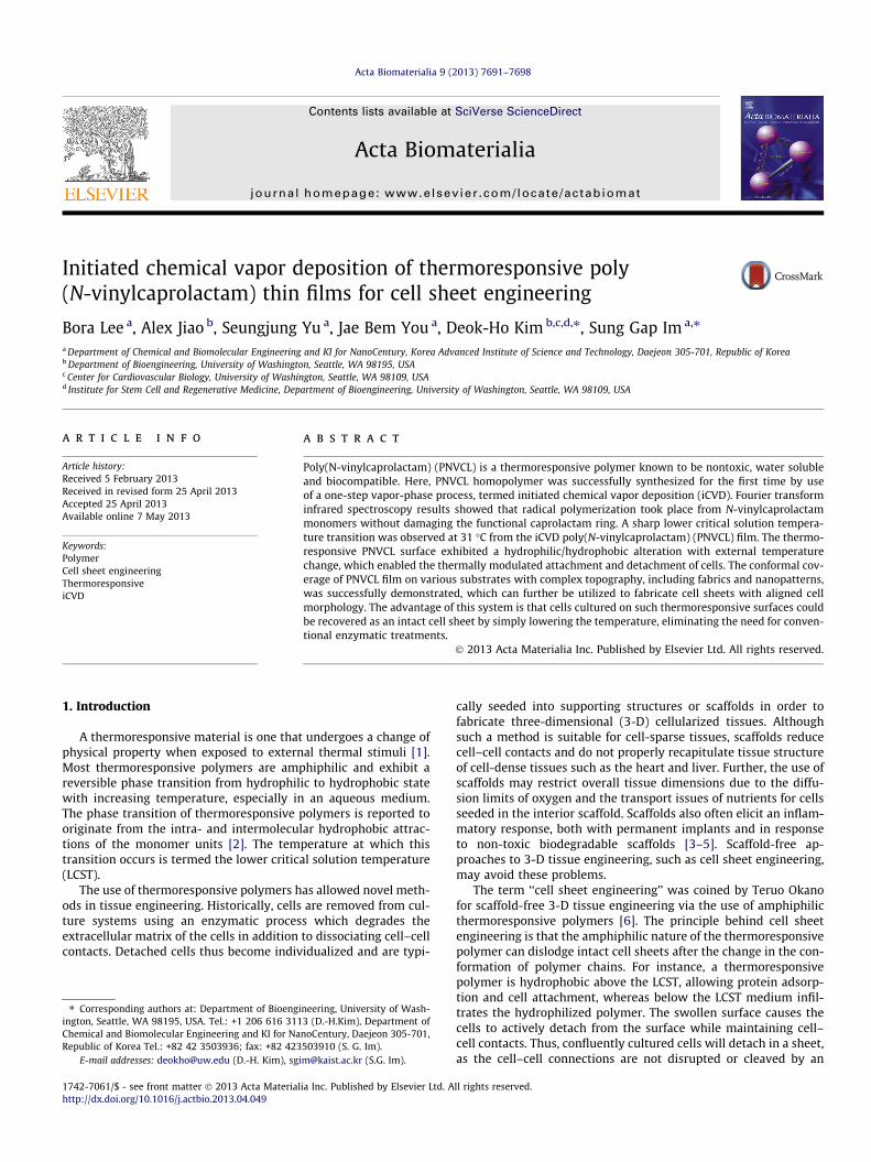
Acta Biomaterialia 9 (2013) 7691–7698
Contents lists available at SciVerse ScienceDirect
Acta Biomaterialia
journal homepage: www.elsevier .com/locate /actabiomat
Initiated chemical vapor deposition of thermoresponsive poly(N-vinylcaprolactam) thin films for cell sheet engineering
1742-7061/$ - see front matter � 2013 Acta Materialia Inc. Published by Elsevier Ltd. All rights reserved.http://dx.doi.org/10.1016/j.actbio.2013.04.049
⇑ Corresponding authors at: Department of Bioengineering, University of Wash-ington, Seattle, WA 98195, USA. Tel.: +1 206 616 3113 (D.-H.Kim), Department ofChemical and Biomolecular Engineering and KI for NanoCentury, Daejeon 305-701,Republic of Korea Tel.: +82 42 3503936; fax: +82 423503910 (S. G. Im).
E-mail addresses: [email protected] (D.-H. Kim), [email protected] (S.G. Im).
Bora Lee a, Alex Jiao b, Seungjung Yu a, Jae Bem You a, Deok-Ho Kim b,c,d,⇑, Sung Gap Im a,⇑a Department of Chemical and Biomolecular Engineering and KI for NanoCentury, Korea Advanced Institute of Science and Technology, Daejeon 305-701, Republic of Koreab Department of Bioengineering, University of Washington, Seattle, WA 98195, USAc Center for Cardiovascular Biology, University of Washington, Seattle, WA 98109, USAd Institute for Stem Cell and Regenerative Medicine, Department of Bioengineering, University of Washington, Seattle, WA 98109, USA
a r t i c l e i n f o
Article history:Received 5 February 2013Received in revised form 25 April 2013Accepted 25 April 2013Available online 7 May 2013
Keywords:PolymerCell sheet engineeringThermoresponsiveiCVD
a b s t r a c t
Poly(N-vinylcaprolactam) (PNVCL) is a thermoresponsive polymer known to be nontoxic, water solubleand biocompatible. Here, PNVCL homopolymer was successfully synthesized for the first time by useof a one-step vapor-phase process, termed initiated chemical vapor deposition (iCVD). Fourier transforminfrared spectroscopy results showed that radical polymerization took place from N-vinylcaprolactammonomers without damaging the functional caprolactam ring. A sharp lower critical solution tempera-ture transition was observed at 31 �C from the iCVD poly(N-vinylcaprolactam) (PNVCL) film. The thermo-responsive PNVCL surface exhibited a hydrophilic/hydrophobic alteration with external temperaturechange, which enabled the thermally modulated attachment and detachment of cells. The conformal cov-erage of PNVCL film on various substrates with complex topography, including fabrics and nanopatterns,was successfully demonstrated, which can further be utilized to fabricate cell sheets with aligned cellmorphology. The advantage of this system is that cells cultured on such thermoresponsive surfaces couldbe recovered as an intact cell sheet by simply lowering the temperature, eliminating the need for conven-tional enzymatic treatments.
� 2013 Acta Materialia Inc. Published by Elsevier Ltd. All rights reserved.
1. Introduction
A thermoresponsive material is one that undergoes a change ofphysical property when exposed to external thermal stimuli [1].Most thermoresponsive polymers are amphiphilic and exhibit areversible phase transition from hydrophilic to hydrophobic statewith increasing temperature, especially in an aqueous medium.The phase transition of thermoresponsive polymers is reported tooriginate from the intra- and intermolecular hydrophobic attrac-tions of the monomer units [2]. The temperature at which thistransition occurs is termed the lower critical solution temperature(LCST).
The use of thermoresponsive polymers has allowed novel meth-ods in tissue engineering. Historically, cells are removed from cul-ture systems using an enzymatic process which degrades theextracellular matrix of the cells in addition to dissociating cell–cellcontacts. Detached cells thus become individualized and are typi-
cally seeded into supporting structures or scaffolds in order tofabricate three-dimensional (3-D) cellularized tissues. Althoughsuch a method is suitable for cell-sparse tissues, scaffolds reducecell–cell contacts and do not properly recapitulate tissue structureof cell-dense tissues such as the heart and liver. Further, the use ofscaffolds may restrict overall tissue dimensions due to the diffu-sion limits of oxygen and the transport issues of nutrients for cellsseeded in the interior scaffold. Scaffolds also often elicit an inflam-matory response, both with permanent implants and in responseto non-toxic biodegradable scaffolds [3–5]. Scaffold-free ap-proaches to 3-D tissue engineering, such as cell sheet engineering,may avoid these problems.
The term ‘‘cell sheet engineering’’ was coined by Teruo Okanofor scaffold-free 3-D tissue engineering via the use of amphiphilicthermoresponsive polymers [6]. The principle behind cell sheetengineering is that the amphiphilic nature of the thermoresponsivepolymer can dislodge intact cell sheets after the change in the con-formation of polymer chains. For instance, a thermoresponsivepolymer is hydrophobic above the LCST, allowing protein adsorp-tion and cell attachment, whereas below the LCST medium infil-trates the hydrophilized polymer. The swollen surface causes thecells to actively detach from the surface while maintaining cell–cell contacts. Thus, confluently cultured cells will detach in a sheet,as the cell–cell connections are not disrupted or cleaved by an
7692 B. Lee et al. / Acta Biomaterialia 9 (2013) 7691–7698
enzymatic process. The resulting cell sheets can be stacked to form3-D tissues without the use of a scaffold [7,8]. Cell sheet engineer-ing is a versatile technology and has been utilized with epithelialcells, ligament cells, hepatocytes, myocardial cells and other celltypes [9–15].
Poly(N-isopropylacrylamide) (PNIPAAm) is one of the best-known thermoresponsive polymers [16], as it has a LCST at 32 �Cwhich falls between room temperature and body temperature.Due to its physiologically relevant LCST, PNIPAAm has triggeredhuge research interest, especially for various biomaterial applica-tions, such as controlled drug delivery [17–21], cell sheet engineer-ing [9,22–24] and bioseparation [25,26]. However, its acrylamidefunctionality is prone to hydrolysis in acidic conditions to releasepotentially toxic small amide molecules from the main polymerchain and cause unwanted side effects during long-term biomedi-cal applications [27]. As an alternative, poly(N-vinylcaprolactam)(PNVCL) is an amphiphilic, water-soluble polymer with an LCSTof approximately 31–34 �C in aqueous medium. Above the LCST,PNVCL is insoluble in water, whereas lowering the temperature be-low the LCST makes it water soluble. This transition is reversibleand extremely fast. The N-vinylcaprolactam (NVCL) functionalityof PNVCL consists of a hydrophilic amide group end-capped witha hydrophobic aliphatic hydrocarbon chain. The cyclic structureof caprolactam ensures the amphiphilic nature of PNVCL, and thehydrolysis of PNVCL only results in the breakage of the caprolac-tam ring with no toxic volatile by-product [28]. In spite of its manyadvantages of PNVCL, the use of PNVCL for biomedical applicationshas not been as widely explored, and few reports exist on its appli-cation as a thin film [29]. Most of the current biomedical applica-tions of PNVCL utilize micro-/nanoparticles [27,30]; theapplication of PNVCL polymer film is very rare. Lim et al. [31] haveapplied a copolymer of PNVCL and PNIPAAm for cell sheet engi-neering, but it is not clear what the role of PNVCL in the PNVCL-co-PNIPAAm polymer was in their work. Most of the PNVCL mate-rials have been synthesized using a liquid phase process compris-ing a complicated multi-step synthesis procedure [32].
In this research, we utilized a vapor-phase deposition processcalled initiated chemical vapor deposition (iCVD) to synthesizePNVCL homopolymer film onto various kinds of substrate. Theone-step iCVD process could fabricate highly pure PNVCL homo-polymer film without a complex purification step. The iCVD pro-cess utilizes a radical polymerization reaction in the vapor-phaseby flowing vaporized initiator and monomer into the vacuum reac-tor. The scheme for the iCVD process is shown in Fig. 1 along withthe polymerization reaction of N-vinylcaprolactam to PNVCL. Sincethe temperature of the substrate is in the range of 15–40 �C, vari-ous vulnerable substrates, such as paper, fabric and membranes,can be coated with iCVD polymer films without any damage[33,34]. Moreover, the vapor-phase process enables conformalcoating of the polymer film onto various complex surface morphol-ogies, including textured surface and nanopatterns [34–37], whichis extremely desirable for the application of generating 3-D struc-tured cell sheets.
The synthesis of PNVCL homopolymer films was successfullyachieved for the first time via the iCVD process. Fourier-transforminfrared (FTIR) spectroscopy and X-ray photoelectron spectroscopy(XPS) analyses indicated that the PNVCL polymer was successfullyformed from the vaporized NVCL monomer with the retention ofthe cyclic amide ring in PNVCL. The molecular weight of the depos-ited polymer was determined by gel-permeation chromatography(GPC) and the conformal coating of PNVCL on a nanopatterned sub-strate was confirmed by scanning electron microscopy (SEM). Thephase transition of PNVCL across the LCST was monitored by bothultraviolet–visible spectroscopy (UV–Vis) and water contact angle(WCA) measurement. The LCST of thermoresponsive polymer wasdetermined by the experimental value of the cloud point tempera-
ture (TCP) through UV–Vis analysis. The cell behavior on the PNVCLsubstrate was observed by bright-field optical microscopy and thebiocompatibility of the iCVD PNVCL polymer was determined bylive/dead assay. Finally, the applicability of PNVCL to cell sheetengineering was also explored for the first time by checking thedetachment of the cell sheet cultured on a PNVCL-coated substrate.
2. Experimental
2.1. iCVD polymerization
NVCL (98%, Aldrich) and tert-butyl peroxide (TBPO; 98%, Al-drich) were purchased and used without further purification asmonomer and initiator, respectively. The iCVD polymerization tookplace in a custom-built iCVD reactor (Daeki Hi-Tech Co. Ltd) de-scribed previously [38]. Briefly, the NVCL monomer was heatedto 80 �C and the formed NVCL vapor was introduced to the reactorat a flow rate of 1.0 sccm. TBPO is volatile enough at room temper-ature to form a vapor, so it was fed into the reactor via meteringvalves at a flow rate of 0.8 sccm without any additional heating.To obtain a PNVCL film with high purity, NVCL and TBPO werefed into the reactor concurrently with a reactor pressure of150 mtorr. The substrate was kept at a temperature of 35 �C to pro-mote monomer adsorption, whereas the filament temperature wasmaintained at 200 �C. The thickness of the PNVCL film was moni-tored in situ using an He–Ne laser (JDS Uniphase). The substrateswere cleaned with isopropanol and deionized (DI) water beforeundergoing 5 min of sonication each and drying with N2 gas. Thesubstrates were then treated with oxygen plasma. Depositing thepolymer immediately after the plasma treatment allowed thegrafting of the polymer to the substrate.
2.2. Characterization of iCVD PNVCL film
FTIR (Alpha FT-IR Spectrometer, Bruker Optics) of PNVCL filmwas measured in normal absorbance mode averaged over 64 scans.All spectra were baseline corrected.
The surface chemical compositions of the PNVCL films depos-ited by iCVD (iCVD PNVCL) were analyzed by XPS (MultiLab2000, Thermo VG Scientific). The spectra were recorded using anAl Ka X-ray radiation source (150 W, 14 kV, KE = 1486.6 eV) undera vacuum base pressure (5 � 10�10 torr). The XPS spectra were col-lected in the range of 0–1100 eV with a resolution of 1.0 eV andwith a pass energy of 100 eV. The binding energy scale was cali-brated with reference to the C1s line at 285.0 eV.
The molecular weight and the polydispersity index (PDI) of thedeposited films were determined by GPC on a system equippedwith a Waters 1515 isocratic high-performance liquid chromatog-raphy pump with a refractive index detector (Waters 2414). Tetra-hydrofuran (JT Baker) was used as an eluent, with a flow rate of1 ml min�1. The temperature of the column oven was set to30 �C. Monodisperse polystyrene standards (Polyscience) wereused to generate the calibration curve. A 30 ll volume of each sam-ple was injected into the column after passing through a 0.2 lm fil-tration membrane.
The morphology of iCVD PNVCL was assessed by SEM using aXL30SFEG (Philips) scanning electron microscope operating at3.00 kV. All the samples were attached to a conducting sampleholder with copper tape. The prepared samples were coated withgold under a vacuum using the sputtering system.
The LCST behavior of the iCVD PNVCL was investigated usingUV–Vis spectroscopy (UV-3600, Shimadzu). The transmittance of0.25 wt.% aqueous polymer solutions was measured at 500 nm atintervals of 2 �C in the range of 25–55 �C. The solution was allowedto be equilibrated at each temperature for 20 min before the trans-mittance was measured.
B. Lee et al. / Acta Biomaterialia 9 (2013) 7691–7698 7693
WCA measurements were performed on a contact angle meter(Phoenix 150, SEO) equipped with an environment chamber (tem-perature range 25–250 �C) using a 2.5 ll DI water droplet. Mea-surements were taken both at room temperature (25 �C) and at55 �C. An Si wafer coated with PNVCL was allowed to equilibrateat each temperature for 20 min before the contact angle wasmeasured.
Fig. 1. (a) Schematic diagram of iCVD polymerization of the PNVCL polymer fromNVCL monomer. (b) Illustrations of the formation process of PNVCL in the iCVDchamber.
2.3. Cell culture
NIH 3T3 mouse fibroblasts were cultured in Dulbecco’s modi-fied Eagle’s medium (DMEM; Invitrogen) on tissue culture polysty-rene (TCPS) dishes at 37 �C in 5% CO2. PNVCL-grafted glasssubstrates were cut and placed in 35 mm diameter TCPS dishesand irradiated with UV at 264 nm for 30 min inside a biosafety cab-inet to sterilize the substrates. PNVCL substrates were then soakedovernight in distilled H2O. Prior to cell seeding, the H2O was aspi-rated and the dry PNVCL substrates were placed in the incubatorused for cell culture. Cultured cells (between passages 10–20) weretrypsinized and seeded onto PNVCL-deposited glass substrates at ahigh density of 1.5 � 105 cells cm�2 to achieve confluency within24 h of culture. After being incubated at 37 �C in 5% CO2, adherentcells were observed at 3, 6 and 24 h post-seeding with a phase con-trast microscope (Eclipse TS100, Nikon).
2.4. Live/dead assay
A live/dead assay (Invitrogen) utilizing Calcein AM as a live cellmarking dye and ethidium homodimer-1 (EthD-1) as a dead cellmarking dye was used according to the manufacturer’s instruc-tions. Briefly, the kit reagents were added to sterile 1� Dulbecco’sPhosphate-Buffered Saline (DPBS) to create a 1 mM Calcein AM and4 mM EthD-1 solution. The cells on PNVCL were washed gentlywith warm 1� DPBS, then 200 ll of the Calcein AM/EthD-1 solu-tion was added to the coverslips and incubated in the dark for30 min at 37 �C and 5% CO2. After incubation, cells were gentlyrinsed with 1� DPBS and then imaged with a high-resolutionwide-field fluorescent microscope (TiE, Nikon).
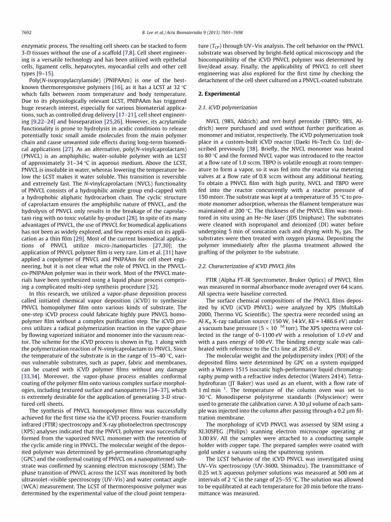
Fig. 2. FTIR spectra of (a) NVCL monomer and (b) iCVD PNVCL polymer. The solidline corresponds to the C@O bond and the dashed line represents the tertiary aminemoiety. Important functional groups were retained in both spectra. Red arrowsmark the C@C double bond and vinyl group in NVCL monomer. These peaks areabsent in the iCVD PNVCL polymer.
2.5. Detachment of cell sheets
For cell sheet transfer experiments, a plunger method previ-ously published by the Okano group [39] was utilized to transferpartial cell sheets onto glass coverslips during the 30 min incuba-tion at 25 �C. Briefly, cells were cultured to confluency on PNVCLsurfaces (within 24 h of seeding). A 7.5% w/v gelatin in sterile 1�DPBS was melted at 60 �C, filter sterilized and poured into a PDMSmold. A Teflon plunger was placed upon the melted gelatin and theensemble was cooled at 4 �C for 10 min. The PDMS mold was thengently removed, leaving a solidified gelatin hydrogel attached tothe Teflon plunger. Substrate medium was aspirated away and200 ll of fresh, serum-free DMEM was added to the substrate sam-ples to prevent the cells from dehydrating. The plunger was thenplaced on top of the substrate and cells, and the scaffold + plungerwas incubated at 25 �C inside a biosafety cabinet for 30 min. Afterincubation, the gelatin plunger was gently removed from the sub-strate surface, capturing partially detached cell sheets. A scalpelwas used to remove the gelatin from the Teflon plunger and thegelatin was placed cell sheet first onto a clean glass coverslip andallowed to attach for 30 min at 25 �C. After attachment, the gelatinplus coverslip was incubated at 37 �C and 5% CO2, and the gelatinwas allowed to melt. Coverslips plus cell sheets were rinsed gentlytwice with PBS, cultured with DMEM at 37 �C and 5% CO2 for 24 hand then imaged.
3. Results and discussion
3.1. FTIR analysis
Fig. 2 shows the FTIR spectra of NVCL monomer and the PNVCLfilm deposited via iCVD. In the FTIR spectrum of the NVCL mono-mer, a characteristic carbonyl peak (C@O) in the amide groupwas detected at 1620 cm�1. The peak for the unsaturated carbonC@C was observed at 1651 cm�1, and the stretching and bendingvibrational peaks of C–H bonds in the vinyl group (@CH and@CH2) were also detected at 3106 and 993 cm�1, respectively.The peak corresponding to C–N bond in the amide group was alsofound at 1478 cm�1. The peak at 1441 cm�1 corresponds to the ali-phatic C–C bond in the caprolactam ring. In the spectrum of iCVDPNVCL, the peaks corresponding to the C@C double bond of the
7694 B. Lee et al. / Acta Biomaterialia 9 (2013) 7691–7698
monomer disappeared, as marked by red arrows in the FTIR spec-trum (Fig. 2b), indicating successful polymerization of the mono-mer by the iCVD process. The retention of the caprolactamfunctionality in the PNVCL polymer was also confirmed from theFTIR spectrum of iCVD polymer. The characteristic carbonyl peak(C@O stretching) in the amide bond was found at 1620 cm�1. Thealiphatic C–H stretching peaks in the caprolactam ring were de-tected at 2931 cm�1. The peak of tertiary amine (C–N) in the amidefunctionality was observed at 2850 cm�1, which is close to that ofNVCL monomer. The retention of caprolactam functionality in theiCVD PNVCL distinctly shows that the iCVD process did not damagethe NVCL monomer during the polymerization process. A broadpeak was observed in the spectrum of the iCVD polymer at3431 cm�1, which corresponds to the O–H stretching vibration.Due to its extremely hygroscopic nature, PNVCL is known to absorbmoisture readily. The peak corresponding to the absorbed moistureis located at 3431 cm�1, which has been observed commonly inprevious reports on PNVCL polymerized by various liquid-phasemethods [28,40].
3.2. XPS survey scan and high resolution analysis
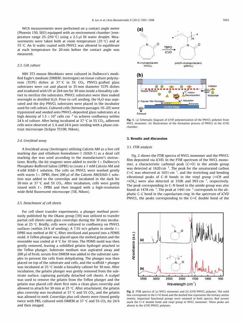
XPS analysis was also performed to determine the chemicalcompositions of the iCVD PNVCL film. As shown in Fig. 3, a repeat-ing unit of PNVCL consists of eight carbons (C), one oxygen (O) andone nitrogen (N) atoms. The characteristic peaks of carbon (C1s at285.0 eV), oxygen (O1s at 531.0 eV) and nitrogen (N1s at 399.4 eV)in PNVCL were all detected in the survey scan spectrum shown inFig. 3a. The atomic ratio of pNVCL film was calculated from the
Fig. 3. The XPS spectra of PNVCL: (a) XPS survey scan of the iCVD PNVCL film. Oxygen, ni(b) O1s, (c) N1s and (d) C1s in PNVCL, respectively.
areas of C1s, O1s and N1s peaks in the XPS survey scan, multipliedby the appropriate sensitivity factors. The measured atomic ratio ofC:O:N was 75.8:13.5:10.7. Compared to the composition predictedfrom the stoichiometry of the NVCL monomer, which is 80.0% of C,10.0% of O and 10.0% of N, an excessive amount of oxygen was de-tected, which most likely originated from water absorption due tothe highly hygroscopic nature of PNVCL. This observation is consis-tent with the FTIR results. XPS high-resolution scans of O, N, and Cwere also performed, and are shown in Fig. 3b–d. In case of the O1score-level XPS spectrum (Fig. 3b), one dominant peak at 531.0 eVcorresponds to the sole oxygen bonding environment in PNVCL,while a small broad shoulder at 533.6 eV was also detected, mostlikely originating from the oxygen of the absorbed water to thePNVCL, which also supports the findings in the XPS survey scanand the FTIR measurement of the PNVCL film. In the N1s core-levelXPS spectrum (Fig. 3c), only one N bonding environment in PNVCLwas observed, at 399.4 eV, representing the N in the amide groupof the caprolatam ring. The C1s core-level XPS spectrum (Fig. 3d)was deconvoluted to three major peaks of C: (1) a peak at287.6 eV, representing C in a carbonyl bond (C@O) (1 in the insetof Fig. 3d); (2) a peak at 285.4 eV, corresponding to C in a C–N bond(2 in the inset of Fig. 3d); and (3) an aliphatic C1s peak at 284.6 eV(3 in the inset of Fig. 3d). The measured atomic ratio from the C1score-level XPS spectrum was 0.19:0.39:1.00, which is consistentwith the theoretical calculation from the chemical formula ofPNVCL, which is 0.20:0.40:1.00 (Table 1b). Both the FTIR and XPSresults clearly demonstrate that the PNVCL polymer was success-fully synthesized by the iCVD polymerization process, maintainingthe caprolactam ring in the PNVCL.
trogen and carbon 1s peaks are recorded in the spectra. XPS high-resolution scans of
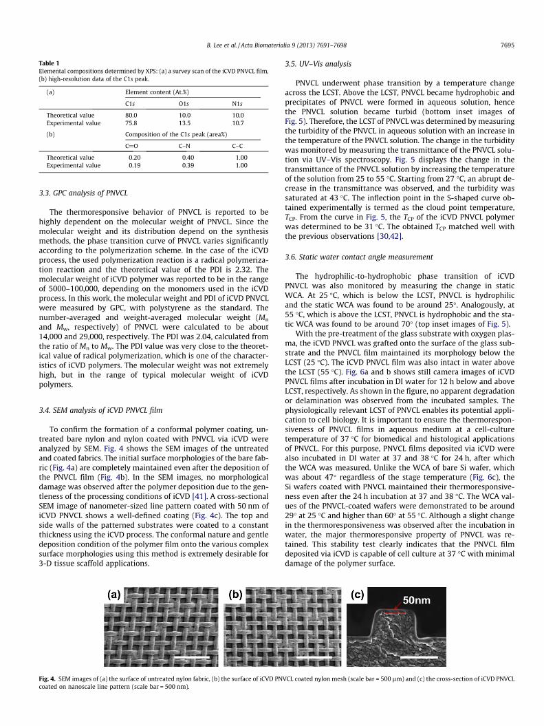
Table 1Elemental compositions determined by XPS: (a) a survey scan of the iCVD PNVCL film,(b) high-resolution data of the C1s peak.
(a) Element content (At.%)
C1s O1s N1s
Theoretical value 80.0 10.0 10.0Experimental value 75.8 13.5 10.7
(b) Composition of the C1s peak (area%)
C@O C–N C–C
Theoretical value 0.20 0.40 1.00Experimental value 0.19 0.39 1.00
B. Lee et al. / Acta Biomaterialia 9 (2013) 7691–7698 7695
3.3. GPC analysis of PNVCL
The thermoresponsive behavior of PNVCL is reported to behighly dependent on the molecular weight of PNVCL. Since themolecular weight and its distribution depend on the synthesismethods, the phase transition curve of PNVCL varies significantlyaccording to the polymerization scheme. In the case of the iCVDprocess, the used polymerization reaction is a radical polymeriza-tion reaction and the theoretical value of the PDI is 2.32. Themolecular weight of iCVD polymer was reported to be in the rangeof 5000–100,000, depending on the monomers used in the iCVDprocess. In this work, the molecular weight and PDI of iCVD PNVCLwere measured by GPC, with polystyrene as the standard. Thenumber-averaged and weight-averaged molecular weight (Mn
and Mw, respectively) of PNVCL were calculated to be about14,000 and 29,000, respectively. The PDI was 2.04, calculated fromthe ratio of Mn to Mw. The PDI value was very close to the theoret-ical value of radical polymerization, which is one of the character-istics of iCVD polymers. The molecular weight was not extremelyhigh, but in the range of typical molecular weight of iCVDpolymers.
3.4. SEM analysis of iCVD PNVCL film
To confirm the formation of a conformal polymer coating, un-treated bare nylon and nylon coated with PNVCL via iCVD wereanalyzed by SEM. Fig. 4 shows the SEM images of the untreatedand coated fabrics. The initial surface morphologies of the bare fab-ric (Fig. 4a) are completely maintained even after the deposition ofthe PNVCL film (Fig. 4b). In the SEM images, no morphologicaldamage was observed after the polymer deposition due to the gen-tleness of the processing conditions of iCVD [41]. A cross-sectionalSEM image of nanometer-sized line pattern coated with 50 nm ofiCVD PNVCL shows a well-defined coating (Fig. 4c). The top andside walls of the patterned substrates were coated to a constantthickness using the iCVD process. The conformal nature and gentledeposition condition of the polymer film onto the various complexsurface morphologies using this method is extremely desirable for3-D tissue scaffold applications.
Fig. 4. SEM images of (a) the surface of untreated nylon fabric, (b) the surface of iCVD PNVcoated on nanoscale line pattern (scale bar = 500 nm).
3.5. UV–Vis analysis
PNVCL underwent phase transition by a temperature changeacross the LCST. Above the LCST, PNVCL became hydrophobic andprecipitates of PNVCL were formed in aqueous solution, hencethe PNVCL solution became turbid (bottom inset images ofFig. 5). Therefore, the LCST of PNVCL was determined by measuringthe turbidity of the PNVCL in aqueous solution with an increase inthe temperature of the PNVCL solution. The change in the turbiditywas monitored by measuring the transmittance of the PNVCL solu-tion via UV–Vis spectroscopy. Fig. 5 displays the change in thetransmittance of the PNVCL solution by increasing the temperatureof the solution from 25 to 55 �C. Starting from 27 �C, an abrupt de-crease in the transmittance was observed, and the turbidity wassaturated at 43 �C. The inflection point in the S-shaped curve ob-tained experimentally is termed as the cloud point temperature,TCP. From the curve in Fig. 5, the TCP of the iCVD PNVCL polymerwas determined to be 31 �C. The obtained TCP matched well withthe previous observations [30,42].
3.6. Static water contact angle measurement
The hydrophilic-to-hydrophobic phase transition of iCVDPNVCL was also monitored by measuring the change in staticWCA. At 25 �C, which is below the LCST, PNVCL is hydrophilicand the static WCA was found to be around 25�. Analogously, at55 �C, which is above the LCST, PNVCL is hydrophobic and the sta-tic WCA was found to be around 70� (top inset images of Fig. 5).
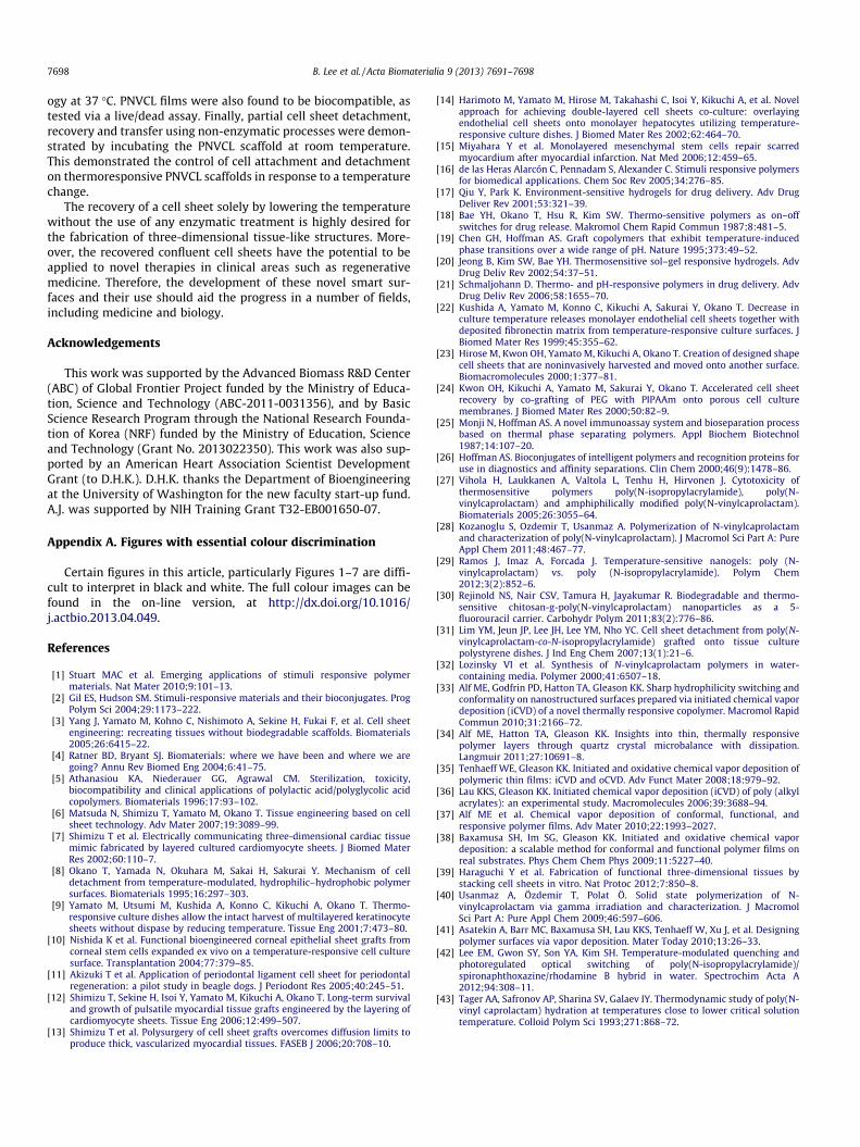
With the pre-treatment of the glass substrate with oxygen plas-ma, the iCVD PNVCL was grafted onto the surface of the glass sub-strate and the PNVCL film maintained its morphology below theLCST (25 �C). The iCVD PNVCL film was also intact in water abovethe LCST (55 �C). Fig. 6a and b shows still camera images of iCVDPNVCL films after incubation in DI water for 12 h below and aboveLCST, respectively. As shown in the figure, no apparent degradationor delamination was observed from the incubated samples. Thephysiologically relevant LCST of PNVCL enables its potential appli-cation to cell biology. It is important to ensure the thermorespon-siveness of PNVCL films in aqueous medium at a cell-culturetemperature of 37 �C for biomedical and histological applicationsof PNVCL. For this purpose, PNVCL films deposited via iCVD werealso incubated in DI water at 37 and 38 �C for 24 h, after whichthe WCA was measured. Unlike the WCA of bare Si wafer, whichwas about 47� regardless of the stage temperature (Fig. 6c), theSi wafers coated with PNVCL maintained their thermoresponsive-ness even after the 24 h incubation at 37 and 38 �C. The WCA val-ues of the PNVCL-coated wafers were demonstrated to be around29� at 25 �C and higher than 60� at 55 �C. Although a slight changein the thermoresponsiveness was observed after the incubation inwater, the major thermoresponsive property of PNVCL was re-tained. This stability test clearly indicates that the PNVCL filmdeposited via iCVD is capable of cell culture at 37 �C with minimaldamage of the polymer surface.
CL coated nylon mesh (scale bar = 500 lm) and (c) the cross-section of iCVD PNVCL
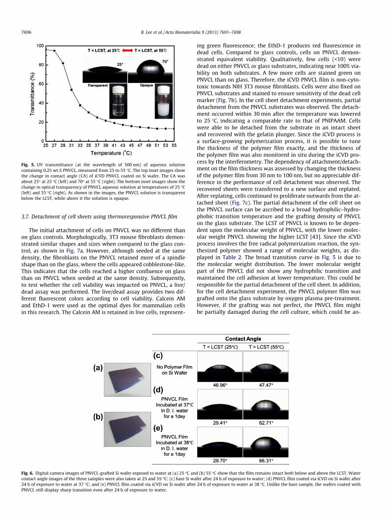
Fig. 5. UV transmittance (at the wavelength of 500 nm) of aqueous solutioncontaining 0.25 wt.% PNVCL, measured from 25 to 55 �C. The top inset images showthe change in contact angle (CA) of iCVD PNVCL coated on Si wafer. The CA wasabout 25� at 25 �C (left) and 70� at 55 �C (right). The bottom inset images show thechange in optical transparency of PNVCL aqueous solution at temperatures of 25 �C(left) and 55 �C (right). As shown in the images, the PNVCL solution is transparentbelow the LCST, while above it the solution is opaque.
7696 B. Lee et al. / Acta Biomaterialia 9 (2013) 7691–7698
3.7. Detachment of cell sheets using thermoresponsive PNVCL film
The initial attachment of cells on PNVCL was no different thanon glass controls. Morphologically, 3T3 mouse fibroblasts demon-strated similar shapes and sizes when compared to the glass con-trol, as shown in Fig. 7a. However, although seeded at the samedensity, the fibroblasts on the PNVCL retained more of a spindleshape than on the glass, where the cells appeared cobblestone-like.This indicates that the cells reached a higher confluence on glassthan on PNVCL when seeded at the same density. Subsequently,to test whether the cell viability was impacted on PNVCL, a live/dead assay was performed. The live/dead assay provides two dif-ferent fluorescent colors according to cell viability. Calcein AMand EthD-1 were used as the optimal dyes for mammalian cellsin this research. The Calcein AM is retained in live cells, represent-
Fig. 6. Digital camera images of PNVCL-grafted Si wafer exposed to water at (a) 25 �C ancontact angle images of the three samples were also taken at 25 and 55 �C: (c) bare Si wa24 h of exposure to water at 37 �C; and (e) PNVCL film coated via iCVD on Si wafer afterPNVCL still display sharp transition even after 24 h of exposure to water.
ing green fluorescence; the EthD-1 produces red fluorescence indead cells. Compared to glass controls, cells on PNVCL demon-strated equivalent viability. Qualitatively, few cells (<10) weredead on either PNVCL or glass substrates, indicating near 100% via-bility on both substrates. A few more cells are stained green onPNVCL than on glass. Therefore, the iCVD PNVCL film is non-cyto-toxic towards NIH 3T3 mouse fibroblasts. Cells were also fixed onPNVCL substrates and stained to ensure sensitivity of the dead cellmarker (Fig. 7b). In the cell sheet detachment experiments, partialdetachment from the PNVCL substrates was observed. The detach-ment occurred within 30 min after the temperature was loweredto 25 �C, indicating a comparable rate to that of PNIPAAM. Cellswere able to be detached from the substrate in an intact sheetand recovered with the gelatin plunger. Since the iCVD process isa surface-growing polymerization process, it is possible to tunethe thickness of the polymer film exactly, and the thickness ofthe polymer film was also monitored in situ during the iCVD pro-cess by the interferometry. The dependency of attachment/detach-ment on the film thickness was assessed by changing the thicknessof the polymer film from 30 nm to 100 nm, but no appreciable dif-ference in the performance of cell detachment was observed. Therecovered sheets were transferred to a new surface and replated.After replating, cells continued to proliferate outwards from the at-tached sheet (Fig. 7c). The partial detachment of the cell sheet onthe PNVCL surface can be ascribed to a broad hydrophilic–hydro-phobic transition temperature and the grafting density of PNVCLon the glass substrate. The LCST of PNVCL is known to be depen-dent upon the molecular weight of PNVCL, with the lower molec-ular weight PNVCL showing the higher LCST [43]. Since the iCVDprocess involves the free radical polymerization reaction, the syn-thesized polymer showed a range of molecular weights, as dis-played in Table 2. The broad transition curve in Fig. 5 is due tothe molecular weight distribution. The lower molecular weightpart of the PNVCL did not show any hydrophilic transition andmaintained the cell adhesion at lower temperature. This could beresponsible for the partial detachment of the cell sheet. In addition,for the cell detachment experiment, the PNVCL polymer film wasgrafted onto the glass substrate by oxygen plasma pre-treatment.However, if the grafting was not perfect, the PNVCL film mightbe partially damaged during the cell culture, which could be an-
d (b) 55 �C show that the film remains intact both below and above the LCST. Waterfer after 24 h of exposure to water; (d) PNVCL film coated via iCVD on Si wafer after24 h of exposure to water at 38 �C. Unlike the bare sample, the wafers coated with
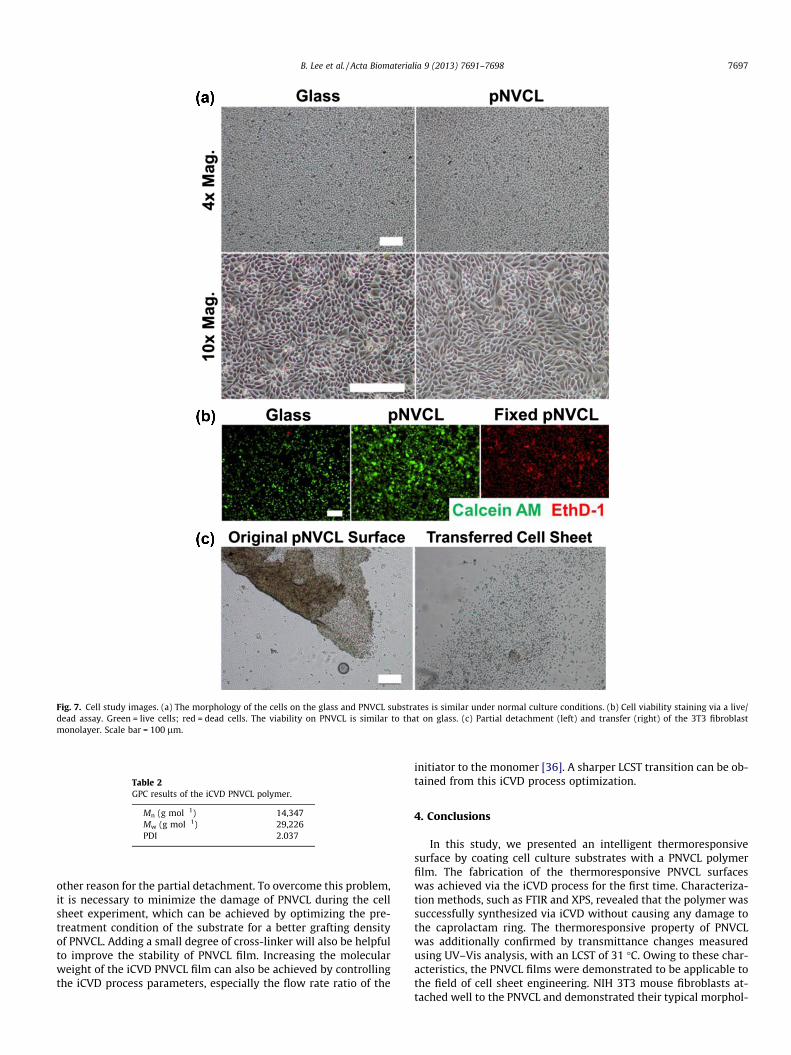
Fig. 7. Cell study images. (a) The morphology of the cells on the glass and PNVCL substrates is similar under normal culture conditions. (b) Cell viability staining via a live/dead assay. Green = live cells; red = dead cells. The viability on PNVCL is similar to that on glass. (c) Partial detachment (left) and transfer (right) of the 3T3 fibroblastmonolayer. Scale bar = 100 lm.
Table 2GPC results of the iCVD PNVCL polymer.
Mn (g mol�1) 14,347Mw (g mol�1) 29,226PDI 2.037
B. Lee et al. / Acta Biomaterialia 9 (2013) 7691–7698 7697
other reason for the partial detachment. To overcome this problem,it is necessary to minimize the damage of PNVCL during the cellsheet experiment, which can be achieved by optimizing the pre-treatment condition of the substrate for a better grafting densityof PNVCL. Adding a small degree of cross-linker will also be helpfulto improve the stability of PNVCL film. Increasing the molecularweight of the iCVD PNVCL film can also be achieved by controllingthe iCVD process parameters, especially the flow rate ratio of the
initiator to the monomer [36]. A sharper LCST transition can be ob-tained from this iCVD process optimization.
4. Conclusions
In this study, we presented an intelligent thermoresponsivesurface by coating cell culture substrates with a PNVCL polymerfilm. The fabrication of the thermoresponsive PNVCL surfaceswas achieved via the iCVD process for the first time. Characteriza-tion methods, such as FTIR and XPS, revealed that the polymer wassuccessfully synthesized via iCVD without causing any damage tothe caprolactam ring. The thermoresponsive property of PNVCLwas additionally confirmed by transmittance changes measuredusing UV–Vis analysis, with an LCST of 31 �C. Owing to these char-acteristics, the PNVCL films were demonstrated to be applicable tothe field of cell sheet engineering. NIH 3T3 mouse fibroblasts at-tached well to the PNVCL and demonstrated their typical morphol-
7698 B. Lee et al. / Acta Biomaterialia 9 (2013) 7691–7698
ogy at 37 �C. PNVCL films were also found to be biocompatible, astested via a live/dead assay. Finally, partial cell sheet detachment,recovery and transfer using non-enzymatic processes were demon-strated by incubating the PNVCL scaffold at room temperature.This demonstrated the control of cell attachment and detachmenton thermoresponsive PNVCL scaffolds in response to a temperaturechange.
The recovery of a cell sheet solely by lowering the temperaturewithout the use of any enzymatic treatment is highly desired forthe fabrication of three-dimensional tissue-like structures. More-over, the recovered confluent cell sheets have the potential to beapplied to novel therapies in clinical areas such as regenerativemedicine. Therefore, the development of these novel smart sur-faces and their use should aid the progress in a number of fields,including medicine and biology.
Acknowledgements
This work was supported by the Advanced Biomass R&D Center(ABC) of Global Frontier Project funded by the Ministry of Educa-tion, Science and Technology (ABC-2011-0031356), and by BasicScience Research Program through the National Research Founda-tion of Korea (NRF) funded by the Ministry of Education, Scienceand Technology (Grant No. 2013022350). This work was also sup-ported by an American Heart Association Scientist DevelopmentGrant (to D.H.K.). D.H.K. thanks the Department of Bioengineeringat the University of Washington for the new faculty start-up fund.A.J. was supported by NIH Training Grant T32-EB001650-07.
Appendix A. Figures with essential colour discrimination
Certain figures in this article, particularly Figures 1–7 are diffi-cult to interpret in black and white. The full colour images can befound in the on-line version, at http://dx.doi.org/10.1016/j.actbio.2013.04.049.
References
[1] Stuart MAC et al. Emerging applications of stimuli responsive polymermaterials. Nat Mater 2010;9:101–13.
[2] Gil ES, Hudson SM. Stimuli-responsive materials and their bioconjugates. ProgPolym Sci 2004;29:1173–222.
[3] Yang J, Yamato M, Kohno C, Nishimoto A, Sekine H, Fukai F, et al. Cell sheetengineering: recreating tissues without biodegradable scaffolds. Biomaterials2005;26:6415–22.
[4] Ratner BD, Bryant SJ. Biomaterials: where we have been and where we aregoing? Annu Rev Biomed Eng 2004;6:41–75.
[5] Athanasiou KA, Niederauer GG, Agrawal CM. Sterilization, toxicity,biocompatibility and clinical applications of polylactic acid/polyglycolic acidcopolymers. Biomaterials 1996;17:93–102.
[6] Matsuda N, Shimizu T, Yamato M, Okano T. Tissue engineering based on cellsheet technology. Adv Mater 2007;19:3089–99.
[7] Shimizu T et al. Electrically communicating three-dimensional cardiac tissuemimic fabricated by layered cultured cardiomyocyte sheets. J Biomed MaterRes 2002;60:110–7.
[8] Okano T, Yamada N, Okuhara M, Sakai H, Sakurai Y. Mechanism of celldetachment from temperature-modulated, hydrophilic–hydrophobic polymersurfaces. Biomaterials 1995;16:297–303.
[9] Yamato M, Utsumi M, Kushida A, Konno C, Kikuchi A, Okano T. Thermo-responsive culture dishes allow the intact harvest of multilayered keratinocytesheets without dispase by reducing temperature. Tissue Eng 2001;7:473–80.
[10] Nishida K et al. Functional bioengineered corneal epithelial sheet grafts fromcorneal stem cells expanded ex vivo on a temperature-responsive cell culturesurface. Transplantation 2004;77:379–85.
[11] Akizuki T et al. Application of periodontal ligament cell sheet for periodontalregeneration: a pilot study in beagle dogs. J Periodont Res 2005;40:245–51.
[12] Shimizu T, Sekine H, Isoi Y, Yamato M, Kikuchi A, Okano T. Long-term survivaland growth of pulsatile myocardial tissue grafts engineered by the layering ofcardiomyocyte sheets. Tissue Eng 2006;12:499–507.
[13] Shimizu T et al. Polysurgery of cell sheet grafts overcomes diffusion limits toproduce thick, vascularized myocardial tissues. FASEB J 2006;20:708–10.
[14] Harimoto M, Yamato M, Hirose M, Takahashi C, Isoi Y, Kikuchi A, et al. Novelapproach for achieving double-layered cell sheets co-culture: overlayingendothelial cell sheets onto monolayer hepatocytes utilizing temperature-responsive culture dishes. J Biomed Mater Res 2002;62:464–70.
[15] Miyahara Y et al. Monolayered mesenchymal stem cells repair scarredmyocardium after myocardial infarction. Nat Med 2006;12:459–65.
[16] de las Heras Alarcón C, Pennadam S, Alexander C. Stimuli responsive polymersfor biomedical applications. Chem Soc Rev 2005;34:276–85.
[17] Qiu Y, Park K. Environment-sensitive hydrogels for drug delivery. Adv DrugDeliver Rev 2001;53:321–39.
[18] Bae YH, Okano T, Hsu R, Kim SW. Thermo-sensitive polymers as on–offswitches for drug release. Makromol Chem Rapid Commun 1987;8:481–5.
[19] Chen GH, Hoffman AS. Graft copolymers that exhibit temperature-inducedphase transitions over a wide range of pH. Nature 1995;373:49–52.
[20] Jeong B, Kim SW, Bae YH. Thermosensitive sol–gel responsive hydrogels. AdvDrug Deliv Rev 2002;54:37–51.
[21] Schmaljohann D. Thermo- and pH-responsive polymers in drug delivery. AdvDrug Deliv Rev 2006;58:1655–70.
[22] Kushida A, Yamato M, Konno C, Kikuchi A, Sakurai Y, Okano T. Decrease inculture temperature releases monolayer endothelial cell sheets together withdeposited fibronectin matrix from temperature-responsive culture surfaces. JBiomed Mater Res 1999;45:355–62.
[23] Hirose M, Kwon OH, Yamato M, Kikuchi A, Okano T. Creation of designed shapecell sheets that are noninvasively harvested and moved onto another surface.Biomacromolecules 2000;1:377–81.
[24] Kwon OH, Kikuchi A, Yamato M, Sakurai Y, Okano T. Accelerated cell sheetrecovery by co-grafting of PEG with PIPAAm onto porous cell culturemembranes. J Biomed Mater Res 2000;50:82–9.
[25] Monji N, Hoffman AS. A novel immunoassay system and bioseparation processbased on thermal phase separating polymers. Appl Biochem Biotechnol1987;14:107–20.
[26] Hoffman AS. Bioconjugates of intelligent polymers and recognition proteins foruse in diagnostics and affinity separations. Clin Chem 2000;46(9):1478–86.
[27] Vihola H, Laukkanen A, Valtola L, Tenhu H, Hirvonen J. Cytotoxicity ofthermosensitive polymers poly(N-isopropylacrylamide), poly(N-vinylcaprolactam) and amphiphilically modified poly(N-vinylcaprolactam).Biomaterials 2005;26:3055–64.
[28] Kozanoglu S, Ozdemir T, Usanmaz A. Polymerization of N-vinylcaprolactamand characterization of poly(N-vinylcaprolactam). J Macromol Sci Part A: PureAppl Chem 2011;48:467–77.
[29] Ramos J, Imaz A, Forcada J. Temperature-sensitive nanogels: poly (N-vinylcaprolactam) vs. poly (N-isopropylacrylamide). Polym Chem2012;3(2):852–6.
[30] Rejinold NS, Nair CSV, Tamura H, Jayakumar R. Biodegradable and thermo-sensitive chitosan-g-poly(N-vinylcaprolactam) nanoparticles as a 5-fluorouracil carrier. Carbohydr Polym 2011;83(2):776–86.
[31] Lim YM, Jeun JP, Lee JH, Lee YM, Nho YC. Cell sheet detachment from poly(N-vinylcaprolactam-co-N-isopropylacrylamide) grafted onto tissue culturepolystyrene dishes. J Ind Eng Chem 2007;13(1):21–6.
[32] Lozinsky VI et al. Synthesis of N-vinylcaprolactam polymers in water-containing media. Polymer 2000;41:6507–18.
[33] Alf ME, Godfrin PD, Hatton TA, Gleason KK. Sharp hydrophilicity switching andconformality on nanostructured surfaces prepared via initiated chemical vapordeposition (iCVD) of a novel thermally responsive copolymer. Macromol RapidCommun 2010;31:2166–72.
[34] Alf ME, Hatton TA, Gleason KK. Insights into thin, thermally responsivepolymer layers through quartz crystal microbalance with dissipation.Langmuir 2011;27:10691–8.
[35] Tenhaeff WE, Gleason KK. Initiated and oxidative chemical vapor deposition ofpolymeric thin films: iCVD and oCVD. Adv Funct Mater 2008;18:979–92.
[36] Lau KKS, Gleason KK. Initiated chemical vapor deposition (iCVD) of poly (alkylacrylates): an experimental study. Macromolecules 2006;39:3688–94.
[37] Alf ME et al. Chemical vapor deposition of conformal, functional, andresponsive polymer films. Adv Mater 2010;22:1993–2027.
[38] Baxamusa SH, Im SG, Gleason KK. Initiated and oxidative chemical vapordeposition: a scalable method for conformal and functional polymer films onreal substrates. Phys Chem Chem Phys 2009;11:5227–40.
[39] Haraguchi Y et al. Fabrication of functional three-dimensional tissues bystacking cell sheets in vitro. Nat Protoc 2012;7:850–8.
[40] Usanmaz A, Özdemir T, Polat Ö. Solid state polymerization of N-vinylcaprolactam via gamma irradiation and characterization. J MacromolSci Part A: Pure Appl Chem 2009;46:597–606.
[41] Asatekin A, Barr MC, Baxamusa SH, Lau KKS, Tenhaeff W, Xu J, et al. Designingpolymer surfaces via vapor deposition. Mater Today 2010;13:26–33.
[42] Lee EM, Gwon SY, Son YA, Kim SH. Temperature-modulated quenching andphotoregulated optical switching of poly(N-isopropylacrylamide)/spironaphthoxazine/rhodamine B hybrid in water. Spectrochim Acta A2012;94:308–11.
[43] Tager AA, Safronov AP, Sharina SV, Galaev IY. Thermodynamic study of poly(N-vinyl caprolactam) hydration at temperatures close to lower critical solutiontemperature. Colloid Polym Sci 1993;271:868–72.
Copyright © 2022 FDOKUMEN