The Toll-Like Receptor Signaling Molecule Myd88 Contributes to Pancreatic Beta-Cell Homeostasis in...
9
The Toll-Like Receptor Signaling Molecule Myd88 Contributes to Pancreatic Beta-Cell Homeostasis in Response to Injury Paul L. Bollyky 1 *, Jeffrey B. Bice 1 , Ian R. Sweet 3 , Ben A. Falk 2 , John A. Gebe 1 , April E. Clark 2 , Vivian H. Gersuk 1 , Alan Aderem 2 , Thomas R. Hawn 3 , Gerald T. Nepom 1 1 Benaroya Research Institute, Seattle, Washington, United States of America, 2 Institute for Systems Biology, Seattle, Washington, United States of America, 3 University of Washington School of Medicine, Seattle, Washington, United States of America Abstract Commensal flora and pathogenic microbes influence the incidence of diabetes in animal models yet little is known about the mechanistic basis of these interactions. We hypothesized that Myd88, an adaptor molecule in the Toll-like-receptor (TLR) pathway, regulates pancreatic b-cell function and homeostasis. We first examined b-cells histologically and found that Myd882/2 mice have smaller islets in comparison to C57Bl/6 controls. Myd882/2 mice were nonetheless normoglycemic both at rest and after an intra-peritoneal glucose tolerance test (IPGTT). In contrast, after low-dose streptozotocin (STZ) challenge, Myd882/2mice had an abnormal IPGTT relative to WT controls. Furthermore, Myd882/2 mice suffer enhanced b- cell apoptosis and have enhanced hepatic damage with delayed recovery upon low-dose STZ treatment. Finally, we treated WT mice with broad-spectrum oral antibiotics to deplete their commensal flora. In WT mice, low dose oral lipopolysaccharide, but not lipotichoic acid or antibiotics alone, strongly promoted enhanced glycemic control. These data suggest that Myd88 signaling and certain TLR ligands mediate a homeostatic effect on b-cells primarily in the setting of injury. Citation: Bollyky PL, Bice JB, Sweet IR, Falk BA, Gebe JA, et al. (2009) The Toll-Like Receptor Signaling Molecule Myd88 Contributes to Pancreatic Beta-Cell Homeostasis in Response to Injury. PLoS ONE 4(4): e5063. doi:10.1371/journal.pone.0005063 Editor: Patricia Bozza, Instituto Oswaldo Cruz and FIOCRUZ, Brazil Received December 16, 2008; Accepted February 19, 2009; Published April 1, 2009 Copyright: ß 2009 Bollyky et al. This is an open-access article distributed under the terms of the Creative Commons Attribution License, which permits unrestricted use, distribution, and reproduction in any medium, provided the original author and source are credited. Funding: This work was supported by grants from the NIH (DK46635, HL18645, and DK53004) and the JDRF (The Center for Translational Research at BRI). PLB is supported by NIH K-08 grant DK080178-01 and an NIH LRP grant. The funders had no role in study design, data collection and analysis, decision to publish, or preparation of the manuscript. Competing Interests: The authors have declared that no competing interests exist. * E-mail: [email protected] Introduction Toll-like receptors (TLRs) recognize structurally conserved microbial products and mediate the initiation of inflammatory and immune defense responses [1]. All TLRs, with the exception of TLR3, can signal via the adaptor Myd88, leading to activation of NF-kB as well as mitogen-activated protein (MAP) kinase pathways. These pathways have been shown to regulate immune response genes such as cytokines and chemokines that modulate both innate and adaptive immunity [2,3]. In conjunction with their role in host defense, TLRs also play concomitant roles limiting tissue damage and promoting wound healing at sites of injury. This has been demonstrated for multiple tissue types including lung [4], liver [5], skin [6], and intestine [7]. Components of microbial pathogens interact with TLRs to promote production of several protective factors relevant to cytoprotection, tissue repair and angiogenesis. These include TGF-b1 [8], VEGF [9], KGF-1 [10] KGF-2 [11], HGF [12] and prostaglandins [13]. In the gut commensal flora-derived TLR ligands have been proposed to play a homeostatic role in steady state conditions [7]. TLRs contribute to liver regeneration [5] and are implicated in the pathogenesis of many chronic GI disorders, including celiac disease [14], inflammatory bowel disease [15], colon cancer [16] and non-alcoholic steatohepatitis [17]. Gut flora has also been suggested to contribute to obesity [18] and diabetes in murine models [19,20]. Although human and mouse b-cells are known to express TLR2, 3, 4 and 9 [21,22], little is known about their role in pancreas b-cell physiology and diabetes. Data are largely limited to autoimmune diabetes models. In this context various bacterial extracts and antibiotics [23,24,25,26,27], as well as viral mimetics [28,29,30,31] have been found to impact the incidence of disease, presumably by impacting on b-cell specific immune tolerance. A recent study examined the role of Myd88 in the incidence of autoimmune diabetes in Non-Obese Diabetic (NOD) mice [32]. Those authors found that NOD Myd882/2 mice were protected from diabetes under sterile pathogen free (SPF) conditions but not under gnotobiotic conditions. Furthermore, the gnotobiotic-reared NOD Myd882/2 mice were protected from diabetes by exposure to SPF gut flora. These data cast new light on the observation that cleanliness in NOD mouse colonies directly relates to diabetes incidence and suggest that gut flora may act as an important tolerizing agent. However, these data are specific to autoimmunity mediated by cytotoxic lymphocytes and do not address any role that Myd882/2 and TLR might regularly play in b-cell development and injury responses under normal (e.g. non- autoimmune prone) circumstances. In this work we have examined the role of TLRs in b-cell physiology in a non-autoimmune prone model, the C57Bl/6 PLoS ONE | www.plosone.org 1 April 2009 | Volume 4 | Issue 4 | e5063
-
Upload
benaroyaresearch -
Category
Documents
-
view
1 -
download
0
Transcript of The Toll-Like Receptor Signaling Molecule Myd88 Contributes to Pancreatic Beta-Cell Homeostasis in...
The Toll-Like Receptor Signaling Molecule Myd88Contributes to Pancreatic Beta-Cell Homeostasis inResponse to InjuryPaul L. Bollyky1*, Jeffrey B. Bice1, Ian R. Sweet3, Ben A. Falk2, John A. Gebe1, April E. Clark2, Vivian H.
Gersuk1, Alan Aderem2, Thomas R. Hawn3, Gerald T. Nepom1
1 Benaroya Research Institute, Seattle, Washington, United States of America, 2 Institute for Systems Biology, Seattle, Washington, United States of America, 3 University of
Washington School of Medicine, Seattle, Washington, United States of America
Abstract
Commensal flora and pathogenic microbes influence the incidence of diabetes in animal models yet little is known about themechanistic basis of these interactions. We hypothesized that Myd88, an adaptor molecule in the Toll-like-receptor (TLR)pathway, regulates pancreatic b-cell function and homeostasis. We first examined b-cells histologically and found thatMyd882/2 mice have smaller islets in comparison to C57Bl/6 controls. Myd882/2 mice were nonetheless normoglycemicboth at rest and after an intra-peritoneal glucose tolerance test (IPGTT). In contrast, after low-dose streptozotocin (STZ)challenge, Myd882/2mice had an abnormal IPGTT relative to WT controls. Furthermore, Myd882/2 mice suffer enhanced b-cell apoptosis and have enhanced hepatic damage with delayed recovery upon low-dose STZ treatment. Finally, we treatedWT mice with broad-spectrum oral antibiotics to deplete their commensal flora. In WT mice, low dose oral lipopolysaccharide,but not lipotichoic acid or antibiotics alone, strongly promoted enhanced glycemic control. These data suggest that Myd88signaling and certain TLR ligands mediate a homeostatic effect on b-cells primarily in the setting of injury.
Citation: Bollyky PL, Bice JB, Sweet IR, Falk BA, Gebe JA, et al. (2009) The Toll-Like Receptor Signaling Molecule Myd88 Contributes to Pancreatic Beta-CellHomeostasis in Response to Injury. PLoS ONE 4(4): e5063. doi:10.1371/journal.pone.0005063
Editor: Patricia Bozza, Instituto Oswaldo Cruz and FIOCRUZ, Brazil
Received December 16, 2008; Accepted February 19, 2009; Published April 1, 2009
Copyright: � 2009 Bollyky et al. This is an open-access article distributed under the terms of the Creative Commons Attribution License, which permitsunrestricted use, distribution, and reproduction in any medium, provided the original author and source are credited.
Funding: This work was supported by grants from the NIH (DK46635, HL18645, and DK53004) and the JDRF (The Center for Translational Research at BRI). PLB issupported by NIH K-08 grant DK080178-01 and an NIH LRP grant. The funders had no role in study design, data collection and analysis, decision to publish, orpreparation of the manuscript.
Competing Interests: The authors have declared that no competing interests exist.
* E-mail: [email protected]
Introduction
Toll-like receptors (TLRs) recognize structurally conserved
microbial products and mediate the initiation of inflammatory
and immune defense responses [1]. All TLRs, with the exception
of TLR3, can signal via the adaptor Myd88, leading to activation
of NF-kB as well as mitogen-activated protein (MAP) kinase
pathways. These pathways have been shown to regulate immune
response genes such as cytokines and chemokines that modulate
both innate and adaptive immunity [2,3].
In conjunction with their role in host defense, TLRs also play
concomitant roles limiting tissue damage and promoting wound
healing at sites of injury. This has been demonstrated for multiple
tissue types including lung [4], liver [5], skin [6], and intestine [7].
Components of microbial pathogens interact with TLRs to
promote production of several protective factors relevant to
cytoprotection, tissue repair and angiogenesis. These include
TGF-b1 [8], VEGF [9], KGF-1 [10] KGF-2 [11], HGF [12] and
prostaglandins [13].
In the gut commensal flora-derived TLR ligands have been
proposed to play a homeostatic role in steady state conditions [7].
TLRs contribute to liver regeneration [5] and are implicated in
the pathogenesis of many chronic GI disorders, including celiac
disease [14], inflammatory bowel disease [15], colon cancer [16]
and non-alcoholic steatohepatitis [17]. Gut flora has also been
suggested to contribute to obesity [18] and diabetes in murine
models [19,20].
Although human and mouse b-cells are known to express
TLR2, 3, 4 and 9 [21,22], little is known about their role in
pancreas b-cell physiology and diabetes. Data are largely limited
to autoimmune diabetes models. In this context various bacterial
extracts and antibiotics [23,24,25,26,27], as well as viral mimetics
[28,29,30,31] have been found to impact the incidence of disease,
presumably by impacting on b-cell specific immune tolerance.
A recent study examined the role of Myd88 in the incidence of
autoimmune diabetes in Non-Obese Diabetic (NOD) mice [32].
Those authors found that NOD Myd882/2 mice were protected
from diabetes under sterile pathogen free (SPF) conditions but not
under gnotobiotic conditions. Furthermore, the gnotobiotic-reared
NOD Myd882/2 mice were protected from diabetes by exposure
to SPF gut flora. These data cast new light on the observation that
cleanliness in NOD mouse colonies directly relates to diabetes
incidence and suggest that gut flora may act as an important
tolerizing agent. However, these data are specific to autoimmunity
mediated by cytotoxic lymphocytes and do not address any role
that Myd882/2 and TLR might regularly play in b-cell
development and injury responses under normal (e.g. non-
autoimmune prone) circumstances.
In this work we have examined the role of TLRs in b-cell
physiology in a non-autoimmune prone model, the C57Bl/6
PLoS ONE | www.plosone.org 1 April 2009 | Volume 4 | Issue 4 | e5063

mouse. We hypothesized that TLR-microbial interactions play a
role in b-cell development and homeostasis and that these effects
might be particularly relevant in settings of injury. This research
highlights an important connection between microbial products
and pancreas b-cell homeostasis in which beta cell resistance to
damage-associated apoptosis is mediated by a TLR dependent
pathway.
Materials and Methods
MiceMyd882/2, TLR22/2, Tlr42/2, mice (129SvJ6C57Bl/6
background) were derived as previously described [2] and
backcrossed for six generations with C57Bl/6 mice. Tlr52/2
mice were derived and backcrossed to a C57Bl/6 background for
eight generations as previously described [31]. TLR32/2
pancreases were provided by Dr. Richard Flavell at Yale
University. Wild type C57Bl/6 mice were purchased from The
Jackson Laboratory (Bar Harbor, ME). Mice were maintained in
the specific pathogen-free AAALAC-accredited animal facility at
the Benaroya Research Institute or at the Institute for Systems
Biology and handled in accordance with institutional guidelines.
All animal work was approved by the Benaroya Research Institute
(BRI) Animal Care and Use Committee (ACUC), and the Institute
for Systems Biology Animal Care and Use Committee.
ReagentsLipopolysaccharide (LPS) (S. minnesota R595)(cat. #L9764),
lipoteichoic acid (LTA) from S. aureus (cat. #L2515), streptozot-
ocin (cat. #S0130), vancomycin (cat. #861987), metronidazole
(cat. #M3761), neomycin (cat. #N1876), ampicillin (cat. #A9518) were all obtained from Sigma-Aldrich (St. Louis, MO).
Blood Glucose MeasurementBlood glucose was performed via saphenous vein bleeds using a
One-Touch FastTake glucometer (LifeScan, Milpitas, CA). For
intraperitoneal glucose tolerance tests (IPGTT), mice were fasted
(given water only) for 8–12 h. Mice were then injected
intraperitoneally with 1.0 mg/ml d-glucose (stock solution in
PBS) at a dose of 1 g/kg body weight. Saphenous blood glucose
readings were taken at 0, 30, 60 and 120 minutes post injection.
Histologic StudiesAnimals were sacrificed under anesthesia and pancreatic and
liver tissues were frozen in Tissue-Tek OCT embedding media
(Sakura Finetek, Torrance, CA). For staining of frozen tissues,
6 mm tissue slices were fixed in 10% neutral-buffered formalin,
processed and embedded in paraffin. Staining was performed with
a polyclonal guinea pig anti-pig insulin antibody (DAKO, Insulin
A0564) at 1:100 after 20 minute citrate heat induced epitope
retrieval with an HRP labeled polymer visualization kit (DAKO,
EnVision+ K4010) following the manufacturer recommended
staining protocol. Slides were then counterstained with hematox-
ylin, cleared and mounted.
Evaluation of b-cell volume and massQuantification of b-cell volume and mass were performed by
point-counting morphometry of insulin-immunostained pancreatic
sections, with minor adaptation to the method described by
Weibel et. al. [33], and later applied by Bonner-Weir and
colleagues [34,35,36]. The number of pancreases used per each
breed of mouse as well as the ages and weights of the mice are
detailed in Table 1. For each pancreas, an average of 6
independent sections at least 100 mm apart were analyzed. Each
section was covered systematically using a digital camera
(Diagnostic Instruments, Sterling Height, MI) attached to a Leica
DM-IRB microscope (Leica Microsystems, Wetzlar, Germany).
Approximately 200 non-overlapping fields from an average of 6
sections were evaluated per pancreas. Between 2–5 pancreases
were evaluated for each breed of mouse in the study (Table 1). The
ImageJ software computer program [37] was used to superimpose
a 99 point grid over each image. The number of intercepts over b-
cells and over non-b-cell pancreatic tissue was counted. The
relative b-cell volume was calculated by dividing the intercepts
over b-cells by the intercepts over total pancreatic tissue. This
number was multiplied by the total mass of the pancreas to arrive
at the total b-cell mass for the pancreas in question. Islet number
was calculated on a per field basis using the same fields as were
used to calculate relative b-cell volume. The definition of an islet
was chosen as a cluster of insulin positive cells with a minimum of
four visible nuclei
Islet isolation and cultureMice were anesthetized by intraperitoneal injection of sodium
pentobarbital (35 mg/230 g body wt). Islets were prepared by
injecting collagenase (10 ml of 0.23 mg/ml liberase; Roche
Molecular Biochemicals, Indianapolis, IN) into the pancreatic
duct and surgically removing the pancreas. The pancreases were
placed into 15-ml conical tubes containing 5 ml of 0.23 mg/ml
liberase and incubated at 37uC for 30 min. The digestate was
filtered (400 mm stainless steel screen), rinsed (Hank’s buffered salt
solution), and purified in a gradient solution of Optiprep
(Nycomed, Oslo, Norway). Islets were cultured for 18–24 h in
RPMI media 1640 supplemented with 10% heat inactivated FBS
before further experimentation. All procedures were approved by
the University of Washington Internal Animal Care and Use
Committee.
Evaluation of Islet sizeIndividual islets were evaluated for size by dithizone staining
[38] and their diameter was scored as ,50, ,100, ,150, ,200,
,250, ,300 using a grid marked at 50 uM increments. A total of
167 islets from 2 WT mice and 198 islets from 3 Myd882/2 mice
were assessed in this manner.
Islet response to glucose challengeIslet function was assessed by monitoring the insulin secretory
response of the purified islets according to the procedure described
by the Edmonton group [39]. Islets were washed twice in 3 mM
Table 1. Characteristics of mouse populations used forhistological analysis of WT and TLR KO strains.
Strain N Age (Wks) Wt (gms)
Mean StDev
WT 5 10 21.6 1.1
MyD882/2 4 10 21 0.8
TLR52/2 3 10 23.5 1.7
TLR42/2 3 10 21.6 0.6
TLR32/2 2 10 ND ND
TLR22/2 3 8 17.6 3.1
Differences between strains were not significant. TLR3 mice were not weighedprior to their being sacrificed.doi:10.1371/journal.pone.0005063.t001
TLR and Beta-Cell Homeostasis
PLoS ONE | www.plosone.org 2 April 2009 | Volume 4 | Issue 4 | e5063
glucose Krebs-Ringer bicarbonate (KBR) solution (2.6 mmol/l
CaCl2/2H2O, 1.2 mmol/l MgSO4/7H2O, 1.2 mmol/l KH2PO4,
4.9 mmol/l KCl, 98.5 mmol/l NaCl, and 25.9 mmol/l NaHCO3
(all from Sigma-Aldrich, St. Louis, MO) supplemented with
20 mmol/l HEPES/NaHEPES (Roche Molecular Biochemicals,
Indianapolis, IN) and 0.1% BSA (Serological, Norcross, GA). 40
islets per condition were placed into a 96 well plate (10 islets per
well in quadruplicate) containing 200 ml of either 3 mM glucose
KRB or 20 mM glucose KRB and incubated for 2 hours at 37uCand 5% CO2. The supernatant was collected for insulin
measurement. Insulin concentrations in these experiments were
analyzed with a human insulin enzyme-linked immunosorbent
assay (ELISA) kit (ALPCO Insulin ELISA kit, Windham, NH).
Streptozotocin TreatmentMice were treated with STZ at 40 mg/kg/day intraperitoneally
for 4 consecutive days. STZ was administered within 10 min. of its
dissolution. Mice in the untreated control group received citrate
buffer (vehicle) alone. IPGTT was performed on fasted mice on
days 0, 11, 18, and 25 following the initiation of the 4 day STZ
treatment course. Animals were monitored for diabetes by
obtaining periodic blood samples from the tail vein of non-fasted
mice and glucose was measured by a blood glucose meter. Mice
were considered diabetic when non-fasting blood glucose levels
were .200 mg/dl for two consecutive days. Mice were weighed
every other day for the determination of percent weight change
and any mice with a .10% weight loss were sacrificed. Weight
loss was calculated as: % weight change = (weight at day X-day 0/
weight at day 0)6100. Animals were monitored clinically for rectal
bleeding, diarrhea, and general signs of morbidity, including
hunched posture and failure to groom. No mice became frankly
diabetic, lost more than 10% of body weight, or died on this
protocol. Mice were anesthetized and pancreases were removed at
several time points following the start of STZ treatment including
days 0, 7, 14, and 21.
TUNEL Staining and apoptosis quantificationFrozen tissues were cut (5 mm), fixed (4% paraformaldehyde),
permeabilized (Triton X-100), and stained using a TUNEL cell
death detection kit (terminal-deoxynucleotidyl-transferase-mediat-
ed dUTP Nick End Labeling, Roche Diagnostics GmbH,
Mannheim, Germany). Positive controls with DNase (Invitrogen,
Carlsbad, CA) and negative controls with Day 0 mice and only
TUNEL label solution were used. For the insulin staining,
polyclonal guinea pig anti-insulin antibody (Abcam, Cambridge,
MA) and AlexaFlour 568-conjugated goat anti-guinea pig
fluorescent antibody (Molecular Probes, Eugene, OR) were
applied in separate incubation steps for 1 hour at 37uC following
TUNEL staining. Slides were coverslipped using VectaShield hard
mount with DAPI (Vector Laboratories, Burlingame, CA). All
pancreatic tissue slides were imaged using a Leica DM-IRB (Leica
Microsystems, Wetzlar, Germany) fluorescent microscope
equipped with a 4-Megapixel CCD SPOT digital camera
(Diagnostic Instruments, Sterling Height, MI) and merged using
SPOT advanced imaging software. Islet morphology was identi-
fied from the insulin and DAPI staining. Apoptotic cells within
islets were counted from the merged images as DAPI nuclei with
positive TUNEL staining. No insulin negative apoptotic cells were
identified.
Glucagon-like peptide 1 (GLP-1) measurementSeparate blood samples taken from fasting mice and then
90 minutes post-glucose challenge were added to ice-cooled tubes
containing EDTA. DPP-IV inhibitor (Cat # DPP4)(Linco, St.
Charles, MO) was added at 10 ul per ml of blood. The samples
were centrifuged within 30 minutes of blood collection and plasma
was removed and stored samples at 220uC. GLP-1 was
subsequently measured using an ELISA kit (Linco, St. Charles,
MO) according to the manufacturer’s instructions.
Depletion of gut commensal microflora andreconstitution of commensal-depleted animals with TLRligands
WT animals were depleted of commensals using a 4-week oral
antibiotic regimen. Mice were provided ampicillin (1 g/L),
vancomycin (500 mg/L), neomycin sulfate (1 g/L), and metroni-
dazole (1 g/L) in drinking water for four weeks prior to beginning
STZ treatment and for the duration of the experiment. This
protocol was based on previously described depletion protocols
[7,40]. For the determination of colonic microflora, fecal matter
was removed from colons using sterile technique, placed in 15 ml
conical tubes with thyoglycolate, and vortexed until homogenous.
Contents were diluted and plated on universal and differential
media for the growth of anaerobes and aerobes in the clinical
microbiology lab in the Department of Laboratory Medicine of
Virginia Mason Medical Center. After counting, colonies were
picked and identified by biochemical analysis, morphologic
appearance, and Gram staining. At week 3, drinking water was
supplemented with 10 mg/ml, or 10 ng/ml of LPS or 100 ng/ml of
S. aureus LTA and was continued in drinking water for the
duration of the experiment. Eight age and gender matched mice
were in each experimental arm. STZ treatment was initiated as
described above.following depletion of intestinal flora.
Statistical AnalysisStatistical analysis was performed using a Student’s t test except
for the comparison of islet size distributions, where a Kolmogorov-
Smirnov (KS) test was used. P values,0.05 were considered
significant. Error bars represent6SEM.
Results
Myd882/2 mice have reduced b-cell volume and isletsize
To test the hypothesis that TLR signaling was critical for b-cell
homeostasis, we first examined the impact of TLR signaling on
pancreatic b-cell size and function in the absence of injury. We
looked for phenotypic differences between WT mice and knock-
out mice for Myd88, TLR2, TLR3, TLR4 and TLR5.
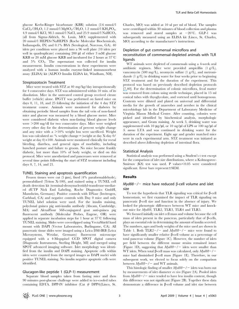
We focused initially on islet cell mass and volume because the cell
mass of islets present in the pancreas, particularly that of b-cells,
plays an essential role in determining the amount of insulin secreted.
The numbers, ages and body weights of the mice used are shown in
Table 1. Both TLR22/2 and Myd882/2 mice were found to
have significantly smaller relative b-cell volume as a percentage of
total pancreas volume (Figure 1C). However, the number of islets
per field between the different mouse strains remained intact
(Figure 1D), suggesting that Myd882/2 islets were smaller than
WT islets. When total b-cell mass was calculated, only Myd882/2
mice had diminished b-cell mass (Figure 1E). Therefore, in our
subsequent work, we elected to focus solely on the comparison
between Myd882/2 and WT animals.
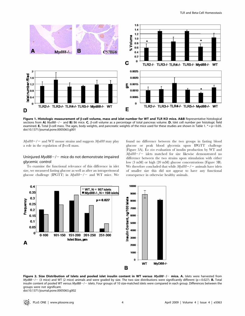
This histologic finding of smaller Myd882/2 islets was born out
by measurements of islet diameter ex vivo (Figure 2A). Pooled islets
from Myd882/2 mice tended to have less insulin content, though
this difference was not significant (Figure 2B). Together these data
demonstrate a difference in b-cell volume and islet size between
TLR and Beta-Cell Homeostasis
PLoS ONE | www.plosone.org 3 April 2009 | Volume 4 | Issue 4 | e5063
Myd882/2 and WT mouse strains and suggests Myd88 may play
a role in the regulation of b-cell mass.
Uninjured Myd882/2 mice do not demonstrate impairedglycemic control
To examine the functional relevance of this difference in islet
size, we measured fasting glucose as well as after an intraperitoneal
glucose challenge (IPGTT) in Myd882/2 and WT mice. We
found no difference between the two groups in fasting blood
glucose or peak blood glycemia upon IPGTT challenge
(Figure 3A). Ex vivo evaluation of insulin production by WT and
Myd882/2 islets matched for size likewise demonstrated no
difference between the two strains upon stimulation with either
low (3 mM) or high (20 mM) glucose concentrations (Figure 3B).
We therefore concluded that while Myd882/2 animals have islets
of smaller size this did not appear to have any functional
consequence in otherwise healthy animals.
Figure 1. Histologic measurement of b-cell volume, mass and islet number for WT and TLR KO mice. A&B Representative histologicalsections from A) Myd882/2 and B) B6 mice. C. b-cell volume as a percentage of total pancreas volume. D. Islet cell number per histologic fieldexamined. E. Total b-cell mass. The ages, body weights, and pancreatic weights of the mice used for these studies are shown in Table 1. * = p,0.05.doi:10.1371/journal.pone.0005063.g001
Figure 2. Size Distribution of Islets and pooled islet insulin content in WT versus Myd882/2 mice. A. Islets were harvested fromMyd882/2 (3 mice) and WT (2 mice) animals and were graded by size. The two size distributions were significantly different (p = 0.027). B. Totalinsulin content of pooled WT versus Myd882/2 islets. Four groups of 10 size-matched islets were compared in each group. Differences between thegroups were not significant.doi:10.1371/journal.pone.0005063.g002
TLR and Beta-Cell Homeostasis
PLoS ONE | www.plosone.org 4 April 2009 | Volume 4 | Issue 4 | e5063

Myd882/2 mice treated with STZ demonstrate impairedglycemic control and transaminitis
We next hypothesized that small decreases in b-cell mass
mediate altered glucose regulation in settings of borderline
glycemic control. We therefore designed experiments to stress
animals into borderline glycemic control conditions to evaluate the
functional relevance of the aforementioned decreases in islet cell
size and mass. We administered 4 days of low-dose (40 mg/kg)
STZ treatment to Myd882/2 and WT controls. While high dose
STZ protocols (e.g. 200 mg/kg) reliably induce diabetes in C57Bl/
6 mice in our experiment, our intention was to administer a sub-
diabetogenic dose which might impact glycemic control of the two
strains of mice differently. Following STZ treatment, we
monitored the mice with weeklyat 0, 7, 14 and 24 days with
IPGTT over a three week period.
We found that Myd882/2 mice developed significant hyper-
glycemia relative to WT controls upon glucose challenge
(Figure 4A–D). This was the case for both the 30 minute and
60 minute IPGTT time points on day 11 (7 days after the final
dose of STZ)(Figure 4A) as well as for the 60 minute time point for
day 18 (14 days after the last dose of STZ)(Figure 4B). The
Myd882/2 mice did, however, recover normal glycemic control
by day 28 (24 days after the final dose of STZ) (Figure 4C)
consistent with healing. Of note, Myd882/2 mice did not reliably
develop hyperglycemic responses to IPGTT upon treatment with
3 days of low-dose STZ treatment (data not shown), suggesting a
threshold with the 4-day regimen.
In order to evaluate whether the enhanced responsiveness of
Myd882/2 mice was a systemic feature of the Myd882/2
phenotype, we looked for evidence of heightened injury in another
cell type susceptible to STZ damage—the hepatocyte. We
evaluated aspartate transaminase (AST) and alanine transaminase
(ALT) for the same time points as were evaluated with IPGTT.
AST and ALT are liver transaminases which, when found in the
peripheral circulation at elevated levels, are indicative of
hepatocellular damage. We predicted that systemic administration
of STZ would have a parallel impact on hepatocytes as on b-cells,
as both cell types are sensitive to STZ. This was indeed the case for
both AST and ALT (Figure 5A,B). These data are consistent with
systemic hyper-responsiveness to STZ treatment in the absence of
Myd882/2 signaling.
Myd882/2 mice demonstrate enhanced b -cellapoptosis following low-dose STZ treatment
The heightened cell damage observed in multiple tissue types in
Myd882/2 animals raised the possibility that low-dose STZ
treatment reflected an increased sensitivity to STZ-induced
apoptosis. STZ is known to induce apoptotic cell death in
hepatocytes as well as b-cells [41,42]. We therefore compared
TUNEL staining in pancreases from low-dose STZ-treated
Myd882/2 mice and WT controls. This was done for 6 sections
from two mice from each group taken before and after low-dose
STZ treatment for 11 days. No TUNEL-positive islet cells were
seen in the sections taken from mice prior to STZ treatment (data
not shown). However following low-dose STZ treatment we
observed an increase in TUNEL positive cells in islets from
Myd882/2 mice (Figure 6A) but not WT controls (Figure 6B).
This difference was significant (Figure 6E). While it is possible that
Myd882/2 mice develop impaired glycemic control more readily
upon STZ treatment because they start with diminished b cell
mass, these data suggest their b-cells are also more likely to
undergo apoptosis under these conditions.
Myd882/2 mice do not have diminished levels of GLP-1To rule out a Myd88-dependent effect mediated by glucagon-
like peptide-1 (GLP-1), a soluble factor known to promote b-cell
mass and insulin gene expression [43,44], we analyzed GLP-1
concentrations in serum from mice both fasting (time = 09) and
following IP glucose challenge (time = 909) (Figure S1). We did not
find any differences in GLP-1 content between the two strains,
suggesting that the findings reported here are not due to
differences in GLP-1 expression.
Low-dose oral LPS promotes glycemic control in animalswith impaired commensal flora
Based on the above data we hypothesized that commensal flora
provide anti-apoptotic signals through Myd882/2. This hypoth-
esis predicts that denuding WT mice of their intestinal flora
enhances sensitivity to low-dose STZ treatment and recapitulates
the Myd882/2 phenotype. Therefore we administered a cocktail
of broad spectrum antibiotics to WT animals for four weeks [7].
We found that these mice were normoglycemic before and after
low-dose STZ treatment in comparison to mice which did not
Figure 3. Response to glucose challenge in vivo and ex vivo. A. Pooled data are shown for IPGTT performed on 47 WT mice and 41 Myd882/2mice. Only the 30 min time-point comparison was significant. All mice were between 10 and 20 weeks of age. The mean age was 15.5 (+/22.1) weeksfor WT mice and 16.0 (+/23.8) weeks for Myd882/2 mice. The mean weight was 24.75 (+/23.5) gms for WT mice and 23.4 (+/23.6) gms for Myd882/2 mice. These differences in age and weight were not significant. 34% of the WT mice were female while 43% of the WT mice were female. IPGTT testresults were not significantly different between the two genders. B. Insulin secretion in response to glucose challenge. Both total insulin content andinsulin response to glucose challenge were determined in paired experiments using islets from the same pair of mice. 10 islets per well were assessedin quadruplicate per condition, per mouse. Data shown are representative of two experiments. There was no significant difference between the twostrains of mice.doi:10.1371/journal.pone.0005063.g003
TLR and Beta-Cell Homeostasis
PLoS ONE | www.plosone.org 5 April 2009 | Volume 4 | Issue 4 | e5063

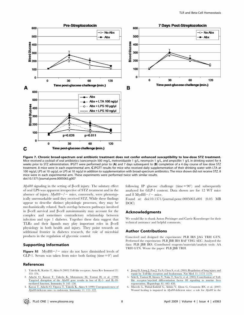
receive antibiotics (Figure 7A–B). Interestingly, we found that four
weeks after the initiation of antibiotics we were still able to culture
limited numbers of viable fecal bacteria. We then attempted to
rescue TLR signaling in ABX treated mice by supplementing
drinking water with LPS or LTA. We found that LPS
supplementation of drinking water in these mice promoted tight
glycemic control (Figure 7C). This effect was significant in the
group of mice which received 10 ng/ml LPS but not in the groups
recieving 10 mg/ml nor the group which received LTA. None of
the mice in these experiments, either those which received
antibiotics nor those which received antibiotics plus TLR ligands,
demonstrated a significant change in their glycemic control from
baseline upon IPGTT following low-dose STZ treatment (data not
shown).
Discussion
The first noteworthy finding of our manuscript is that TLRs and
the signaling molecule Myd88 in particular may contribute to the
regulation of islet mass. The histologic measurements reported
here point to a loss of approximately 40% more islet volume in
Myd882/2 animals as compared to WT controls on the same
C57Bl/6 background. As TLR represent a major molecular
interface between hosts and microbial flora, this finding may help
explain an isolated report of diminished islet mass in gnotobiotic
rats [45]. As no specific individual TLR was associated here
individually with diminished islet mass, it may be that a
combination of TLR make a contribution towards maintaining
islet homeostasis in WT animals.
The second finding of this work is that Myd882/2 mice were
more likely than WT controls to develop hyperglycemia upon b-
cell insult. Myd882/2 mice became relatively hyperglycemic
upon IPGTT following treatment with a low-dose STZ regimen.
This is in contrast to the intact glycemic control seen here in
healthy, uninjured Myd882/2 mice upon fasting IPGTT and
evaluation of their islets ex vivo. It may be that Myd882/2 animals
maintain adequate functional reserve to maintain glycemic control
under normal circumstances and their reduced islet mass only
become functionally relevant upon injury. This is consistent with
models of diabetes in which the large majority of b-cell mass must
be lost before hyperglycemia is observed.
The third notable finding of this work is that Myd882/2 mice also
have an increased rate of apoptosis upon low-dose STZ treatment,as
Myd882/2 mice had greater numbers of apoptotic TUNEL positive
cells per islet than WT controls. Both enhanced islet injury upon STZ
treatment and diminished islet mass may contribute to the
hyperglycemia seen here in Myd882/2 mice. The conclusion that
Myd882/2 suffer greater injury upon STZ treatment is buttressed
by the fact that they also displayed transaminitis indicative of
hepatocyte damage. Myd88 mediated activation of NF-kB is known to
promote cell survival via production of anti-apoptotic Bcl-2 family
molecules [46,47,48,49]. However, Myd88 also binds the Fas-
associated Death Domain (FADD) and promotes induction of
apoptosis via a pathway involving caspase 8 [50]. In the context of
the pancreas, TLR42/2 mice were found to have reduced apoptosis
in a surgical model of pancreatitis [51]. Whether Myd88 signaling
contributes toward anti- or pro-apoptotic pathways is dependant on
the particular TLR involved and other contextual cues [52]. While
TLR have been found to contribute to the maintenance of tissue
integrity at multiple sites of host-microbial interface [4,5,6,7], these
data extend this model to two tissue types not always considered to be
part of that interface, the pancreatic islet and liver.
The enhanced sensitivity to low-dose STZ was not recapitulated
in WT mice denuded of intestinal flora upon the long-term
administration of broad-spectrum antibiotics. Since this treatment
did not render the intestines of these mice entirely sterile, it may be
that even small quantities of intestinal bacteria are adequate.
Alternatively, endogenous TLR ligands might suffice to maintain
islet homeostasis upon STZ treatment in the setting of competent
Myd88 signaling.
Our data do not contradict the recent finding that the absence
of Myd88 leads to changes in the penetrance of diabetes in NOD
mice raised in either SPF or gnotobiotic conditions. In our work
diabetes is induced by a toxin whereas diabetes in NOD mice is
mediated by an autoimmune phenomenon. Together these data
emphasize that effects of different TLR ligands and gut microbiota
on diabetes are likely to be complex, multifactorial and dependent
on the genetic underpinnings of the diabetes in question.
Our data also do not directly contradict the recent report that
Myd882/2 mice developed an increased incidence of autoim-
mune diabetes relative to WT controls one to two months after
low-dose STZ treatment. Notably those authors did not find
relative hyperglycemia in Myd882/2 mice in the weeks
immediately following low-dose STZ treatment but they measured
Figure 4. IPGTT for WT and Myd882/2 mice treated with STZ.Data shown are for 8 WT mice and 8 Myd882/2 mice who receivedlow-dose STZ treatment for 4 days. IPGTT data are shown for this samegroup of mice 7 days (A), 14 days (B) and 24 days (C) followingcompletion of the STZ treatment course. All mice were between 12 and16 weeks of age and were evenly split between male and femalegenders. The mean weight was 21.3 (+/22.3) gms for WT mice and 24.8(+/24.1) gms for Myd882/2 mice. This difference in weight was notsignificant. Data shown are representative of two experiments.doi:10.1371/journal.pone.0005063.g004
TLR and Beta-Cell Homeostasis
PLoS ONE | www.plosone.org 6 April 2009 | Volume 4 | Issue 4 | e5063

blood glucose in fasting mice rather then upon glucose challenge
via IPGTT as we have done here.
We were intrigued to find that low-dose oral LPS has a salutary
effect on glycemic control. These data are consistent with reports
that endogenous gram negative bacteria tonically prime insulin
production in rats [53] and consistent with data that treatment of
islets ex vivo with LPS promotes b-cell insulin production [21]. In
that study the tonic effects of LPS on insulin secretion were dose
dependant, with the lower concentrations being more beneficial.
These effects were observed at LPS concentrations of 1–100 ng/
ml, which is similar to that used to stimulate macrophages. At
higher concentrations of LPS (e.g. 5–10 mg/ml) cytotoxic effect on
islets have been observed [54,55] and in vivo are associated with
sepsis-induced hypoglycemia [56]. In sum, low-dose LPS appears
to promotes insulin production and release while high dose LPS
leads to stress-related responses. The dose-dependent nature of
LPS effects on b-cells may be clinically relevant during acute
endotoxemic events and in settings of inflammation. Chronic
inflammation and basal endotoxemia have been correlated with
obesity and type 2 diabetes in humans and rats [19,57]. Dose-
dependant effects of endotoxin on b-cells may also be relevant to
the viability and function of transplanted islets. In the most
commonly followed transplant protocol, endotoxins are infused
into the portal circulation of recipients [58], where levels of
endotoxin have been shown to be higher relative to peripheral
circulation [59].
We interpret the effects observed upon low-dose oral LPS
treatment as functionally distinct from the role described here for
Figure 6. TUNEL staining of pancreatic sections from WT and Myd882/2 mice following treatment with STZ. Data are shown for sectionstaken from A. Myd882/2 and B. WT mice on day 7 following completion of a low-dose STZ treatment. TUNEL staining is shown in green, insulin stainingis shown in red and DAPI is shown in blue. C. Pooled data for the number of TUNEL staining cells per islet in Myd882/2 versus WT mice.doi:10.1371/journal.pone.0005063.g006
Figure 5. Transaminase Levels Following STZ Treatment. Data shown are for A. AST and B. ALT for 8 WT mice and 8 Myd882/2 mice whoreceived low-dose STZ treatment for 4 days. The animals in this experiment were the same set of mice described in Figure 4 and data for the sametime points 7, 14, and 24 days post completion of the low-dose STZ treatment course are shown.doi:10.1371/journal.pone.0005063.g005
TLR and Beta-Cell Homeostasis
PLoS ONE | www.plosone.org 7 April 2009 | Volume 4 | Issue 4 | e5063
Myd88 signaling in the setting of b-cell injury. The salutary effect
of oral LPS was apparent irrespective of STZ treatment and in the
absence of injury. Myd882/2 mice, conversely, were phenotyp-
ically unremarkable until they received STZ. While these findings
appear to describe distinct physiologic processes, they may be
mechanistically related. Such overlap between pathways involved
in b-cell survival and b-cell autoimmunity may account for the
complex and sometimes contradictory relationship between
infection and type 1 diabetes. Together these data suggest that
TLRs and their ligands may play important roles in b-cell
physiology in both health and injury. They point towards an
additional frontier in diabetes research, the role of microbial
products in the regulation of glycemic control.
Supporting Information
Figure S1 Myd882/2 mice do not have diminished levels of
GLP-1. Serum was taken from mice both fasting (time = 09) and
following IP glucose challenge (time = 909) and subsequently
analyzed for GLP-1 content. Data shown are for 12 WT mice
and 8 Myd882/2 mice.
Found at: doi:10.1371/journal.pone.0005063.s001 (0.05 MB
DOC)
Acknowledgments
We would like to thank Anton Preisinger and Carrie Rosenberger for their
technical assistance and helpful comments.
Author Contributions
Conceived and designed the experiments: PLB IRS JAG TRH GTN.
Performed the experiments: PLB JBB IRS BAF VHG AEC. Analyzed the
data: PLB JBB IRS. Contributed reagents/materials/analysis tools: AA
TRH GTN. Wrote the paper: PLB JBB TRH.
References
1. Takeda K, Kaisho T, Akira S (2003) Toll-like receptors. Annu Rev Immunol 21:
335–376.
2. Adachi O, Kawai T, Takeda K, Matsumoto M, Tsutsui H, et al. (1998)
Targeted disruption of the Myd88 gene results in loss of IL-1- and IL-18-
mediated function. Immunity 9: 143–150.
3. Kawai T, Adachi O, Ogawa T, Takeda K, Akira S (1999) Unresponsiveness of
Myd88-deficient mice to endotoxin. Immunity 11: 115–122.
4. Jiang D, Liang J, Fan J, Yu S, Chen S, et al. (2005) Regulation of lung injury and
repair by Toll-like receptors and hyaluronan. Nat Med 11: 1173–1179.
5. Seki E, Tsutsui H, Iimuro Y, Naka T, Son G, et al. (2005) Contribution of Toll-
like receptor/myeloid differentiation factor 88 signaling to murine liver
regeneration. Hepatology 41: 443–450.
6. Macedo L, Pinhal-Enfield G, Alshits V, Elson G, Cronstein BN, et al. (2007)
Wound healing is impaired in Myd88-deficient mice: a role for Myd88 in the
Figure 7. Chronic broad-spectrum oral antibiotic treatment does not confer enhanced susceptibility to low-dose STZ treatment.Mice received a cocktail of oral antibiotics (vancomycin 500 mg/L, metronidazole 1 g/L, neomycin 1 g/L, and ampicillin 1 g/L in drinking water) for 4weeks prior to STZ administration. IPGTT were performed prior to (A) and 7 days subsequent to (B) completion of a 4 day course of low dose STZtreatment. 8 mice were in each experimental arm. C.IPGTT results for mice who received daily supplementation of their drinking water with LTA at100 ng/ml, LPS at 10 ug/ml, or LPS at 10 ng/ml in addition to supplementation with broad-spectrum antibiotics. The mice shown did not receive STZ. 8mice were in each experimental arm. These experiments were performed twice with similar results.doi:10.1371/journal.pone.0005063.g007
TLR and Beta-Cell Homeostasis
PLoS ONE | www.plosone.org 8 April 2009 | Volume 4 | Issue 4 | e5063
regulation of wound healing by adenosine A2A receptors. Am J Pathol 171:
1774–1788.7. Rakoff-Nahoum S, Paglino J, Eslami-Varzaneh F, Edberg S, Medzhitov R
(2004) Recognition of commensal microflora by toll-like receptors is required for
intestinal homeostasis. Cell 118: 229–241.8. van Tol EA, Holt L, Li FL, Kong FM, Rippe R, et al. (1999) Bacterial cell wall
polymers promote intestinal fibrosis by direct stimulation of myofibroblasts.Am J Physiol 277: G245–G255.
9. Li M, Carpio DF, Zheng Y, Bruzzo P, Singh V, et al. (2001) An essential role of
the NF-kappa B/Toll-like receptor pathway in induction of inflammatory andtissue-repair gene expression by necrotic cells. J Immunol 166: 7128–7135.
10. Putnins EE, Sanaie AR, Wu Q, Firth JD (2002) Induction of keratinocyte growthfactor 1 Expression by lipopolysaccharide is regulated by CD-14 and toll-like
receptors 2 and 4. Infect Immun 70: 6541–6548.11. Sanale AR, Firth JD, Uitto VJ, Putnins EE (2002) Keratinocyte growth factor
(KGF)-1 and -2 protein and gene expression in human gingival fibroblasts.
J Periodontal Res 37: 66–74.12. Sugiyama A, Arakaki R, Ohnishi T, Arakaki N, Daikuhara Y, et al. (1996)
Lipoteichoic acid and interleukin 1 stimulate synergistically production ofhepatocyte growth factor (scatter factor) in human gingival fibroblasts in culture.
Infect Immun 64: 1426–1431.
13. Uematsu S, Matsumoto M, Takeda K, Akira S (2002) Lipopolysaccharide-dependent prostaglandin E(2) production is regulated by the glutathione-
dependent prostaglandin E(2) synthase gene induced by the Toll-like receptor 4/Myd88/NF-IL6 pathway. J Immunol 168: 5811–5816.
14. Thomas KE, Sapone A, Fasano A, Vogel SN (2006) Gliadin stimulation ofmurine macrophage inflammatory gene expression and intestinal permeability
are Myd88-dependent: role of the innate immune response in Celiac disease.
J Immunol 176: 2512–2521.15. Franchimont D, Vermeire S, El Housni H, Pierik M, Van Steen K, et al. (2004)
Deficient host-bacteria interactions in inflammatory bowel disease? The toll-likereceptor (TLR)-4 Asp299gly polymorphism is associated with Crohn’s disease
and ulcerative colitis. Gut 53: 987–992.
16. Clevers H (2004) At the crossroads of inflammation and cancer. Cell 118:671–674.
17. Rivera CA, Adegboyega P, van Rooijen N, Tagalicud A, Allman M, et al. (2007)Toll-like receptor-4 signaling and Kupffer cells play pivotal roles in the
pathogenesis of non-alcoholic steatohepatitis. J Hepatol 47: 571–579.18. Ley RE, Turnbaugh PJ, Klein S, Gordon JI (2006) Microbial ecology: human
gut microbes associated with obesity. Nature 444: 1022–1023.
19. Cani PD, Amar J, Iglesias MA, Poggi M, Knauf C, et al. (2007) Metabolicendotoxemia initiates obesity and insulin resistance. Diabetes 56: 1761–1772.
20. Membrez M, Blancher F, Jaquet M, Bibiloni R, Cani PD, et al. (2008) Gutmicrobiota modulation with norfloxacin and ampicillin enhances glucose
tolerance in mice. FASEB J 22: 2416–2426.
21. Vives-Pi M, Somoza N, Fernandez-Alvarez J, Vargas F, Caro P, et al. (2003)Evidence of expression of endotoxin receptors CD14, toll-like receptors TLR4
and TLR2 and associated molecule MD-2 and of sensitivity to endotoxin (LPS)in islet beta cells. Clin Exp Immunol 133: 208–218.
22. Wen L, Peng J, Li Z, Wong FS (2004) The effect of innate immunity onautoimmune diabetes and the expression of Toll-like receptors on pancreatic
islets. J Immunol 172: 3173–3180.
23. Takahashi K, Satoh J, Seino H, Zhu XP, Sagara M, et al. (1993) Prevention oftype I diabetes with lymphotoxin in BB rats. Clin Immunol Immunopathol 69:
318–323.24. Sai P, Rivereau AS (1996) Prevention of diabetes in the nonobese diabetic mouse
by oral immunological treatments. Comparative efficiency of human insulin and
two bacterial antigens, lipopolysacharide from Escherichia coli and glycoproteinextract from Klebsiella pneumoniae. Diabetes Metab 22: 341–348.
25. Balasa B, van Gunst K, Sarvetnick N (2000) The microbial productlipopolysaccharide confers diabetogenic potential on the T cell repertoire of
BDC2.5/NOD mice: implications for the etiology of autoimmune diabetes. Clin
Immunol 95: 93–98.26. Alyanakian MA, Grela F, Aumeunier A, Chiavaroli C, Gouarin C, et al. (2006)
Transforming growth factor-beta and natural killer T-cells are involved in theprotective effect of a bacterial extract on type 1 diabetes. Diabetes 55: 179–185.
27. Brugman S, Klatter FA, Visser JT, Wildeboer-Veloo AC, Harmsen HJ, et al.(2006) Antibiotic treatment partially protects against type 1 diabetes in the Bio-
Breeding diabetes-prone rat. Is the gut flora involved in the development of type
1 diabetes? Diabetologia 49: 2105–2108.28. van der WN, Kroese FG, Rozing J, Hillebrands JL (2007) Viral infections as
potential triggers of type 1 diabetes. Diabetes Metab Res Rev 23: 169–183.29. Sobel DO, Ewel CH, Zeligs B, Abbassi V, Rossio J, et al. (1994) Poly I:C
induction of alpha-interferon in the diabetes-prone BB and normal Wistar rats.
Dose-response relationships. Diabetes 43: 518–522.30. Serreze DV, Hamaguchi K, Leiter EH (1989) Immunostimulation circumvents
diabetes in NOD/Lt mice. J Autoimmun 2: 759–776.31. Uematsu S, Jang MH, Chevrier N, Guo Z, Kumagai Y, et al. (2006) Detection
of pathogenic intestinal bacteria by Toll-like receptor 5 on intestinal CD11c+lamina propria cells. Nat Immunol 7: 868–874.
32. Wen L, Ley RE, Volchkov PY, Stranges PB, Avanesyan L, et al. (2008) Innateimmunity and intestinal microbiota in the development of Type 1 diabetes.
Nature 455: 1109–1113.
33. Weibel ER, Staubli W, Gnagi HR, Hess FA (1969) Correlated morphometricand biochemical studies on the liver cell. I. Morphometric model, stereologic
methods, and normal morphometric data for rat liver. J Cell Biol 42: 68–91.
34. Montana E, Bonner-Weir S, Weir GC (1993) Beta cell mass and growth aftersyngeneic islet cell transplantation in normal and streptozocin diabetic C57BL/6
mice. J Clin Invest 91: 780–787.
35. Pick A, Clark J, Kubstrup C, Levisetti M, Pugh W, et al. (1998) Role of apoptosis
in failure of beta-cell mass compensation for insulin resistance and beta-cell
defects in the male Zucker diabetic fatty rat. Diabetes 47: 358–364.
36. Xu G, Stoffers DA, Habener JF, Bonner-Weir S (1999) Exendin-4 stimulates
both beta-cell replication and neogenesis, resulting in increased beta-cell massand improved glucose tolerance in diabetic rats. Diabetes 48: 2270–2276.
37. Collins TJ (2007) ImageJ for microscopy. Biotechniques 43: 25–30.
38. Ricordi C, Gray DW, Hering BJ, Kaufman DB, Warnock GL, et al. (1990) Isletisolation assessment in man and large animals. Acta Diabetol Lat 27: 185–195.
39. Shapiro AM, Lakey JR, Ryan EA, Korbutt GS, Toth E, et al. (2000) Islet
transplantation in seven patients with type 1 diabetes mellitus using aglucocorticoid-free immunosuppressive regimen. N Engl J Med 343: 230–238.
40. Fagarasan S, Muramatsu M, Suzuki K, Nagaoka H, Hiai H, et al. (2002) Critical
roles of activation-induced cytidine deaminase in the homeostasis of gut flora.Science 298: 1424–1427.
41. O’Brien BA, Harmon BV, Cameron DP, Allan DJ (1996) Beta-cell apoptosis isresponsible for the development of IDDM in the multiple low-dose
streptozotocin model. J Pathol 178: 176–181.
42. Saini KS, Thompson C, Winterford CM, Walker NI, Cameron DP (1996)Streptozotocin at low doses induces apoptosis and at high doses causes necrosis
in a murine pancreatic beta cell line, INS-1. Biochem Mol Biol Int 39:1229–1236.
43. Wideman RD, Yu IL, Webber TD, Verchere CB, Johnson JD, et al. (2006)
Improving function and survival of pancreatic islets by endogenous productionof glucagon-like peptide 1 (GLP-1). Proc Natl Acad Sci U S A 103:
13468–13473.
44. Drucker DJ (2003) Glucagon-like peptide-1 and the islet beta-cell: augmentationof cell proliferation and inhibition of apoptosis. Endocrinology 144: 5145–5148.
45. Heald KA, Jay TR, Topham D, Webberley J, Downing R (1996) The effect ofgnotobiotic rearing on porcine islet isolation and function. Transplant Proc 28:
824–825.
46. Gelman AE, Zhang J, Choi Y, Turka LA (2004) Toll-like receptor ligandsdirectly promote activated CD4+ T cell survival. J Immunol 172: 6065–6073.
47. Francois S, El Benna J, Dang PM, Pedruzzi E, Gougerot-Pocidalo MA, et al.
(2005) Inhibition of neutrophil apoptosis by TLR agonists in whole blood:involvement of the phosphoinositide 3-kinase/Akt and NF-kappaB signaling
pathways, leading to increased levels of Mcl-1, A1, and phosphorylated Bad.J Immunol 174: 3633–3642.
48. Akira S, Uematsu S, Takeuchi O (2006) Pathogen recognition and innate
immunity. Cell 124: 783–801.
49. Lombardo E, Alvarez-Barrientos A, Maroto B, Bosca L, Knaus UG (2007)
TLR4-mediated survival of macrophages is Myd88 dependent and requires
TNF-alpha autocrine signalling. J Immunol 178: 3731–3739.
50. Aliprantis AO, Yang RB, Weiss DS, Godowski P, Zychlinsky A (2000) The
apoptotic signaling pathway activated by Toll-like receptor-2. EMBO J 19:3325–3336.
51. Sawa H, Ueda T, Takeyama Y, Yasuda T, Shinzeki M, et al. (2007) Role of toll-
like receptor 4 in the pathophysiology of severe acute pancreatitis in mice. SurgToday 37: 867–873.
52. Salaun B, Romero P, Lebecque S (2007) Toll-like receptors’ two-edged sword:
when immunity meets apoptosis. Eur J Immunol 37: 3311–3318.
53. Cornell RP (1985) Endogenous gut-derived bacterial endotoxin tonically primes
pancreatic secretion of insulin in normal rats. Diabetes 34: 1253–1259.
54. Arnush M, Heitmeier MR, Scarim AL, Marino MH, Manning PT, et al. (1998)
IL-1 produced and released endogenously within human islets inhibits beta cell
function. J Clin Invest 102: 516–526.
55. Saitoh N, Awaya A, Sakudo A, SungWook S, Saeki K, et al. (2004) Serum
thymic factor prevents LPS-induced pancreatic cell damage in mice via up-
regulation of Bcl-2 expression in pancreas. Microbiol Immunol 48: 629–638.
56. Oguri S, Motegi K, Iwakura Y, Endo Y (2002) Primary role of interleukin-1
alpha and interleukin-1 beta in lipopolysaccharide-induced hypoglycemia inmice. Clin Diagn Lab Immunol 9: 1307–1312.
57. Creely SJ, McTernan PG, Kusminski CM, Fisher M, Da Silva NF, et al. (2007)
Lipopolysaccharide activates an innate immune system response in humanadipose tissue in obesity and type 2 diabetes. Am J Physiol Endocrinol Metab
292: E740–E747.
58. Ryan EA, Paty BW, Senior PA, Bigam D, Alfadhli E, et al. (2005) Five-yearfollow-up after clinical islet transplantation. Diabetes 54: 2060–2069.
59. Jacob AI, Goldberg PK, Bloom N, Degenshein GA, Kozinn PJ (1977) Endotoxinand bacteria in portal blood. Gastroenterology 72: 1268–1270.
TLR and Beta-Cell Homeostasis
PLoS ONE | www.plosone.org 9 April 2009 | Volume 4 | Issue 4 | e5063