Recombinant Modified Vaccinia Virus Ankara Expressing Glycoprotein E2 of Chikungunya Virus Protects...
12
Recombinant Modified Vaccinia Virus Ankara Expressing Glycoprotein E2 of Chikungunya Virus Protects AG129 Mice against Lethal Challenge Petra van den Doel 1 , Asisa Volz 2 , Jouke M. Roose 1 , Varsha D. Sewbalaksing 1 , Gorben P. Pijlman 3 , Ingeborg van Middelkoop 3 , Vincent Duiverman 4 , Eva van de Wetering 4 , Gerd Sutter 2 , Albert D. M. E. Osterhaus 1,5 , Byron E. E. Martina 1,5 * 1 Department of Viroscience, Erasmus Medical Center, Rotterdam, The Netherlands, 2 Institute for Infectious Diseases and Zoonoses, University of Munich LMU, Munich, Germany, 3 Laboratory of Virology, Wageningen University, Wageningen, The Netherlands, 4 Erasmus Medical Center Laboratory Animal Science Center (EDC), Rotterdam, The Netherlands, 5 Artemis One Health, Utrecht, The Netherlands Abstract Chikungunya virus (CHIKV) infection is characterized by rash, acute high fever, chills, headache, nausea, photophobia, vomiting, and severe polyarthralgia. There is evidence that arthralgia can persist for years and result in long-term discomfort. Neurologic disease with fatal outcome has been documented, although at low incidences. The CHIKV RNA genome encodes five structural proteins (C, E1, E2, E3 and 6K). The E1 spike protein drives the fusion process within the cytoplasm, while the E2 protein is believed to interact with cellular receptors and therefore most probably constitutes the target of neutralizing antibodies. We have constructed recombinant Modified Vaccinia Ankara (MVA) expressing E3E2, 6KE1, or the entire CHIKV envelope polyprotein cassette E3E26KE1. MVA is an appropriate platform because of its demonstrated clinical safety and its suitability for expression of various heterologous proteins. After completing the immunization scheme, animals were challenged with CHIV-S27. Immunization of AG129 mice with MVAs expressing E2 or E3E26KE1 elicited neutralizing antibodies in all animals and provided 100% protection against lethal disease. In contrast, 75% of the animals immunized with 6KE1 were protected against lethal infection. In conclusion, MVA expressing the glycoprotein E2 of CHIKV represents as an immunogenic and effective candidate vaccine against CHIKV infections. Citation: van den Doel P, Volz A, Roose JM, Sewbalaksing VD, Pijlman GP, et al. (2014) Recombinant Modified Vaccinia Virus Ankara Expressing Glycoprotein E2 of Chikungunya Virus Protects AG129 Mice against Lethal Challenge. PLoS Negl Trop Dis 8(9): e3101. doi:10.1371/journal.pntd.0003101 Editor: Ann M. Powers, Centers for Disease Control and Prevention, United States of America Received September 30, 2013; Accepted July 7, 2014; Published September 4, 2014 Copyright: ß 2014 van den Doel et al. This is an open-access article distributed under the terms of the Creative Commons Attribution License, which permits unrestricted use, distribution, and reproduction in any medium, provided the original author and source are credited. Funding: This work was performed within the framework of Dutch Top Institute Pharma (project nr T4-301). The funders had no role in study design, data collection and analysis, decision to publish, or preparation of the manuscript. Competing Interests: The authors have declared that no competing interests exist, apart from Albert Osterhaus, who is a part time employee (CEO) of Viroclinics BV (for details go to www.erasmusmc.nl). The stated competing interest does not alter the authors’ adherence to all the journal’s policies on sharing data and materials. * Email: [email protected] Introduction Chikungunya virus (CHIKV) belongs to the family Togaviridae, genus Alphavirus. The virus was first isolated in 1952 during an epidemic of arthralgic disease in Tanzania, and CHIKV is now known as a mosquito-borne virus endemic in many parts of Sub- Saharan Africa as well as in Asia [1]. Epidemic CHIKV is maintained mainly as an urban transmission cycle, involving humans as amplification hosts and the peri-domestic mosquitoes Aedes aegypti, and since recently also Aedes albopictus. Large outbreaks have been reported since 2004, with the first description originating from Kenya [2,3] and subsequent introduction of the virus to islands in the Indian Ocean [4,5] and India [6,7]. These epidemics resulted in up to six million cases, with regular importation of the virus in Europe, Southeast Asia and the Americas [8]. The first autochthonous CHIKV infections in Europe occurred in Italy in 2007 and France in 2010. The widespread dissemination of CHIKV is associated with amino acid mutations in the glycoprotein E1 (A226V), which facilitated adaptation of CHIKV to the Aedes albopictus, a mosquito that survives in temperate climates and is widely distributed [9]. While patients with CHIKV infection may exhibit a variety of symptoms such as rash, chills, headache, nausea, photophobia, and vomiting, it is characterized by acute high fever, and severe polyarthralgia [10]. Persistent arthralgia can remain for years [11– 14] and it has been shown to be a major cause of long-term discomfort [15]. Fatalities following CHIKV infection are rare, but have been shown to occur [16–18] as a result of severe neurologic disease [19–21]. To date, CHIKV continues to pose a threat to several countries in the world and continues to cause outbreaks in India and Southeast Asia, resulting in persistent morbidity and consequently considerable economic losses. Therefore, vaccine development remains a high priority. Currently, there are no licensed CHIKV vaccines available, although there are several experimental candidate vaccines are under investigation. CHIKV is composed of a single-stranded, positive-sense RNA genome of about 12 kb and contains two open-reading frames (ORFs). These two ORFs encode four non-structural proteins PLOS Neglected Tropical Diseases | www.plosntds.org 1 September 2014 | Volume 8 | Issue 9 | e3101
Transcript of Recombinant Modified Vaccinia Virus Ankara Expressing Glycoprotein E2 of Chikungunya Virus Protects...

Recombinant Modified Vaccinia Virus Ankara ExpressingGlycoprotein E2 of Chikungunya Virus Protects AG129Mice against Lethal ChallengePetra van den Doel1, Asisa Volz2, Jouke M. Roose1, Varsha D. Sewbalaksing1, Gorben P. Pijlman3,
Ingeborg van Middelkoop3, Vincent Duiverman4, Eva van de Wetering4, Gerd Sutter2,
Albert D. M. E. Osterhaus1,5, Byron E. E. Martina1,5*
1 Department of Viroscience, Erasmus Medical Center, Rotterdam, The Netherlands, 2 Institute for Infectious Diseases and Zoonoses, University of Munich LMU, Munich,
Germany, 3 Laboratory of Virology, Wageningen University, Wageningen, The Netherlands, 4 Erasmus Medical Center Laboratory Animal Science Center (EDC), Rotterdam,
The Netherlands, 5 Artemis One Health, Utrecht, The Netherlands
Abstract
Chikungunya virus (CHIKV) infection is characterized by rash, acute high fever, chills, headache, nausea, photophobia,vomiting, and severe polyarthralgia. There is evidence that arthralgia can persist for years and result in long-termdiscomfort. Neurologic disease with fatal outcome has been documented, although at low incidences. The CHIKV RNAgenome encodes five structural proteins (C, E1, E2, E3 and 6K). The E1 spike protein drives the fusion process within thecytoplasm, while the E2 protein is believed to interact with cellular receptors and therefore most probably constitutes thetarget of neutralizing antibodies. We have constructed recombinant Modified Vaccinia Ankara (MVA) expressing E3E2, 6KE1,or the entire CHIKV envelope polyprotein cassette E3E26KE1. MVA is an appropriate platform because of its demonstratedclinical safety and its suitability for expression of various heterologous proteins. After completing the immunization scheme,animals were challenged with CHIV-S27. Immunization of AG129 mice with MVAs expressing E2 or E3E26KE1 elicitedneutralizing antibodies in all animals and provided 100% protection against lethal disease. In contrast, 75% of the animalsimmunized with 6KE1 were protected against lethal infection. In conclusion, MVA expressing the glycoprotein E2 of CHIKVrepresents as an immunogenic and effective candidate vaccine against CHIKV infections.
Citation: van den Doel P, Volz A, Roose JM, Sewbalaksing VD, Pijlman GP, et al. (2014) Recombinant Modified Vaccinia Virus Ankara Expressing Glycoprotein E2 ofChikungunya Virus Protects AG129 Mice against Lethal Challenge. PLoS Negl Trop Dis 8(9): e3101. doi:10.1371/journal.pntd.0003101
Editor: Ann M. Powers, Centers for Disease Control and Prevention, United States of America
Received September 30, 2013; Accepted July 7, 2014; Published September 4, 2014
Copyright: � 2014 van den Doel et al. This is an open-access article distributed under the terms of the Creative Commons Attribution License, which permitsunrestricted use, distribution, and reproduction in any medium, provided the original author and source are credited.
Funding: This work was performed within the framework of Dutch Top Institute Pharma (project nr T4-301). The funders had no role in study design, datacollection and analysis, decision to publish, or preparation of the manuscript.
Competing Interests: The authors have declared that no competing interests exist, apart from Albert Osterhaus, who is a part time employee (CEO) ofViroclinics BV (for details go to www.erasmusmc.nl). The stated competing interest does not alter the authors’ adherence to all the journal’s policies on sharingdata and materials.
* Email: [email protected]
Introduction
Chikungunya virus (CHIKV) belongs to the family Togaviridae,
genus Alphavirus. The virus was first isolated in 1952 during an
epidemic of arthralgic disease in Tanzania, and CHIKV is now
known as a mosquito-borne virus endemic in many parts of Sub-
Saharan Africa as well as in Asia [1]. Epidemic CHIKV is
maintained mainly as an urban transmission cycle, involving
humans as amplification hosts and the peri-domestic mosquitoes
Aedes aegypti, and since recently also Aedes albopictus. Large
outbreaks have been reported since 2004, with the first description
originating from Kenya [2,3] and subsequent introduction of the
virus to islands in the Indian Ocean [4,5] and India [6,7]. These
epidemics resulted in up to six million cases, with regular
importation of the virus in Europe, Southeast Asia and the
Americas [8]. The first autochthonous CHIKV infections in
Europe occurred in Italy in 2007 and France in 2010. The
widespread dissemination of CHIKV is associated with amino acid
mutations in the glycoprotein E1 (A226V), which facilitated
adaptation of CHIKV to the Aedes albopictus, a mosquito that
survives in temperate climates and is widely distributed [9].
While patients with CHIKV infection may exhibit a variety of
symptoms such as rash, chills, headache, nausea, photophobia, and
vomiting, it is characterized by acute high fever, and severe
polyarthralgia [10]. Persistent arthralgia can remain for years [11–
14] and it has been shown to be a major cause of long-term
discomfort [15]. Fatalities following CHIKV infection are rare, but
have been shown to occur [16–18] as a result of severe neurologic
disease [19–21]. To date, CHIKV continues to pose a threat to
several countries in the world and continues to cause outbreaks in
India and Southeast Asia, resulting in persistent morbidity and
consequently considerable economic losses. Therefore, vaccine
development remains a high priority. Currently, there are no
licensed CHIKV vaccines available, although there are several
experimental candidate vaccines are under investigation.
CHIKV is composed of a single-stranded, positive-sense RNA
genome of about 12 kb and contains two open-reading frames
(ORFs). These two ORFs encode four non-structural proteins
PLOS Neglected Tropical Diseases | www.plosntds.org 1 September 2014 | Volume 8 | Issue 9 | e3101

(nsP1, nsP2, nsP3 and nsP4), which are important for virus
replication, and five structural proteins (C, E1, E2, E3 and 6K).
The capsid (C) proteins surround the RNA molecule and together
form the nucleocapsid. The E1/E2 heterodimers are embedded in
the viral membrane and the average CHIKV particle contains
around 80 E1/E2 spikes projecting from the viral envelope.
Alphaviruses enter susceptible cells after attachment to cellular
receptors on many different cell types. The E1 spike protein drives
the fusion process within the cytoplasm, while the E2 protein is
believed to interact with cellular receptors and therefore most
probably constitute the target of neutralizing antibodies [22–24].
Recombinant Modified Vaccinia virus Ankara (MVA) is among
the most promising live viral vector systems, because of its well-
established biological and clinical safety, together with its
suitability for expression of various heterologous proteins. MVA
was originally developed as a safe and effective vaccine against
smallpox by employing serial passages in primary chicken embryo
cells [25,26]. After more than 570 passages in avian tissue culture
MVA had lost the ability to undergo multiple rounds of productive
replication in a broad range animal species including humans [27].
Furthermore, when used as viral vaccine MVA has been shown to
induce immune responses against many different recombinant
antigens and provide protection against multiple viral diseases
[28]. The immunogenicity of recombinant proteins is higher or at
least similar to what is achieved during a productive infection with
replicating vaccinia virus vectors [29] and makes MVA an
attractive platform for vaccine development.
Here we describe for the first time the construction and
evaluation of three different recombinant modified vaccinia
Ankara (MVA) viruses expressing 6KE1, E3E2, or the entire
CHIKV envelope polyprotein E3E26KE1. The three vaccine
candidates were evaluated in the AG129 mouse model to assess
induction of protective immunity against CHIKV-S27.
Materials and Methods
Ethics statementThe animal experiments described in this paper were conducted
according to Dutch guidelines for animal experimentation after
approval (EMC 2514, 122-11-27) by the Animal Welfare
Committee of the Erasmus Medical Centre, Rotterdam, The
Netherlands. Research plans were reviewed by the local ethical
review committee, which considered the benefit of an experiment
against the cost of distress inflicted to the animals during the
experiments. Appropriate practices and procedures as defined in
the Biosafety in microbiological and biomedical laboratories (US
Dept. of Health and Human Services) were used in sample
handling. Samples were stored at 280uC in a biosafety level-3
(BSL-3) facility at the Erasmus Medical Centre Rotterdam, The
Netherlands.
Cells and virusesVero E6 cells were cultured in DMEM with 10% heat
inactivated fetal bovine serum (HI-FBS), supplemented with
penicillin/streptomycin/L-glutamine (2 mM) (psg; Biowhittaker
for all), sodium bicarbonate and 10 mM hepes buffer. Baby
hamster kidney BHK-21 cells (clone 13, ATCC CCL-10) were
grown in DMEM supplemented with 10% HI-FBS and psg. Aedespseudoscutelaris insect cells (AP-61) were grown in Leibovitz-15
medium (BioWhittaker) supplemented with 5% tryptose phosphate
broth medium (MP Biomedicals), psg and 5% HI-FBS. Cells were
grown and maintained at 27uC. Primary chicken embryo
fibroblast cells (CEF) were isolated from 11-day old embryonated
chicken eggs and maintained in DMEM (BioWhittaker) supple-
mented with psg, non-essential amino acids (Sigma) and 10% HI-
FBS.
The African prototype CHIKV-S27 strain (genbank acc.nr.
AF369024), the CHIKV-IND/NL10, and LS3 [30] strains were
grown on Vero E6 cells and virus titers were determined after
titration of virus stocks on Vero E6 cells. To this end, 96-wells
plates (greiner) were seeded with 56104 Vero cells per well. A
serial ten-fold dilution of virus stocks was made in octaplicates on
the seeded plates, and plates were subsequently incubated for four
days at 37uC. The Vero E6 plates were scored microscopically for
cytopathic changes (cpe). Viral titers were expressed as TCID50
per ml as calculated with the Karber method [31]. The highly
attenuated vaccinia virus (VACV) strain MVA (clonal isolate F6,
CEF passage 584) was used for this study. MVA is derived from
VACV chorioallantois virus Ankara (CVA) by more than 570
passages in CEF. During this serial passage MVA incurred six
major deletions and many smaller mutations in its genome and
developed a severe restriction in host range so that it is unable to
replicate in most mammalian cells [32,33].
Generation of recombinant virusRNA of the African prototype CHIKV-S27 strain was isolated
using the High Pure RNA isolation kit (Roche). cDNA of 6KE1,
E3E2, and E3E26KE1 were obtained with SuperscriptIII (Invi-
trogen) and the respective products were amplified with high
fidelity polymerase PFU (Roche). Primers were flanked by BamHI
and NotI restriction sites (Italic and underlined nucleotides). The
following primers were used for specific amplification of 6KE1
(Fw: CGCGGATCCGCCGCCACCATGGCGGCCACATAC-
CAAGAG; Rev: AGCTTTGTTTAAACTTAGTGCCTGCT-
GAACGACACGCA), E3E2 (Fw: CGCGGATCCGCCGCCAC-
CATGGAAGAGTGGAGTCTTG; Rev: AGCTTTGTTTAA-ACTTATTTAGCTGTTCTGATGCAGCA), and E3E26KE1
(Fw: CGCGGATCCGCCGCCACCATGGAAGAGTGGAGT-
CTTG; Rev: AGCTTTGTTTAAACTTAGTGCCTGCTGA-
ACGACACGCA). The resulting PCR products were cloned
directionally into the MVA vector plasmid pIIIRedH5 and placed
under the transcriptional control of the modified vaccinia virus
early/late promoter PmH5 [34].
Author Summary
The Chikungunya virus (CHIKV) represents a threat toEurope. This is supported by its recent introduction andautochthonous transmission in Italy and France in 2007and 2010 respectively. CHIKV causes an acute febrile illnessand severe joint pains that may last for several months insome patients. In absence of a proven effective treatment,safe and effective vaccines represent the best way tocontrol outbreaks of CHIKV. This study describes thedevelopment of a vaccine candidate based on theModified Vaccinia Ankara virus (MVA). MVA has been usedto develop vaccines against several viruses and based onthe safety and efficacy records of MVA vaccines, variouscandidate vaccines are currently in clinical trials. MVAexpressing structural proteins of CHIKV were constructedand tested in AG129 mice for immunogenicity andprotective ability. All animals that received MVA expressingE2 developed neutralizing antibodies and were protectedagainst lethal challenge. Our study shows that recombi-nant MVA expressing the E2 protein of CHIKV represents apotential candidate vaccine for prevention of CHIKVinfections in travellers or endemic areas.
MVA-Based Chikungunya Vaccine
PLOS Neglected Tropical Diseases | www.plosntds.org 2 September 2014 | Volume 8 | Issue 9 | e3101

Recombinant MVA expressing 6KE1, E3E2, and E3E26KE1
(MVA-6KE1, MVA-E3E2, MVA-E3E26KE1) were generated
using the principles of homologous recombination targeting the
site of major deletion III within the MVA genome for insertion of
recombinant gene sequences. Primary CEF were infected with
MVA followed by transfection of the infected cells with
pIIIRedH5 DNA, using FuGENE transfection reagent (Roche).
Clonal isolates of recombinant MVA were obtained by multiple
rounds of plaques purification on CEF cells and screening for the
transient expression of the fluorescent marker mCHERRY. In
each round, the MVA genome at the insertion site was analyzed
by PCR and recombinant gene expression was confirmed by
immunostaining of BHK-21 cells infected with selected recombi-
nant viruses. Clonal recombinant MVAs expressing mCHERRY
were further propagated on CEF until loss of the transient marker
mCHERRY. To this end, limiting dilutions of the recombinant
viruses were done and wells with the highest virus dilution
displaying cpe were selected for further analysis. To generate the
vaccine batches for the pre-clinical animal studies, all recombinant
MVA were propagated in a multistep amplification process on
Figure 1. Analysis of candidate vaccines. Different MVA constructs were checked for presence of the specific transgene E3E2 (panel A),E3E26KE1 (panel B), and 6KE1 (panel C) using different sets of primers (Table S1). PCRs were conducted on DNA extracted from uninfected BHK-21cells (lane 1, negative pcr), BHK-21 cells infected with wildtype MVA (lane 2) and recombinant MVA DNA (lanes 3, 5, 7, 9, 11 and 13), as positivecontrols shuttle vector containing the three respective transgenes (lane 4, 6, 8, 10,12 and 14) were used as a template. Amplicons were separated on1% agarose gel with Sybrsafe staining and were visualised under blue light transillumination. M Smartladder, kb molecular weight.doi:10.1371/journal.pntd.0003101.g001
MVA-Based Chikungunya Vaccine
PLOS Neglected Tropical Diseases | www.plosntds.org 3 September 2014 | Volume 8 | Issue 9 | e3101
CEF cells, purified by ultracentrifugation through 20% sucrose,
and reconstituted in TE buffer, pH 9.0. Expression of the
respective genes was confirmed by Western blot analysis of lysates
of infected BHK-21 cells. Six different PCR primer sets were used
to confirm the genetic integrity of the recombinant viruses (see
Table S1). The sequence of all inserted recombinant genes was
confirmed to be identical to the original CHIKV-S27 sequence
using the 31306l automatic sequencer (ABI).
Immunoperoxidase stainingCells were fixed in methanol, washed with PBS, blocked for
endogenous peroxidase with hydrogen peroxide and after two
washes with PBS cells were subsequently stained with
polyclonal rabbit anti-E1 or rabbit anti-E2 [35] for 1 hour at
37uC. Cells were washed twice with PBS and primary antibody
was detected using peroxidase labeled goat anti-rabbit IgG
(Invitrogen) after incubation for 45 min at 37uC and 3-amino-
9-ethylcarbazole (AEC) substrate for 10 min at RT. Amount of
cells expressing the recombinant proteins were counted using a
light microscope.
Electron microscopyTo investigate whether the recombinant MVA-E3E26KE1 lead
to generation of VLPs, BHK-21 and Hela cells were infected with
the recombinant MVA, using an MOI of 1 and 10. Supernatant
were collected 24 hours and 48 hours after infection. Supernatant
were centrifuged for 10 minutes at 1200 rpm and 10 ml was used
for EM analysis. Briefly, Copper 400 square mesh grids (Veco)
were treated by Argon gas discharge and loaded with 5 ml culture
fluid of MVA-infected cells for 30 sec at room temp. Excess liquid
was removed using filter paper and the grids were treated with 2%
uranyl acetate for 30 sec, excess uranyl acetate was carefully
removed using filter paper. The grids were air dried and analyzed
with a JEOL JEM 1011 transmission electron microscope at the
Wageningen Electron Microscopy Centre.
Vaccination—challenge experimentsMice lacking the IFN-a/b/c receptor (A129 mice) [36] have
been described previously to be susceptible to CHIKV infection
[37]. The AG129 mice were purchased from B&K Universal (UK)
at the age of six weeks. Mice were maintained under specific
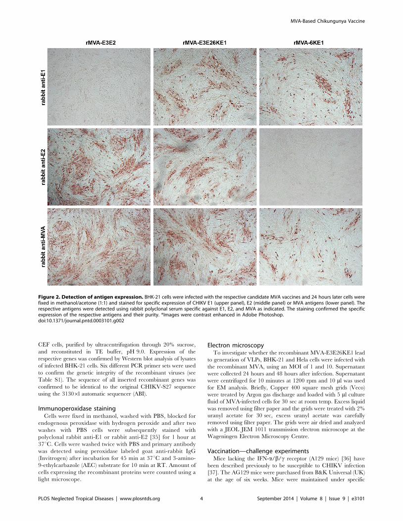
Figure 2. Detection of antigen expression. BHK-21 cells were infected with the respective candidate MVA vaccines and 24 hours later cells werefixed in methanol/acetone (1:1) and stained for specific expression of CHIKV E1 (upper panel), E2 (middle panel) or MVA antigens (lower panel). Therespective antigens were detected using rabbit polyclonal serum specific against E1, E2, and MVA as indicated. The staining confirmed the specificexpression of the respective antigens and their purity. *Images were contrast enhanced in Adobe Photoshop.doi:10.1371/journal.pntd.0003101.g002
MVA-Based Chikungunya Vaccine
PLOS Neglected Tropical Diseases | www.plosntds.org 4 September 2014 | Volume 8 | Issue 9 | e3101

Ta
ble
1.
Ne
utr
aliz
atio
nti
ters
agai
nst
CH
IKV
-S2
7an
dC
HIK
V-I
ND
/NL1
0.
An
ima
l#
Da
y0
Da
y2
1D
ay
63
Da
y0
Da
y2
1D
ay
63
Sta
tus
aft
er
cha
lle
ng
e
MV
AV
acci
ne
Ch
alle
ng
evi
rus
6K
E1
CH
IKV
-S2
7C
HIK
V-I
ND
/NL1
0
Mo
use
1,
10
,1
01
0,
10
,1
02
0su
rviv
ed
Mo
use
2,
10
,1
02
0,
10
,1
02
0su
rviv
ed
Mo
use
3,
10
,1
01
0,
10
,1
01
0su
rviv
ed
Mo
use
4,
10
,1
01
0,
10
,1
01
0su
rviv
ed
Mo
use
5,
10
,1
0,
10
,1
0,
10
,1
0d
ead
Mo
use
6,
10
,1
0,
10
,1
0,
10
10
surv
ive
d
Mo
use
7,
10
,1
0,
10
,1
0,
10
,1
0su
rviv
ed
Mo
use
8,
10
,1
01
0,
10
,1
01
0d
ead
MV
AV
acci
ne
Ch
alle
ng
evi
rus
E3
E2
CH
IKV
-S2
7C
HIK
V-I
ND
/NL1
0
Mo
use
1,
10
10
20
,1
01
02
0su
rviv
ed
Mo
use
2,
10
,1
02
0,
10
10
20
surv
ive
d
Mo
use
3,
10
,1
02
0,
10
10
10
surv
ive
d
Mo
use
4,
10
,1
02
0,
10
10
20
surv
ive
d
Mo
use
5,
10
10
10
,1
01
02
0su
rviv
ed
Mo
use
6,
10
10
10
,1
01
01
0su
rviv
ed
Mo
use
7,
10
,1
01
0,
10
,1
01
0su
rviv
ed
Mo
use
8,
10
10
20
,1
01
02
0su
rviv
ed
MV
AV
acci
ne
Ch
alle
ng
evi
rus
6K
E1
E3
E2
CH
IKV
-S2
7C
HIK
V-I
ND
/NL1
0
Mo
use
1,
10
10
80
,1
02
08
0su
rviv
ed
Mo
use
2,
10
10
80
,1
01
08
0su
rviv
ed
Mo
use
3,
10
10
16
0,
10
20
16
0su
rviv
ed
Mo
use
4,
10
10
80
,1
01
08
0su
rviv
ed
Mo
use
5,
10
20
40
,1
02
08
0su
rviv
ed
Mo
use
6,
10
10
40
,1
02
04
0su
rviv
ed
Mo
use
7,
10
20
80
,1
02
01
60
surv
ive
d
Mo
use
8,
10
20
40
,1
02
04
0su
rviv
ed
MV
AV
acci
ne
Ch
alle
ng
evi
rus
Wil
dty
pe
CH
IKV
-S2
7C
HIK
V-I
ND
/NL1
0
Mo
use
1,
10
,1
0,
10
,1
0,
10
,1
0d
ead
Mo
use
2,
10
,1
0,
10
,1
0,
10
,1
0d
ead
Mo
use
3,
10
,1
0,
10
,1
0,
10
,1
0d
ead
MVA-Based Chikungunya Vaccine
PLOS Neglected Tropical Diseases | www.plosntds.org 5 September 2014 | Volume 8 | Issue 9 | e3101

pathogen-free conditions and were allowed to adjust to the facility
for one week before experiments were performed. Four groups of
mice (n = 8 for each group) were immunized with the MVA-
6KE1, MVA-E3E2 and E3E26KE1 twice at 3-week intervals.
Wildtype (wt) MVA was used as a negative control. All MVA
stocks had a concentration of 108 TCID50/ml and 50 ml was
injected into the quadriceps muscles of the left leg of each animal.
Blood was collected on day 0, 21, 63, 70 and 77.
Six weeks after the last immunization (day 63), all animals were
challenged by intraperitoneal inoculation of 103 TCID50 of
CHIKV-S27 in a total volume of 100 ml and animals were
checked daily for clinical signs of infection, such as lethargy and
hind limb weakness. Animals were then sacrificed either 14 days
post challenge or earlier if a humane end point (immobility and
paralysis) was reached. At the end of the experiment, the survival
rates were analyzed and compared between groups.
Immunogenicity studiesTo characterize the antibody responses triggered by immuni-
zation with the recombinant MVA vaccines, a virus neutralization
test was performed. To this end, sera of immunized mice were
heat-inactivated and diluted (1:10 to 1:2560) in triplicate in 96-
wells plate and 100 TCID50 of CHIKV-S27, CHIKV-IND/NL10
or CHIKV-LS3 was added to each well. After one hour of
incubation at 37uC, 16104 Vero E6 cells were added to each well
and plates were incubated for another four days. Neutralizing
titres were determined microscopically and expressed as the
reciprocal of the highest serum dilution still giving 100%
suppression of cpe.
Determination of viral loadIn order to quantify relative numbers of viral RNA in the
respective tissues, 100 mL of organ homogenate was added to
400 mL of lysis buffer (Roche). Viral RNA was then extracted from
spleen, liver, kidney, spinal cord, and brain samples using the
automated MagnaPure method (Total nucleic acid isolation kit,
Roche Diagnostics, the Netherlands) according to the manufac-
turer’s instructions, and quantified using a one-step RT-PCR
TaqMan protocol (EZ-kit, Applied Biosystems) and an ABI
PRISM 7500 detection instrument. The primers and probe used
for CHIKV RNA quantification were essentially as described [38].
Specifically, CHIKV-forw AAGCTCCGCGTCCTTTAC-
CAAG; CHIKV-rev CCAAATTGTCCTGGTCTTCCT; and
Probe: Fam-CCAATGTCTTCAGCCTGGACACCTTT-Tamra
were used. Results are expressed as TCID50 equivalents per gram
of tissue.
Immuno-histochemistryTissues were removed and fixed in 10% neutral-buffered
formalin, embedded in paraffin and sectioned at 4 mm. Slides
were stained with hematoxylin and eosin (HE) and analyze by light
microscopy. Subsequently, 4-mm thick paraffin sections were
processed for immunohistochemistry. To this end, sections were
deparaffinized in xylene, rehydrated in descending concentrations
of ethanol and incubated for 10 min in 3% H2O2 diluted in PBS
to block endogenous peroxidase activity. Antigen exposure was
performed by incubation for 15 min at 121uC in citrate buffer
(0.01 M, pH 6.0). Sections were incubated overnight at 4uC with
rabbit-anti-CHIKV capsid (1:5000), and the primary antibody was
detected with secondary goat anti-rabbit IgG-PO (1:100; Dako,
The Netherlands). Sections were counterstained with Mayer’s
hematoxylin and mounted with Kaiser’s glycerin-gelatin and
analyzed using a light microscope.
Ta
ble
1.
Co
nt.
An
ima
l#
Da
y0
Da
y2
1D
ay
63
Da
y0
Da
y2
1D
ay
63
Sta
tus
aft
er
cha
lle
ng
e
Mo
use
4,
10
,1
0,
10
,1
0,
10
,1
0d
ead
Mo
use
5,
10
,1
0,
10
,1
0,
10
,1
0d
ead
Mo
use
6,
10
,1
0,
10
,1
0,
10
,1
0d
ead
Mo
use
7,
10
,1
0,
10
,1
0,
10
,1
0d
ead
Mo
use
8,
10
,1
0,
10
,1
0,
10
,1
0d
ead
do
i:10
.13
71
/jo
urn
al.p
ntd
.00
03
10
1.t
00
1
MVA-Based Chikungunya Vaccine
PLOS Neglected Tropical Diseases | www.plosntds.org 6 September 2014 | Volume 8 | Issue 9 | e3101

MVA-Based Chikungunya Vaccine
PLOS Neglected Tropical Diseases | www.plosntds.org 7 September 2014 | Volume 8 | Issue 9 | e3101

Statistical analysisDifferences in Kaplan-Meier survival curves between the groups
were assessed using the log-rank test. All statistical analyses were
performed using GraphPad Prism version 4 software (Graphpad
Software, San Diego, USA). Differences between viral loads were
assessed using the student’s t test. P values#0.05 were considered
to be statistically significant.
Results
Characterization of the different candidate vaccinesThe first step in generation of the candidate vaccines was
selection of recombinant MVA that contained CHIKV-S27 gene
sequences of interest. These recombinant viruses expressed the
mCHERRY marker and contained the expected CHIKV gene.
Next the recombinant viruses were passaged until they were
mCHERRY-free and all constructs underwent thorough quality
control PCR (Figure 1; Table S1). In previous studies, genetic
instabilities have been recognized with MVA vector viruses that
contain recombinant sequences encoding for heterologous viral
glycoproteins [39,40]. The genetic stability of final MVA-6KE1,
MVA-E3E2 and E3E26KE1 recombinant viruses was evaluated
after passing the viruses multiple times on CEF (data not shown)
(Figure 1). Synthesis of the recombinant proteins E1 and E2 was
confirmed in an immunostaining assay (Figure 2).
In order to investigate whether the MVA-E3E26KE1 construct
resulted in production of VLPs, supernatant of cells infected with
the MVA construct were collected several time points after
infection. Extensive screening with electron microscopy revealed
no presence of VLPs, suggesting that MVA-E3E26KE1 did not
result in generation of VLPs.
MVA-E3E2 and MVA-E3E26KE1 induce neutralizingantibodies against homologous and heterologous CHIKV
As shown in table 1, immunization with MVA-6KE1 and
MVA-E3E2 induced low levels of neutralizing antibodies (range:
10–20) against both CHIKV-S27 and CHIKV-IND/NL10, 56
days post immunization. In contrast, recombinant MVA express-
ing the structural envelope cassette E3E26KE1 induced signifi-
cantly higher levels of neutralizing antibodies compared to the
other two candidate vaccines (P,0.05), with titers ranging from
40–160. Only MVA-E3E26KE1 induced neutralizing antibodies
in 100% of the animals after one vaccination (day 21). Similar
titers were obtained in neutralization assay using the LS2 strain
(not shown).
MVA vaccines protect against lethal challenge withCHIKV-S27
In order to study the protective efficacy of the different
recombinant MVA candidate vaccines, all the mice were
challenged intra-peritoneally with a lethal dose of CHIKV-S27.
The survival rates of the eight animals were monitored in each
group after challenge. All mock-vaccinated (MVA-wt) mice died
within five days post infection (Figure 3A). In the groups of mice
vaccinated with MVA-E3E2 and MVA-E3E26KE1 respectively,
all animals were protected against lethal infection. In contrast,
75% (6/8) of the animals immunized with 6KE1 were protected
against lethal infection caused by challenge with CHIKV-S27. A
clear booster response could be seen in all the groups (Figure 3E).
The booster response was significantly higher in the group
vaccinated with MVA-6KE1 Compared to the other groups.
The anamnestic response in animals that received MVA-
E3E26KE1 was two-fold lower than those immunized with
MVA-E3E2, indicating that these two candidate vaccines provid-
ed similar levels of protection.
MVA vaccines reduce viral load in organs of challengedanimals
When animals reached the humane-endpoint after challenge,
they were sacrificed and several tissues were collected to quantify
virus titers. High levels of CHIKV RNA were detected in the liver
and spleen (average: 105 TCID50 equivalents/g tissue), and
moderate amounts of viral RNA were found in the brain (average:
102 TCID50 equivalents/g tissue) of mock vaccinated animals.
However, no viral RNA was detected in the kidney or spinal cord
of the infected animals (Figure 3 B–D). Six out of 8 animals
immunized with MVA-E3E26KE1 and challenged with CHIK-
S27 had very low levels viral RNA detectable in the spleen (range:
12 to 68 TCID50 equivalents/g tissue) and only 2 out of 8 animals
had low RNA levels in the liver. No RNA was detected in the
brain of these, indicating that MVA-E3E26KE1 provided high
level protection against virus replication in the periphery and the
brain. By contrast, most animals immunized with MVA-6KE1 and
MVA-E3E2 and challenged with CHIKV-S27 had virus RNA in
the liver, spleen and brain. The two animals that died after
immunization with MVA-6KE1 had high levels of viral RNA in
these organs (Figure 3). Infectious virus could not be recovered
from any of the animals that received either MVA-E3E2 or MVA-
E3E26KE1. On the other hand, low levels of infectious virus were
isolated from the spleen of all the animals that received the MVA-
6KE1 vaccine. However, the exact amount infectious virus in the
spleen of these animals could not be determined because of
toxicity in cell culture.
Histological analysisImmunohistochemical analyses were performed on tissues of
animals that died as a result of CHIKV challenge and tissues
collected from animals that survive the infection (day 14). No
significant abnormalities were observed in the HE staining of the
liver, brain, spinal cord, or leg skeletal muscle of neither the sick
nor the healthy animals. In majority of the sick animals,
neutrophils were seen in the sinusoids in the liver (Figure 4A).
However, in the spleen extensive lymphocytic necrosis was
observed in the white pulp of the control animals and in the
MVA-6KE1 vaccinated animals that succumbed to infection
(Figure 4C). This pathological finding was not observed in mice
that survived the infection. Furthermore, abundant CHIKV
antigens were detected in endothelial cells of all examined organs.
Figure 3. Survival of mice after vaccination and challenge infection with CHIKV. (A). Mice (n = 8) were immunized intra-muscularly withMVA-6KE1, MVA-E3E2, MVA-E3E26KE1 or MVA-wt. Subsequently, the mice were challenged intra-peritoneally with 1000 TCID50 CHIKV-S27. Thesurvival rates of the mice after challenge are depicted as Kaplan-Meier curves. Differences between the curves were determined by the log-rank test.(B, C, D). Vial RNA copies were determined in spleen (B), liver (C) and brain (D) samples of animals that succumbed to the infection and survivors (day14 post challenge). (E) Neutralizing antibody titers were determined on day 0, 21, 63, 70, and 77. Animals were immunized on day 0 and 21 andchallenged on day 63. A clear booster response is seen after challenge, where a higher response was measured in MVA-6KE1 immunized group. Theresults are expressed as TCID50 equivalents per gram of tissue; *indicates a statistically significant result (P,0.05) and ** indicates highly significantresults (P,0.001) as determined by the Student’s t test.doi:10.1371/journal.pntd.0003101.g003
MVA-Based Chikungunya Vaccine
PLOS Neglected Tropical Diseases | www.plosntds.org 8 September 2014 | Volume 8 | Issue 9 | e3101

Figure 4. Histopathology of several tissues staining positive for CHIKV antigen. Panels A–E show representative staining of the liver,spleen, and brain of CHIKV infected AG129 mice. Hematoxylin and eosin-stained sections of the liver from all groups showed no abnormalities (A;406objective). Endothelial and Kupffer cells stained positive with anti-CHIKV capsid antibody (B, 406objective). Massive depletion of lymphocytes
MVA-Based Chikungunya Vaccine
PLOS Neglected Tropical Diseases | www.plosntds.org 9 September 2014 | Volume 8 | Issue 9 | e3101

In addition, antigen was detected in Kupffer cells in the liver
(Figure 4B), macrophages and megakaryocytes in the spleen
(Figure 4D) and adipocytes in the muscles of all mice that
succumbed to infection. Few cells of the choroid plexus and
endothelial cells were found antigen positive in the brain
(Figure 4E), whereas no antigens were found in the spinal cord
of any mice (not shown). Furthermore, no CHIKV antigen was
detected in any organs of the animals that survived the infection.
The negative immunohistochemical results obtained in the spleen
of surviving animals that received MVA-6KE1, suggest low levels
of virus in these animals.
Discussion
In the present study, we have evaluated recombinant MVA
expressing the structural genes of CHIKV-S27 for induction of
protective immunity. Immunization of AG129 mice with MVAs
expressing E2 or the entire CHIKV envelope polyprotein cassette
E3E26KE1 provided 100% protection against lethal disease.
Development of neutralizing antibodies correlated with protection
against lethal infection in vivo.
To date, there are no licensed drugs or vaccines against CHIKV
for use in humans. Safety and immunogenicity are major concerns
when developing new vaccines. Recombinant vector vaccines such
as MVA represent attractive alternatives for safe and effective
vaccines. One theoretical disadvantage of an extensive use of the
MVA system is the development of anti-MVA immunity. Several
studies have shown that MVA is effective in eliciting protection
against several virus infections and is not affected by anti-MVA
immunity [41–43]. In this study, we have constructed several
recombinant MVA vaccines that express E1, E2, or a combination
of E1E3. We have chosen to clone the structural proteins from the
West African CHIKV strain S27. E1 of S27 differs from la
Reunion strain (LR2006) in only three amino acids (A226V,
M270V, and V323A). These mutations are conserved. The E2 of
S27 differed in six amino acids however (G57K, G79E, I211T,
M266R, T312M, and A344T). The CHIK-NL10 strain was 100%
identical to the LR2006 in the E1 protein and had only two
mutations in the E2 (S191T and K252Q) compared to LR2006.
However, because the S27 strain replicates faster in vitro and it is
more virulent than NL10 in mice (not shown), we decided to use
the S27 strain as the protein donor for the vaccine and challenge
virus. A dose of 56106 TCID50 of recombinant MVA expressing
E3E26KE1 rendered 100% protection against disease in chal-
lenged mice and the data suggest that presence of neutralizing
antibodies correlates with protection. The antibody neutralization
titers against a heterologous strain of CHIKV, which is more
similar to LR2006, suggest that a vaccine based on the S27 strain
would provide sufficient cross-protection against a different
CHIKV strain. The relatively low levels of neutralizing antibody
titers obtained against S27, NL10 and LS3 is unlikely to be
explained by differences between the viruses. The most likely
explanation is the relatively low dose of the vaccine that was
administered, which was 1–2 log lower than what is normally
given to reach high levels of antibody titers (108 TCID50). The LS3
CHIKV strain is a synthetic virus based on the consensus sequence
of several CHIKV strains. It is interesting to note that low levels of
virus RNA could still be detected in 75% (6/8) of the spleens of
animals immunized with E3E26KE1. Whether infectious virus
persists is not known. It has been shown that CHIKV may persist
in macrophages of humans and macaques long after acute
infection [44]. Our results are in agreement with a recent study,
which showed that MVA expressing C, E1, and E2 protected mice
against lethal challenge with CHIKV [34]. Several other
experimental vaccine candidates have been described for CHIKV,
including inactivated [45–48], subunit protein [45,49], virus-like
particle (VLP) [50,51], live-attenuated virus [37,52,53], DNA
[54,55] or vector vaccines [56–58]. All the platforms that express
E1 and E2 rendered mice complete protection. It is known that
vaccines based on inactivated virus or subunit proteins need to be
adjuvanted to achieve satisfying immunogenicity. However, the
use of adjuvants in humans is conflicting and non-adjuvanted
vaccines are preferable. Live-attenuated vaccines have been shown
to be safe and effective for several viral infections, but are
associated with side-effects and there is fear for reversion to a
virulent phenotype. Consistently, live-attenuated CHIKV vaccines
have been shown to be more immunogenic in animal models and
humans, but were associated with some side-effects [52]. DNA
vaccines have so far not been particularly effective at generating
antibody responses in humans [59], which is a concern as
antibodies are believed to be required for protection against
CHIKV infections [60,61]. The role cell-mediated immunity
cannot be excluded however. In this study we used AG129 mice,
which are deficient in IFN-a, b, and c receptor. It has been
suggested that IFN-a and IFN-b system mainly inhibits early
spread of virus from the primary site of infection, whereas the IFN-
c system may play a more important role in later stages of viral
infection, e.g., viral persistence [36].
Our study further indicates that E2 is sufficient to induce
complete protection in the mouse model. However, further studies
are needed to see whether immunization with a single dose and
whether use of a lower dose would provide clinical protection
against homologous and heterologous strains of CHIKV.
Supporting Information
Table S1 Sequence of primers and information about the
amplified product.
(DOCX)
Acknowledgments
We thank Penelope Koraka, Lisette Provacia, Eurydice Martina, Astrid
Freudenstein and Minoushka Oduber for their technical assistance.
Author Contributions
Conceived and designed the experiments: PvdD BEEM GS AV.
Performed the experiments: PvdD JMR VDS IvM VD EvdW BEEM.
Analyzed the data: PvdD BEEM. Contributed reagents/materials/analysis
tools: AV GPP. Wrote the paper: PvdD GS ADMEO BEEM.
was observed in the spleen of animals that died after challenge with CHIKV (C; HE staining, 406objective). Antigen was mainly located in endothelialcells in the spleen (D). Epithelia cells of the choroid plexus (E) in the brain were scarcely stained and no antigen was found in the neuropil of the brain.Examples of positively stained cells are indicated by block arrows.doi:10.1371/journal.pntd.0003101.g004
MVA-Based Chikungunya Vaccine
PLOS Neglected Tropical Diseases | www.plosntds.org 10 September 2014 | Volume 8 | Issue 9 | e3101

References
1. Charrel RN, de Lamballerie X, Raoult D (2007) Chikungunya outbreaks–the
globalization of vectorborne diseases. N Engl J Med 356: 769–771.
2. Sergon K, Njuguna C, Kalani R, Ofula V, Onyango C, et al. (2008)Seroprevalence of Chikungunya virus (CHIKV) infection on Lamu Island,
Kenya, October 2004. Am J Trop Med Hyg 78: 333–337.
3. Sutherland LJ, Cash AA, Huang YJ, Sang RC, Malhotra I, et al. (2011)Serologic evidence of arboviral infections among humans in Kenya. Am J Trop
Med Hyg 85: 158–161.
4. Parola P, de Lamballerie X, Jourdan J, Rovery C, Vaillant V, et al. (2006) Novelchikungunya virus variant in travelers returning from Indian Ocean islands.
Emerg Infect Dis 12: 1493–1499.
5. Simon F, Parola P, Grandadam M, Fourcade S, Oliver M, et al. (2007)Chikungunya infection: an emerging rheumatism among travelers returned from
Indian Ocean islands. Report of 47 cases. Medicine (Baltimore) 86: 123–137.
6. Yergolkar PN, Tandale BV, Arankalle VA, Sathe PS, Sudeep AB, et al. (2006)
Chikungunya outbreaks caused by African genotype, India. Emerg Infect Dis 12:1580–1583.
7. Ravi V (2006) Re-emergence of chikungunya virus in India. Indian J Med
Microbiol 24: 83–84.8. Pialoux G, Gauzere BA, Jaureguiberry S, Strobel M (2007) Chikungunya, an
epidemic arbovirosis. Lancet Infect Dis 7: 319–327.
9. Tsetsarkin KA, Vanlandingham DL, McGee CE, Higgs S (2007) A singlemutation in chikungunya virus affects vector specificity and epidemic potential.
PLoS Pathog 3: e201.
10. Borgherini G, Poubeau P, Staikowsky F, Lory M, Le Moullec N, et al. (2007)
Outbreak of chikungunya on Reunion Island: early clinical and laboratoryfeatures in 157 adult patients. Clin Infect Dis 44: 1401–1407.
11. Borgherini G, Poubeau P, Jossaume A, Gouix A, Cotte L, et al. (2008) Persistent
arthralgia associated with chikungunya virus: a study of 88 adult patients onreunion island. Clin Infect Dis 47: 469–475.
12. Chow A, Her Z, Ong EK, Chen JM, Dimatatac F, et al. (2011) Persistent
arthralgia induced by Chikungunya virus infection is associated with interleukin-6 and granulocyte macrophage colony-stimulating factor. J Infect Dis 203: 149–
157.
13. Higgs S, Ziegler SA (2010) A nonhuman primate model of chikungunya disease.J Clin Invest 120: 657–660.
14. Schilte C, Staikovsky F, Couderc T, Madec Y, Carpentier F, et al. (2013)
Chikungunya Virus-associated Long-term Arthralgia: A 36-month ProspectiveLongitudinal Study. PLoS Negl Trop Dis 7: e2137.
15. Queyriaux B, Simon F, Grandadam M, Michel R, Tolou H, et al. (2008)
Clinical burden of chikungunya virus infection. Lancet Infect Dis 8: 2–3.
16. Gerardin P, Fianu A, Malvy D, Mussard C, Boussaid K, et al. (2011) Perceivedmorbidity and community burden after a Chikungunya outbreak: the
TELECHIK survey, a population-based cohort study. BMC Med 9: 5.
17. Mavalankar D, Shastri P, Bandyopadhyay T, Parmar J, Ramani KV (2008)Increased mortality rate associated with chikungunya epidemic, Ahmedabad,
India. Emerg Infect Dis 14: 412–415.
18. Renault P, Josseran L, Pierre V (2008) Chikungunya-related fatality rates,Mauritius, India, and Reunion Island. Emerg Infect Dis 14: 1327.
19. Jaffar-Bandjee MC, Ramful D, Gauzere BA, Hoarau JJ, Krejbich-Trotot P,
et al. (2010) Emergence and clinical insights into the pathology of Chikungunyavirus infection. Expert Rev Anti Infect Ther 8: 987–996.
20. Rampal, Sharda M, Meena H (2007) Neurological complications in Chikungu-
nya fever. J Assoc Physicians India 55: 765–769.
21. Tandale BV, Sathe PS, Arankalle VA, Wadia RS, Kulkarni R, et al. (2009)Systemic involvements and fatalities during Chikungunya epidemic in India,
2006. J Clin Virol 46: 145–149.
22. Kielian M, Rey FA (2006) Virus membrane-fusion proteins: more than one wayto make a hairpin. Nat Rev Microbiol 4: 67–76.
23. Kam YW, Lum FM, Teo TH, Lee WW, Simarmata D, et al. (2012) Early
neutralizing IgG response to Chikungunya virus in infected patients targets a
dominant linear epitope on the E2 glycoprotein. EMBO Mol Med 4: 330–343.24. Lee CY, Kam YW, Fric J, Malleret B, Koh EG, et al. (2011) Chikungunya virus
neutralization antigens and direct cell-to-cell transmission are revealed by
human antibody-escape mutants. PLoS Pathog 7: e1002390.25. Sutter G, Staib C (2003) Vaccinia vectors as candidate vaccines: the
development of modified vaccinia virus Ankara for antigen delivery. Curr Drug
Targets Infect Disord 3: 263–271.
26. Mayr A, Munz E (1964) [Changes in the vaccinia virus through continuingpassages in chick embryo fibroblast cultures] Veranderung von Vaccinevirus
durch Dauerpassagen in Huhnerembryofibroblasten-Kulturen. Zentralbl Bak-teriol Orig 195: 24–35.
27. Moss B, Carroll MW, Wyatt LS, Bennink JR, Hirsch VM, et al. (1996) Host
range restricted, non-replicating vaccinia virus vectors as vaccine candidates.Adv Exp Med Biol 397: 7–13.
28. Volz A, Sutter G (2013) Protective efficacy of Modified Vaccinia virus Ankara in
preclinical studies. Vaccine.
29. Sutter G, Wyatt LS, Foley PL, Bennink JR, Moss B (1994) A recombinant vectorderived from the host range-restricted and highly attenuated MVA strain of
vaccinia virus stimulates protective immunity in mice to influenza virus. Vaccine12: 1032–1040.
30. Scholte FEM, Tas A, Martina BEE, Cordioli P, Narayanan K, et al. (2013)
Characterization of Synthetic Chikungunya Viruses Based on the ConsensusSequence of Recent E1-226V Isolates. PLoS One 8.
31. Brownie C, Statt J, Bauman P, Buczynski G, Skjolaas K, et al. (2011) Estimatingviral titres in solutions with low viral loads. Biologicals 39: 224–230.
32. Antoine G, Scheiflinger F, Dorner F, Falkner FG (1998) The complete genomicsequence of the modified vaccinia Ankara strain: comparison with other
orthopoxviruses. Virology 244: 365–396.
33. Sutter G, Moss B (1992) Nonreplicating vaccinia vector efficiently expresses
recombinant genes. Proc Natl Acad Sci U S A 89: 10847–10851.
34. Garcia-Arriaza J, Cepeda V, Hallengard D, Sorzano COS, Kummerer BM, et
al. (2014) A Novel Poxvirus-Based Vaccine, MVA-CHIKV, Is HighlyImmunogenic and Protects Mice against Chikungunya Infection. Journal of
Virology 88: 3527–3547.
35. Metz SW, Geertsema C, Martina BE, Andrade P, Heldens JG, et al. (2011)
Functional processing and secretion of Chikungunya virus E1 and E2
glycoproteins in insect cells. Virol J 8: 353.
36. van den Broek MF, Muller U, Huang S, Aguet M, Zinkernagel RM (1995)
Antiviral defense in mice lacking both alpha/beta and gamma interferonreceptors. J Virol 69: 4792–4796.
37. Partidos CD, Weger J, Brewoo J, Seymour R, Borland EM, et al. (2011) Probingthe attenuation and protective efficacy of a candidate chikungunya virus vaccine
in mice with compromised interferon (IFN) signaling. Vaccine 29: 3067–3073.
38. Werneke SW, Schilte C, Rohatgi A, Monte KJ, Michault A, et al. (2011) ISG15
is critical in the control of Chikungunya virus infection independent of UbE1Lmediated conjugation. PLoS Pathog 7: e1002322.
39. Wyatt LS, Shors ST, Murphy BR, Moss B (1996) Development of a replication-deficient recombinant vaccinia virus vaccine effective against parainfluenza virus
3 infection in an animal model. Vaccine 14: 1451–1458.
40. Wyatt LS, Earl PL, Xiao W, Americo JL, Cotter CA, et al. (2009) Elucidating
and minimizing the loss by recombinant vaccinia virus of human immunode-
ficiency virus gene expression resulting from spontaneous mutations and positiveselection. J Virol 83: 7176–7184.
41. Harrop R, Drury N, Shingler W, Chikoti P, Redchenko I, et al. (2008)Vaccination of colorectal cancer patients with TroVax given alongside
chemotherapy (5-fluorouracil, leukovorin and irinotecan) is safe and inducespotent immune responses. Cancer Immunol Immunother 57: 977–986.
42. Gomez CE, Najera JL, Krupa M, Esteban M (2008) The poxvirus vectors MVAand NYVAC as gene delivery systems for vaccination against infectious diseases
and cancer. Curr Gene Ther 8: 97–120.
43. Lai L, Kwa SF, Kozlowski PA, Montefiori DC, Nolen TL, et al. (2012)
SIVmac239 MVA vaccine with and without a DNA prime, similar prevention of
infection by a repeated dose SIVsmE660 challenge despite different immuneresponses. Vaccine 30: 1737–1745.
44. Labadie K, Larcher T, Joubert C, Mannioui A, Delache B, et al. (2010)Chikungunya disease in nonhuman primates involves long-term viral persistence
in macrophages. Journal of Clinical Investigation 120: 894–906.
45. Kumar M, Sudeep AB, Arankalle VA (2012) Evaluation of recombinant E2
protein-based and whole-virus inactivated candidate vaccines against chikungu-nya virus. Vaccine 30: 6142–6149.
46. Harrison VR, Eckels KH, Bartelloni PJ, Hampton C (1971) Production andevaluation of a formalin-killed Chikungunya vaccine. J Immunol 107: 643–647.
47. Nakao E, Hotta S (1973) Immunogenicity of purified, inactivated chikungunyavirus in monkeys. Bull World Health Organ 48: 559–562.
48. Tiwari M, Parida M, Santhosh SR, Khan M, Dash PK, et al. (2009) Assessmentof immunogenic potential of Vero adapted formalin inactivated vaccine derived
from novel ECSA genotype of Chikungunya virus. Vaccine 27: 2513–2522.
49. Khan M, Dhanwani R, Rao PV, Parida M (2012) Subunit vaccine formulations
based on recombinant envelope proteins of Chikungunya virus elicit balanced
Th1/Th2 response and virus-neutralizing antibodies in mice. Virus Res 167:236–246.
50. Akahata W, Yang ZY, Andersen H, Sun S, Holdaway HA, et al. (2010) A virus-like particle vaccine for epidemic Chikungunya virus protects nonhuman
primates against infection. Nat Med 16: 334–338.
51. Metz SW, Gardner J, Geertsema C, Le TT, Goh L, et al. (2013) Effective
Chikungunya Virus-like Particle Vaccine Produced in Insect Cells. PLoS NeglTrop Dis 7: e2124.
52. Edelman R, Tacket CO, Wasserman SS, Bodison SA, Perry JG, et al. (2000)Phase II safety and immunogenicity study of live chikungunya virus vaccine TSI-
GSD-218. Am J Trop Med Hyg 62: 681–685.
53. Plante K, Wang E, Partidos CD, Weger J, Gorchakov R, et al. (2011) Novelchikungunya vaccine candidate with an IRES-based attenuation and host range
alteration mechanism. PLoS Pathog 7: e1002142.
54. Mallilankaraman K, Shedlock DJ, Bao H, Kawalekar OU, Fagone P, et al.
(2011) A DNA vaccine against chikungunya virus is protective in mice andinduces neutralizing antibodies in mice and nonhuman primates. PLoS Negl
Trop Dis 5: e928.
55. Muthumani K, Lankaraman KM, Laddy DJ, Sundaram SG, Chung CW, et al.
(2008) Immunogenicity of novel consensus-based DNA vaccines againstChikungunya virus. Vaccine 26: 5128–5134.
MVA-Based Chikungunya Vaccine
PLOS Neglected Tropical Diseases | www.plosntds.org 11 September 2014 | Volume 8 | Issue 9 | e3101

56. Chattopadhyay A, Wang E, Seymour R, Weaver SC, Rose JK (2013) A chimeric
vesiculo/alphavirus is an effective alphavirus vaccine. J Virol 87: 395–402.
57. Wang D, Suhrbier A, Penn-Nicholson A, Woraratanadharm J, Gardner J, et al.
(2011) A complex adenovirus vaccine against chikungunya virus provides
complete protection against viraemia and arthritis. Vaccine 29: 2803–2809.
58. Wang E, Kim DY, Weaver SC, Frolov I (2011) Chimeric Chikungunya viruses
are nonpathogenic in highly sensitive mouse models but efficiently induce a
protective immune response. J Virol 85: 9249–9252.
59. Lu S, Wang S, Grimes-Serrano JM (2008) Current progress of DNA vaccine
studies in humans. Expert Rev Vaccines 7: 175–191.60. Kam YW, Lee WW, Simarmata D, Harjanto S, Teng TS, et al. (2012)
Longitudinal analysis of the human antibody response to Chikungunya virus
infection: implications for serodiagnosis and vaccine development. J Virol 86:13005–13015.
61. Warter L, Lee CY, Thiagarajan R, Grandadam M, Lebecque S, et al. (2011)Chikungunya virus envelope-specific human monoclonal antibodies with broad
neutralization potency. J Immunol 186: 3258–3264.
MVA-Based Chikungunya Vaccine
PLOS Neglected Tropical Diseases | www.plosntds.org 12 September 2014 | Volume 8 | Issue 9 | e3101