Domains of herpes simplex virus I glycoprotein B that function in virus penetration, cell-to-cell...
14
VIROLOGY 186, 99-l 12 (1992) Domains of Herpes Simplex Virus I Glycoprotein B that Function in Virus Penetration, Cell-to-Cell Spread, and Cell Fusion DAVID NAVARRO, PEDRO PAZ, AND LENORE PEREIRA’ Division of Oral Biology, School of Dentistry, University of California San Francisco, San Francisco, California 94 143-05 12 Received August 21, 199 1; accepted September 13, 199 1 Herpes simplex virus 1 glycoprotein B (gB) is one of 10 glycoproteins in the virion envelope and in the membranes of infected cells. It is required for infection of cells in culture and functions in penetration of the cell by fusing the virion envelope with the plasma membrane. In studies to map the functional domains on HSV-1 gB, we reported that epitopes of potent neutralizing antibodies cluster in three major antigenic domains, Dl, D2, and D5a. Dl contains continuous epitopes in the very amino terminus of gB. D2 comprises discontinuous epitopes that are assembled on gB derivatives 457 amino acids in length. D5a contains discontinuous epitopes that map between amino acids 600 and 690. We have now analyzed the function of these domains in virion infectivity by a detailed examination of the effects of 16 neutraliz- ing antibodies on virion adsorption, penetration, plaque development, and cell fusion. Our results are as follows. (i) Ten antibodies with complement-independent neutralizing activity blocked penetration of virions into cells but not their adsorption to the cell surface. Treating cell-bound, neutralized virus with the fusogenic agent polyethylene glycol promoted their entry into cells. (ii) Ten antibodies with complement-dependent and -independent neutralizing activity interfered with plaque development by preventing spread of virus from infected to neighboring uninfected cells. (iii) Nine neutralizing antibodies, all complement-independent, prevented cell fusion induced by strain HFEM syn. We conclude that domains mapping in three regions of gB function in penetration of virions into cells, and that most neutralizing antibodies to these domains also block cell-to-cell spread of virus and cell fusion. The findings that three complement-independent neutralizing antibodies that blocked penetration did not inhibit plaque development, and that only one of these blocked cell fusion, indicate that the cell-to-cell spread of virus and cell fusion are related processes, but not identical to the penetration function. D 1992 Academic Press, Inc. INTRODUCTION Herpes simplex virus type 1 (HSV-1) glycoprotein B (gB) is one of eight well-characterized glycoproteins in the viral envelope and the membranes of infected cells (Roizman et a/., 1984; Spear 1976). Homologues of gB have been mapped in the genomes of HSV-2 (Bzik et a/., 1986; Stuve et al., 1987) varicella zoster virus (Da- vidson and Scott, 1986) Epstein-Barr virus (Pellett et a/., 1985a), human cytomegalovirus (Cranage et a/., 1986), bovine herpesvirus 1 (Lawrence et al., 1986; Misra et al., 1988; Whitbeck et al., 1988) and 2 (Conraths et a/., 1987) equine herpesvirus 4 (Riggio et a/., 1989) and pseudorabies virus (Robbins 1987). Nu- cleotide sequence analysis of the HSV-1 gB gene from strains F and KOS predicts a transmembrane glycopro- tein containing a hydrophilic extracellular domain and a highly charged intracellular carboxy tail (Bzik et al., 1984b; Pellett et al., 1985b). HSV-1 gB forms dimers, which appear to be the functional form of the molecule (Claesson-Welsh and Spear, 1986; Haffey and Spear, 1980; Sarmiento and Spear, 1979). Although deletion of other glycoproteins from the HSV-1 genome has lit- ’ To whom reprint requests should be addressed. tle effect on virus infectivity in cell culture (Longnecker et al., 1987; Longnecker and Roizman 1987; Weber et al., 1987), gB, gD, and gH are required for virus growth (Cai et al., 1987; Desai et al,, 1988; Ligas and Johnson, 1988; Little et a/., 198 1; Sarmiento et al., 1979). HSV-1 gB mediates penetration of virions into cells by fusing the viral envelope with the cell membrane. When temperature-sensitive viruses with mutations mapping to the gB gene are grown at nonpermissive temperature, they attach to cells but fail to penetrate (Haffey and Spear 1980; Little et al., 1981; Sarmiento eta/., 1979) as do virions lacking gB (Cai eta/., 1988a). Treatment of the cells with the fusogenic agent polyeth- ylene glycol after these mutant viruses have attached enhances their infectivity. Viruses with mutations in the ectodomain of gB display a slower rate of entry into cells (Bzik et a/., 1984a; Highlander eta/., 1989). HSV-1 gB may also function in virus-induced cell-cell fusion. Nucleotide sequence analysis of gB demonstrated that a mutant with a syncytial phenotype, HSV- 1(HFEM)tsB5, contains a single amino acid change in the cytoplasmic domain (Bzik et a/., 1984a). The mechanism by which HSV-1 spreads from cell to cell is not known, but most likely involves fusion between virions emerging from infected cells, or virion 99 0042-6822/92 $3.00 Copyright 0 1992 by Academic Press, Inc. All rights of reproduction in any form reserved.
Transcript of Domains of herpes simplex virus I glycoprotein B that function in virus penetration, cell-to-cell...

VIROLOGY 186, 99-l 12 (1992)
Domains of Herpes Simplex Virus I Glycoprotein B that Function in Virus Penetration, Cell-to-Cell Spread, and Cell Fusion
DAVID NAVARRO, PEDRO PAZ, AND LENORE PEREIRA’
Division of Oral Biology, School of Dentistry, University of California San Francisco, San Francisco, California 94 143-05 12
Received August 21, 199 1; accepted September 13, 199 1
Herpes simplex virus 1 glycoprotein B (gB) is one of 10 glycoproteins in the virion envelope and in the membranes of infected cells. It is required for infection of cells in culture and functions in penetration of the cell by fusing the virion envelope with the plasma membrane. In studies to map the functional domains on HSV-1 gB, we reported that epitopes of potent neutralizing antibodies cluster in three major antigenic domains, Dl, D2, and D5a. Dl contains continuous epitopes in the very amino terminus of gB. D2 comprises discontinuous epitopes that are assembled on gB derivatives 457 amino acids in length. D5a contains discontinuous epitopes that map between amino acids 600 and 690. We have now analyzed the function of these domains in virion infectivity by a detailed examination of the effects of 16 neutraliz- ing antibodies on virion adsorption, penetration, plaque development, and cell fusion. Our results are as follows. (i) Ten antibodies with complement-independent neutralizing activity blocked penetration of virions into cells but not their adsorption to the cell surface. Treating cell-bound, neutralized virus with the fusogenic agent polyethylene glycol promoted their entry into cells. (ii) Ten antibodies with complement-dependent and -independent neutralizing activity interfered with plaque development by preventing spread of virus from infected to neighboring uninfected cells. (iii) Nine neutralizing antibodies, all complement-independent, prevented cell fusion induced by strain HFEM syn. We conclude that domains mapping in three regions of gB function in penetration of virions into cells, and that most neutralizing antibodies to these domains also block cell-to-cell spread of virus and cell fusion. The findings that three complement-independent neutralizing antibodies that blocked penetration did not inhibit plaque development, and that only one of these blocked cell fusion, indicate that the cell-to-cell spread of virus and cell fusion are related processes, but not identical to the penetration function. D 1992 Academic Press, Inc.
INTRODUCTION
Herpes simplex virus type 1 (HSV-1) glycoprotein B (gB) is one of eight well-characterized glycoproteins in the viral envelope and the membranes of infected cells (Roizman et a/., 1984; Spear 1976). Homologues of gB have been mapped in the genomes of HSV-2 (Bzik et a/., 1986; Stuve et al., 1987) varicella zoster virus (Da- vidson and Scott, 1986) Epstein-Barr virus (Pellett et a/., 1985a), human cytomegalovirus (Cranage et a/., 1986), bovine herpesvirus 1 (Lawrence et al., 1986; Misra et al., 1988; Whitbeck et al., 1988) and 2 (Conraths et a/., 1987) equine herpesvirus 4 (Riggio et a/., 1989) and pseudorabies virus (Robbins 1987). Nu- cleotide sequence analysis of the HSV-1 gB gene from strains F and KOS predicts a transmembrane glycopro- tein containing a hydrophilic extracellular domain and a highly charged intracellular carboxy tail (Bzik et al., 1984b; Pellett et al., 1985b). HSV-1 gB forms dimers, which appear to be the functional form of the molecule (Claesson-Welsh and Spear, 1986; Haffey and Spear, 1980; Sarmiento and Spear, 1979). Although deletion of other glycoproteins from the HSV-1 genome has lit-
’ To whom reprint requests should be addressed.
tle effect on virus infectivity in cell culture (Longnecker et al., 1987; Longnecker and Roizman 1987; Weber et al., 1987), gB, gD, and gH are required for virus growth (Cai et al., 1987; Desai et al,, 1988; Ligas and Johnson, 1988; Little et a/., 198 1; Sarmiento et al., 1979).
HSV-1 gB mediates penetration of virions into cells by fusing the viral envelope with the cell membrane. When temperature-sensitive viruses with mutations mapping to the gB gene are grown at nonpermissive temperature, they attach to cells but fail to penetrate (Haffey and Spear 1980; Little et al., 1981; Sarmiento eta/., 1979) as do virions lacking gB (Cai eta/., 1988a). Treatment of the cells with the fusogenic agent polyeth- ylene glycol after these mutant viruses have attached enhances their infectivity. Viruses with mutations in the ectodomain of gB display a slower rate of entry into cells (Bzik et a/., 1984a; Highlander eta/., 1989). HSV-1 gB may also function in virus-induced cell-cell fusion. Nucleotide sequence analysis of gB demonstrated that a mutant with a syncytial phenotype, HSV- 1 (HFEM)tsB5, contains a single amino acid change in the cytoplasmic domain (Bzik et a/., 1984a).
The mechanism by which HSV-1 spreads from cell to cell is not known, but most likely involves fusion between virions emerging from infected cells, or virion
99 0042-6822/92 $3.00 Copyright 0 1992 by Academic Press, Inc. All rights of reproduction in any form reserved.

100 NAVARRO, PAZ, AND PEREIRA
glycoproteins in the plasma membrane of infected cells, and the membranes of neighboring uninfected cells. Evidence that gB promotes cell-to-cell spread emerged from studies on temperature-sensitive mu- tants in gB, tsB5, and tsJ12, which fail to form regular plaques at nonpermissive temperature (Haffey and Spear, 1980; Little eta/., 1981; Sarmiento et al., 1979). Several glycoproteins are involved in virus spread, in- sofar as monoclonal antibodies to gB, gD, and gH in- hibit plaque development (Buckmaster et a/., 1984; Highlander et al., 1987, 1988; Minson et al., 1986).
In the present study, we conducted a thorough anal- ysis of the functional domains on HSV-1 gB by examin- ing the effects of neutralizing monoclonal antibodies on the processes of virion attachment, penetration, plaque development, and cell fusion. We found that complement-independent neutralizing antibodies had no effect on adsorption but prevented penetration by interfering with fusion of the virion envelope with the cell membrane. Several antibodies interfered with plaque development, with cell fusion induced by HSV- 1 (HFEM)syn, or both. The epitopes of antibodies hav- ing these effects mapped in three regions of the extra- cellular portion of gB: domain Dl , located at the very amino terminus; domain D2, which is centrally located and dependent on the conformation of the molecule; and domain D5a, which maps adjacent to the mem- brane anchor sequence.
MATERIAL AND METHODS
Viruses, cells, and media
Vero cells obtained from ATCC were maintained in Dulbecco’s minimum essential medium (DMEM) sup- plemented with nonessential amino acids and 10% fe- tal calf serum (Gibco). The isolation and properties of HSV-1 strains F and HFEM syn have been published elsewhere (Ejercito et a/., 1968; Manservigi et a/., 1977). Virus stocks were grown and titrated in Vero cells.
Preparation and purification of monoclonal antibodies
Monoclonal antibodies to HSV-l(F) gB were derived from hybridomas produced by fusion of NS-1 myeloma cells with spleen cells of BALB/c mice immunized with cell extracts of HSV-1 -infected cells or with immunoaf- finity-purified gB as previously described (Pereira eta/., 1980). Properties of the antibodies were reported else- where (Chapsal and Pereira, 1988; Pereira eta/., 1982; Pereira et al., 1981). The IgG fraction was purified from ascites fluids by affinity chromatography on protein A- Sepharose (Affi-gel protein A MAPS II Kit, Bio-Rad).
Each antibody was purified on a new affinity column to avoid possible cross-contamination. IgG bound to pro- tein A was eluted according to the specified instruc- tions of the manufacturer and dialyzed against phos- phate-buffered saline, pH 7.2. Antibodies were lyophi- lized and resuspended in PBS at a concentration of 2 mg/ml as determined by spectrophotometry. Antibod- ies were tested by immunofluorescence on HSV-1 (F)- infected cells fixed at 18 hr postinfection prior to use in various assays.
Purification of radiolabeled virus
Purified virions were isolated using published proce- dures (Heine et al., 1974; Spear and Roizman, 1972). Briefly, Vero cells in roller bottles were infected with HSV-1 (F) at 1 to 5 PFU per cell. Cells were labeled with [35S]methionine (50 &i/ml) from 5 to 20 hr postinfec- tion in DMEM without serum and containing l/10 the normal concentration of methionine. Virus was har- vested from the infected cells and purified by centrifu- gation on dextran gradients (Dextran T-l 0, Pharmacia), and infectivity titers were determined on Vero cells. The specific activity of the radiolabeled virions was monitored by liquid scintillation counting. Typical virus preparations usually contained 1 to 5 X lo* PFU/ml and approximately 1 X 1 O6 to 1 X 10’ cpm/ml.
Virus neutralization assays
Virus neutralization assays using HSV-1 strains F and HFEM syn were performed on Vero cells in 24-well plates, using approximately 250-500 PFU as a viral input and affinity-purified monoclonal antibodies at concentrations ranging from 0.1 to 200 pg/ml. Neutral- ization titers (antibody concentration required to achieve a 50% reduction in number of plaques formed) were determined with or without 10% normal guinea pig serum as a source of complement.
Inhibition of virus attachment assay
Assays for inhibition of virus attachment were per- formed as published (Fuller and Spear 1985) with some modification. Briefly, Vero cell monolayers in 24-well plates (Costar) were treated for 15 min at 4’ with PBS containing 0.1% glucose and 1% bovine serum albu- min and then washed once with PBS. Radiolabeled pu- rified HSV-1 (F) virions (5 x 1 O4 to 1 O5 PFU) were incu- bated for 1 hr at 37” with different concentrations of antibody (50, 250, and 500 pg/ml) in a total volume of 250 ~1. As a control for nonspecific interference with attachment, virions were incubated with a nonneutra- lizing antibody to gB (H 1727). As a positive control for inhibition of attachment, virions were preincubated with 100 pg/ml of heparan sulfate, which blocks virion
FUNCTIONAL DOMAINS OF HSV-1 gB 101
binding to cells (WuDunn and Spear 1989). Samples were then added in duplicate to the cell monolayers and incubated for 2 hr at 4”. The inocula were re- moved, and the monolayers were washed for 10 min twice with PBS containing 1 O/O BSA. The residual inoc- ula and washes were combined, forming the unbound virus fraction, and added to aqueous counting scintilla- tion solution. The monolayers were then solubilized with 250 ~1 of PBS, pH 7.2, containing 1% Nonidet P-40 and 1% sodium dodecyl sulfate and transferred to liquid scintillation vials. The cpm in the bound and the unbound fractions were quantitated by liquid scintilla- tion counting. Total recovery was approximately 85 to 90% of the total input (in cpm), and variation of dupli- cate samples was usually less than 15%.
Postattachment virus neutralization assay
Postattachment virus neutralization assays were performed using published procedures with modifica- tions (Highlander et al., 1987). HSV-l(F) stock virus (250-500 PFU) in 0.5 ml of DMEM was adsorbed to Vero cells in 24-well plates for 2 hr at 4”. Cell mono- layers were washed twice with PBS, overlaid with DMEM containing the appropriate antibody, and incu- bated for 2 hr at 4”. Cell monolayers were again washed twice with PBS, overlaid with DMEM contain- ing 0.5% methylcellulose or 0.1% pooled human gam- maglobulins, and incubated for 72 hr at 37” until plaques were fully developed. As a control, similar sam- ples of virus were treated with the appropriate antibody for 2 hr at 4” before adsorption to cells. Antibody con- centrations were sufficient to neutralize 50 to 609/o of the input virus. Cells were then stained with crystal violet, and the number of plaques obtained with control virus pretreated with antibody was compared with the number of plaques obtained when the antibody was added after adsorption.
Polyethylene glycol assay
Purified HSV-1 (F) virions (approximately 1 X 1 O6 PFU) were incubated with 300 to 400 pg/ml of the appro- priate antibody at 37” for 1 hr. Serial 1 O-fold dilutions of virions were then plated on Vero cell monolayers in 12-well plates for 1 hr at 37”. After attachment, cells were washed once in PBS, exposed briefly to polyethyl- ene glycol (PEG) 8000 (509/o in PBS or 1 g/2 ml), then washed twice in PBS and treated again with PEG (33% in PBS or 1 g/3 ml). Control cells were treated identi- cally except for exposure to PEG. Cells were then washed, and overlay medium containing 0.1% pooled human gammaglobulins was added. After 72 hr, cells were fixed and stained with crystal violet for plaque counting.
Reduction of plaque development assay
Vero cells grown in 12- or 6-well plates were infected with either HSV-1 (F) or HSV-1 (HFEM) syn at 100-200 PFU per well for 4 hr at 37” in DMEM with 1% FCS. Cells were then washed with PBS and overlaid with diluted antibody (10, 50, 100, 250, and 500 pg/ml) and 0.5% methylcellulose in DMEM. After 72 hr, infected- cell monolayers were fixed and stained with crystal vio- let, and plaque diameters were measured. Reduction of plaque size by 509/o was considered positive inhibi- tion. An average of 30 plaques were measured from each well tested.
Inhibition of syncytia formation assay
Vero cells grown in 24-well plates were infected with HSV-1 (HFEM)syn at 20 PFU/cell at 37”. After 2 hr, the appropriate dilutions of antibodies (50, 100, 250, and 500 pg/mI) were added to each inoculum in a total vol- ume of 500 ~1. After incubation for 24 hr at 37”, mono- layers were fixed with methanol, stained with crystal violet, and examined microscopically for the presence of syncytia.
RESULTS
Antigenic structure of HSV-1 (F) glycoprotein B (gB)
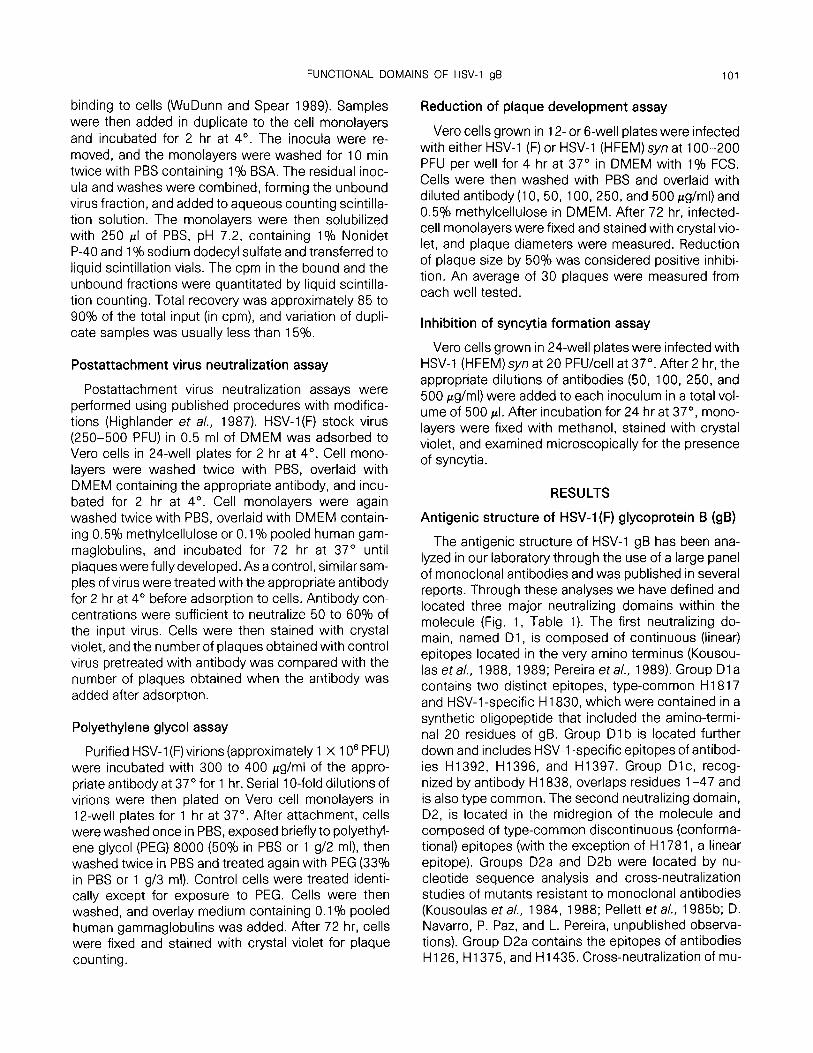
The antigenic structure of HSV-1 gB has been ana- lyzed in our laboratory through the use of a large panel of monoclonal antibodies and was published in several reports. Through these analyses we have defined and located three major neutralizing domains within the molecule (Fig. 1, Table 1). The first neutralizing do- main, named Dl, is composed of continuous (linear) epitopes located in the very amino terminus (Kousou- las et a/., 1988, 1989; Pereira et al., 1989). Group Dl a contains two distinct epitopes, type-common H 18 17 and HSV-1 -specific H 1830, which were contained in a synthetic oligopeptide that included the amino-termi- nal 20 residues of gB. Group Dl b is located further down and includes HSV-1 -specific epitopes of antibod- ies H1392, H1396, and H1397. Group Dlc, recog- nized by antibody H 1838, overlaps residues l-47 and is also type common. The second neutralizing domain, D2, is located in the midregion of the molecule and composed of type-common discontinuous (conforma- tional) epitopes (with the exception of H 1781, a linear epitope). Groups D2a and D2b were located by nu- cleotide sequence analysis and cross-neutralization studies of mutants resistant to monoclonal antibodies (Kousoulas et a/., 1984, 1988; Pellett et al., 1985b; D. Navarro, P. Paz, and L. Pereira, unpublished observa- tions). Group D2a contains the epitopes of antibodies H 126, H1375, and H1435. Cross-neutralization of mu-

102 NAVARRO, PAZ, AND PEREIRA
FIG. 1. Topographic map of the antigenic and functronal domarns on HSV-1 gf3 modified from Qadn et al. (1991). Domains on gB are designated by filled ellipses on the linear diagram of the glycoprotein. Amino acid numbers are shown below the diagram. Epitopes within each domain are listed in boxes. Location of the transmembrane (TM) domain. Enclosed boxes indicate the epitopes formed on gB dimers.
tant R126 showed that epitope H 1815 partially over- laps that of H 126 and also clusters in group D2a. Epi- topes in group D2b, determined by cross-neutraliza-
tion of mutants R233 and R126, include epitope H1819, which partially overlaps H233 and, to a lesser degree, H 126. Residues critical to the assembly of
TABLE 1
PROPERTIES OF NEUTRALIZING MONOCLONAL ANTIBODIES TO HSV-1 gB
Antigenic domain’ Antibody lsotype6
Serotype specificityC
Neutd fl- c
Epitope locatiot?
Dla
Dlb
TC TS TS
-
+ +
Dlc D2a
H1817 H1830 H1392 H1396 H1397 H1838 H126
IgGl IgG2a lgG2a IgGl IgGl IgGl lg2b
+ + - -
l-20 aa l-20 aa
Ab 16-50 aa
Thr,, l-47 aa
Ask
D2b
D2c D5a
H1375 H1435 H1815 H233 H1819 H1781 H1695 H420 H1373
IgGl IgGl IgG2a IgG2a lgG2a IgGl IgG2a IgGl IgG2a
TS TS TC TC TC TC TC TC TC TC TC TC TC
- -
263-283 aa’ His 298 273-298 aag 440-457 aa 600-690 aa 600-690 aa 600-690 aa
a Di and D2c epitopes are continuous; D2a. D2b. and D5a epitopes are discontinuous. * Determined by radial immunodiffusion. c TC (type common), TS (type specific). d Neutralization with (+) or without (-) complement (C’). e Epitopes mapped previously (Pellett et al., 1985; Kousoulas et al., 1988; Kousoulas et a/., 1989; Pereira et al., 1989; Qadri et al., 1991).
Cross-neutralization studies of antibody-resistant mutant viruses R126 and R233 (Kousoulas et a/., 1984; Pellett et al., 1985) showed that ‘epitope H1815 partially overlaps amino acid 273 and that gepitope H1819 partially overlaps amino acid 298 and, to a lesser degree, amino acid 273.

FUNCTIONAL DOMAINS OF HSV-1 gB 103
TABLE 2
TITERS OF COMPLEMENT-INDEPENDENT NEUTRALIZING ANTIBODIES TO HSV-1 gB IN PLAQUE REDUCTION ASSAYS
Monoclonal Concentratior? (pg/ml) required antibody for 50% reduction in no. of plaques
H1817 1.6 H1838 40.0 H126 0.4 H1375 3.2 H1435 1.6 H1815 8.0 H233 0.8 H1819 8.0 H1781 160.0 H1695 80.0
’ Concentration of immunoaffinity-purified antibodies required to neutralize 50% of the viral input (250-500 PFU) of HSV-1 (F). Titers are average of three experiments.
some epitopes in domain D2 map between amino acids 273 and 298 on gB monomers comprising the amino-terminal 457 residues (Pereira et a/., 1989; Qa- dri eT al., 1991). The third neutralizing domain, D5a, is composed of conformational epitopes that were as- sembled in a gB derivative 690 amino acids long, which was dimeric in conformation (Qadri era/., 1991).
Monoclonal antibodies to gB that exhibit complement-independent neutralizing activity
In the first set of experiments, we determined whether complement was required for neutralization of HSV-1 (F) by the panel of 16 monoclonal antibodies to gB. Our hypothesis was that epitopes recognized by complement-independent neutralizing antibodies are likely to be located in regions of gB that mediate virion penetration into cells. Neutralizing activity was ana- lyzed in the presence or absence of 10% normal guinea pig serum as a source of complement. Affinity- purified antibodies were used so that the specific activ- ity (neutralization titer) of each antibody could be accu- rately determined. We found that 10 of 16 antibodies showed significant neutralizing activity in the absence of complement with titers ranging from 0.4 to 160 pg/ ml, the concentration required for greater than 50% plaque reduction (Tables 1 and 2). Epitopes recog- nized by the complement-independent neutralizing an- tibodies were located in three different regions of the molecule. Most of the antibodies with the highest neu- tralizing activity, H126, H1375, H1435, H1815, H233, and H1819, mapped in domain D2. Antibodies H1817 and H1838 to domain Dl and antibody H1695 to do- main D5a also displayed complement-independent
neutralizing activity. Most of these antibodies to HSV- 1 (F) neutralized strain HFEM syn with comparable effi- ciency; the exception, H 1695, required a higher con- centration of antibody to neutralize HFEM (200 /*g/ml). With the exception of H420 and H 1373, most antibod- ies recognizing type-common epitopes displayed com- plement-independent neutralizing activity. Type-spe- cific antibodies H1392, H1396, H1397, and H1830 neutralized virus only when complement was added to the reaction mixture and high amounts of antibodies (approximately 200 pg/ml) were used. As expected, H 1392, H 1396, and H 1397 failed to neutralize HFEM syn, in agreement with our previous finding that these antibodies do not react with HFEM in immunoblots of infected cell extracts (Chapsal and Pereira, 1988).
Complement-independent neutralizing antibodies to gB have no effect on virion attachment to cell surfaces
The next set of experiments was designed to test whether the 10 complement-independent neutralizing antibodies to different domains on gB prevent attach- ment of virions to cells. For this analysis, purified radio- labeled HSV-l(F) virions at a concentration below the estimated saturable limits were mixed with antibodies prior to plating onto cells. Affinity-purified antibodies were used at concentrations ranging from 50 to 500 pg/ml, which was in excess of the amount needed for neutralization. Virus and antibody mixtures were then plated on Vero cells at 4”, a temperature at which viri- ons attach to the cell surface but do not penetrate. The relative inhibition of virus attachment after antibody treatment was determined by comparison with attach- ment of untreated virus. Virions treated with heparan sulfate (100 pg/ml) were used as a positive control to demonstrate blocking of virus attachment to the cell surface. Antibody H1727, which reacts with gB but has no neutralizing activity, was a control for nonspe- cific interference with attachment. Our results showed that none of the antibodies inhibited viral attachment (Table 3). Heparan sulfate-treated virions attached poorly to the cell surface (approximately 20°b of the control), demonstrating that the interaction between HSV-1 virions and the surface of cells was a specific one. Results of these experiments indicate that the neutralizing activity of the antibodies tested does not involve inhibition of virion attachment to cells.
Complement-independent neutralizing antibodies block penetration of virions into cells
Results obtained in the experiments described above indicated that the complement-independent neutralizing antibodies failed to block attachment and

104 NAVARRO, PAZ, AND PEREfRA
TABLE 3
EFFECT OF COMPLEMENT-INDEPENDENT MONOCLONAL ANTIBODIES TO
HSV-1 gB ON ATFACHMENT OF RADIOLABELED HSV-l(F) VIRIONS TO
CELLS
Monoclonal antibody
Percentage of control attachmenta at Ab cont. (pglml)
50 250 500
H1817 98.8 116.4 116.8 H1838 80.0 93.0 116.0
H126 102.3 102.5 113.6
H1375 111.0 113.0 110.0
H1435 110.0 126.0 120.0
H1815 109.0 116.0 115.0 H233 99.1 106.0 119.0 H1819 80.0 94.0 99.0 H1781 90.0 106.4 107.7 H1695 100.4 116.6 108.2 H1727’ 89.5 91.6 112.0
n/ore. Purified radiolabeled HSV-1 virions (5 X 1 O4 PFU) were incu- bated with antibody at 37” for 1 hr and plated on Vero cells (0.5 PFU/cell) at 4’ for 2 hr. Each value IS the average of duplicate sam- ples Radioactivity (cpm) bound to cells usually varied less than 15% between duplicate samples, Radioactivity bound of virus control (no antibody) was 600 cpm (average of 4 samples) and of heparan sul- fate-treated virus (100 pglml of heparan sulfate) was 123 cpm (aver- age of 4 samples). Experiments performed twice gave similar re- sults.
a Calculated as cpm of antibody-treated viruskpm of untreated virus control.
b Nonneutralizing antibody to HSV-1 gB.
suggested that they may block penetration of virions into cells after attachment. To test this hypothesis, we compared the neutralizing activity of antibodies before and after virion attachment to the cell surface by a plaque reduction assay. For these experiments, anti- bodies were used at concentrations that neutralized approximately half of the input virus (Table 2). Our find- ings indicate that the neutralizing antibodies efficiently blocked infectivity, even after virion attachment (Table 4). Antibodies with the highest titers, such as H126 and H233, were most effective in neutralizing attached virus (neutralization ratios of 0.99 and 0.96, respec- tively). Antibodies with low neutralizing activity, such as H 1781, were less effective after attachment (neutral- ization ratio of 0.53). These results showed that the complement-independent neutralizing antibodies to HSV-1 gB inhibit a step following virion attachment, most likely penetration or fusion of the virion envelope with the cell membrane.
Studies on HSV-(tsB5), a temperature-sensitive mu- tant in gB, have shown that the fusogenic agent poly- ethylene glycol (PEG) promotes entry of virions made at nonpermissive temperature that attached to cells but
failed to enter (Sarmiento et a/., 1979). It was recently demonstrated that treatment of cells with PEG en- hanced the infectivity of cell-bound virions treated with monoclonal antibodies to gD or gH (Fuller et al., 1989; Fuller and Spear 1987). To determine whether comple- ment-independent neutralizing antibodies to gB inter- fere with fusion between the viral envelope and the cell membrane, we determined whether PEG treatment would promote the infectivity of antibody-treated virus that was attached to cells.
For these experiments, purified HSV-1 (F) virions were mixed with the appropriate antibodies at concen- trations that would neutralize a large fraction of the input virus. We used a higher titer of virus (approxi- mately 106) than was used for plaque neutralization assays, so that reversal of neutralization by exposure to PEG could be easily measured. Virus samples were serially diluted and plated on Vero cells. After attach- ment, the cells were briefly treated with PEG or mock- treated as described under the Materials and Methods section, and plaques were allowed to form. We found that PEG reversed a fraction of the neutralizing activity of antibodies to gB, as indicated by the increase in plaque-forming ability of the cell-bound virus after PEG treatment (Table 5). The degree to which PEG en- hanced plaque formation varied among the different antibodies. The maximal increase (approximately 7-to lo-fold) was observed for most of the antibodies to epitopes in domain 02. A low-level but reproducible
TABLE 4
COMPARISON OF THE NEUTRALIZING ACTIVITY OF COMPLEMENT-
INDEPENDENT ANTIBODIES TO HSV-1 gB BEFORE AND AFTER VIRUS AT-
TACHMENT
Percent neutralizationa
Antigenic domain
Monoclonal antibody
Before attachment
After attachment Ratiob
Dla Dlc D2a
D2b
D2c D5a
H1817 68.2 67.0 0.98 H1838 66.1 53.2 0.80 H126 66.2 66.0 0.99 H1375 71.2 69.2 0.97 H1435 50.1 45.7 0.91 H1815 49.6 41.9 0.84 H233 67.8 65.2 0.96 H1819 50.2 39.2 0.78 H1781 64.5 34.4 0.53 H1695 66.7 55.5 0.85
’ Percentage neutralization of HSV-1 (F) (250-500 PFU) before or after attachment of virus to Vero cells. Antibodies were added at concentrations required to neutralize approximately 50 to 60% of the viral input. Numbers are average of duplicate samples. Experiments performed twice gave similar results.
’ Ratio of virus neutralization (after/before) attachment.

FUNCTIONAL DOMAINS OF HSV-1 gB 105
TABLE 5
EFFECTOFPOLYETHYLENEGLYCOLONTHEINFECTIVIP~OFHSV-~(F)VIRI- ONS TREATED WITH COMPLEMENT-INDEPENDENT NEUTRALIZING ANTIBOD- IES TO HSV-1 gB
Monoclonal antibody
HSV-1 titers (PFU/ml)
PEG + PEG -
H1817 1.5 x lo4 6.5 x lo3 H1838 9.2 x lo5 3.7 x lo5 H126 1.9 x lo4 1.4 x lo3 H1375 3.5 x lo4 4.0 x lo3 H1435 1.1 x lo4 1.8 x lo3 H1815 4.1 x lo4 1.0 x lo4 H233 3.3 x lo4 5.1 x lo3 H1819 1.0 x lo5 2.0 x lo4 H1781 8.0 X lo5 4.0 x lo5 H1695 6.3 x lo4 4.2 X lo4 H1727a 1.1 x lo6 1.2 x lo6 Control 1.0 x lo6 1.1 x lo6
Note. Purified HSV-1 (F) virions (approximately 1 X 1 06) were mixed with antlbodies (400 pg/ml) or not (Control). Tenfold dilutions of the samples were plated on Vero cells, incubated at 37“ for 1 hr. then treated with PEG (+) or mock-treated (-). Residual infectivity was determined by enumeration of plaques. Experiments performed three times gave similar results.
a Nonneutralrzing antibody to HSV-1 gB.
increase in titer (approximately twofold) was observed for antibodies to domains Dl and D5a. These experi- ments showed that PEG treatment of neutralized virus enhances plaque formation, further indicating that complement-independent neutralizing antibodies to gB interfere with penetration or fusion with the cell membrane. Of the three functional domains, D2 ap- pears to play a dominant role in this process.
Monoclonal antibodies to different domains inhibit plaque development
It is generally agreed that infectious foci (plaques) develop when HSV-1 infection spreads from infected to neighboring uninfected cells. The next set of experi- ments was designed to test whether neutralizing anti- bodies to different domains on HSV-1 gB would inter- fere with plaque development when added to cultures of already infected cells. For this analysis, Vero cells were infected with HSV-1 (F), different concentrations of antibodies were added, plaques were allowed to de- velop, and the diameter of the infectious foci was mea- sured. Results of these experiments (Fig. 2 and Table 6) are as follows.
(i) Most of the monoclonal antibodies (10 of 16) in- terfered with plaque development to varying degrees. All three major neutralizing domains-Dl, D2, and D5a-were represented among these antibodies.
Some correlation was observed between complement- independent neutralizing activity and the degree of in- terference with plaque development. The most potent neutralizing antibodies in domain D2, H 126, H233, and H1435, completely blocked the development of plaques at concentrations ranging between 250 and 500 pg/ml. Microscopic examination of these cultures revealed the presence of very small foci of infected cells, most likely abortive plaques, which resemble the foci observed with neutralizing antibodies to gH (Buck- master et a/., 1984). Antibodies to gB that limited the development of plaques by strain F also limited plaque development of HFEM syn, with the exception of anti- bodies H 1392, H 1396, and H 1397, which recognize domain Dl b. We reported elsewhere that these anti- bodies react with different epitopes in the first 50 resi- dues of HSV-1 gB (Kousoulas et a/., 1988; Pereira et al., 1989) but do not recognize strains HFEM (Chap- sal and Pereira, 1988) or KOS (Kousoulas et a/., 1988). Complement-independent neutralizing antibody H 1695, whose epitope is assembled on the dimer-de- pendent domain D5a, also inhibited plaque develop- ment to a significant degree.
(ii) Threecomplement-independent neutralizinganti- bodies, H1838 to domain Dl c, H 1815 to domain D2a, and H1781 to domain D2c, which had effectively blocked virion penetration into cells (see Tables 3 and 4) failed to inhibit plaque development even when pres- ent in excess of the concentration required for virus neutralization.
(iii) Three of the complement-dependent neutraliz- ing antibodies, H1392, H1396, and H1397, inhibited plaque development by HSV-l(F) in the absence of complement. This finding suggests that inhibition of cell-to-cell spread did not result from neutralization of virus released into the medium, but from blocking a function of gB which did not require complement.
Monoclonal antibodies to gB that inhibit cell fusion by HSV-1 (HFEM) syn
We next conducted experiments to determine whether neutralizing antibodies to HSV-1 gB would prevent cell fusion induced by HSV-1 (HFEM) syn. Vero cells infected with 20 PFU/cell were treated with differ- ent concentrations of antibodies in the overlay me- dium. After 24 hr, monolayers were stained with crystal violet and the plates were examined microscopically for inhibition of cell fusion. The results are summarized in Table 6 and representative examples are shown in Fig. 3. In the absence of neutralizing antibody, or in the presence of certain neutralizing antibodies to gB, the monolayers were extensively fused. However, 8 out of 16 neutralizing antibodies whose epitopes map in do-

106 NAVARRO, PAZ, AND PERElRA
50 Pg 256 i.4 10 Pg 250 k4
FIG. 2. Effect of neutralizing monoclonal antibodres to HSV-1 gB on plaque development. Vero ceils were Infected wrth HSV-l(F) at 100 to 200 PFU/well. After 4 hr, cells were overlaid with antrbodies in DMEM containrng 0.5% methylcellulose. Plaques were allowed to develop and the diameters were measured. Antibodies in the overlay are shown to the left and to the rrght of each row of three wells. Concentrations of antibodies are indicated at bottom. Representative antibodies are shown from each domain. (A) Domains Dla, Dl b, and D2a. (6) Domains D2b. D2c, D5a. Negative antibody (H1727).
mains Dl , D2, and D5a strongly suppressed cell fusion at concentrations ranging from 100 to 500 pg/ml. The complement-independent antibodies with the highest anti-fusion activity, H 126, H 1435, H 1815, H233, and H1819, reacted with domain D2. These were also the most effective in reducing the plaque number and limit- ing plaque development (Tables 3 and 6). We found a direct correlation between complement-independent neutralization and the ability to suppress fusion, al- though the latter effect required higher amounts of anti- bodies than the former. One complement-dependent neutralizing antibody to domain Dl , HI 830, inhibited
fusion but to a much lesser extent than any of the com- plement-independent antibodies. The complement- dependent neutralizing antibodies to domain D5a, which neutralize strain HFEM syn, failed to inhibit cell fusion.
DISCUSSION
In this study, we conducted an in-depth analysis of the neutralizing epitopes on HSV-1 gB to correlate the antigenic structure of this glycoprotein with its function in virion penetration, cell-to-cell spread, and ceil fusion

FUNCTIONAL DOMAINS OF HSV-1 gB 107
TABLE 6
EFFECTOF NEUTRALIZINGANTIBODIESTO HSV-1 gB ON PLAQUE DEVELOPMENTANDCELLFUSION
Plaque development by Cell fusion by HSV- HSV-1 (F) 1 (HFEM)synb
Monoclonal Minimal Ab Minimal Ab antibody Inhibition cont. (pg/ml) Inhibition cont. (pg/ml)
H1817 + 100 + 100 H1830 - f 500 H1392* + 250 -
H1396* + 500 -
H1397* + 100 -
H1838 - -
H126 + 50 + 100 H1375 + 500 + 250 H1435 + 250 + 100 H1815 - + 100 H233 + 100 + 100 H1819 + 500 + 100 H1781 - -
H1695 + 250 + 250 H420 - -
H1373 - -
a Experiments on inhibition of plaque development were done with HSV-1 strains F and HFEM syn. Antibodies were tested at concen- trations of 10, 50, 100, 250, and 500 pglml. Experiments were per- formed 3 times with similar results.
b Experiments on inhibition of cell fusion were carried out with strain HFEM syn and were performed three times with similar re- sults.
(+) Reduction of at least 50% of the plaque size or complete inhibi- tion of syncytial development. An average of 10 plaques was mea- sured per sample.
(-) No effect on plaque development or cell fusion. (+) Small but measurable reduction of plaque size or cell fusion. (*) Antibodies that fail to react with HFEM (Chapsal and Pereira,
1988).
We previously reported the mapping of epitopes recog- nized by a large panel of monoclonal antibodies to HSV-1 gB using a variety of strategies (Kousoulas eta/., 1988; Pellett eta/., 1985b; Pereira eta/., 1989; Qadri et a/., 1991). Our findings are summarized in a topo- graphic map of the molecule that defines three major neutralizing domains, Dl, D2, and D5a (Fig. 1). De- tailed investigation of the biological properties of 16 neutralizing antibodies showed that each domain func- tions in infectivity and that domain D2, which is recog- nized by most of the complement-independent neutral- izing antibodies in the panel, plays a predominant role in this process.
Penetration of virus into cells
Evidence from this study and others indicates that the mechanism by which the complement-indepen-
dent neutralizing antibodies to HSV-1 gB, gD, and gH block infection involves interference with virion pene- tration into cells. First, complement-independent neu- tralizing antibodies to gB had no effect on virus attach- ment to the cell membrane, even at concentrations in excess of that required for neutralization (Table 2). These results are consistent with studies on HSV-1 glycoproteins gD, gH (Fuller et a/., 1989; Fuller and Spear 1987; Gompels and Minson, 1986; Highlander et al., 1987) and gB (Highlander et al., 1988), further supporting the view that gB is not absolutely required for attachment of virions to the cell surface. Second, complement-independent neutralizing antibodies that recognized different domains on gB prevented virus from entering cells, even after it had attached to the cell surface. A similar finding was reported for another neutralizing antibody to gB (Highlander et al,, 1988). Third, polyethylene glycol (PEG), a membrane fuso- genie agent, increased the ability of antibody-treated virions that were already bound to the cell surface to infect cells. Experiments with PEG indicated that neu- tralizing antibodies to gD and gH also act at the level of virion entry into cells (Fuller et al., 1989; Fuller and Spear, 1987). In these studies, PEG enhanced the in- fectivity of virus treated with neutralizing antibodies to gD and gH to a greater degree than we showed for gB. This could be due to differences in experimental condi- tions, particularly the binding affinity of the antibodies, the concentration of PEG, and the cell type used in the assay. Taken together, our results strongly indicate that neutralizing antibodies to different domains on gB interfere with fusion of the virion envelope to the cell membrane, an event that is required for viral penetra- tion, and that this block in infectivity is reversible by use of a membrane fusogen.
For several reasons, it is not easily determined whether discontinuous epitopes on gB recognized by neutralizing antibodies overlap the site that functions in virion penetration, are adjacent, or are distant from it. If neutralizing antibodies bind to an epitope adjacent to the functional site, they may prevent penetration by steric hindrance of receptor binding. Studies on the attachment protein of rhinoviruses indicate that neutral- izing antibodies bind to the rim of the receptor-binding canyon on the molecule, thus blocking access to the cell-surface receptor (Colonno et a/., 1988). Alterna- tively, antibody binding may alter the conformation of a molecule so that the actual functional site, which may be located distal from the antibody binding site, is less accessible to the cell surface receptor. For the com- plement-independent neutralizing antibodies to do- main D2, we found a good correlation between the neutralization titer, their ability to block infectivity after virion attachment to cells, and an increase in the

NAVARRO, PAZ, AND PEREIRA
t
H233
FIG. 3. Effect of monoclonal antibodres to HSV-1 gB on cell-to-cell fusion induced by HSV-l(HFEM) syn. Vero cells were infected at 20 PFWcell. Antrbodies were added to the cells 2 hr after adsorption, and cell monolayers were examined 24 hr later for inhibition of syncytia formation. Virus control (no antibody); complement-independent neutralizrng antibody to domain D5a, H420 (500 pglml); complement-indepen- dent neutralizing antibodies to domain D2a, H126 (100 pg/ml); and to domain D2b. H233 (100 @g/ml).
plaque-forming efficiency of antibody-treated virions after PEG treatment. Moreover, mutants resistant to neutralization by most of these antibodies have single amino acid changes mapping between residues 274 and 298 (Kousoulas et al., 1984, 1988; Pellett et al., 1985b). The results of the present study and the loca- tion of the amino acid changes which confer neutraliza- tion resistance strongly indicate that epitopes on gB recognized by these antibodies either overlap or are proximal to the functional domain of the molecule.
The epitopes recognized by our panel of monoclonal antibodies that block penetration cluster into domains Dl, D2, and D5a (Pereira et a/., 1989; Qadri er a/., 1991). D2 is a conformation-dependent domain, which contains epitopes of potent complement-independent neutralizing antibodies. This domain is proximal to anti- genie site III on strain KOS, which was mapped by an amino acid change at position 305 in a mutant that entered cells more slowly (Highlander et a/., 1989; Marlin et a/., 1986). Domain D2 on strain F (Pereira et
a/., 1989, Qadri et al., 1991) encompasses antigenic site I on strain KOS, which is recognized by antibodies that slow the rate of entry (Highlander et a/., 1988). Domain Dl maps at the amino terminus of HSV-1 (F) gB (Kousoulas et a/., 1988; Pereira et al., 1989). Antibod- ies H 1817 and H 1838 to Dl a and Dl c display comple- ment-independent neutralizing activity and block pene- tration. Epitopes in domain Dl b overlap antigenic site IV of strain KOS gB (Highlander eta/., 1988). We previ- ously reported that antibodies H1392, H1396, and H1397 to epitopes in domain Dl b fail to recognize strain KOS due to different amino acids in this region (Kousoulas et a/., 1988). The finding that two HSV-1 strains have evolved penetration functions at the amino terminus of gB underscores the importance of this region in infectivity. In domain D5a, antibody H 1695, which recognizes a dimer-dependent discon- tinuous epitope (Qadri et a/., 1991) blocked penetra- tion, although only at high concentrations. KOS mu- tants in antigenic site II, which overlaps domain D5a,

FUNCTIONAL DOMAINS OF HSV-1 gB 109
have altered rates of entry (Highlander et al., 1988; Highlander et al., 1989; Marlin et a/., 1986). Linker-in- sertions in this region of strain KOS gB render the mole- cule nonfunctional with respect to the activities re- quired for infectivity (Cai et a/., 1987). In summary, the results of our analysis of the biological activity of a panel of neutralizing antibodies to different epitopes indicate that several regions on the extracellular do- main contribute to virion penetration and suggest that they may act coordinately.
It is possible that each of the HSV-1 virus glycopro- teins that is required for infection in cell culture, and also those that are not essential, interacts indepen- dently with a receptor on the cell surface. Two recent studies support this view. First, in polarized cells that are infected with HSV-1, two asymmetrically distrib- uted cell receptors interact with different viral glyco- proteins (Sears et a/., 1991). gC is involved with attach- ment to the receptor on the apical cell surface. Sec- ond, studies with soluble forms of HSV-1 gD indicate that a saturable receptor exists for this glycoprotein (Johnson et a/., 1990; Johnson and Spear 1989). How- ever, it has also been proposed that the binding of viri- ons to a cell-surface receptor may involve the forma- tion of a glycoprotein complex (Ruyechan et al., 1979). At the present time, however, the cell surface receptor for HSV-1 remains unknown (Mirda et al,, 1991). By analogy, it was demonstrated that immunoglobulin Fc receptor activity of HSV-1 depends on a complex of two viral glycoproteins, gE and gl (Johnson and Feen- stra, 1987; Johnson et al., 1988). Although each of these glycoproteins has some Fc-binding activity, effi- cient binding occurs only when both glycoproteins are expressed (Hanke et al., 1990). It is possible that other HSV-1 glycoproteins form a functional complex after virion attachment to the cell surface. If such a complex were formed, then the neutralizing activity of some of the antibodies that prevent penetration may not di- rectly involve occupation of the functional site of each of the glycoproteins, but rather may involve interfer- ence with their complex formation by occupation of their cognitive sites for each other.
Cell-to-cell spread of virus
We evaluated the role of HSV-1 gB in cell-to-cell spread of virus by analyzing the effect of neutralizing antibodies in the panel on the development of plaques by HSV-1 strains HFEM syn and F after infection. Sev- eral laboratories have reported that neutralizing anti- bodies to gB, gD, and gH interfere with plaque develop- ment (Buckmaster et a/., 1984; Highlander et al., 1987, 1988; Minson et a/., 1986). In these studies, it was proposed that spread of infection to uninfected cells
does not depend on release of virus but rather on the biological activities of HSV-1 glycoproteins in the cell membrane of infected cells. This conclusion was based on the finding that three complement-indepen- dent neutralizing antibodies (H1838, H1815, and H1781) were unable to inhibit plaque development by HSV-1 (F), whereas those that required complement (H1392, H1396, and H1397) were able to do so.
In our study, we found that 10 of 16 antibodies with complement-dependent and -independent neutralizing activity interfered with plaque development. With one exception (H1815), those recognizing domain D2 blocked plaque development almost completely. All three major neutralizing domains-Dl, D2, and D5a -were represented among these antibodies (Kousou- las et al., 1988; Pellett et a/., 1985b; Pereira et a/., 1989; Qadri eta/., 1991) indicating that more than one domain on gB functions in promoting spread of virus from infected to uninfected cells. When comparing the capacity of neutralizing antibodies to gB to reduce plaque number with their capacity to limit plaque devel- opment, we found that whereas small amounts of the potent antibodies neutralized virus (Table 2) much larger amounts were required to inhibit plaque develop- ment (Table 6). The level of antibody required for this activity suggests that the high level of gB expressed in the plasma membrane is necessary for plaque spread and that gB is an essential component in promoting cell-to-cell transmission of infection. Recent analysis of antibody-resistant mutant viruses with amino acid changes in domain D2 of gB showed that the mutants enter cells but that their plaque development at 39” is severely limited (D. Navarro, P. Paz, I. Qadri, and L. Pereira, manuscript in preparation). These results con- firm that HSV-1 gB in the plasma membrane of infected cells has the potential to promote virus spread and are consistent with the results of studies on gD and gH which indicate that virion release is not required for the infection of neighboring cells.
Virus-induced cell-to-cell fusion
Our finding that antibodies to three antigenic do- mains on HSV-1 gB suppress cell-to-cell fusion in- duced by HSV-1 strain HFEM syn constitutes the first such report and directly implicates gB in the fusion process. Mutations within the cy-toplasmic tail of HSV- 1 gB have been identified as responsible for the syncy- tial phenotype (Bzik et a/., 1984a). However, syn muta- tions map in genes encoding other HSV-1 glycopro- teins and membrane-associated proteins (Debroy et al., 1985; Hutchinson et al., 1991; Ruyechan et al., 1979) and it has been proposed that several of the envelope glycoproteins may form a functional complex

110 NAVARRO. PAZ, AND PEREIRA
and that mutations in any one of them would yield the syncytial phenotype (Ruyechan et al., 1979).
Studies from several laboratories indicate that HSV- 1 gD and gH may also be involved in syncytia forma- tion. First, virosomes comprised of several HSV-1 gly- coproteins appear to fuse with cells (Johnson et al., 1984) and cell lines constitutively expressing gD can be induced to form polykaryocytes (Campadelli-Fiume eta/., 1988). Second, monoclonal antibodies to gD and gH were reported to prevent cell-to-cell fusion induced by several syncytial strains of HSV-1 (Fuller et al., 1989; Fuller and Spear 1987; Highlander et al., 1987). It has not yet been established whether fusion between the viral envelope and the cell membrane of a susceptible cell is similar to the membrane-related events leading to the extensive cell fusion induced by syncytial strains of HSV-1. In the present study, we identified several antibodies that dramatically suppressed the syncytia formation induced by HSV-1 (HFEM)syn. A direct corre- lation was found between neutralizing activity and the ability to prevent fusion of infected cells, suggesting that these processes are similar and involve the same functional domains on gB. The gB epitopes involved in fusion activity map within all three major neutralizing domains, Dl, D2, and D5a. Insofar as antibodies with anti-fusion activity recognize the extracellular domain of the gB molecule, it appears that this domain is active in the fusion event. Our findings support the studies on linker insertion mutants in gB showing that insertions in the ectodomain strongly prevented syncytia formation in a complementation assay (Cai et al., 198813). If gB forms a structural complex with other HSV-1 glycopro- teins in the virion envelope and the plasma membrane of infected cells, then contacts at the carboxy terminus may stabilize this structure. Assuming that this com- plex also functions in fusion, it is likely that certain re- gions of the extracellular domains of the glycoproteins contact each other transiently in the fusion process. Thus, both mutations in the carboxy terminus that per- turb the interactions of the glycoproteins with each other and antibodies to the extracellular domain that block their functional association may inhibit fusion. Our studies revealed that antibodies blocking plaque development and those blocking cell fusion recognize overlapping domains on gB. However, the finding that not all antibodies which limit cell fusion have the capac- ity to block plaque development suggests that these processes may be related but are not identical. Experi- ments are in progress to select recombinants with conditional-lethal mutations in functional domains on gB. Following penetration, such mutants should lack the ability to infect neighboring cells or to produce nor- mal plaques at the nonpermissive temperature.
ACKNOWLEDGMENTS
These studies were supported by Public Health Service Grants DE-08275 from the National Institutes of Dental Research, Al-30873 and Al-23592 from the National Institute of Allergy and Infectious Diseases. D.N. was supported by a fellowship from the Spanish Min- istry of Education and Science.
REFERENCES
BUCKMASTER, E. A., GOMPELS, U., and MINSON, A. (1984). Character- ization and physical mapping of an HSV-1 glycoprotein of approxi- mately 115 X 1 O3 molecular weight. virology 139, 408-413.
BZIK, D. J., DEBROY, C., Fox, B. A., PEDERSON, N. E., and PERSON, S. (1986). The nucleotide sequence of the gB glycoprotein gene of HSV-2 and comparison with the corresponding gene of HSV-1. Virology 155, 322-333.
BZIK, D. J., Fox, B. A., DELUCA, N. A., and PERSON, S. (1984a). Nu- cleotide sequence of a region of the herpes simplex virus type 1 gB glycoprotein gene: Mutations affecting rate of virus entry and cell fusion. Virology 137, 185-l 90.
BZIK. D. J., Fox, B. A., DELUCA, N. A., and PERSON, S. (1984b). Nu- cleotide sequence specifying the glycoprotein gene, gB, of herpes simplex virus type 1. Virology 133, 301-314.
CAI, W., Gu, B., and PERSON, S. (1988a). Role of glycoprotein B of herpes simplex virus type 1 in viral entry and cell fusion. J. viral. 62, 2596-2604.
CAI, W., PERSON, S., DEBROY, C.. and Gu, B. (1988b). Functional re- gions and structural features of the gB glycoprotein of herpes sim- plex virus type 1. J. MO/. Biol. 201, 575-588.
CAI, W., PERSON, S., WARNER, S. C., ZHOU. J., and DELUCA, N. A. (1987). Linker-insertion nonsense and restriction-site deletion mu- tations of the gB glycoprotein gene of herpes simplex virus type 1. J. Viral. 61, 714-721.
CAMPADELLI-FIUME, G., AVITABILE. E.. FINI. S.. STIRPE, D., ARSENAKIS, M., and ROIZMAN, B. (1988). Herpes simplexvirus glycoprotein D is sufficient to induce spontaneous pHindependent fusion in a cell line that constitutively expresses the glycoprotein. Virology 166, 598-602.
CHAPSAL, J. M., and PEREIRA, L. (1988). Characterization of epitopes on native and denatured forms of herpes simplex virus glycopro- tein B. Virology 164, 427-434.
CLAESSON-WELSH, L., and SPEAR, P. G. (1986). Oligomerization of herpes simplex virus glycoprotein B. /. Viral 60, 803-806.
COLONNO, R. J., CONDFIA, J. H., MIZUTANI, S., CALLAHAN, P. L., DAVIES, M. E., and MUREKO, M. A. (1988). Evidence for the direct involve- ment of the rhinovirus canyon in receptor binding. Proc. Nat/. Acad. Sci. USA 85, 5449-5453.
CONRATHS, F., ACKERMANN, M., LUDWIG, H., PAULI, G., and PEREIRA, L. (1987). Conserved antigenic and functional domains on glycopro- tein B of herpes simplex virus 1 and bovine herpesvirus 2. Arch. Viral. 96, 309-318.
CRANAGE, M. P., KOUSARIDES, T., BANKIER, A. T., SATCHWELL, S., WES- TON, K., TOMLINSON, P., BARRELL, B., HART, H., BELL, S. E., MINSON, A. C., and SMITH, G. L. (1986). Identification of the human cytomeg- alovirus glycoprotein B gene and induction of neutralizing antibod- ies via its expression in recombinant vaccinia virus. EMBO 1. 5, 3057-3063.
DAVIDSON, A. J., and Scorr, 1. E. (1986). The complete DNA se- quence of varicella-zoster virus. /. Gen. Viral. 67, 1759-l 816.
DEBROY, C., PEDERSON, N.. and PERSON, S. (1985). Nucleotide se- quence of a herpes simplex virus type 1 gene that causes cell fusion. virology 145, 36-48.

FUNCTIONAL DOMAINS OF HSV-1 gB 111
DESAI, P. J.. SCHAFFER, P. A., and MINSON, A. C. (1988). Excretion of non-infectious virus particles lacking glycoprotein H by a tempera- ture-sensitive mutant of herpes simplex virus type 1: Evidence that gH is essential for virion infectivity. 1. Gen. Viol. 69, 1147-1 158.
EJERCITO, P. M., KIEFF, E. D., and ROIZMAN, B. (1968). Characteriza- tion of herpes simplex virus strains differing in their effect on social behavior of infected cells. J. Gen. I/ire/. 3, 357-364.
FULLER, A. 0.. SANTOS, R. E., and SPEAR, P. G. (1989). Neutralizing antibodies specific for glycoprotein H of herpes simplex virus per- mit viral attachment to cells but prevent penetration. /. Viral. 63(8), 3435-3443.
FULLER, A. O., and SPEAR, P. G. (1985). Specificities of monoclonal and polyclonal antibodies that inhibit adsorption of herpes simplex virus to cells and lack of Inhibition by potent neutralizing antibod- ies. J. Viol. 55, 475-482.
FULLER, A. O., and SPEAR, P. G. (1987). Anti-glycoprotein D antibod- ies that permit adsorption but block infection by herpes simplex virus 1 prevent virion-cell fusion at the cell surface. Proc. Nat/. Acad. Sci. USA 84, 5454-5458.
GOMPELS, U., and MINSON, A. (1986). The properties and sequence of glycoprotein H of herpes simplex virus type 1. Virology 153, 230-247.
HAFFEY, M., and SPEAR, P. G. (1980). Alterations in glycoprotein gB specified by mutants and their partial revertants in herpes simplex virus type 1 and relationshlp to other mutant phenotypes. J. Virof. 35, 114-128.
HANKE, T., GRAHAM, F. L., LULITANOND. V., and JOHNSON, D. C. (1990). Herpes slmplexvirus IgG Fc receptors induced using recombinant adenovtrus vectors expressing glycoproteins E and I. Viroiogy 177,437-444.
HEINE, 1. W., HONES% R. W.. CASSAI, E., and ROIZMAN, B. (1974). Proteins specified by herpes simplex virus. XII. The vlnon polypep- trdes of type 1 strains. /. Viral. 14, 640-651.
HIGHLANDER, S. L., CAI, W.. PERSON, S., LEVINE, M., and GLORIOSO, I. C. (1988). Monoclonal antibodies define a domain on herpes simplex virus glycoprotein B involved in virus penetration. J. Viral. 62,1881-1888.
HIGHLANDER, S. L., DORNEY. D. J., GAGE, P. J., HOLLAND, T. C., CAI, W., PERSON, S., LEVINE, M.. and GLORIOSO, J. C. (1989). Identification of mar mutations in herpes simplex virus type 1 glycoprotein B which alter antigenic structure and function in virus penetration. J. Viral. 63,730-738.
HIGHLANDER, S. L., SUTHERLAND, S. L., GAGE, P. J., JOHNSON, D. C.. LEVINE, M., and GLORIOSO, J. C. (1987). Neutralizing monoclonal antibodies specific for herpes simplex virus glycoprotein D inhibit virus penetration. /. Viral. 61, 3356-3364.
HUTCHINSON, L., GOLDSMITH, K., PRIMORAC, S., ROOP, C., GRAHAM, F. L., and JOHNSON, D. C. (1991). Characterization of two novel HSV-1 glycoproteins, gK and gL, involved in membrane fusion. Abst. Internat. Herpesvirus Workshop, Pacific Grove, CA, p. 43.
JOHNSON, D. C., BURKE, R. L., and GREGORY, T. (1990). Soluble forms of herpes simplex virus glycoprotein D bind to a limited number of cell surface receptors and Inhibit virus entry into cells. J. Viral. 64, 2569-2576.
JOHNSON, D. C., and FEENSTRA, V. (1987). Identification of a novel herpes simplex virus type l-Induced glycoprotein which com- plexes with gE and binds immunoglobulin. J. Viol. 61, 2208- 2216.
JOHNSON, D. C., FRAME, M. C., LIGAS, M. W., CROSS, A. M.. and STOW, N. G. (1988). Herpes simplex virus lmmunoglobulin G Fc receptor activity depends on a complex of two viral glycoproteins, gE and gl. J. Viral. 62, 1347-l 354.
JOHNSON, D. C., WITTELS, M., and SPEAR, P. G. (1984). Binding to cells
of virosomes containing herpes simplex virus type 1 glycoproteins and evidence for fusion. J. Viral. 52, 238-247.
JOHNSON, R. M., and SPEAR, P. G. (1989). Herpes slmplexvirus glyco- protein D mediates interference with herpes simplex virus infec- tion. /. viral. 63, 819-827.
Kousoul~s, K.. ARSENAKIS, M., and PEREIRA, L. (1989). A subset of type-specific epitopes map in the amino terminus of herpes sim- plex virus 1 glycoprotein B. J. Gen. t&o/. 70, 735-741.
Kousoul~s. K. G., Hue, B., and PEREIRA, L. (1988). Antibody resistant mutations in cross-reactive and type-specific epitopes of herpes simplex virus 1 glycoprotein B map in separate domains. Virology 166,423-431.
Kousoul~s, K. G., PELLETT, P. E., PEREIRA, L., and ROIZMAN, B. (1984). Mutations affecting conformation or sequence of neutralizing epi- topes identified by reactivity of viable plaques segregated from syn and ts domains of HSV-1 (F) gB gene. Virology 135, 379-394.
LAWRENCE, W. C.. D’URSO. R. C., KUNDEL, C. A., WHITBECK, J. C., and BELLO. L. 1. (1986). Map location of the gene for a 130.000-dalton glycoprotein of bovine herpesvirus 1. /. viral. 60, 405-414.
LIGAS, M. W., and JOHNSON, D. C. (1988). A herpes simplex virus mutant in which glycoprotein D sequences are replaced by P-ga- lactosidase sequences binds to but is unable to penetrate into cells. J. Viral. 62. 1486-l 494.
LITTLE, S. P., JOFRE, J. T., COURTNEY, R. J., and SCHAFFER, P. A. (1981). A virion-associated glycoprotein essential for infectivity of herpes simplex virus type 1. Virology 115, 149-l 60.
LONGNECKER, R., CHAT~ER~EE, S.. WHITLEY, R. J., and ROIZMAN, B. (1987). Identification of a herpes simplex virus 1 glycoprotein gene within a gene cluster dispensable for growth in cell culture. Proc. Nat/ Acad. Sci. USA 84, 4303-4307.
LONGNECKER, R., and ROIZMAN. B. (1987). Clustering of genes dispen- sable for growth in culture in the S component of the HSV-1 ge- nome. Science 236, 573-576.
MANSERVIGI, R.. SPEAR, P. G., and BUCHAN, A. (1977). Cell fusion induced by herpes simplex virus is promoted and suppressed by different viral glycoproteins. Proc. Nat/. Acad. Sci. USA 74, 3913- 3917.
MARLIN, S. D., HIGHLANDER, S. L., HOLLAND, T. C., LEVINE, M.. and GLORIOSO, J. C. (1986). Antigenic variation (mar mutations) in herpes simplex virus glycoprotein B can induce temperature de- pendent alterations in gB processing and virus production. J. Viol. 59,142-153.
MINSON, A. C., HODGMAN, T. C.. DIGARD, P., HANCOCK, D. C., BELL, S. E., and BUCKMASTER, E. A. (1986). An analysis of the biological properties of monoclonal antibodies against glycoprotein D of herpes simplex virus and identification of amino acid substitution that confer resistance to neutralization. /. Gen. viral. 67, lOOl- 1013.
MIRDA, D. P., NAVARRO, D.. PAZ, P., LEE, P. L., PEREIRA, L., and WIL-
LIAMS, L. T. (1991). The fibroblast growth factor receptor is not required for herpes simplex virus type 1 infection.). Virot., in press.
MISRA, V., NELSON, R., and SMITH, M. (1988). Sequence of a bovine herpesvirus type-l glycoprotein gene that is homologous to the herpes simplex gene for the glycoprotein gB. tirology 166, 542- 549.
,
PELLETT, P. E., BIGGIN, M. D., BARRELL. B., and ROIZMAN, B. (1985a). Epstein-Barr virus genome may encode a protein showing signifi- cant amino acid and predicted secondary structure homology with glycoprotein B of herpes simplex virus 1. /. viral. 56, 807-813.
PELLET, P. E., Kousoums, K. G., PEREIRA, L., and ROIZMAN, B. (1985b). Anatomy of the herpes simplex virus 1 strain F glycopro- tein B gene: Primary sequence and predicted protein Structure of

112 NAVARRO, PAZ, AND PEREIRA
the wild type and of monoclonal antibody-resistant mutants. /. Viral. 53, 243-253.
PEREIRA, L., ALI, M., Kousoul~s, K., Huo, B., and BANKS, T. (1989). Domain structureof herpessimplexvirus 1 glycoproteln B: Neutral- izing epitopes map in regions of continuous and discontinuous residues. Virology 172, 1 l-24.
PEREIRA, L., DONDERO. D., GALLO, D., DEVLIN, V., and WOODIE, J. D. (1982). Serological analysis of herpes simplex virus types 1 and 2 with monoclonal antibodies. infect. Immun. 35, 363-367.
PEREIRA, L., DONDERO, D., NORRILD, B., and ROIZMAN, B. (1981). Dif- ferential immunologic and electrophoretic properties of glycopro- teins gA and gB of HSV-2 produced In HEp-2 and Vero cells. Proc. Nat/. Acad. Sci. USA 78, 5202-5206.
PEREIRA, L., KLASSEN, T.. and BARINGER, J. R. (1980). Type common and type specific monoclonal antibody to herpes simplex virus 1. Infect. Immun. 29, 724-732.
QADRI, I., GIMENO. C., NAVARRO, D., and PEREIRA, L. (1991). Mutations in discontinuous domains of herpes simplexvirus 1 glycoprotein B affectthe antigenic properties, dimerization, and cell-surface trans- port of the molecule. virology 180, 135-l 52.
RIGGIO, M. P., CULLINANE, A. A.. and ONIONS, D. E. (1989). Identifica- tion and nucleotide sequence of the glycoprotein gB gene of equine herpesvirus 4. /. Viral. 63, 1 123-l 133.
ROBBINS, A. K., DORNEY, D. S., WATHEN, M. W., WHEALY, M. E., GOLD, C., WATSON, R. J., HOLLAND, L. E., WEED, S. D.. LEVINE, M., GLOR- IOSO. J. C., and ENQUIST, L. W. (1987). The pseudorabiesvirus gll is closely related to the gB glycoprotein gene of Herpes simplex virus. /. !YIro/. 61, 2691-2701.
ROIZMAN. B.. NORRILD, B., CHAN, C., and PEREIRA, L. (1984). Identifica- tion and preliminary mapping with monoclonal antibodies of a herpes simplex 2 glycoprotein lacking a known type 1 counterpart. Virology 133, 242-247.
RUYECHAN. T. W., MORSE, L. S.. KNIPE, D. M., and ROIZMAN, B. (1979). Molecular genetics of herpes simplex virus. II. Mapplng of the major viral glycoproteins and of the genetic loci specifying the social behavior of infected cells. /. Viral. 29, 677-697.
SARMIENTO, M., HAFFEY, M., and SPEAR, P. G. (1979). Membrane pro- teins specified by herpes simplex viruses. Ill. Role of glycoprotein VP7 (B,) In virion infectivity. /. viral. 29, 1 149-l 158.
SARMIENTO, M., and SPEAR, P. G. (1979). Membrane proteins speci- fied by herpes simplex viruses. IV. Conformation of the virion gly- coprotein designated VP7 (BJ, /. Viral. 29, 1 159-l 167.
SEARS, A. E., MCGWIRE. B. S.. and ROIZMAN, B. (1991). Infection of polarized MDCK cells with herpes simplexvirus 1: two asymmetri- cally distributed cell receptors interact with different viral proteins. Proc. Nat/. Acad. Sci. USA 88, 5087-5091.
SPEAR, P. G. (1976). Membrane proteins specified by herpes simplex viruses. I. Identification of four glycoprotein precursors and their products in type 1 -infected cells. /. Viral. 17, 991-l 008.
SPEAR, P. G.. and ROIZMAN, B. (1972). Proteins specified by herpes simplex virus. V. Purification and structural proteins of the herpes- virion. /. Viral. 9, 143-159.
STUVE, L. L., BROWN-SHIMER, S., PACHL. C., NAJARIAN, R., DINA, D., and BURKE, R. L. (1987). Structure and expression of the herpes simplex virus type 2 glycoprotein gB gene. 1. Viral. 61, 326-335.
WEBER. P. C., LEVINE, M., and GLORIOSO, J. C. (1987). Rapid identifica- tion of nonessential genes of herpes simplex virus type 1 by Tn5 mutagenesis. Science 236, 576-579.
WHITBECK. J. C., BELLO, L. J., and LAWRENCE, W. C. (1988). Compari- son of the bovine herpesvirus 1 gl gene and the herpes simplex virus tvoe 1 aB aene. J. Viral. 62. 3319-3327.
WUDUNN,’ D., &d-SPEAR, P. G. (1989). Initial interaction of herpes simplex virus with cells is binding to heparan sulfate. /. Viral. 63, 52-58.