Alteration of ribosomal protein maps in herpes simplex virus type 1 infection
13
771 (2002) 237–249 Journal of Chromatography B, www.elsevier.com / locate / chromb Review Alteration of ribosomal protein maps in herpes simplex virus type 1 infection * ´ Jean-Jacques Diaz , Stephane Giraud, Anna Greco ´ ´ INSERM U369, Faculte de Medecine Lyon-R.T.H. Laennec, 7, Rue Guillaume Paradin, 69372 Lyon Cedex 08, France Abstract At present, the effect of herpes simplex virus infection on the entire proteomes of infected cells is very poorly documented. Following several studies performed over the past few years, the modifications of a sub-cellular fraction induced by herpes simplex virus type 1 can be documented. These studies were performed in order to characterize the virally-induced modifications of a major component of the translational apparatus, the ribosomes. The very basic nature of most of the ribosomal proteins renders them very difficult to separate using isoelectric focusing (IEF). Therefore these studies were achieved using several different but related two-dimensional electrophoretic systems which allowed several two-dimensional ribosomal protein maps to be built. Comparison of the ribosomal protein maps built from non-infected cells with those built from infected cells demonstrated that infection by herpes simplex virus type 1 (HSV-1) induces important modifications of ribosomes: (i) non-reversible phosphorylation of ribosomal protein S6; (ii) unusual phosphorylation of several proteins of the small and the large subunits; and (iii) association of viral and cellular proteins to the ribosomal fraction. An overview of these published studies is presented in this review. 2002 Published by Elsevier Science B.V. Keywords: Reviews; Ribosomal protein maps; Proteomics; Herpes simplex virus; Poly(A)-binding proteins; Viral proteins Contents 1. Introduction ............................................................................................................................................................................ 238 2. Methods of analysis of proteins ................................................................................................................................................ 239 2.1. Analysis of total proteins ................................................................................................................................................. 239 2.1.1. Infection of cells and radioactive labeling of proteins ............................................................................................. 239 2.1.2. Purification of total proteins ................................................................................................................................. 239 2.1.3. Isoelectric focusing for separation of total proteins ................................................................................................ 239 2.2. Analysis of ribosomal proteins ......................................................................................................................................... 240 2.2.1. Purification of ribosomes ..................................................................................................................................... 240 2.2.2. Extraction, alkylation and lyophilization of ribosomal proteins ............................................................................... 240 2.2.3. Separation of ribosomal proteins by 2D-PAGE ...................................................................................................... 240 3. Modifications of ribosomal protein maps after HSV-1 infection .................................................................................................. 241 3.1. Identification of phosphorylated ribosomal proteins ........................................................................................................... 242 3.2. Identification of non-ribosomal proteins associated with ribosomes ..................................................................................... 243 3.3. Functions of phosphorylated ribosomal proteins ................................................................................................................ 245 *Corresponding author. Tel.: 133-4-7877-8717; fax: 133-4-7877-8736. E-mail address: [email protected] (J.-J. Diaz). 1570-0232 / 02 / $ – see front matter 2002 Published by Elsevier Science B.V. PII: S1570-0232(02)00038-7
-
Upload
centreleonberard -
Category
Documents
-
view
1 -
download
0
Transcript of Alteration of ribosomal protein maps in herpes simplex virus type 1 infection
771 (2002) 237–249Journal of Chromatography B,www.elsevier.com/ locate /chromb
Review
Alteration of ribosomal protein maps in herpes simplex virus type 1infection
* ´Jean-Jacques Diaz , Stephane Giraud, Anna Greco´ ´INSERM U369, Faculte de Medecine Lyon-R.T.H. Laennec, 7, Rue Guillaume Paradin, 69372 Lyon Cedex 08, France
Abstract
At present, the effect of herpes simplex virus infection on the entire proteomes of infected cells is very poorlydocumented. Following several studies performed over the past few years, the modifications of a sub-cellular fractioninduced by herpes simplex virus type 1 can be documented. These studies were performed in order to characterize thevirally-induced modifications of a major component of the translational apparatus, the ribosomes. The very basic nature ofmost of the ribosomal proteins renders them very difficult to separate using isoelectric focusing (IEF). Therefore thesestudies were achieved using several different but related two-dimensional electrophoretic systems which allowed severaltwo-dimensional ribosomal protein maps to be built. Comparison of the ribosomal protein maps built from non-infected cellswith those built from infected cells demonstrated that infection by herpes simplex virus type 1 (HSV-1) induces importantmodifications of ribosomes: (i) non-reversible phosphorylation of ribosomal protein S6; (ii) unusual phosphorylation ofseveral proteins of the small and the large subunits; and (iii) association of viral and cellular proteins to the ribosomalfraction. An overview of these published studies is presented in this review. 2002 Published by Elsevier Science B.V.
Keywords: Reviews; Ribosomal protein maps; Proteomics; Herpes simplex virus; Poly(A)-binding proteins; Viral proteins
Contents
1. Introduction ............................................................................................................................................................................ 2382. Methods of analysis of proteins ................................................................................................................................................ 239
2.1. Analysis of total proteins ................................................................................................................................................. 2392.1.1. Infection of cells and radioactive labeling of proteins............................................................................................. 2392.1.2. Purification of total proteins ................................................................................................................................. 2392.1.3. Isoelectric focusing for separation of total proteins ................................................................................................ 239
2.2. Analysis of ribosomal proteins ......................................................................................................................................... 2402.2.1. Purification of ribosomes ..................................................................................................................................... 2402.2.2. Extraction, alkylation and lyophilization of ribosomal proteins ............................................................................... 2402.2.3. Separation of ribosomal proteins by 2D-PAGE ...................................................................................................... 240
3. Modifications of ribosomal protein maps after HSV-1 infection .................................................................................................. 2413.1. Identification of phosphorylated ribosomal proteins ........................................................................................................... 2423.2. Identification of non-ribosomal proteins associated with ribosomes..................................................................................... 2433.3. Functions of phosphorylated ribosomal proteins ................................................................................................................ 245
*Corresponding author. Tel.: 133-4-7877-8717; fax: 133-4-7877-8736.E-mail address: [email protected] (J.-J. Diaz).
1570-0232/02/$ – see front matter 2002 Published by Elsevier Science B.V.PI I : S1570-0232( 02 )00038-7
771 (2002) 237–249238 J.-J. Diaz et al. / J. Chromatogr. B
3.4. Functions of non-ribosomal proteins associated with ribosomes.......................................................................................... 2463.4.1. US11 protein ...................................................................................................................................................... 2463.4.2. VP19C,VP26...................................................................................................................................................... 2463.4.3. PABP1, PABP2 ................................................................................................................................................... 246
3.5. Persistence of ribosomal protein synthesis after infection ................................................................................................... 2474. Conclusions ............................................................................................................................................................................ 2475. Nomenclature ......................................................................................................................................................................... 247Acknowledgements ...................................................................................................................................................................... 248References .................................................................................................................................................................................. 248
1. Introduction individual, the latent virus persists within infectedneurons of either trigeminal, sacral or vagal ganglia,
The molecular mechanisms governing the interac- in an apparently inactive state and may be reacti-tions between herpes simplex viruses and their vated by numerous stimuli. The frequency and theeucaryotic host cells remain to be fully elucidated. intensity of the reactivation are highly variable fromThe tremendous progress made recently in one infected individual to another [9].proteomics offers a unique opportunity to point out During productive infection following either thethe numerous changes in host-cells’ gene expression initial contact with the virus or a reactivation, theinduced during productive infection with herpes physiology and morphology of the infected cells aresimplex viruses. However, at present, the effect of profoundly modified.Viral genome expression occursherpes simplex virus infection on the entire following three sequential steps that are coordinatelyproteomes of infected cells is very poorly docu- regulated and leads to the synthesis of very early (a),mented. Following several studies performed over early (b) and late (g) proteins. Expression of a
the past few years, the modifications of a sub-cellular genes, controlled by the transcriptional transactivatorfraction induced by herpes simplex virus type 1 factor a-TIF brought into the cell by the virions,(HSV-1) can be documented. Indeed, since the end activates transcription of b genes. Then, both a andof the 1980s several experiments have been con- some b gene products stimulate the expression of g
ducted in order to analyze the alterations of ribosom- genes as well as viral DNA synthesis. Finally,al protein maps of cells infected with HSV-1. These expression of a and b genes is inhibited by late viralanalyses were initially conducted in Madjar’s labora- proteins. Expression of the viral genome is accom-tory [1–5] and were continued in this laboratory panied by a shut-off of the synthesis of most host[6,7]. An overview of these studies and of the proteins that occurs in two stages, a primary shut-offtechniques used is presented in this review. This occurring very early after infection and a secondaryintroduction summarizes the experimental data re- shut-off occurring simultaneously with the synthesisported in the literature before the first experiments of b genes (for review see Ref. [10]). Experimentsand explains the rationale underlying these experi- performed since the discovery of the virus, in thements. The second section details the techniques first quarter of the last century, up to the late 1980sused for the preparation and analyses of ribosomal strongly supported the hypothesis that the virally-proteins, while the final section overviews the most induced shut-off, at least the secondary one, resultedsignificant published results. in part from translational control [11–14]. For exam-
HSV-1 was the first human herpesvirus discovered ple, it was known that at this time of infection, anand is considered a prototype of the alphaherpes- increasing amount of non-translating polyribosomesvirinae subfamily [8]. Primary infection occurs gen- accumulate in infected cells [15]. Remarkably, theerally through the oral or genital mucosal tissues by virally-induced inhibition of host protein synthesis iscontact of a susceptible seronegative individual with selective since several cellular proteins are stillsomeone excreting HSV-1. A fundamental property produced even late in infection [6].of HSV-1 is its ability to become latent after primary Other experiments performed during this periodinfection. During the entire lifetime of the infected demonstrated that another virally-induced modifica-
771 (2002) 237–249 239J.-J. Diaz et al. / J. Chromatogr. B
tion of the host cell concerned nucleoli and more was replaced 1 h before harvesting, by methionine-particularly ribosomes. Soon after infection, nucleoli, and cysteine-free minimum essential medium sup-which are the site of ribosome synthesis, increase in plemented with 1% inactivated and dialyzed FCS
35size, localize close to the nuclear membrane and containing a mixture of [ S]methionine and35 6finally become fragmented in small pieces [10]. In [ S]cysteine at a final concentration of 1.85310
6addition, the phosphorylation of the ribosomal pro- and 0.925310 Bq/ml, respectively.tein S6 is stimulated by infection [16,17].
Taken together, all the above data suggested that 2.1.2. Purification of total proteinscomplex regulatory mechanisms involving transcrip- After infection and labeling, the medium wastional and translational controls take place in infected discarded and cells were scraped off and washedcells and supported the hypothesis that ribosomes twice with ice-cold PBS (130 mM NaCl, 4 mMmight be sequentially modified during infection. To Na HPO ?2H O, 1.5 mM KH PO ). Harvested cells2 4 2 2 4
verify this hypothesis, a first group of experiments were resuspended in 50 mM Tris–HCl buffer at pHwas initiated to analyze ribosome and protein syn- 7.5 containing 25 mM KCl and 5 mM MgCl (3002
6thesis modification of human cells during the course ml for 5310 cells). At this stage, total proteins wereof infection with HSV-1 [5]. These experiments extracted using the acetic acid extraction methodallowed identification of some of the ribosomal previously described [19–21]. This method allows aproteins which are phosphorylated after infection and fraction of proteins to be obtained which is highlydetermination of their kinetics of phosphorylation soluble in isoelectric focusing (IEF) buffer and[2,5,18]. Other experiments performed since then devoid of nucleic acids. Briefly, the cell suspensionallowed us to identify all the ribosomal proteins was adjusted to 0.2 M magnesium usingwhich are phosphorylated during infection, to iden- (CH COO) Mg?4H O before addition of 2 vol. of3 2 2
tify non-ribosomal proteins which appear associated cold glacial acetic acid. After 1 h at 4 8C withwith ribosomes, and finally to demonstrate that occasional vigorous agitation, the precipitated nu-synthesis of ribosomal proteins persists late in in- cleic acids were removed by centrifugation at 7500 gfection, while that of most of the other cellular at 4 8C for 10 min. The supernatant was kept at 4 8Cproteins is inhibited. during the time required for the second extraction of
the pellet. For this, the pellet was incubated for 30min in 200 ml of 0.1 M (CH COO) Mg?4H O and 23 2 2
2. Methods of analysis of proteins vol. of cold glacial acetic acid. The precipitatednucleic acids were removed by centrifugation. Both
The following methods were used for all the supernatants were pooled and dialyzed three timesexperiments reported here. against 1000 vol. of 1 M acetic acid and stored at
280 8C.2.1. Analysis of total proteins
2.1.3. Isoelectric focusing for separation of total2.1.1. Infection of cells and radioactive labeling of proteins
35proteins The acetic acid soluble fraction of S-labeledHeLa cells were plated into Petri dishes and proteins was separated as described [6]. Aliquots of
incubated at 37 8C for 16–18 h. Pre-confluent cells each sample containing 20 mg of protein [22] werewere infected with HSV-1, macroplaque strain, at a lyophilized and resuspended in IEF buffer containingmultiplicity of infection of 20 plaque-forming units 8 M urea, 4% CHAPS and 40 mM Tris base. The(PFU) per cell. The medium (M 199) was sup- sample was then diluted with 8 vol. of a solutionplemented with 1% inactivated fetal calf serum containing 8 M urea, 2% CHAPS, 20 mM(FCS). After 1 h at 33 8C, the inoculum was replaced dithioerythritol (DTE), 0.5% (v/v) immobilized pHby M 199 containing 1% inactivated FCS and the gradient (IPG) buffer, pH 4–7 (Amersham Phar-infected cells were incubated at 37 8C for different macia Biotech). IEF was carried out in theperiods of time. For radioactive labeling, the medium IPGphorE using linear pH 4–7 IPG DryStrips
771 (2002) 237–249240 J.-J. Diaz et al. / J. Chromatogr. B
(Amersham Pharmacia Biotech). IEF was performed top of 1 ml of sucrose cushion made of 1 M sucrose,at 20 8C with 40 000 Vh after 14 h of rehydration. At 0.5 M KCl, 5 mM MgCl , 50 mM Tris–HCl, pH 7.4.2
the end of the focusing time, the IPG strips con- After centrifugation for 2 h at 245 000 g at 4 8C, thetaining focused proteins were equilibrated immedi- supernatant was removed from the pellet of ribo-ately for 15 min in 5 ml of 50 mM Tris–HCl, pH 8.8 somes. The ribosome pellet was quickly washedcontaining 6 M urea, 30% (v/v) glycerol, 2% (w/v) twice with cold water, then resuspended in 300 ml ofsodium dodecyl sulfate (SDS), 65 mM DTE and buffer A. The amount of ribosomes was estimated bytraces of bromophenol blue. The strips were then measuring the optical density at 260 nm, assumingincubated for 15 min in 5 ml of the same solution that 14 A units correspond to |1 mg of ribosomes260
than that used for equilibration except that the DTE and 0.5 mg of total ribosomal proteins [26].was replaced by 135 mM iodoacetamide. At the endof the incubation times, the strips were loaded onto a 2.2.2. Extraction, alkylation and lyophilization of1 mm thick 12.5% (w/v) polyacrylamide slab gel. ribosomal proteinsThe second dimension was performed at 20 8C in the Proteins were first extracted by the acetic acidpresence of SDS with 650 Vh [23]. Proteins were procedure as described above. Then, all the SHvisualized by silver staining of the gels [24]. Gels groups of the proteins were blocked without changewere dried and submitted to autoradiography. of the net charge of the proteins by reduction by
DTE and alkylation by iodoacetamide according to2.2. Analysis of ribosomal proteins Madjar and Traut [27]. Briefly, the dried proteins
were resuspended in 6 M guanidine hydrochloride,The method used to purify ribosomes and 0.5 M Tris–HCl, pH 8.5, and 10 mM DTE at room
ribosomal proteins allows a mixture of total ribosom- temperature under nitrogen for 30 min, thenal proteins (TP80S) free of RNA molecules to be alkylated in the presence of 0.04 M iodoacetamideprepared. This procedure originally developed by under nitrogen for 1.5 h. The mixture was dialyzedFraenkel-Conrat [19] was modified and described in against 1 M acetic acid to eliminate contaminatinggreat detail in Refs. [20,25,26]. It permits the salts and proteins were lyophilized in a SpeedVacsubsequent separation and analysis of ribosomal concentrator [26].proteins by two-dimensional polyacrylamide gelelectrophoresis (2D-PAGE). 2.2.3. Separation of ribosomal proteins by 2D-
PAGE2.2.1. Purification of ribosomes Except for Sa, S12, P0, P1 and P2, the mammalian
Ribosomes were purified after infection and/or eucaryotic ribosomal proteins are very basic. Their35 6S-labeling. For this, 15310 mock-infected and pI range from 8.83 to 13.46 for rat S21 and L41,HSV-1-infected HeLa cells were washed three times respectively [28]. This intrinsic property means it iswith cold PBS, kept at 4 8C, and scraped off the flask not easy to analyse them by IEF. However, afterin 10 ml of PBS. After centrifugation of the cell many attempts to obtain a reproducible separation ofsuspension at 500 g for 5 min at 4 8C, cells were very basic proteins by IEF, Gorg and colleagues [29]resuspended in 0.3 ml of cold 0.25 M sucrose, 25 have been able to separate under steady state con-mM KCl, 5 mM MgCl , 50 mM Tris–HCl, pH 7.4 ditions ribosomal proteins of HeLa cells, revealing2
(buffer A), containing 0.7% NP-40, and left on ice highly reproducible 2-D patterns. Unfortunately,for 10 min to disrupt the cell membranes. Nuclei these systems can not yet be routinely used since thewere then spun down by centrifugation at 750 g for identities of the ribosomal proteins visible in these10 min at 4 8C and removed. The postnuclear 2-D patterns are still not known.supernatant was centrifuged at 12 500 g for 10 min Several 2-D electrophoretic systems, dedicated toat 4 8C to spin down the mitochondria. The KCl the separation of ribosomal proteins, have beenconcentration of the postmitochondrial supernatant developed which allow the reproducible separationwas precisely adjusted to 0.5 M. of all procaryotic as well as eucaryotic ribosomal
The postmitochondrial supernatant was layered on proteins. The first systems were developed by
771 (2002) 237–249 241J.-J. Diaz et al. / J. Chromatogr. B
Kaltschmidt and Wittmann [30] for separation of II and III. Therefore, a rectangle can be drawn, eachEscherichia coli ribosomal proteins. Since these corner of which corresponds to the position of thepioneer analyses of ribosomal proteins by 2-D same protein [32].PAGE, numerous studies have been carried out using Systems I and IV are used for their ability indifferent electrophoretic systems. From these studies, resolving acidic proteins that do not enter the firstreviewed in Refs. [31,32], a uniform nomenclature dimension in systems II and III. In systems I and IV,for mammalian ribosomal proteins has been pro- the different phosphorylated and non-phosphorylatedposed [31]. In the same period, Madjar and his forms of a basic protein are in the same spot. On thecolleagues developed a procedure called the ‘‘meth- other hand, in systems II and III, the differentod of four corner’’ [32]. This method, which in- phosphorylated forms of a basic protein can bevolved the separation of proteins in four different but separated. This could allow the determination of therelated 2-D electrophoretic systems, for the first time number of phosphate residues incorporated per phos-allowed the identification of all the ribosomal pro- phorylated ribosomal protein [34,35]. Severalteins, without their individual separation and purifi- ribosomal protein maps have been established usingcation. In addition, this method presented the advan- these four systems [21,27,32,36,37]. A map oftages of identifying each of the ribosomal proteins human ribosomal proteins obtained in this laboratoryaccording to the uniform nomenclature and establish- using system II is presented in Fig. 1. At present, theing a correlation between the position of one given sequences of 78 and 77 ribosomal proteins have beenprotein in one of the four gels with that of the same determined from rat and human, respectively. Asprotein in other gel systems used previously [21]. reported in great detail in Ref. [28], these sequencesThe description and some of the applications of this have been determined either by direct sequencing ofmethod have been reviewed previously [33]. the proteins or deduced from the sequences of
The four systems are: system I (acidic–SDS), nucleotides in cDNAs. A compilation of the primarysystem II (basic–SDS), system III (basic–acidic), structure of the |80 mammalian ribosomal proteinsand system IV (acidic–acidic). In brief, the first has been reported [28]. The electrophoretic geldimensional separation is performed in identical systems used for most of these identifications wereconditions for systems I and IV, and for systems II very similar, if not identical, to that presented in Fig.and III. It is carried out in tube gels containing 4% 1 (see for example identification of rat ribosomal(w/v) polyacrylamide and 8 M urea at pH 5.5 for protein L10 in Ref. [38]). However, sequencing ofsystems I and IV or at pH 8.6 for systems II and III. all the proteins visible on Fig. 1 has not yet beenThe second dimensional separation is performed in achieved.identical conditions for systems I and II and forsystems III and IV. It is carried out in slab gelscontaining 6 M urea in either 12.5% (w/v) poly- 3. Modifications of ribosomal protein maps afteracrylamide at pH 6.75 in the presence of SDS HSV-1 infection(systems I and II) or in 18% (w/v) polyacrylamide atpH 4.5 in the absence of SDS (systems III and IV). The HSV-1 induced alterations of ribosomal pro-
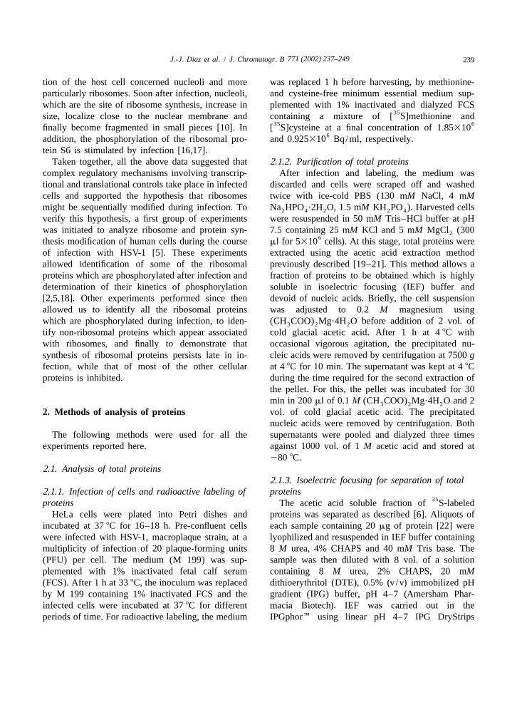
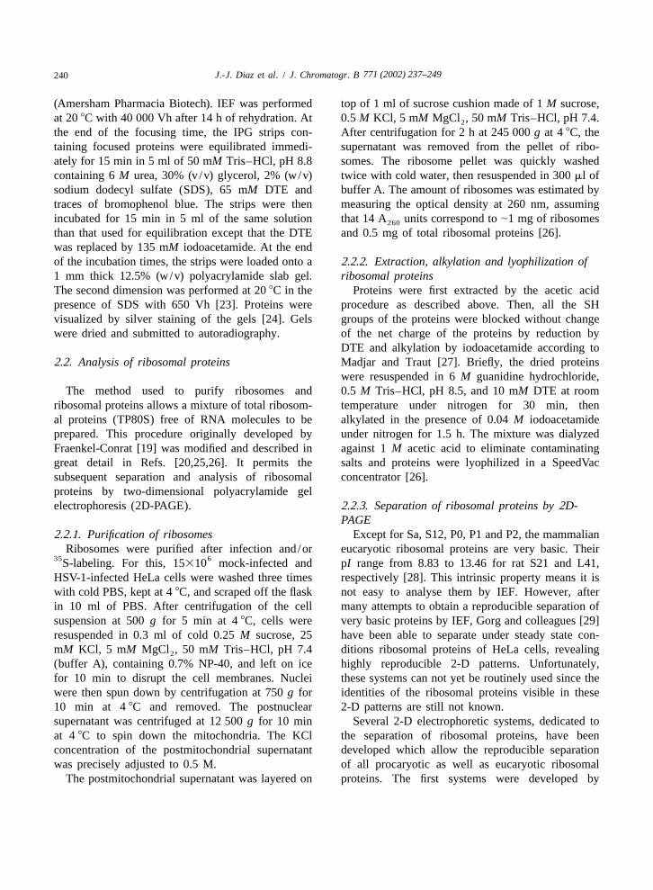
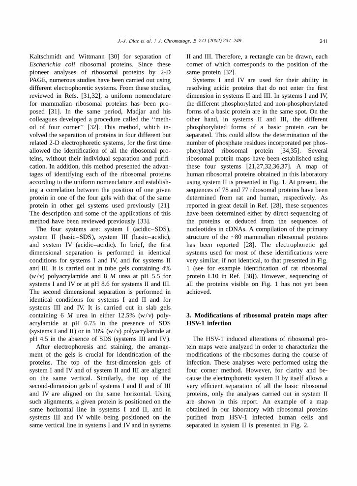
After electrophoresis and staining, the arrange- tein maps were analyzed in order to characterize thement of the gels is crucial for identification of the modifications of the ribosomes during the course ofproteins. The top of the first-dimension gels of infection. These analyses were performed using thesystem I and IV and of system II and III are aligned four corner method. However, for clarity and be-on the same vertical. Similarly, the top of the cause the electrophoretic system II by itself allows asecond-dimension gels of systems I and II and of III very efficient separation of all the basic ribosomaland IV are aligned on the same horizontal. Using proteins, only the analyses carried out in system IIsuch alignments, a given protein is positioned on the are shown in this report. An example of a mapsame horizontal line in systems I and II, and in obtained in our laboratory with ribosomal proteinssystems III and IV while being positioned on the purified from HSV-1 infected human cells andsame vertical line in systems I and IV and in systems separated in system II is presented in Fig. 2.

771 (2002) 237–249242 J.-J. Diaz et al. / J. Chromatogr. B
Fig. 1. Two-dimensional ribosomal protein map of basic ribosomal proteins purified from HeLa cells. An amount corresponding to 2.5 A260
units of ribosomes was separated in the two-dimensional electrophoretic system II. The first dimension was run at constant voltage (72 V)during 16 h, from the anode to the cathode, in 1.25-mm inside diameter tube gels. The second dimension was performed at 20 8C with 1000Vh using a maximum power of 6 W per gel. At the end of the electrophoresis ribosomal proteins were visualized by Coomassie brilliant bluestaining and numbered according to the uniformed nomenclature [31]. L and S stand for proteins belonging to the large and the smallribosomal subunit, respectively.
3.1. Identification of phosphorylated ribosomal map similar to those obtained in the above study isproteins presented in Fig. 2. All together, these experiments
demonstrated that HSV-1 induces the phosphoryla-´At the beginning of the study performed by Masse tion of five ribosomal proteins. Four of them, S2, Sa,
and his colleagues [5], it was not known whether the S3a and S6, belong to the 40S subunit and only one,phosphorylation of ribosomal proteins other than S6 L30, belongs to the 60S subunit. Using the fourcould be stimulated after infection by HSV-1. There- corner method, identification of the phosphorylatedfore to identify these proteins, the following ex- forms of S2, Sa, S3a and S6 was unambiguous.perimental strategy was developed. Mock-infected However, identification of the phosphorylated formand HSV-1 human infected cells were labelled with of L30, initially designated v2 [5], required the32P for 90 min at different times after infection. further following experiments [2]. Ribosomal pro-Some of these experiments were carried out after a teins from infected cells were separated in system IIdeprivation of serum because S6 phosphorylation and transferred to polyvinylidene difluoride (PVDF)was known to be stimulated by addition of serum membranes. Ribosomal protein L30 and its suspected[35]. Ribosomes were purified from mock-infected phosphorylated derivative (L30a, Fig. 2) were sub-and infected cells and ribosomal proteins were jected to an in situ CNBr cleavage [39]. Theanalyzed by the four corner method described above. hydrolysis products were eluted and separatedAn example of an HSV-1-modified ribosomal protein through high-resolution discontinuous SDS–PAGE
771 (2002) 237–249 243J.-J. Diaz et al. / J. Chromatogr. B
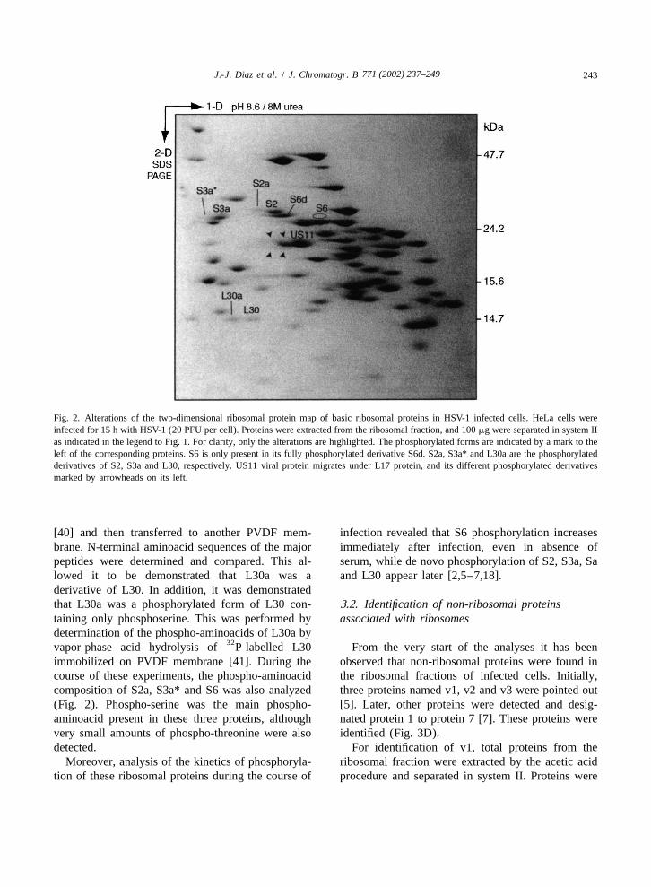
Fig. 2. Alterations of the two-dimensional ribosomal protein map of basic ribosomal proteins in HSV-1 infected cells. HeLa cells wereinfected for 15 h with HSV-1 (20 PFU per cell). Proteins were extracted from the ribosomal fraction, and 100 mg were separated in system IIas indicated in the legend to Fig. 1. For clarity, only the alterations are highlighted. The phosphorylated forms are indicated by a mark to theleft of the corresponding proteins. S6 is only present in its fully phosphorylated derivative S6d. S2a, S3a* and L30a are the phosphorylatedderivatives of S2, S3a and L30, respectively. US11 viral protein migrates under L17 protein, and its different phosphorylated derivativesmarked by arrowheads on its left.
[40] and then transferred to another PVDF mem- infection revealed that S6 phosphorylation increasesbrane. N-terminal aminoacid sequences of the major immediately after infection, even in absence ofpeptides were determined and compared. This al- serum, while de novo phosphorylation of S2, S3a, Salowed it to be demonstrated that L30a was a and L30 appear later [2,5–7,18].derivative of L30. In addition, it was demonstratedthat L30a was a phosphorylated form of L30 con- 3.2. Identification of non-ribosomal proteinstaining only phosphoserine. This was performed by associated with ribosomesdetermination of the phospho-aminoacids of L30a by
32vapor-phase acid hydrolysis of P-labelled L30 From the very start of the analyses it has beenimmobilized on PVDF membrane [41]. During the observed that non-ribosomal proteins were found incourse of these experiments, the phospho-aminoacid the ribosomal fractions of infected cells. Initially,composition of S2a, S3a* and S6 was also analyzed three proteins named v1, v2 and v3 were pointed out(Fig. 2). Phospho-serine was the main phospho- [5]. Later, other proteins were detected and desig-aminoacid present in these three proteins, although nated protein 1 to protein 7 [7]. These proteins werevery small amounts of phospho-threonine were also identified (Fig. 3D).detected. For identification of v1, total proteins from the
Moreover, analysis of the kinetics of phosphoryla- ribosomal fraction were extracted by the acetic acidtion of these ribosomal proteins during the course of procedure and separated in system II. Proteins were
771 (2002) 237–249244 J.-J. Diaz et al. / J. Chromatogr. B
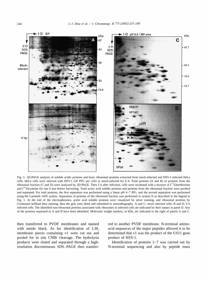
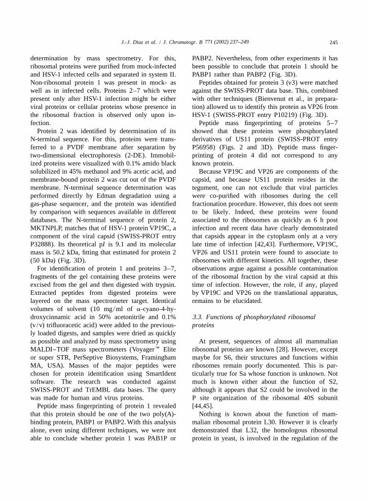
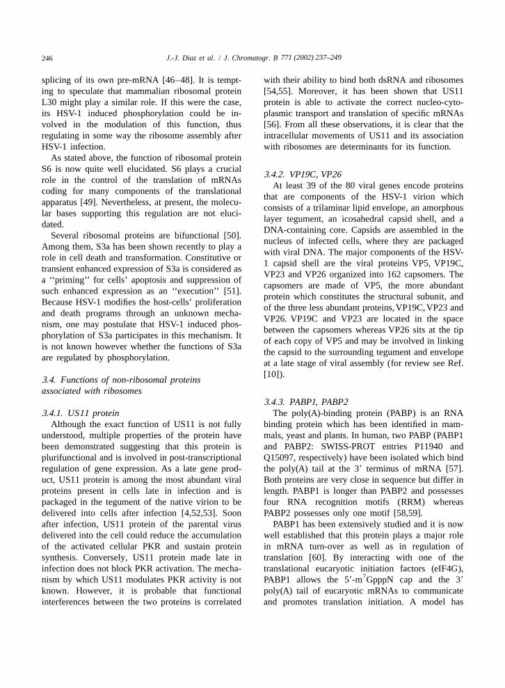
Fig. 3. 2D-PAGE analysis of soluble acidic proteins and basic ribosomal proteins extracted from mock-infected and HSV-1-infected HeLacells. HeLa cells were infected with HSV-1 (20 PFU per cell) or mock-infected for 6 h. Total proteins (A and B) or proteins from the
35ribosomal fraction (C and D) were analyzed by 2D-PAGE. Then 5 h after infection, cells were incubated with a mixture of [ S]methionine35and [ S]cysteine for one h just before harvesting. Total acetic acid soluble proteins and proteins from the ribosomal fraction were purified
and separated. For total proteins, the first separation was performed using a linear pH 4–7 IPG, and the second separation was performedusing the Laemmli–SDS system. Separation of proteins of the ribosomal fraction was performed in system II as described in the legend toFig. 1. At the end of the electrophoreses, acetic acid soluble proteins were visualized by silver staining, and ribosomal proteins byCoomassie brilliant blue staining, then the gels were dried and submitted to autoradiography. A and C, mock infected cells; B and D, 6 hinfected cells. The identified non-ribosomal proteins associated with ribosomes in infected cells are indicated by their names in panel D. Anyof the proteins separated in A and B have been identified. Molecular weight markers, in kDa, are indicated to the right of panels A and C.
then transferred to PVDF membranes and stained red to another PVDF membrane. N-terminal amino-with amido black. As for identification of L30, acid sequences of the major peptides allowed it to bemembrane pieces containing v1 were cut out and determined that v1 was the product of the US11 genepooled for in situ CNBr cleavage. The hydrolysis product of HSV-1.products were eluted and separated through a high- Identification of proteins 1–7 was carried out byresolution discontinuous SDS–PAGE then transfer- N-terminal sequencing and also by peptide mass
771 (2002) 237–249 245J.-J. Diaz et al. / J. Chromatogr. B
determination by mass spectrometry. For this, PABP2. Nevertheless, from other experiments it hasribosomal proteins were purified from mock-infected been possible to conclude that protein 1 should beand HSV-1 infected cells and separated in system II. PABP1 rather than PABP2 (Fig. 3D).Non-ribosomal protein 1 was present in mock- as Peptides obtained for protein 3 (v3) were matchedwell as in infected cells. Proteins 2–7 which were against the SWISS-PROT data base. This, combinedpresent only after HSV-1 infection might be either with other techniques (Bienvenut et al., in prepara-viral proteins or cellular proteins whose presence in tion) allowed us to identify this protein as VP26 fromthe ribosomal fraction is observed only upon in- HSV-1 (SWISS-PROT entry P10219) (Fig. 3D).fection. Peptide mass fingerprinting of proteins 5–7
Protein 2 was identified by determination of its showed that these proteins were phosphorylatedN-terminal sequence. For this, proteins were trans- derivatives of US11 protein (SWISS-PROT entryferred to a PVDF membrane after separation by P56958) (Figs. 2 and 3D). Peptide mass finger-two-dimensional electrophoresis (2-DE). Immobil- printing of protein 4 did not correspond to anyized proteins were visualized with 0.1% amido black known protein.solubilized in 45% methanol and 9% acetic acid, and Because VP19C and VP26 are components of themembrane-bound protein 2 was cut out of the PVDF capsid, and because US11 protein resides in themembrane. N-terminal sequence determination was tegument, one can not exclude that viral particlesperformed directly by Edman degradation using a were co-purified with ribosomes during the cellgas-phase sequencer, and the protein was identified fractionation procedure. However, this does not seemby comparison with sequences available in different to be likely. Indeed, these proteins were founddatabases. The N-terminal sequence of protein 2, associated to the ribosomes as quickly as 6 h postMKTNPLP, matches that of HSV-1 protein VP19C, a infection and recent data have clearly demonstratedcomponent of the viral capsid (SWISS-PROT entry that capsids appear in the cytoplasm only at a veryP32888). Its theoretical pI is 9.1 and its molecular late time of infection [42,43]. Furthermore, VP19C,mass is 50.2 kDa, fitting that estimated for protein 2 VP26 and US11 protein were found to associate to(50 kDa) (Fig. 3D). ribosomes with different kinetics. All together, these
For identification of protein 1 and proteins 3–7, observations argue against a possible contaminationfragments of the gel containing these proteins were of the ribosomal fraction by the viral capsid at thisexcised from the gel and then digested with trypsin. time of infection. However, the role, if any, playedExtracted peptides from digested proteins were by VP19C and VP26 on the translational apparatus,layered on the mass spectrometer target. Identical remains to be elucidated.volumes of solvent (10 mg/ml of a-cyano-4-hy-droxycinnamic acid in 50% acetonitrile and 0.1% 3.3. Functions of phosphorylated ribosomal(v /v) trifluoracetic acid) were added to the previous- proteinsly loaded digests, and samples were dried as quicklyas possible and analyzed by mass spectrometry using At present, sequences of almost all mammalianMALDI–TOF mass spectrometers (VoyagerE Elite ribosomal proteins are known [28]. However, exceptor super STR, PerSeptive Biosystems, Framingham maybe for S6, their structures and functions withinMA, USA). Masses of the major peptides were ribosomes remain poorly documented. This is par-chosen for protein identification using SmartIdent ticularly true for Sa whose function is unknown. Notsoftware. The research was conducted against much is known either about the function of S2,SWISS-PROT and TrEMBL data bases. The query although it appears that S2 could be involved in thewas made for human and virus proteins. P site organization of the ribosomal 40S subunit
Peptide mass fingerprinting of protein 1 revealed [44,45].that this protein should be one of the two poly(A)- Nothing is known about the function of mam-binding protein, PABP1 or PABP2. With this analysis malian ribosomal protein L30. However it is clearlyalone, even using different techniques, we were not demonstrated that L32, the homologous ribosomalable to conclude whether protein 1 was PAB1P or protein in yeast, is involved in the regulation of the
771 (2002) 237–249246 J.-J. Diaz et al. / J. Chromatogr. B
splicing of its own pre-mRNA [46–48]. It is tempt- with their ability to bind both dsRNA and ribosomesing to speculate that mammalian ribosomal protein [54,55]. Moreover, it has been shown that US11L30 might play a similar role. If this were the case, protein is able to activate the correct nucleo-cyto-its HSV-1 induced phosphorylation could be in- plasmic transport and translation of specific mRNAsvolved in the modulation of this function, thus [56]. From all these observations, it is clear that theregulating in some way the ribosome assembly after intracellular movements of US11 and its associationHSV-1 infection. with ribosomes are determinants for its function.
As stated above, the function of ribosomal proteinS6 is now quite well elucidated. S6 plays a crucial
3.4.2. VP19C, VP26role in the control of the translation of mRNAs
At least 39 of the 80 viral genes encode proteinscoding for many components of the translational
that are components of the HSV-1 virion whichapparatus [49]. Nevertheless, at present, the molecu-
consists of a trilaminar lipid envelope, an amorphouslar bases supporting this regulation are not eluci-
layer tegument, an icosahedral capsid shell, and adated.
DNA-containing core. Capsids are assembled in theSeveral ribosomal proteins are bifunctional [50].
nucleus of infected cells, where they are packagedAmong them, S3a has been shown recently to play a
with viral DNA. The major components of the HSV-role in cell death and transformation. Constitutive or
1 capsid shell are the viral proteins VP5, VP19C,transient enhanced expression of S3a is considered as
VP23 and VP26 organized into 162 capsomers. Thea ‘‘priming’’ for cells’ apoptosis and suppression of
capsomers are made of VP5, the more abundantsuch enhanced expression as an ‘‘execution’’ [51].
protein which constitutes the structural subunit, andBecause HSV-1 modifies the host-cells’ proliferation
of the three less abundant proteins,VP19C,VP23 andand death programs through an unknown mecha-
VP26. VP19C and VP23 are located in the spacenism, one may postulate that HSV-1 induced phos-
between the capsomers whereas VP26 sits at the tipphorylation of S3a participates in this mechanism. It
of each copy of VP5 and may be involved in linkingis not known however whether the functions of S3a
the capsid to the surrounding tegument and envelopeare regulated by phosphorylation.
at a late stage of viral assembly (for review see Ref.[10]).
3.4. Functions of non-ribosomal proteinsassociated with ribosomes
3.4.3. PABP1, PABP23.4.1. US11 protein The poly(A)-binding protein (PABP) is an RNA
Although the exact function of US11 is not fully binding protein which has been identified in mam-understood, multiple properties of the protein have mals, yeast and plants. In human, two PABP (PABP1been demonstrated suggesting that this protein is and PABP2: SWISS-PROT entries P11940 andplurifunctional and is involved in post-transcriptional Q15097, respectively) have been isolated which bindregulation of gene expression. As a late gene prod- the poly(A) tail at the 39 terminus of mRNA [57].uct, US11 protein is among the most abundant viral Both proteins are very close in sequence but differ inproteins present in cells late in infection and is length. PABP1 is longer than PABP2 and possessespackaged in the tegument of the native virion to be four RNA recognition motifs (RRM) whereasdelivered into cells after infection [4,52,53]. Soon PABP2 possesses only one motif [58,59].after infection, US11 protein of the parental virus PABP1 has been extensively studied and it is nowdelivered into the cell could reduce the accumulation well established that this protein plays a major roleof the activated cellular PKR and sustain protein in mRNA turn-over as well as in regulation ofsynthesis. Conversely, US11 protein made late in translation [60]. By interacting with one of theinfection does not block PKR activation. The mecha- translational eucaryotic initiation factors (eIF4G),
7nism by which US11 modulates PKR activity is not PABP1 allows the 59-m GpppN cap and the 39
known. However, it is probable that functional poly(A) tail of eucaryotic mRNAs to communicateinterferences between the two proteins is correlated and promotes translation initiation. A model has
771 (2002) 237–249 247J.-J. Diaz et al. / J. Chromatogr. B
been proposed in which interaction of PABP1 with 4. ConclusionseIF4G induces the circularization of mRNAs [61].This model has been confirmed using atomic force Ribosomal protein maps have not yet been estab-microscopy [62]. lished using IEF. This is mainly due to the very basic
In contrast to PABP1, PABP2 interacts slightly nature of ribosomal proteins which renders themwith the poly(A) tail of mRNAs and does not interact very difficult to separate using this method. How-significantly with eIF4G [58,59,63]. The exact func- ever, several two-dimensional electrophoretic sys-tion of PABP2 is still not fully determined, however, tems have been developed over the last two decadesit has been proposed very recently that PABP2 could which allow a reproducible separation of allbe a component of a so-called ‘‘Pioneer initiation ribosomal proteins from procaryotic as well as fromcomplex’’. This complex, which is probably involved eucaryotic cells. Among them, four different butonly in the first round of translation, might act as a related systems were developed by Madjar and hisquality control mechanism to eliminate mRNAs colleagues which allowed several 2-D ribosomalwhich prematurely terminate translation [64]. protein maps to be built. Over the past few years the
PABPs are thus multifunctional proteins which modifications of ribosomes and protein synthesisparticipate in the regulation of translation initiation, after infection by HSV-1 have been analyzed. Thistranslation accuracy, degradation and/or stability of has been achieved by comparison of the ribosomalmRNA. protein maps built from non-infected cells with those
built from infected cells. These comparisons demon-strated that infection by HSV-1 induces important
3.5. Persistence of ribosomal protein synthesis modifications of ribosomes: (i) non-reversible phos-after infection phorylation of ribosomal protein S6; (ii) unusual
phosphorylation of several proteins of small andSynthesis of ribosomal proteins was analyzed in large subunits; and (iii) association of viral and
cells infected with HSV-1 under different experimen- cellular proteins to the ribosomal fraction. The roletal conditions [1–3,6]. For this, the HSV-1 induced that these modifications may play in the regulation of
35alterations of S-labelled ribosomal protein maps viral and cellular gene expression remains to bewere analyzed during the course of infection. An determined. However, these modifications are corre-example of such analysis is presented in Fig. 3. The lated with changes in the cellular protein synthesisrate of synthesis of proteins present in the ribosomal occurring during the course of infection. This ob-fraction was estimated in mock-infected cells and in servation led us to hypothesize that ribosomes them-cells infected for 6 h corresponding to the early stage selves participate in the translational regulation ofof infection (Fig. 3C and D). In parallel, the rate of a viral and cellular gene expression. The finding thatsub-set of total cellular proteins was also estimated several viral and cellular proteins with mRNA-bind-(Fig. 3A and B). ing activities associate with ribosomes after infection
As expected for total cellular proteins, the syn- and that the synthesis of these modified ribosomesthesis rate of host proteins decreased dramatically in persists even late in infection, strongly supports this6-h infected cells (compare Fig. 3A and B). The hypothesis. Indeed, one might postulate that theseglobal synthesis rate of this set of proteins was modified, and newly synthesized ribosomes allow thedecreased to |40% of that of mock-infected cells. specific translation of some class of viral and/orThis reflected the typical HSV-1-induced shut-off of cellular mRNAs whose translation is determinant forthe majority of host protein syntheses. Conversely, the outcome of infection.the synthesis of all the basic ribosomal proteinsseparated in system II remained very efficient (com-pare Fig. 3C and D). Indeed the synthesis of 5. Nomenclatureribosomal proteins was sustained until 9 h post-infection and finally decreased for most of them at 2-D two-dimensional15 h post-infection [6,7]. 2-DE two-dimensional electrophoresis
771 (2002) 237–249248 J.-J. Diaz et al. / J. Chromatogr. B
[14] R.W. Honess, B. Roizman, Proc. Natl. Acad. Sci. USA 72DTE dithioerythritol(1975) 1276.HSV-1 herpes simplex virus type 1
[15] S. Silverstein, D.L. Engelhardt, Virology 95 (1979) 334.IEF isoelectric focusing [16] M.L. Fenwick, M.J. Walker, J. Gen. Virol. 45 (1979) 397.IPG immobilized pH gradient [17] I.M. Kennedy, W.S. Stevely, D.P. Leader, J. Virol. 39 (1981)
359.PABP poly(A)-binding protein´[18] T. Masse, D. Garcin, B. Jacquemont, J.-J. Madjar, Eur. J.PAGE polyacrylamide gel electrophoresis
Biochem. 194 (1990) 287.PFU plaque forming unit [19] H. Fraenkel-Conrat, Virology 4 (1957) 1.PVDF polyvinylidene difluoride [20] J.R. Waller, J.I. Harris, Proc. Natl. Acad. Sci. USA 47 (1961)
18.SDS sodium dodecyl sulfate[21] J.-J. Madjar, M. Arpin, M. Buisson, J.P. Reboud, Mol. Gen.VP viral protein
Genet. 171 (1979) 121.[22] M.M. Bradford, Anal. Biochem. 72 (1976) 248.[23] U.K. Laemmli, Nature 227 (1970) 680.[24] B.R. Oakley, D.R. Kirsch, N.R. Morris, Anal. Biochem. 105
Acknowledgements (1980) 361.[25] J.-J. Madjar, in: J.E. Celis (Ed.), Cell Biology: A Laboratory
Handbook, Academic Press, London, 1994, p. 657.The authors would like to express their gratitude[26] A. Greco, J.-J. Madjar, in: J.E. Celis (Ed.), Cell Biology: Ato Professor J.-J. Madjar who has elaborated many of
Laboratory Handbook, Academic Press, London, 1998, p.the studies described in this review and who continu- 135.ously provided much helpful advice during develop- [27] J.-J. Madjar, R.R. Traut, Mol. Gen. Genet. 179 (1980) 89.
¨[28] I.G. Wool, Y.-L. Chan, A. Gluck, in: J.W.B. Hershey, M.B.ment of the other experiments.Mathews, N. Sonenberg (Eds.), Translational Control, ColdSpring Harbor Laboratory Press, Cold Spring Harbor, NY,1996, p. 685.
[29] A. Gorg, C. Obermaier, G. Boguth, A. Csordas, J.J. Diaz, J.J.ReferencesMadjar, Electrophoresis 18 (1997) 328.
[30] E. Kaltschmidt, H.G. Wittmann, Anal. Biochem. 36 (1970)[1] A. Greco, A.M. Laurent, J.-J. Madjar, Mol. Gen. Genet. 256 401.
(1997) 320. [31] E.H. McConkey, H. Bielka, J. Gordon, S.M. Lastick, A. Lin,[2] D. Simonin, J.-J. Diaz, K. Kindbeiter, L. Denoroy, J.-J. K. Ogata, J.P. Reboud, R.R. Traugh, J.R. Warner, H. Welfe, I.
Madjar, Electrophoresis 16 (1995) 854. Wool, Mol. Gen. Genet. 169 (1979) 1.´[3] D. Simonin, J.-J. Diaz, T. Masse, J.-J. Madjar, J. Gen. Virol. [32] J.-J. Madjar, S. Michel, A.J. Cozzone, J.P. Reboud, Anal.
78 (1997) 435. Biochem. 92 (1979) 174.´[4] J.-J. Diaz, D. Simonin, T. Masse, P. Deviller, K. Kindbeiter, [33] M.J. Dunn, in: A. Chrambach, M.J. Dunn, B.J. Radola
L. Denoroy, J.-J. Madjar, J. Gen. Virol. 74 (1993) 397. (Eds.), Advances in Electrophoresis, VCH, Weinheim, 1987,´[5] T. Masse, D. Garcin, B. Jacquemont, J.-J. Madjar, Mol. Gen. p. 1.
Genet. 220 (1990) 1. [34] B. Buendia, A. Person-Fernandez, G. Beaud, J.-J. Madjar,´[6] A. Greco, N. Bausch, Y. Coute, J.-J. Diaz, Electrophoresis 21 Eur. J. Biochem. 162 (1987) 95.
(2000) 2522. [35] J.-J. Diaz, O. Gandrillon, D. Hentzen, D. Leguellec, J.[7] A. Greco, W. Bienvenut, J.C. Sanchez, K. Kindbeiter, D. Samarut, J.-J. Madjar, Oncol. Res. 1 (1989) 163.
Hochstrasser, J.-J. Madjar, J.-J. Diaz, Proteomics 1 (2001) [36] J.-J. Madjar, K. Nielsen-Smith, M. Frahm, D.J. Roufa, Proc.545. Natl. Acad. Sci. USA 79 (1982) 1003.
[8] B. Roizman, in: B. Roizman, R.J. Whitley, C. Lopez (Eds.), [37] J.-J. Madjar, A. Fournier, Eur. J. Biochem. 163 (1987) 577.The Human Herpesviruses, Raven Press, New York, 1993, p. [38] Y.-L. Chan, J.-J. Diaz, L. Denoroy, J.-J. Madjar, I.G. Wool,1. Biochem. Biophys. Res. Commun. 225 (1996) 952.
[9] R.J. Whitley, J.W. Gnann Jr., in: B. Roizman, R.J. Whitley, C. [39] M. Scott, D.L. Crimmins, D.W. McCourt, J.J. Tarrand, M.C.Lopez (Eds.), The Human Herpesviruses, Raven Press, New Eyerman, M.H. Nahm, Biochem. Biophys. Res. Commun.York, 1993, p. 69. 155 (1988) 1353.
¨[10] B. Roizman, A.E. Sears, in: B. Roizman, R.J. Whitley, C. [40] H. Schagger, G. Von Jagow, Anal. Biochem. 166 (1987) 368.Lopez (Eds.), The Human Herpesviruses, Raven Press, New [41] E. Hildebrandt, V.A. Fried, Anal. Biochem. 177 (1989) 407.York, 1993, p. 11. [42] G. Elliott, P. O’Hare, J. Virol. 73 (1999) 4110.
[11] M.L. Fenwick, Virology 77 (1977) 860. [43] M. Miranda-Saksena, P. Armati, R.A. Boadle, D.J. Holland,[12] M.L. Fenwick, M.J. Walker, J. Gen. Virol. 41 (1978) 37. A.L. Cunningham, J. Virol. 74 (2000) 1827.[13] E. Harris-Hamilton, S.L. Bachenheimer, J. Virol. 53 (1985) [44] U.A. Bommer, F. Noll, G. Lutsch, H. Bielka, FEBS Lett. 111
144. (1980) 171.
771 (2002) 237–249 249J.-J. Diaz et al. / J. Chromatogr. B
[45] F. Noll, U.A. Bommer, G. Lutsch, H. Theise, H. Bielka, [56] J.-J. Diaz, M. Duc Dodon, N. Schaerer-Uthurralt, D.FEBS Lett. 87 (1978) 129. Simonin, K. Kindbeiter, L. Gazzolo, J.-J. Madjar, Nature 379
[46] H. Mao, S.A. White, J.R. Williamson, Nat. Struct. Biol. 6 (1996) 273.(1999) 1139. [57] D.R. Gallie, Gene 216 (1998) 1.
[47] J. Vilardell, S.J. Yu, J.R. Warner, Mol. Cell 5 (2000) 761. [58] E. Wahle, A. Lustig, P. Jeno, P. Maurer, J. Biol. Chem. 268[48] J. Vilardell, P. Chartrand, R.H. Singer, J.R. Warner, RNA 6 (1993) 2937.
(2000) 1773. [59] A. Nemeth, S. Krause, D. Blank, A. Jenny, P. Jeno, A.[49] O. Meyuhas, Eur. J. Biochem. 267 (2000) 6321. Lustig, E. Wahle, Nucleic Acids Res. 23 (1995) 4034.[50] I.G. Wool, Trends Biochem. Sci. 21 (1996) 164. [60] C. Grosset, C.Y. Chen, N. Xu, N. Sonenberg, H. Jacquemin-[51] H. Naora, Immunol. Cell Biol. 77 (1999) 197. Sablon, A.B. Shyu, Cell 103 (2000) 29.[52] R.J. Roller, B. Roizman, J. Virol. 66 (1992) 3624. [61] D.R. Gallie, Plant Mol. Biol. 32 (1996) 145.[53] R.J. Roller, L.L. Monk, D. Stuart, B. Roizman, J. Virol. 70 [62] S.E. Wells, P.E. Hillner, R.D. Vale, A.B. Sachs, Mol. Cell 2
(1996) 2842. (1998) 135.[54] K.A. Cassady, M. Gross, B. Roizman, J. Virol. 72 (1998) [63] H. Imataka, A. Gradi, N. Sonenberg, EMBO J. 17 (1998)
8620. 7480.[55] K.A. Cassady, M. Gross, B. Roizman, J. Virol. 72 (1998) [64] Y. Ishigaki, X. Li, G. Serin, L.E. Maquat, Cell 106 (2001)
7005. 607.