Antiviral activity of extracts from Brazilian seaweeds against herpes simplex virus
Upload
universityofcaliforniaCategory
view
1download
0
Published Ahead of Print 29 April 2009. 2009, 83(14):7252. DOI: 10.1128/JVI.00153-09. J. Virol.
Robert D. Wojahn and Curt M. HorvathAparna Ramachandran, Jason J. Rodriguez, Glen Barber, Jean-Patrick Parisien, Darja Bamming, Akihiko Komuro, MDA5 and LGP2Interference with Antiviral RNA Helicases A Shared Interface Mediates Paramyxovirus
http://jvi.asm.org/content/83/14/7252Updated information and services can be found at:
These include:
REFERENCEShttp://jvi.asm.org/content/83/14/7252#ref-list-1at:
This article cites 32 articles, 15 of which can be accessed free
CONTENT ALERTS more»articles cite this article),
Receive: RSS Feeds, eTOCs, free email alerts (when new
http://journals.asm.org/site/misc/reprints.xhtmlInformation about commercial reprint orders: http://journals.asm.org/site/subscriptions/To subscribe to to another ASM Journal go to:
on Novem
ber 1, 2013 by guesthttp://jvi.asm
.org/D
ownloaded from
on N
ovember 1, 2013 by guest
http://jvi.asm.org/
Dow
nloaded from
on Novem
ber 1, 2013 by guesthttp://jvi.asm
.org/D
ownloaded from
on N
ovember 1, 2013 by guest
http://jvi.asm.org/
Dow
nloaded from
on Novem
ber 1, 2013 by guesthttp://jvi.asm
.org/D
ownloaded from
on N
ovember 1, 2013 by guest
http://jvi.asm.org/
Dow
nloaded from
on Novem
ber 1, 2013 by guesthttp://jvi.asm
.org/D
ownloaded from
on N
ovember 1, 2013 by guest
http://jvi.asm.org/
Dow
nloaded from
on Novem
ber 1, 2013 by guesthttp://jvi.asm
.org/D
ownloaded from
on N
ovember 1, 2013 by guest
http://jvi.asm.org/
Dow
nloaded from
JOURNAL OF VIROLOGY, July 2009, p. 7252–7260 Vol. 83, No. 140022-538X/09/$08.00�0 doi:10.1128/JVI.00153-09Copyright © 2009, American Society for Microbiology. All Rights Reserved.
A Shared Interface Mediates Paramyxovirus Interference withAntiviral RNA Helicases MDA5 and LGP2�
Jean-Patrick Parisien,1 Darja Bamming,1 Akihiko Komuro,1 Aparna Ramachandran,1Jason J. Rodriguez,1 Glen Barber,2 Robert D. Wojahn,1 and Curt M. Horvath1*
Department of Biochemistry, Molecular Biology, and Cell Biology, Northwestern University, Evanston, Illinois 60208,1 andDepartment of Microbiology and Immunology, University of Miami School of Medicine, Miami, Florida 331362
Received 22 January 2009/Accepted 23 April 2009
Diverse members of the Paramyxovirus family of negative-strand RNA viruses effectively suppress host innateimmune responses through the actions of their V proteins. The V protein mediates interference with theinterferon regulatory RNA helicase MDA5 to avoid cellular antiviral responses. Analysis of the interactioninterface revealed the MDA5 helicase C domain as necessary and sufficient for association with V proteins fromhuman parainfluenza virus type 2, parainfluenza virus type 5, measles virus, mumps virus, Hendra virus, andNipah virus. The identified �130-residue region is highly homologous between MDA5 and the related antiviralhelicase LGP2, but not RIG-I. Results indicate that the paramyxovirus V proteins can also associate withLGP2. The V protein interaction was found to disrupt ATP hydrolysis mediated by both MDA5 and LGP2.These findings provide a potential mechanistic basis for V protein-mediated helicase interference and identifyLGP2 as a second cellular RNA helicase targeted by paramyxovirus V proteins.
Detection of pathogens by the mammalian innate immunesystem is mediated by pattern recognition receptors. For vi-ruses, nucleic acids are often the trigger for innate responsesthat culminate in antiviral gene expression, including the pro-duction of type I interferon (IFN-�/�). Foreign nucleic acidsoutside the cell can be recognized by transmembrane Toll-likereceptors at the cell surface or in the lumen of endocyticvesicles (9). Intracellular nucleic acids are recognized by cyto-plasmic receptor proteins (24). In both cases, receptor bindingto the nucleic acid ligand triggers a signal transduction cascadethat activates immediate transcriptional responses, includingthe induction of the antiviral cytokines in the IFN family.
An important class of receptors for the detection of cytosolicnonself RNAs is represented by the proteins encoded by reti-noic acid-inducible gene I (RIG-I) and melanoma differentia-tion-associated gene 5 (MDA5) (24). These proteins sharefunctional domains, including an amino-terminal protein inter-action motif that functions in signal transduction and is ho-mologous to the caspase activation and recruitment domain(CARD) (8) and a C-terminal DECH-box family RNA heli-case domain. The helicase domains share approximately 30 to40% overall sequence identity. Experimental and structuralstudies have demonstrated a third functional region at theextreme C terminus of RIG-I that forms a zinc-mediated foldand functions as a regulatory domain for the recognition anddiscrimination of RNA 5� ends (6, 20). Current evidence sup-ports a model in which binding to RNA ligand induces achange in accessibility to the CARD, allowing it to interactwith downstream signaling proteins, including the mitochon-drial resident IPS-1 (also identified as MAVS, VISA, andCardiff [12, 15, 21, 28]). This association coordinates a serine
kinase-mediated cascade that activates latent transcription fac-tors, including NF-�B and IFN regulatory factor 3, culminatingin the expression of IFN-� and a number of other crucialantiviral effector genes (8).
A third helicase protein, LGP2, resembles RIG-I andMDA5 in the helicase domain, also exhibiting 30 to 40% se-quence identity. This overall level of similarity may underesti-mate the relatedness of these proteins, as they are more iden-tical in their key helicase sequence motifs and encompassingdomains (2). Significantly, LGP2 lacks the N-terminal CARDhomology region. As a result, LGP2 expression is not typicallyassociated with the ability to directly activate downstream sig-naling. Ectopic expression of LGP2 interferes with double-stranded RNA (dsRNA) and virus-induced antiviral signaling,while reduction of LGP2 expression by RNA interference re-sults in enhanced IFN-� synthesis and antiviral responses. Ex-pression of LGP2, like that of RIG-I and MDA5, is induced byvirus infection, nucleic acid transfection, and IFN stimulation,suggesting it has the properties of a negative feedback inhibitor(14, 19, 20, 29).
The importance of these RNA helicase proteins in antiviralresponses is validated by the phenotypes of mice harboringtargeted disruptions in these genes (9). Deficiency in RIG-Ileads to a widespread enhancement in replication of manyRNA virus types, due to suppressed IFN biosynthesis andantiviral responses (11). Cells deficient in RIG-I exhibit a gen-eral defect in the ability to respond to foreign dsRNAs orsingle-stranded RNAs (ssRNAs) bearing phosphorylated 5�ends. MDA5 deficiency showed a more specific phenotype,resulting in increased susceptibility to picornavirus infectionand insensitivity to the synthetic dsRNA analog poly(I:C) (7).
LGP2 deficiency leads to a more complex phenotype (27).LGP2-deficient mice are more sensitive to IFN induction bycytosolic poly(I:C), an MDA5 ligand, and more resistant tovesicular stomatitis virus, a virus that triggers RIG-I signaling.Negative regulation of the IFN response remains intact overall,
* Corresponding author. Mailing address: Pancoe ENH Pavilion,Rm. 4401, 2200 Campus Drive, Evanston, IL 60208. Phone: (847)491-5530. Fax: (847) 491-4400. E-mail: [email protected].
� Published ahead of print on 29 April 2009.
7252

indicating that LGP2 may not be the primary negative regula-tor of type I IFN production. However, LGP2 deficiency re-sults in a suppressed IFN response to infection with encepha-lomyocarditis virus, a picornavirus that has been demonstratedto trigger MDA5-mediated antiviral signaling. These data sug-gest that LGP2 executes both negative and positive regulatoryfunctions related to RIG-I and MDA5 signaling. The disparateeffects of LGP2 deficiency are difficult to interpret without abetter mechanistic understanding of LGP2 functions in cellularinnate antiviral immunity.
Further affirmation of the antiviral role of the helicase pro-teins derives from the fact that individual viruses have evolvedmeans to evade or disrupt their activity, either by direct inter-ference or by antagonizing the signaling intermediates. Thelarge Paramyxovirus family of negative-strand RNA viruses iswell known to evade IFN antiviral responses. Most viruseswithin this family encode a protein, called V, that is essentialfor IFN signaling evasion. In addition to suppressing theactivity of signal transducer and activator of transcription(STAT) proteins to compromise IFN signal transduction,paramyxovirus V proteins can also limit the induction of IFNgene expression by interfering with the MDA5 protein (1, 3).Associations between the V protein C-terminal domain (CTD), a60- to 70-residue zinc binding fold that is the conserved hall-mark of V proteins, and the MDA5 helicase domain preventssignal transduction downstream of poly(I:C), a synthetic RNAligand that is a known MDA5 activator (1, 3). This inhibitionwas observed for all tested V proteins of the large Paramyxo-virus family (3). Despite the potential importance of the Vprotein-MDA5 interface as an antiviral target, the mechanisticbasis and consequences for MDA5 interference by V proteinsare poorly understood and difficult to reconcile with the lack ofphenotype in MDA5-deficient mice. Recent evidence indicatesthat MDA5 is involved in the detection of Sendai virus-defec-tive interfering (DI) genomes, providing plausible biologicalrelevance to the observed V protein interference that may nothave been apparent from gene disruption studies (31).
Identification of the MDA5 target recognized by paramyxo-virus V proteins led to the discovery that LGP2 is also a targetfor V protein antagonism. Results demonstrate that the heli-case C domain of both proteins functions as a V protein bind-ing region common to both MDA5 and LGP2 but absent inRIG-I. We demonstrate that interaction with the V proteininterferes with the catalytic activity of both target helicases,providing a biochemical basis for V protein helicase antago-nism.
MATERIALS AND METHODS
Cells and virus. HEK293T cells were cultured in Dulbecco’s modified Eagle’smedium containing 10% cosmic calf serum (HyClone). Parainfluenza virus type5 (PIV5) was obtained from Robert Lamb (Northwestern University, Evanston,IL) and was propagated and titers were determined in Vero cells.
Plasmids, immunoprecipitations, and immunoblotting. To construct helicaseprotein fragment expression vectors, PCR-amplified segments of MDA5, LGP2,or RIG-I cDNAs were subcloned into plasmid pEF-FLAG to generate an N-terminal in-frame epitope tag, unless otherwise specified in the text. All resultingplasmids were verified by DNA sequencing and expression analysis to detect theepitope-tagged protein fragment. Expression vectors for hemagglutinin (HA)-tagged paramyxovirus V proteins and the glutathione S-transferase (GST)–mea-sles virus and mumps virus V protein fragment fusions have been describedelsewhere (17, 26). For immunoprecipitation experiments 5 �g of FLAG-heli-case vector and 5 �g of HA-V (or GST-V) vector were transfected by the CaPO4
method in HEK293T cells. Twenty-four hours later, cells were washed once withice-cold phosphate-buffered saline and subsequently lysed with whole-cell extractbuffer (WCEB) as described previously (26). For immunoprecipitation, lysateswere prepared in WCEB and precleared with Sepharose beads. Protein com-plexes were purified by overnight incubation with FLAG M2 affinity resin(Sigma) and washed with WCEB. Alternatively, GST fusions were collected onglutathione Sepharose (Sigma). After elution with sodium dodecyl sulfate, pro-teins were separated by sodium dodecyl sulfate-polyacrylamide gel electrophore-sis and processed for immunoblotting. For immunoblotting, proteins were sep-arated, transferred to nitrocellulose, probed with antibodies recognizing FLAG,HA, or GST (Sigma), and visualized by enhanced chemiluminescence (NEN LifeSciences). Rabbit antiserum recognizing the PIV5 P and V proteins was gener-ated using a GST-V protein purified from Escherichia coli (Cocalico Biologicals,Inc.) and screened by immunoblotting.
ATP hydrolysis. For ATP hydrolysis assays FLAG-tagged helicase proteins inthe presence or absence of PIV5, mumps virus, measles virus, or Nipah virus Vproteins were immunoprecipitated as described above and eluted with FLAGtripeptide (Sigma). Purified protein or helicase-V protein complexes were incu-bated with 2 �g poly(I:C) in 50-�l ATPase reaction mixtures containing 5 mMmorpholinopropanesulfonic acid (pH 6.5), 0.3 mM MgCl2, 0.2 mM dithiothreitolfor 10 min at room temperature. The reaction was started by addition of 0.5 mMATP (Sigma), 0.66 nM [�-32P]ATP (Perkin-Elmer) and incubated for 1 h at 37°C.Aliquots (10%) of the reaction mixtures were loaded onto thin-layer chroma-tography plates (Sigma) and analyzed by phosphorimaging with a MolecularDynamics Storm scanner and subsequently quantified using ImageQuant soft-ware. The remaining reaction mixture was analyzed by sodium dodecyl sulfate-polyacrylamide gel electrophoresis and immunoblotting using anti-FLAG anti-body (Sigma) and antisera detecting PIV5 V protein or HA epitope tag. For theexperiment shown in Fig. 6A, below, 293T cells were transfected with 2 �gMDA5 expression plasmid by calcium phosphate precipitation and after 24 hinfected with 10 PFU/ml PIV5 for 24 h prior to lysis. For the experiment shownin Fig. 6B, 293T cells were infected with 10 PFU/ml PIV5 for 24 h and thentransfected with 8 �g LGP2 expression plasmid using polyethyleneimine (Poly-sciences) for 24 h prior to lysis.
RESULTS
LGP2 and MDA5 share a V-protein binding region in thehelicase domain. Diverse paramyxovirus V proteins are capa-ble of disrupting IFN production by binding to the MDA5protein through protein interactions between the V proteinCTD and the MDA5 helicase domain. In contrast, the relatedRIG-I protein is not inhibited by V proteins (1, 3). To gainfurther insights into the underlying selectivity of these proteininteractions, coimmunoprecipitation experiments were usedto map the interaction sites. FLAG epitope-tagged MDA5,RIG-I, their N-terminal CARD or C-terminal helicase do-main, or LGP2 was expressed along with HA-tagged measlesvirus V protein (Fig. 1A and B). Following FLAG immuno-precipitations, HA immunoblot assays confirmed that neitherRIG-I nor any fragment of RIG-I was able to coprecipitate theV protein. Both MDA5 and the MDA5 helicase domain spe-cifically bound to the V protein, but the MDA5 CARD regiondid not. The full-length LGP2 protein was also tested in thisassay and was found to efficiently coprecipitate with the Vprotein (Fig. 1B).
To map the minimum region required for V protein inter-actions, a comprehensive nested series of MDA5 fragmentswas constructed sequentially, designed to encompass the entirehelicase domain with both N-terminal and C-terminal dele-tions (Fig. 1A). Coimmunoprecipitation experiments using N-terminal deletions revealed that the C-terminal half of thehelicase domain was able to coprecipitate with measles virus V(Fig. 1C), ultimately defining the amino-terminal boundarybetween residues 701 and 716 (Fig. 1D, compare 3E to 3F).Similar analysis mapped the C-terminal boundary between res-
VOL. 83, 2009 PARAMYXOVIRUS INTERFERENCE WITH MDA5 AND LGP2 7253

FIG. 1. Map of V protein interaction regions of MDA5 and LGP2. (A) Diagrammatic representations of the LGP2, MDA5, and RIG-Iproteins. (Top) Box diagrams illustrate the key features of the proteins, with CARD and helicase regions highlighted. Roman numerals depict the
7254 PARISIEN ET AL. J. VIROL.
idues 800 and 830 (Fig. 1E, compare C830 to C800). Theseresults tentatively identified the V protein target as approxi-mately 130 amino acids in length, between MDA5 residues 701and 830, which is analogous to LGP2 residues 351 to 479.
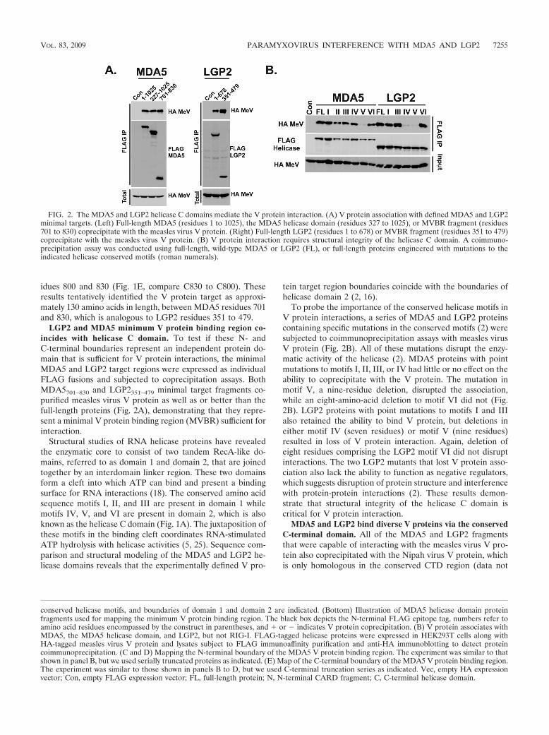
LGP2 and MDA5 minimum V protein binding region co-incides with helicase C domain. To test if these N- andC-terminal boundaries represent an independent protein do-main that is sufficient for V protein interactions, the minimalMDA5 and LGP2 target regions were expressed as individualFLAG fusions and subjected to coprecipitation assays. BothMDA5701–830 and LGP2351–479 minimal target fragments co-purified measles virus V protein as well as or better than thefull-length proteins (Fig. 2A), demonstrating that they repre-sent a minimal V protein binding region (MVBR) sufficient forinteraction.
Structural studies of RNA helicase proteins have revealedthe enzymatic core to consist of two tandem RecA-like do-mains, referred to as domain 1 and domain 2, that are joinedtogether by an interdomain linker region. These two domainsform a cleft into which ATP can bind and present a bindingsurface for RNA interactions (18). The conserved amino acidsequence motifs I, II, and III are present in domain 1 whilemotifs IV, V, and VI are present in domain 2, which is alsoknown as the helicase C domain (Fig. 1A). The juxtaposition ofthese motifs in the binding cleft coordinates RNA-stimulatedATP hydrolysis with helicase activities (5, 25). Sequence com-parison and structural modeling of the MDA5 and LGP2 he-licase domains reveals that the experimentally defined V pro-
tein target region boundaries coincide with the boundaries ofhelicase domain 2 (2, 16).
To probe the importance of the conserved helicase motifs inV protein interactions, a series of MDA5 and LGP2 proteinscontaining specific mutations in the conserved motifs (2) weresubjected to coimmunoprecipitation assays with measles virusV protein (Fig. 2B). All of these mutations disrupt the enzy-matic activity of the helicase (2). MDA5 proteins with pointmutations to motifs I, II, III, or IV had little or no effect on theability to coprecipitate with the V protein. The mutation inmotif V, a nine-residue deletion, disrupted the association,while an eight-amino-acid deletion to motif VI did not (Fig.2B). LGP2 proteins with point mutations to motifs I and IIIalso retained the ability to bind V protein, but deletions ineither motif IV (seven residues) or motif V (nine residues)resulted in loss of V protein interaction. Again, deletion ofeight residues comprising the LGP2 motif VI did not disruptinteractions. The two LGP2 mutants that lost V protein asso-ciation also lack the ability to function as negative regulators,which suggests disruption of protein structure and interferencewith protein-protein interactions (2). These results demon-strate that structural integrity of the helicase C domain iscritical for V protein interaction.
MDA5 and LGP2 bind diverse V proteins via the conservedC-terminal domain. All of the MDA5 and LGP2 fragmentsthat were capable of interacting with the measles virus V pro-tein also coprecipitated with the Nipah virus V protein, whichis only homologous in the conserved CTD region (data not
conserved helicase motifs, and boundaries of domain 1 and domain 2 are indicated. (Bottom) Illustration of MDA5 helicase domain proteinfragments used for mapping the minimum V protein binding region. The black box depicts the N-terminal FLAG epitope tag, numbers refer toamino acid residues encompassed by the construct in parentheses, and � or indicates V protein coprecipitation. (B) V protein associates withMDA5, the MDA5 helicase domain, and LGP2, but not RIG-I. FLAG-tagged helicase proteins were expressed in HEK293T cells along withHA-tagged measles virus V protein and lysates subject to FLAG immunoaffinity purification and anti-HA immunoblotting to detect proteincoimmunoprecipitation. (C and D) Mapping the N-terminal boundary of the MDA5 V protein binding region. The experiment was similar to thatshown in panel B, but we used serially truncated proteins as indicated. (E) Map of the C-terminal boundary of the MDA5 V protein binding region.The experiment was similar to those shown in panels B to D, but we used C-terminal truncation series as indicated. Vec, empty HA expressionvector; Con, empty FLAG expression vector; FL, full-length protein; N, N-terminal CARD fragment; C, C-terminal helicase domain.
FIG. 2. The MDA5 and LGP2 helicase C domains mediate the V protein interaction. (A) V protein association with defined MDA5 and LGP2minimal targets. (Left) Full-length MDA5 (residues 1 to 1025), the MDA5 helicase domain (residues 327 to 1025), or MVBR fragment (residues701 to 830) coprecipitate with the measles virus V protein. (Right) Full-length LGP2 (residues 1 to 678) or MVBR fragment (residues 351 to 479)coprecipitate with the measles virus V protein. (B) V protein interaction requires structural integrity of the helicase C domain. A coimmuno-precipitation assay was conducted using full-length, wild-type MDA5 or LGP2 (FL), or full-length proteins engineered with mutations to theindicated helicase conserved motifs (roman numerals).
VOL. 83, 2009 PARAMYXOVIRUS INTERFERENCE WITH MDA5 AND LGP2 7255

shown). To demonstrate the generality of this MVBR as atarget for diverse paramyxovirus V proteins, MDA5701–830 andLGP2351–479 were tested for their ability to interact with Vproteins from parainfluenza virus 5, human parainfluenza virus2, mumps virus, measles virus, Nipah virus, and Hendra virus(Fig. 3A). All of the tested V proteins were able to coprecipi-tate with both MDA5 and LGP2 MVBR fragments.
Conversely, to verify that V proteins contact the helicasetarget fragments by means of their conserved CTD zinc fingerregions, GST fusion proteins that express the mumps virus ormeasles virus CTD, or the control measles virus N-terminaldomain (17), were used in coprecipitation assays (Fig. 3B). Nointeraction was observed between the full-length helicase orMVBR and the V protein N-terminal domain, but both mea-sles virus and mumps virus V protein CTD fusion proteinscoprecipitated with both full-length MDA5 (residues 1 to1025) or full-length LGP2 (residues 1 to 678) and the MVBRsMDA5701–830 and LGP2351–479. It was observed that the targetfragments were more efficiently coprecipitated than the respec-tive full-length helicase proteins, suggesting that the isolateddomain may be more accessible to the V protein CTD.
LGP2 and MDA5 MVBRs select V protein from PIV5-in-fected cells. The abilities of the defined helicase target frag-ments to interact with physiologically expressed V protein inthe context of virus infection were also tested (Fig. 4). Cellsexpressing the tagged full-length helicase proteins or MVBRwere infected with PIV5, lysates were subjected to immuno-precipitation with FLAG immunoaffinity resin, and eluateswere probed with antiserum that recognized the PIV5 P and Vproteins. The V protein, but not the P protein, was specificallycoprecipitated by either full-length MDA5 or LGP2 as well as
with their respective minimal target regions. Together, thesedata define �130-amino-acid regions of MDA5 and LGP2 thatcontain the binding targets for paramyxovirus V proteins.
V protein interferes with MDA5 and LGP2 enzymatic activ-ities. The experimental results defined the helicase C domainas the site of V protein association with MDA5 and LGP2.MDA5 interference by V proteins has been validated withdemonstration of V-mediated signaling interference leading to
FIG. 3. Targeting of MDA5 and LGP2 via the MVBR is a property of diverse paramyxovirus V proteins and is mediated by the conserved CTD.(A) Diverse paramyxovirus V proteins coprecipitate with the minimal MDA5 or LGP2 target fragments. Another coprecipitation experiment wasdone using V proteins from PIV5 (P5), human parainfluenza virus type 2 (H2), mumps virus (Mu), measles virus (Me), Hendra virus (He), orNipah virus (Ni). Expression of FLAG-tagged green fluorescent protein (GFP) combined with measles virus V protein served as a control fornonspecific interactions. (B) The V protein cysteine-rich C-terminal domain is sufficient to mediate association with the LGP2 and MDA5 MVBRfragments. FLAG-tagged full-length or target fragment of MDA5 (left) or LGP2 (right) was expressed along with glutathione S-transferase fusionproteins containing the measles virus (Me) or mumps virus (Mu) C-terminal domain, or as a control the measles virus N-terminal domain.Following FLAG or GST affinity purification, samples were probed for coprecipitation with antiserum specific to GST or FLAG, as indicated.
FIG. 4. MVBR reacts with V protein from infected cells. Parallelplates of HEK293T cells were transfected with expression vectors forthe indicated FLAG-tagged helicase proteins or fragments and theninfected with 1 PFU/cell of PIV5 (�) or mock infected (). Celllysates were subjected to FLAG purification and immunoblotting withantiserum that recognizes the PIV5 V protein and P protein. Only Vprotein coprecipitated.
7256 PARISIEN ET AL. J. VIROL.

disruption of IFN-� promoter activity. LGP2 plays a morecomplex and poorly understood role, as both a negative regu-lator of dsRNA signaling and a positive regulator of antiviralresponses (13, 14, 19, 20, 27, 29). While MDA5 and LGP2fundamentally differ in their antiviral signaling capacities, atthe catalytic level they both use RNA-dependent ATP hydro-lysis to drive enzymatic activity (2, 10).
The biochemical consequences for V protein interactionswith both MDA5 and LGP2 were examined at the enzymaticlevel with ATP hydrolysis assays (Fig. 5). Our previous studiesdemonstrated that FLAG-immunopurified helicases have spe-cific ATP hydrolysis activity (2). Epitope-tagged MDA5,LGP2, and RIG-I proteins were expressed in HEK293T cells inthe absence or presence of the PIV5 V protein, and lysateswere subjected to immunoaffinity purification with anti-FLAGresin. Immunoblotting detected copurified V protein in com-plex with both MDA5 and LGP2, but not RIG-I. FLAG elu-
ates were subjected to an ATP hydrolysis assay in the presenceof poly(I:C). MDA5- and LGP2-dependent ATP hydrolysisactivity was significantly diminished in the V protein-contain-ing reactions (Fig. 5A). Although the precise stoichiometry isdifficult to evaluate under these experimental conditions, thespecificity of V protein-mediated inhibition of LGP2 is shownby dose-dependent interference (Fig. 5B). Similar effects onthe helicase catalytic activity were observed for measles virus,mumps virus, and Nipah virus V proteins (Fig. 5C). Thesefindings demonstrate that the V protein interferes with theenzymatic activity of both MDA5 and LGP2.
To verify the ATP hydrolysis interference in a more nativecontext, a similar experiment was carried out with physiologi-cally expressed V protein from PIV5 infection (Fig. 6). Again,both MDA5 and LGP2, but not RIG-I, were capable of copre-cipitating the V protein from the infected cells. Analysis ofATP hydrolysis in the purified and eluted material demon-
FIG. 5. V protein interferes with ATP hydrolysis by MDA5 and LGP2. FLAG-tagged MDA5, LGP2, and RIG-I were immunoaffinity purifiedfrom HEK293T extracts with or without coexpressed V protein, and eluates were subjected to ATP hydrolysis analysis in the presence of poly(I:C).Top panels demonstrate copurification of V protein (PIV5V) with LGP2 and MDA5 but not RIG-I by immunoblotting. Middle panels demonstrateATP hydrolysis activities by autoradiography, with positions of the origin, ATP, and free phosphate indicated. Bottom panels contain graphicalrepresentations of ATP hydrolysis activities, quantified by phosphorimaging analysis. Graphs plot the ratio of Pi to ATP, expressed as thepercentage of the ATP hydrolysis activity without V protein. (A) Plasmids coding for FLAG-tagged LGP2, MDA5, or RIG-I RNA helicases werecotransfected with plasmids coding for HA-tagged PIV5 V protein or empty vector plasmid at a 1:1 ratio into HEK293T cells. Con, no-proteincontrol; WT, wild type. (B) Four micrograms of cDNA plasmids coding for FLAG-tagged LGP2 helicase were cotransfected with empty vector() or 2 �g, 8 �g, or 16 �g of plasmid coding for HA-tagged PIV5 V protein. Total amounts of transfected DNA were equalized with empty vectorfor each reaction. (C) Plasmids coding for FLAG-tagged LGP2, MDA5, or RIG-I RNA helicases were cotransfected with empty vector controlor plasmids coding for HA-tagged measles virus, mumps virus, or Nipah viurs V protein at a 1:3 ratio into HEK293T cells. *NS, nonspecific band.
VOL. 83, 2009 PARAMYXOVIRUS INTERFERENCE WITH MDA5 AND LGP2 7257

strated interference with both MDA5 and LGP2, but notRIG-I, reducing the enzymatic activity of these helicases byapproximately 60% of uninfected controls.
DISCUSSION
RNA helicase proteins are essential for a vast array of cel-lular processes, and the recent identification of RIG-I, MDA5,and LGP2 as essential for recognition of and response to viral
pathogens establishes their importance for innate antiviral im-munity (24). The significant role of these proteins in antiviralsignaling makes them prime targets for virus-encoded hostevasion and antagonism. Antagonism of MDA5 signaling is aunique property attributed to paramyxovirus V proteins andresults in a disengagement of MDA5-mediated IFN-� pro-moter activation (1, 3). Here, LGP2 is identified as a secondhelicase protein targeted by paramyxovirus V proteins.
Interaction mapping has defined an MVBR that is necessary
FIG. 6. Interference with ATP hydrolysis in infected cells. Results are for an experiment similar to that shown in Fig. 5, but FLAG-tagged MDA5,LGP2, RIG-I, or control GFP were immunoaffinity purified from HEK293T cells that were also infected with PIV5 and eluates were subjected to ATPhydrolysis analysis in the presence of poly(I:C). Top panels demonstrate copurification of V protein (PIV5V) with MDA5 (A) or LGP2 (B). Middlepanels demonstrate ATP hydrolysis activities by autoradiography, and bottom panels contain graphical representations of ATP hydrolysis activities,quantified by phosphorimaging analysis. Graphs plot the ratio of Pi to ATP, expressed as a percentage of the ATP hydrolysis activity without V protein.
7258 PARISIEN ET AL. J. VIROL.

and sufficient for contact with MDA5, encompassed by resi-dues 701 to 830. The analogous region of LGP2, encompassedby residues 351 to 479, was also found to be targeted byparamyxovirus V proteins. Sequence analysis of the identifiedMVBR reveals unusually high conservation between MDA5and LGP2, with 57% identity and 78% similarity. In contrast,RIG-I is only 37% identical and 58% similar to LGP2 and 41%identical and 58% similar to MDA5 in this region, the degreeof homology observed when comparing the full-length pro-teins.
RNA helicase catalytic activity is thought to arise from thecoordinated action of two structural domains, known as do-main 1 and domain 2 (5, 18). These two lobes encompass all ofthe conserved sequence motifs that contribute to ATP bindingand hydrolysis as well as RNA interaction and unwinding.Domain 2, also referred to as the helicase C domain, repre-sents a widely recognized structural element of diverse RNAand DNA helicase families. The boundaries of the MVBRsdefined here coincide exactly with the helicase C domains.Results from ATP hydrolysis assays indicate that the V proteininteraction can interfere with enzymatic activity, and disrup-tion of the MVBR by deletion mutagenesis abrogates V pro-tein interactions. Although the relationship between enzymaticactivity of the helicase domain and signal transduction by theseproteins remains poorly understood, the disruption of helicasedomain structure and function provides a reasonable mecha-nistic basis for the observed V protein antagonism.
The boundaries of the MDA5 MVBR characterized here,residues 701 to 830, are in close agreement with those definedusing yeast two-hybrid methods (4). The site of PIV5 V proteininteraction with MDA5 was mapped to MDA5 residues 676 to816. This study also observed that PIV5 V protein-mediatedMDA5 interference is directed at the level of RNA-dependentMDA5 oligomerization. This is not inconsistent with the ob-served disruption of ATPase activity, as the detection of RNAsubstrate is a prerequisite for MDA5-mediated ATP hydroly-sis. Prevention of RNA access by V proteins might contributeto the observed loss of enzymatic activity in the presence of Vprotein. However, the weak RNA binding capacity of MDA5complicates analysis of RNA interactions, and we have notobserved V-dependent loss of LGP2 RNA contact. Furtherwork is required to determine the precise point of V proteinantagonism for both MDA5 and LGP2.
MDA5 and LGP2 MVBR fragments are targeted by the Vprotein C-terminal domain, a zinc binding domain that exhibitshigh amino acid conservation among all paramyxovirus V pro-teins. In agreement with the generality of V-mediated MDA5interference (1, 3), we demonstrated that the MDA5 andLGP2 MVBRs are recognized by V proteins from measlesvirus, PIV5, mumps virus, human parainfluenza virus type 2,Nipah virus, and Hendra virus, as well as by the isolated CTDsfrom measles virus and mumps virus V proteins. The expressedMVBR fragments can selectively copurify the PIV5 V proteinfrom infected cells, demonstrating complex formation at phys-iologically relevant levels of V protein expression.
Mice deficient in RIG-I expression are more susceptibleto infection with Sendai virus, but MDA5-deficient miceretain normal resistance. Therefore, intracellular detectionof paramyxovirus infection is considered widely to be mediatedprimarily by RIG-I (11). As a consequence, the precise role for
MDA5 in antiviral responses to paramyxovirus infection re-mains enigmatic. Recent studies have demonstrated a specificrole for MDA5 signaling in response to Sendai virus Cantell, astrain that is known to accumulate DI genomes (31). DI ge-nomes are known to be potent stimulators of IFN biosynthesisand antiviral responses that can activate RIG-I-dependentpathways (22, 23, 30, 32), but available evidence suggests thatDI particles function as a paramyxovirus-associated molecularpattern that can stimulate dendritic cell activation via MDA5.Dendritic cells derived from MDA5-deficient mice were in-efficiently activated by Sendai virus Cantell, and sole expres-sion of Sendai virus V protein can inhibit direct early den-dritic cell activation by DI particles (31). These data providebiological relevance to the reported MDA5 interference byV proteins and identify DI genomes as a potential naturalMDA5 activator.
At our current level of understanding, the V protein antag-onism of LGP2 remains difficult to reconcile with prior studiesof LGP2 action. Characterized as a negative feedback regula-tor of IFN synthesis and antiviral responses, analysis of LGP2-deficient mice has revealed phenotypes related to both nega-tive and positive regulation of IFN responses and antiviralsignaling (14, 19, 20, 27, 29). Further experimentation will beneeded to fully appreciate the exact roles for LGP2 in detec-tion of and response to paramyxoviruses and the precise ad-vantages of V-mediated interference. Considering that expres-sion of both MDA5 and LGP2 proteins is strongly controlledby virus-induced and IFN-responsive signaling pathways, theiraccumulation at late times following infections or in special-ized cell types may function as a modifier of initial antiviralresponses. Common mechanisms for targeting MDA5 andLGP2 and a shared role in detection and response to DI RNAgenomes provide an additional suggestion of collaboration be-tween these two helicases. Together with the fact that bothLGP2 deficiency and MDA5 deficiency result in greater sus-ceptibility to picornavirus infections (11, 27), it is tempting tospeculate that LGP2 might act directly or indirectly in concertwith the MDA5 pathway to modulate antiviral responses.
ACKNOWLEDGMENTS
We acknowledge Aristobolo M. Silva and members of the Horvathand Barber laboratories for critical comments and helpful discussions.
This work was supported by NIH grants AI050707 and AI073919 toC.M.H.
REFERENCES
1. Andrejeva, J., K. S. Childs, D. F. Young, T. S. Carlos, N. Stock, S. Good-bourn, and R. E. Randall. 2004. The V proteins of paramyxoviruses bind theIFN-inducible RNA helicase, mda-5, and inhibit its activation of the IFN-beta promoter. Proc. Natl. Acad. Sci. USA 101:17264–17269.
2. Bamming, D., and C. M. Horvath. 2009. Regulation of signal transduction byenzymatically inactive antiviral RNA helicase proteins MDA5, RIG-I andLGP2. J. Biol. Chem. 284:9700–9712.
3. Childs, K., N. Stock, C. Ross, J. Andrejeva, L. Hilton, M. Skinner, R.Randall, and S. Goodbourn. 2007. mda-5, but not RIG-I, is a common targetfor paramyxovirus V proteins. Virology 359:190–200.
4. Childs, K. S., J. Andrejeva, R. E. Randall, and S. Goodbourn. 2009. Mech-anism of mda-5 inhibition by paramyxovirus V proteins. J. Virol. 83:1465–1473.
5. Cordin, O., J. Banroques, N. K. Tanner, and P. Linder. 2006. The DEAD-box protein family of RNA helicases. Gene 367:17–37.
6. Cui, S., K. Eisenacher, A. Kirchhofer, K. Brzozka, A. Lammens, K. Lam-mens, T. Fujita, K. K. Conzelmann, A. Krug, and K. P. Hopfner. 2008. TheC-terminal regulatory domain is the RNA 5�-triphosphate sensor of RIG-I.Mol. Cell 29:169–179.
7. Gitlin, L., W. Barchet, S. Gilfillan, M. Cella, B. Beutler, R. A. Flavell, M. S.
VOL. 83, 2009 PARAMYXOVIRUS INTERFERENCE WITH MDA5 AND LGP2 7259

Diamond, and M. Colonna. 2006. Essential role of mda-5 in type I IFNresponses to polyriboinosinic:polyribocytidylic acid and encephalomyocardi-tis picornavirus. Proc. Natl. Acad. Sci. USA 103:8459–8464.
8. Hiscott, J., R. Lin, P. Nakhaei, and S. Paz. 2006. MasterCARD: a pricelesslink to innate immunity. Trends Mol. Med. 12:53–56.
9. Ishii, K. J., S. Koyama, A. Nakagawa, C. Coban, and S. Akira. 2008. Hostinnate immune receptors and beyond: making sense of microbial infections.Cell Host Microbe 3:352–363.
10. Kang, D. C., R. V. Gopalkrishnan, Q. Wu, E. Jankowsky, A. M. Pyle, andP. B. Fisher. 2002. mda-5: an interferon-inducible putative RNA helicasewith double-stranded RNA-dependent ATPase activity and melanomagrowth-suppressive properties. Proc. Natl. Acad. Sci. USA 99:637–642.
11. Kato, H., O. Takeuchi, S. Sato, M. Yoneyama, M. Yamamoto, K. Matsui, S.Uematsu, A. Jung, T. Kawai, K. J. Ishii, O. Yamaguchi, K. Otsu, T. Tsu-jimura, C. S. Koh, C. Reis e Sousa, Y. Matsuura, T. Fujita, and S. Akira.2006. Differential roles of MDA5 and RIG-I helicases in the recognition ofRNA viruses. Nature 441:101–105.
12. Kawai, T., K. Takahashi, S. Sato, C. Coban, H. Kumar, H. Kato, K. J. Ishii,O. Takeuchi, and S. Akira. 2005. IPS-1, an adaptor triggering RIG-I- andMda5-mediated type I interferon induction. Nat. Immunol. 6:981–988.
13. Komuro, A., D. Bamming, and C. M. Horvath. 2008. Negative regulation ofcytoplasmic RNA-mediated antiviral signaling. Cytokine 43:350–358.
14. Komuro, A., and C. M. Horvath. 2006. RNA- and virus-independent inhibi-tion of antiviral signaling by RNA helicase LGP2. J. Virol. 80:12332–12342.
15. Meylan, E., J. Curran, K. Hofmann, D. Moradpour, M. Binder, R. Barten-schlager, and J. Tschopp. 2005. Cardif is an adaptor protein in the RIG-Iantiviral pathway and is targeted by hepatitis C virus. Nature 437:1167–1172.
16. Nishino, T., K. Komori, D. Tsuchiya, Y. Ishino, and K. Morikawa. 2005.Crystal structure and functional implications of Pyrococcus furiosus hefhelicase domain involved in branched DNA processing. Structure 13:143–153.
17. Ramachandran, A., J. P. Parisien, and C. M. Horvath. 2008. STAT2 is aprimary target for measles virus V protein-mediated alpha/beta interferonsignaling inhibition. J. Virol. 82:8330–8338.
18. Rocak, S., and P. Linder. 2004. DEAD-box proteins: the driving forcesbehind RNA metabolism. Nat. Rev. Mol. Cell Biol. 5:232–241.
19. Rothenfusser, S., N. Goutagny, G. DiPerna, M. Gong, B. G. Monks, A.Schoenemeyer, M. Yamamoto, S. Akira, and K. A. Fitzgerald. 2005. TheRNA helicase Lgp2 inhibits TLR-independent sensing of viral replication byretinoic acid-inducible gene-I. J. Immunol. 175:5260–5268.
20. Saito, T., R. Hirai, Y. M. Loo, D. Owen, C. L. Johnson, S. C. Sinha, S. Akira,T. Fujita, and M. Gale, Jr. 2007. Regulation of innate antiviral defenses
through a shared repressor domain in RIG-I and LGP2. Proc. Natl. Acad.Sci. USA 104:582–587.
21. Seth, R. B., L. Sun, C. K. Ea, and Z. J. Chen. 2005. Identification andcharacterization of MAVS, a mitochondrial antiviral signaling protein thatactivates NF-�B and IRF 3. Cell 122:669–682.
22. Strahle, L., D. Garcin, and D. Kolakofsky. 2006. Sendai virus defective-interfering genomes and the activation of interferon-beta. Virology 351:101–111.
23. Strahle, L., J. B. Marq, A. Brini, S. Hausmann, D. Kolakofsky, and D.Garcin. 2007. Activation of the beta interferon promoter by unnatural Sen-dai virus infection requires RIG-I and is inhibited by viral C proteins. J. Vi-rol. 81:12227–12237.
24. Takeuchi, O., and S. Akira. 2008. MDA5/RIG-I and virus recognition. Curr.Opin. Immunol. 20:17–22.
25. Tanner, N. K., and P. Linder. 2001. DExD/H box RNA helicases: fromgeneric motors to specific dissociation functions. Mol. Cell 8:251–262.
26. Ulane, C. M., A. Kentsis, C. D. Cruz, J. P. Parisien, K. L. Schneider, andC. M. Horvath. 2005. Composition and assembly of STAT-targeting ubiq-uitin ligase complexes: paramyxovirus V protein carboxyl terminus is anoligomerization domain. J. Virol. 79:10180–10189.
27. Venkataraman, T., M. Valdes, R. Elsby, S. Kakuta, G. Caceres, S. Saijo, Y.Iwakura, and G. N. Barber. 2007. Loss of DExD/H box RNA helicase LGP2manifests disparate antiviral responses. J. Immunol. 178:6444–6455.
28. Xu, L. G., Y. Y. Wang, K. J. Han, L. Y. Li, Z. Zhai, and H. B. Shu. 2005.VISA is an adapter protein required for virus-triggered IFN-beta signaling.Mol. Cell 19:727–740.
29. Yoneyama, M., M. Kikuchi, K. Matsumoto, T. Imaizumi, M. Miyagishi, K.Taira, E. Foy, Y. M. Loo, M. Gale, Jr., S. Akira, S. Yonehara, A. Kato, andT. Fujita. 2005. Shared and unique functions of the DExD/H-box helicasesRIG-I, MDA5, and LGP2 in antiviral innate immunity. J. Immunol. 175:2851–2858.
30. Young, D. F., L. Didcock, S. Goodbourn, and R. E. Randall. 2000. Paramyxo-viridae use distinct virus-specific mechanisms to circumvent the interferonresponse. Virology 269:383–390.
31. Yount, J. S., L. Gitlin, T. M. Moran, and C. B. Lopez. 2008. MDA5 partic-ipates in the detection of paramyxovirus infection and is essential for theearly activation of dendritic cells in response to Sendai virus defective inter-fering particles. J. Immunol. 180:4910–4918.
32. Yount, J. S., T. A. Kraus, C. M. Horvath, T. M. Moran, and C. B. Lopez.2006. A novel role for viral-defective interfering particles in enhancing den-dritic cell maturation. J. Immunol. 177:4503–4513.
7260 PARISIEN ET AL. J. VIROL.
Copyright © 2022 FDOKUMEN