Complete genome sequence of avian paramyxovirus type 3 reveals an unusually long trailer region
13
Complete Genome Sequence of Avian Paramyxovirus (APMV) Serotype 5 Completes the Analysis of Nine APMV Serotypes and Reveals the Longest APMV Genome Arthur S. Samuel 1 , Anandan Paldurai 1 , Sachin Kumar 1 , Peter L. Collins 2 , Siba K. Samal 1 * 1 Virginia-Maryland Regional College of Veterinary Medicine, University of Maryland, College Park, Maryland, United States of America, 2 Laboratory of Infectious Diseases, National Institute of Allergy and Infectious Diseases, Bethesda, Maryland, United States of America Abstract Background: Avian paramyxoviruses (APMV) consist of nine known serotypes. The genomes of representatives of all APMV serotypes except APMV type 5 have recently been fully sequenced. Here, we report the complete genome sequence of the APMV-5 prototype strain budgerigar/Kunitachi/74. Methodology/Principal Findings: APMV-5 Kunitachi virus is unusual in that it lacks a virion hemagglutinin and does not grow in the allantoic cavity of embryonated chicken eggs. However, the virus grew in the amniotic cavity of embryonated chicken eggs and in twelve different established cell lines and two primary cell cultures. The genome is 17,262 nucleotides (nt) long, which is the longest among members of genus Avulavirus, and encodes six non-overlapping genes in the order of 39N-P/V/W-M-F-HN-L-59 with intergenic regions of 4–57 nt. The genome length follows the ‘rule of six’ and contains a 55-nt leader sequence at the 39end and a 552 nt trailer sequence at the 59 end. The phosphoprotein (P) gene contains a conserved RNA editing site and is predicted to encode P, V, and W proteins. The cleavage site of the F protein (G-K-R-K-K- RQF) conforms to the cleavage site motif of the ubiquitous cellular protease furin. Consistent with this, exogenous protease was not required for virus replication in vitro. However, the intracerebral pathogenicity index of APMV-5 strain Kunitachi in one-day-old chicks was found to be zero, indicating that the virus is avirulent for chickens despite the presence of a polybasic F cleavage site. Conclusions/Significance: Phylogenetic analysis of the sequences of the APVM-5 genome and proteins versus those of the other APMV serotypes showed that APMV-5 is more closely related to APMV-6 than to the other APMVs. Furthermore, these comparisons provided evidence of extensive genome-wide divergence that supports the classification of the APMVs into nine separate serotypes. The structure of the F cleavage site does not appear to be a reliable indicator of virulence among APMV serotypes 2–9. The availability of sequence information for all known APMV serotypes will facilitate studies in epidemiology and vaccinology. Citation: Samuel AS, Paldurai A, Kumar S, Collins PL, Samal SK (2010) Complete Genome Sequence of Avian Paramyxovirus (APMV) Serotype 5 Completes the Analysis of Nine APMV Serotypes and Reveals the Longest APMV Genome. PLoS ONE 5(2): e9269. doi:10.1371/journal.pone.0009269 Editor: Jean-Pierre Vartanian, Institut Pasteur, France Received December 10, 2009; Accepted January 27, 2010; Published February 17, 2010 This is an open-access article distributed under the terms of the Creative Commons Public Domain declaration which stipulates that, once placed in the public domain, this work may be freely reproduced, distributed, transmitted, modified, built upon, or otherwise used by anyone for any lawful purpose. Funding: This research was supported by National Institute of Allergy and Infectious Diseases (NIAID) contract no. N01A060009 (85% support) and NIAID, National Institutes of Health (NIH) Intramural Research Program (15% support). The funders had no role in study design, data collection and analysis, decision to publish, or preparation of the manuscript. Competing Interests: The authors have declared that no competing interests exist. * E-mail: [email protected] Introduction Paramyxoviruses constitute a large family that includes important respiratory and systemic pathogens of humans and animals [1]. Some members of the family are responsible for major diseases, others cause inapparent infections, and the pathogenic potential of others remains unknown. Family Paramyxoviridae is divided into two subfamilies: Paramyxovirinae and Pneumovirinae. Subfamily Paramyxovirinae comprises five genera: Rubulavirus {including mumps viruses [MuV], human parainfluenza virus types [HPIV 2 and 4], parainfluenza virus 5 [PIV5] (formerly simian virus type 5 [SV5]}, Respirovirus {including Sendai viruses [SeV], human parainfluenza virus types 1 and 3 [HPIV 1 and 3]}, Morbillivirus {including measles virus [MeV], canine distemper viruses [CDV]}, Henipavirus {including Hendra [HeV] and Nipah [NiV] viruses}, and Avulavirus {including nine serotypes of avian paramyxoviruses [APMV-1 to -9]}. Subfamily Pneumovirinae is divided into two genera: Pneumovirus {including human respira- tory syncytial virus [HRSV] and bovine respiratory syncytial virus [BRSV]} and Metapneumovirus {human metapneumovirus [HMPV] and avian metapneumovirus [AMPV]}[2]. Members of the family Paramyxoviridae are pleomorphic, enveloped viruses containing a negative-sense, single-stranded RNA genome. The viral genomes vary in length between 13 to 19 kb and contain 6–10 tandemly linked genes that encode at least 7 and as many as 12 different proteins [1]. For members of subfamily Paramyxovirinae, efficient RNA replication requires that the nucleotide (nt) length of the genome is an even multiple of six, known as ‘rule of six’, reflecting the precise packaging of the polynucleotide in the nucleocapsid [3,4]. The 39 and 59 ends of PLoS ONE | www.plosone.org 1 February 2010 | Volume 5 | Issue 2 | e9269
-
Upload
independent -
Category
Documents
-
view
2 -
download
0
Transcript of Complete genome sequence of avian paramyxovirus type 3 reveals an unusually long trailer region
Complete Genome Sequence of Avian Paramyxovirus(APMV) Serotype 5 Completes the Analysis of Nine APMVSerotypes and Reveals the Longest APMV GenomeArthur S. Samuel1, Anandan Paldurai1, Sachin Kumar1, Peter L. Collins2, Siba K. Samal1*
1 Virginia-Maryland Regional College of Veterinary Medicine, University of Maryland, College Park, Maryland, United States of America, 2 Laboratory of Infectious Diseases,
National Institute of Allergy and Infectious Diseases, Bethesda, Maryland, United States of America
Abstract
Background: Avian paramyxoviruses (APMV) consist of nine known serotypes. The genomes of representatives of all APMVserotypes except APMV type 5 have recently been fully sequenced. Here, we report the complete genome sequence of theAPMV-5 prototype strain budgerigar/Kunitachi/74.
Methodology/Principal Findings: APMV-5 Kunitachi virus is unusual in that it lacks a virion hemagglutinin and does notgrow in the allantoic cavity of embryonated chicken eggs. However, the virus grew in the amniotic cavity of embryonatedchicken eggs and in twelve different established cell lines and two primary cell cultures. The genome is 17,262 nucleotides(nt) long, which is the longest among members of genus Avulavirus, and encodes six non-overlapping genes in the order of39N-P/V/W-M-F-HN-L-59 with intergenic regions of 4–57 nt. The genome length follows the ‘rule of six’ and contains a 55-ntleader sequence at the 39end and a 552 nt trailer sequence at the 59 end. The phosphoprotein (P) gene contains aconserved RNA editing site and is predicted to encode P, V, and W proteins. The cleavage site of the F protein (G-K-R-K-K-RQF) conforms to the cleavage site motif of the ubiquitous cellular protease furin. Consistent with this, exogenous proteasewas not required for virus replication in vitro. However, the intracerebral pathogenicity index of APMV-5 strain Kunitachi inone-day-old chicks was found to be zero, indicating that the virus is avirulent for chickens despite the presence of apolybasic F cleavage site.
Conclusions/Significance: Phylogenetic analysis of the sequences of the APVM-5 genome and proteins versus those of theother APMV serotypes showed that APMV-5 is more closely related to APMV-6 than to the other APMVs. Furthermore, thesecomparisons provided evidence of extensive genome-wide divergence that supports the classification of the APMVs intonine separate serotypes. The structure of the F cleavage site does not appear to be a reliable indicator of virulence amongAPMV serotypes 2–9. The availability of sequence information for all known APMV serotypes will facilitate studies inepidemiology and vaccinology.
Citation: Samuel AS, Paldurai A, Kumar S, Collins PL, Samal SK (2010) Complete Genome Sequence of Avian Paramyxovirus (APMV) Serotype 5 Completes theAnalysis of Nine APMV Serotypes and Reveals the Longest APMV Genome. PLoS ONE 5(2): e9269. doi:10.1371/journal.pone.0009269
Editor: Jean-Pierre Vartanian, Institut Pasteur, France
Received December 10, 2009; Accepted January 27, 2010; Published February 17, 2010
This is an open-access article distributed under the terms of the Creative Commons Public Domain declaration which stipulates that, once placed in the publicdomain, this work may be freely reproduced, distributed, transmitted, modified, built upon, or otherwise used by anyone for any lawful purpose.
Funding: This research was supported by National Institute of Allergy and Infectious Diseases (NIAID) contract no. N01A060009 (85% support) and NIAID,National Institutes of Health (NIH) Intramural Research Program (15% support). The funders had no role in study design, data collection and analysis, decision topublish, or preparation of the manuscript.
Competing Interests: The authors have declared that no competing interests exist.
* E-mail: [email protected]
Introduction
Paramyxoviruses constitute a large family that includes
important respiratory and systemic pathogens of humans and
animals [1]. Some members of the family are responsible for major
diseases, others cause inapparent infections, and the pathogenic
potential of others remains unknown. Family Paramyxoviridae is
divided into two subfamilies: Paramyxovirinae and Pneumovirinae.
Subfamily Paramyxovirinae comprises five genera: Rubulavirus
{including mumps viruses [MuV], human parainfluenza virus
types [HPIV 2 and 4], parainfluenza virus 5 [PIV5] (formerly
simian virus type 5 [SV5]}, Respirovirus {including Sendai viruses
[SeV], human parainfluenza virus types 1 and 3 [HPIV 1 and 3]},
Morbillivirus {including measles virus [MeV], canine distemper
viruses [CDV]}, Henipavirus {including Hendra [HeV] and Nipah
[NiV] viruses}, and Avulavirus {including nine serotypes of avian
paramyxoviruses [APMV-1 to -9]}. Subfamily Pneumovirinae is
divided into two genera: Pneumovirus {including human respira-
tory syncytial virus [HRSV] and bovine respiratory syncytial
virus [BRSV]} and Metapneumovirus {human metapneumovirus
[HMPV] and avian metapneumovirus [AMPV]}[2].
Members of the family Paramyxoviridae are pleomorphic,
enveloped viruses containing a negative-sense, single-stranded
RNA genome. The viral genomes vary in length between 13 to
19 kb and contain 6–10 tandemly linked genes that encode at least
7 and as many as 12 different proteins [1]. For members of
subfamily Paramyxovirinae, efficient RNA replication requires that
the nucleotide (nt) length of the genome is an even multiple of six,
known as ‘rule of six’, reflecting the precise packaging of the
polynucleotide in the nucleocapsid [3,4]. The 39 and 59 ends of
PLoS ONE | www.plosone.org 1 February 2010 | Volume 5 | Issue 2 | e9269
the genome contain short extragenic sequences known as
the ‘leader’ and ‘trailer’, regions, respectively. The viral RNA
polymerase begins transcription at the 39 end and proceeds
downstream in a sequential manner generating individual mRNAs
by a start-stop mechanism guided by gene-start (GS) and gene-end
(GE) signals that flank each gene. Non-coding intergenic sequences
(IGS) are present between gene boundaries and are not copied into
mRNAs. During RNA replication, the GS and GE signals
are ignored and a complementary copy of the genome (the
antigenome) is synthesized, which serves as the template for
synthesis of progeny genome.
All members of family Paramyxoviridae encode a nucleoprotein
(N), a phosphoprotein (P), a matrix protein (M), a fusion
glycoprotein (F), an attachment glycoprotein that in the case of
the APMVs is a hemagglutinin-neuraminidase (HN), and a large
polymerase protein (L) [1]. The N protein binds the entire length
of the viral genomic and antigenomic RNAs to form a stable
nucleocapsid that is the template for transcription and RNA
replication. The P protein associates with monomeric N protein
destined for nucleocapsid assembly and serves as a polymerase co-
factor essential for RNA synthesis [5]. The M protein lines the
inner membrane of the viral envelope and plays a major role
in virion morphogenesis [6,7]. The F protein mediates viral
penetration and syncytium formation. F is synthesized as an
inactive precursor F0 that is activated by cleavage by host protease
into F1 and F2 subunits. HN initiates infection by binding to sialic
acid-containing host-cell surface receptors. HN protein also
possesses neuraminidase activity that prevents self-aggregation by
progeny viruses. The large protein (L) is the viral RNA dependent
RNA polymerase. In addition, most members of subfamily
Paramyxovirinae encode two additional accessory proteins, V and
W (or I, in case of genus Rubulavirus) from the P gene by RNA
editing during transcription. RNA editing involves the co-
transcriptional insertion of one or more non-templated G residues
into the nascent P mRNA by the viral polymerase at a conserved
RNA editing motif located midway along the P gene. This results
in translational frameshifts that access alternative open reading
frames downstream, generating the V and W/I proteins. These
are proteins whose N-terminal domain is identical to that of P, but
each of which have a unique C-terminal domain encoded by the
alternative reading frame. The V protein has a conserved cysteine
rich zinc finger binding motif in its unique C-terminal domain that
is necessary for the activity of V in regulating RNA synthesis and
antagonizing the host interferon system [8,9].
Most of the paramyxoviruses that are isolated from avian hosts
are classified in genus Avulavirus of subfamily Paramyxovirinae: the
only exceptions are the avian metapneumoviruses, which are
classified in genus Metapneumovirus of subfamily Pneumovirinae due
to substantial differences in genome organization and sequence
relatedness [1]. Genus Avulavirus is composed of nine recognized
serotypes (APMV-1 through APMV-9), a classification that is
based on Hemagglutination Inhibition (HI) and Neuraminidase
Inhibition (NI) assays [10]. APMV-1 is composed of the many
naturally-occurring strains of Newcastle disease virus (NDV) and
is the only well-characterized serotype, owing to the high
morbidity, mortality, and economic loss caused by highly virulent
strains. APMV-2 and -3 also have been reported to cause
significant disease in poultry [11,12], whereas the pathogenic
potential of APMV-4 to -9 is generally unknown [13]. Recently,
APMV-3 infections in Neophema species of birds was shown to
cause 70% mortality [14]. Infections from APMV-4, -8 and -9
appear to be restricted to ducks and geese. APMV-6 and -7
infections in turkeys cause drops in egg production and induce
respiratory disease. Currently, our knowledge on the APMV
serotypes is expanding with the availability of complete genome
sequences of prototypes of all APMV serotypes except APMV-5
[15,16,17,18,19,20,21,22].
APMV-5 has not been well-characterized but appears to differ
from all the other APMV serotypes in two major attributes: (1) the
inability to grow in allantoic cavity of embryonated chicken eggs,
and, (2) the failure to cause hemagglutination with chicken RBC
[23]. APMV-5 was first isolated from an epizootic outbreak
involving budgerigars (Melopsittacus undulatus) at Kunitachi, Tokyo,
Japan in 1974 [23]. This virus was serologically and antigenically
distinct from the other previously known APMV serotypes and
serves as the prototype virus for this serotype. APVM-5 causes a
disease in budgerigars that is characterized by depression,
dyspnoea, diarrhea, torticollis and very high mortality. A second
outbreak occurred among budgerigars in Brisbane in Queensland,
Australia. Experimental infection of APMV-5 isolated from this
outbreak caused no signs of disease in young and adult chickens
and pigeons, but caused acute fatal enteritis among immature
budgerigars [24]. The third known outbreak of APMV-5 disease
occurred in budgerigars from UK in 1993 [25]. The clinical signs
consisted mainly of vomiting and diarrhea followed by death. Of
all the APMVs examined to date, only APMV-1 and APMV-5
have been associated with 100% mortality [23].
As an initial step towards understanding the molecular biology
and biological characteristics of APMV-5, we have determined the
complete genome sequence of APMV-5 prototype strain APMV-5/
budgerigar/Kunitachi/74 (GeneBank accession no. GU206351).
An understanding of the molecular and biological characteristics of
APMV-5 is important for characterizing the sequence and antigenic
relationships within and among the APMV serotypes and for
developing vaccines and diagnostic reagents against these viruses.
Materials and Methods
2.1. Virus and CellsAPMV-5 strain budgerigar/Kunitachi/74 was kindly provided
by Dr. Ian Brown, the Veterinary Laboratories Agency, Wey-
bridge, Surrey, UK. The virus was propagated in the African
green monkey kidney (Vero) cell line. Vero cells were grown in
Dulbecco’s minimum essential medium (DMEM) containing 10%
fetal bovine serum (FBS) and incubated at 37uC under 5% CO2.
Cell monolayers were infected with a 103 dilution of the original
virus stock; after 2h of adsorption, the viral inoculum was replaced
with maintenance medium containing 2% FBS. The infected cells
were observed daily for cytopathic effects (CPE), which was
evident beginning on day 3. The infected cell culture supernatant
was examined daily for hemagglutination (HA) activity using
chicken RBCs, but this failed to detect virus growth. On day 5
post-infection, when CPE was extensive, the infected cells were
scraped into the cell culture medium and pelleted, and the cell
pellet was resuspended in a small volume of collected medium and
subjected to three cycles of freezing and thawing. This suspension
was clarified by low speed centrifugation and the collected
medium was pooled and stored at 280uC as virus stock. The
virus titer was determined by plaque assay in Vero cells using 0.8%
methylcellulose overlay and staining with 1% crystal violet 5 days
post-infection. Growth of the virus was evaluated in twelve
established cell lines namely, Vero, chicken embryo fibroblast (DF-
1), Madin-Darby Canine Kidney (MDCK), human epidermoid
carcinoma (HEp-2), Baby Hamster Kidney (BHK-21), Bovine
Turbinate (BT), Pig Kidney (PK-15), Quail fibrosarcoma (QT-35),
Rabbit Kidney (RK-13), Human cervical carcinoma (HeLa),
Madin-Darby Bovine Kidney (MDBK), and duck embryo (CCL-
141), all the cell lines were obtained from the American Type
Complete Genome Sequence APMV5
PLoS ONE | www.plosone.org 2 February 2010 | Volume 5 | Issue 2 | e9269
Culture Collection (ATCC, Manassas, VA). Growth was also
evaluated in two primary cell cultures, namely chicken embryo
fibroblast (CEF) and chicken embryo kidney (CEK) cells, which
were cultured from 10 and 20-day-old specific pathogen free (SPF)
embryonated chicken eggs, respectively, using standard procedures.
A total of three serial passages of the virus were made in each cell
line to examine virus growth. At each passage level, the cells were
collected on day 5-x post-infection, when CPE was apparent and
freeze-thawed as described above, and the one-tenth of the collected
medium was used to inoculate the next passage. The virus failed to
grow in the allantoic cavity of 9-day-old embryonated chicken eggs,
but was successfully propagated in the amniotic cavity of 8-day-old
embryonated chicken eggs. The infected amniotic fluid was
harvested 3 days post-inoculation and the virus was titered using
the plaque assay described above. The growth of virus in each
cell type was observed with and without the presence of 10%
allantoic fluid, or 1–5 mg/ml of acetyl trypsin (Invitrogen, USA), or
1–5 mg/ml of a-chymotrypsin (Sigma), as potential sources of
protease for cleavage of the F protein if necessary. All research
involving animals have been approved by the members of the
institutional animal care and use committee (IACUC).
2.2. Virus RNA Isolation and Sequence AnalysisViral RNA was isolated from infected Vero cells using RNeasy
kit according to the manufacturer’s instructions (QIAGEN, USA).
Reverse transcription (RT) was performed using Superscript II RT
(Invitrogen) and an oligo dT primer. The resulting first strand
cDNA was PCR-amplified (Recombinant Taq DNA polymerase,
Invitrogen) using degenerate consensus primers as the forward
primers and either oligo dT or a degenerate consensus primer as
the reverse primers. The degenerate consensus forward and
reverse primers were designed using published sequences of the
viral genomes of members of genera Avulavirus, Rubulavirus,
Respirovirus and Morbillivirus (accession numbers given below).
Specifically, the N, P, M, F, HN and L nucleotide sequences of
selected viruses were aligned, and highly-conserved sequence
segments were identified and used to design minimally-degenerate
primers of 22–24 nt in length. The following degenerate consensus
forward primers genes (N = A/C/G/T, S = G/C, M = A/C,
W = A/T, K = G/T, R = A/G) were used in conjunction
with oligo dT: (i) the N gene forward primer AP-5N380F
(59-NTKCGTCWCTTGCTKCACGAARCA-39), which would
prime at APMV-5 nt 382–405, within the N gene, yielding a
1350 bp length amplicon; (ii) the P gene forward primer AP-5PF
2315 (59-KCACWCTGCNACASATCAGMRTCC-39), which
would prime at APMV-5 nt 2315–2338, within the P gene,
yielding a 1305 bp length amplicon; (iii) the M gene forward
primer AP-5MF 4465 (59-TGGWGTCTACAAARCTCA-
TATCTG-39), which would prime at APMV-5 nt 4465–4488,
within the M gene, yielding a 952 bp length amplicon; (iv) the F
gene forward primer AP-5FF 5981 (59-TTGGTTTGGCAW-
CTRCTRCACARR-39), which would prime at APMV-5
nt 5961–5984, within the F gene, yielding a 1505 bp length
amplicon; (v) the HN gene forward primer AP-5HNF 7888
(59- TTAGATARRRTTWCTGTTRAGGTA-39), which would
prime at APMV-5 nt 7888–7911, within the HN gene, yielding a
1824 bp length amplicon; and (vi) the L gene forward primer AP-
5LF 14337 (59- CTRTTTACRTCAGCWGCKCRAGAC-39),
which would prime at APMV-5 nt 14337–14360, within the L
gene, yielding a 2368 bp length amplicon. In addition, a second
internal L gene forward primer AP-5L1F (59-CCCTWTKKT-
CTCTCATWGATACTA-39), which would prime at APMV-5 nt
10618–10642, within the L gene, was used in conjunction with
reverse primer AP-5L1R (59- WCTTKRTCCTCKCAWTGC-
TGWTCT-39), which would prime at APMV-5 nt 13595–13619,
within the L gene, yielding a 3002 bp length amplicon. PCR was
carried out using a cycling sequence of 95uC for 5 min followed by
35 cycles of 95uC for 1 min, 45uC for 1 min and 72uC for 2 min,
which was then followed by a final extension of 72uC for 10 min.
New sets of primers were designed from the sequence analysis of
these virus-specific cDNAs for determining the remaining
sequence. PCR products for determining the sequences of the 39
and 59 termini of the genome were made by rapid amplification of
cDNA ends (39 and 59 RACE, respectively) from the viral RNA
using manufacturer’s protocol (Invitrogen) with modification from
Subbiah et al., 2008. Briefly purified viral RNA from infected cells
was ligated using T4 RNA ligase to a 59-phosphorylated and 39
blocked RNA oligonucleotide adapter (59-CCAAAACGC-
CAUUUCCACCUUCUCUUC-39). The ligated RNA was sub-
jected to RT using adapter 2 (59-GAAGAGAAGGTGGAAA-
TGGCGTTTTGG-39) as the primer. The cDNA was PCR
amplified using adapter 2 as the forward primer and a gene
specific reverse primer obtained from the N gene (59-GTT-
TAATCTTAAGAGCCCTCCTGTC-39), which would prime at
APMV-5 nt 200–224, within the N gene. For determining the 59
end of the genome, a 59RACE kit (Invitrogen) was utilized.
Purified viral RNA was reverse transcribed using an L gene
specific primer (59-CTAACGCGCCTTCTTACATCCGACGT-
TCAT-39), which would prime at APMV-5 nt 16423–16452,
within the L gene. The resulting cDNA was subjected to a tailing
reaction with dCTP using terminal deoxynucleotidyl transferase,
and then was PCR amplified using same L gene specific primer
and an anchored oligo dG primer. PCR products were gel purified
and were sequenced directly or were cloned into the TOPO TA
cloning vector (Invitrogen) and positive clones were sequenced
using M13 forward and M13 reverse primers. DNA sequencing
was performed in a 3130xl genetic analyzer (Applied Biosystems
Inc, USA) according to the manufacturer’s instruction. The entire
genome sequence was determined at least three times: once by
analysis of cloned cDNAs and twice by direct sequencing of
uncloned RT-PCR products, providing a consensus sequence.
2.3. Sequence and Phylogenetic Tree AnalysisSequence analysis, BLAST searches, and prediction of ORFs
were carried out using the SeqMan and EditSeq programs, and
PCR primers were designed using the PrimerSelect program in
DNASTAR Lasergene 8 (software suite for sequence analysis,
version 8.0.2(13) 412). Bootstrap values in the phylogenetic trees
were calculated using 1000 replicas. Construction of phylogenetic
trees and divergence analysis was performed by maximum
parsimony method using MEGA 4 software (Molecular Evolu-
tionary Genetics Analysis) [26].
2.4. Database Accession NumbersThe complete genome sequence of APMV-5 strain budgerigar/
Kunitachi/74 was submitted to GenBank (accession number
GU206351). Accession numbers for other paramyxovirus sequences
were as follows. Avulaviruses: APMV-1, AF077761; APMV-2,
EU338414; APMV-3, EU403085; APMV-4KR, EU877976;
APMV-4HK, FJ177514; APMV-6TW, NC 003043; APMV-
6HK, EU622637; APMV-6FE, EF569970; APMV-7, FJ231524;
APMV-8DEL, FJ215863; APMV-8WAK, FJ215864; APMV-9,
EU910942. Rubulaviruses: hPIV-2 NC_003443; PIV5 (also known
as SV-5) NC_006430; MuV NC_002200; simian virus 41 (SV41)
NC_006428. Respiroviruses: hPIV-1, NC_003461; hPIV-3,
NC_001796; SeV, NC_001552, BPIV-3, NC_002161. Henipa-
viruses: NiV, NC_002728; HeV, NC_001906. Morbilliviruses:
CDV, NC_001921; MeV, AF266288; phocine distemper virus
Complete Genome Sequence APMV5
PLoS ONE | www.plosone.org 3 February 2010 | Volume 5 | Issue 2 | e9269

(PDV), NC_006383; rinderpest virus (RPV), NC_006296; peste
des petits ruminants virus (PPRV), NC_006383; dolphin mor-
billivirus (DMV), NC_005283; other paramyxovirus: Atlantic
salmon paramyxovirus (ASPV), EF646380; Beilong virus (BeV),
NC_007803; Fer-de-Lance virus (FDLV), NC_005084; J virus (JV),
NC_007454; Menangle virus (MenV), NC_007620; Mossman
(MoV), NC_005339; Tupaia paramyxovirus (TpV), NC_002199;
Pneumoviruses: HRSV, NC001781; BRSV, NC001989. Metap-
neumoviruses: AMPV, NC007652; HMPV, NC004148.
Results
3.1. Growth Characteristics of APMV-5APMV-5 strain Kunitachi produced CPE in Vero cells,
resulting in cell rounding and detachment as well as the formation
of large syncytia observed 3 days post-infection. The virus
produced distinct plaques in Vero cells under methylcellulose
overlay. Supplementation with 10% fresh allantoic fluid, acetyl
trypsin (1–5 mg/ml), or a-chymotrypsin (1–5 mg/ml) did not
enhance the growth of the virus, indicating the lack of a
requirement for external protease for efficient cleavage of the F
protein. Consistent with previous reports [16,18,20], APMV-5 did
not cause hemagglutination of chicken, turkey, guinea pig, horse
or human-O-group RBCs. We note that weak hemagglutinin (HA)
activity (up to 24 HAU) was observed when we purified and
concentrated the APMV-5 strain Kunitachi virus (not shown).
However, we observed a similar level of weak HA activity with
other purified preparations of viruses that are known to lack HA
activity and were analyzed in parallel as negative controls, namely
blue tongue virus, infectious bovine rhinotracheitis, and infectious
bronchitis virus, and even uninfected Vero cell lysates gave a titer
of up to 22 HAU. Thus, we concluded that this weak HA activity
was a non-specific artifact of using highly concentrated virus.
These results identified strain Kunitachi as the only APMV
analyzed to date that appears to lack a typical virion HA activity.
Twelve different cell lines from mammalian and avian species
(BHK-21 HEp-2, Vero, HeLa, MDCK, DF-1, MDBK, PK-15,
RK-13, CCL-141, QT-35 and BT) and two primary chicken cell
cultures (CEF and CEK) were evaluated to identify cell substrates
that can support the replication of APMV-5. The virus was
subjected to three passages in each cell line, and the peak titer was
determined by plaque assay. The virus grew most efficiently in
chicken DF-1 cells, followed by Vero and BHK-21 cells. The peak
titers (PFU/ml) of the different cell lines tested were: Vero,
6.56107; BHK-21, 4.66105; DF-1, 4.461010; HeLa, 36105;
MDCK, 2.86105; MDBK, 36103; HEp-2, 1.16103; QT-35,
26102; CEK, 1.36103; and CEF, 2.56103. Virus growth was not
detectable in PK-15, RK-13, BT and CCL-141 cells using plaque
assay but RT-PCR analysis of the infected cell culture supernatant
and the cell pellet indicated a low level of virus replication in each
of these cell cultures. Interestingly, APMV-5 failed to grow in the
allantoic cavity of 9-day-old embryonated chicken eggs, which is
contrary to all other APMV serotypes. But APMV-5 grew in the
amniotic cavity of 8-day-old embryonated chicken eggs, yielding a
titer of 1.26103 PFU/ml. The virus did not cause death of the
chicken embryos in a mean death time (MDT) assay even after
168 h post infection. The intracerebral pathogenicity index (ICPI)
in one-day-old chicks for APMV-5 strain Kunitachi was zero,
indicating the virus is non pathogenic for chickens.
3.2. Determination of the Complete Genome Sequenceof APMV-5
We determined a complete consensus sequence of the genome
of APMV-5 strain Kunitachi (GenBank accession no. GU206351).
RT-PCR products spanning much of the genome were prepared
using degenerate PCR primers derived from consensus sequences
identified by sequence alignment of multiple members of the
Avulavirus, Rubulavirus, Respirovirus, and Morbillivirus genera (see
Materials and Methods). Based on the sequences of these initial
cDNAs, additional primers were designed to complete the analysis.
The entire sequence was confirmed in uncloned PCR products,
providing a consensus sequence.
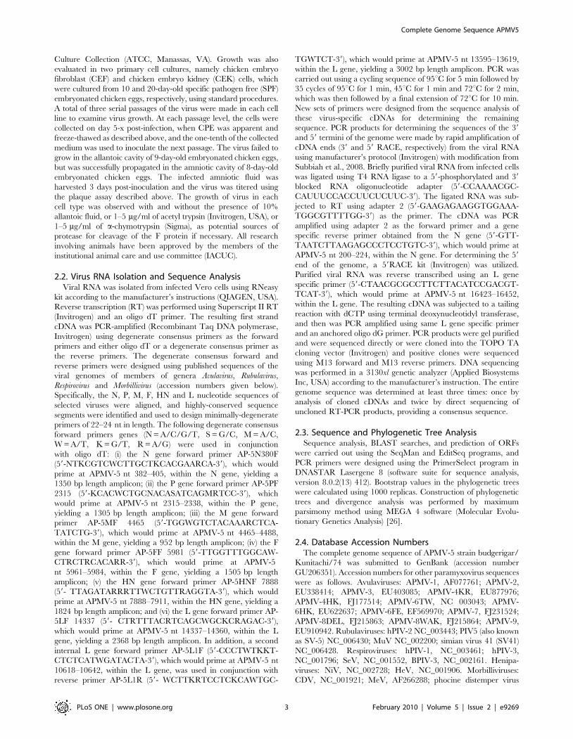
The genome organization of APMV-5 strain Kunitachi is 39-
leader-N-P/V/W-M-F-HN-L-trailer-59 (Fig 1), similar to the
organization of the other members of genus Avulavirus except for
APMV-6, which has an additional small hydrophobic (SH) protein
gene between the F and HN genes [19]. Thus, each of the APMV
serotypes contains six genes except for APMV-6, which has seven.
Major features of the genome of APMV-5 strain Kunitachi are
summarized in Fig. 1 and are compared with prototype members
of the other APMV serotypes. The APMV-5 genome consists of
17,262 nt and thus is the longest among the APMVs. The genome
nt length is a multiple of six, conforming to the ‘‘rule of six’’, as is
the case for all members of subfamily Paramyxovirinae analyzed to
date [4,27].
Although the genome length of APMV-5 is substantially greater
than for the other serotypes, this was not reflected in increased
lengths for the encoded proteins: the amino acid lengths predicted
for the APMV-5 proteins are very similar to those of the cognate
proteins in the other APMV serotypes (Fig. 1). Instead, the
increased genome length of APMV-5 was mostly due to the
presence of unusually long 39(downstream) untranslated regions
(UTR) in the P (458 nt), M (531 nt), and HN (378 nt) genes, as
well as the presence of a long trailer region (below). The UTRs of
all the APMVs sequenced to date vary greatly in length. The
length of 59 (upstream) UTRs of the APMV-5 N, P, M, F, HN and
L genes are 67, 55, 123, 165, 112 and 67, nt, respectively
(Figure 1). For comparison, the range of nt lengths of the 59 UTRs
of the N, P, M, F, HN and L genes of the other eight APMV
serotypes are 48–85, 42–113, 34–113, 12–101, 43–103, and 11–
112, respectively. The lengths of the 39 UTRs of the APMV-5 N,
P, M, F, HN and L genes are 191, 458, 531, 228, 378 and 133,
respectively. For comparison, the nt lengths of the 39 UTRs of the
N, P, M, F, HN and L genes of the eight other APMV serotypes
range between (88–210), (91–245), (106–221), (42–204), (80–292)
and (69–291), respectively. Consistent with the longer lengths of
some of the UTRs of APMV-5, the percentage of the APMV-5
genome that encodes proteins is 81%, which is less than average
coding percentage (92%) of other members of subfamily
Paramyxovirinae [1,28].
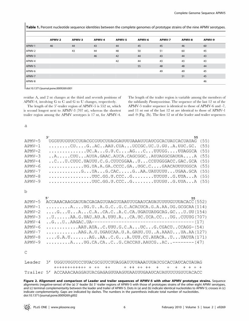
The genome of APMV-5 has 44%, 48%, 42%, 42%, 55%,
48%, 48% and 44% nt sequence identity with the genomes of
APMV-1, -2, -3, -4, -6, -7, -8 and -9, respectively (Table 1). The
GC content of the genome of APMV-5 is 40%, which is similar to
APMV-7 (39%) but significantly lower compared to the GC
contents of the genomes of APMV-1 (46%), APMV-2 (47%),
APMV-3 (46%), APMV-4 (47%), APMV-6 (46%), APMV-8 (43%)
and APMV-9 (45%).
The 39-leader sequence of APMV-5 consists of 55 nt, a length
that is conserved among all of the APMV and among almost all of
the members of the subfamily Paramyxovirinae. The APMV-5 leader
has a high degree of nucleotide sequence identity with those of the
other APMVs (Fig. 2a). The sequence of the first 12 nt of the
leader region of APMV-5 (39-UGGUUUGUUCCU-59, negative-
sense) is identical to that of APMV-2, -6, -7, -8 and -9. The first
8 nt of the leader sequence are the same for all the members of
genus Avulavirus sequenced to date except for a 1-nt change in the
third position of APMV-3, where residue G is replaced with
Complete Genome Sequence APMV5
PLoS ONE | www.plosone.org 4 February 2010 | Volume 5 | Issue 2 | e9269
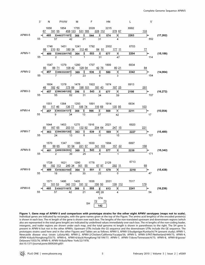
Figure 1. Gene map of APMV-5 and comparison with prototype strains for the other eight APMV serotypes (maps not to scale).Individual genes are indicated by rectangles, with the gene names given at the top of the Figure. The amino acid length(s) of the encoded protein(s)is shown in each box. The nt length of the gene is shown over each box. The lengths of the non-translated upstream and downstream regions (whichalso are represented in the total gene length) are indicated by underlined values immediately over each box. The nt lengths of the non-coding leader,intergenic, and trailer regions are shown under each map, and the total genome nt length is shown in parentheses to the right. The SH gene ispresent in APMV-6 but not in the other APMVs. Upstream UTRs include the GS sequence and the downstream UTRs include the GE sequence. Theprototypes strains used here and in the other Figures and Tables are as follows: APMV-5, APMV-5/budgerigar/Kunitachi/74 (present study); APMV-1,Newcastle disease virus (strain LaSota/46); APMV-2, APMV-2/Chicken/California/Yucaipa/56; APMV-3, APMV-3/PKT/Netherland/449/75; APMV-4,APMV-4/duck/Hongkong/D3/75; APMV-6, APMV-6/duck/HongKong/18/199/77; APMV-7, APMV-7/dove/Tennessee/4/75; APMV-8, APMV-8/goose/Delaware/1053/76; APMV-9, APMV-9/duck/New York/22/1978.doi:10.1371/journal.pone.0009269.g001
Complete Genome Sequence APMV5
PLoS ONE | www.plosone.org 5 February 2010 | Volume 5 | Issue 2 | e9269
residue A, and 2 nt changes at the third and seventh positions of
APMV-4, involving G to C and G to U changes, respectively.
The length of the 59-trailer region of APMV-5 is 552 nt, which
is second longest next to APMV-3 (707 nt), whereas the shortest
trailer region among the APMV serotypes is 17 nt, for APMV-4.
The length of the trailer region is variable among the members of
the subfamily Paramyxovirinae. The sequence of the last 12 nt of the
APMV-5 trailer sequence is identical to those of APMV-6 and -7,
and 11 nt out of the last 12 nt are identical to those of APMV-1
and -9 (Fig. 2b). The first 12 nt of the leader and trailer sequences
Table 1. Percent nucleotide sequence identities between the complete genomes of prototype strains of the nine APMV serotypes.
APMV-2 APMV-3 APMV-4 APMV-5 APMV-6 APMV-7 APMV-8 APMV-9
APMV-1 46 44 43 44 45 45 46 60
APMV-2 43 44 48 50 51 60 45
APMV-3 46 42 44 43 44 43
APMV-4 42 44 43 43 43
APMV-5 55 48 48 44
APMV-6 49 49 45
APMV-7 51 45
APMV-8 46
doi:10.1371/journal.pone.0009269.t001
Figure 2. Alignment and comparison of Leader and trailer sequences of APMV-5 with other APMV prototype strains. Sequencealignments (negative-sense) of the (a) 39-leader (b) 59-trailer regions of APMV-5 with those of prototypes strains of the other eight APMV serotypes,and (c) terminal complementarity between the leader and trailer of APMV-5. Dots in (a) and (b) indicate identical nucleotides to APMV-5; crosses in (c)indicate complementarity. Gaps are indicated by dashes. The numbers in the parenthesis indicate total number of nucleotides.doi:10.1371/journal.pone.0009269.g002
Complete Genome Sequence APMV5
PLoS ONE | www.plosone.org 6 February 2010 | Volume 5 | Issue 2 | e9269
of APMV-5 are 100% complementary to each other, which is
suggestive of conserved promoter elements at the 39 termini of the
genome and antigenome (Fig. 2c).
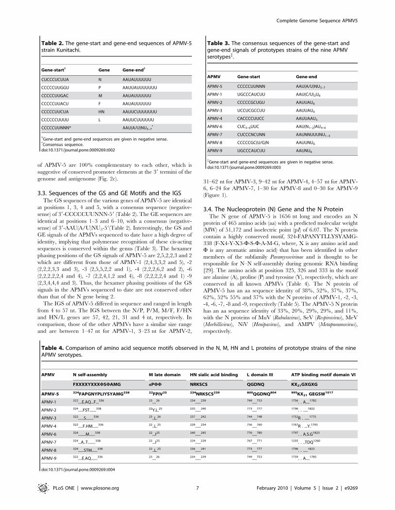
3.3. Sequences of the GS and GE Motifs and the IGSThe GS sequences of the various genes of APMV-5 are identical
at positions 1, 3, 4 and 5, with a consensus sequence (negative-
sense) of 39-CCCCCUUNNN-59 (Table 2). The GE sequences are
identical at positions 1–3 and 6–10, with a consensus (negative-
sense) of 39-AAU(A/U)NU5-59(Table 2). Interestingly, the GS and
GE signals of the APMVs sequenced to date have a high degree of
identity, implying that polymerase recognition of these cis-acting
sequences is conserved within the genus (Table 3). The hexamer
phasing positions of the GS signals of APMV-5 are 2,5,2,2,3 and 2
which are different from those of APMV-1 (2,4,3,3,2 and 5), -2
(2,2,2,3,3 and 3), -3 (2,5,5,2,2 and 1), -4 (2,2,2,6,2 and 2), -6
(2,2,2,2,2,4 and 4), -7 (2,2,4,1,2 and 4), -8 (2,2,2,2,4 and 1) -9
(2,3,4,4,4 and 3). Thus, the hexamer phasing positions of the GS
signals in the APMVs sequenced to date are not conserved other
than that of the N gene being 2.
The IGS of APMV-5 differed in sequence and ranged in length
from 4 to 57 nt. The IGS between the N/P, P/M, M/F, F/HN
and HN/L genes are 57, 42, 21, 31 and 4 nt, respectively. In
comparison, those of the other APMVs have a similar size range
and are between 1–47 nt for APMV-1, 3–23 nt for APMV-2,
31–62 nt for APMV-3, 9–42 nt for APMV-4, 4–57 nt for APMV-
6, 6–24 for APMV-7, 1–30 for APMV-8 and 0–30 for APMV-9
(Figure 1).
3.4. The Nucleoprotein (N) Gene and the N ProteinThe N gene of APMV-5 is 1656 nt long and encodes an N
protein of 465 amino acids (aa) with a predicted molecular weight
(MW) of 51,172 and isoelectric point (pI) of 6.07. The N protein
contain a highly conserved motif, 324-FAPANYTLLYSYAMG-
338 (F-X4-Y-X3-W-S-W-A-M-G, where, X is any amino acid and
W is any aromatic amino acid) that has been identified in other
members of the subfamily Paramyxovirinae and is thought to be
responsible for N–N self-assembly during genomic RNA binding
[29]. The amino acids at position 325, 326 and 333 in the motif
are alanine (A), proline (P) and tyrosine (Y), respectively, which are
conserved in all known APMVs (Table 4). The N protein of
APMV-5 has an aa sequence identity of 38%, 52%, 37%, 37%,
62%, 52% 55% and 37% with the N proteins of APMV-1, -2, -3,
-4, -6, -7, -8 and -9, respectively (Table 5). The APMV-5 N protein
has an aa sequence identity of 33%, 20%, 29%, 29%, and 11%,
with the N proteins of MuV (Rubulavirus), SeV (Respirovirus), MeV
(Morbillivirus), NiV (Henipavirus), and AMPV (Metapneumovirus),
respectively.
Table 2. The gene-start and gene-end sequences of APMV-5strain Kunitachi.
Gene-start{ Gene Gene-end{
CUCCCUCUUA N AAUAUUUUUU
CCCCCUUGGU P AAUUAUUUUUUU
CCCCCUUGAC M AAUAUUUUUU
CCCCCUUACU F AAUAUUUUUU
CCCCCUUCUA HN AAUUCUUUUUUU
CCCCCCUUUU L AAUUCUUUUUU
CCCCCUUNNN* AAU(A/U)NU5–7*
{Gene-start and gene-end sequences are given in negative sense.*Consensus sequence.doi:10.1371/journal.pone.0009269.t002
Table 3. The consensus sequences of the gene-start andgene-end signals of prototypes strains of the nine APMVserotypes{.
APMV Gene-start Gene-end
APMV-5 CCCCCUUNNN AAU(A/U)NU5–7
APMV-1 UGCCCAUCUU AAU(C/U)2U6
APMV-2 CCCCCGCUGU AAUUAU6
APMV-3 UCCUCGCCUU AAUUAU6
APMV-4 CACCCCUUCC AAUUAAU5
APMV-6 CUC5–6UUC AAU(N1–2)AU4–6
APMV-7 CUCCCNCUNN AAUNNUUUNU1–3
APMV-8 CCCCCGC(U/G)N AAUUNU6
APMV-9 UGCCCAUCUU AAUNU6
{Gene-start and gene-end sequences are given in negative sense.doi:10.1371/journal.pone.0009269.t003
Table 4. Comparison of amino acid sequence motifs observed in the N, M, HN and L proteins of prototype strains of the nineAPMV serotypes.
APMV N self-assembly M late domain HN sialic acid binding L domain III ATP binding motif domain VI
FXXXXYXXXWSWAMG aPWW NRKSCS QGDNQ KX21GXGXG
APMV-5 324FAPGNYPLIYSYAMG338 22FPIV25 234NRKSCS239 800QGDNQ804 995KX21 GEGSW1017
APMV-1 322....E.AQ...F...336 23....26 234......239 749.....753 1756. . A....1782
APMV-2 324.....FST.......338 22Y.L.25 235......240 773.....777 1796. . .....1822
APMV-3 322......S........336 23..L.26 237......242 744.....748 1752R . .....1775
APMV-4 322.....F.HM......336 22..L.25 229......234 756.....760 1767R . ...Y.1793
APMV-6 324........M......338 22...I25 240......245 776.....780 1797. . A.S.G1823
APMV-7 324...A..T........338 22...I25 224......229 767.....771 1235. . ..TDQ1260
APMV-8 324......STM......338 22..L.25 236......241 773.....777 1796. . .....1822
APMV-9 322....E.AQ.......336 23....26 234......239 749.....753 1759. . A....1785
doi:10.1371/journal.pone.0009269.t004
Complete Genome Sequence APMV5
PLoS ONE | www.plosone.org 7 February 2010 | Volume 5 | Issue 2 | e9269
3.5. The Phosphoprotein (P) Gene and the P, V, and WProteins
The P gene of APMV-5 is 1854 nt long and encodes a P pro-
tein of 446 aa with a MW of 48,906 and pI of 5.92. The predicted
P protein contains 38 potential sites for phosphorylation, as
predicted by the NetPhos 2.0 program of Expasy proteomics
server. These include 29 potential sites of serine phosphorylation
(28S, 31S, 65S, 67S, 80S, 93S, 98S, 112S, 131S, 138S, 159S,
164S, 171S, 194S, 233S, 240S, 280S, 288S, 301S, 308S, 349S,
379S, 383S, 388S, 410S, 415S, 425S, 427S and 431S), 7 potential
sites of threonine phosphorylation (40T, 133T, 156T, 185T,
198T, 222T and 419T), and 2 potential sites for tyrosine
phosphorylation (36Y and 193Y). The P protein of APMV-5 has
an aa sequence identity of 20%, 26%, 19%, 22%, 28%, 22%, 27%
and 21% with the P proteins of APMV-1, -2, -3, -4, -6, -7, -8 and
-9, respectively (Table 5). The P protein of APMV-5 has an aa
sequence identity of 16%, 12%, 11%, 10%, and 9%, with the P
proteins of MuV (Rubulavirus), SeV (Respirovirus), MeV (Morbillivi-
rus), NiV (Henipavirus), and AMPV (Metapneumovirus), respectively.
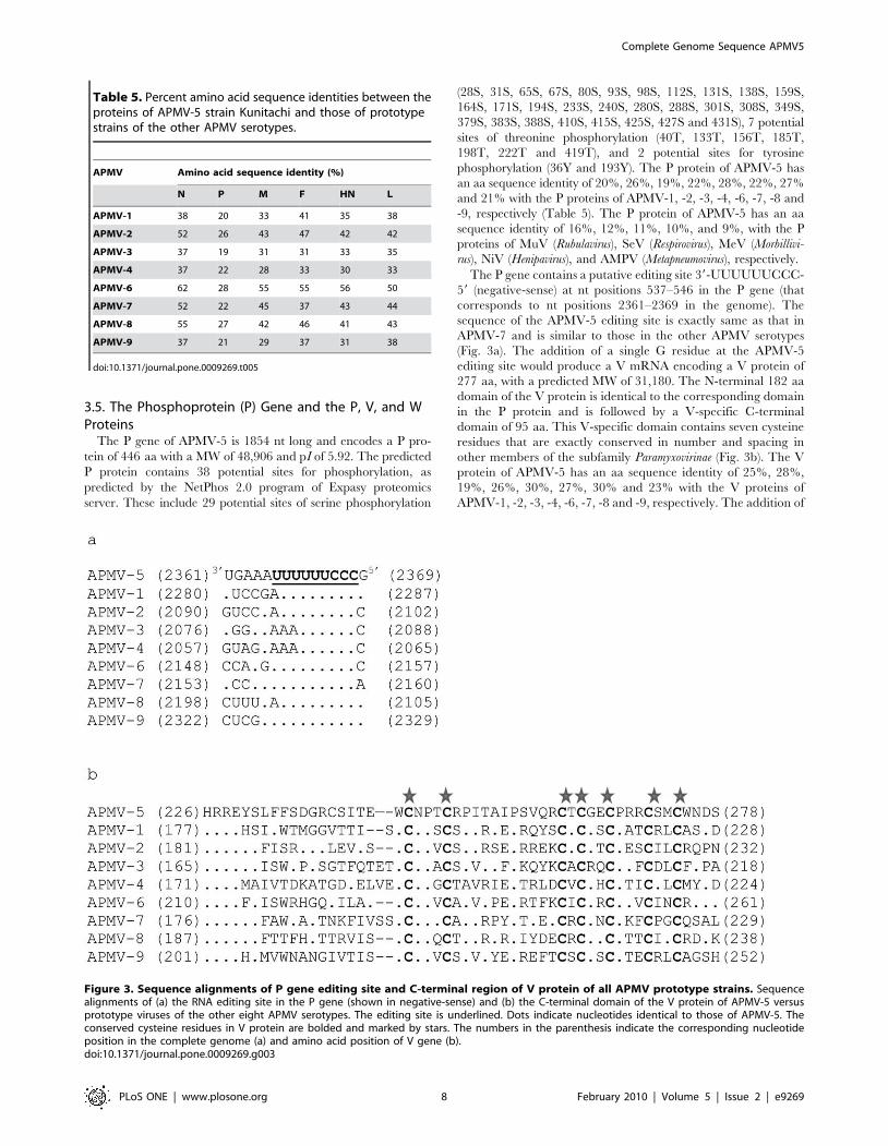
The P gene contains a putative editing site 39-UUUUUUCCC-
59 (negative-sense) at nt positions 537–546 in the P gene (that
corresponds to nt positions 2361–2369 in the genome). The
sequence of the APMV-5 editing site is exactly same as that in
APMV-7 and is similar to those in the other APMV serotypes
(Fig. 3a). The addition of a single G residue at the APMV-5
editing site would produce a V mRNA encoding a V protein of
277 aa, with a predicted MW of 31,180. The N-terminal 182 aa
domain of the V protein is identical to the corresponding domain
in the P protein and is followed by a V-specific C-terminal
domain of 95 aa. This V-specific domain contains seven cysteine
residues that are exactly conserved in number and spacing in
other members of the subfamily Paramyxovirinae (Fig. 3b). The V
protein of APMV-5 has an aa sequence identity of 25%, 28%,
19%, 26%, 30%, 27%, 30% and 23% with the V proteins of
APMV-1, -2, -3, -4, -6, -7, -8 and -9, respectively. The addition of
Table 5. Percent amino acid sequence identities between theproteins of APMV-5 strain Kunitachi and those of prototypestrains of the other APMV serotypes.
APMV Amino acid sequence identity (%)
N P M F HN L
APMV-1 38 20 33 41 35 38
APMV-2 52 26 43 47 42 42
APMV-3 37 19 31 31 33 35
APMV-4 37 22 28 33 30 33
APMV-6 62 28 55 55 56 50
APMV-7 52 22 45 37 43 44
APMV-8 55 27 42 46 41 43
APMV-9 37 21 29 37 31 38
doi:10.1371/journal.pone.0009269.t005
Figure 3. Sequence alignments of P gene editing site and C-terminal region of V protein of all APMV prototype strains. Sequencealignments of (a) the RNA editing site in the P gene (shown in negative-sense) and (b) the C-terminal domain of the V protein of APMV-5 versusprototype viruses of the other eight APMV serotypes. The editing site is underlined. Dots indicate nucleotides identical to those of APMV-5. Theconserved cysteine residues in V protein are bolded and marked by stars. The numbers in the parenthesis indicate the corresponding nucleotideposition in the complete genome (a) and amino acid position of V gene (b).doi:10.1371/journal.pone.0009269.g003
Complete Genome Sequence APMV5
PLoS ONE | www.plosone.org 8 February 2010 | Volume 5 | Issue 2 | e9269

two G residues at the editing site would produce a W mRNA
encoding a W protein of 187 aa, with a predicted MW of 20,727.
The putative W protein contains an N-terminal domain of 182 aa
that is identical to that of P followed by five additional residues
that are unique to W. The W protein of APMV-5 has an aa
sequence identity of 16%, 22%, 15%, 28%, 28%, 22%, 22% and
24% with the W proteins of APMV-1, -2, -3, -4, -6, -7, -8 and -9,
respectively.
3.6. The Matrix (M) Gene and the M ProteinThe M gene of APMV-5 is 1755 nt long and encodes a M
protein of 366 aa with a predicted MW of 39,999. The pI value of
the M protein is 9.31: this basic nature is thought to mediate ionic
interactions with the acidic N protein [1]. The M proteins of some
members of the subfamily Paramyxovirinae contains a motif (242-
KKTNRKGADRSVLQIKEKVRK-262 in APMV-5) that con-
sists of a bipartite clustering of basic residues. This motif is thought
to serve as a nuclear localization signal (NLS) for the protein
[6,30], although the significance of this for infection remains
unclear. A protein–protein interaction domain called the ‘late
domain’, which contains the motif FPIV and is involved in
assembly and budding of viruses, was first identified in the
paramyxovirus SV5 [31]. The M protein of APMV-5 contains a
potential late domain motif FPIV at aa positions 22–25. Similar
motifs are present in the M proteins of other APMV, namely: 22-
YPLI-25, APMV-2; 23-FPLI-26, APMV-3; 22-FPLI-25, APMV-
4; 22-FPLV-25, APMV-6; 22-FPII-25, APMV-7; 22-FPLV-25,
APMV-8; and 23-FPIV-26, APMV-9 (Table 5). In the late domain
motif, the amino acid proline (P) in the second position is
conserved in all the members of genera Avulavirus and Rubulavirus,
with the consensus pattern a-P-W-W (a represents an aliphatic
amino acid and W represents an aromatic amino acid). However,
this motif is not observed in the M proteins of members of genera
Respirovirus, Morbillivirus and Henipavirus of the subfamily Paramyx-
ovirinae. The M protein of APMV-5 has an aa sequence identity of
33%, 43%, 31%, 28%, 55%, 45%, 42% and 29% with the M
proteins of APMV-1, -2, -3, -4, -6, -7, -8 and -9, respectively
(Table 5). The APMV-5 M protein has an aa sequence identity of
28%, 22%, 20%, 21%, and 14%, with the M proteins of MuV
(Rubulavirus), SeV (Respirovirus), MeV (Morbillivirus), NiV (Henipa-
virus), and AMPV (Metapneumovirus), respectively.
3.7. The Fusion (F) Gene and the F ProteinThe F gene of APMV-5 is 2028 nt long and encodes a F protein
of 544 aa with a predicted MW of 58,724 and pI of 8.34. The
F protein is predicted to be a type I transmembrane protein
similar to the F proteins of the other members of the family
Paramyxoviridae. It contains a signal sequence of 25 aa (positions
1–25) at the N-terminal end and a predicted transmembrane
domain located at aa sequence positions 494–516. The putative
cleavage site of the APMV-5 F0 protein precursor is G-K-R-K-K-RQF (aa positions 104–110), with the residue F representing the
newly-created N-terminus of the F1 fusion peptide. The cleavage
site sequence of APMV-5 conforms to the favored sequence motif
for cleavage by the intracellular protease furin, R-X-K/R-RQF
[32]. Interestingly, the cleavage site of APMV-5 contains five
tandem lysine and arginine residues (KRKKR), which is the
greatest number of basic residues for any of the APMV (Table 6)
except for some strains of APMV-1 [33]. The F1 polypeptide of
APMV-5 contains heptad repeats at aa positions 134–182 and
455–485, as predicted by the LearnCoil-VMF program, which
correspond to the characteristic HRA and HRB heptad repeats
that are found in paramyxovirus F proteins and are thought to be
important for the conformational changes that lead to membrane
fusion [34]. The APMV-5 F protein contains five potential N-
linked glycosylation sites located at aa position 74 of the F2 subunit
and at aa positions 189, 359, 440 and 464 of the F1 subunit, as
predicted by the NetNGlyc 1.0 program of the Expasy proteomics
server. The F protein of APMV-5 has an aa sequence identity of
41%, 47%, 31%, 33%, 55%, 37%, 46% and 37% with the F
proteins of APMV-1, -2, -3, -4, -6, -7, -8 and -9, respectively
(Table 5). The APMV-5 F protein has an aa sequence identity of
33%, 24%, 27%, 24%, and 19%, with the F proteins of MuV
(Rubulavirus), SeV (Respirovirus), MeV (Morbillivirus), NiV (Henipavirus)
and AMPV (Metapneumovirus), respectively.
3.8. The Hemagglutinin–Neuraminidase (HN) Gene andthe HN Protein
The HN gene of APMV-5 is 2215 nt long and encodes a HN
protein of 574 aa with a predicted MW of 63,911 and pI of 6.78.
The HN protein is predicted to be a type II integral membrane
protein and has a predicted hydrophobic signal/anchor domain
located at aa residues 21 and 43 at the N-terminus. There are six
potential N-linked glycosylation sites predicted at aa positions 58,
119, 148, 278, 346 and 383. The HN protein contains the unique
motif N-R-K-S-C-S at aa positions 234–239, which is thought to
be involved in sialic acid binding at the cell surface [35,36]. This
motif is conserved in the HN proteins of all APMVS sequenced to
date (Table 4). Comparing the seven conserved residues of the
proposed neuraminidase active site found in the HN protein of
NDV [37,38], the corresponding residues in the APMV-5 HN are:
R(174), S(202), E(399), R(414), R(504), Y(532) and E(550). In
addition, the APMV-5 HN protein has eleven conserved cysteine
residues at aa positions 186, 196, 238, 247, 251, 344, 461, 467,
471, 534 and 545. The HN protein of APMV-5 has an aa
sequence identity of 35%, 42%, 33%, 30%, 56%, 43%, 41% and
31% with the HN proteins of APMV-1, -2, -3, -4, -6, -7, -8 and -9,
respectively (Table 5). APMV-5 HN protein has an aa sequence
identity of 33%, 24%, 11%, 16%, and 8%, with the HN proteins
of MuV (Rubulavirus), SeV (Respirovirus), MeV (Morbillivirus), NiV
(Henipavirus) and AMPV (Metapneumovirus), respectively.
Table 6. Comparison of the putative cleavage sites of the F0
proteins of prototypes strains of the nine APMV serotypes,and requirements for exogenous protease.
APMVF0 proteincleavage site{
Requirement forexogenous protease{
APMV-5 GKRKKRQFVG 2
APMV-1(Avirulent) GGRQGRQLIG +
APMV-1(Virulent) GRRQKRQFIG 2
APMV-2 DKPASRQFVG 2
APMV-3 ARPRGRQLFG +
APMV-4 ADIQPRQFIG 2
APMV-6 PAPEPRQLIG 2
APMV-7 TLPSSRQFAG 2
APMV-8 TYPQTRQLIG +
APMV-9 RIREGRQIFG +
{Basic amino acids (R/K) are underlined and in bold, and the downward arrowindicates the predicted site of cleavage.{Virus replication in cell culture requires the supplementation of 10% v/v freshallantoic fluid or acetylated trypsin (1 mg/ml).
Note: APMV-1 (Avirulent) denotes strain LaSota, APMV-1 (Virulent) denotesstrain GB Texas, APMV-2 to APMV-9 includes all the prototype strains.doi:10.1371/journal.pone.0009269.t006
Complete Genome Sequence APMV5
PLoS ONE | www.plosone.org 9 February 2010 | Volume 5 | Issue 2 | e9269
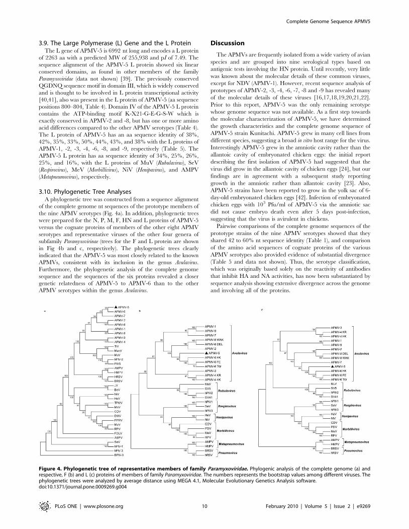
3.9. The Large Polymerase (L) Gene and the L ProteinThe L gene of APMV-5 is 6992 nt long and encodes a L protein
of 2263 aa with a predicted MW of 255,938 and pI of 7.49. The
sequence alignment of the APMV-5 L protein showed six linear
conserved domains, as found in other members of the family
Paramyxoviridae (data not shown) [39]. The previously conserved
QGDNQ sequence motif in domain III, which is widely conserved
and is thought to be involved in L protein transcriptional activity
[40,41], also was present in the L protein of APMV-5 (aa sequence
positions 800–804, Table 4). Domain IV of the APMV-5 L protein
contains the ATP-binding motif K-X21-G-E-G-S-W which is
exactly conserved in APMV-2 and -8, but has one or more amino
acid differences compared to the other APMV serotypes (Table 4).
The L protein of APMV-5 has an aa sequence identity of 38%,
42%, 35%, 33%, 50%, 44%, 43%, and 38% with the L proteins of
APMV-1, -2, -3, -4, -6, -8, and -9, respectively (Table 5). The
APMV-5 L protein has aa sequence identity of 34%, 25%, 26%,
25%, and 16%, with the L proteins of MuV (Rubulavirus), SeV
(Respirovirus), MeV (Morbillivirus), NiV (Henipavirus), and AMPV
(Metapneumovirus), respectively.
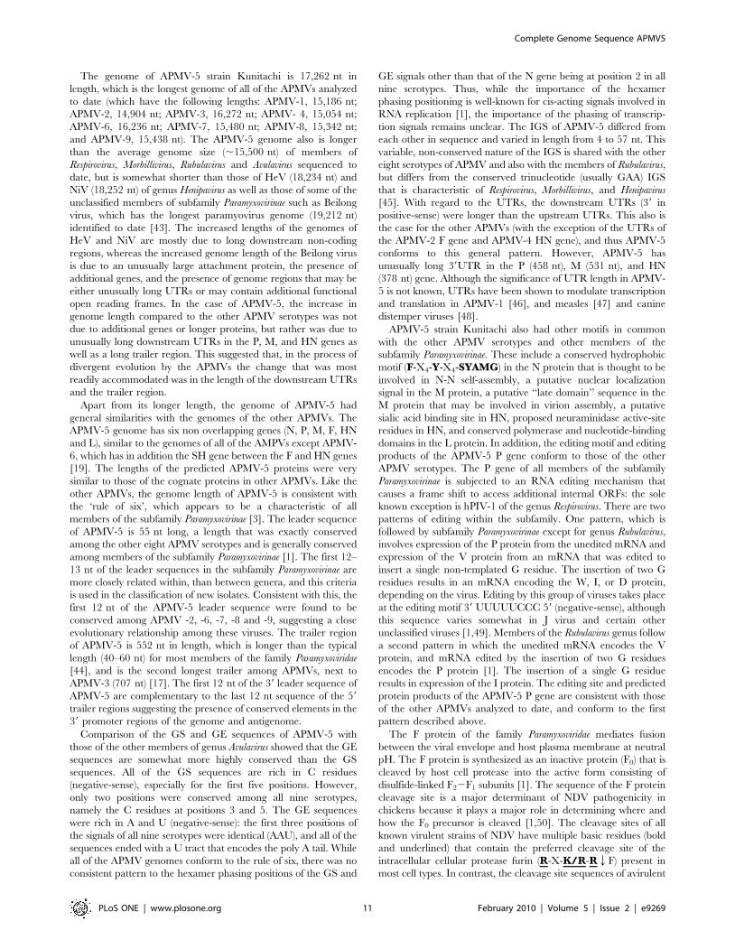
3.10. Phylogenetic Tree AnalysesA phylogenetic tree was constructed from a sequence alignment
of the complete genome nt sequences of the prototype members of
the nine APMV serotypes (Fig. 4a). In addition, phylogenetic trees
were prepared for the N, P, M, F, HN and L proteins of APMV-5
versus the cognate proteins of members of the other eight APMV
serotypes and representative viruses of the other four genera of
subfamily Paramyxovirinae (trees for the F and L protein are shown
in Fig 4b and c, respectively). The phylogenetic trees clearly
indicated that the APMV-5 was most closely related to the known
APMVs, consistent with its inclusion in the genus Avulavirus.
Furthermore, the phylogenetic analysis of the complete genome
sequence and the sequences of the six proteins revealed a closer
genetic relatedness of APMV-5 to APMV-6 than to the other
APMV serotypes within the genus Avulavirus.
Discussion
The APMVs are frequently isolated from a wide variety of avian
species and are grouped into nine serological types based on
antigenic tests involving the HN protein. Until recently, very little
was known about the molecular details of these common viruses,
except for NDV (APMV-1). However, recent sequence analysis of
prototypes of APMV-2, -3, -4, -6, -7, -8 and -9 has revealed many
of the molecular details of these viruses [16,17,18,19,20,21,22].
Prior to this report, APMV-5 was the only remaining serotype
whose genome sequence was not available. As a first step towards
the molecular characterization of APMV-5, we have determined
the growth characteristics and the complete genome sequence of
APMV-5 strain Kunitachi. APMV-5 grew in many cell lines from
different species, suggesting a broad in vitro host range for the virus.
Interestingly APMV-5 grew in the amniotic cavity rather than the
allantoic cavity of embryonated chicken eggs: the initial report
describing the first isolation of APMV-5 had suggested that the
virus did grow in the allantoic cavity of chicken eggs [24], but our
findings are in agreement with a subsequent study reporting
growth in the amniotic rather than allantoic cavity [23]. Also,
APMV-5 strains have been reported to grow in the yolk sac of 6-
day-old embryonated chicken eggs [42]. Infection of embryonated
chicken eggs with 103 Pfu/ml of APMV-5 via the amniotic sac
did not cause embryo death even after 5 days post-infection,
suggesting that the virus is avirulent in chickens.
Pairwise comparisons of the complete genome sequences of the
prototype strains of the nine APMV serotypes showed that they
shared 42 to 60% nt sequence identity (Table 1), and comparison
of the amino acid sequences of cognate proteins of the various
APMV serotypes also provided evidence of substantial divergence
(Table 5 and data not shown). Thus, the serotype classification,
which was originally based solely on the reactivity of antibodies
that inhibit HA and NA activities, has now been substantiated by
sequence analysis showing extensive divergence across the genome
and involving all of the proteins.
Figure 4. Phylogenetic tree of representative members of family Paramyxoviridae. Phylogenic analysis of the complete genome (a) andrespective, F (b) and L (c) proteins of members of family Paramyxoviridae. The numbers represents the bootstrap values among different viruses. Thephylogenetic trees were analyzed by average distance using MEGA 4.1, Molecular Evolutionary Genetics Analysis software.doi:10.1371/journal.pone.0009269.g004
Complete Genome Sequence APMV5
PLoS ONE | www.plosone.org 10 February 2010 | Volume 5 | Issue 2 | e9269
The genome of APMV-5 strain Kunitachi is 17,262 nt in
length, which is the longest genome of all of the APMVs analyzed
to date (which have the following lengths: APMV-1, 15,186 nt;
APMV-2, 14,904 nt; APMV-3, 16,272 nt; APMV- 4, 15,054 nt;
APMV-6, 16,236 nt; APMV-7, 15,480 nt; APMV-8, 15,342 nt;
and APMV-9, 15,438 nt). The APMV-5 genome also is longer
than the average genome size (,15,500 nt) of members of
Respirovirus, Morbillivirus, Rubulavirus and Avulavirus sequenced to
date, but is somewhat shorter than those of HeV (18,234 nt) and
NiV (18,252 nt) of genus Henipavirus as well as those of some of the
unclassified members of subfamily Paramyxovirinae such as Beilong
virus, which has the longest paramyovirus genome (19,212 nt)
identified to date [43]. The increased lengths of the genomes of
HeV and NiV are mostly due to long downstream non-coding
regions, whereas the increased genome length of the Beilong virus
is due to an unusually large attachment protein, the presence of
additional genes, and the presence of genome regions that may be
either unusually long UTRs or may contain additional functional
open reading frames. In the case of APMV-5, the increase in
genome length compared to the other APMV serotypes was not
due to additional genes or longer proteins, but rather was due to
unusually long downstream UTRs in the P, M, and HN genes as
well as a long trailer region. This suggested that, in the process of
divergent evolution by the APMVs the change that was most
readily accommodated was in the length of the downstream UTRs
and the trailer region.
Apart from its longer length, the genome of APMV-5 had
general similarities with the genomes of the other APMVs. The
APMV-5 genome has six non overlapping genes (N, P, M, F, HN
and L), similar to the genomes of all of the AMPVs except APMV-
6, which has in addition the SH gene between the F and HN genes
[19]. The lengths of the predicted APMV-5 proteins were very
similar to those of the cognate proteins in other APMVs. Like the
other APMVs, the genome length of APMV-5 is consistent with
the ‘rule of six’, which appears to be a characteristic of all
members of the subfamily Paramyxovirinae [3]. The leader sequence
of APMV-5 is 55 nt long, a length that was exactly conserved
among the other eight APMV serotypes and is generally conserved
among members of the subfamily Paramyxovirinae [1]. The first 12–
13 nt of the leader sequences in the subfamily Paramyxovirinae are
more closely related within, than between genera, and this criteria
is used in the classification of new isolates. Consistent with this, the
first 12 nt of the APMV-5 leader sequence were found to be
conserved among APMV -2, -6, -7, -8 and -9, suggesting a close
evolutionary relationship among these viruses. The trailer region
of APMV-5 is 552 nt in length, which is longer than the typical
length (40–60 nt) for most members of the family Paramyxoviridae
[44], and is the second longest trailer among APMVs, next to
APMV-3 (707 nt) [17]. The first 12 nt of the 39 leader sequence of
APMV-5 are complementary to the last 12 nt sequence of the 59
trailer regions suggesting the presence of conserved elements in the
39 promoter regions of the genome and antigenome.
Comparison of the GS and GE sequences of APMV-5 with
those of the other members of genus Avulavirus showed that the GE
sequences are somewhat more highly conserved than the GS
sequences. All of the GS sequences are rich in C residues
(negative-sense), especially for the first five positions. However,
only two positions were conserved among all nine serotypes,
namely the C residues at positions 3 and 5. The GE sequences
were rich in A and U (negative-sense): the first three positions of
the signals of all nine serotypes were identical (AAU), and all of the
sequences ended with a U tract that encodes the poly A tail. While
all of the APMV genomes conform to the rule of six, there was no
consistent pattern to the hexamer phasing positions of the GS and
GE signals other than that of the N gene being at position 2 in all
nine serotypes. Thus, while the importance of the hexamer
phasing positioning is well-known for cis-acting signals involved in
RNA replication [1], the importance of the phasing of transcrip-
tion signals remains unclear. The IGS of APMV-5 differed from
each other in sequence and varied in length from 4 to 57 nt. This
variable, non-conserved nature of the IGS is shared with the other
eight serotypes of APMV and also with the members of Rubulavirus,
but differs from the conserved trinucleotide (usually GAA) IGS
that is characteristic of Respirovirus, Morbillivirus, and Henipavirus
[45]. With regard to the UTRs, the downstream UTRs (39 in
positive-sense) were longer than the upstream UTRs. This also is
the case for the other APMVs (with the exception of the UTRs of
the APMV-2 F gene and APMV-4 HN gene), and thus APMV-5
conforms to this general pattern. However, APMV-5 has
unusually long 39UTR in the P (458 nt), M (531 nt), and HN
(378 nt) gene. Although the significance of UTR length in APMV-
5 is not known, UTRs have been shown to modulate transcription
and translation in APMV-1 [46], and measles [47] and canine
distemper viruses [48].
APMV-5 strain Kunitachi also had other motifs in common
with the other APMV serotypes and other members of the
subfamily Paramyxovirinae. These include a conserved hydrophobic
motif (F-X4-Y-X4-SYAMG) in the N protein that is thought to be
involved in N-N self-assembly, a putative nuclear localization
signal in the M protein, a putative ‘‘late domain’’ sequence in the
M protein that may be involved in virion assembly, a putative
sialic acid binding site in HN, proposed neuraminidase active-site
residues in HN, and conserved polymerase and nucleotide-binding
domains in the L protein. In addition, the editing motif and editing
products of the APMV-5 P gene conform to those of the other
APMV serotypes. The P gene of all members of the subfamily
Paramyxovirinae is subjected to an RNA editing mechanism that
causes a frame shift to access additional internal ORFs: the sole
known exception is hPIV-1 of the genus Respirovirus. There are two
patterns of editing within the subfamily. One pattern, which is
followed by subfamily Paramyxovirinae except for genus Rubulavirus,
involves expression of the P protein from the unedited mRNA and
expression of the V protein from an mRNA that was edited to
insert a single non-templated G residue. The insertion of two G
residues results in an mRNA encoding the W, I, or D protein,
depending on the virus. Editing by this group of viruses takes place
at the editing motif 39 UUUUUCCC 59 (negative-sense), although
this sequence varies somewhat in J virus and certain other
unclassified viruses [1,49]. Members of the Rubulavirus genus follow
a second pattern in which the unedited mRNA encodes the V
protein, and mRNA edited by the insertion of two G residues
encodes the P protein [1]. The insertion of a single G residue
results in expression of the I protein. The editing site and predicted
protein products of the APMV-5 P gene are consistent with those
of the other APMVs analyzed to date, and conform to the first
pattern described above.
The F protein of the family Paramyxoviridae mediates fusion
between the viral envelope and host plasma membrane at neutral
pH. The F protein is synthesized as an inactive protein (F0) that is
cleaved by host cell protease into the active form consisting of
disulfide-linked F22F1 subunits [1]. The sequence of the F protein
cleavage site is a major determinant of NDV pathogenicity in
chickens because it plays a major role in determining where and
how the F0 precursor is cleaved [1,50]. The cleavage sites of all
known virulent strains of NDV have multiple basic residues (bold
and underlined) that contain the preferred cleavage site of the
intracellular cellular protease furin (R-X-K/R-RQF) present in
most cell types. In contrast, the cleavage site sequences of avirulent
Complete Genome Sequence APMV5
PLoS ONE | www.plosone.org 11 February 2010 | Volume 5 | Issue 2 | e9269
NDV strains characteristically have one or a few basic residues
immediately upstream of the cleavage site that do not create a
furin cleavage site. The ability of the NDV F protein to be cleaved
by furin provides the opportunity for viral replication in a wide
variety of tissues, which is essential for systemic spread of the virus.
The presence of a non-furin cleavage site as in avirulent NDV
strains restricts viral replication to the respiratory and enteric
tracts, where secretory proteases necessary for F cleavage are
present. The ability of NDV F to be cleaved also is affected by the
amino acid that immediately follows the cleavage site and becomes
the N-terminal residue of the F1 subunit: the presence of leucine
in this position is associated with less efficient cleavage [34].
Surprisingly, however, some of the other APMV serotypes do not
conform to the NDV paradigm. For example, for a number of the
other eight serotypes, the sequence of the cleavage site does not
predict whether exogenous protease is needed for growth in vitro.
Specifically, the F protein cleavage sites of APMV-2 (PASRQF),
APMV-4 (IQPRQF) and APMV-7 (PSSRQF) all contain a single
basic residue (R) immediately upstream of the putative cleavage
site, but do not require exogenous protease supplementation for
growth in cell culture [16,18,20]. Other APMV serotypes do
conform more closely with the NDV paradigm: specifically, they
have a non-furin cleavage site and do require exogenous protease
for replication in vitro. These viruses are APMV-3 (PRGRQL),
APMV-8 (PQTRQL), and APMV-9 (REGRQI). It also is
noteworthy that these three serotypes have leucine or isoleucine
immediately downstream of the cleavage site. APMV-6 also has a
single basic residue immediately upstream of the putative cleavage
site (PEPRQL), but the requirement for trypsin varies with the
strain (S. Xiao, P.L.C. and S.K.S, unpublished data). APMV-5
strain Kunitachi contains five tandem basic amino acid residues at
the putative cleavage site (105KRKKRQF110), and thus contains
even more basic residues than most of the virulent strains of NDV
sequenced to date. The presence of this highly basic cleavage
sequence in APMV-5 may be responsible for the high pathoge-
nicity of this virus in budgerigars, where it can cause up to 100%
mortality [23]. However, it is interesting that this same virus was
completely avirulent in chicken eggs. This is suggestive of a host
range difference that is not ameliorated by the presence of a multi-
basic cleavage site that otherwise would confer virulence in
chickens. Similarly, some of the other APMV serotypes that did
not require trypsin for cleavage in vitro nonetheless had low
virulence in chickens (e.g. APMV-2, -4, and -7). Thus, for a
number of the APMV serotypes, the cleavage site sequence did not
predict the cleavage phenotype in vitro nor virulence in vivo.
All members of genus Avulavirus (APMV-1 to 9) have both
hemagglutination and neuraminidase activities except for APMV-
5 [23]. Although the APMV-5 HN protein has neuraminidase
activity, it neither hemagglutinated nor hemadsorbed RBCs of
chickens, turkeys, guinea pigs, horses, or human O-group, either at
37uC or at 4uC. The sequence of the APMV-5 stain Kunitachi
HN protein provides evidence of the expected globular head,
N-terminal-proximal signal/anchor domain, putative sialic acid
binding motif (NRKSCS), seven conserved residues of the
proposed neuraminidase active site, and eleven conserved cysteine
residues that have been shown to be conserved among type II
integral membrane proteins of subfamily Paramyxovirinae [38].
Thus, the lack of HA activity for APMV-5 cannot be ascribed to
an obvious difference in HN structure. Non-hemagglutinating
highly virulent APMV-1 strains which grow in the allantoic cavity,
have been reported from cormorants (also a psittacine bird) in
Canada [51]. These observations indicate that the HA activity of
the HN protein is not required for replication of APMVs. APMV-
5 failed to grow in the allantoic cavity of embryonated chicken
eggs, in contrast to all hemagglutinating APMVs, which grow in
the allantoic cavity. It is tempting to suggest that there is a link
between these observations: specifically, that the lack of a strong
HA in APMV-5 renders the virus unable to bind to receptors
present on cells of the allantoic cavity. However, this hypothesis
requires further study.
In summary, APMV-5 Kunitachi strain differs significantly
from all other APMVs in the following ways; (i) having isolated
only from budgerigars, (ii) the presence of an HN protein with no
HA activity, (iii) the inability to grow in the allantoic cavity of
embryonated chicken eggs, (iv) the presence of unusually long
39UTRs in P, M and HN genes, and (v) the longest genome
among the APMVs. The sequence alignment and phylogenetic
analysis showed that APMV-5 clusters with the other APMVs,
justifying its classification in the genus Avulavirus. Phylogenetically,
APMV-5 is more closely related to APMV-6 than to the other
known APMVs. Further sequence analysis of additional APMV-5
strains will help us understand the antigenic and genetic variation
among APMV-5 strains. Future studies on the epidemiology and
pathogenicity of this virus in different avian species will be
necessary to define the role of this virus in avian health.
Acknowledgments
We thank Drs. Ian Brown and D.J. Alexander, Veterinary Laboratories
Agency, Weybridge, Surrey, UK for providing APMV-5 strain Kunitachi.
We thank Sunil Khattar, Sa Xiao and Madhuri Subbiah for helpful
discussions in the experiments. We also thank Daniel Rockemann, Flavia
Militino Dias and all other laboratory members for their excellent technical
assistance and help.
Author Contributions
Conceived and designed the experiments: ASS PLC SKS. Performed the
experiments: ASS AP SK. Analyzed the data: ASS AP SK. Contributed
reagents/materials/analysis tools: PLC SKS. Wrote the paper: ASS PLC
SKS.
References
1. Lamb R, Parks G (2007) Paramyxoviridae: the viruses and their replication. In:
Knipe DM, Howley PM, Griffin DE, Lamb RA, Martin MA, Roizman B,
Straus SE, eds. Philadelphia: Lippincott Williams & Wilkins. pp 1449–1496.
2. Lamb RA, Collins PL, Kolakofsky D, Melero JA, Nagai Y, et al. (2005) Family
Paramyxoviridae. In: Fauquet CM, ed. Virus Taxonomy: The Classification and
Nomenclature of Viruses, The Eighth Report of the International Committee in
Taxonomy of Viruses. Press EA, editor.
3. Calain P, Roux L (1993) The rule of six, a basic feature for efficient replication of
Sendai virus defective interfering RNA. J Virol 67: 4822–4830.
4. Kolakofsky D, Pelet T, Garcin D, Hausmann S, Curran J, et al. (1998)
Paramyxovirus RNA synthesis and the requirement for hexamer genome length:
the rule of six revisited. J Virol 72: 891–899.
5. Curran J, Marq JB, Kolakofsky D (1995) An N-terminal domain of the Sendai
paramyxovirus P protein acts as a chaperone for the NP protein during the
nascent chain assembly step of genome replication. J Virol 69: 849–855.
6. Peeples ME (1991) Paramyxovirus M proteins : pulling it all together and taking
it on the road.; Kingsbury DW, ed. New york: Plenum press. pp 427–456.
7. Lamb RA, Choppin PW (1977) The synthesis of Sendai virus polypeptides in
infected cells. III. Phosphorylation of polypeptides. Virology 81: 382–397.
8. Lin Y, Horvath F, Aligo JA, Wilson R, He B (2005) The role of simian virus 5 V
protein on viral RNA synthesis. Virology 338: 270–280.
9. Goodbourn S, Didcock L, Randall RE (2000) Interferons: cell signalling,
immune modulation, antiviral response and virus countermeasures. J Gen Virol
81: 2341–2364.
10. Alexander DJ (2003) Avian paramyxoviruses 2–9.; Saif YM, ed. Ames: Iowa
State University Press. pp 88–92.
11. Alexander DJ (1982) Avian paramyxoviruses-other than Newcastle disease virus.
World’s Poul Sci J 38: 97–104.
12. Beck I, Gerlach H, Burkhardt E, Kaleta EF (2003) Investigation of several
selected adjuvants regarding their efficacy and side effects for the production of a
Complete Genome Sequence APMV5
PLoS ONE | www.plosone.org 12 February 2010 | Volume 5 | Issue 2 | e9269
vaccine for parakeets to prevent a disease caused by a paramyxovirus type 3.
Vaccine 21: 1006–1022.13. Alexander DJ, Collins MS (1982) Pathogenecity of PMV-3/Parakeet/Nether-
land/449/75 for chickens. Avian Pathol 11: 179–185.
14. Jung A, Grund C, Muller I, Rautenschlein S (2009) Avian paramyxovirusserotype 3 infection in Neopsephotus, Cyanoramphus, and Neophema species.
J Avian Med Surg 23: 205–208.15. Krishnamurthy S, Samal SK (1998) Nucleotide sequences of the trailer,
nucleocapsid protein gene and intergenic regions of Newcastle disease virus
strain Beaudette C and completion of the entire genome sequence. J Gen Virol79 ( Pt 10): 2419–2424.
16. Subbiah M, Xiao S, Collins PL, Samal SK (2008) Complete sequence of thegenome of avian paramyxovirus type 2 (strain Yucaipa) and comparison with
other paramyxoviruses. Virus Res 137: 40–48.17. Kumar S, Nayak B, Collins PL, Samal SK (2008) Complete genome sequence of
avian paramyxovirus type 3 reveals an unusually long trailer region. Virus Res.
18. Nayak B, Kumar S, Collins PL, Samal SK (2008) Molecular characterizationand complete genome sequence of avian paramyxovirus type 4 prototype strain
duck/Hong Kong/D3/75. Virology Journal 5.19. Chang PC, Hsieh ML, Shien JH, Graham DA, Lee MS, et al. (2001) Complete
nucleotide sequence of avian paramyxovirus type 6 isolated from ducks. J Gen
Virol 82: 2157–2168.20. Xiao S, Paldurai A, Nayak B, Subbiah M, Collins PL, et al. (2009) Complete
genome sequence of avian paramyxovirus type 7 (strain Tennessee) andcomparison with other paramyxoviruses. Virus Res 145: 80–91.
21. Paldurai A, Subbiah M, Kumar S, Collins PL, Samal SK (2009) Completegenome sequences of avian paramyxovirus type 8 strains goose/Delaware/
1053/76 and pintail/Wakuya/20/78. Virus Res 142: 144–153.
22. Samuel AS, Kumar S, Madhuri S, Collins PL, Samal SK (2009) Completesequence of the genome of avian paramyxovirus type 9 and comparison with
other paramyxoviruses. Virus Res 142: 10–18.23. Nerome K, Nakayama M, Ishida M, Fukumi H (1978) Isolation of a new avian
paramyxovirus from budgerigar (Melopsittacus undulatus). J Gen Virol 38:
293–301.24. Mustaffa-Babjee A, Spradbrow PB, Samuel JL (1974) A pathogenic paramyxo-
virus from a budgerigar (Melopsittacus undulatus). Avian Dis 18: 226–230.25. Gough RE, Manvell RJ, Drury SE, Naylor PF, Spackman D, et al. (1993)
Deaths in budgerigars associated with a paramyxovirus-like agent. Vet Rec 133:123.
26. Kumar S, Tamura K, Nei M (2004) MEGA3: Integrated software for Molecular
Evolutionary Genetics Analysis and sequence alignment. Brief Bioinform 5:150–163.
27. Samal SK, Collins PL (1996) RNA replication by a respiratory syncytial virusRNA analog does not obey the rule of six and retains a nonviral trinucleotide
extension at the leader end. J Virol 70: 5075–5082.
28. Miller PJ, Boyle DB, Eaton BT, Wang LF (2003) Full-length genome sequenceof Mossman virus, a novel paramyxovirus isolated from rodents in Australia.
Virology 317: 330–344.29. Yu M, Hansson E, Shiell B, Michalski W, Eaton BT, et al. (1998) Sequence
analysis of the Hendra virus nucleoprotein gene: comparison with othermembers of the subfamily Paramyxovirinae. J Gen Virol 79 ( Pt 7): 1775–1780.
30. Coleman NA, Peeples ME (1993) The matrix protein of Newcastle disease virus
localizes to the nucleus via a bipartite nuclear localization signal. Virology 195:596–607.
31. Schmitt AP, Leser GP, Morita E, Sundquist WI, Lamb RA (2005) Evidence for anew viral late-domain core sequence, FPIV, necessary for budding of a
paramyxovirus. J Virol 79: 2988–2997.
32. Hosaka M, Nagahama M, Kim WS, Watanabe T, Hatsuzawa K, et al. (1991)
Arg-X-Lys/Arg-Arg motif as a signal for precursor cleavage catalyzed by furin
within the constitutive secretory pathway. J Biol Chem 266: 12127–12130.
33. Servan de Almeida R, Maminiaina OF, Gil P, Hammoumi S, Molia S, et al.
(2009) Africa, a reservoir of new virulent strains of Newcastle disease virus?
Vaccine 27: 3127–3129.
34. Morrison TG (2003) Structure and function of a paramyxovirus fusion protein.
Biochim Biophys Acta 1614: 73–84.
35. Varghese JN, Laver WG, Colman PM (1983) Structure of the influenza virus
glycoprotein antigen neuraminidase at 2.9 A resolution. Nature 303: 35–40.
36. Mirza AM, Deng R, Iorio RM (1994) Site-directed mutagenesis of a conserved
hexapeptide in the paramyxovirus hemagglutinin-neuraminidase glycoprotein:
effects on antigenic structure and function. J Virol 68: 5093–5099.
37. Takimoto T, Taylor GL, Crennell SJ, Scroggs RA, Portner A (2000)
Crystallization of Newcastle disease virus hemagglutinin-neuraminidase glyco-
protein. Virology 270: 208–214.
38. Langedijk JP, Daus FJ, van Oirschot JT (1997) Sequence and structure
alignment of Paramyxoviridae attachment proteins and discovery of enzymatic
activity for a morbillivirus hemagglutinin. J Virol 71: 6155–6167.
39. Poch O, Blumberg BM, Bougueleret L, Tordo N (1990) Sequence comparison of
five polymerases (L proteins) of unsegmented negative-strand RNA viruses:
theoretical assignment of functional domains. J Gen Virol 71 ( Pt 5): 1153–1162.
40. Schnell MJ, Conzelmann KK (1995) Polymerase activity of in vitro mutated
rabies virus L protein. Virology 214: 522–530.
41. Malur AG, Choudhary SK, De BP, Banerjee AK (2002) Role of a highly
conserved NH(2)-terminal domain of the human parainfluenza virus type 3
RNA polymerase. J Virol 76: 8101–8109.
42. Purchase HG (January 1990) A Laboratory Manual for the Isolation and
Identification of Avian Pathogens Kendall/Hunt Publishing Company. . pp 240.
43. Li Z, Yu M, Zhang H, Magoffin DE, Jack PJ, et al. (2006) Beilong virus, a novel
paramyxovirus with the largest genome of non-segmented negative-stranded
RNA viruses. Virology 346: 219–228.
44. Shioda T, Iwasaki K, Shibuta H (1986) Determination of the complete
nucleotide sequence of the Sendai virus genome RNA and the predicted amino
acid sequences of the F, HN and L proteins. Nucleic Acids Res 14: 1545–1563.
45. Nylund S, Karlsen M, Nylund A (2007) The complete genome sequence of the
Atlantic salmon paramyxovirus (ASPV). Virology.
46. Yan Y, Rout SN, Kim SH, Samal SK (2009) Role of untranslated regions of the
hemagglutinin-neuraminidase gene in replication and pathogenicity of newcastle
disease virus. J Virol 83: 5943–5946.
47. Takeda M, Ohno S, Seki F, Nakatsu Y, Tahara M, et al. (2005) Long
untranslated regions of the measles virus M and F genes control virus replication
and cytopathogenicity. J Virol 79: 14346–14354.
48. Anderson DE, von Messling V (2008) Region between the canine distemper
virus M and F genes modulates virulence by controlling fusion protein
expression. J Virol 82: 10510–10518.
49. Jack PJ, Boyle DB, Eaton BT, Wang LF (2005) The complete genome sequence
of J virus reveals a unique genome structure in the family Paramyxoviridae.
J Virol 79: 10690–10700.
50. Panda A, Huang Z, Elankumaran S, Rockemann DD, Samal SK (2004) Role of
fusion protein cleavage site in the virulence of Newcastle disease virus. Microb
Pathog 36: 1–10.
51. Weingartl HM, Riva J, Kumthekar P (2003) Molecular characterization of avian
paramyxovirus 1 isolates collected from cormorants in Canada from 1995 to
2000. J Clin Microbiol 41: 1280–1284.
Complete Genome Sequence APMV5
PLoS ONE | www.plosone.org 13 February 2010 | Volume 5 | Issue 2 | e9269