A novel high-resolution melting analysis-based method for< i> Yersinia pseudotuberculosis genotyping

INTERNATIONAL JOURNAL OF SYSTEMATIC BACTERIOLOGY, Apr. 1984, p. 166-172 OO20-7713/84/020166-07$02.00/0
Vol. 34, No. 2
Yersinia aldovae (Formerly Yersinia enterocolitica-Like Group X2): a New Species of Enterobacteriaceae Isolated from Aquatic
Ecosystems HERVE BERCOVIER,'t ARNOLD G. STEIGERWALT,2 ANNIE GUIYOULE,' GERALDINE HUNTLEY-CARTER,3
AND DON J. BRENNER2*
Centre National des Yersinia, fnstitut Pasteur, 15724 Paris, Cedek 15, France,' and Molecular Biology Laboratory, Biotechnology Branch,2 and Enteric Laboratory Section, Enteric Diseases Branch13 Division of Bacterial Diseases, Center
for Infectious Diseases, Centers for Disease Control, Atlanta, Georgia 30333
Previously, a group of 40 Yersinia enterocolitica-like strains that were isolated from water and fish were called group X2. These strains produced acid from L-rhamnose, did not ferment sorbose, cellobiose, melibiose, or raffinose, and rarely fermented sucrose (5% in 48 h, 10% in 7 days). This pattern of reactions separated group X2 strains from Yersinia enterocolitica, Yersinia intermedia, Yersinia frederiksenii, and Yersinia kristensenii. Positive reactions for acetoin production (Voges-Proskauer test), ornithine decarbox- ylase, and lack of acid production from melibiose distinguished group X2 strains from both Yersinia pseudotuberculosis and Yersinia pestis. Group X2 strains exhibited variable reactions only in tests for citrate utilization, hydrolysis of Tween 80, and acid production from maltose. Genetically, group X2 strains formed a single deoxyribonucleic acid hybridization group with an average level of relatedness of 86% or more (86% as determined by the S1 method at 60°C or by the hydroxyapatite method at 75°C and 92% as determined by the hydroxyapatite method at 60°C). The level of divergence among related sequences in 60°C reactions was 0.5%, as determined by the hydroxyapatite method. The relatedness of group X2 strains to other Yersinia species was 42 to 73% in 60°C reactions. The divergence in these reactions was 11.0 to 15.5%, and the relatedness to other yersiniae in 75°C reactions was 21 to 38%. Group X2 strains were 11 to 32% related to 67 species of the Enterobacteriaceae that belonged to genera other than Yersinia. On the basis of these biochemical and genetic data, we believe that group X2 represents a single new species in the genus Yersinia. The name Yersinia aldovae sp. nov. is proposed for this species. The type strain of Y. aldovae is strain CNY 6005 (= CDC 669-83 = ATCC 35236).
Strains that resembled Yersinia enterocolitica but were atypical by virtue of acid production from L-rhamnose and negative reactions for fermentation of sorbose, cellobiose, melibiose, and usually sucrose were described as group X2 strains by Bercovier et al. (3). Subsequently, Brenner et al. showed that strain CNY 6005T (T = type strain), represent- ing group X2, was substantially related to, but genetically distinct from, all Yersinia species, as determined by deoxyri- bonucleic acid (DNA) hybridization (8).
In this report we describe the biochemical characteristics and sources of 40 group X2 strains and the levels of DNA relatedness among 28 of these strains. We show that group X2 strains do, in fact, constitute a new species in the genus Yersinia. The name Yersinia aldovae sp. nov. is proposed for this new species.
MATERIALS AND METHODS
Bacterial strains. The 40 Y. aldovae strains which we studied were sent to the Centre National des Yersinia (H. H. Mollaret, Institut Pasteur, Paris, France) from Czechoslova- kia and Norway as atypical Y. enterocolitica-like strains. The origins and serological and phage-typing characteristics of these strains are shown in Table 1. The strains represent- ing other Yersinia species and other genera of the Enterobac- teriaceae that were used in the DNA hybridization studies
* Corresponding author. t Present address: Department of Clinical Microbiology, The
Hebrew University, Hadassah Medical School, Jerusalem, Israel.
have all been deposited in the American Type Culture Collection, Rockville, Md. All bacteria were cultivated either at 28°C (most Yersinia strains) or at 36°C and were maintained on nutrient agar.
Cultural and biochemical characteristics. The colonial mor- phology of Y. aldovae was observed after 2 days of aerobic growth at 28°C on petri plates containing nutrient agar. The flagella of Y. aldovae strain CNY 6005= were studied by light microscopy after cells grown overnight at 28°C on semisolid nutrient agar were stained with Rhodes flagellum strain. The biochemical tests were done as previously described (1, 2, 12). Unless otherwise stated, the biochemical tests were done at 28°C. Hafnia-specific bacteriophage 1672 of Guinee and Valkenburg was used as described previously (11).
DNA methods. DNA was isolated by the methods of Brenner et al. and Hickman et al. (7, 13). The guanine-plus- cytosine content of Y. aldovae strain CNY 6005T DNA was determined by optical denaturation of high-molecular-weight DNA diluted in 0.1X SSC (1 x SSC is 0.15 M NaCl plus 0.015 M sodium citrate) (14). Using the equation of Owen et al. (15), we calculated the base compositions; these were deter- mined in triplicate, and Escherichia coli B DNA was includ- ed as a control in each determination (the thermal denatur- ation midpoint of Escherichia coli B DNA in 0.1 X SSC was 75.5OC). DNAs from Y. aldovae strains CNY 6005= and CNY 7618 were labeled with 32P04 in vivo or with 3H in vitro (7, 13). DNA hybridization was done first at 60°C by using the S1 endonuclease method and diethylaminoethyl cellulose filters, as described by Popoff and Coynault (16). In these experiments DNA from Y. aldovae CNY 6005T was labeled
166

VOL. 34, 1984 YERSZNZA ALDOVAE SP. NOV. 167
TABLE 1. Origins and serological and phage typing characteristics of the 40 Y. aldovae strains studied
Countryb Sent by: Serogroup Laboratory strain no. CNY strain no.a Habitat
1 6005T Drinking water C E. Aldova NT' 2 6679 Drinking water C E. Aldova 17 3 4 5 6 7 8 9
10 11 12
13 14 15 16 17 18 19 20 21 22 23 24 25 26 27 28 29 30 31 32 33 34 35 36 37 38 39 40
7089 7090 7091 7092 7096 7109 7111 7112 7328 7330
7331 7332 7618 7622 7632 7683 7898 7927 7928 7970 7971 8285 8286 8287 8288 8290 8516 8619 8626 863 1 8632 8780 8784 8789 8790 8791 8793 9553
Surface water Drinking water Drinking water Drinking water River Water Surface water Surface water Drinking water Swimming pool Surface water Surface water Fish Fish Fish Water Water Water Water Water Water Water Water Water Water Water Drinking water Drinking water Drinking water Drinking water Drinking water Drinking water Drinking water Drinking water Drinking water Drinking water Drinking water Drinking water
C C C C C C C C C
C C C N N N C N N N N N N N N N N C C C C C C C C C C C C
E. Aldova E. Aldova E. Aldova E. Aldova E. Aldova E. Aldova E. Aldova E. Aldova E. Aldova
E. Aldova E. Aldova E. Aldova G. Kapperud G. Kapperud G. Kapperud E. Aldova G. Langeland G. Langeland G. Langeland G. Langeland G. Langeland G. Langeland G. Langeland G. Langeland G. Langeland G. Langeland E. Aldova E. Aldova E. Aldova E. Aldova E. Aldova E. Aldova E. Aldova E. Aldova E. Aldova E. Aldova E. Aldova E. Aldova
17 17 17
NT NT NT 17
7,8 798
NT NT NT NT NT NT 22
NT NT NT NT NT 6,31 6,31 NT 6,31 NT 17
NT NT NT NT 6,30 21
NT 17 17
6,30 NT
Phage type
XO XO XO
a CNY, Centre National des Yersinia (H. H. Mollaret, Institut Pasteur, Paris, France). C, Czechoslovakia; N, Norway. NT, Untypable.
in vitro with [3H]thymidine. Subsequently, DNA hybridiza- tion was done by the hydroxyapatite method at 60 and 75"C, using DNAs from Y. aldovae strains CNY 6005T and CNY 7618 that were labeled with 32P04 in vivo.
RESULTS
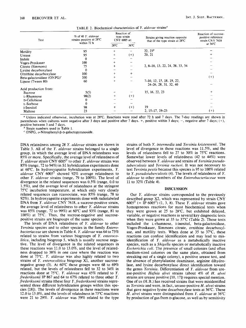
Phenotypic characteristics, The 40 strains of Y. aldovae examined gave mostly homogeneous reactions in biochemi- cal tests done at 28°C (Table 2). The characteristics common to all of the strains studied are listed below. A total of 36 strains did not ferment sucrose; 2 strains fermented sucrose within 48 h, and 2 strains fermented sucrose after 4 days. Utilization of citrate (Simmons), acid production from malt- ose, and hydrolysis of Tween 80 were independently vari- able reactions in the 40 Y. aldovae strains. Two strains were nonmotile at 28"C, and two other strains gave negative reactions for urease. A single strain gave a negative test for i- inositol. Like other Yersinia species, especially the L-rham- nose-fermenting species Yersinia intermedia and Yersinia frederiksenii, when Y. aldovae was incubated at 36 2 1"C, it exhibited negative or delayed positive reactions in tests for
L-rhamnose, D-xylose, i-inositol, the Voges-Proskauer reac- tion, Simmons citrate, ornithine decarboxylase, beta-galac- tosidase (o-nitrophenyl-P-D-galactopyranoside), and, of course, motility. These data (except the motility, methyl red, Voges-Proskauer, beta-xylosidase, and Simmons citrate data) are not shown for all strains, but the results of the 36 k 1°C reactions for representative strains CNY 6005T and CNY 7618 are shown in Table 2.
Of the 40 Y. aldovae strains, 17 were typable in slide agglutination tests for 0 antigens by the scheme of Wauters et al. (19). Eight strains were serogroup 0:17 strains, fol- lowed by decreasing numbers of strains in serogroups 0:6,31, 0:6,30, 0:7,8, 0:21, and 0:22 (Table 1). All but 1 of the 40 strains belonged to either phage type X, or phage type X,; these phage types consist of bacteriophages that are isolated from sewage, not of specific temperate bacterio- phages that are isolated from Y. enterocolitica (Table 1).
Genetic characteristics. The guanine-plus-cytosine content of DNA from Y. aldovae CNY 600ST was 48 mol%. DNA relatedness studies were done by using radiolabeled DNAs from Y. aldovae CNY 6005T and CNY 7618. The levels of
168 BERCOVIER ET AL. INT. J . SYST. BACTERIOL.
TABLE 2. Biochemical characteristics of Y . aldovae strainsu
Reaction of Reaction of sucrose- type strain
CNY 6005 at:
28°C 36°C
-
Strains giving reaction opposite that of the type strain at 28°C
positive reference strain CNY 7618
% of 40 Y . aldovae Test strains positive at 28°C
within 72 h at 36°C
- Motility 95 + - 22, 23b Urease 95 Indole 0
+ (+I 20, 21 + - - -
Voges-Pros kauer 100 Citrate (Simmons) 75 L,ysine decarboxylase 0 Ornithine decarboxylase 100 Be ta-galac tosidase (ONPG)' 100 Lipase (Tween 80) 60
Acid production from: Sucrose L-Rhamnose D-Cellobiose L-Sorbose i-Inositol Maltose
- - + + + + + + - 7-10, 12, 15, 18, 19, 22,
- 2,-8-10, 13, 22, 24, 28, 33, 34 -
(+ I
- - - -
-
24-26, 28, 31, 32, 40 -
Unless indicated otherwise, incubation was at 28°C. Reactions were read after 72 h and 7 days. The 7-day readings are shown in parentheses when cultures were negative after 3 days and positive after 7 days. +, positive within 3 days; -, negative after 7 days; (+), positive between 3 and 7 days.
Strain numbers used in Table 1. ONPG, o-Nitrophenyl-P-D-galactopyranoside.
DNA relatedness among 28 Y. aldovae strains are shown in Table 3. All of the Y. aldovae strains belonged to a single group, in which the average level of DNA relatedness was 85% or more. Specifically, the average level of relatedness of Y. aldovae strain CNY 6005T to other Y. aldovae strains was 85% (range, 72 to 94%) in S1 hybridization experiments done at 60°C. In hydrox apatite hybridization experiments, Y. aldovae CNY 6005 showed 92% average relatedness to other Y. aldovae strains (range, 79 to 100%). The level of divergence in the related sequences was 0.5% (range, 0.0 to 1.5%), and the average level of relatedness at the stringent 75°C incubation temperature, at which only very closely related sequences can reassociate, was 85% (range, 78 to 92%). In hydroxyapatite experiments done with radiolabeled DNA from Y. aldovae CNY 7618, a sucrose-positive strain, the average level of relatedness to other Y. aldovae strains was 85% (range, 72 to 99%) at 60°C and 93% (range, 81 to 100%) at 75°C. Thus, the sucrose-negative' and sucrose- positive strains are biogroups of the same species.
The levels of DNA relatedness of Y. aldovae to other Yersinia species and to other species in the family Entero- bacteriaceae are shown in Table 4. Y. aldovae was 64 to 73% related to strains from various biogroups of Y . enteroco- litica, including biogroup 5 , which is usually sucrose nega- tive. The level of divergence in the related sequences in these reactions was 11.0 to 13.0%, and the level of related- ness dropped to 38% in one case where the reaction was done at 75°C. Y. aldovae was also highly related to two strains of Y. enterocolitica biogroup X1, another sucrose- negative group (3). At 60°C these groups were 68 to 69% related, but the levels of relatedness fell to 32 to 34% in reactions done at 75°C. Y. aldovae was 45% related to Y. frederiksenii IP 867 and 64 to 67% related to three other Y. frederiksenii strains (these four Y. frederiksenii strains repre- sented three different hybridization groups within this spe- cies [ls]). The levels of divergence in these reactions were 12.0 to 15.0%, and the levels of relatedness in 75°C reactions were 21 to 29%. Y. aldovae was 59% related to the type
Y
strains of both Y. intermedia and Yersinia kristensenii. The level of divergence in these reactions was 11.5%, and the levels of relatedness fell to 27 to 30% in 75°C reactions. Somewhat lower levels of relatedness (42 to 44%) were observed between Y. aldovae and strains of Yersinia pseudo- tuberculosis and Yersinia ruckeri. It was not necessary to test Yersinia pestis because this species is 95 to 100% related to Y. pseudotuberculosis (4). The levels of relatedness of Y. aldovae to other members of the Enterobacteriaceae were 11 to 32% (Table 4).
DISCUSSION
Our Y. aldovae strains corresponded to the previously described group X2, which was represented by strain CNY 600ST (= IP 6005T) (1, 3, 8). These Y. aldovae strains gave homogeneous reactions for most biochemical tests when they were grown at 25 to 2 8 T , but exhibited delayed, variable, or negative reactions in several key diagnostic tests when they were grown at 35 to 37°C (Table 2). These tests included the L-rhamnose, i-inositol, beta-galactosidase, Voges-Proskauer, Simmons citrate, ornithine decarboxyl- ase, and motility tests. When done at 35 to 37"C, these reactions can confuse identification and may lead to mis- identification of Y. aldovae as a metabolically inactive species, such as a Shigella species or metabolically inactive Escherichia coti. The presence of small colonies (and often medium-sized colonies on the same plate, obtained from streaking out of a single colony), a positive urease test, and the absence of phenylalanine deaminase , arginine dihydro- lase, and lysine decarboxylase direct identification toward the genus Yersinia. Differentiation of Y. aldovae from ure- ase-positive Hafnia alvei strains (about 4% of H . alvei strains are urease positive [lo, 171) requires special mention. One of us (H.B.) has received strains that were misidentified as Yersinia and were, in fact, urease-positive H . alvei strains that gave negative lysine decarboxylase tests at 36°C. These H. alvei strains were distinguished from Y. aldovae at 36°C by production of gas from D-glucose, as well as by sensitivity

VOL. 34, 1984 YERSINIA ALDOVAE S P . NOV. 169
TABLE 3. Levels of intraspecies DNA relatedness of Y. aldovae strainsa
Source of labeled DNA
Y . aldovae CNY 7618 RBR (%, HA method) at:
Source of unlabeled Y. aldovae DNA
Y. aldovae CNY 6005T
RBR (%) at 75°C RBR (92) at 60°C
S1 method HA method D (HA method)
CNY 6005= CNY 6679 CNY 7089 CNY 7090 CNY 7091 CNY 7092 CNY 7096 CNY 7109 CNY 7111 CNY 7112 CNY 7328 CNY 7330 CNY 7331 CNY 7332 CNY 7618 CNY 7622 CNY 7632 CNY 7683 CNY 7898 CNY 7927 CNY 7928 CNY 7970 CNY 7971 CNY 8285 CNY 8286 CNY 8287 CNY 8288 CNY 8290
100 89 85 80 89 89 83 84 94 88 93 84 89 87 88 78 74 89 78 72 79 87 74 89 92 92 88 89
100 ND 94
ND 100 ND
99 100 ND
98 ND ND
89 ND
94 83 79
ND 83
ND 99
100 ND ND
83 ND ND ND
0.0 ND 0.0 ND ND ND ND ND ND ND ND ND 0.0 ND 0.0 ND 1.5 ND 1.5 ND ND ND ND ND ND ND ND ND
100 ND
91 ND
89 ND
83 84
ND 83
ND ND
84 ND
84 89 86
ND 78
ND 80 83
ND ND
92 ND ND ND
60°C 75°C
80 ND ND ND
81 ND
74 86
ND 72
ND ND
80 ND 100 88 99
ND 92
ND ND
84 ND ND
99 ND ND ND
88 ND ND ND
91 ND
96 98
ND 92
ND ND
90 ND 100 92 96
ND 94
ND 81 95
ND ND 100 ND ND ND
a S1 method, Diethylaminoethyl cellulose S1 nuclease hybridization assay; HA method, hydroxyapatite hybridization assay; RBR, relative binding ratio; D, divergence. For the S1 nuclease assay the following formula was used: relative binding ratio = (percentage of heterologous DNA bound to diethylaminoethyl cellulose filter)/(percentage of homologous DNA bound to diethylaminoethyl cellulose filter) x 100. For the hydroxyapatite assay the formula used was: relative binding ratio = (percentage of heterologous DNA bound to hydroxyapatite)/(percentage of homologous DNA bound to hydroxyapatite) x 100. Divergence was calculated on the assumption that each 1°C decrease in the stability of a heterologous DNA duplex compared with the stability of a homologous DNA duplex was caused by approximately 1% unpaired nucleotide bases within the heteroduplex. Divergence is expressed to the nearest 0.5%. 3H-labeled DNA from Y . alduvae CNY 600ST was used in the S1 nuclease experiments. Each reaction was done twice on duplicate filters. The values given are averages of four values, which did not differ by more than 2%. The average level of homologous binding was 81%. Approximately 3% of the labeled DNA bound to diethylaminoethyl cellulose filters in the absence of unlabeled DNA. This control value was subtracted from the heterologous binding values before normalization. DNAs from Y . alduvae strains CNY 6005= and CNY 7618, labeled in vivo with 3zP04, were used in the hydroxyapatite hybridization experiments. The average level of binding in homologous reactions was 75% for both labeled DNA preparations. Each reaction was done twice. Approximately 5% of the labeled DNA bound to the hydroxyapatite in the absence of unlabeled DNA. This control value was subtracted from the heterologous binding values before normalization.
ND, Not done.
to a Hafnia-specific bacteriophage (11). At 28"C, the reac- tions described above, as well as a positive lysine decarbox- ylase test and a negative test for acid production from D- sorbitol, distinguished H. alvei from Y. aldovae.
The variability in biochemical test results for Y . aldovae obtained at 36 k 1°C compared with the results obtained at 28°C is found in most Yersinia species (2, 3, 5, 6, 8, 18). These phenotypic characteristics, especially motility at 25 to 28°C but not at 36 5 1"C, strengthen the phenotypic resem- blance of Y. aldovae to other Yersinia species. Practical biochemical tests that are useful for differentiating Y. aldo- vae from other Yersinia species are shown in Table 5. Y. aldovae is easily separated from the other Yersinia species that are sucrose negative or that contain sucrose-negative strains. Y. aldovae is separated from Y. kristensenii by its positive L-rhamnose reaction, negative reactions for cellobi- ose and sorbose, and positive Voges-Proskauer reaction. Y. aldovae is distinguished from Y . pseudotuberculosis on the
bases of its positive ornithine decarboxylase and D-sorbitol reactions and its negative melibiose reaction and from Y. pestis by the same tests plus its positive urease reaction. It is separated from biogroup 5 of Y. enterocolitica (which con- tains sucrose-negative strains) by its positive D-sorbitol and L-rhamnose reactions and its negative cellobiose reaction. Y. aldovae is differentiated from Y. ruckeri by its positive reactions for urease and D-sorbitol. It is also simple to differentiate Y. aldovae from the sucrose-positive Yersinia species by its negative sucrose reaction, as well as its negative reactions for sorbose and cellobiose (from Y. en- terocolitica biogroups 1 through 4, Y. intermedia, and Y. frederiksenii), negative indole reaction (from Y. intermedia, and Y. frederiksenii), negative melibiose reaction (from Y. intermedia), and positive L-rhamnose reaction (from Y. enterocolitica biogroups 1 through 4).
DNA hybridization studies showed that the Y. aldovae strains are highly interrelated, forming a good genospecies

INT. J . SYST. BACTERIOL. 170 BERCOVIER ET AL.
TABLE 4. Levels of DNA relatedness of Y. aldovae to species of Yersinia and other genera in the Enterobacteriaceae" Relatedness to 32P0,-labeled Y . aldovae CNY 600ST DNA
Source of unlabeled DNA RBR(%) D(%) RBR (%)
at 75°C
P'. enterocolitica 2525-75 (biogroup
P'. enterocolitica 497-70 (biogroup 1) Y. enterocolitica 500-70 (biogroup 3) Yersinia biogroup X1 strain IP 7706 Yersinia biogroup X1 strain IP 7702 Y. enterocolitica 1P 178 (biogroup 5) Y. frederiksenii IP 5960 Y. frederiksenii IP 6175 Y . frederiksenii 2851-77 Y . enterocolitica IP 211 (biogroup 2) Y. kristensenii IP 1474 Y. intermedia IP 3953 Y. frederiksenii IP 867 Y. ruckeri 4535-69 Y . pseudotuberculosis P62 Serratia fonticola 4556-71 Ssrratia liquefaciens 446-68 Erwinia cypripedii EC155 Erwinia rhapontici ER106 Erwinia carotovora ATCC 495 Enterobacter aerogenes 1627-66 Erwinia nigrifuens EN104 Enterobacter agglomerans 2780-70 Enterobacter agglomerans 1429-71 Serratia rubidaea 934-72 Serratia plymuthica 392 Rszhnella aquatilis 1327-79 Kluyvera cryocrescens 409-78 Escherichia blattae 9005-74 Obesumbacterium proteus 4302-74 Erwinia carnegieana EC186 Enterobacter agglomerans 6003-71 Enterobacter agglomerans 1600-71 Enterobacter agglomerans 5378-71 Enterobacter cloacae 1347-71 Klebsiella oxytoca ATCC 13182 Enterobacter agglomerans 5422-69 Citrobacter freundii 460-61 Hszjnia alvei I 5632-72 Morganella morganii ATCC 25830
unknown)
at 600C - ~
73
70 70 69 68 68 67 67 64 64 59 59 45 44 42 32 30 28 28 27 27 25 25 25 25 25 25 25 25 24 24 24 24 24 24 24 23 23 23 23
12.5
11.0 38 11.0
32 34
29 12.5
14.5 15.0 13.0 11.5 30 11.5 27 12.0 21 15.5 14.0
Relatedness to 32P04-labeled Y . aldovae CNY 6005' DNA
Source of unlabeled DNA - RBR(%) D(%) RBR(%) at 60°C at 75°C
Enterobacter amnigenus 1325-79 Kluyvera ascorbata 408-78 Erwinia salicis ES102 Erwinia amylovora EA178 Enterobacter agglomerans 219-71 Enterobacter agglomerans 4388-71 Enterobacter agglomerans 3482-71 En terobacter agglomeruns 3 123-70 Serratia marcescens 868-57 Citrobacter amalonaticus ATCC
Cedecea neteri 621-75 Salmonella typhimurium LT2 Enterobacter agglomerans 1741-71 Serratia $curia 1165-77 Cedecea davisae 3278-77 Cedecea lapagei 485-76 Erwinia mallotivora NCPPB 2851 Enterobacter agglomerans 6070-69 Erwinia quercina EQ102 Klebsiella pneurnoniae 2 Shigella boydii C13 1610-55 Edwardsiella tarda 3592-64 Escherichia vulneris 2898-73 Proteus vulgaris PR1 Enterobacter skazakii 4562-70 Escherichia hermannii 980-73 Citrobacter diversus 1066-71 Providencia rustigianii 132-68 Enterobacter gergoviae CIP 7601 Hafnia alvei 111 329-73 Enterobacter agglomerans 1645-71 Providencia alcalifaciens 3370-67 Providencia stuartii 2896-68 Providencia rettgeri 1163 Hafnia alvei I1 4510-75 Erwinia chrysanthemi SR32 Escherichia coli K-12 Proteus myxofaciens ATCC 19692 Proteus mirabilis PR14 Erwinia rubrifaciens ER105
25406
23 23 22 22 22 22 22 22 22 22
22 22 21 21 21 21 20 20 19 19 19 19 19 19 18 18 18 18 17 17 16 15 15 15 14 14 12 12 11 11
RBR. Relative binding ratio; D, divergence. See Table 3 , footnote a , for experimental conditions.
(Table 3). Although strain CNY 7618 produced acid from sucrose, the levels of relatedness between this strain and other Y. aldovae strains were similar to the levels of related- ness between the more typical (sucrose-negative) strain CNY 6005T and other Y. aldovae strains. This proves that the sucrose-positive biogroup is part of Y. aldovae. The results of DNA hybridization studies done with labeled DNAs from other Yersinia species and unlabeled DNAs from group X2 strains CNY 6005T and IP 5850 have been published previously (8). In these studies Y. enterocolitica was 53 to 65% related to Y. aldovae at 60°C and 28 to 36% related at 75°C; Y. kristensenii was 60 to 66% related to Y. aldovae at 60°C and 33 to 35% related at 75°C; Y. frederik- senii was 43 to 56% related to Y. aldovae at 60°C and 14 to 23% related at 75°C; and Y. intermedia was 51 to 57% related to Y. aldovae at 60°C and 29 to 31% related at 75°C. Where determined, the levels of divergence in related sequences were 10 to 14% (8).
Xn the present study the reciprocal hybridization experi- ments yielded similar results (Table 4). The levels of diver-
gence in related sequences were 11 to 15%; the levels of relatedness in 75°C reactions were 21 to 38%, and the levels of relatedness in 60°C reactions were 59% (to Y. kristen- senii), 45 to 67% (to Y.frederiksenii), 59% (to Y. intermedia), 42% (to Y. pseudotuberculosis), and 64 to 73% (to Y. enterocolitica). The level of relatedness between Y. aldovae and Y. enterocolitica (as determined with labeled Y. aldovae DNA) is close to the lower level of relatedness observed between strains of a single species (70% or more). The higher levels of relatedness observed between these species when Y. aldovae DNA rather than labeled Y. enterocolitica DNA was used (64 to 74% compared with 53 to 65%) might have been due to differences in genome size between strains of these two species ( Y . enterocolitica is 12 to 14% smaller than Y. aldovae). The fact that Y. aldovae and Y. enterocolitica are separate species was proven by the substantial levels of divergence present in DNA heteroduplexes (10 to 15%) compared with the levels observed in reactions between strains of Y. aEdovae (0.0 to 1.5% [Table 31) or Y. enteroco- litica (0.0 to 4.0% [S]) and by the substantially lower levels of

VOL. 34, 1984 YERSINIA ALDOVAE SP. NOV. 171
TABLE 5. Biochemical differentiation of Y. aldovae from other Yersinia speciesa
Test
Reaction of
Y. enterocolitica
Biogroup 1 through 4
Y . inter- Y. fred- BiogrouPs media eriksenii
lensenii Y. ruckeri Y . aldovae Y. pestis '' kris- Y. pseudo-
Voges-Proskauer + Sucrose - Cellobiose - Indole - Sorbose -
L-Rhamnose + Melibiose -
D-Sorbitol + Ornithine decarboxylase + Urease +
+ + + + + + + + + V + + + + +
+ + +
+ + + + + + + + +
- - -
~~ ~~ -
The data for Y . aldovae are from Table 2 , the data for Y . ruckeri are from reference 9, and the data for the other yersiniae are from refer- ences 3 through 6 and 18. All of the data are for 3 days of incubation at 28°C (2 days of incubation at 25°C for Y. rzickeri). +, 90% or more of the strains positive; -, less than 10% positive; V, 10 to 89% positive; ND, test not done.
relatedness observed in 75°C reactions between these spe- cies (24 to 36% [Table 41 [8]) than in reactions among strains of Y. aldovae (78 to 100% [Table 31) or Y . enterocolitica (68% to 97% [8]). For that matter, the level of relatedness among strains of either species at 60°C (average for Y. aldovae, 85 to 92% [Table 31; average for Y . enterocolitica, 85% [S]) was substantially higher than the levels obtained in reactions between strains of Y. aldovae and Y. enterocolitica (69% average relatedness at 60°C with labeled Y. alduvae DNA and unlabeled Y. enterocolitica DNAs [Table 41; 60% average relatedness at 60°C with labeled Y. enterocolitica DNA and unlabeled Y . aldovae DNAs [S]).
The levels of relatedness of Y. aldovae to other members of the Enterobacteriaceae were substantially lower than the levels of relatedness to yersiniae, but nonetheless were significant (1 1 to 32% [Table 41). The uanine-plus-cytosine content of Y. aldovae strain CNY 6005 DNA was 48 mol%, a value which is similar to the guanine-plus-cytosine con- tents of other Yersinia species. Therefore, we conclude that, on the basis of phenotypic and genetic characteristics, Y . aldovae is a new species of the Enterobacteriaceae and does indeed belong in the genus Yersinia.
It must be emphasized that, as with most other yersiniae, biochemical characterization is more definitive when test preparations are incubated at 25 to 28°C (the optimal growth temperature) than when they are incubated at 36 2 1°C. The results of biochemical tests done at 36 t 1°C are frequently misleading, because several important diagnostic tests give false-negative or delayed positive results. These false-nega- tive findings can result in misidentification at the species level or even at the genus level. Results from this study and our previous studies have clearly shown that all Yersinia species are readily identified when they are studied at 28°C
The 40 strains of Y. aldovae which we studied were all isolated from aquatic ecosystems in Czechoslovakia or Nor- way. One previously studied strain (strain IP 5850) was isolated from soil in France (8). Y. aldovae has not been implicated in animal infections and has not been isolated from humans. It may be isolated from many of the same water sources as Y. enterocolitica. Therefore, clinical and environmental microbiologists should be able to distiguish these species from one another.
We propose the name Yersinia aldovae sp. nov. (al. do' vae. M. L. gen. n. aldovae in honor of Eva Aldova, the
?r
(3-6, 9, 18).
Czechoslovakian microbiologist who first isolated this bacte- rium) for the Yersinia group formerly referred to as group X2. The type strain of Y . aldovae is strain CNY 6005 (= ATCC 35236 = IP 6005 = A1 19955 = CDC 669-83); it was isolated by E. Aldova from drinking water in Czechoslova- kia.
Description of Yersinia aldovae sp. nov. The phenotypic characteristics of Y. aldovae fit the definition of the Entero- bacteriaceae. Y . aldovae cells are gram negative, nonspore- forming, non-encapsulated, straight, and rod shaped and have peritrichous or degeneratively peritrichous flagella when they are grown at 28"C, but not when they are grown at 37°C; 2 to 10 flagella are usually present. Y. aldovae CNY 6005T grown for 48 h at 28°C aerobically on nutrient agar produces both small (diameter, 0.5 mm) and medium-sized (diameter, 2 mm) colonies. The colonies are translucent and have smooth, entire edges.
All Y . aldovae strains give positive results in tests for beta- galactosidase (o-nitrophenyl-P-D-galactopyranoside), cata- lase, methyl red (28 and 36"C), nitrate reduction to nitrite by type B nitrate reductase, ornithine decarboxylase, the Voges-Proskauer reaction at 28"C, and acid production from L-arabinose, esculin, D-fructose, galactose, N-acetyl-glucos- amine, D-glucose, glycerol, D-mannitol, D-mannose, L-rham- nose, ribose, D-sorbitol, trehalose, and D-xylose. All Y. aldovae strains give negative results in tests for arginine dihydrolase, citrate (Simmons) (36"C), deoxyribonuclease, gas from D-glucose, gelatin liquefaction (film), HZS (Kligler), indole, lysine decarboxylase, malonate, motility at 36"C, oxidase, phenylalanine deaminase, tetrathionate reductase, tryptophan deaminase, the Voges-Proskauer reaction at 36"C, beta-xylosidase (36"C), and acid production from adonitol, amygdalin, D-arabinose, arbutin, D-cellobiose, dul- citol, erythritol, lactose, melibiose, melizitose, alpha-meth- yl-D-glucoside, alpha-methyl-D-mannoside, alpha-methyl- xyloside, raffinose, salicin, L-sorbose, and L-xylose. At least 95% of the strains are positive in tests for i-inositol, motility at 28"C, and urease. All but two of our strains are sucrose negative after 48 h.
The guanine-plus-cytosine content of the DNA of the type strain is 48 mol%.
All of our Y . aldovae strains except strain IP 5850 were isolated from aquatic ecosystems (drinking water, river water, fish); strain IP 5850 was isolated from soil. None was associated with disease in humans or animals.
172 BERCOVIER ET AL. INT. J. SYST. BACTERIOL.
The type strain is CNY 6005 (= ATCC 35236 = IP 6005 =
Description of the type strain. Some characteristics of the type strain are given in Table 2. Other characteristics are as given above for the species. This strain is serologically untypable and belongs to phage type X,.
A1 19955 = CDC 669-83).
ACKNOWLEDGMENT
We are grateful to H. H. Mollaret for making his culture collec- tion and laboratory facilities available to us and for his continuing advice and encouragement during the course of this study.
1.
2.
3.
4.
5.
6.
LITERATURE CITED
Bercovier, H., J. M. Alonso, Z. N. Bentaiba, J. Brault, and H. H. Mollaret. 1979. Contribution to the definition and taxono- my of Yersinia enterocolitica. Contrib. Microbiol. Immunol.
Bercovier, H., J. Brault, N. Barre, M. Treignier, J. M. Alonso, and H. H. Mollaret. 1978. Biochemical, serological and phage typing characteristics of 459 I’ersinia strains isolated from a terrestrial ecosystem. Curr. Microbiol. 1:353-357. Bercovier, H., D. J. Brenner, J. Ursing, A. G. Steigerwalt, G. R. Fanning, J. M. Alonso, G. P. Carter, and H. H. Mollaret. 1980. Characterization of Yersinia enterocolitica sensu stricto. Curr. Microbiol. 4:201-206. Bercovier, H., H. H. Mollaret, J. M. Alonso, J. Brault, G. R. Fanning, A. G. Steigerwalt, and D. J. Brenner. 1980. Intra- and interspecies relatedness of Yersinia pestis by DNA hybridiza- tion and its relationship to Yersinia pseudotuberculosis. Curr. Microbiol. 4:225-229. Bercovier, H., J. Ursing, D. J. Brenner, A. G. Steigerwalt, G. R. Fanning, G. P. Carter, and H. H. Mollaret. 1980. Yersinia kristensenii: a new species of Enterobacteriaceae composed of sucrose-negative strains (formerly called atypical Yersinia en- terocofitica or Yersinia enterocolitica-like). Curr. Microbiol. 4:219-224. Brenner, D. J., H. Bercovier, J. Ursing, J. M. Alonso, A. G. Steigerwalt, G. R. Fanning, G. P. Carter, and H. H. Mollaret. 1980. Yersinia intermedia: a new species of Enterobacteriaceae composed of rhamnose-positive, raf€inose-positive strains (for- merly called Yersinia enterocolitica or Yersinia enterocolitica- like). Curr. Microbiol. 4:207-212.
5:12-22.
7. Brenner, D. J., A. C. McWhorter, J. K. Leete-Knutson, and A. G. Steigerwalt. 1982. Escherichia vulneris: a new species of Enterobacteriaceae associated with human wounds. J. Clin. Microbiol. 15: 1133-1140.
8. Brenner, D. J., J. Ursing, H. Bercovier, A. G. Steigerwalt, G. R. Fanning, J. M. Alonso, and H. H. Mollaret. 1980. Deoxyribonu- cleic acid relatedness in Yersinia enterocolitica and Yersinia enterocofitica-like organisms. Curr. Microbiol. 4:195-200.
9. Ewing, W. H., A. J. Ross, D. J. Brenner, B. R. Davis, and G. R. Fanning. 1978. Yersinia ruckeri sp. n., the RM bacterium. Int. J. Syst. Bacteriol. 28:37-44.
10. Farmer, J. J., 111. 1983. Biochemical chart for identifying the named species and “enteric groups” of Enterobacteriaceae. Centers for Disease Control, Atlanta, Ga.
11. Guinee, P. A. M., and J. J. Valkenburg. 1968. Diagnostic value of a Hafnia-specific bacteriophage. J. Bacteriol. 96564.
12. Hickman, F. W., and J. J. Farmer 111. 1978. Salmonella typhi: identification, antibiograms, serology, and bacteriophage typ- ing. Am. J. Med. Technol. 44:1149-1159.
13. Hickman, F. W., J. J. Farmer 111, D. G. Hollis, G. R. Fanning, A. G. Steigerwalt, R. E. Weaver, and D. J. Brenner. 1982. Identification of Vibrio hollisae sp. nov. from patients with diarrhea. J. Clin. Microbiol. 15395-401.
14. Mandel, M., and J. Marmur. 1968. Use of ultraviolet absor- bance-temperature profile for determining the guanine plus cytosine content of DNA. Methods Enzymol. 12B:195-206.
15. Owen, R. J., L. R. Hill, and S. P. Lapage. 1969. Determination of DNA base compositions from melting profiles in dilute buffers. Biopolymers 7506-516.
16. Popoff, M., and C. Coynault. 1980. Use of DEAE-cellulose filters in the S1 nuclease method for bacterial deoxyribonucleic acid hybridization. Ann. Microbiol. (Paris) 131A:151-155.
17. Richard, C., and J. M. Alonso. 1976. Une entkrobacterie me- connue: Enterobacter hafniae. Bull. Inst. Pasteur (Paris)
18. Ursing, J., D. J. Brenner, H. Bercovier, G. R. Fanning, A. G. Steigerwalt, J. Brault, and H. H. Mollaret. 1980. Yersiniafreder- iksenii: a new species of Enterobacteriaceae composed of rhamnose-positive strains (formerly called atypical Yersinia enterocolitica or Yersinia enterocolitica-like). Curr. Microbiol.
19. Wauters, G., L. Le Minor, A. M. Chalon, and J. Lassen. 1972. SupplCment au schema antigknique de Yersinia enterocolitica. Ann. Inst. Pasteur (Paris) 122:951-956.
74:339-352.
4: 2 13-2 17.
Copyright © 2022 FDOKUMEN