Identification and Molecular Characterization of Yersinia ruckeri Isolated from mass mortalities of...
12
Global Journal of Fisheries and Aquaculture Researches, Vol.1, (2): pp. 01 -17 (2014) Massive Publisher House M.P.H. Egypt www.mphegypt.com ISSN 2356-6329 Identification and Molecular Characterization of Yersinia ruckeri Isolated from mass mortalities of Cultured Nile tilapia at Kafr El- Sheikh Governorate Hany M. R. Abdel-Latif;* Riad H. Khalil; Talaat T. Saad and Rehab Y. El-bably Department of Poultry and Fish diseases, Faculty of Veterinary Medicine, Alexandria University, Edfina, Behera, P.O. Box: 22758, Egypt *Corresponding author. E-mail: [email protected] ABSTRACT Yersiniosis is an infectious disease which produces high mortalities and severe economic losses in freshwater fish farms. Heavy mortalities were found in pond cultured Oreochromis niloticus at Kafr El-sheikh. A total number of one hundred and fifty freshly dead fish were sampled and transferred as soon as possible to the lab for clinical examination and bacteriological identification. The examined fish showed the septicemic signs of Yersiniosis which appeared as generalized erythema, petechial hemorrhages on the skin, fin erosions, protruded hemorrhagic vent with red erythematic hemorrhages on mouth and lips. Postmortem examination revealed engorged gall bladder, hemorrhagic gills, liver, gastrointestinal tract (GIT) and darkened congested spleen. After bacterial isolation from spleen, kidney, liver and heart on specific culture media, sure diagnosis was achieved by conventional biochemical tests, serotyping and polymerase chain reaction (PCR). Antibiogram was conducted on bacterial isolates in order to find the most suitable antibiotic for controlling this bacterial infection. From our study, it was found that sharp decrease in water temperature with intensive fish culture was considered as co-factors for appearance of signs of yersiniosis and the recommended antibiotic for controlling this bacterial infection is ciprofloxacin or Sulphamethoxazole-Trimethoprim combination. Key words: Oreochromis niloticus - Yersinia ruckeri - Septicemic signs - Polymerase chain reaction (PCR). 1) INTRODUCTION Enteric Redmouth Disease (ERM) is a septicemic disease caused by Yersinia ruckeri "Y. ruckeri", a gram negative, ‘rod’ shaped bacterium of the family Enterobacteriaceae. The disease was previously called Hagermann redmouth, and causes significant economic losses in the salmonid farming industry. The disease was first described in rainbow trout, Oncorhynchus mykiss, in 1958 in the United States
Transcript of Identification and Molecular Characterization of Yersinia ruckeri Isolated from mass mortalities of...

Global Journal of Fisheries and Aquaculture Researches, Vol.1, (2): pp. 01 -17 (2014)
Massive Publisher House M.P.H. Egypt www.mphegypt.com ISSN 2356-6329
Identification and Molecular Characterization of Yersinia ruckeri Isolated from mass mortalities of Cultured Nile tilapia at Kafr El-Sheikh Governorate
Hany M. R. Abdel-Latif; * Riad H. Khalil; Talaat T. Saad and Rehab Y.
El-bably Department of Poultry and Fish diseases, Faculty of Veterinary Medicine,
Alexandria University, Edfina, Behera, P.O. Box: 22758, Egypt
*Corresponding author. E-mail: [email protected] ABSTRACT
Yersiniosis is an infectious disease which produces high mortalities and severe economic losses in freshwater fish farms. Heavy mortalities were found in pond cultured Oreochromis niloticus at Kafr El-sheikh. A total number of one hundred and fifty freshly dead fish were sampled and transferred as soon as possible to the lab for clinical examination and bacteriological identification. The examined fish showed the septicemic signs of Yersiniosis which appeared as generalized erythema, petechial hemorrhages on the skin, fin erosions, protruded hemorrhagic vent with red erythematic hemorrhages on mouth and lips. Postmortem examination revealed engorged gall bladder, hemorrhagic gills, liver, gastrointestinal tract (GIT) and darkened congested spleen. After bacterial isolation from spleen, kidney, liver and heart on specific culture media, sure diagnosis was achieved by conventional biochemical tests, serotyping and polymerase chain reaction (PCR). Antibiogram was conducted on bacterial isolates in order to find the most suitable antibiotic for controlling this bacterial infection. From our study, it was found that sharp decrease in water temperature with intensive fish culture was considered as co-factors for appearance of signs of yersiniosis and the recommended antibiotic for controlling this bacterial infection is ciprofloxacin or Sulphamethoxazole-Trimethoprim combination. Key words: Oreochromis niloticus - Yersinia ruckeri - Septicemic signs - Polymerase
chain reaction (PCR). 1) INTRODUCTION
Enteric Redmouth Disease (ERM) is a septicemic disease caused by Yersinia ruckeri "Y. ruckeri", a gram negative, ‘rod’ shaped bacterium of the family Enterobacteriaceae. The disease was previously called Hagermann redmouth, and causes significant economic losses in the salmonid farming industry. The disease was first described in rainbow trout, Oncorhynchus mykiss, in 1958 in the United States
Abdel-Latif, H. M. R. et. al, (2014)
2
(Bullock et al., 1977) and has become endemic widely in fish populations throughout in North America, Australia, South Africa and Europe. Although the infection with this bacterium has been reported in other fish species, salmonids and especially rainbow trout are most susceptible to ERM (Furones et al., 1993). In Egypt, Y. ruckeri was firstly isolated from both apparently healthy and moribund Nile tilapia from the Nile River in Giza Province (Hussein et al., 1997) Nile Delta region from apparently healthy and diseased African catfish and common carp, with relatively low prevalence (12.5, 8 and 5% respectively) (Abd-El Latief, 2001) and from earthen pond cultured Nile tilapia at a semi-intensive fish farm in Sharkiya Province, Lower Egypt (Eissa et al., 2008).
There are many stresses as excessive stocking, pollution and oxygen depletion that play a key role in the outbreak. Severe epidemics are observed at water temperatures of 15 - 20 °C and the incubation period of infection is 5-10 days (Roberts, 1983). Its mode of transmission has been related to wild or farmed carrier fish and other putative vectors, such as aquatic invertebrates and birds (Willumsen, 1989). Clinically, the disease is characterized by the development of acute or chronic entero-septicemia, with the presence of congestive or hemorrhagic zones on the skin, gills, mouth and intestine, and exophthalmia is also frequently evident (Rigos and Stevenson, 2001).
With respect to the most recent diagnostic methods for ERM, polymerase chain reaction (PCR) represents a widely-used alternative to traditional identification methods. Although pathogenicity and virulence genes have been used as target regions, the rRNA molecule is now accepted as a tool for identification purposes. Recent phylogenetic studies of the genus Yersinia, based on 16S rRNA sequencing, have shown that this genus represents a tight cluster within the family Enterobacteriaceae, with Y. ruckeri forming a separate subline within the Yersinia five-subline cluster (Ibrahim et al., 1993 & Altinok et al., 2001). This suggests that, by taking advantage of the differences at the level of 16S rRNA sequences, specific oligonucleotides that can be used in a PCR assay for diagnostic purposes can be designed (Gibello et al., 1999). The present work focused mainly on isolation and molecular identification of Y. ruckeri from mass mortality of cultured O. niloticus during outbreak in the winter season at private farms in Kafr-El sheikh governorate.
2) MATERIAL AND METHODS 2. 1. Fish and sample collection:-
In our investigation, a total number of one hundred and fifty (150) earthen pond cultured O. niloticus of different body weight averaged (130 ± 5 g) were collected. They showed typical lesions and other signs of Yersiniosis, freshly dead and in the moribund state from private fish farms at Kafr el sheikh governorate, Egypt during outbreak in winter season where there is a sudden abrupt sharp fluctuation in water temperature ranged (17-18 °C). Fish were transferred to the laboratory of the Department of Poultry and fish diseases, Faculty of veterinary medicine, Alexandria

Identification and Molecular Characterization of Yersinia ruckeri Isolated ………
3
University as soon as possible. The collected fish specimens were subjected to full clinical, postmortem (PM) lesions, and bacteriological examinations. 2. 2. Reference strain:-
Yersinia ruckeri (ATCC® 29473™) strain (Ewing et al., 1978) was purchased from the American Type Culture Collection (ATCC) (P. O. Box 1549 Manassas, VA 20108 USA), that was isolated from moribund Rainbow trout, Oncorhynchus mykiss, with red mouth disease, USA.
2. 3. Gross clinical and Postmortem (PM) examination:-
Clinical examination of naturally infected fish was performed to investigate any clinical abnormalities (Amlacher, 1970), while Necropsy was performed on freshly dead and moribund fishes for detection of PM lesions (Conroy and Herman, 1981).
2. 4. Bacterial culture and identification:-
The fish surfaces were swapped with 70 % ethyl alcohol for surface disinfection and then inocula were taken from liver, Kidney, spleen, lower part of intestine and heart under complete aseptic condition to be cultured on tryptic soya broth (Difco, Detroit, MI, USA) and incubated at 20 0C for 24 - 48 hrs then sub-cultured on selective Yersinia agar media (Tobback et al., 2007). After incubation period, a single colony from each suspected isolate was picked up and re-streaked on a new plate of its perused selective culture media and re-incubated at the same conditions. When pure colonies have been grown, a loopful of each pure culture was streaked onto a nutrient agar slope or semisolid agar medium to be used as stock culture for further biochemical and serological identification. Bacterial isolates were stored in 15 % glycerol and kept at – 80 0C for further molecular identification.
The criteria used for identification of the isolates are based on colonial characteristics (colony morphology and arrangement) and gram staining of the microorganisms (Austin and Austin, 1999). Bacterial isolates were presumptively identified using conventional biochemical tests including catalase with 3% hydrogen peroxide solution, cytochrome oxidase with oxidase strips (Remel), motility in a motility test medium (BD Biosciences, MA), citrate utilization using simmons's citrate (Remel), sugar utilization using triple sugar iron (TSI, Remel), oxidation/fermentation of glucose using of basal media with glucose as the sole carbohydrate source (BD Biosciences), and esculin hydrolysis using bile esculin agar (Remel). Further biochemical testing was performed using API20E and 20NE tests (Bio-Me´rieux, NC), with isolates incubated at 20 °C. Results were interpreted at 24-48 hours according to the manufacturer’s instructions. Isolates were presumptively identified as Yersinia ruckeri according to the standard criteria of Ross et al. (1966), Ewing et al. (1978) and Austin and Austin (2007).
2. 5. Molecular identification of the bacterial isolates:- 2. 5. 1. Extraction of bacterial genomic DNA:-
It is useful to use the bacterial samples from slope agar directly. The genomic DNA was extracted from 100 µl of the bacterial suspension (a single colony
Abdel-Latif, H. M. R. et. al, (2014)
4
of each of the isolated bacteria suspended in 100 µl of sterile saline) using Gene JET genomic DNA extraction kit following the manufacturer protocol (Fermentas, #K0721).The principle of this reaction depends on digestion of the bacterial cell wall with Proteinase K. The lysate is then mixed with ethanol and loaded on the purification column where the DNA binds to the silica membrane. Impurities are effectively removed by washing the column with the prepared wash buffers. Genomic DNA is then eluted under low ionic strength conditions with the Elution Buffer. The extracted DNA was amplified using oligonucleotide primer set specifically for Yersinia ruckeri (Gibello et al., 1999).
2. 5. 2. Polymerase Chain reaction (PCR) techniques:-
DNA from eight Y. ruckeri isolates was tested. Forward and reverse primers were used to detect protease Yrp1 gene; Yrp 1F (forward primer) sequence is 5′- TATTCAACTGAAAGTGTA-3′ and Yrp 1R (reverse primer) sequence is 5′- ATAGCTCATAATACTGA-3′. The isolated DNA was amplified using dream Taq green PCR master mix kit following the manufacturer protocol (Fernandez et al., 2003).
The polymerase chain reaction mixture was carried out in a 50 µl which contain the following; DNA template (5-20 ng/ µl) (3 µl), PCR Master Mix (9 µl), Primer forward (0.1-0.5 µM) (0.5 µl), Primer reverse (0.1-0.5 µM) (0.5 µl) and nuclease free water (37 µl). Negative (no DNA template) and positive (50 ng of purified DNA from Y. ruckeri) controls were included in each batch of PCR mixtures. Thermal cycling was done in the thermal cycler (Techne, 3500, UK) was programmed as follows; One cycle for 5 minutes at 94oC to denature the template DNA followed by 25 cycles of denaturation, annealing and extension at 94 oC for 30 seconds, 40oC for 30 seconds and 72 oC for 60 seconds, respectively. The 25 cycles were followed by: a final cycle of extension at 72 oC for 10 minutes to ensure that the entire PCR product is double strand DNA and at the end of cycling the tubes were stored at – 20 oC until being used. The PCR products were electrophoresed in 2% agarose gel (Invitrogen), stained with ethidium bromide, visualized with ultraviolet light, and photographed using a Kodak EDAS system (Eastman Kodak, Rochester, NY). Isolates were considered positive when a 961 bp band was detected. 2. 6. Antibiogram test:-
The antibiotic discs were obtained from (Oxoid, England). The graduated rule to 0.5 mm was used for reading the diameter of the zones of inhibition twice at right angles. Antibiotic discs used were Ciprofloxacin (CIP) (5 mg), Gentamycin (CN) (10 mg), Flumequine (30 mg) (UB), Enrofloxacin (5 mg) (ENR), Doxycycline (30 mg) (DO), Sulphamethoxazole-Trimethoprim (23.7+1.25 mg) (SXT), Oxytetracycline (30 mg) (OT), Amoxicillin (10 mg) (AMK) and Erythromycin (15 mg) (E). Susceptibility to several antibiotics was determined using the disc diffusion technique (Barry and Thornsberry, 1991). The inoculum was prepared in 0.85% saline, and turbidity was adjusted to 0.5 McFarland's standard (approximately 2 x 10⁸ CFU / ml). Petri dishes of Mueller-Hinton ager supplemented with 1% NaCl were streaked with 1 ml of the prepared inoculum, then put the different antimicrobials
Identification and Molecular Characterization of Yersinia ruckeri Isolated ………
5
discs and incubated at 25 oC for 24-48 hours. The diameter of the zones of inhibition (point at which no growth is visible) was read twice at right angles with a ruler graduated to 0.5 mm.
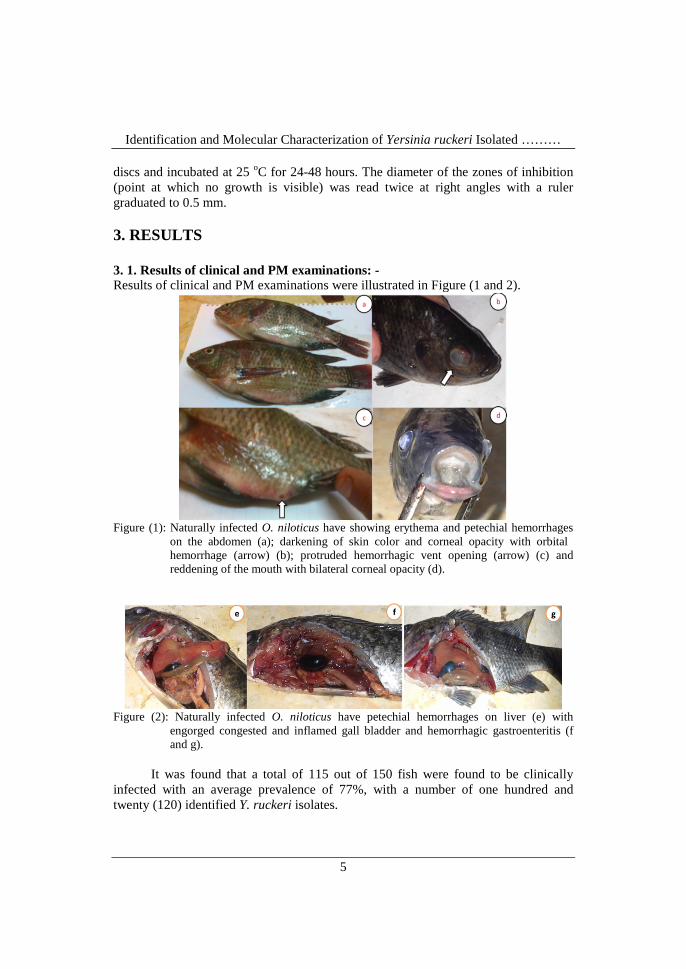
3. RESULTS 3. 1. Results of clinical and PM examinations: - Results of clinical and PM examinations were illustrated in Figure (1 and 2).
Figure (1): Naturally infected O. niloticus have showing erythema and petechial hemorrhages
on the abdomen (a); darkening of skin color and corneal opacity with orbital hemorrhage (arrow) (b); protruded hemorrhagic vent opening (arrow) (c) and reddening of the mouth with bilateral corneal opacity (d).
Figure (2): Naturally infected O. niloticus have petechial hemorrhages on liver (e) with
engorged congested and inflamed gall bladder and hemorrhagic gastroenteritis (f and g).
It was found that a total of 115 out of 150 fish were found to be clinically
infected with an average prevalence of 77%, with a number of one hundred and twenty (120) identified Y. ruckeri isolates.
Abdel-Latif, H. M. R. et. al, (2014)
6
3. 2. Results of bacterial isolation and identification:-
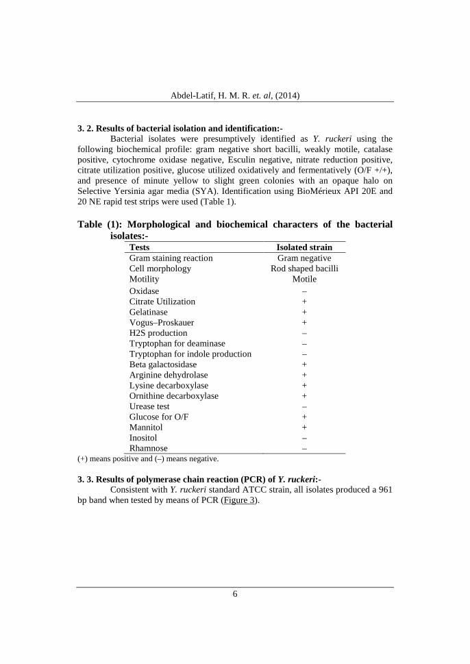
Bacterial isolates were presumptively identified as Y. ruckeri using the following biochemical profile: gram negative short bacilli, weakly motile, catalase positive, cytochrome oxidase negative, Esculin negative, nitrate reduction positive, citrate utilization positive, glucose utilized oxidatively and fermentatively (O/F +/+), and presence of minute yellow to slight green colonies with an opaque halo on Selective Yersinia agar media (SYA). Identification using BioMérieux API 20E and 20 NE rapid test strips were used (Table 1). Table (1): Morphological and biochemical characters of the bacterial
isolates:- Tests Isolated strain Gram staining reaction Gram negative Cell morphology Rod shaped bacilli Motility Motile Oxidase – Citrate Utilization + Gelatinase + Vogus–Proskauer + H2S production – Tryptophan for deaminase – Tryptophan for indole production – Beta galactosidase + Arginine dehydrolase + Lysine decarboxylase + Ornithine decarboxylase + Urease test – Glucose for O/F + Mannitol + Inositol – Rhamnose –
(+) means positive and (–) means negative. 3. 3. Results of polymerase chain reaction (PCR) of Y. ruckeri:-
Consistent with Y. ruckeri standard ATCC strain, all isolates produced a 961 bp band when tested by means of PCR (Figure 3).

Identification and Molecular Characterization of Yersinia ruckeri Isolated ………
7
Figure (3): Ethidium bromide stained agarose gel of PCR product representing amplification of
Yrp1gene (961bp) in two isolates as estimated by 100 bp ladder (L). Lanes (1 & 2) are samples, Lane (3) is positive control (DNA from ATCC Y. ruckeri 29473) while lane (4) is negative control (no DNA template).
3. 4. Results of antimicrobial susceptibility of Y. ruckeri isolates:-
Antimicrobial susceptibility tests against 3 isolates of Y. ruckeri showed that the bacterial isolates were markedly sensitive to Ciprofloxacin and Sulphamethoxazole-Trimethoprim combination, moderately sensitive to Enrofloxacin and Doxycycline and resistant to other antibiotics. 4) DISCUSSION
Bacterial diseases, from the epizootiological point of view, affect nearly all cultured, wild marine and freshwater fishes to a limit that exceeds all other disease causes combined. Moreover, bacterial diseases ranked first one among all the causative agents causing pisciculture problems (Meyer, 1991).
The prevalence of Yersiniosis among the entire Nile tilapia, with mortalities approaching 77%, as well as the severity of the lesions on individual fish, suggests the high virulence of the Y. ruckeri isolates retrieved from the clinical cases as well as the possible suppression of the fish’s resistance during the winter season with sharp decrease in water temperature. In fact, Y. ruckeri was able to adhere to solid supports such as algae and sediment samples, and proved that surface colonization of fish tanks by Y. ruckeri biofilms is a potential source of recurrent infection for extended periods of time (Coquet et al., 2002). The association of ERM with abrupt fluctuations in water temperature (Danley et al., 1999 & Rigos and Stevenson, 2001), overcrowding and harvest stress, makes this pre-eminent. Despite the fact that Y. ruckeri has been described primarily as a salmonid pathogen, there are increasing numbers of reports of its isolation from diseased non-salmonid species (Bullock and Cipri ano 1990; Hussein et al., 1997 and Berc et al., 1999).

Abdel-Latif, H. M. R. et. al, (2014)
8
The external and internal examinations of O. niloticus during the outbreak showing the characteristic clinicopathological picture of Yersiniosis, and these results in non salmonid fish were in concordance with that obtained with (Abd-El Latief, 2001 and Eissa et al., 2008). These results may be attributed to the virulence factors of Y. ruckeri, whereas it produces some extra-cellular products (ECP) such as cytotoxins and haemolysins (Romalde and Toranzo, 1993).
Furthermore, a 47-kDa metalloprotease (Yrp1 protease) contributes to the
virulence of this bacterium, involved in the colonization, invasion of different tissues (muscle proteins), and may lead to membrane alterations and pores in the capillary vessels that may result in the leaking of blood from these vessels and hence cause the typical hemorrhages especially around the mouth and intestine (Fernandez et al., 2003). Also, the siderophore, ruckerbactin, is the iron uptake system, from iron binding proteins as transferrin, involved in the virulence of Y. ruckeri (Fernandez et al., 2004).
Delay in diagnosis of fish disease leads to increase the economic losses due to fish mortalities, cost of treatment and loss of ration due to the fish become off food. All of the isolates were motile, and this result suggests that Y. ruckeri has morphological characteristics similar to those in strains isolated from salmon species and may be potentially pathogenic or virulent to the eels (Davies and Frerichs, 1989). Y. ruckeri can be isolated after cultivation in culture media such as TSA, BHIA and selective Yersinia agar (SYA) media by incubating for 48 hours at 22- 25°C. The comparison of SYA with the other culture media suggested that there were no significant differences between them and SYA can be used for epidemiological studies owing to its advantages such as high sensitivity and shorter time requirement (Austin and Austin, 1993).
Several researches based on Molecular diagnosis using PCR techniques for identification of Y. ruckeri isolated from salmonid fishes was done before (Gibello et al., 1999; Fernandez et al., 2003 and Fadaeifard et al., 2014), but little were done on non salmonid fish (Eissa et al., 2008). Our study illustrate that the primer sequence showed maximum identity with the sequence of Y. ruckeri with 100% homology and the Yrp1 gene - PCR amplified the correctly sized products for the isolates identified as virulent strains of Y. ruckeri.
In the antimicrobial susceptibility tests, all of the isolates showed marked sensitivity to ciprofloxacin and Sulphamethoxazole-Trimethoprim combination, and these results were similar to that obtained by Joon Joh et al. (2010). 5) CONCLUSION
The current study confirm the fact that the fish diseases are mainly stress related, whereas, the sharp decrease in water temperature during the winter season act mainly as predisposing factor for incidence of Yersiniosis. Moreover, PCR can be beneficially help in confirmation of diagnosis of Y. ruckeri. Certain antibiotics like Ciprofloxacin and Sulphonamides-Trimethoprime combination can treat Yersinia
Identification and Molecular Characterization of Yersinia ruckeri Isolated ………
9
infections. In future work, we will act hopefully to produce a vaccine from the local strains of Y. ruckeri to be used prior winter season to build up immunity against that harmful pathogen in order to reduce the heavy mortalities. REFERENCES Abd-El Latief, J. I. (2001). Yersinia microorganisms as the causative agent of enteric
redmouth disease in delta Nile fishes. MVSc thesis, Faculty of Veterinary Medicine, Cairo University.
Altinok, I., Grizzle, J. M. and Liu, Z. (2001). Detection of Yersinia ruckeri in rainbow trout blood by use of the polymerase chain reaction. Diseases of Aquatic Organisms (44): 29–34.
Amlacher, E. (1970). Textbook of fish diseases, T.E.S. Publication, New Jersey. USA, P. 117-135.
Austin, B. and Austin, D. A. (1993). Bacterial Fish Pathogens: Disease of Famed and wild Fish, 2nd ed. Chichester: Ellis Horwood.
Austin, B. and Austin, D. A. (1999). Bacterial fish pathogens: disease of farmed and wild fish, 3rd edn (revised). Springer-Praxis series in aquaculture and fisheries. Springer-Verlag, London.
Austin, B. and Austin, D. A. (2007). Bacterial Fish Pathogens: Disease in Farmed and Wild Fish (4th ed)., Springer Praxis, Chichester.
Barry, A. and Thornsberry, C. (1991). Susceptibility test and diffusion test procedures. In: Manual of Clinical Microbiology, Washington DC, pp. 1526-1542.
Berc, A., Mata, E., Kozariae, Z. and Petrinec-kozariae, Z. (1999). Yersinia ruckeri septicemia in experimentally infected carp (Cyprinus carpio L.) fingerlings. Acta Veterinaria Hungarica, (47): 161-172.
Bullock, G. L. and Cipriano, R. C. (1990). Enteric redmouth disease of salmonids. Fish disease leaflet No 82, US Fish and Wildlife Service, National Fisheries Research Center, Lee town, West Virginia.
Bullock, G. L., Stuckey, H. M. and Shotts, E. B. J. R. (1977). Early records of North American and Australian outbreaks of enteric redmouth disease. Fish Health News (6): 96–97.
Conroy, D. A. and Herman, L. R. (1981). Text book of fish diseases. T.F.H. publ., West Sylvania.
Coquet, L., Cosette, P., Quillet, L., Petit, F., Junter, G. A. and Jouenne, T. (2002). Occurrence and phenotypic characterization of Yersinia ruckeri strains with biofilm-forming capacity in a rainbow trout farm. Applied and Environmental Microbiology, (68): 470-475.
Danley, M. L., Goodwin, A. E. and Killian, H. S. (1999). Epizootics in farm-raised channel catfish, Ictalurus punctatus (Rafinesque), caused by the enteric redmouth bacterium Yersinia ruckeri. Journal of Fish Diseases, (22): 451- 456.
Davies, R. L. and Frerichs, G. N. (1989). Morphological and biochemical differences among isolates of Yersinia ruckeri obtained from wide
Abdel-Latif, H. M. R. et. al, (2014)
10
geographical areas. J. Fish Dis., (12): 357-365. Eissa, A. E., Moustafa, M. Abdelaziz, M. and Ezzeldeen, N. A. (2008). Yersinia
ruckeri infection in cultured Nile tilapia, Oreochromis niloticus, at a semi-intensive fish farm in Lower Egypt. African Journal of Aquatic Science, 33(3): 283-286.
Ewing, W. H., Ross, A. J., Brenner, D. J. and Fanning, G. R. (1978). Yersinia ruckeri sp. nov., the red mouth (RM) bacterium. International Journal of Systemic Bacteriology, (28): 37-44.
Fadaeifard, F., Sharifzadeh, A., Raissy, M., Mazrooi, H., Safari, S. and Moumeni, M. (2014). Molecular identification of Yersinia ruckeri isolates by polymerase chain reaction test in rainbow trout, Oncorhynchus mykiss. European Journal of Experimental Biology, 4(1):1-4.
Fernandez, L., Lopez, J. R., Secades, P., Menendez, A., Marquez, I. and Guijarro, J. A. (2003). In vitro and in vivo studies of the Yrp1 protease from Yersinia ruckeri and its role in protective immunity against enteric red mouth disease of salmonids. Applied and Environmental Microbiology, (69): 7328–7335.
Fernandez, L., Marquez, I. and Guijarro, J. A. (2004). Identification of specific in vivo-induced (ivi) genes in Yersinia ruckeri and analysis of ruckerbactin, a catecholate siderophore iron acquisition system. Applied and Environmental Microbiology, (70): 5199-5207.
Furones, M. D., Rodgers, C. J. and Munn, C. B. (1993). Yersinia ruckeri, the causal agent of enteric redmouth disease (ERM) in fish. Annual Review of Fish Diseases, (3): 105-125.
Gibello, A., Blanco, M. M., Moreno, M. A., Cutuli, M. T., Domenech, A., Domínguez, L. and Fernández-Garayzábal, J. F. (1999). Development of a PCR assay for detection of Yersinia ruckeri in tissues of inoculated and naturally infected trout. Applied Environmental Microbiology, (65): 346-350.
Hussein, M. M., Elkhatibe, N. R. and Riad, E. M. (1997). Studies on enteric redmouth disease among freshwater fish. Veterinary Medical Journal (Giza), (45): 549-559.
Ibrahim, A., Goebel, B. M. and Liesack, W. (1993). The phylogeny of the genus Yersinia based on 16S rDNA sequences. FEMS Microbiological Letters, (114): 173-178.
Joon Joh, S., Hee Kweon, C., Jeong Kim, M., Su Kang, M., Jang, H. and Hun Kwon, J. (2010). Characterization of Yersinia ruckeri isolated from the farm-cultured eel Anguilla japonica in Korea. Korean J. Vet. Res., 50 (1): 29-35.
Meyer, F. P. (1991). Aquaculture disease and health management. J. Anim. Scin., (69): 4201 – 4208.
Rigos, G. and Stevenson, R. (2001). The effect of antibiotic treatment on the establishment of persistent infection with Yersinia ruckeri serovar II in rainbow trout, Oncorhynchus mykiss (Walbaum). Aquaculture International, (9): 247-253.
Roberts, M. S. (1983). A report of an epizootic in hatchery-reared rainbow trout, Salmo gairdneri Richardson, at an English trout farm, caused by Yersinia

Identification and Molecular Characterization of Yersinia ruckeri Isolated ………
11
ruckeri. Journal of Fish Diseases, (6): 551-552. Romalde, J. L., Planas, E., Sotelo, J. M. and Toranzo, A. E. (2003). First
description of Yersinia ruckeri serotype O2 in Spain. Bulletin of the European Association of Fish Pathologists, (23): 135-138.
Romalde, J. L. and Toranzo, A. E. (1993). Pathological activities of Yersinia-ruckeri, the enteric redmouth (ERM) bacterium. FEMS Microbiology Letters, (112): 291-300.
Ross, A. J., Rucker, R. R., and Ewing, W. H. (1966). Description of a bacterium associated with redmouth disease of rainbow trout (Salmo gairdneri). Canadian Journal of Microbiology, (12): 763-770.
Tobback, E., Decostere, A., Hermans, K., Haesebrouk, F. and Chiers, K. (2007). Yersinia ruckeri infections in salmonid fish. Journal of Fish Diseases, (30): 257-268.
Willumsen, B. (1989). Birds and wild fish as potential vectors of Yersinia ruckeri. Journal of Fish Diseases, (12): 275–277.
Abdel-Latif, H. M. R. et. al, (2014)
12
�ـ�ـ� ا��ــ��ــ�� ��ـ�ـ��ــ�ــ�ــ�ـ�ـ�ـا�� وا�ـ� ـ��ـ� ــ�و�ــ" ـا�ــ! رو ــ�ري
0ــ�ق ــ.ـ�ـ� &ــ� أ�ــ!ــ�ك ا�ـ+ـ�ـ*ـ� ا�ــ�ـ�ـ�ــ� ا�ـ!ـ(ـ�ـ�ر)ــ' &ــ� ـمـ$ ن 4ا�ـ3ـ�ـ�مـ2ـ�&ـ1ـ" ـ0ـ
ـ5 ، ر�ـ�ض =ـ(ـ$ >ـ�ــ�ـ; ، ھـ�نـ� مـ8ـ�ـ� ر7ـ6 )ـ+ـ5ا��ـ*ـ�ــ� ـ? �ـ ـ? طـ�ـ طـ�ـ
ا�ــ+ـ�Bــ�ــ�� ــ��ــ�ر=ــ�ب ، .94CA"D -د6A<4" ا (".@94 ا%$4?<*ر>9 - ;:946 ا+845 ا+4567-ي -012 أ.-اض ا+*وا() وا%$#"ك
.F- -ا+6D7-ه
G4A 4"د>9 ;467-هFH2ا -I"41J4#"ك و$K+ L6M; قOPQ 871< ض .@*ي-. Oض ا+6-$6<6" ھ-.$#"ك ا+G45:7 ا+<G4:6 ا+H41#\ر^G4A 9 أد O GA -67; ]A"Qا+#\ارع ا+YJ .96?#1ل WFA ا+HV"ء +STO و(
0H44A _644V+ا-P; 944CA"D.44*د ^6<44"ت أ^ a44Jو ١٥٠ "M<*44T 944eA"Q 9?#44$ 044f g44DP+44-اء ا)% W44#@#:+ "44h:eQ "44h6:^ *44)ا%$44#"ك و g44DA *44>^ ."44h6:^ G)O44+O<-H?7+وا G44?6>6:;%ي +#44-ضأاO.*44+441#0 اH+44-اض ا^
ا+44hH"ب و44n-وز ا+44V-ج - T#44-ار وl44:^ 9446A\Q m44en ا+k:44* و G44A W44;jf ا+\^LQ"44أا+i644hf W44M. "6>644$-6 وا+44hH"ب -ا+#44-اره v644; G44A وأ.YH44ء 44eHT"نأT#44-ار G44A ا+044P. ا+g6t44VH ا+r44sO< GD<-44VH و(O44د أو
*4@n .]."4w+ن اO4:+ 94QO+ -46wf4"ل وD5+ا G4A 4"نeHTد اO4)و m4. 96#4xh+4"ه ا>e+06 وا+?7* واy"6t+ا+@4\ل اjf 044f;446* ا+g6t44VH ^44) .44) ا+?447* وا+44D5"ل وا+e:844 وا+?:l44:^ l44 ا+#"44n} ا+6H?7+"n 944z"t->44" ا+44H?7-ي
47HJ"ر $:W4^"Pf ~:41 ا+7:#4-ة. W4#^ 04f ا47HJ"ر أط->[ ا%7HJ"رات ا+?96I"6#6 وا+L6>4FH ا+641-و+G)O وA 944<-6H?7+ه ا+@44*وى اa44ج ھY44@+ يO446T 44"دx. 8441Q446"ر أHJ% ا+#@44\و�ت W44?+ 9644$"1T هa4444* أن ھ)O
iH>H41Q 94$ه ا+*راa40. .4) ھ<-nOM6#<ا-f لO1;OM6."P:$ 1"$6) أو;O:A1"$96 +:761-وT -M;ا+#@\و�ت ا$4#"ك ھY."4^ "4#ن .41"^*ان H4$\راع ا+#?PtQK+ L4M"ض ا+GA *<*V در(4T 9-اره ا+#46"ه .m4 ا%ن ا%أ
^-اض .-ض ا+6-$6<6".أ+OhCر -':ــ5ا�ـــ�ت ا�ـ!ـ�ــCــا�
G5:7+ا G:6>+ي ->-$6<6" رو;"ري -اO.*+1#0 اH+ا+7:#-ة. -ا^-اض ا W^"Pf ~:1:$