Purification methods of mammalian catechol-O-methyltransferases
15
ELSEVIER Journal of Chromatography B, 684 (1996) 147-161 JOURNALOF CHROMATOGRAPHY B: BIOMEDICAL APPLICATIONS Review Purification methods of mammalian catechol-O-methyltransferases Carola Tilgmann, Ismo Ulmanen* Target Protein Laboratory, Orion-Farmos, Orion Research, PL 56, 00014 University of Helsinki, Finland Abstract The protein purification strategies used for obtaining homogeneous rat and human soluble catechol-O-methyltransferase (S-COMT) polypeptides are reviewed, Expression and purification of recombinant rat and human S-COMT in Escherichia coli and for human S-COMT in baculovirus-infected insect cells made it possible to elucidate the S-COMT polypeptides in more detail. The application of these purification methods has allowed the crystallization of the rat S-COMT protein and the analysis of the kinetic properties of the enzyme in great detail. The availability of the pure S-COMT protein together with the structural data has also greatly enhanced the development of more potent COMT inhibitors, Keywords: Reviews; Enzymes; CatechoI-O-methyltransferase Contents 1. Introduction ............................................................................................................................................................................ 147 2. Measurement of COMT enzyme activity ................................................................................................................................... 148 3. Rat and human native S-COMT ............................................................................................................................................... 149 3.1. Purification of S-COMT from rat liver ............................................................................................................................... 149 3.2. Purification of human placental S-COMT .......................................................................................................................... 150 3.3. Properties of the native rat and human S-COMT proteins .................................................................................................... 150 4. Expression of recombinant S-COMT proteins ............................................................................................................................ 153 4.1. Expression of recombinant S-COMT proteins in E. coli ..................................................................................... 153 4.2. Expression of recombinant human S-COMT fusion protein in E. coli ..................................................................... 153 4.3. Expression of recombinant human S-COMT in insect cells ................................................................................................. 153 5. Recombinant S-COMT proteins ............................................................................................................................................... 153 5.1. Purification of recombinant S-COMT protein from E. coli for structural and functional studies .............................................. 153 5.2. Purification of recombinant human S-COMT from GST-fusion protein ................................................................................ 156 5.3. Purification of recombinant human S-COMT from insect cells ............................................................................................ 157 6. Conclusions ............................................................................................................................................................................ 158 7. List of abbreviations ................................................................................................................................................................ 159 References .................................................................................................................................................................................. 159 1. Introduction In mammals, catecholamines Corresponding author. are metabolized mainly by three enzymes: catechol-O-methyltransfer- ase, (COMT) monoamine oxidase (MAO) and phenolsulphotransferase (PST) [1]. The COMT en- zyme and its activity in catalyzing O-methylation of catecholamines were discovered in 1957 by Axelrod 0378-4347/96/$15.00 © 1996 Elsevier Science B.V. All rights reserved PII S0378-4347(96)00117-X
-
Upload
independent -
Category
Documents
-
view
0 -
download
0
Transcript of Purification methods of mammalian catechol-O-methyltransferases

ELSEVIER Journal of Chromatography B, 684 (1996) 147-161
JOURNAL OF CHROMATOGRAPHY B: BIOMEDICAL APPLICATIONS
Review
Purification methods of mammalian catechol-O-methyltransferases
Carola Tilgmann, Ismo Ulmanen* Target Protein Laboratory, Orion-Farmos, Orion Research, PL 56, 00014 University o f Helsinki, Finland
Abstract
The protein purification strategies used for obtaining homogeneous rat and human soluble catechol-O-methyltransferase (S-COMT) polypeptides are reviewed, Expression and purification of recombinant rat and human S-COMT in E s c h e r i c h i a
col i and for human S-COMT in baculovirus-infected insect cells made it possible to elucidate the S-COMT polypeptides in more detail. The application of these purification methods has allowed the crystallization of the rat S-COMT protein and the analysis of the kinetic properties of the enzyme in great detail. The availability of the pure S-COMT protein together with the structural data has also greatly enhanced the development of more potent COMT inhibitors,
K e y w o r d s : Reviews; Enzymes; CatechoI-O-methyltransferase
Contents
1. Introduction ............................................................................................................................................................................ 147 2. Measurement of COMT enzyme activity ................................................................................................................................... 148 3. Rat and human native S-COMT ............................................................................................................................................... 149
3.1. Purification of S-COMT from rat liver ............................................................................................................................... 149 3.2. Purification of human placental S-COMT .......................................................................................................................... 150 3.3. Properties of the native rat and human S-COMT proteins .................................................................................................... 150
4. Expression of recombinant S-COMT proteins ............................................................................................................................ 153 4.1. Expression of recombinant S-COMT proteins in E. coli . . . . . . . . . . . . . . . . . . . . . . . . . . . . . . . . . . . . . . . . . . . . . . . . . . . . . . . . . . . . . . . . . . . . . . . . . . . . . . . . . . . . . 153 4.2. Expression of recombinant human S-COMT fusion protein in E. coli ..................................................................... 153 4.3. Expression of recombinant human S-COMT in insect cells ................................................................................................. 153
5. Recombinant S-COMT proteins ............................................................................................................................................... 153 5.1. Purification of recombinant S-COMT protein from E. coli for structural and functional studies .............................................. 153 5.2. Purification of recombinant human S-COMT from GST-fusion protein ................................................................................ 156 5.3. Purification of recombinant human S-COMT from insect cells ............................................................................................ 157
6. Conclusions ............................................................................................................................................................................ 158 7. List of abbreviations ................................................................................................................................................................ 159 References .................................................................................................................................................................................. 159
1. Introduction
In m a m m a l s , c a t e c h o l a m i n e s
Corresponding author.
are m e t a b o l i z e d
m a i n l y by three enzymes : c a t echo l -O-me thy l t r an s f e r -
ase, ( C O M T ) m o n o a m i n e ox idase ( M A O ) and
p h e n o l s u l p h o t r a n s f e r a s e ( P S T ) [1]. The C O M T en-
z y m e and its ac t iv i ty in ca ta lyz ing O - m e t h y l a t i o n of
c a t e c h o l a m i n e s were d i s c o v e r e d in 1957 by Axe l rod
0378-4347/96/$15.00 © 1996 Elsevier Science B.V. All rights reserved P I I S 0 3 7 8 - 4 3 4 7 ( 9 6 ) 0 0 1 1 7 - X

148 C. Tilgmann, 1. Ulmanen / J. Chromatogr. B 684 (1996) 147-161
[2]. The major roles of COMT include its participa- tion in the inactivation of the neurotransmitters in the central nervous system [3-5], and as a barrier enzyme limiting the access of catecholamines to certain biological compartments [6,7]. COMT also has important roles in the metabolism of catecholes- trogens and melanin, and in the inactivation of toxic and carcinogenic compounds as well as catechol- containing drugs [8-12].
Since the 1960s the enzyme has been intensively studied, and numerous articles dealing with different aspects of COMT proteins have been published (reviewed in [13-15]). COMT received new atten- tion in the late 1980s when new and more selective inhibitors were discovered. One of the most promis- ing clinical uses of these new inhibitors will proba- bly be in the treatment of Parkinson's disease [15- 18].
Developments in molecular biology and gene technology have allowed the production and isolation of large amounts of recombinant proteins for bio- chemical, pharmacological and structural studies. This has resulted in cloning and characterization of rat and human COMT cDNAs and genes [19-25].
On the basis of the subcellular localization [26,27], cloning and nucleotide sequencing, two major classes of the COMT enzyme, a soluble cytosolic form (S-COMT) and a rough endoplasmic reticulum membrane-bound form (MB-COMT), have been characterized [19-21,27,30,31]. S- and MB- COMT are coded by a single gene using two separate promoters [22,23,28]. The MB-COMT se- quence revealed that it contains the S-COMT poly- peptide and an amino terminal extension with 17 and 24 amino- acid-long hydrophobic signal-anchor pep- tides in rat and man, respectively [19-21,29[.
Immunocytochemical studies have revealed inten- sive reactions for COMT in the rat liver and kidney [32], which is in agreement with the high COMT activity in these organs [33]. The localization of COMT in many mammalian organs has been exten- sively reviewed by Creveling and Hartman [34]. More recent studies of the S-COMT localization, using antibodies against pure recombinant rat S- COMT protein [27], have confirmed and extended the earlier observations [32,35,36]. High levels of both human S- and MB-COMT expressed in E. coli [37] and in SF9 insect cells [27] have also allowed
more precise and extensive kinetic and structural studies [38,39].
This review will focus on the strategies that have been used to purify rat and human S-COMT poly- peptides to elucidate the structure and properties of the COMT enzyme and its gene. The information obtained has also been used to design new and more potent inhibitors for COMT.
2. Measurement of COMT enzyme activity
COMT catalyses the transfer of the methyl group from S-adenosyl-L-methionine (SAM) in the pres- ence of magnesium ions to an acceptor catechol substrate liberating the O-methylated catechol and S-adenosyl-L-homocysteine (SAH) [40]. The methyl group from SAM is transferred predominantly to position 3 of a catechol and a minor proportion of the 4-O-methylated product is formed. COMT activi- ty in biological materials is usually measured in vitro by determination of the amount of the 3- and 4-0- methylated end-products after termination of the enzyme reaction.
Separation of the reaction products by solvent extraction and detection by fluorometry [40] or radiochemical techniques [41-46] have been used. However, the introduction of HPLC techniques coupled with UV detection [47], fluorometric [48,49], radiochemical [50] and electrochemical de- tection [51,52] have improved the sensitivity and specificity of the analysis. The most recent studies on the purification of S-COMT from different sources for structural studies applied COMT activity assay using reversed-phase (RP)-HPLC. The reaction prod- ucts were monitored either by UV absorbance at 254 nm or detected electrochemically by redox potential [51,53].
The COMT activity was first detected in rat liver extracts [40]. Earlier studies on the tissue distribution of COMT activity have been extensively reviewed by Guldberg and Marsden [13]. Since then, COMT activity has been identified in various organisms and species: prokaryotes, yeast, plants and different mammals including primates [13,49,54-65]. In mammals, COMT activity has been found in most tissues, the highest levels of the enzyme residing in the liver and kidney. The degree of liver COMT

C. Tilgmann, I. Ulmanen / J. Chromatogr. B 684 (1996) 147-161 149
activity varies among different species being highest in rat and progressively decreasing in cow, pig, mouse, guinea pig, man, cat and rabbit [14].
3. Rat and human native S-COMT
3.1. Purification o f S -COMT f rom rat liver
In spite of the extensive literature on COMT purifications, until 1990 the primary structure of the protein has not been used to establish the homo- geneity of the purified COMT. Because most of the COMT activity is generally found in the soluble fraction of the tissue lysates [66], the starting materi- al for COMT purification usually has been the supernatant of the tissue homogenates (cytosolic fraction). Since the highest COMT activity has been measured from rat liver [67], this tissue has also been most frequently used as the source of the material for S-COMT purification [40,67-83]. The COMT enzyme has been enriched by various bio- chemical separation methods such as differential centrifugation, ammonium sulphate fractionation, adsorption, size exclusion, and anion- or cation- exchange chromatography. The earlier purification data suggested that the lability of the S-COMT protein increases with its specific activity [80,82,84] and therefore more rapid and effective purification methods were needed to recover an active pure protein.
A new approach to purify rat liver S-COMT was worked out by Tilgmann and Kalkkinen [85], in order to purify the protein for structural analysis. This purification method is summarized in Fig. 1. Using this scheme about 1330-fold purification of S-COMT with a yield of 11% from rat liver was obtained. SDS-PAGE analysis showed that the final COMT-active fraction of the anion-exchange chro- matography Mono-Q (HR5/5, Pharmacia column) still contained several proteins. Further, RP-chroma- tography (TSK, TMS 250 4×0 .46 cm) of the Mono- Q fraction revealed three protein peaks. Highest COMT activity correlated with one of the RP-peaks, which on the basis of one- and two-dimensional SDS-PAGE, contained mainly a 25-103 polypeptide (Fig. 2). To indisputably identify the COMT protein, purified by RP-chromatography, N-terminal se-
[ C'e~n~,, 7:d 0 ~ '
1
Fig. 1. The outline of the purification steps used for rat liver S-COMT,
( ~ (~) M W
J 67.0
4 3 . 0
_ _ 3 0 . 0
20.1
~_ 1 4 . 4
Fig. 2. Two-dimensional gel electrophoresis of enzymatically active rat liver S-COMT fraction eluted from RP-chromatography TSK TMS 250 C1 (3×0.46 cm) column. A 10-/zg amount of purified rat S-COMT was separated by isoelectric focusing in capillary rod gels containing pH 4.0-7.5 carrier ampholytes, after which the gels were equilibrated with SDS-PAGE buffer. The second-dimension electrophoresis was performed by a 12.5% SDS-PAGE and the gel was stained by Coomassie Brilliant blue.

150 C. Tilgmann, 1. Ulmanen / J. Chromatogr. B 684 (1996) 147-161
quence analysis was carried out. That analysis gave no positive data suggesting that the N-terminus of the protein is blocked. A blocked N-terminus has also been reported for purified S-COMT from pig liver [86].
To determine the primary structure of the COMT enzyme the protein purified by RP-chromatography was alkylated, digested with trypsin and the peptides were separated by RP-chromatography. Ninety-two per cent of the amino acid sequence obtained from the tryptic peptides of the purified rat liver S-COMT corresponded with the nucleotide sequence of puta- tive COMT cDNA clone from rat liver [19,85].
A greatly simplified purification method described by Bertocci et al. [86] employed immunoaffinity chromatography. By these means enough pig liver S-COMT was obtained for amino acid sequence analysis. The drawback of this method was a rather low purification efficiency (154-fold), compared to the 1330-fold purification of rat liver S-COMT obtained with the method described by Tilgmann and Kalkkinen [85]. The purification attempts of native S-COMT from rat and pig liver made it obvious that by this means it was not feasible to obtain enough active protein for structural studies. The S-COMT protein purified by RP-chromatography could be used for amino acid sequence analysis but it was in a denatured state and thus not suitable for kinetic or structural studies [85].
Mono-Q column) the human enzyme was 1452-fold purified with a yield of 9%, which was roughly the same as for the rat liver S-COMT. However, the human S-COMT fraction from the Mono-Q column contained several contaminating proteins. Therefore, the COMT enzyme had to be further purified by RP-chromatography. SDS-PAGE analysis revealed that the RP-fraction containing the COMT activity still consisted of two polypeptides of 26.103 and 40.103. The 26.103 protein was identified as COMT based on Western blotting using a specific anti- COMT antiserum [85]. To be able to separate the 26.103 COMT protein from the 40.103, the RP- fraction containing these two proteins was alkylated before RP-chromatography. Both alkylated and RP- separated 26 and 40.103 proteins were cleaved by trypsin, the peptides separated by RP-chromatog- raphy and subjected to sequence analysis. The co- eluting 40-103 protein was identified as the human sphingolipid activator protein 1 precursor [92].
The sequences obtained from the 26-103 protein were 82% identical with the sequences of rat and pig liver S-COMT, confirming that the purified protein was human S-COMT. N-terminal sequence analysis of the purified human placental S-COMT showed no positive data, suggesting that its N-terminus is blocked, like that of rat and pig liver S-COMT [85,86].
3.2. Purification of human placental S-COMT
Although the main research interest has been directed to rat liver S-COMT, the COMT enzyme has also been partially purified and characterized from human placenta [87,88], liver [89] and brain [90]. According to these studies the human enzyme differs from the rat enzyme in activity, mass, kinetic properties, stability and response to certain enzyme inhibitors [74].
Essentially the same methods which were used for purification of rat liver S-COMT [85] were applied for separation of human placental S-COMT [91]. The minor modifications included omission of the acetate precipitation step and performing chromato- graphic separations in the presence of 20 mM cysteine. After the last non-denaturing chromato- graphic step (anion-exchange chromatography,
3.3. Properties of the native rat and human S- COMT proteins
According to several reports the purified COMT enzyme is unstable and loses 50-70% of its activity at 4°C within 24 h [68,72,93]. However, Tilgmann and Kalkkinen [85] reported that the semi-purified rat liver S-COMT protein (material in the second-last purification step) without addition of reductants was stable for about 96 h at room temperature. A half-life of 5.5 days at 4°C for the highly purified rat liver S-COMT recorded by Korkolainen and Nissinen [83] is in accordance with the observed high stability of the enzyme.
The purification of human placental S-COMT showed that it is distinctly less stable than the rat liver enzyme. In practice it turned out that human S-COMT activity was completely lost at the very

C. Tilgmann, 1. Ulmanen / J. Chromatogr. B 684 (1996) 147-161 151
Table 1
Comparison of the amino acid sequences of the human (H), rat (R) and porcine (P) MB- and S-COMT amino acid sequences derived from the cloned cDNAs
M B - C O M T -
R - ~ ...... LAAVSLGLLLLA'LLLLLRIILGWGLVTIFWFEyIrLOPVHNLI -43 [ ~ = = ====== ======= ==== = = ===========
H EAPPLL _ LLGLVLLVVLLLLLP,/IWGWGLCLIGWNEFILQPIHNLL -50
S - C O M T - - - -
R -~DTKEQRILRYVQQNAKPGDPQSVLEAIDTYCTQKEWAMNVGDAKGQIM -93 ::''1:::::.. . . . . . . . . . . . . . . . . . . . . . . . . . . . . .
H -~;DTKEQRILNHVLQHAEPGNAQSVLEAIDTYCEQKEWAMNVGDKKGKIV -i00 . . o . o . , . . . ,
P - KERAMHVGRKKCQIV -15
R - DAVIREYSPSLVLELGAYCGYSAVIhMARII.QPCARLLTMEMNPDYAAITQ -143 ° - • • .
..... :::::::::::::::::::::: : ::::.:.:.::: :::::
H - DAVIQE|IQPSVLLELGA¥CGYSAVRMARLLSPGARLITIEINPDCAAITQ -150 ~ _ i ~ ------------------------------'''------ -- -- .......................... : : : : : : • : : : : : : . :
R - QMLNFAGLQDK~'TILNGASQDLIPQLKK}~YDVD~T.DMVFLDHW-KDRYIPD .19~
...... ::::., ::::::::::::::::::::::::::::::::::
H - RMVDFAGVKDKVTLV~GASQDIIPQLKKKYDVDTLDMVFLDHWKDRYLPD -200
R TLLLEKCGLLRKGTVLLADNVIVPGTPDFLAYVRGZSSFECTHYSSYLEY . 243 ::::: ::::::''': ...........................
R - M-KWDGLERAIYOGPSSPDKS____ -------..__ __ _
H REWDGLEKAIYKGPGSEAGP _ 271 " ' ° ' ' ' " * ' o - . i . o
The putative signal peptides of the MB-COMT are underlined, The translation-initiation methionines for the MB- and S-COMT are boxed. Identical amino acids are shown by two dots and similar amino acids by one dot.
152 C. Tilgmann, 1. Ulmanen / J. Chromatogr. B 684(1996) 147-161
beginning of the purification if cysteine (20 mM) was omitted from the buffers [91]. Similarly the use of reducing agents like mercaptoethanol, dithiothrei- tol or cysteine by other investigators has stabilized the enzyme and allowed the purification and partial characterization of human COMT [68,82,87,93-95]. It can be concluded that since reducing agents can restore COMT activity, the observed rapid inactiva- tion of purified S-COMT is most likely a result of the oxidation of sulphydryl group(s) of the protein molecule [68].
The protein purification has revealed some hetero- geneity in COMT activity. Multiple COMT forms have also been demonstrated for a variety of tissues and animal species, suggesting the presence of isozymes [96,97]. In most cases the degree of homogeneity of the COMT enzyme has been demon- strated by SDS-PAGE or by sedimentation analysis in the ultracentrifuge. On the basis of analysis by SDS-PAGE, size exclusion chromatography or suc- rose gradient centrifugation, the S-COMT from the rat liver, pig liver and pig brain has a molecular mass of about 24.103 [68,77,98]. Molecular masses of 52-103 [87] and 49-103 [88] for human placental S-COMT enzyme have also been reported. Both human placental and rat liver S-COMT-activity eluted on size exclusion chromatography as a single symmetrical peak of 25.103 . Analysis with native PAGE showed a molecular mass of 25.103 for purified rat liver S-COMT and 26.103 for the human placental S-COMT. These techniques, and more recently, chemical cross-linking, indicated that S- COMT is a monomeric enzyme [85,91,99].
In addition to the difference in molecular mass between the rat liver and human placental S-COMT, chromatofocusing revealed that the two proteins have slight difference in isoelectric points (5.1 and 5.3;
[85,91]). Earlier other investigators have reported isoelectric points from 4.6 to 5.3 for rat liver S- COMT [74,79,97,100,101] 5.5 for human liver S- COMT [74] and 5.0 for human brain S-COMT [90]. The differences in size and isoelectric point between human and rat liver S-COMT proteins obviously reflect the differences in the primary structures of the polypeptides - - direct sequence analysis of the tryptic peptides from both enzymes indicated 80% homology (Table 1).
Quantitative amino acid analysis has suggested that the pig liver COMT contains five cysteines [98] and the rat liver COMT contains one [7]. However, the protein sequence analysis has established the presence of four conserved cysteine residues in the rat and human S-COMT proteins [85,91]. In addition to this the human S-COMT has three more cysteines. The 165 amino acids predicted from the porcine incomplete cDNA clones [86] show 82% identity with the corresponding human sequence. Three of the cysteine residues are also found in the porcine sequence (Table 1). Comparison of the cDNA sequence of rat liver S-COMT [19] with the human placental S-COMT cDNA sequence [20] showed that in these polypeptides altogether 44 amino acids are different. Computer based sequence comparisons of methyltransferases have revealed two consensus sequences (LDv/IGXGXG and LRPGGXL) in mam- malian methyltransferases, corresponding to the amino acids LELGAYCG and LRKGTVL in rat and human S-COMT [102,103].
COMT has been proposed to be associated with a carbohydrate moiety [104], which was later reported to be a short polysaccharide [7]. However, SDS- PAGE analysis before and after endoglycosidase-H treatment suggested that no N-linked glycans exist in rat or human COMT enzyme (Tilgmann and Kal-
Table 2 Plasmids and bacterial strains used for the expression of rat and human recombinant S-COMT
Construct Bacterial strain Induction Product Ref.
pRCX2 BL-21 (DE3) IPTG Rat-S-COMT [31 ] pHCX12 Human S-COMT
(authentic) pOGIA95 DH-5 IPTG Human S-COMT
(fusion) pDS56/RBSII SG13009 IPTG Human S-COMT a
Lundstr6m et al., unpublished data [105]
a Contains 24 amino acids from the MB-COMT sequence in the amino terminus.

c. Tilgmann, 1. Ulmanen / J. Chromatogr. B 684 (1996) 147-161 153
kkinen, unpublished results). This is in agreement with the observed inability of the endoglycosidase treatment to alter the mobility of porcine liver S- COMT in SDS-PAGE [86]. The deduced sequences of the S-COMT clones reveal no N-glycosylation sites, confirming that the protein is non-glycosylated.
1 2 3
6!
41
4. Expression of recombinant S-COMT proteins
4.1. Expression of recombinant S-COMT proteins in E. coli
E. coli is a promising host for expression of the recombinant S-COMT protein since it completely lacks endogenous COMT enzyme. The human and rat S-COMT expression plasmids have been con- structed containing the S-COMT-coding sequence from rat liver cDNA [19] and human placental cDNA clones [20,21] as summarized in Table 2.
The bacteria carrying the rat COMT expression construct revealed a 25.10 3 intensively stained poly- peptide (representing about 10% of the stained proteins) which reacted with the anti-COMT an- tiserum in Western blotting. Similarly bacteria con- taining the human COMT vectors produced a CBB- stained and immunoreactive 25-26.103 polypeptide [37,105]. All the recombinant S-COMT enzymes produced in bacteria showed COMT enzyme activi- ty.
3(
14,3
Fig. 3. SDS-PAGE of the human S-COMT-GST fusion protein. Lane (1): molecular mass standard; lane (2): uninduced bacteria; lane (3): bacteria induced with 0.5 mM IPTG for 3 h. An aliquot of the bacterial lysate was analysed in a 10% SDS-PAGE and the gel was stained with Coomassie Brilliant blue.
4.3. Expression of recombinant human S-COMT in insect cells
The human S-COMT sequence [20] was expressed in insect cells using the baculovirus expression system [27,106,107]. The infected cells were grown as described [27]. The baculovirus expression system produced enzymatically active recombinant S-COMT and the protein also reacted with COMT antiserum in Western blotting [27].
4.2. Expression of recombinant human S-COMT fusion protein in E. coli
To enhance the purification of human S-COMT, we have expressed recombinant human S-COMT in E. coli using the GST-fusion protein system (Til- gmann, Lundstr6m and Ulmanen, unpublished re- sults). The coding sequence of the human placental S-COMT [20] was inserted into the pGEX-2T vector (Pharmacia, Biotech). SDS-PAGE analysis of the IPTG-induced bacterial lysates revealed that the GST-COMT fusion protein was expressed at high levels (Fig. 3). Densitometric scanning of the stained gels indicated that about 10-15% of the bacterial protein consisted of the recombinant polypeptide. The fusion polypeptide was fully soluble and en- zymatically active.
5. Recombinant S-COMT proteins
5. I. Purification of recombinant S-COMT protein from E. coli for structural and functional studies
Cultivation of 8 1 of E. coli containing the pRCX2 (rat) and pHCX12 (human) expression constructs yielded about 16 g wet weight of bacterial cells, which was the starting material for the recombinant protein purification (the purification scheme used for recombinant S-COMT protein purification is outlined in Fig. 4). Briefly, the bacterial ceils were disrupted by sonication and the resulting lysates were clarified by ammonium sulphate precipitation or ultrafiltra- tion. The concentrated samples were fractionated by size exclusion chromatography, from which the COMT activity of rat and human recombinant pro-

154 C. Tilgmann, 1. Ulmanen / J. Chromatogr. B 684 (1996) 147-161
E. col/ ceJJs
I J Harvesting of the I e"s J
Disruption of the ~ ~ [ 10 000 x g, ZO' cells by sonication
Ammonium sulphate J precipitation J J
(60 % saturation) I
Size-exclusion j chromatography Bk>Gel P-IO0 (5 x 90 cm)
Clarification of the supernatant
10 000 x g, ZO'
Centrifugation
10 000 x g, 15'
Anion-exchange chromatography I
Concentration by I HL 16/10 Q-Sepha- ultrafiltration, . Q rose gel filtration, Bio- | Ge P 6DG (1.S-Z0 cm)~
Anion-exchange i ~ M chromatography Reversed-phase H T Mono-Q HR S/S
chromatography Vydac 218 (0.3 x 1 S cm) TPBS
Fig. 4. Purification scheme of recombinant rat S-COMT protein produced in E. coll.
teins eluted as single symmetrical peaks corre- sponding to a molecular mass of about 25.103. Due to large quantities of contaminating protein remain- ing after the size exclusion chromatography, a high capacity anion-exchange chromatography (HL 16/10 column, Pharmacia) was carried out before the final purification step on a high resolution anion-exchange chromatography (Mono-Q HR 5/5, Pharrnacia) [37]. The recombinant rat S-COMT eluted as three sepa- rate peaks from the anion-exchange Mono-Q column having slightly different specific activities. In SDS- PAGE, all three peaks gave a single band with a mobility of 25.103 . The 25.103 protein from all three peaks also reacted with anti-COMT antiserum on Western blots (Fig. 5). In RP-chromatography, the three enzymatically active fractions from the Mono- Q column gave single peaks with identical retention times (Fig. 6).
To determine the atomic structure of the S-COMT,
1 2 3
S - C O M T
Fig. 5. Western blot analysis of three different fractions of recombinant rat S-COMT eluted from an anion-exchange chroma- tography column (Mono-Q HR 5/5, Pharmacia). SDS-PAGE was run on a 12.5% gel and the protein detected by anti-COMT antiserum.
a highly pure, homogeneous and enzymatically ac- tive protein was required. Thus, for crystallization of rat recombinant S-COMT, the anion-exchange chro- matography fraction from Mono-Q-column contain- ing the highest specific COMT activity was used. Although all protein purification parameters applied indicated that recombinant S-COMT protein was in a homogenous form, the isoelectric focusing gel analy- sis of the pure S-COMT revealed multiple protein bands. The different, possibly conformational, forms were not analysed further, but the material was used for crystallization. To increase the structural homo- geneity of the S-COMT protein and to enhance the formation of suitable crystals, the crystallization was performed in the presence of a COMT inhibitor 1,5-dinitrocatechol, magnesium and the co-substrate S-adenosyl-L-methionine. These procedures led to the successful crystallization and subsequent solving of the atomic structure of recombinant rat S-COMT at 2.0 A resolution [38,108].
Purification of E. coil-produced human recombi- nant S-COMT revealed that in contrast to the corresponding rat protein, the activity of the human S-COMT eluted as a single peak from the anion- exchange chromatography Mono-Q column [37]. However, the human S-COMT protein was still contaminated with bacterial proteins and was purified further by RP-chromatography. In RP-chromatog- raphy, three dominant peaks were eluted and the

C. Tilgmann, I. Ulmanen / J. Chromatogr. B 684 (1996) 147-161 155
2~ 3e 35
('4 <
~'~
* . l _ , . , . t J ~ l _ , . , ~ t i . l . ~ . t . i I A l . ~ l * l ~ l k J . J 1 . 1 . 1 . 1 . 1 ' ~ * - ' - | ~ ' ~ ' ~ s e , D .
B
. . . . . . . . ! . . . . . . . . . . . . . . ~ . . . . . . . . . . .
'dt- Tim* 0 - - - 3 4 . 5 m t n t S . I I l * O ~
2 1 4 r i m
C i . . . .
i . . . . . . . . . . . . . . ! . . . . . . . . ; . . . . . . . .
. . . . . . . . . . . . . . . . . . . . . . . . . . . i . . . . . . . . . . . . . . . . . . . . . . . . . . . . . . . i
-'7"'~' ~:e: *" ,! ' " ; ; t r.. 2 S 3 1 1 5 21P
T ~ I t I I - . . ~ I . i I l r * ( ~ . l l l l t ¢ )
T I M E ( m i n )
Fig. 6. RP-chromatography of three different fractions of purified rat recombinant S-COMT protein eluted from anion-exchange chromatography (Mono-Q HR 5/5, Pharmacia) colunm. RP-chromatography was performed on a TSK TMS 250 C1 (0,46×3 cm) column. A linear gradient of acetonitrile (20-60% in 40 min) in 0.1% trifluoroacetic acid was used for elution with a flow of 1.0 ml/min. Chromatography was monitored at the sensitivity of -0 .01 -1 .0 AUFS.
presence of S-COMT protein was identified by Western blotting. The immunologically active RP- peaks gave a single band of 26 kDa in Coomassie Brilliant blue stained SDS-PAGE. Internal amino acid sequence analysis from the tryptic peptides also
confirmed the identity of the recombinant S-COMT proteins. Unfortunately the recombinant human S- COMT could not be purified by these means to homogeneity in active form, and thus did not allow crystallization attempts.

156 C, Tilgmann, 1. Ulmanen / J. Chromatogr. B 684 (1996) 147-161
5.2. Purification of recombinant human S-COMT from GST-fusion protein
To obtain sufficient amounts of recombinant human S-COMT for structural studies, human S- COMT was expressed as a glutathione S-transferase (GST) fusion protein in E. coli. After the induction with IPTG, the bacteria from a 5-1 flask culture medium were harvested by centrifugation. The cells were disrupted in MTPBS buffer (phosphate buf- fered saline with 1% Triton X-100, 0.2 mM phenyl- methylsulphonyl fluoride, 0.1% fl-mercaptoethanol) by sonication at 100 watts (at 4°C) and the lysate was clarified by centrifugation (10 000 g, 20 min at 4°C). The lysate was applied on a glutathione- Sepharose 4B-affinity-column (20 ml, Pharmacia) in MTPBS buffer and the column was washed with 10 column volumes of the same buffer. Recombinant S-COMT was then cleaved from the fusion protein in the column with 40 U thrombin for 1 h and with additional 20 U thrombin for 1 h. Fusion protein was eluted from the column with the cleavage buffer containing 50 mM Tris-HC1, (pH 8.0), 150 mM NaCI, 20 mM L-cysteine and 0.1% fl-mercap- toethanol. Aliquots from the eluted fraction were analysed by SDS-PAGE and stained with Coomassie Brilliant blue (Fig. 7). The fractions containing the fusion protein were pooled, concentrated, and the buffer changed by ultrafiltration (Omega Filter NMWL, Filtron).
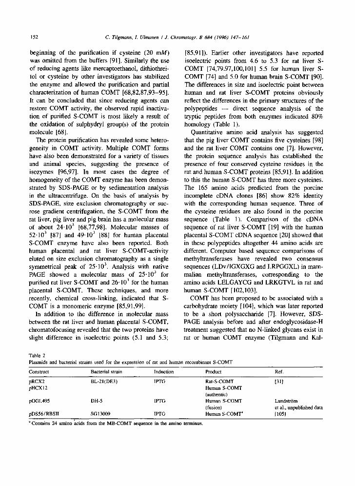
Further purification was performed on an anion- exchange Mono-Q chromatography column (HR5/5, Pharmacia). The column was equilibrated with 20 mM Tris-HCl, (pH 6.05), containing 0.1% fl-mer- captoethanol, and the protein eluted with a linear gradient of NaCI (0-0.7 M) in 40 min (Fig. 8). The Mono-Q fractions were analysed by 12.5% SDS- PAGE and the peak containing COMT-activity and a single 26.103 band (Fig. 9, lane 2) was analysed further by RP-chromatography. From the RP-col- umn, the human recombinant S-COMT eluted as a single peak (Fig. 10), which by analysis in SDS- PAGE showed only the 26.103 S-COMT protein (Fig. 10, insert). The active homogeneous recombi- nant human S-COMT peak from the Mono-Q col- umn is used for ongoing crystallization studies.
The other fractions eluted from the Mono-Q column contained two species of polypeptides with
1 5 1 4 1 3 1 2 1 1 1 0 9 8 7 6 5 4 3 2 1
MW
43.0
30.0
20.1
14.4
16 17 18 19 20 21 22 23 24 25 26 27 28 29 30 MW ~ ~
43 .0
3 0 . 0 ~
20.1
14.4
Fig. 7. SDS-PAGE analysis of recombinant human S-COMT protein eluted from glutathione-Sepharose affinity chromatography column (Pharmacia). The bound GST-COMT fusion protein was eluted from the colunm by the cleavage of the fusion protein with thrombin and every third fraction eluted was analysed by a 12.5% SDS-PAGE. Lanes 1 and 16: low molecular mass standard (Pharmacia). Lanes 2-7: fractions collected during the washing of the column prior to the enzyme cleavage. Lanes from 8 on: fractions eluted after the enzymatic cleavage with thrombin. The gel was stained by Coomassie Brilliant blue.
approximately 1000 Da difference in the apparent molecular mass (Fig. 9, lane 3). We assume that the polypeptide with the higher molecular mass has an extended C-terminus of ca. 7 amino acids. This heterogeneity is probably due to the suppression of the S-COMT translation stop codon, which on the basis of DNA-sequence, could lead to the extension of 7 amino acids and the termination of translation in the stop-codon of the pGEX-2T vector sequence. Similar heterogeneity for this reason has been re- ported in the case of the human recombinant Troponin C protein produced by the same GST- fusion-system [109]. Another possibility is the exten- sion of the C-terminus of the bacteria-produced S- COMT by ribosomal frame-shifting as observed by Vilbois et al. [110].
C. Tilgmann, 1. Ulmanen I J. Chromatogr. B 684 (1996) 147-161
3 2 1
157
U-~t
. - 4 43.0
0
*ZO ,...j
IK)
'l
¢,,I C¢.,
'.'O. l--. C"~ 6"~1 *1" .,--*
CC* ,,JL'I ¢"-I
I I I I I I I I I I I I I I I I | I I I I I I I | I | I I1"i, ,~.., I f 3 t ~
T ~ ~ ¢"~ ,".J
TIME (min) Fig. 8. Anion-exchange chromatography of the recombinant human S-COMT eluted from the glutathione-Sepharose affinity chromatography colunm. The Mono-Q, HR 5/5 column (Phar- macia) was eluted with a linear gradient of NaCI (0-0.7 M in 40 rain) in 20 mM Tris-HCI, (pH 6.05), containing 0.1% ~8-mercap- toethanol with a flow of 1.0 ml/min. Chromatography was monitored at the sensitivity of 0.25 AUFS.
30.0
20.1
I 14.4 Fig. 9. SDS-PAGE analysis of recombinant human S-COMT enzyme eluted from the anion-exchange chromatography shown in Fig. 8. The fractions were analysed on a 12.5% SDS-PAGE followed by Coomassie Brilliant blue staining. Lane (1): low molecular mass standard (Pbarmacia); lane (2): the fraction from the peak 1; lane (3): the fraction from the peaks 2 and 3.
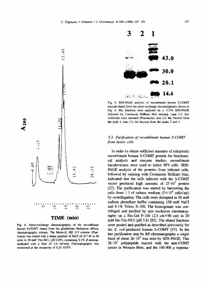
5.3. Purification o f recombinant human S-COMT f rom insect cells
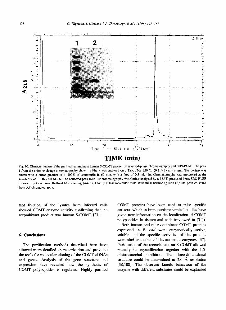
In order to obtain sufficient amounts of eukaryotic recombinant human S-COMT protein for biochemi- cal analysis and enzyme studies, recombinant baculoviruses were used to infect SF9 cells. SDS- PAGE analysis of the proteins from infected cells, followed by staining with Coomassie Brilliant blue, indicated that the cells infected with the S-COMT vector produced high amounts of 25.103 protein [27]. The purification was started by harvesting the cells from 1 1 of culture medium (5)<106 ceUs/ml) by centrifugation. The cells were disrupted in 50 mM sodium phosphate buffer containing 150 mM NaC1 and 0.1% Triton X-100. The homogenate was cen- trifuged and purified by size exclusion chromatog- raphy on a Bio-Gel P-100 (2.5 cm)<90 cm) in 20 mM bis-Tris-HCl (pH 5.8) [85]. The eluted fractions were pooled and purified as described previously for the E. coli-produced human S-COMT [37]. In the last purification step by RP-chromatography a single band of about 26.103 was seen by SDS-PAGE. This 26.103 polypeptide reacted with the anti-COMT serum in Western blots, and the 100 000 g superna-
158 C. Tilgmann, I. Ulmanen I J. Chromatogr. B 684 (1996) 147-161
¢,1 ,<
I
I °
b.j
I -
'J3
I
i I . . l I i
1
I
~ 1 , , , , I . . . . 1 i , ,
' t
2
!i~!iiiii!!iiiiiiii~iill i~! !i i !i
2 t ~3nmt
T l :~e
. . . . . . , . . . . . . . . . . . . . . . . . . . . . . . ~ T r " 7 . . . . . . .
2(~ 7,0 4~. 50
TIME (win) tZig. 10. Characterization of the purified recombinant human S-COMT protein by reversed-phase chromatography and SDS-PAGE. The peak 1 from the anion-exchange chromatography shown in Fig. 8 was analysed on a TSK TMS 250 C1 (0.21 x3 cm) column• The protein was eluted with a linear gradient of 3-100% of acetonitrile in 60 min, with a flow of 0.5 ml/min. Chromatography was monitored at the sensitivity of -0 .02-2.0 AUFS. The collected peak from RP-chromatography was further analysed by a 12.5% precasted Phast SDS-PAGE followed by Coomassie Brilliant blue staining (insert). Lane (1): low molecular mass standard (Pharmacia); lane (2): the peak collected from RP-chromatography.
tant fraction of the lysates from infected cells showed COMT enzyme activity confirming that the recombinant product was human S-COMT [27].
6. Conclusions
The purification methods described here have allowed more detailed characterization and provided the tools for molecular cloning of the COMT cDNAs and genes. Analysis of the gene structure and expression have revealed how the synthesis of COMT polypeptides is regulated. Highly purified
COMT proteins have been used to raise specific antisera, which in immunohistochemical studies have given new information on the localization of COMT polypeptides in tissues and cells (reviewed in [31]).
Both human and rat recombinant COMT proteins expressed in E. coli were enzymatically active, soluble and the specific activities of the proteins were similar to that of the authentic enzymes [37]. Purification of the recombinant rat S-COMT allowed recently its crystallization together with the 1,5- dinitrocatechol inhibitor. The three-dimensional structure could be determined at 2.0 ,~ resolution [38,108]. The observed kinetic behaviour of the enzyme with different substrates could be explained

c. Tilgmann, I. Ulmanen I J. Chromatogr. B 684 (1996) 147-161 159
on the basis o f the a tomic structure o f recombinant
rat S - C O M T [39]. The a tomic structure and the
sequence compar i son reveal that all residues that are
important for the binding of the substrates and for
the catalyt ic react ion are conserved in human and rat
C O M T proteins [38,108].
Unti l recently, the kinetic constants o f the C O M T
enzyme have not been established. The use of tight-
binding inhibitors and recombinant S - C O M T protein,
has a l lowed the determinat ion o f the actual enzyme
concentra t ions and the accurate measurement o f the
kinetic parameters and the substrate selectivity [39].
This study proposed a new kinetic react ion mecha-
nism for the C O M T enzyme. Unders tanding of the
structure and the catalyt ic mechan i sm of C O M T
enzyme will help the deve lopment o f more potent
and cl inical ly interest ing COMT-inhib i tors .
7. List of abbreviations
C O M T
S - C O M T
M B - C O M T
c D N A
RP
S D S - P A G E
P C R
G S T
IPTG
CBB
Catechol -O-methyl t ransferase
Soluble C O M T
Membrane -bound C O M T
Complemen ta ry D N A
reversed-phase
Sod ium dodecyl sulphate
acrylamide gel e lectrophoresis
Po lymerase chain react ion
Gluta thione S-transferase
Isopropyl- /3-D-galactopyranoside
Coomass i e Bri l l iant blue
poly-
References
[1] H. Blaschko and E. Muscholl (Editors), Handb. Exp. Pharm. XXXIII, Springer-Verlag, Heidelberg, New York, 1972.
[2] J. Axelrod, Science, 126 (1957) 400. [3] U. Trendelenburg, Naunyn-Schmiedeberg's Arch. Phar-
macol., 339 (1989) 293. [4] J. Cooper, F. Bloom and R. Roth (Editors), The Biochemical
Basis of Neuropharmacology, Oxford University Press, New York, 1991.
[5] E. Jucker (Editor), Progr. in Drug Res., Birckhauser Verlag, Basel, 1992.
[6] O. Castren and S. Saarikoski, Acta. Obstet. Gynec. Scand., 53 (1974) 41.
[7] A. Liss (Editor), Perspectives in Psychopharmacology: a collection of papers in honor of Earl Usdin, 1988.
[8] J. Axelrod and A.B. Lemer, Biochim. Biophys. Acta, 71 (1963) 650.
[9] P. Ball, R. Knuppen, M. Haupt and H. Breuer, J. Clin. Endocrinol. Met., 34 (1972) 736.
[10] B.T. Zhu, E.L. Ezell and J.G. Liehr, J. Biol. Chem., 269 (1994) 292.
[11] R. Weinshilboum (Editor), Selected Topics in Clinical En- zymology, Walter de Gruyter, Berlin, 1984.
[12] M. Sandier and H. Smith (Editors), Design of Enzyme Inhibitors as Drugs, Oxford University Press, Oxford, 1994.
[13] H. Guldberg and C. Marsden, Pharmacol. Rev., 27 (1975) 135.
[14] D. Park (Editor), Neuromethods 5, Neurotransmitter Enzyme, Humana Press, Clifton, USA, 1986.
[15] R M~innisto and S. Kaakkola, Trends. Pharmacol. Sci., 10 (1989) 54.
[16] G. Bemardi, M. Carpenter, G. Di Chiara and P. Stanzione (Editors), The Basal Ganglia III, Plenum, New York, 1991.
[17] I.J. Kopin, Ann. Rev. Pharmacol. Toxicol., 32 (1993) 467. [18] S. Kaakkola, H. Teravainen, S. Ahtila and A. Gordin,
Neurology, 44 (1994) 77. [19] M. Salminen, K. Lundstrfm, C. Tilgmann, R. Savolainen, N.
Kalkkinen and I. Ulmanen, Gene, 93 (1990) 241. [20] K. Lundstr6m, M. Salminen, A. Jalanko, R. Savolainen and
I. Ulmanen, DNA and Cell Biol., 10 (1991) 181. [21] B. Bertocci,V. Miggiano, M. Da Prada, Z. Dembic, H. Lahm
and P. Malherbe, Proc. Natl. Acad. Sci. USA, 88 (1991) 1416.
[22] J. Tenhunen, M. Salminen, A. Jalanko, S. Ukkonen and I. Ulmanen, DNA and Cell Biol., 12 (1993) 253.
[23] J. Tenhunen, M. Salminen, K. Lundstr6m, T. Kiviluoto, R. Savolainen and I. Ulmanen, Eur. J. Biochem., 223 (1994) 1049.
[24] R. Winqvist, K. Lundstr6m, M. Salminen, M. Laatikainen and I. Ulmanen, Cytogenet. Cell Genet., 59 (1992) 253.
[25] M. Grossman, B. Emanuel and M. Budarf, Genomics, 12 (1992) 822.
[26] J. Roth, Rev. Physiol. Biochem. Pharmacol., 120 (1992) 1. [27] C. Tilgmann, K. Melen, K. Lundstr6m, A. Jalanko, I.
Julkunen, N. Kalkkinen and I. Ulmanen, Eur. J. Biochem., 207 (1992) 813.
[28] J. Tenhunen and I. Ulmanen, Biochem. J., 296 (1993) 595. [29] I. Ulmanen and K. Lundstr6m, Eur. J. Biochem., 202 (1991)
1013. [30] N. Smit, C. Tilgmann, T. Karhunen, R. Slingerland, I.
Ulmanen, W. Westerhof and S. Pavel, Pigment Cell Res., 7 (1994) 403.
[31] K. Lundstr6m, J. Tenhunen, C. Tilgmann. T. Karhunen, P. Panula and I. Ulmanen, Biochim. Biophys. Acta, 1251 (1995) 1.
[32] T. Karhunen, C. Tilgmann, I. Ulmanen, I. Julkunen and P. Panula, J. Histochem. Cytochem., 42 (1994) 1079.
[331 J. Axelrod, W. Albers and C. Clemente, J. Neurochem., 5 (1959) 68.
[34] C. Creveling and B. Hartman (Editors), Biochemistry of SAM and Related Compounds, Macmillan Press, London, 1982.

160 C. Tilgmann, 1. Ulmanen / J. Chromatogr. B 684 (1996) 147-161
[35] T. Karhunen, C. Tilgmann, I. Ulmanen and P. Panula, Neuroscience Lett., 187 (1995) 1.
[36] T. Karhunen, C. Tilgmann, I. Ulmanen and E Panula, Int. J. Devl. Neuroscience, 13 (1995) in press.
[37] K. Lundstr6m, C. Tilgmann, J. Per'~inen, N. Kalkkinen and I. Ulmanen, Biochim. Biophys. Acta, 1129 (1992) 149.
[38] J. Vidgren, L. Svensson and A. Liljas, Nature, 368 (1994) 354.
[39] T. Lotta, J. Vidgren, C. Tilgmann, I. Ulmanen, K. Melen, I. Julkunen and J. Taskinen, Biochemistry, 34 (1995) 4202.
[40] J. Axelrod and R. Tomchick, J. Biol. Chem., 233 (1958) 702. [41] G. Bates, C. Edman, J. Porter, J. Johnston and P. MacDonald,
Clin. Chim. Acta, 94 (1979) 63. [42] R. Borchardt, C. Cheng, P. Cooke and C. Creveling, Life
Sci., 14 (1974) 1089. [43] P. Gulliver and K. Tipton, Biochem. Pharmacol., 27 (1978)
773. [44] M. Levitt, C. Hunter and M. Baron, Neuropsychobiology, 8
(1982) 276. [45] F. Raymond and R. Weinshilboum, Clin. Chim. Acta, 58
(1975) 185. [46] G. Zurcher and M. DaPrada, J. Neurochem., 52 (1982) 191. [47] E. Pennings and G. van Kempen, Anal. Biochem., 98 (1979)
452. [48] H. Nohta, S. Noma and Y. Ohkura, J. Chromatogr., 308
(1984) 93. [49] N. Smit, S. Pavei, A. Kammeyer and W. Westerhof, Anal.
Biochem., 190 (1990) 286. [50] E. Nissinen, Anal. Biochem., 144 (1985) 247. [51] E. Nissinen and P. M~innistiS, Anal. Biochem., 137 (1984) 69. [52] R. Shoup, G. Davis and P. Kissinger, Anal. Chem., 52 (1980)
4 8 3 . [53] E. Schultz and E. Nissinen, Biomed. Chromatography, 3
(1989) 64. [54] O'Brien, G. Semenuk, G. Coleman and S. Spector, Clin.
Exp. Pharmacol. Physiol., 3 (1976) 9. [55] G. De Luca, I. Barberi, P. Ruggeri and R. Di Giorgio, Ital. J.
Biochem., 25 (1976) 213. [56] J. Roth, X. Breakefield and C. Castiglione, Life. Sci., 19
(1976) 1705. [57] R. Weinshilboum, Life Sci., 22 (1978) 625. [58] J. Feldman, D. Reintgen and H. Seigler, Diabetologia, 17
(1979) 249. [59] J. Bidart, M. Assicot and C. Bohuon, Res. Commun. Chem.
Pathol. Pharmacol., 34 (1981) 47. [60] M. Baron, M. Levitt, C. Hunter, R. Gruen and L. Asnis, Biol.
Psychiatry, 17 (1982) 265. [61] J. Bidart, P. Motte, M. Assicot, C. Bohuon and D. Bellet,
Clin. Immunol. Immunopathol., 26 (1983) 1. [62] M. Casey and P. MacDonald, Am. J. Obstet. Gynecol., 145
(1983) 453. [63] M. Spatz, N. Kaneda, C. Sumi, I. Nagatsu, C. Creveling and
T. Nagatsu, Brain Res., 381 (1986) 363. [64] E. Barnea, N. MacLusky, A. DeCherney, F. Naftolin and D.
Phil, Am. J. Perinatol., 5 (1988) 121. [65] E. Nissinen, R. Tuominen, V. Perhoniemi and S. Kaakkola,
Life Sci., 42 (1988) 2609.
[66] P. Molinoff and J. Axelrod, Annu. Rev. Biochem., 40 (1971) 465.
[67] E Anderson and A. D'Iorio, Biochem. Pharmacol., 18 (1968) 1893.
[68] B. Assicot and C. Bohuon, Ear. J. Biochem., 12 (1970) 490. [69] L. Flohe and K. Schwabe, Biochim. Biophys. Acta, 220
(1970) 469. [70] B. Nicodejevic, S. Senoh, J. Daly and C. Creveling, J.
Pharmacol. Exp. Ther., 174 (1970) 83. [71] C. Creveling, N. Morris, H. Shimizu, H. Ong and J. Daly,
Mol. Pharmacol., 8 (1972) 398. [72] R. Borehardt, C. Cheng and D. Thakker, Biochem. Biophys.
Res. Commun., 63 (1975) 69. [73] G. Marzullo and A. Friedhoff, Life Sci., 17 (1975) 933. [74] H. White and J. Chen Wu, Biochem. J., 145 (1975) 135. [75] E Gulliver and C. Wharton, Biochem. Pharmacol., 25 (1976)
2033. [76] J. Tong and A. D'Iorio, Can. J. Biochem., 55 (1977) ll08. [77] E Gulliver and K. Tipton, J. Neurochem., 32 (1979) 1525. [78] G. Marzullo and A. Friedhoff (Editors), Transmeth.,
Elsevier, Amsterdam, 1979. [79] M. Grossman, C. Creveling, R. Rybczynski, M. Braverman,
C. Isersky and X. Breakefield, J. Neurochem., 44 (1985) 421.
[80] J. Veser and W. May, Chromatographia, 22 (1986) 404. [81] J. Veser and R. Martin, Eur. J. Biochem., 154 (1986) 657. [82] J. Rhee and M. Choi, Korean Biochem. J., 21 (1988) 60. [83] T. Korkolainen and E. Nissinen, Biomed. Chromatogr., 3
(1989) 127. [84] N. Ayoubi, C. Boehlert and C. Creveling, Fed. Proc., 46
(1987) 2277. [85] C. Tilgmann and N. Kalkkinen, FEBS Lett., 264 (1990) 95. [86] B. Bertocci, G. Garotta, M. Da Prada, H. Lahm, G. Zureher,
G. Virgallita and V. Miggiano, Biochim. Biophys. Acta, 1080 (1991) 103.
[87] R. Gugler, R. Knuppen and H. Breuer, Biochim. Biophys. Acta, 220 (1970) 10.
[88] E Darmenton, L. Cronenberger and H. Pacheco, Biochimie, 58 (1976) 1401.
[89] P. Ball, R. Knuppen, M. Haupt and H. Breuer., Eur. J. Biochem., 26 (1972) 560.
[90] D. Jeffery and J. Roth, J. Neurochem., 44 (1985) 881. [91] C. Tilgmann and N. Kalkkinen, Biochem. Biophys. Res.
Comm., 174 (1991) 995. [92] N. Dewji, D. Wenger and J. O'Brien, Proc. Natl. Acad. Sci.
USA, 84 (t987) 8652. [93] P. Ball, R. Knuppen and H Breuer, Eur. J. Biochem., 21
(1971) 517. [94] P. Ball, R. Knuppen and H. Breuer, J. Neural. Transm., 32
(1971) 359. [95] L. Flohe and H. Hennies (Editors), Structure and Function of
Monoamine Enzymes, Marcel Dekker, New York, 1977. [96] B. Assicot and C. Bohuon, Biochimie, 53 (1971) 871. [97] M. Huh and A. Friedhoff, J. Biol. Chem., 254 (1979) 299. [98] P. Gulliver and K. Tipton, Eur. J. Biochem., 88 (1978) 439. [99] K. Melen, T. Ronni, B. Broni, R. Krug, C. yon Bonsdorff
and I. Julkunen, J. Biol. Chem., 267 (1992) 25898.

C. Tilgmann, 1. Ulmanen / J. Chromatogr. B 684 (1996) 147-161 161
[100] E. Usdin and S. Snyder (Editors), Frontiers of Catechol- amine Research, Pergamon Press, UK, 1973.
[101] W. Heydom, G. Creed, C. Creveling and D. Jacobowitz, Neurochem. Int., 8 (1986) 581.
[102] M. Fujioka, Int. J. Biochem., 24 (1992) 1917. [103] T. Gomi, K. Tanihara, T. Date and M. Fujioka, Int. J.
Biochem., 24 (1992) 1639. [104] E. Usdin and R. Kvetnansky (Editors), The Role of
Catecholamines and other Neurotransmitters in Stress, Gordon and Breach, New York, 1984.
[105] P. Malherbe, B. Bertocci, P. Caspers, P. Zurcher and M. Da Prada, J. Neurochem., 58 (1992) 1782.
[106] Y. Matsuura, R. Possee, H. Overton and D. Bishop, J. Gen. Virol., 68 (1987) 1233.
[107] M. Summers and G. Smith, Tex. Agric. Exp. Stn. Bull., 1555 (1987) 56.
[108] J. Vidgren, C. Tilgmann, K. Lundstr6m and A. Liljas, Proteins: Struct., Funct., Genet., 11 (1991) 233.
[109] P. Pollesello, M. Ovaska, J. Kaivola, C. Tilgmann, K. Lundstr6m, N. Kalkkinen, I. Ulmanen, E. Nissinen and J. Taskinen, J. Biol. Chem., 269 (1994) 28584.
[110] F. Vilbois, P. Caspers, M. DaPrada, G. Lang, C. Karrer, H.-W. Lahm and A.M. Cesura, Eur. J. Biochem., 222 (1994) 377.