Mammalian Wnt3a is released on lipoprotein particles
13
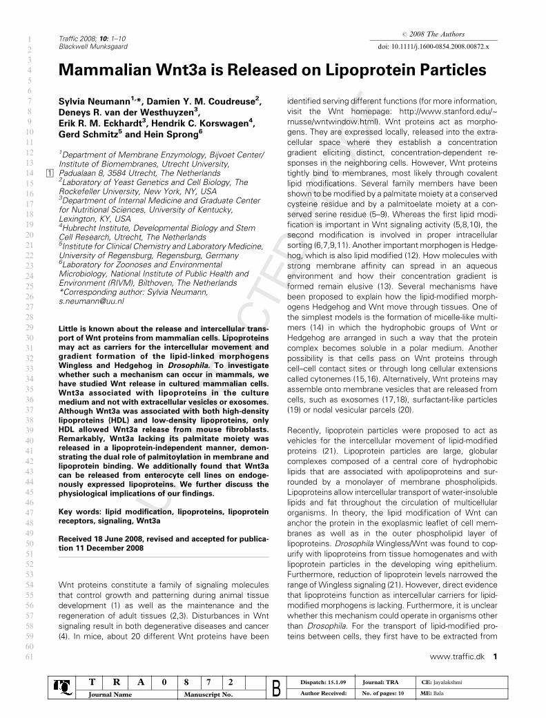
# 2008 The Authors doi: 10.1111/j.1600-0854.2008.00872.x Traffic 2008; 10: 1–10 Blackwell Munksgaard Mammalian Wnt3a is Released on Lipoprotein Particles Sylvia Neumann 1, *, Damien Y. M. Coudreuse 2 , Deneys R. van der Westhuyzen 3 , Erik R. M. Eckhardt 3 , Hendrik C. Korswagen 4 , Gerd Schmitz 5 and Hein Sprong 6 1 Department of Membrane Enzymology, Bijvoet Center/ Institute of Biomembranes, Utrecht University, Padualaan 8, 3584 Utrecht, The Netherlands 1 2 Laboratory of Yeast Genetics and Cell Biology, The Rockefeller University, New York, NY, USA 3 Department of Internal Medicine and Graduate Center for Nutritional Sciences, University of Kentucky, Lexington, KY, USA 4 Hubrecht Institute, Developmental Biology and Stem Cell Research, Utrecht, The Netherlands 5 Institute for Clinical Chemistry and Laboratory Medicine, University of Regensburg, Regensburg, Germany 6 Laboratory for Zoonoses and Environmental Microbiology, National Institute of Public Health and Environment (RIVM), Bilthoven, The Netherlands *Corresponding author: Sylvia Neumann, [email protected] Little is known about the release and intercellular trans- port of Wnt proteins from mammalian cells. Lipoproteins may act as carriers for the intercellular movement and gradient formation of the lipid-linked morphogens Wingless and Hedgehog in Drosophila. To investigate whether such a mechanism can occur in mammals, we have studied Wnt release in cultured mammalian cells. Wnt3a associated with lipoproteins in the culture medium and not with extracellular vesicles or exosomes. Although Wnt3a was associated with both high-density lipoproteins (HDL) and low-density lipoproteins, only HDL allowed Wnt3a release from mouse fibroblasts. Remarkably, Wnt3a lacking its palmitate moiety was released in a lipoprotein-independent manner, demon- strating the dual role of palmitoylation in membrane and lipoprotein binding. We additionally found that Wnt3a can be released from enterocyte cell lines on endoge- nously expressed lipoproteins. We further discuss the physiological implications of our findings. Key words: lipid modification, lipoproteins, lipoprotein receptors, signaling, Wnt3a Received 18 June 2008, revised and accepted for publica- tion 11 December 2008 Wnt proteins constitute a family of signaling molecules that control growth and patterning during animal tissue development (1) as well as the maintenance and the regeneration of adult tissues (2,3). Disturbances in Wnt signaling result in both degenerative diseases and cancer (4). In mice, about 20 different Wnt proteins have been identified serving different functions (for more information, visit the Wnt homepage: http://www.stanford.edu/~ rnusse/wntwindow.html). Wnt proteins act as morpho- gens. They are expressed locally, released into the extra- cellular space where they establish a concentration gradient eliciting distinct, concentration-dependent re- sponses in the neighboring cells. However, Wnt proteins tightly bind to membranes, most likely through covalent lipid modifications. Several family members have been shown to be modified by a palmitate moiety at a conserved cysteine residue and by a palmitoelate moiety at a con- served serine residue (5–9). Whereas the first lipid modi- fication is important in Wnt signaling activity (5,8,10), the second modification is involved in proper intracellular sorting (6,7,9,11). Another important morphogen is Hedge- hog, which is also lipid modified (12). How molecules with strong membrane affinity can spread in an aqueous environment and how their concentration gradient is formed remain elusive (13). Several mechanisms have been proposed to explain how the lipid-modified morph- ogens Hedgehog and Wnt move through tissues. One of the simplest models is the formation of micelle-like multi- mers (14) in which the hydrophobic groups of Wnt or Hedgehog are arranged in such a way that the protein complex becomes soluble in a polar medium. Another possibility is that cells pass on Wnt proteins through cell–cell contact sites or through long cellular extensions called cytonemes (15,16). Alternatively, Wnt proteins may assemble onto membrane vesicles that are released from cells, such as exosomes (17,18), surfactant-like particles (19) or nodal vesicular parcels (20). Recently, lipoprotein particles were proposed to act as vehicles for the intercellular movement of lipid-modified proteins (21). Lipoprotein particles are large, globular complexes composed of a central core of hydrophobic lipids that are associated with apolipoproteins and sur- rounded by a monolayer of membrane phospholipids. Lipoproteins allow intercellular transport of water-insoluble lipids and fat throughout the circulation of multicellular organisms. In theory, the lipid modification of Wnt can anchor the protein in the exoplasmic leaflet of cell mem- branes as well as in the outer phospholipid layer of lipoproteins. Drosophila Wingless/Wnt was found to cop- urify with lipoproteins from tissue homogenates and with lipoprotein particles in the developing wing epithelium. Furthermore, reduction of lipoprotein levels narrowed the range of Wingless signaling (21). However, direct evidence that lipoproteins function as intercellular carriers for lipid- modified morphogens is lacking. Furthermore, it is unclear whether this mechanism could operate in organisms other than Drosophila. For the transport of lipid-modified pro- teins between cells, they first have to be extracted from www.traffic.dk 1 1 2 3 4 5 6 7 8 9 10 11 12 13 14 15 16 17 18 19 20 21 22 23 24 25 26 27 28 29 30 31 32 33 34 35 36 37 38 39 40 41 42 43 44 45 46 47 48 49 50 51 52 53 54 55 56 57 58 59 60 61 T R A 0 8 7 2 Journal Name Manuscript No. B Dispatch: 15.1.09 Journal: TRA CE: Jayalakshmi Author Received: No. of pages: 10 ME: Bala
-
Upload
independent -
Category
Documents
-
view
0 -
download
0
Transcript of Mammalian Wnt3a is released on lipoprotein particles
# 2008 The Authors
doi: 10.1111/j.1600-0854.2008.00872.xTraffic 2008; 10: 1–10Blackwell Munksgaard
Mammalian Wnt3a is Released on Lipoprotein Particles
Sylvia Neumann1,*, Damien Y. M. Coudreuse2,
Deneys R. van der Westhuyzen3,
Erik R. M. Eckhardt3, Hendrik C. Korswagen4,
Gerd Schmitz5 and Hein Sprong6
1Department of Membrane Enzymology, Bijvoet Center/Institute of Biomembranes, Utrecht University,Padualaan 8, 3584 Utrecht, The Netherlands12Laboratory of Yeast Genetics and Cell Biology, TheRockefeller University, New York, NY, USA3Department of Internal Medicine and Graduate Centerfor Nutritional Sciences, University of Kentucky,Lexington, KY, USA4Hubrecht Institute, Developmental Biology and StemCell Research, Utrecht, The Netherlands5Institute for Clinical Chemistry and Laboratory Medicine,University of Regensburg, Regensburg, Germany6Laboratory for Zoonoses and EnvironmentalMicrobiology, National Institute of Public Health andEnvironment (RIVM), Bilthoven, The Netherlands*Corresponding author: Sylvia Neumann,[email protected]
Little is known about the release and intercellular trans-
port of Wnt proteins from mammalian cells. Lipoproteins
may act as carriers for the intercellular movement and
gradient formation of the lipid-linked morphogens
Wingless and Hedgehog in Drosophila. To investigate
whether such a mechanism can occur in mammals, we
have studied Wnt release in cultured mammalian cells.
Wnt3a associated with lipoproteins in the culture
medium and not with extracellular vesicles or exosomes.
Although Wnt3a was associated with both high-density
lipoproteins (HDL) and low-density lipoproteins, only
HDL allowed Wnt3a release from mouse fibroblasts.
Remarkably, Wnt3a lacking its palmitate moiety was
released in a lipoprotein-independent manner, demon-
strating the dual role of palmitoylation in membrane and
lipoprotein binding. We additionally found that Wnt3a
can be released from enterocyte cell lines on endoge-
nously expressed lipoproteins. We further discuss the
physiological implications of our findings.
Key words: lipid modification, lipoproteins, lipoprotein
receptors, signaling, Wnt3a
Received 18 June 2008, revised and accepted for publica-
tion 11 December 2008
Wnt proteins constitute a family of signaling molecules
that control growth and patterning during animal tissue
development (1) as well as the maintenance and the
regeneration of adult tissues (2,3). Disturbances in Wnt
signaling result in both degenerative diseases and cancer
(4). In mice, about 20 different Wnt proteins have been
identified serving different functions (for more information,
visit the Wnt homepage: http://www.stanford.edu/~
rnusse/wntwindow.html). Wnt proteins act as morpho-
gens. They are expressed locally, released into the extra-
cellular space where they establish a concentration
gradient eliciting distinct, concentration-dependent re-
sponses in the neighboring cells. However, Wnt proteins
tightly bind to membranes, most likely through covalent
lipid modifications. Several family members have been
shown to bemodified by a palmitate moiety at a conserved
cysteine residue and by a palmitoelate moiety at a con-
served serine residue (5–9). Whereas the first lipid modi-
fication is important in Wnt signaling activity (5,8,10), the
second modification is involved in proper intracellular
sorting (6,7,9,11). Another important morphogen is Hedge-
hog, which is also lipid modified (12). How molecules with
strong membrane affinity can spread in an aqueous
environment and how their concentration gradient is
formed remain elusive (13). Several mechanisms have
been proposed to explain how the lipid-modified morph-
ogens Hedgehog and Wnt move through tissues. One of
the simplest models is the formation of micelle-like multi-
mers (14) in which the hydrophobic groups of Wnt or
Hedgehog are arranged in such a way that the protein
complex becomes soluble in a polar medium. Another
possibility is that cells pass on Wnt proteins through
cell–cell contact sites or through long cellular extensions
called cytonemes (15,16). Alternatively, Wnt proteins may
assemble onto membrane vesicles that are released from
cells, such as exosomes (17,18), surfactant-like particles
(19) or nodal vesicular parcels (20).
Recently, lipoprotein particles were proposed to act as
vehicles for the intercellular movement of lipid-modified
proteins (21). Lipoprotein particles are large, globular
complexes composed of a central core of hydrophobic
lipids that are associated with apolipoproteins and sur-
rounded by a monolayer of membrane phospholipids.
Lipoproteins allow intercellular transport of water-insoluble
lipids and fat throughout the circulation of multicellular
organisms. In theory, the lipid modification of Wnt can
anchor the protein in the exoplasmic leaflet of cell mem-
branes as well as in the outer phospholipid layer of
lipoproteins. Drosophila Wingless/Wnt was found to cop-
urify with lipoproteins from tissue homogenates and with
lipoprotein particles in the developing wing epithelium.
Furthermore, reduction of lipoprotein levels narrowed the
range ofWingless signaling (21). However, direct evidence
that lipoproteins function as intercellular carriers for lipid-
modified morphogens is lacking. Furthermore, it is unclear
whether this mechanism could operate in organisms other
than Drosophila. For the transport of lipid-modified pro-
teins between cells, they first have to be extracted from
www.traffic.dk 1
1
2
3
4
5
6
7
8
9
10
11
12
13
14
15
16
17
18
19
20
21
22
23
24
25
26
27
28
29
30
31
32
33
34
35
36
37
38
39
40
41
42
43
44
45
46
47
48
49
50
51
52
53
54
55
56
57
58
59
60
61
T R A 0 8 7 2
Journal Name Manuscript No. BDispatch: 15.1.09 Journal: TRA CE: Jayalakshmi
Author Received: No. of pages: 10 ME: Bala
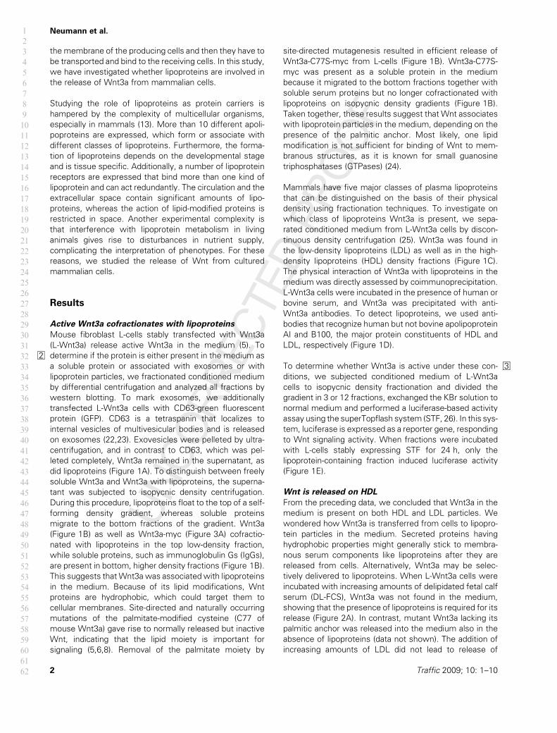
the membrane of the producing cells and then they have to
be transported and bind to the receiving cells. In this study,
we have investigated whether lipoproteins are involved in
the release of Wnt3a from mammalian cells.
Studying the role of lipoproteins as protein carriers is
hampered by the complexity of multicellular organisms,
especially in mammals (13). More than 10 different apoli-
poproteins are expressed, which form or associate with
different classes of lipoproteins. Furthermore, the forma-
tion of lipoproteins depends on the developmental stage
and is tissue specific. Additionally, a number of lipoprotein
receptors are expressed that bind more than one kind of
lipoprotein and can act redundantly. The circulation and the
extracellular space contain significant amounts of lipo-
proteins, whereas the action of lipid-modified proteins is
restricted in space. Another experimental complexity is
that interference with lipoprotein metabolism in living
animals gives rise to disturbances in nutrient supply,
complicating the interpretation of phenotypes. For these
reasons, we studied the release of Wnt from cultured
mammalian cells.
Results
Active Wnt3a cofractionates with lipoproteins
Mouse fibroblast L-cells stably transfected with Wnt3a
(L-Wnt3a) release active Wnt3a in the medium (5). To
2 determine if the protein is either present in the medium as
a soluble protein or associated with exosomes or with
lipoprotein particles, we fractionated conditioned medium
by differential centrifugation and analyzed all fractions by
western blotting. To mark exosomes, we additionally
transfected L-Wnt3a cells with CD63-green fluorescent
protein (GFP). CD63 is a tetraspanin that localizes to
internal vesicles of multivesicular bodies and is released
on exosomes (22,23). Exovesicles were pelleted by ultra-
centrifugation, and in contrast to CD63, which was pel-
leted completely, Wnt3a remained in the supernatant, as
did lipoproteins (Figure 1A). To distinguish between freely
soluble Wnt3a and Wnt3a with lipoproteins, the superna-
tant was subjected to isopycnic density centrifugation.
During this procedure, lipoproteins float to the top of a self-
forming density gradient, whereas soluble proteins
migrate to the bottom fractions of the gradient. Wnt3a
(Figure 1B) as well as Wnt3a-myc (Figure 3A) cofractio-
nated with lipoproteins in the top low-density fraction,
while soluble proteins, such as immunoglobulin Gs (IgGs),
are present in bottom, higher density fractions (Figure 1B).
This suggests thatWnt3a was associatedwith lipoproteins
in the medium. Because of its lipid modifications, Wnt
proteins are hydrophobic, which could target them to
cellular membranes. Site-directed and naturally occurring
mutations of the palmitate-modified cysteine (C77 of
mouse Wnt3a) gave rise to normally released but inactive
Wnt, indicating that the lipid moiety is important for
signaling (5,6,8). Removal of the palmitate moiety by
site-directed mutagenesis resulted in efficient release of
Wnt3a-C77S-myc from L-cells (Figure 1B). Wnt3a-C77S-
myc was present as a soluble protein in the medium
because it migrated to the bottom fractions together with
soluble serum proteins but no longer cofractionated with
lipoproteins on isopycnic density gradients (Figure 1B).
Taken together, these results suggest that Wnt associates
with lipoprotein particles in the medium, depending on the
presence of the palmitic anchor. Most likely, one lipid
modification is not sufficient for binding of Wnt to mem-
branous structures, as it is known for small guanosine
triphosphatases (GTPases) (24).
Mammals have five major classes of plasma lipoproteins
that can be distinguished on the basis of their physical
density using fractionation techniques. To investigate on
which class of lipoproteins Wnt3a is present, we sepa-
rated conditioned medium from L-Wnt3a cells by discon-
tinuous density centrifugation (25). Wnt3a was found in
the low-density lipoproteins (LDL) as well as in the high-
density lipoproteins (HDL) density fractions (Figure 1C).
The physical interaction of Wnt3a with lipoproteins in the
medium was directly assessed by coimmunoprecipitation.
L-Wnt3a cells were incubated in the presence of human or
bovine serum, and Wnt3a was precipitated with anti-
Wnt3a antibodies. To detect lipoproteins, we used anti-
bodies that recognize human but not bovine apolipoprotein
AI and B100, the major protein constituents of HDL and
LDL, respectively (Figure 1D).
To 3determine whether Wnt3a is active under these con-
ditions, we subjected conditioned medium of L-Wnt3a
cells to isopycnic density fractionation and divided the
gradient in 3 or 12 fractions, exchanged the KBr solution to
normal medium and performed a luciferase-based activity
assay using the superTopflash system (STF, 26). In this sys-
tem, luciferase is expressed as a reporter gene, responding
to Wnt signaling activity. When fractions were incubated
with L-cells stably expressing STF for 24 h, only the
lipoprotein-containing fraction induced luciferase activity
(Figure 1E).
Wnt is released on HDL
From the preceding data, we concluded that Wnt3a in the
medium is present on both HDL and LDL particles. We
wondered how Wnt3a is transferred from cells to lipopro-
tein particles in the medium. Secreted proteins having
hydrophobic properties might generally stick to membra-
nous serum components like lipoproteins after they are
released from cells. Alternatively, Wnt3a may be selec-
tively delivered to lipoproteins. When L-Wnt3a cells were
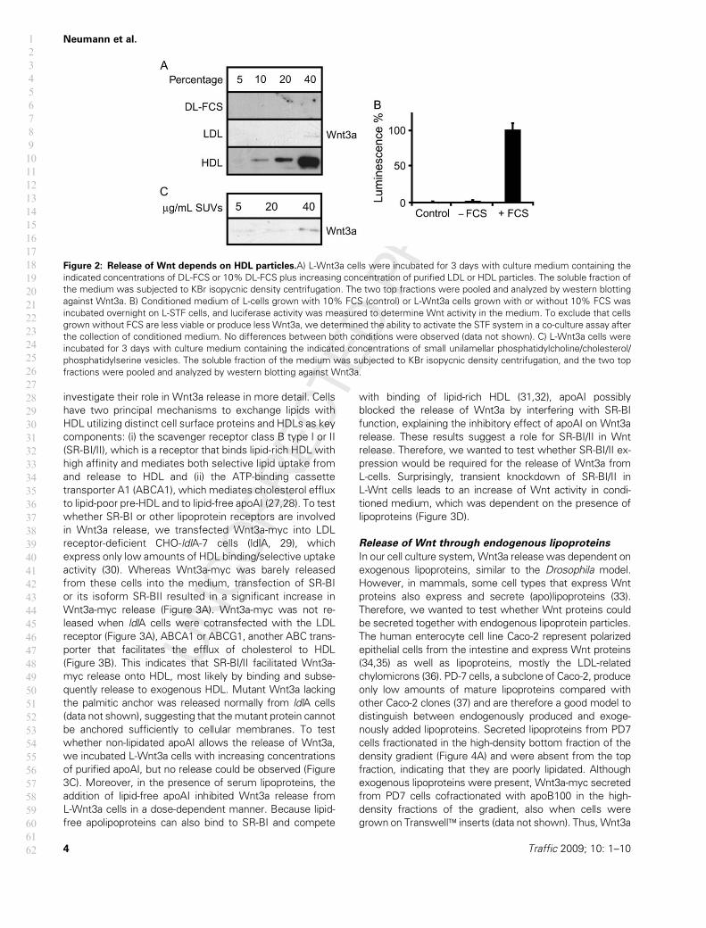
incubated with increasing amounts of delipidated fetal calf
serum (DL-FCS), Wnt3a was not found in the medium,
showing that the presence of lipoproteins is required for its
release (Figure 2A). In contrast, mutant Wnt3a lacking its
palmitic anchor was released into the medium also in the
absence of lipoproteins (data not shown). The addition of
increasing amounts of LDL did not lead to release of
2 Traffic 2009; 10: 1–10
Neumann et al.1
2
3
4
5
6
7
8
9
10
11
12
13
14
15
16
17
18
19
20
21
22
23
24
25
26
27
28
29
30
31
32
33
34
35
36
37
38
39
40
41
42
43
44
45
46
47
48
49
50
51
52
53
54
55
56
57
58
59
60
61
62

Wnt3a from cells either. However, Wnt3a release into the
medium was only restored by the addition of HDL (Figure
2A). To confirm that lipoproteins are required for the
release of functional Wnt3a, conditioned medium from
L-Wnt3a cells was produced with and without 10% FCS.
Only medium containing lipoproteins exhibited activity,
whereas medium without lipoproteins only showed minor
activity (Figure 2B). To exclude that structures or particles
that generally have the capability to sequester hydrophobic
molecules lead to the extraction of Wnt3a from plasma
membranes, we tested whether small unilamellar vesicles
(SUVs) were capable to solubilize Wnt3a. However, re-
lease of Wnt3a after incubation with vesicles was not
observed (Figure 2C).
Release of Wnt3a is facilitated by the
SR-BI/II receptor
Our results argue against an unspecific release of Wnt3a
on lipoproteins or other serum components but indicate
the specific use of HDL particles. This prompted us to
Figure 1: Association of Wnt3a with lipoprotein particles.A)L-cells stably expressing Wnt3a (L-Wnt3a) were transiently transfected
with CD63-GFP and incubated with culture medium containing 10% FCS and 0.5% human serum for 3 days. Cells were scraped and
pooled with the 10 000� gpellet from themedium (cells). The supernatant was centrifuged for 1 h at 120 000� gand separated into pellet
(exosomes) and supernatant (S120). B) Twelve fractions were taken from top (T) to bottom (B) of KBr isopycnic density gradients of the
S120 from media of L-cells stably or transiently (mock) expressing Wnt3a or mutant Wnt3a-C77S-myc (L-Wnt3a-myc) and analyzed by
western blotting for themigration ofWnt proteins and compared themwith lipoprotein-associated proteins (h-apoB100) or soluble proteins
(hIgGs). C) S120 from media of L-Wnt3a cells were analyzed by discontinuous KBr density fractionation to separate different classes of
lipoproteins and analyzed for the presence of Wnt3a and apolipoproteins (h-apoAI and h-apoB100) by western blotting. D) L-Wnt3a cells
were incubated with culture medium containing either 10% FCS (�) or 5% FCS and 5% human serum (þ) for 3 days. Wnt3a from the
medium was adsorbed to protein A–Sepharose with rabbit antisera against Wnt3a or myc as a control (IP). Immunoprecipitates were
subjected to SDS–PAGE and western blotting. Coprecipitated human lipoproteins were detected using antisera against h-apoA1 and h-
apoB100, respectively. The relative amounts of lipoproteins and Wnt3a in the media prior immunoprecipitation (load) were determined on
a fraction of the media by western blotting. Coprecipitation was specific because immunoprecipitation with a control antibody (GAPDH) did
not pull down either h-apoAI or h-apoB100. E) To measure which fraction of the medium contained active Wnt3a, conditioned medium of
L-cells or L-Wnt3a cells was subjected to a KBr gradient (as in Figure 1B) and the fractions were exchanged to normal medium. L-cells
stably expressing the superTopFlash system (L-STF) were incubated with fractions overnight, and luciferase activity was determined.
Traffic 2009; 10: 1–10 3
Release of Mammalian Wnt1
2
3
4
5
6
7
8
9
10
11
12
13
14
15
16
17
18
19
20
21
22
23
24
25
26
27
28
29
30
31
32
33
34
35
36
37
38
39
40
41
42
43
44
45
46
47
48
49
50
51
52
53
54
55
56
57
58
59
60
61
62
investigate their role in Wnt3a release in more detail. Cells
have two principal mechanisms to exchange lipids with
HDL utilizing distinct cell surface proteins and HDLs as key
components: (i) the scavenger receptor class B type I or II
(SR-BI/II), which is a receptor that binds lipid-rich HDL with
high affinity and mediates both selective lipid uptake from
and release to HDL and (ii) the ATP-binding cassette
transporter A1 (ABCA1), which mediates cholesterol efflux
to lipid-poor pre-HDL and to lipid-free apoAI (27,28). To test
whether SR-BI or other lipoprotein receptors are involved
in Wnt3a release, we transfected Wnt3a-myc into LDL
receptor-deficient CHO-ldlA-7 cells (ldlA, 29), which
express only low amounts of HDL binding/selective uptake
activity (30). Whereas Wnt3a-myc was barely released
from these cells into the medium, transfection of SR-BI
or its isoform SR-BII resulted in a significant increase in
Wnt3a-myc release (Figure 3A). Wnt3a-myc was not re-
leased when ldlA cells were cotransfected with the LDL
receptor (Figure 3A), ABCA1 or ABCG1, another ABC trans-
porter that facilitates the efflux of cholesterol to HDL
(Figure 3B). This indicates that SR-BI/II facilitated Wnt3a-
myc release onto HDL, most likely by binding and subse-
quently release to exogenous HDL. Mutant Wnt3a lacking
the palmitic anchor was released normally from ldlA cells
(data not shown), suggesting that themutant protein cannot
be anchored sufficiently to cellular membranes. To test
whether non-lipidated apoAI allows the release of Wnt3a,
we incubated L-Wnt3a cells with increasing concentrations
of purified apoAI, but no release could be observed (Figure
3C). Moreover, in the presence of serum lipoproteins, the
addition of lipid-free apoAI inhibited Wnt3a release from
L-Wnt3a cells in a dose-dependent manner. Because lipid-
free apolipoproteins can also bind to SR-BI and compete
with binding of lipid-rich HDL (31,32), apoAI possibly
blocked the release of Wnt3a by interfering with SR-BI
function, explaining the inhibitory effect of apoAI on Wnt3a
release. These results suggest a role for SR-BI/II in Wnt
release. Therefore, we wanted to test whether SR-BI/II ex-
pression would be required for the release of Wnt3a from
L-cells. Surprisingly, transient knockdown of SR-BI/II in
L-Wnt cells leads to an increase of Wnt activity in condi-
tioned medium, which was dependent on the presence of
lipoproteins (Figure 3D).
Release of Wnt through endogenous lipoproteins
In our cell culture system,Wnt3a release was dependent on
exogenous lipoproteins, similar to the Drosophila model.
However, in mammals, some cell types that express Wnt
proteins also express and secrete (apo)lipoproteins (33).
Therefore, we wanted to test whether Wnt proteins could
be secreted together with endogenous lipoprotein particles.
The human enterocyte cell line Caco-2 represent polarized
epithelial cells from the intestine and express Wnt proteins
(34,35) as well as lipoproteins, mostly the LDL-related
chylomicrons (36). PD-7 cells, a subclone of Caco-2, produce
only low amounts of mature lipoproteins compared with
other Caco-2 clones (37) and are therefore a good model to
distinguish between endogenously produced and exoge-
nously added lipoproteins. Secreted lipoproteins from PD7
cells fractionated in the high-density bottom fraction of the
density gradient (Figure 4A) and were absent from the top
fraction, indicating that they are poorly lipidated. Although
exogenous lipoproteins were present, Wnt3a-myc secreted
from PD7 cells cofractionated with apoB100 in the high-
density fractions of the gradient, also when cells were
grown on Transwell� inserts (data not shown). Thus,Wnt3a
Figure 2: Release of Wnt depends on HDL particles.A) L-Wnt3a cells were incubated for 3 days with culture medium containing the
indicated concentrations of DL-FCS or 10% DL-FCS plus increasing concentration of purified LDL or HDL particles. The soluble fraction of
the medium was subjected to KBr isopycnic density centrifugation. The two top fractions were pooled and analyzed by western blotting
against Wnt3a. B) Conditioned medium of L-cells grown with 10% FCS (control) or L-Wnt3a cells grown with or without 10% FCS was
incubated overnight on L-STF cells, and luciferase activity was measured to determine Wnt activity in the medium. To exclude that cells
grown without FCS are less viable or produce less Wnt3a, we determined the ability to activate the STF system in a co-culture assay after
the collection of conditioned medium. No differences between both conditions were observed (data not shown). C) L-Wnt3a cells were
incubated for 3 days with culture medium containing the indicated concentrations of small unilamellar phosphatidylcholine/cholesterol/
phosphatidylserine vesicles. The soluble fraction of the medium was subjected to KBr isopycnic density centrifugation, and the two top
fractions were pooled and analyzed by western blotting against Wnt3a.
4 Traffic 2009; 10: 1–10
Neumann et al.1
2
3
4
5
6
7
8
9
10
11
12
13
14
15
16
17
18
19
20
21
22
23
24
25
26
27
28
29
30
31
32
33
34
35
36
37
38
39
40
41
42
43
44
45
46
47
48
49
50
51
52
53
54
55
56
57
58
59
60
61
62
could be released as a soluble protein or be associated to the
poorly lipidated apoB100. To test the interaction between
Wnt3a-myc and lipoproteins under these conditions, we
tested whether apoB100 can be coprecipitated withWnt3a-
myc. We observed that apoB100 is not pulled down in the
absence of Wnt3-myc or with a control antibody but
specifically coprecipitated with anti-myc antibody, indicating
a direct interaction between Wnt3a-myc and apoB100
(Figure 4B). In contrast, the parental Caco-2 cells secrete
lipoproteins that are fully lipidated and fractionate in the top
fraction of a density gradient. In Caco-2 cells stably express-
ing Wnt3a-myc (Caco-2-Wnt3a), Wnt3a-myc cofractionates
with lipoproteins and is absent from the high-density bottom
fraction (Figure 4C). These experiments show that Wnt3a-
myc associated with endogenously produced lipoproteins,
which may have taken place in the secretory pathway or at
the cell surface.
Discussion
Although many components of the Wnt signaling cascade
have been identified and characterized, only little is known
about the release of Wnt into the extracellular space and
its trafficking to neighboring cells, particularly in mammals.
Several mechanisms have been proposed for the transport
of lipid-modified proteins through the aqueous environ-
ment of cells and tissues. Recently, it was suggested that
Wingless, a Wnt homologue in Drosophila, is released and
transported through lipoproteins (21). In this study, we
observed that Wnt3a was released from mammalian cells
by lipoproteins present in the culture medium and that the
protein was active under these conditions. The release of
Wnt3a occurred specifically to HDL particles, and ectopical
expression of SR-BI/II facilitated SR-BI/II Wnt release;
however, knockdown of SR-BI/II had the same effect.
Furthermore, Wnt3a was released on newly synthesized
lipoprotein particles from enterocyte cell lines.
Lipid modifications are required for the
association to lipoproteins
Members of the Wnt family have been shown to be lipid
modified by palmitic acid (16:0) at a conserved cysteine
residue (C77 in Wnt3a) (5) and by palmitoleic acid (16:1) at
a conserved serine residue (S209 in Wnt3a) (6). Whether
all members of this family carry a lipid anchor is not clear;
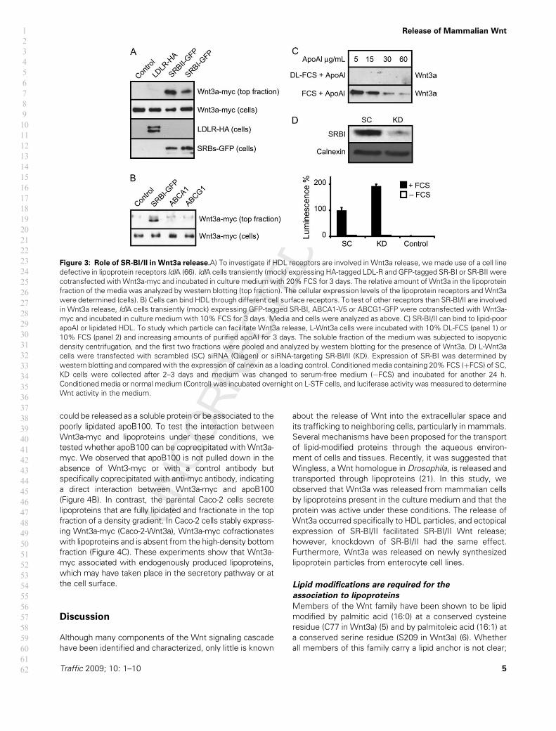
Figure 3: Role of SR-BI/II in Wnt3a release.A) To investigate if HDL receptors are involved in Wnt3a release, we made use of a cell line
defective in lipoprotein receptors ldlA (66). ldlA cells transiently (mock) expressing HA-tagged LDL-R and GFP-tagged SR-BI or SR-BII were
cotransfected withWnt3a-myc and incubated in culture mediumwith 20% FCS for 3 days. The relative amount of Wnt3a in the lipoprotein
fraction of the media was analyzed by western blotting (top fraction). The cellular expression levels of the lipoprotein receptors and Wnt3a
were determined (cells). B) Cells can bind HDL through different cell surface receptors. To test of other receptors than SR-BI/II are involved
in Wnt3a release, ldlA cells transiently (mock) expressing GFP-tagged SR-BI, ABCA1-V5 or ABCG1-GFP were cotransfected with Wnt3a-
myc and incubated in culture medium with 10% FCS for 3 days. Media and cells were analyzed as above. C) SR-BI/II can bind to lipid-poor
apoAI or lipidated HDL. To study which particle can facilitate Wnt3a release, L-Wnt3a cells were incubated with 10% DL-FCS (panel 1) or
10% FCS (panel 2) and increasing amounts of purified apoAI for 3 days. The soluble fraction of the medium was subjected to isopycnic
density centrifugation, and the first two fractions were pooled and analyzed by western blotting for the presence of Wnt3a. D) L-Wnt3a
cells were transfected with scrambled (SC) siRNA (Qiagen) or siRNA-targeting SR-BI/II (KD). Expression of SR-BI was determined by
western blotting and compared with the expression of calnexin as a loading control. Conditioned media containing 20% FCS (þFCS) of SC,
KD cells were collected after 2–3 days and medium was changed to serum-free medium (�FCS) and incubated for another 24 h.
Conditioned media or normal medium (Control) was incubated overnight on L-STF cells, and luciferase activity was measured to determine
Wnt activity in the medium.
Traffic 2009; 10: 1–10 5
Release of Mammalian Wnt1
2
3
4
5
6
7
8
9
10
11
12
13
14
15
16
17
18
19
20
21
22
23
24
25
26
27
28
29
30
31
32
33
34
35
36
37
38
39
40
41
42
43
44
45
46
47
48
49
50
51
52
53
54
55
56
57
58
59
60
61
62
however, the critical residues for lipid modifications are
highly conserved between family members and species.
Nonetheless, only a number of Wnt proteins have been
characterized in respect to the lipid anchor so far (5–9). As
proteins with different lipid anchors associated with lip-
oproteins in Drosophila, we expect the association of Wnt
proteins with lipoproteins also to occur with Wnt proteins
other than Wnt3a. However, very recently, it was shown
that WntD, a Drosophila Wnt protein that does not have a
mammalian homologue, is not lipid modified (38). Removal
of the palmitate moiety allows normal release of Wnt to
the cell culture medium but impairs its function, whereas
removal of the palmitoleate moiety impedes transport of
Wnt from the endoplasmic reticulum (ER) to the plasma
membrane (5–11), indicating several distinct roles of the
lipid modifications. Despite the normal release from cells
of mutant WntC77S, which lacks the palmitate moiety,
into the medium, we find that mutant WntC77S was
present as a soluble protein, in contrast to wild-type
Wnt3a, which was associated with lipoproteins. This
indicates that the palmitoleate moiety on Ser209 by itself
is not able to anchor Wnt3a to lipoproteins. Probably,
a single lipid modification is not sufficient to efficiently
target proteins to membranous structures as is the case
for cytosolic small GTPases (24). However, it cannot be
excluded that the removal of the lipid anchor leads to
a structural rearrangement of Wnt, which impairs signaling
activity and lipoprotein binding.
Lipoproteins and lipoprotein receptors
In theory, all classes of lipoproteins have the capability to
carry lipid-modified proteins in the phospholipid monolayer
surrounding the fat core. A possible role for lipoprotein
receptors is to bring lipoprotein particles in close vicinity to
the exoplasmic leaflet of cell membranes, which contain
Wnt. Then, Wnt can be transferred to the lipoprotein
particle, either by passive diffusion or by an active, but
yet unidentified, protein machinery. In our experiments,
Wnt is released from cells only to HDL but not to LDL.
However, once associated to lipoproteins, Wnt could
freely exchange between lipoprotein particles, similar to
other proteins, like the small apolipoproteins apoE and
apoC (39–41). From ldlA cells, the release of Wnt was
facilitated by the SR-BI/II receptor, and several other
lipoprotein receptors tested did not show similar effects.
In contrast, knockdown of SR-BI/II in L-Wnt cells resulted
in an increase of Wnt release. The SR-BI/II knockout
mouse does not show phenotypes related to impaired
Wnt signaling (42), which would be in line with our results
in L-cells. Both cell lines, ldlA and L-cells, are not very well
characterized in terms of lipoprotein receptor expression.
For ldlA cells, it is known that they have a defect in LDL
receptor expression (29,43) and express only low amounts
of HDL binding/selective uptake activity (30); therefore,
these cells probably adapted to a situation with impaired
lipid uptake from lipoproteins. It is difficult to predict,
however, whether upon knockdown of SR-BI/II in L-cells,
other lipoprotein receptors would be upregulated and
also facilitate Wnt release. Furthermore, it is known that
membrane structure is dependent on SR-BI/II expression,
as shown in Sf9 cells and for the formation and organiza-
tion of microvilli in adrenal gland cells (44–46). Hereby, the
lack of SR-BI/II leads to a decrease in membrane
thickness, possibly because of a decrease in membrane
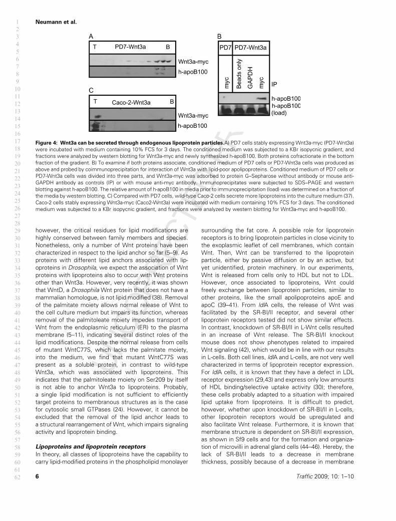
Figure 4: Wnt3a can be secreted through endogenous lipoprotein particles.A) PD7 cells stably expressing Wnt3a-myc (PD7-Wnt3a)
were incubated with medium containing 10% FCS for 3 days. The conditioned medium was subjected to a KBr isopycnic gradient, and
fractions were analyzed by western blotting for Wnt3a-myc and newly synthesized h-apoB100. Both proteins cofractionate in the bottom
fraction of the gradient. B) To examine if both proteins associate, conditioned medium of PD7 cells or PD7-Wnt3a cells was produced as
above and probed by coimmunoprecipitation for interaction of Wnt3a with lipid-poor apolipoproteins. Conditioned medium of PD7 cells or
PD7-Wnt3a cells was divided into three parts, and Wnt3a-myc was adsorbed to protein G–Sepharose without antibody or mouse anti-
GAPDH antibody as controls (IP) or with mouse anti-myc antibody. Immunoprecipitates were subjected to SDS–PAGE and western
blotting against h-apoB100. The relative amount of h-apoB100 in media prior to immunoprecipitation (load) was determined on a fraction of
the media by western blotting. C) Compared with PD7 cells, wild-type Caco-2 cells secrete more lipoproteins into the culture medium (37).
Caco-2 cells stably expressing Wnt3a-myc (Caco2-Wnt3a) were incubated with medium containing 10% FCS for 3 days. The conditioned
medium was subjected to a KBr isopycnic gradient, and fractions were analyzed by western blotting for Wnt3a-myc and h-apoB100.
6 Traffic 2009; 10: 1–10
Neumann et al.1
2
3
4
5
6
7
8
9
10
11
12
13
14
15
16
17
18
19
20
21
22
23
24
25
26
27
28
29
30
31
32
33
34
35
36
37
38
39
40
41
42
43
44
45
46
47
48
49
50
51
52
53
54
55
56
57
58
59
60
61
62
cholesterol (45). It is possible that extraction of Wnt from
the plasma membrane is facilitated under these condi-
tions. Zhai et al. showed that Drosophila Wnt1 is present
in cholesterol-rich, detergent-resistant membranes (9),
which indicates that Wnt is organized in specialized
membrane domains. Therefore, interference with mem-
brane lipid composition may result in different membrane
anchoring.
We find that transfection of ldlA cells with SR-BII leads to
more efficient Wnt release than that with SR-BI. Hereby,
sorting in the endocytic system, which was found to be
essential for Wnt release (47–51), could be responsible for
this effect. SR-BII preferentially localized to endocytic
compartments and the plasmamembrane (52), suggesting
a role in HDL recycling. Possibly, Wnt can also be released
on recycling lipoprotein particles other than HDL (53,54).
Whereas LDL particles and their associated proteins are
taken up through the LDL receptor and targeted for
degradation to the lysosome, HDL can be recycled by
SR-BI/II or ABCA1 (52,55,56). Thus, if Wnt would have
been released onto LDL, both proteins would have been
targeted to lysosomal degradation.
Certainly, the cell culture system we used is poorly
understood. Therefore, it would have to be tested whether
other lipoprotein receptors are also involved in Wnt
release, for example from the large family of LDL receptor-
related proteins or other scavenger receptors. Further-
more, it is important to decipher the mechanism, howWnt
is transferred to lipoprotein particles. Possibly, a change in
membrane environment is required, for example, during
recycling. Finally, the release should be studied in vivo.
Perhaps, the use of inducible reporter genes for Wnt
signaling in combination with inducible, tissue-specific
knockdowns of lipoproteins and their receptors will give
more insight in Wnt transport in vivo.
Wnt release through endogenously expressed
lipoproteins
Different from the Drosophila model, where lipoproteins
are synthesized in the fat body and Wnt is produced in
other cell types, some mammalian cell types producing
Wnt are also able to synthesize lipoproteins (34,35). We
find first evidence that in these cells, Wnt is present on
endogenously produced lipoproteins. In contrast to the
fibroblast system, Wnt3a cofractionated together with
poorly lipidated lipoproteins in conditioned medium from
PD7 cells, instead of being released onto exogenous
lipoproteins, possibly because of the lack of expression
of receptors or because of the sidedness in these cells as
known, for example for SR-BI/II that expresses apically
(57), whereas lipoproteins are secreted basolaterally (58)
together with Wnt3a (data not shown). Whether in these
cells the association of Wnt3a with lipoprotein particles
occurs in the secretory pathway or as a consequence of
extraction from the plasma membrane through secreted
lipoproteins has to be further investigated. Nevertheless,
lipid modification of Wnt and lipidation of lipoproteins take
place in the ER. Therefore, it is likely that association of
both proteins occurs in the same compartment. This
pathway would give cells the opportunity to flexibly react
to changes in nutrition. In 4mammals, the lipid-modified
hormone Ghrelin was found to be associated to very low-
density lipoprotein and HDL particles in serum (59). Ghrelin
is a small peptide hormone carrying an octanoate moiety
(60) and is secreted from the stomach and the intestine
upon starvation to induce food uptake. This is another
example of regulating fat metabolism by the action of lipid-
modified proteins.
Other lipid-modified proteins
A rising number of extracellular proteins have been iden-
tified as containing a lipid anchor. For all of these proteins,
it is unclear how they are transported through an aqueous
environment. Lipoproteins have the potential to be a vehi-
cle for a variety of lipid-modified proteins. In Drosophila,
the lipid-modified morphogens Hedgehog andWingless as
well as GPI-anchored proteins were found to be associated
with lipophorin (21), indicating that the incorporation of
lipid-modified proteins into the phospholipid monolayer of
a lipoprotein is rather unspecific. In contrast, our findings
suggest that the transfer of the lipid-modified protein
Wnt3a occurs specifically to HDL. The fact that not all
lipoprotein receptors facilitated the release of Wnt implies
that, in addition to recruiting the lipoprotein to the mem-
brane, a specific interaction between either the lipid-
modified protein and the lipoprotein or the lipoprotein
receptor could be involved. Theoretically, lipid-modified
proteins and lipoprotein receptors could also be laterally
restricted to distinct membrane domains, depending on
the physical properties of the lipid modification and the
transmembrane domains of the receptor. Thus, the inter-
action between lipoproteins and lipid-modified proteins
may be rather unspecific, although in the context of lateral
organization of membranes combined with individual pro-
tein–protein interactions, specificity in uptake can be
obtained. Whether specificity is only relevant for efficient
release or also serves additional functions remains to be
determined. As many different proteins are associated to
lipoprotein particles (21,61–63), the role of lipoproteins and
their associated proteins should be reconsidered.
Materials and Methods
MaterialsChemicals, if not indicated otherwise, were from Sigma and used in the
highest purity available. Organic solvents were from Riedel de Haen, and
cell culture media and reagents were from Invitrogen. Cell culture plastics
were from Costar. Mouse anti-myc 9E10, rabbit anti-myc A14 and rabbit
anti-hemagglutinin (HA) Y11 antibodies were purchased from Santa Cruz
Biotechnology. Rabbit anti-calnexin H-70 was from Santa Cruz Biotechnol-
ogy. Rabbit anti-mouse SR-BI NB400-104 was from Novus Biologicals.
Rabbit anti-human apolipoprotein AI was from Calbiochem, and antisera
against apolipoprotein B100 were from Calbiochem or from Santa Cruz
Traffic 2009; 10: 1–10 7
Release of Mammalian Wnt1
2
3
4
5
6
7
8
9
10
11
12
13
14
15
16
17
18
19
20
21
22
23
24
25
26
27
28
29
30
31
32
33
34
35
36
37
38
39
40
41
42
43
44
45
46
47
48
49
50
51
52
53
54
55
56
57
58
59
60
61
62
Biotechnology (C1.4). The mouse antibody against GFP JL8 was from
Clontech. Rabbit5 anti-calnexin antiserum (Zhang et al., 1997) was a kind gift
of Ineke Braakman (Utrecht University, The Netherlands). Rabbit anti-
Wnt3a antiserum was a kind gift from Roel Nusse (Stanford University,
Stanford, CA, USA). Mouse anti-glyceraldehyde 3-phosphate dehydroge-
nase (GAPDH) was from Ambion (Applied Biosystems). Anti6 -human IgG
(hIgG) was a kind gift of Jeanette Leusen (University Medical Center
Utrecht, The Netherlands). Horseradish peroxidase-conjugated secondary
goat antibodies were from Pierce. Myc-peptide was synthesized using an
Applied Biosystems 431A peptide synthesizer.
Sera and lipoprotein preparationFCS (Hyclone) was delipidated (DL-FCS) by solvent extraction. Therefore,
serum was mixed with an equal volume of a 2:1 mixture of diisopropyl
ether: n-butanol, and stirred at room temperature for 30 min, and phases
were separated by centrifugation at 5000 � g for 30 min. The aqueous
phase was re-extracted with an equal volume of diisopropyl ether and
subsequently dialyzed against PBS. This treatment resulted in the total loss
of intact lipoprotein particles, as judged by the loss of proteins from the top
fraction of a KBr gradient centrifugation that was analyzed by SDS–PAGE
and Coomassie brilliant blue staining. The cholesterol content was reduced
to less than 5% of untreated serum, as determined using the Amplex Red
Cholesterol assay kit (Molecular Probes). Human serum was obtained from
healthy fasting human donors, and HDL and LDL were prepared by
discontinuous density centrifugation as previously described (25), dialyzed
against PBS and stored under nitrogen at 48C. The protein contents of the
preparations were determined using the BCA assay (Pierce Chemical Co.).
SUVs were generated by sonication on ice (64) of egg phosphatidylcholine:
cholesterol:egg phosphatidylserine (50:50:1, mol/mol).
PlasmidspcDNA3-LDLR-HA was kindly provided by Jurgen Gent (Utrecht University,
The Netherlands). The construction of pEGFP-SRBI and pEGFP-SRBII has
been described (52). pcDNA3.1/V5-His-TOPO-ABCA1 and pEGFP-ABCG1
were from Gerd Schmitz (Regensburg, Germany). pEGFP-CD63 was from
Gillian Griffiths (Oxford, UK). pcDNA3-Wnt3a was from Damien Coudreuse
(Rockefeller University, New York, NY, USA). A myc tag was appended at
the carboxy-terminus of Wnt3a by polymerase chain reaction (PCR) and
ligated into pcDNA3 (pcDNA3-Wnt3a-myc). Cysteine 77 of Wnt3a was
mutated into a serine by PCR mutagenesis, yielding pcDNA3-Wnt3a-C77S-
myc. All synthetic complementary DNAs were verified by restriction
analysis and dye termination sequencing.
Cell culture and transfectionMutant Chinese hamster ovary ldlA (clone 7) cells that are LDL receptor
deficient and express very little SR-BI or HDL binding/selective uptake
activity (30,65) were kindly provided by Monty Krieger (MIT Department
of Biology, Cambridge, UK) and Jurgen Gent. ldlA and mouse L-cells
[American Type Culture Collection (ATCC)] were grown in DMEM contain-
ing 4 mM L-glutamine, 4.5 g/L glucose and 10% FCS (culture medium) and
were maintained at 378C with 5% CO2. Control L-cells were stably
transfected with pcDNA3 and maintained in culture medium containing
0.4 mg/mL geneticin. L-Wnt3a cells (ATCC) were cultured in culture
medium with 0.4 mg/mL geneticin. For Wnt activity measurements,
L-Wnt3a were kindly provided by Daniele Tauriello (Utrecht University,
The Netherlands), and cells were cultured in medium containing 50 mg/mL
zeocin (Invitrogen). L-cells stably transfected with the superTOPflash
system (L-STF) were kindly provided by Roel Nusse and cultured in medium
containing 0.8 mg/mL geneticin.
For transient overexpression, cells were transfected with Lipofectamine
2000 (Invitrogen) and expression was induced by addition of 2 mM butyrate
during all incubation times. For transient knockdown, cells were transfected
with small interfering RNA (siRNA) oligonucleotides targeting SR-BI/II
(sense: AGG UCA ACA UCA CCU UCA A; Qiagen) using oligofectamine
(Invitrogen). Transfection was repeated after 2–3 days of incubation with
siRNA. Protein expression was determined by western blot after 2–3 days.
Wnt conditioned mediumUnless indicated otherwise, in experiments detecting human lipoproteins
by western blotting, human sera were used. Cells on 3-cm dishes were
transiently (mock) transfected using Lipofectamine 2000 (Invitrogen) and
grown in Optimem (Invitrogen) with DL-FCS, FCS, purified HDL or LDL or
lipid-free apoA1 at 378C with 5% CO2 for 3 days. Cell 7debris was removed
by centrifugation at 10 000 � g, and the supernatant was centrifuged for
1–3 h at 39 000 r.p.m. at 48C in a SW41Ti rotor (Beckman). Lipoprotein
particles were separated from soluble proteins by isopycnic density
centrifugation. In short, KBr was added to the supernatant to a final
concentration of 0.33 g/mL, and the sample was spun for 2 days at
37 000 r.p.m. at 48C in a SW41Ti rotor. Twelve fractions were taken from
the top. The proteins were precipitated with trichloroacetic acid. In some
cases, the proteins from the supernatant were directly precipitated with
trichloroacetic acid.
Wnt activity measurementsL-cells or L-cells stably transfected with Wnt3a were grown on 15-cm
dishes. Protein expression was induced with 2 mM butyrate for 3–5 days
when cells where 60% confluent. Conditioned medium (20 mL) was
collected, and cell debris was removed by centrifugation. KBr gradients
were performed and divided in 3 (pooled) or 12 fractions. Fractions were
exchanged to normal medium using an Amicon column (Millipore) to normal
medium without FCS. L-cells stably transfected with STF were grown on a
24-well and incubated overnight with a 1:1 dilution of a fraction, conditioned
medium or normal medium in a final volume of 300 mL. Cells were lysed, and
luciferase activity was measured using the BrightGlo system (Promega).
Cells transiently knocked down for SR-BI/II were incubated with Optimem
containing 20% FCS during the incubations with siRNA. After 2–3 days,
conditioned medium was collected and medium was exchanged to serum-
free medium for 24 h. Cell debris was removed by centrifugation. Wnt
activity was determined as above.
ImmunoprecipitationConditioned medium of cells was subjected to ultracentrifugation at
120 000 � g in a SW41Ti rotor (Beckman), and the supernatant was
precleared during a 2-h incubation with 0.25 volumes of Sepharose CL4B
beads. A fraction of each supernatant was used to determine relative
amounts of apoA1 and apoB by western blotting. The remainder was
incubated with anti-Wnt3a, anti-myc or anti-GAPDH adsorbed to protein A–
Sepharose or protein G–Sepharose (GE Healthcare). Immunoprecipitates
were washed at least 10 times with 5 volumes of PBS (without Ca2þ and
Mg2þ), 0.5%BSA and once with PBS (without Ca2þ and Mg2þ). Proteins
were eluted from beads using either 150 mM NaCl, 2 mM ethylenediami-
netetraacetic acid, 100 mM Tris–Cl (pH 8.3), 0.5% Nonidet P-40, 0.5%
sodiumdeoxycholate and 0.1% sodium dodecyl sulfate or Laemmli sample
buffer containing 5% b-mercaptoethanol, or proteinswere eluted in a 1 mg/mL
solution of myc peptide.
Acknowledgments
We are grateful to Daniele Tauriello, Madelon Maurice, Jurgen Gent, Monty
Krieger and Roel Nusse for their generosity in providing reagents. We also
thank our colleagues Gerrit van Meer and Maarten Egmond for support,
helpful discussion and critical reading of the manuscript.
References
1. Logan CY, Nusse R. The Wnt signaling pathway in development and
disease. Annu Rev Cell Dev Biol 2004;20:781–810.
2. Stoick-Cooper CL, Moon RT, Weidinger G. Advances in signaling in
vertebrate regeneration as a prelude to regenerative medicine. Genes
Dev 2007;21:1292–1315.
8 Traffic 2009; 10: 1–10
Neumann et al.1
2
3
4
5
6
7
8
9
10
11
12
13
14
15
16
17
18
19
20
21
22
23
24
25
26
27
28
29
30
31
32
33
34
35
36
37
38
39
40
41
42
43
44
45
46
47
48
49
50
51
52
53
54
55
56
57
58
59
60
61
62

3. de Lau W, Barker N, Clevers H. WNT signaling in the normal intestine
and colorectal cancer. Front Biosci 2007;12:471–491.
4. Sancho E, Batlle E, Clevers H. Signaling pathways in intestinal
development and cancer. Annu Rev Cell Dev Biol 2004;20:695–723.
5. Willert K, Brown JD, Danenberg E, Duncan AW, Weissman IL, Reya T,
Yates JR III, Nusse R. Wnt proteins are lipid-modified and can act as
stem cell growth factors. Nature 2003;423:448–452.
6. Takada R, Satomi Y, Kurata T, Ueno N, Norioka S, Kondoh H, Takao T,
Takada S. Monounsaturated fatty acid modification of Wnt protein: its
role in Wnt secretion. Dev Cell 2006;11:791–801.
7. Galli LM, Barnes TL, Secrest SS, Kadowaki T, Burrus LW. Porcupine-
mediated lipid-modification regulates the activity and distribution of Wnt
proteins in the chick neural tube. Development 2007;134:3339–3348.
8. Kurayoshi M, Yamamoto H, Izumi S, Kikuchi A. Post-translational
palmitoylation and glycosylation of Wnt-5a are necessary for its signal-
ling. Biochem J 2007;402:515–523.
9. Zhai L, Chaturvedi D, Cumberledge S. Drosophila wnt-1 undergoes
a hydrophobic modification and is targeted to lipid rafts, a process that
requires porcupine. J Biol Chem 2004;279:33220–33227.
10. Komekado H, Yamamoto H, Chiba T, Kikuchi A. Glycosylation and
palmitoylation of Wnt-3a are coupled to produce an active form of Wnt-
3a. Genes Cells 2007;12:521–534.
11. Tanaka K, Okabayashi K, Asashima M, Perrimon N, Kadowaki T. The
evolutionarily conserved porcupine gene family is involved in the
processing of the Wnt family. Eur J Biochem 2000;267:4300–4311.
12. Porter JA, Young KE, Beachy PA. Cholesterol modification of hedgehog
signaling proteins in animal development. Science 1996;274:255–259.
13. Mann RS, Culi J. Developmental biology: morphogens hitch a greasy
ride. Nature 2005;435:30–33.
14. Zeng X, Goetz JA, Suber LM, Scott WJ Jr, Schreiner CM, Robbins DJ.
A freely diffusible form of Sonic hedgehog mediates long-range
signalling. Nature 2001;411:716–720.
15. Hsiung F, Ramirez-Weber FA, Iwaki DD, Kornberg TB. Dependence of
Drosophila wing imaginal disc cytonemes on Decapentaplegic. Nature
2005;437:560–563.
16. Ramirez-Weber FA, Kornberg TB. Cytonemes: cellular processes that
project to the principal signaling center in Drosophila imaginal discs.
Cell 1999;97:599–607.
17. Fevrier B, Raposo G. Exosomes: endosomal-derived vesicles shipping
extracellular messages. Curr Opin Cell Biol 2004;16:415–421.
18. Greco V, Hannus M, Eaton S. Argosomes: a potential vehicle for the
spread of morphogens through epithelia. Cell 2001;106:633–645.
19. Eliakim R, DeSchryver-Kecskemeti K, Nogee L, Stenson WF, Alpers
DH. Isolation and characterization of a small intestinal surfactant-like
particle containing alkaline phosphatase and other digestive enzymes.
J Biol Chem 1989;264:20614–20619.
20. Tanaka Y, Okada Y, Hirokawa N. FGF-induced vesicular release of Sonic
hedgehog and retinoic acid in leftward nodal flow is critical for left-right
determination. Nature 2005;435:172–177.
21. Panakova D, Sprong H, Marois E, Thiele C, Eaton S. Lipoprotein
particles are required for Hedgehog and Wingless signalling. Nature
2005;435:58–65.
22. Wubbolts R, Leckie RS, Veenhuizen PT, Schwarzmann G, Mobius W,
Hoernschemeyer J, Slot JW, Geuze HJ, Stoorvogel W. Proteomic and
biochemical analyses of human B cell-derived exosomes. Potential
implications for their function and multivesicular body formation. J Biol
Chem 2003;278:10963–10972.
23. Escola JM, Kleijmeer MJ, Stoorvogel W, Griffith JM, Yoshie O, Geuze
HJ. Selective enrichment of tetraspan proteins on the internal vesicles
of multivesicular endosomes and on exosomes secreted by human
B-lymphocytes. J Biol Chem 1998;273:20121–20127.
24. Behnia R, Munro S. Organelle identity and the signposts for membrane
traffic. Nature 2005;438:597–604.
25. Redgrave TG, Roberts DC, West CE. Separation of plasma lipoproteins
by density-gradient ultracentrifugation. Anal Biochem 1975;65:42–49.
26. Mikels AJ, Nusse R. Purified Wnt5a protein activates or inhibits beta-
catenin-TCF signaling depending on receptor context. PLoS Biol 2006;
4:e115.
27. Van Eck M, Bos IS, Hildebrand RB, Van Rij BT, Van Berkel TJ. Dual
role for scavenger receptor class B, type I on bone marrow-derived
cells in atherosclerotic lesion development. Am J Pathol 2004;165:
785–794.
28. Wang N, Silver DL, Costet P, Tall AR. Specific binding of ApoA-I,
enhanced cholesterol efflux, and altered plasma membrane morphol-
ogy in cells expressing ABC1. J Biol Chem 2000;275:33053–33058.
29. Sege RD, Kozarsky K, Nelson DL, Krieger M. Expression and regulation
of human low-density lipoprotein receptors in Chinese hamster ovary
cells. Nature 1984;307:742–745.
30. Acton SL, Scherer PE, Lodish HF, KriegerM. Expression cloning of SR-BI,
a CD36-related class B scavenger receptor. J Biol Chem 1994;269:
21003–21009.
31. Xu S, Laccotripe M, Huang X, Rigotti A, Zannis VI, Krieger M.
Apolipoproteins of HDL can directly mediate binding to the scavenger
receptor SR-BI, an HDL receptor that mediates selective lipid uptake.
J Lipid Res 1997;38:1289–1298.
32. Liadaki KN, Liu T, Xu S, Ishida BY, Duchateaux PN, Krieger JP, Kane J,
Krieger M, Zannis VI. Binding of high density lipoprotein (HDL) and
discoidal reconstituted HDL to the HDL receptor scavenger receptor
class B type I. Effect of lipid association and APOA-I mutations on
receptor binding. J Biol Chem 2000;275:21262–21271.
33. Neumann S, Harterink M, Sprong H. Hitch-hiking between cells on
lipoprotein particles. Traffic 2007;8:331–338.
34. Holcombe RF, Marsh JL, Waterman ML, Lin F, Milovanovic T, Truong
T. Expression of Wnt ligands and Frizzled receptors in colonic mucosa
and in colon carcinoma. Mol Pathol 2002;55:220–226.
35. Gregorieff A, Pinto D, Begthel H, Destree O, Kielman M, Clevers H.
Expression pattern of Wnt signaling components in the adult intestine.
Gastroenterology 2005;129:626–638.
36. Levy E, Yotov W, Seidman EG, Garofalo C, Delvin E, Menard D. Caco-2
cells and human fetal colon: a comparative analysis of their lipid
transport. Biochim Biophys Acta 1999;1439:353–362.
37. Salvini S, Charbonnier M, Defoort C, Alquier C, Lairon D. Functional
characterization of three clones of the human intestinal Caco-2 cell line
for dietary lipid processing. Br J Nutr 2002;87:211–217.
38. Ching W, Hang HC, Nusse R. Lipid-independent secretion of a Dro-
sophila Wnt protein. J Biol Chem 2008. 8
39. Shearer GC, Couser WG, Kaysen GA. Nephrotic livers secrete normal
VLDL that acquire structural and functional defects following interac-
tion with HDL. Kidney Int 2004;65:228–237.
40. Havel RJ, Kane JP, Kashyap ML. Interchange of apolipoproteins
between chylomicrons and high density lipoproteins during alimentary
lipemia in man. J Clin Invest 1973;52:32–38.
41. Mahley RW. Apolipoprotein E: cholesterol transport protein with
expanding role in cell biology. Science 1988;240:622–630.
42. Rigotti A, Trigatti BL, Penman M, Rayburn H, Herz J, Krieger M. A
targeted mutation in the murine gene encoding the high density lipo-
protein (HDL) receptor scavenger receptor class B type I reveals its key
role in HDL metabolism. Proc Natl Acad Sci U S A 1997;94:12610–
12615.
43. Sege RD, Kozarsky KF, Krieger M. Characterization of a family of
gamma-ray-induced CHO mutants demonstrates that the ldlA locus is
diploid and encodes the low-density lipoprotein receptor. Mol Cell Biol
1986;6:3268–3277.
44. Reaven E, Nomoto A, Cortez Y, Azhar S. Consequences of over-
expression of rat scavenger receptor, SR-BI, in an adrenal cell model.
Nutr Metab 2006;3:43.
Traffic 2009; 10: 1–10 9
Release of Mammalian Wnt1
2
3
4
5
6
7
8
9
10
11
12
13
14
15
16
17
18
19
20
21
22
23
24
25
26
27
28
29
30
31
32
33
34
35
36
37
38
39
40
41
42
43
44
45
46
47
48
49
50
51
52
53
54
55
56
57
58
59
60
61
62
45. Williams DL, Wong JS, Hamilton RL. SR-BI is required for microvillar
channel formation and the localization of HDL particles to the surface of
adrenocortical cells in vivo. J Lipid Res 2002;43:544–549.
46. Parathath S, Connelly MA, Rieger RA, Klein SM, Abumrad NA, De La
Llera-Moya M, Iden CR, Rothblat GH, Williams DL. Changes in plasma
membrane properties and phosphatidylcholine subspecies of insect
Sf9 cells due to expression of scavenger receptor class B, type I, and
CD36. J Biol Chem 2004;279:41310–41318.
47. Bartscherer K, Pelte N, Ingelfinger D, Boutros M. Secretion of Wnt
ligands requires Evi, a conserved transmembrane protein. Cell 2006;
125:523–533.
48. Banziger C, Soldini D, Schutt C, Zipperlen P, Hausmann G, Basler K.
Wntless, a conserved membrane protein dedicated to the secretion of
Wnt proteins from signaling cells. Cell 2006;125:509–522.
49. Yang PT, Lorenowicz MJ, Silhankova M, Coudreuse DY, Betist MC,
Korswagen HC. Wnt signaling requires retromer-dependent recycling
of MIG-14/Wntless in Wnt-producing cells. Dev Cell 2008;14:140–147.
50. Franch-Marro X, Wendler F, Guidato S, Griffith J, Baena-Lopez A,
Itasaki N, Maurice MM, Vincent JP. Wingless secretion requires
endosome-to-Golgi retrieval of Wntless/Evi/Sprinter by the retromer
complex. Nat Cell Biol 2008;10:170–177.
51. Coudreuse DY, Roel G, Betist MC, Destree O, Korswagen HC. Wnt
gradient formation requires retromer function in Wnt-producing cells.
Science 2006;312:921–924.
52. Eckhardt ER, Cai L, Sun B, Webb NR, van der Westhuyzen DR. High
density lipoprotein uptake by scavenger receptor SR-BII. J Biol Chem
2004;279:14372–14381.
53. Fazio S, Linton MF, Swift LL. The cell biology and physiologic relevance
of ApoE recycling. Trends Cardiovasc Med 2000;10:23–30.
54. Roosendaal SD, Kerver J, Schipper M, Rodenburg KW, Van der Horst
DJ. The complex of the insect LDL receptor homolog, lipophorin
receptor, LpR, and its lipoprotein ligand does not dissociate under
endosomal conditions. FEBS J 2008;275:1751–1766.
55. Neufeld EB, Stonik JA, Demosky SJ Jr, Knapper CL, Combs CA,
Cooney A, Comly M, Dwyer N, Blanchette-Mackie J, Remaley AT,
Santamarina-Fojo S, Brewer HB Jr. The ABCA1 transporter modulates
late endocytic trafficking: insights from the correction of the genetic
defect in Tangier disease. J Biol Chem 2004;279:15571–15578.
56. Hassan HH, Bailey D, Lee DY, Iatan I, Hafiane A, Ruel I, Krimbou L,
Genest J. Quantitative analysis of ABCA1-dependent compartmental-
ization and trafficking of apolipoprotein A-I: implications for determining
cellular kinetics of nascent HDL biogenesis. J Biol Chem 2008. 9
57. Cai L, Eckhardt ERM, Shi W, Zhao Z, Nasser M, de Villiers WJS, van der
Westhuyzen DR. Scavenger receptor class B type I reduces cholesterol
absorption in cultured enterocyte CaCo-2 cells. J Lipid Res 2004;45:
253–262.
58. van Greevenbroek MM, Voorhout WF, Erkelens DW, van Meer G, de
Bruin TW. Palmitic acid and linoleic acid metabolism in Caco-2 cells:
different triglyceride synthesis and lipoprotein secretion. J Lipid Res
1995;36:13–24.
59. De Vriese C, Hacquebard M, Gregoire F, Carpentier Y, Delporte C.
Ghrelin interacts with human plasma lipoproteins. Endocrinology 2007;
148:2355–2362.
60. Kaiya H, Kojima M, Hosoda H, Koda A, Yamamoto K, Kitajima Y,
MatsumotoM, Minamitake Y, Kikuyama S, Kangawa K. Bullfrog ghrelin
is modified by n-octanoic acid at its third threonine residue. J Biol Chem
2001;276:40441–40448.
61. Sprong H, Suchanek M, van Dijk SM, van Remoortere A, Klumperman
J, Avram D, van der Linden J, Leusen JH, van Hellemond JJ, Thiele C.
Aberrant receptor-mediated endocytosis of Schistosoma mansoni
glycoproteins on host lipoproteins. PLoS Med 2006;3:e253.
62. O’Brien KD. Inflammatory proteins on HDL: what are we measuring?
Transl Res 2007;150:150–152.
63. Vaisar T, Pennathur S, Green PS, Gharib SA, Hoofnagle AN, Cheung
MC, Byun J, Vuletic S, Kassim S, Singh P, Chea H, Knopp RH, Brunzell
J, Geary R, Chait A et al. Shotgun proteomics implicates protease
inhibition and complement activation in the antiinflammatory properties
of HDL. J Clin Invest 2007;117:746–756.
64. Smith AJ, Timmermans-Hereijgers JLPM, Roelofsen B, Wirtz KWA,
van Blitterswijk WJ, Smit JJM, Schinkel AH, Borst P. The human
MDR3 P-glycoprotein promotes translocation of phosphatidylcholine
through the plasma membrane of fibroblasts from transgenic mice.
FEBS Lett 1994;354:263–266.
65. Acton S, Rigotti A, Landschulz KT, Xu S, Hobbs HH, Krieger M.
Identification of scavenger receptor SR-BI as a high density lipoprotein
receptor. Science 1996;271:518–520.
66. Kingsley DM, Krieger M. Receptor-mediated endocytosis of low
density lipoprotein: somatic cell mutants define multiple genes
required for expression of surface-receptor activity. Proc Natl Acad
Sci U S A 1984;81:5454–5458.
10 Traffic 2009; 10: 1–10
Neumann et al.1
2
3
4
5
6
7
8
9
10
11
12
13
14
15
16
17
18
19
20
21
22
23
24
25
26
27
28
29
30
31
32
33
34
35
36
37
38
39
40
41
42
43
44
45
46
47
48
49
50
51
52
53
54
55
56
57
58
59
60
61
62

Author Query Form
Journal: Traffic
Article: tra_872
Dear Author,
During the copy-editing of your paper, the following queries arose. Please respond to these by marking up yourproofs with the necessary changes/additions. Please write your answers on the query sheet if there is insufficientspace on the page proofs. Please write clearly and follow the conventions shown on the attached corrections sheet.If returning the proof by fax do not write too close to the paper’s edge. Please remember that illegible mark-upsmay delay publication.Many thanks for your assistance.
QueryNo.
Query Remark
1 Please check the edits made to the affiliations and provide the department (if
any) for affiliations 2, 4, 5 and 6. Also clarify whether ‘Department of Internal
Medicine’ and ‘Graduate Center for Nutritional Sciences’ of affiliation 3 can be
treated as two different affiliations.
2 Please check the edits made to the sentence ‘To determine . . .’
3 Please note that the usage of ‘superTopflash system’ is found to inconsistent
as ‘superTopflash system’ and ‘superTOPflash system’. Please check.
4 Please note that ‘VLDL’ has been spelt out as ‘very low-density lipoprotein’ in
the sentence ‘In mammals, the . . .’. Please check if this is correct.
5 Please note that ‘Zhang et al., 1997’ is cited in the text but not been listed.
Hence, please provide the reference details in the list and change the citation
accordingly as numbered citation.
6 Please note that ‘hIgG’ has been defined as ‘human IgG’ in the sentence
‘Anti-human . . .’. Please check if this is correct.
7 Please note that as per style, centrifugation values should be expressed in
terms of ‘g-values’. Hence, please provide the ‘g-value’ for ‘39 000 r.p.m.’ in
the sentence ‘Cell debris was . . .’ and in the subsequent ‘r.p.m.’ values.
8 Please provide the volume number and page range for reference 38.
9 Please provide the volume number and page range for reference 56.

USING E-ANNOTATION TOOLS FOR ELECTRONIC PROOF CORRECTION Required Software Adobe Acrobat Professional or Acrobat Reader (version 7.0 or above) is required to e-annotate PDFs. Acrobat 8 Reader is a free download: http://www.adobe.com/products/acrobat/readstep2.htmlOnce you have Acrobat Reader 8 on your PC and open the proof, you will see the Commenting Toolbar (if it does not appear automatically go to Tools>Commenting>Commenting Toolbar). The Commenting Toolbar looks like this:
Note Tool — For making notes at specific points in the text Marks a point on the paper where a note or question needs to be addressed.
Replacement text tool — For deleting one word/section of text and replacing it
How to use it: 1. Right click into area of either inserted
text or relevance to note 2. Select Add Note and a yellow speech
bubble symbol and text box will appear 3. Type comment into the text box 4. Click the X in the top right hand corner
of the note box to close.
Strikes red line through text and opens up a replacement text box.
How to use it: 1. Select cursor from toolbar 2. Highlight word or sentence 3. Right click 4. Select Replace Text (Comment) option 5. Type replacement text in blue box 6. Click outside of the blue box to close
Cross out text tool — For deleting text when there is nothing to replace selection Strikes through text in a red line.
How to use it: 1. Select cursor from toolbar 2. Highlight word or sentence 3. Right click 4. Select Cross Out Text
Approved tool — For approving a proof and that no corrections at all are required.
How to use it: 1. Click on the Stamp Tool in the toolbar 2. Select the Approved rubber stamp from
the ‘standard business’ selection 3. Click on the text where you want to rubber
stamp to appear (usually first page) Page 1 of 2

Highlight tool — For highlighting selection that should be changed to bold or italic. Highlights text in yellow and opens up a text box.
How to use it: 1. Select Highlighter Tool from the
commenting toolbar 2. Highlight the desired text 3. Add a note detailing the required change
Attach File Tool — For inserting large amounts of text or replacement figures as a files. Inserts symbol and speech bubble where a file has been inserted.
How to use it: 1. Click on paperclip icon in the commenting toolbar 2. Click where you want to insert the attachment 3. Select the saved file from your PC/network 4. Select appearance of icon (paperclip, graph, attachment or
tag) and close
Pencil tool — For circling parts of figures or making freeform marks Creates freeform shapes with a pencil tool. Particularly with graphics within the proof it may be useful to use the Drawing Markups toolbar. These tools allow you to draw circles, lines and comment on these marks.
How to use it: 1. Select Tools > Drawing Markups > Pencil Tool 2. Draw with the cursor 3. Multiple pieces of pencil annotation can be grouped together 4. Once finished, move the cursor over the shape until an arrowhead appears
and right click 5. Select Open Pop-Up Note and type in a details of required change 6. Click the X in the top right hand corner of the note box to close.
Help For further information on how to annotate proofs click on the Help button to activate a list of instructions:
Page 2 of 2