cancers - MDPI
12
Citation: Durmo, R.; Filice, A.; Fioroni, F.; Cervati, V.; Finocchiaro, D.; Coruzzi, C.; Besutti, G.; Fanello, S.; Frasoldati, A.; Versari, A. Predictive and Prognostic Role of Pre-Therapy and Interim 68Ga-DOTATOC PET/CT Parameters in Metastatic Advanced Neuroendocrine Tumor Patients Treated with PRRT. Cancers 2022, 14, 592. https://doi.org/10.3390/ cancers14030592 Academic Editor: Elif Hindie Received: 19 December 2021 Accepted: 22 January 2022 Published: 25 January 2022 Publisher’s Note: MDPI stays neutral with regard to jurisdictional claims in published maps and institutional affil- iations. Copyright: © 2022 by the authors. Licensee MDPI, Basel, Switzerland. This article is an open access article distributed under the terms and conditions of the Creative Commons Attribution (CC BY) license (https:// creativecommons.org/licenses/by/ 4.0/). cancers Article Predictive and Prognostic Role of Pre-Therapy and Interim 68Ga-DOTATOC PET/CT Parameters in Metastatic Advanced Neuroendocrine Tumor Patients Treated with PRRT Rexhep Durmo 1,2, *, Angelina Filice 1 , Federica Fioroni 3 , Veronica Cervati 4 , Domenico Finocchiaro 3 , Chiara Coruzzi 1 , Giulia Besutti 5 , Silvia Fanello 6 , Andrea Frasoldati 7 and Annibale Versari 1 1 Nuclear Medicine Unit, Azienda USL-IRCCS of Reggio Emilia, 42123 Reggio Emilia, Italy; [email protected] (A.F.); [email protected] (C.C.); [email protected] (A.V.) 2 PhD Program in Clinical and Experimental Medicine (CEM), University of Modena and Reggio Emilia, 41125 Modena, Italy 3 Medical Physics Unit, Azienda USL-IRCCS of Reggio Emilia, 42123 Reggio Emilia, Italy; [email protected] (F.F.); [email protected] (D.F.) 4 Nuclear Medicine Unit, Azienda Ospedaliero-Universitaria di Parma, 43126 Parma, Italy; [email protected] 5 Radiology Unit, Azienda USL-IRCCS of Reggio Emilia, 42123 Reggio Emilia, Italy; [email protected] 6 Medical Oncology Unit, Azienda USL-IRCCS of Reggio Emilia, 42123 Reggio Emilia, Italy; [email protected] 7 Department of Endocrinology and Metabolism, Azienda USL-IRCCS of Reggio Emilia, 42123 Reggio Emilia, Italy; [email protected] * Correspondence: [email protected]; Tel.: +39-0522296284 Simple Summary: Although a significant improvement has been achieved in the management of metastatic neuroendocrine tumor (NET), disease progression is observed in 20–30% of patients treated with peptide receptor radionuclide therapy (PRRT). Therefore, the early identification of patients who are at high risk of treatment failure is important to avoid futile therapy toxicities. The aim of this study was to identify biomarkers derived from baseline and interim 68Ga-DOTATOC PET/CT in patients undergoing PRRT. In 46 metastatic NET patients with available baseline and interim PET, only baseline total tumor volume (bTV) was able to discriminate responders to PRRT (partial response or stable disease) vs. non-responders. Patients with high bTV had also the worst overall survival. bTV, an imaging biomarker, integrated in the initial workup of NET patients could improve risk stratification and contribute to a tailored therapy approach. Abstract: Peptide receptor radionuclide therapy (PRRT) is an effective therapeutic option in patients with metastatic neuroendocrine tumor (NET). However, PRRT fails in about 15–30% of cases. Iden- tification of biomarkers predicting the response to PRRT is essential for treatment tailoring. We aimed to evaluate the predictive and prognostic role of semiquantitative and volumetric parameters obtained from the 68Ga-DOTATOC PET/CT before therapy (bPET) and after two cycles of PRRT (iPET). A total of 46 patients were included in this retrospective analysis. The primary tumor was 78% gastroenteropancreatic (GEP), 13% broncho-pulmonary and 9% of unknown origin. 35 patients (76.1%) with stable disease or partial response after PRRT were classified as responders and 11 (23.9%) as non-responders. Logistic regression analysis identified that baseline total volume (bTV) was associated with therapy outcome (OR 1.17; 95%CI 1.02–1.32; p = 0.02). No significant association with PRRT response was observed for other variables. High bTV was confirmed as the only variable independently associated with OS (HR 12.76, 95%CI 1.53–107, p = 0.01). In conclusion, high bTV is a negative predictor for PRRT response and is associated with worse OS rates. Early iPET during PRRT apparently does not provide information useful to change the management of NET patients. Keywords: neuroendocrine tumor; GEP-NET; PET/CT; PRRT; DOTATOC Cancers 2022, 14, 592. https://doi.org/10.3390/cancers14030592 https://www.mdpi.com/journal/cancers
-
Upload
khangminh22 -
Category
Documents
-
view
2 -
download
0
Transcript of cancers - MDPI

�����������������
Citation: Durmo, R.; Filice, A.;
Fioroni, F.; Cervati, V.; Finocchiaro,
D.; Coruzzi, C.; Besutti, G.; Fanello,
S.; Frasoldati, A.; Versari, A.
Predictive and Prognostic Role of
Pre-Therapy and Interim
68Ga-DOTATOC PET/CT
Parameters in Metastatic Advanced
Neuroendocrine Tumor Patients
Treated with PRRT. Cancers 2022, 14,
592. https://doi.org/10.3390/
cancers14030592
Academic Editor: Elif Hindie
Received: 19 December 2021
Accepted: 22 January 2022
Published: 25 January 2022
Publisher’s Note: MDPI stays neutral
with regard to jurisdictional claims in
published maps and institutional affil-
iations.
Copyright: © 2022 by the authors.
Licensee MDPI, Basel, Switzerland.
This article is an open access article
distributed under the terms and
conditions of the Creative Commons
Attribution (CC BY) license (https://
creativecommons.org/licenses/by/
4.0/).
cancers
Article
Predictive and Prognostic Role of Pre-Therapy and Interim68Ga-DOTATOC PET/CT Parameters in Metastatic AdvancedNeuroendocrine Tumor Patients Treated with PRRTRexhep Durmo 1,2,*, Angelina Filice 1, Federica Fioroni 3, Veronica Cervati 4, Domenico Finocchiaro 3 ,Chiara Coruzzi 1 , Giulia Besutti 5, Silvia Fanello 6, Andrea Frasoldati 7 and Annibale Versari 1
1 Nuclear Medicine Unit, Azienda USL-IRCCS of Reggio Emilia, 42123 Reggio Emilia, Italy;[email protected] (A.F.); [email protected] (C.C.); [email protected] (A.V.)
2 PhD Program in Clinical and Experimental Medicine (CEM), University of Modena and Reggio Emilia,41125 Modena, Italy
3 Medical Physics Unit, Azienda USL-IRCCS of Reggio Emilia, 42123 Reggio Emilia, Italy;[email protected] (F.F.); [email protected] (D.F.)
4 Nuclear Medicine Unit, Azienda Ospedaliero-Universitaria di Parma, 43126 Parma, Italy; [email protected] Radiology Unit, Azienda USL-IRCCS of Reggio Emilia, 42123 Reggio Emilia, Italy; [email protected] Medical Oncology Unit, Azienda USL-IRCCS of Reggio Emilia, 42123 Reggio Emilia, Italy;
[email protected] Department of Endocrinology and Metabolism, Azienda USL-IRCCS of Reggio Emilia, 42123 Reggio Emilia,
Italy; [email protected]* Correspondence: [email protected]; Tel.: +39-0522296284
Simple Summary: Although a significant improvement has been achieved in the management ofmetastatic neuroendocrine tumor (NET), disease progression is observed in 20–30% of patients treatedwith peptide receptor radionuclide therapy (PRRT). Therefore, the early identification of patientswho are at high risk of treatment failure is important to avoid futile therapy toxicities. The aim ofthis study was to identify biomarkers derived from baseline and interim 68Ga-DOTATOC PET/CTin patients undergoing PRRT. In 46 metastatic NET patients with available baseline and interimPET, only baseline total tumor volume (bTV) was able to discriminate responders to PRRT (partialresponse or stable disease) vs. non-responders. Patients with high bTV had also the worst overallsurvival. bTV, an imaging biomarker, integrated in the initial workup of NET patients could improverisk stratification and contribute to a tailored therapy approach.
Abstract: Peptide receptor radionuclide therapy (PRRT) is an effective therapeutic option in patientswith metastatic neuroendocrine tumor (NET). However, PRRT fails in about 15–30% of cases. Iden-tification of biomarkers predicting the response to PRRT is essential for treatment tailoring. Weaimed to evaluate the predictive and prognostic role of semiquantitative and volumetric parametersobtained from the 68Ga-DOTATOC PET/CT before therapy (bPET) and after two cycles of PRRT(iPET). A total of 46 patients were included in this retrospective analysis. The primary tumor was78% gastroenteropancreatic (GEP), 13% broncho-pulmonary and 9% of unknown origin. 35 patients(76.1%) with stable disease or partial response after PRRT were classified as responders and 11 (23.9%)as non-responders. Logistic regression analysis identified that baseline total volume (bTV) wasassociated with therapy outcome (OR 1.17; 95%CI 1.02–1.32; p = 0.02). No significant associationwith PRRT response was observed for other variables. High bTV was confirmed as the only variableindependently associated with OS (HR 12.76, 95%CI 1.53–107, p = 0.01). In conclusion, high bTV is anegative predictor for PRRT response and is associated with worse OS rates. Early iPET during PRRTapparently does not provide information useful to change the management of NET patients.
Keywords: neuroendocrine tumor; GEP-NET; PET/CT; PRRT; DOTATOC
Cancers 2022, 14, 592. https://doi.org/10.3390/cancers14030592 https://www.mdpi.com/journal/cancers

Cancers 2022, 14, 592 2 of 12
1. Introduction
Neuroendocrine tumors (NETs) are relatively rare neoplasms that originate fromendocrine cells, mostly of the gastroenteropancreatic tract (GEP) and the pulmonary system.Due to their indolent natural course, NETs are identified as locally advanced or with distantmetastasis in 40–50% of patients [1].
Peptide receptor radionuclide therapy (PRRT) is an effective systemic therapeuticoption for patients with advanced, metastatic, or unresectable NETs with high somatostatinreceptor (SSTR) expression. PRRT consists in the intravenous systemic administration ofsomatostatin analogs (SSAs) labeled with a βminus (β-) emitting radioisotope (90Y and177Lu), that binds SSTRs overexpressed in tumors with high affinity and specificity. Thecompound is internalized by endocytosis and retained in lysosomes of cells allowing thedelivery of cytotoxic radiation directly on target cells, therefore producing the breakdownof intracellular DNA chains and cell death [2]. Although a significant improvement hasbeen reported in progression free survival (PFS) and overall survival (OS), disease pro-gression is still observed in 20–30% of patients treated with PRRT [3]. Moreover, PRRTcan be associated with hematologic, renal, and hepatic toxicities [4]. Therefore, the earlyidentification of patients who are at high risk of treatment failure represents an unmet need.
Positron emission tomography/computed tomography with 68Ga-DOTA-labelledsomatostatin analogues (68Ga-DOTA-peptide PET/CT) is the elective imaging techniquefor diagnosis and management of NETs [5]. In clinical practice PET/CT plays a pivotalrole also for properly selecting patients as candidates for PRRT. 68Ga-DOTA-peptide PETimaging is used for both evaluation of somatostatin receptor expression and correct stagingof disease [6].
In addition, PET derived parameters, such as the semiquantitative standardizeduptake values (SUVs), have been widely studied as prognostic and predictive factors inNET patients treated with PRRT [7]. In several other malignancies, FDG-PET-derivedmetabolic tumor volume (MTV) and total lesion glycolysis (TLG), which is the productof SUVmean × MTV, have shown a major role in prognosis and treatment monitoringcompared to SUVs parameters [8]. MTV and TLG provide a direct estimation of the whole-body tumor burden and have been validated as quantitative imaging biomarkers in severaltumors [9]. Furthermore, early changes in tumor burdens are a promising index of responseto treatment and could be the basis of a more individualized treatment approach [10,11].
Hence, the aim of this study is to evaluate the predictive and prognostic value ofsemiquantitative and volumetric parameters calculated from 68Ga-DOTATOC PET/CTperformed before PRRT. Moreover, we assessed the potential role of the change (∆) be-tween baseline and interim PET parameters after two cycles of PRRT for early predictionof response.
2. Materials and Methods2.1. Study Design
We conducted a retrospective subgroup analysis of patients enrolled in a prospective,monocentric, non-randomized phase II clinical trial (EudraCT:2013-002605-65). All patientssigned a written informed consent form. For this study we included biopsy-proven,unresectable, metastatic GEP or bronchopulmonary or unknown primary site NET patients,who were treated with PRRT in our institution (Azienda USL-IRCCS of Reggio Emilia,Italy). All patients underwent screening with 68Ga-DOTATOC PET/CT to confirm theadequate expression of SSTR type 2 on tumor sites and an interim PET/CT scan 7 weeksafter the second cycle of PRRT. The association between PET parameters and outcomes(PRRT response and OS) was evaluated.
2.2. Ga-DOTATOC PET/CT
A baseline 68Ga-DOTATOC PET/CT (bPET) 1–3 weeks before PRRT and after twocycles of PRRT (interimPET) was performed with a hybrid scanner (Discovery STE; GEHealthcare). Image acquisition started 60 ± 10 min after administration of 2 MBq/kg of
Cancers 2022, 14, 592 3 of 12
68Ga-DOTATOC. All DOTATOC-avid lesions were semiautomatically segmented by anexperienced board-certified nuclear medicine physician (V.C.) using a commercial software(PET VCAR, GE Healthcare). Subsequently, all regions of physiological or non-diseaserelated uptake were manually removed. From the remaining volume-of interests (VOIs),containing all 68Ga-DOTATOC avid tumor lesions, maximum standardized uptake value(SUVmax), mean standardized uptake value (SUVmean), the somatostatin receptor-derivedtumor volume (TV) and total lesion SSR expression (Total Lesion Activity-TLA, definedas SUVmean × TV) were measured. Based on the experience of metabolic tumor volumein FDG PET/CT studies a threshold above 41% of SUVmax was used to calculate TV. Thewhole-body tumor volume (bTV) was calculated by summing TV measurements of alllesions in each patient. The whole-body TLA (bTLA) was calculated by summing TLA ofall lesions. Also, the ratio of the lesion SUVmax to the SUVmax in the spleen (SUVratio T/S)was calculated. Differences (∆) in SUVmax, SUVmean, bTV, bTLA and SUVratio T/S wereevaluated by calculating the percentage in variation of each parameter using the followingformula: Delta = (interimPET − bPET)/bPET × 100.
2.3. PRRT
PRRT was performed according to the joint International Atomic Energy Agency(IAEA; Vienna, Austria), European Association of Nuclear Medicine (EANM; Vienna,Austria), and Society of Nuclear Medicine and Molecular Imaging (SNMMI; Reston, VA,USA) practical guidance ([12]). Briefly, the inclusion criteria for treatment with PRRT werehistologically confirmed, unresectable, metastatic GEP or bronchopulmonary or unknownprimary site NET patients; high expression of somatostatin receptor on baseline 68Ga-DOTATOC PET/CT (defined as greater than or equal to that of normal liver); a glomerularfiltration rate greater than 50 mL/min/1.73 m2; a white blood cell count greater than2.5 × 109/L; a platelet count greater than 90 × 109/L; bilirubin levels less than 2.5 mg/dLand ECOG performance status ≤ 2. A fractionated treatment protocol, with an activityof 1850–2590 MBq 90Y-DOTATOC or 3700–5550 MBq 177Lu-DOTATOC per treatmentcycle every eight weeks, aimed at four to six courses with an interval of eight weeks, wasfollowed. The activity prescription was determined on the basis of the Biological EffectiveDose (BED) delivered to kidneys and on the basis of the absorbed dose to bone marrow,considered as the main organs at risk. Dosimetry was scheduled during the first courseof therapy after a therapeutic administration of 177Lu-DOTATOC. The cumulative doselimit to kidneys was set to 46 Gy of BED for patients with no risk factors, and at 28 Gy forpatients with risk factors, while the absorbed dose limit to bone marrow was set to 2 Gy forboth. Documented disease progression at any time led to the cessation of PRRT and theclassification of the patient as having progressive disease.
2.4. Assessment of Treatment Response
Response assessment was performed three months after completion of PRRT courses.Combination of anatomical imaging (CT/MRI), functional (PET) and clinical data in amultidisciplinary tumor board setting was used to define response. Response Evaluationcriteria in Solid Tumors (RECIST version 1.1) criteria was used in determining anatomicalresponse to PRRT. PET response was assessed using the European Organization for Re-search and Treatment of Cancer (EORTC) criteria. For the study analysis, responders weredefined as patients with a complete/partial response or stable disease, and non-respondershad progressive disease.
2.5. Statistical Analysis
Quantitative variables were expressed as median with range or mean with SD. Cate-gorical variables are presented with absolute and relative frequencies. Mann-Whitney Utest was used for comparing continuous variables between responders and non-responders.Chi-square test was used to analyze differences in discrete variables between respondersand non-responders. Binary logistic regression was performed to identify factors predictive

Cancers 2022, 14, 592 4 of 12
of treatment. Receiver operating characteristics (ROC) analysis was performed to identifyoptimal cut-off values for PET/CT quantitative parameters. The area under the curves(AUC), sensitivity, specificity and accuracy were reported. Survival curves were estimatedaccording to the Kaplan-Meier method. A log-rank test was used to compare survivalcurves. Cox proportional-hazards model was used for univariate and multivariate survivalanalysis and results were reported as hazard ratio (HR), 95% confidence interval (95%CI)and p-Value based on statistical Wald-test. OS was defined as the time (months) from thefirst PRRT cycle to death from any cause. Surviving patients were censored at the last dateof follow-up. All statistical tests were two-sided and p < 0.05 was considered a statisticallysignificant result. Statistical analysis was performed using IBM SPS Statistics 25 (IBM, NewYork, NY, USA) and MedCalc Statistical Software 14.8.1 (Ostend, Belgium) for Windows.
3. Results3.1. Patients
A total of 46 patients with available bPET and iPET were included in this retrospectiveanalysis. The median age was 60 (range 25–85) and 21 patients (46%) were female. Primarytumor was for 78% GEP, 13% broncho-pulmonary and 9% of unknown origin. All patientshad stage IV disease. Liver and lymph nodes were the most frequent sites of metastasis.Patients’ main characteristics are reported in Table 1.
Table 1. Main characteristics of the study population.
Patients (n = 46)
Age, median (range) in years 60 (25–85)Gender, n (%)
Male 25 (54%)Female 21 (46%)
Primary tumour, n (%)GEP 36 (78%)
Stomach 3 (8%)Pancreas 8 (22%)Intestine 25 (70%)
Broncho-Pulmonary 6 (13%)Unkown 4 (9%)
GEP NETs WHO grade, n (%)G1 ((Ki-67 0–2%) 21 (46%)G2 (Ki-67 3–20%) 19 (41%)G3 (Ki-67 >20%) 2 (4%)
NA 4 (9%)Metastasis, n
Liver 21Lymph nodes 23
Bone 15Lung 7Other 9
Cycles of PRRT, n (%)2 2 (4%)3 1 (2%)4 5 (11%)5 11 (24%)6 27 (59%)
Type of PRRTOnly 177Lu 5 (11%)177Lu + 90Y 41(89%)
Abbreviations: GEP: gastroenteropancreatic; PRRT: peptide receptor radionuclide therapy; NET:neuroendocrine tumor.

Cancers 2022, 14, 592 5 of 12
3.2. PRRT and PET/CT
Forty-three patients (94%) received four to sixcycles of PRRT. Five patients (11%) weretreated with 177Lu-DOTATOC radiopeptide, while the 41 remaining subjects underwent acombination of 90Y-DOTATOC and 177Lu-DOTATOC therapy.
The observed response rates were 21% (n = 10) partial response, 55.1% (n = 25) stabledisease, and 23.9% (n = 11) progressive disease for the entire cohort. Considering GEP NETcohort response rates were 27% (n = 10) partial response, 51% (n = 18) stable disease, and22% (n = 8) progressive disease. Complete response was not observed. For the analysis ofthis study, 35 patients (76.1%) with either stable disease or partial response after PRRT wereclassified as responders and 11 (23.9%) patients with progressive disease as non-responders.
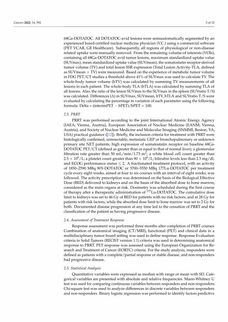
The median values of SUVmax, SUVmean, SUVt/s, bTV and bTLA were 34 (IQR,23.1–55.8), 9.9 (IQR, 7.6–16.4), 1.1 (IQR, 0.7–2.3), 143.8 mL (IQR, 32.9–354), and 1834 (IQR,342–6309), respectively. The median percentage in variation between baseline and inter-imPET after two cycles of PRTT reported as ∆SUVmax, ∆SUVmean, ∆SUVratioT/S, ∆bTV,∆bTLA were 1.1% (IQR, −21.2–22.1), 4.2% (IQR, −17.8–39.4), −1.1% (IQR, −41.5–24.5),32.4% (IQR, −10.2–70.6), 25% (−12.5–80.9), respectively. Figure 1 shows a baseline andinterim PET.
Cancers 2022, 14, x 6 of 12
Figure 1. Baseline (A) and interim (B) PET scan of a 54-year-old patient with G1 pancreatic NET.
Red arrows indicate the primary pancreatic tumor and the most representative liver metastasis. In-
terim 68Ga-DOTATOC PET after the second PRRT cycle shows disease progression. The patient
was treated with a total of six PRRT courses (four cycles of 177Lu-DOTATOC + 2 cycles of 90Y-
DOTATOC) for a total of 28,534 MBq activity and classified as partial response at the assessment
following treatment.
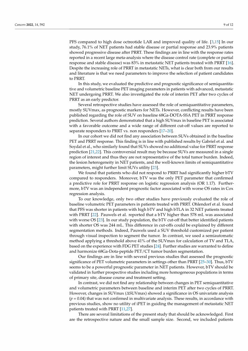
The bTV and bTLA values of patients that did not respond to PRRT were significantly
higher than those of patients with response to PRRT therapy (496.2 IQR 218.3–2029.4 vs.
77.6 IQR 31–186.6, p < 0.001 and 6078.3 IQR 2813–18,959 vs. 1341 IQR 272.3–3865, p = 0.001,
respectively: Figure 2).
(a) (b)
Figure 1. Baseline (A) and interim (B) PET scan of a 54-year-old patient with G1 pancreatic NET. Redarrows indicate the primary pancreatic tumor and the most representative liver metastasis. Interim68Ga-DOTATOC PET after the second PRRT cycle shows disease progression. The patient was treatedwith a total of six PRRT courses (four cycles of 177Lu-DOTATOC + 2 cycles of 90Y-DOTATOC) for atotal of 28,534 MBq activity and classified as partial response at the assessment following treatment.
The bTV and bTLA values of patients that did not respond to PRRT were significantlyhigher than those of patients with response to PRRT therapy (496.2 IQR 218.3–2029.4 vs.77.6 IQR 31–186.6, p < 0.001 and 6078.3 IQR 2813–18,959 vs. 1341 IQR 272.3–3865, p = 0.001,respectively: Figure 2).

Cancers 2022, 14, 592 6 of 12
Cancers 2022, 14, x 6 of 12
Figure 1. Baseline (A) and interim (B) PET scan of a 54-year-old patient with G1 pancreatic NET.
Red arrows indicate the primary pancreatic tumor and the most representative liver metastasis. In-
terim 68Ga-DOTATOC PET after the second PRRT cycle shows disease progression. The patient
was treated with a total of six PRRT courses (four cycles of 177Lu-DOTATOC + 2 cycles of 90Y-
DOTATOC) for a total of 28,534 MBq activity and classified as partial response at the assessment
following treatment.
The bTV and bTLA values of patients that did not respond to PRRT were significantly
higher than those of patients with response to PRRT therapy (496.2 IQR 218.3–2029.4 vs.
77.6 IQR 31–186.6, p < 0.001 and 6078.3 IQR 2813–18,959 vs. 1341 IQR 272.3–3865, p = 0.001,
respectively: Figure 2).
(a) (b)
Figure 2. Box Plot comparing bTV (a) and bTLA (b) between PRRT responder and non-responderpatients. “F” and “#” symbols represent outlier cases.
No association between semiquantitative PET parameters and response were observed.Percentage variations (∆) of PET semiquantitative and volumetric parameters were notsignificantly different between PRRT responders and non-responders. PET parameters andcomparison between responders vs. non-responders are summarized in Table 2.
Table 2. Characteristic of calculated PET parameters. Data of all patients (n = 46) and a comparisonbetween responders (n = 35) vs. non-responders (n = 11) groups are reported. Values are expressed inmean (SD) and median (IQR). Mann-Whitney U test was used to compare variables. The p-Valuesshown in boldface correspond to p < 0.05.
PET Parameters All(n = 46)
Responders(n = 35)
Non-Responders(n = 11) p Value
SUVmax0.58Mean (SD) 40 (24.9) 41.5 (27.1) 35.5 (35.5)
Median (IQR) 34 (23.1–55.8) 34.5 (23.7–56.2) 33.5 (21.8–53.9)SUVmean
0.57Mean (SD) 11.6 (5.9) 10.3 (4.1) 11.9 (6.3)Median (IQR) 9.9 (7.6–16.4) 9.9 (7.2–17.3) 10(7.8–13.5)SUVratio T/S
0.93Mean (SD) 1.8 (0.9) 1.4 (0.7) 1.9 (2.2)Median (IQR) 1.1 (0.7–2.3) 1.2 (0.6–2.3) 1 (0.8–2.2)
bTV<0.001Mean (SD) 371 (665.5) 143.7 (177.6) 1073.5 (1061,4)
Median (IQR) 143.8 (32.9–354) 77.6 (31–186.8) 496.2 (218.3–2029.4)bTLA
0.001Mean (SD) 5339.5 (8171.4) 3108.13 (4971.1) 12,236.4 (11,959)Median (IQR) 1834 (342–6309) 1341 (272.3–3865) 6078.3 (2813–18,959)
∆SUVmax0.89Mean (SD) 23.9 (117.7) 25.2 (129,6) 18.1 (62)
Median (IQR) 1.1 (−21.2–22.1) −2.5 (−21.2–22.2) 3.5 (−26.6–22.1)∆SUVmean
0.84Mean (SD) 25.5 (79.2) 45.6 (128) 19 (55,9)Median (IQR) 4.2 (−17.8–39.4) 6.2 (−16.5–25.4) −10.2 (−27.4–47.6)∆SUVratioT/S
0.42Mean (SD) 1.3 (2) 1.9 (0.8) 1.4 (0.9)Median (IQR) −1.1 (−41.5–24.5) −11.5 (−40.5–20.9) 1.9 (−46.8–32.3)

Cancers 2022, 14, 592 7 of 12
Table 2. Cont.
PET Parameters All(n = 46)
Responders(n = 35)
Non-Responders(n = 11) p Value
∆TV0.51Mean (SD) 61 (170) 27.1 (55,6) 71.5 (756,7)
Median (IQR) 32.4 (−10.2–70.6) 32.4 (−5.5–69.2) 32.4 (−23.9–79.9)∆TLA
0.92Mean (SD) 143 (668,4) 45.7 (79,1) 171.9 (756.7)Median (IQR) 25 (−12.5–80.9) 24.3 (−0.4–80) 36 (−22.5–111)
Abbreviations: SUV: standardized uptake value; T/S: tumor/spleen; TV: total volume; TLA: total lesion activity.
Logistic regression analysis of bPET derived parameters and ∆PET values identifiedthat only bTV was associated with therapy outcome (OR 1.17; 95%CI 1.02–1.32; p = 0.02).No significant association with PRRT response was observed for other variables (Table 3).
Table 3. Logistic Regression Analysis.
PET Parameters Univariate Analysis
OR (95%CI) p Value
SUVmax 1.06 (0.99–1.13) 0.09
SUVmean 0.8 (0.55–1.15) 0.23
SUVratio T/S 0.84 (0.52–1.35) 0.48
bTV 1.17 (1.02–1.32) 0.02
bTLA 0.99 (0.93–1.01) 0.08
∆SUVmax 0.97 (0.93–1.01) 0.25
∆SUVmean 1.007 (0.98–1.01) 0.259
∆SUVratioT/S 0.99 (0.98–1.01) 0.839
∆TV 0.99 (0.98–1.01) 0.922
∆TLA 0.99 (0.98–1.01) 0.70Abbreviations: SUV: standardized uptake value; T/S: tumor/spleen; TV: total volume; TLA: total lesion activity.The p-Values shown in boldface correspond to p < 0.05.
3.3. Overall Survival Analysis
During the follow-up period (mean 31 months; ranged from eight to 86 months), seven(15.2%) patients died. Mean survival time was 69.4 (SD, 5.3) months, the median was nonreached for the entire cohort.
ROC analysis showed a best cut-off point for bTV of >244.5 mL (sensitivity 85.7%;specificity 79.5%) and AUC of 0.77 (95% CI 0.62–0.88, p = 0.036) for identification of patientswith worse OS. For bTLA best cut-off point was 2659 (sensitivity 85.7%; specificity 64.1%)and AUC of 0.71 (95% CI 0.56–0.84, p = 0.092). The complete ROC curves analysis of PETparameters is shown in Table S1.
In univariate analysis, higher bTV and bTLA were associated with lower survivalprobability (HR 13 95%CI 2.6–64.1, p = 0.001; HR 9.08 95%CI 1.09–75.76, p = 0.04) (Figure 3).
Moreover, a difference in SUVmax above 5.5% between iPET and bPET (∆SUVmax)was associated with a favourable outcome (HR 0.15 95% CI0.03–0.67; p = 0.04). In Coxmultivariate analysis, only a high bTV was confirmed as an independent prognostic factor(HR 12.76 95%CI 1.53–107, p = 0.01) (Table 4).

Cancers 2022, 14, 592 8 of 12
Cancers 2022, 14, x 8 of 12
Table 3. Logistic Regression Analysis.
PET Parameters Univariate Analysis
OR (95%CI) p Value
SUVmax 1.06 (0.99–1.13) 0.09
SUVmean 0.8 (0.55–1.15) 0.23
SUVratio T/S 0.84 (0.52–1.35) 0.48
bTV 1.17 (1.02–1.32) 0.02
bTLA 0.99 (0.93–1.01) 0.08
ΔSUVmax 0.97 (0.93–1.01) 0.25
ΔSUVmean 1.007 (0.98–1.01) 0.259
ΔSUVratioT/S 0.99 (0.98–1.01) 0.839
ΔTV 0.99 (0.98–1.01) 0.922
ΔTLA 0.99 (0.98–1.01) 0.70
Abbreviations: SUV: standardized uptake value; T/S: tumor/spleen; TV: total volume; TLA: total
lesion activity. The p-Values shown in boldface correspond to p < 0.05.
3.3. Overall Survival Analysis
During the follow-up period (mean 31 months; ranged from eight to 86 months),
seven (15.2%) patients died. Mean survival time was 69.4 (SD, 5.3) months, the median
was non reached for the entire cohort.
ROC analysis showed a best cut-off point for bTV of >244.5 mL (sensitivity 85.7%;
specificity 79.5%) and AUC of 0.77 (95% CI 0.62–0.88, p = 0.036) for identification of pa-
tients with worse OS. For bTLA best cut-off point was 2659 (sensitivity 85.7%; specificity
64.1%) and AUC of 0.71 (95% CI 0.56–0.84, p = 0.092). The complete ROC curves analysis
of PET parameters is shown in Table S1.
In univariate analysis, higher bTV and bTLA were associated with lower survival
probability (HR 13 95%CI 2.6–64.1, p = 0.001; HR 9.08 95%CI 1.09–75.76, p = 0.04) (Figure
3).
(a) (b)
Figure 3. (a) Kaplan-Meier curves for overall survival (OS) among patients with high- and low-bTV
defined on the basis of the ROC curve (244 mL). (b) Kaplan-Meier curves for OS among patients
with high- and low-bTLA defined on the basis of the ROC curve (2658 mL*SUV).
Figure 3. (a) Kaplan-Meier curves for overall survival (OS) among patients with high- and low-bTVdefined on the basis of the ROC curve (244 mL). (b) Kaplan-Meier curves for OS among patients withhigh- and low-bTLA defined on the basis of the ROC curve (2658 mL*SUV).
Table 4. Univariate and multivariate Cox regression analysis for OS.
PET ParameterUnivariate Analysis Multivariate Analysis
HR (95%CI) p Value HR (95%CI) p Value
SUVmax(<22.02) 0.99 (0.12–8.20) 0.99 - -
SUVmean(≤5.45) 0.31 (0.06–1.51) 0.26 - -
SUVratioT/S(≤1.31) 0.84 (0.08–8.23) 0.87 - -
bTV(>244.48) 13 (2.6–64.1) 0.001 12.76(1.53–107) 0.01
bTLA(>2658.62) 9.08 (1.09–75.76) 0.04 7.15 (0.96–68.1) 0.98
∆SUVmax (>5.5598) 0.15 (0.03–0.67) 0.04 0.17 (0.02–1.46) 0.1
∆SUVmean(>24.4984) 0.65 (0.05–7.87) 0.68 - -
∆SUVratioT/S(>−0.75) 0.14 (0.017–1.28) 0.08 - -
∆TV(≤−15.876) 0.23(0.02–1.86) 0.354 - -
∆TLA(>80.2) 0.45 (0.082–2.48) 0.2737 - -
Abbreviations: SUV: standardized uptake value; T/S: tumor/spleen; TV: total volume; TLA: total lesion activity.The p-Values shown in boldface correspond to p < 0.05.
4. Discussion
After about 20 years of retrospective and phase I/II trials, PRRT was finally approvedby regulatory authorities. Since its approval for the treatment of inoperable or metastaticGEP-NETs with progressive disease by the European Medicine Agency in 2017, PRRT hasbeen widely used [13,14]. The phase III NETTER-1 trial has established that PRRT prolongs
Cancers 2022, 14, 592 9 of 12
PFS compared to high dose octreotide LAR and improved quality of life. [3,15] In ourstudy, 76.1% of NET patients had stable disease or partial response and 23.9% patientsshowed progressive disease after PRRT. These findings are in line with the response ratesreported in a recent large meta-analysis where the disease control rate (complete or partialresponse and stable disease) was 83% in metastatic NET patients treated with PRRT [16].Despite the increasing role of PRRT in metastatic NETs, what is clear both from our resultsand literature is that we need parameters to improve the selection of patient candidatesto PRRT.
In this study, we evaluated the predictive and prognostic significance of semiquantita-tive and volumetric baseline PET imaging parameters in patients with advanced, metastaticNET undergoing PRRT. We also investigated the role of interim PET after two cycles ofPRRT as an early predictor.
Several retrospective studies have assessed the role of semiquantitative parameters,mostly SUVmax, as prognostic markers for NETs. However, conflicting results have beenpublished regarding the role of SUV on baseline 68Ga-DOTA-SSA PET in PRRT responseprediction. Several authors demonstrated that a high SUVmax in baseline PET is associatedwith a favorable outcome and a wide range of different cut-off values are reported toseparate responders to PRRT vs. non responders [17–20].
In our cohort we did not find any association between SUVs obtained in the baselinePET and PRRT response. This finding is in line with published results by Gabriel et al. andSoydal et al., who similarly found that SUVs showed no additional value for PRRT responseprediction [21,22]. This controversial result may be because SUVs are measured in a singleregion of interest and thus they are not representative of the total tumor burden. Indeed,the lesion heterogeneity in NET patients, and the well-known limits of semiquantitativeparameters, might further limit SUVs utility [23].
We found that patients who did not respond to PRRT had significantly higher bTVcompared to responders. Moreover, bTV was the only PET parameter that confirmeda predictive role for PRRT response on logistic regression analysis (OR 1.17). Further-more, bTV was an independent prognostic factor associated with worse OS rates in Coxregression analysis.
To our knowledge, only two other studies have previously evaluated the role ofbaseline volumetric PET parameters in patients treated with PRRT. Ohlendorf et al. foundthat PFS was shorter in patients with high bTV and high bTLA in 32 NET patients treatedwith PRRT [22]. Pauwels et al. reported that a bTV higher than 578 mL was associatedwith worse OS [23]. In our study population, the bTV cut-off that better identified patientswith shorter OS was 244 mL. This difference in cut-offs could be explained by differentsegmentation methods. Indeed, Pauwels used a SUV threshold customized per patientthrough visual inspection to segment the tumor. In contrast, we used a semiautomaticmethod applying a threshold above 41% of the SUVmax for calculation of TV and TLA,based on the experience with FDG PET studies [24]. Further studies are warranted to defineand harmonize 68Ga-Dota-peptide PET/CT tumor burden segmentation.
Our findings are in line with several previous studies that assessed the prognosticsignificance of PET volumetric parameters in settings other than PRRT [25–30]. Thus, bTVseems to be a powerful prognostic parameter in NET patients. However, bTV should bevalidated in further prospective studies including more homogeneous populations in termsof primary site, disease course and treatment setting.
In contrast, we did not find any relationship between changes in PET semiquantitativeand volumetric parameters between baseline and interim PET after two cycles of PRRT.However, changes in SUVmax (∆SUVmax) showed a significance in OS univariate analysis(p = 0.04) that was not confirmed in multivariate analysis. These results, in accordance withprevious studies, show no utility of iPET in guiding the management of metastatic NETpatients treated with PRRT [11,27].
There are several limitations of the present study that should be acknowledged. Firstare the retrospective nature and the small sample size. Second, we included patients

Cancers 2022, 14, 592 10 of 12
with different NET sites. Furthermore, we did not evaluate FDG PET findings in ouranalysis. It is known that some patients with grade 2–3 NET can show higher affinity forFDG rather than 68Ga-DOTATOC PET [28]. This can result in an underestimation of theactual tumor burden by 68Ga-DOTATOC PET. A combination of these two tracers can beconsidered to address this issue. In addition, it should be noted that the generalizability ofour findings with 68Ga-DOTATOC to the other often used 68Ga-DOTATATE PET ligandneed to be confirmed. Indeed, while 68Ga-DOTATATE shows only affinity to SSTR2,68Ga-DOTATOC also exhibits some affinity to SSTR5. However, despite differences inreceptor affinities, a head-to-head comparison showed no clinically significant differencebetween the two tracers [29]. Finally, it is worth mentioning that other PET tracers like64Cu-DOTATATE, providing better resolution and potentially a better lesion detectionrate than 68Ga-DOTATOC, may improve tumor burden quantification [30]. However, the64Cu-labeled tracers are not in use in the clinical routine and require further work.
5. Conclusions
In metastatic NET patients addressed to PRRT, higher total body tumor volumemeasured from baseline 68Ga-DOTATOC PET/CT scans is a negative predictor for therapyresponse and is associated with worse OS rates. Interim 68Ga-DOTATOC PET/CT aftertwo cycles of PRRT did not allow the identification of patients with poorer prognosis thatwould justify a change in treatment strategy.
Supplementary Materials: The following are available online at https://www.mdpi.com/article/10.3390/cancers14030592/s1, Table S1: ROC curve analysis for OS.
Author Contributions: Conceptualization and supervision A.V. and A.F. (Angelina Filice); dataacquisitions, V.C. and C.C.; data analysis, F.F. and D.F.; writing—original draft preparation, R.D.;revision of the article for important intellectual content, A.F. (Andrea Frasoldati), S.F. and G.B. Allauthors have read and agreed to the published version of the manuscript.
Funding: This research received no external funding.
Institutional Review Board Statement: The study was conducted according to the guidelines of theDeclaration of Helsinki and approved by the Ethics Committee of AUSL-IRCCS of Reggio Emilia(protocol code 24871).
Informed Consent Statement: Informed consent was obtained from all subjects involved in the study.
Data Availability Statement: The data presented in this study are available on request from thecorresponding author. The data are not publicly available due to privacy-related issues.
Conflicts of Interest: A.V.: Novartis and Advanced Accelerator Applications for advisory role. Theother authors declare no conflict of interest.
References1. Dasari, A.; Shen, C.; Halperin, D.; Zhao, B.; Zhou, S.; Xu, Y.; Shih, T.; Yao, J.C. Trends in the incidence, prevalence, and survival
outcomes in patients with neuroendocrine tumors in the United States. JAMA Oncol. 2017, 3, 1335–1342. [CrossRef]2. Eychenne, R.; Bouvry, C.; Bourgeois, M.; Loyer, P.; Benoist, E.; Lepareur, N. Overview of Radiolabeled Somatostatin Analogs for
Cancer Imaging and Therapy. Molecules 2020, 25, 4012. [CrossRef] [PubMed]3. Strosberg, J.; El-Haddad, G.; Wolin, E.; Hendifar, A.; Yao, J.; Chasen, B.; Mittra, E.; Kunz, P.L.; Kulke, M.H.; Jacene, H.; et al. Phase
3 Trial of 177 Lu-Dotatate for Midgut Neuroendocrine Tumors. N. Engl. J. Med. 2017, 376, 125–135. [CrossRef] [PubMed]4. Bodei, L.; Kidd, M.; Paganelli, G.; Grana, C.M.; Drozdov, I.; Cremonesi, M.; Lepensky, C.; Kwekkeboom, D.J.; Baum, R.P.;
Krenning, E.P.; et al. Long-term tolerability of PRRT in 807 patients with neuroendocrine tumours: The value and limitations ofclinical factors. Eur. J. Nucl. Med. Mol. Imaging 2015, 42, 5–19. [CrossRef] [PubMed]
5. Shah, M.H.; Goldner, W.S.; Halfdanarson, T.R.; Bergsland, E.; Berlin, J.D.; Halperin, D.; Chan, J.; Kulke, M.H.; Benson, A.B.;Blaszkowsky, L.S.; et al. Neuroendocrine and adrenal tumors, version 2.2018 featured updates to the nccn guidelines. JNCCN J.Natl. Compr. Cancer Netw. 2018, 16, 693–702. [CrossRef] [PubMed]
6. Filice, A.; Fraternali, A.; Frasoldati, A.; Asti, M.; Grassi, E.; Massi, L.; Sollini, M.; Froio, A.; Erba, P.A.; Versari, A. RadiolabeledSomatostatin Analogues Therapy in Advanced Neuroendocrine Tumors: A Single Centre Experience. J. Oncol. 2012, 2012, 320198.[CrossRef] [PubMed]

Cancers 2022, 14, 592 11 of 12
7. Albertelli, M.; Dotto, A.; Di Dato, C.; Malandrino, P.; Modica, R.; Versari, A.; Colao, A.; Ferone, D.; Faggiano, A. PRRT: Identikit ofthe perfect patient. Rev. Endocr. Metab. Disord. 2021, 22, 563–579. [CrossRef] [PubMed]
8. Cottereau, A.S.; Versari, A.; Loft, A.; Casasnovas, O.; Bellei, M.; Ricci, R.; Bardet, S.; Castagnoli, A.; Brice, P.; Raemaekers, J.; et al.Prognostic value of baseline metabolic tumor volume in early-stage Hodgkin lymphoma in the standard arm of the H10 trial.Blood 2018, 131, 1456–1463. [CrossRef] [PubMed]
9. Hofheinz, F.; Li, Y.; Steffen, I.G.; Lin, Q.; Lili, C.; Hua, W.; van den Hoff, J.; Zschaeck, S. Confirmation of the prognostic value ofpretherapeutic tumor SUR and MTV in patients with esophageal squamous cell carcinoma. Eur. J. Nucl. Med. Mol. Imaging 2019,46, 1485–1494. [CrossRef] [PubMed]
10. Ohlendorf, F.; Werner, R.A.; Henkenberens, C.; Ross, T.L.; Christiansen, H.; Bengel, F.M.; Derlin, T. Predictive and prognosticimpact of blood-based inflammatory biomarkers in patients with gastroenteropancreatic neuroendocrine tumors commencingpeptide receptor radionuclide therapy. Diagnostics 2021, 11, 504. [CrossRef] [PubMed]
11. Ortega, C.; Wong, R.K.S.; Schaefferkoetter, J.; Veit-Haibach, P.; Myrehaug, S.; Juergens, R.; Laidley, D.; Anconina, R.; Liu, A.; Metser,U. Quantitative 68 Ga-DOTATATE PET/CT Parameters for the Prediction of Therapy Response in Patients with ProgressiveMetastatic Neuroendocrine Tumors Treated with 177 Lu-DOTATATE. J. Nucl. Med. 2021, 62, 1406–1414. [CrossRef] [PubMed]
12. Zaknun, J.J.; Bodei, L.; Mueller-Brand, J.; Pavel, M.E.; Baum, R.P.; Hörsch, D.; O’Dorisio, M.S.; O’Dorisiol, T.M.; Howe, J.R.;Cremonesi, M.; et al. The joint IAEA, EANM, and SNMMI practical guidance on peptide receptor radionuclide therapy (PRRNT)in neuroendocrine tumours. Eur. J. Nucl. Med. Mol. Imaging 2013, 40, 800–816. [CrossRef] [PubMed]
13. CHMP Lutathera, INN-Lutetium (177Lu) Oxodotreotide. Available online: https://www.ema.europa.eu/en/documents/product-information/lutathera-epar-product-information_en.pdf (accessed on 23 January 2022).
14. Cives, M.; Strosberg, J. Radionuclide Therapy for Neuroendocrine Tumors. Curr. Oncol. Rep. 2017, 19, 1–9. [CrossRef] [PubMed]15. Strosberg, J.; Wolin, E.; Chasen, B.; Kulke, M.; Bushnell, D.; Caplin, M.; Baum, R.P.; Kunz, P.; Hobday, T.; Hendifar, A.; et al.
Health-related quality of life in patients with progressive midgut neuroendocrine tumors treated with 177 lu-dotatate in thephase III netter-1 trial. J. Clin. Oncol. 2018, 36, 2578–2584. [CrossRef] [PubMed]
16. Wang, L.; Lin, L.; Wang, M.; Li, Y. The therapeutic efficacy of 177Lu-DOTATATE/DOTATOC in advanced neuroendocrine tumors.Medicine 2020, 99, e19304. [CrossRef] [PubMed]
17. Öksüz, M.Ö.; Winter, L.; Pfannenberg, C.; Reischl, G.; Müssig, K.; Bares, R.; Dittmann, H. Peptide receptor radionuclide therapyof neuroendocrine tumors with 90Y-DOTATOC: Is treatment response predictable by pre-therapeutic uptake of 68Ga-DOTATOC?Diagn. Interv. Imaging 2014, 95, 289–300. [CrossRef] [PubMed]
18. Kratochwil, C.; Stefanova, M.; Mavriopoulou, E.; Holland-Letz, T.; Dimitrakopoulou-Strauss, A.; Afshar-Oromieh, A.; Mier, W.;Haberkorn, U.; Giesel, F.L. SUV of [68Ga]DOTATOC-PET/CT Predicts Response Probability of PRRT in Neuroendocrine Tumors.Mol. Imaging Biol. 2014, 17, 313–318. [CrossRef] [PubMed]
19. Gabriel, M.; Oberauer, A.; Dobrozemsky, G.; Decristoforo, C.; Putzer, D.; Kendler, D.; Uprimny, C.; Kovacs, P.; Bale, R.; Virgolini,I.J. 68Ga-DOTA-Tyr3-octreotide PET for assessing response to somatostatin-receptor-mediated radionuclide therapy. J. Nucl. Med.2009, 50, 1427–1434. [CrossRef] [PubMed]
20. Soydal, Ç.; Peker, A.; Özkan, E.; Küçük, Ö.N.; Kir, M.K. The role of baseline Ga-68 DOTATATE positron emission tomogra-phy/computed tomography in the prediction of response to fixed-dose peptide receptor radionuclide therapy with lu-177DOTATATE. Turk. J. Med. Sci. 2016, 46, 409–413. [CrossRef]
21. Werner, R.A.; Ilhan, H.; Lehner, S.; Papp, L.; Zsótér, N.; Schatka, I.; Muegge, D.O.; Javadi, M.S.; Higuchi, T.; Buck, A.K.; et al.Pre-therapy Somatostatin Receptor-Based Heterogeneity Predicts Overall Survival in Pancreatic Neuroendocrine Tumor PatientsUndergoing Peptide Receptor Radionuclide Therapy. Mol. Imaging Biol. 2019, 21, 582–590. [CrossRef] [PubMed]
22. Ohlendorf, F.; Henkenberens, C.; Brunkhorst, T.; Ross, T.L.; Christiansen, H.; Bengel, F.M.; Derlin, T. Volumetric 68Ga-DOTA-TATE PET/CT for assessment of whole-body tumor burden as a quantitative imaging biomarker in patients with metastaticgastroenteropancreatic neuroendocrine tumors. Q. J. Nucl. Med. Mol. Imaging 2020. [CrossRef] [PubMed]
23. Pauwels, E.; Van Binnebeek, S.; Vandecaveye, V.; Baete, K.; Vanbilloen, H.; Koole, M.; Mottaghy, F.M.; Haustermans, K.; Clement,P.M.; Nackaerts, K.; et al. Inflammation-based index and 68ga-dotatoc pet-derived uptake and volumetric parameters predictoutcome in neuroendocrine tumor patients treated with 90y-dotatoc. J. Nucl. Med. 2020, 61, 1014–1020. [CrossRef] [PubMed]
24. Meignan, M.; Sasanelli, M.; Casasnovas, R.O.; Luminari, S.; Fioroni, F.; Coriani, C.; Masset, H.; Itti, E.; Gobbi, P.G.; Merli, F.; et al.Metabolic tumour volumes measured at staging in lymphoma: Methodological evaluation on phantom experiments and patients.Eur. J. Nucl. Med. Mol. Imaging 2014, 41, 1113–1122. [CrossRef] [PubMed]
25. Tirosh, A.; Papadakis, G.Z.; Millo, C.; Hammoud, D.; Sadowski, S.M.; Herscovitch, P.; Pacak, K.; Marx, S.J.; Yang, L.; Nockel, P.;et al. Prognostic Utility of Total 68Ga-DOTATATE-Avid Tumor Volume in Patients with Neuroendocrine Tumors. Gastroenterology2018, 154, 998–1008.e1. [CrossRef] [PubMed]
26. Abdulrezzak, U.; Kurt, Y.K.; Kula, M.; Tutus, A. Combined imaging with 68Ga-DOTA-TATE and 18F-FDG PET/CT on the basisof volumetric parameters in neuroendocrine tumors. Nucl. Med. Commun. 2016, 37, 874–881. [CrossRef] [PubMed]
27. Haug, A.R.; Auernhammer, C.J.; Wängler, B.; Schmidt, G.P.; Uebleis, C.; Göke, B.; Cumming, P.; Bartenstein, P.; Tiling, R.; Hacker,M. 68Ga-DOTATATE PET/CT for the early prediction of response to somatostatin receptor-mediated radionuclide therapy inpatients with well-differentiated neuroendocrine tumors. J. Nucl. Med. 2010, 51, 1349–1356. [CrossRef] [PubMed]
https://www.ema.europa.eu/en/documents/product-information/lutathera-epar-product-information_en.pdf

Cancers 2022, 14, 592 12 of 12
28. Binderup, T.; Knigge, U.; Johnbeck, C.B.; Loft, A.; Berthelsen, A.K.; Oturai, P.; Mortensen, J.; Federspiel, B.; Langer, S.W.; Kjaer, A.18F-FDG PET is Superior to WHO Grading as a Prognostic Tool in Neuroendocrine Neoplasms and Useful in Guiding PRRT: AProspective 10-Year Follow-up Study. J. Nucl. Med. 2021, 62, 808–815. [CrossRef] [PubMed]
29. Poeppel, T.D.; Binse, I.; Petersenn, S.; Lahner, H.; Schott, M.; Antoch, G.; Brandau, W.; Bockisch, A.; Boy, C. 68Ga-DOTATOCversus 68Ga-DOTATATE PET/CT in functional imaging of neuroendocrine tumors. J. Nucl. Med. 2011, 52, 1864–1870. [CrossRef][PubMed]
30. Johnbeck, C.B.; Knigge, U.; Loft, A.; Berthelsen, A.K.; Mortensen, J.; Oturai, P.; Langer, S.W.; Elema, D.R.; Kjaer, A. Head-to-HeadComparison of 64Cu-DOTATATE and 68Ga-DOTATOC PET/CT: A Prospective Study of 59 Patients with NeuroendocrineTumors. J. Nucl. Med. 2017, 58, 451–457. [CrossRef] [PubMed]