The larval blood cells of Drosophila willistoni
15
THE LARVAL BLOOD CELLS OF DROSOPHILA JIrILLISTONI M. T. M. RIZKI’ Department of Zoology, Dartmouth College, Hunover, New Hampshire TWENTYTHREE FIGURES INTRODUCTION The larvae homozygous for the lethal factor 42P display “melanotic sclerosis ” (description will appear elsewhere). It was noted that these larvae have a marked anemic condi- tion at the time of melanosis. The haeinolymph is almost free of haemocytes. Since the blood cells of insects are known to participate in the processes which lead to puparium forma- tion (Dennell, ’47) it was considered desirable to undertake a detailed study of the normal blood picture of Drosophilcc zcjillistoizi. Such an investigation would serve as a future reference for possible differences or abnormalities in the blood of mutant phenotypes. The author wishes to express his gratitude to the Depart- ment of Zoology, Dartmouth College, for providing the facili- ties which made this study possible. He would also like to thank Professors J. H. Gerould and L. C. Dunn and Dr. S. Hughes-Schrader for helpful suggestions in the preparation of the manuscript. MATERIAL AND METHODS A standard stock of D. wiZZistoni was used for this investi- gation, Larvae of known age and stage were washed with R. Melville Cramer Research Fellow. 397 THE JOURNAL OF EXPERIMENTAL ZOOLOGY, VOL. 123, NO. 3 AUGUST 1953
-
Upload
independent -
Category
Documents
-
view
1 -
download
0
Transcript of The larval blood cells of Drosophila willistoni

THE LARVAL BLOOD CELLS O F DROSOPHILA JIrILLISTONI
M. T. M. RIZKI’ Department of Zoology, Dartmouth College, Hunover, New Hampshire
TWENTYTHREE FIGURES
INTRODUCTION
The larvae homozygous for the lethal factor 42P display “melanotic sclerosis ” (description will appear elsewhere). It was noted that these larvae have a marked anemic condi- tion at the time of melanosis. The haeinolymph is almost free of haemocytes. Since the blood cells of insects are known to participate in the processes which lead to puparium forma- tion (Dennell, ’47) it was considered desirable to undertake a detailed study of the normal blood picture of Drosophilcc zcjillistoizi. Such an investigation would serve as a future reference for possible differences or abnormalities in the blood of mutant phenotypes.
The author wishes to express his gratitude to the Depart- ment of Zoology, Dartmouth College, for providing the facili- ties which made this study possible. He would also like to thank Professors J. H. Gerould and L. C. Dunn and Dr. S. Hughes-Schrader for helpful suggestions in the preparation of the manuscript.
MATERIAL AND METHODS
A standard stock of D. wiZZistoni was used for this investi- gation, Larvae of known age and stage were washed with
R. Melville Cramer Research Fellow. 397
THE JOURNAL OF EXPERIMENTAL ZOOLOGY, VOL. 123, NO. 3 AUGUST 1953

398 M. T. M. RIZKI
insect Ringer solution, and the excess Ringer solution was absorbed by placing the larvae on a piece of absorbent tissue. A larva was then transferred to a drop of immersion oil (Crown) on a clean slide. Using two pair of fine watchmakers forceps the body of the larva was carefully opened so that the blood flowed out onto the slide without breaking the gut and fat bodies. Care was taken to hold the larva at the pos- terior region (8th segment) and tear off the skin. As soon as a drop of blood formed on the slide the larva was removed and a coverslip placed on the preparation of the blood. Such preparations were examined with a phase microscope under dark contrast.
Owing to the difficulty in obtaining blood from the small first instar larvae and the small number of blood cells present in these larvae, the observations on this material are limited. Special methods were adapted to study the blood cells of the first instar larva in addition to the method described above. The larvae were washed and dried as usual and flattened only slightly with a coverslip so that the body was not broken. The circulating blood cells were then observed in the body under the oil immersion phase microscope.
Smears were also prepared by pipetting the blood from larvae opened under oil. Smears were stained with Giemsa and Wright’s stain. Paraffin sections of larvae which had been fixed in acetic alcohol were stained with Feulgen-fast green and Azure-B, and the blood cells in such preparations were examined.
OBSERVATIONS
The terminology utilized in the classification of the haemo- cytes of D. willistoni is borrowed from Yeager’s study on Prodenia eridania (Yeager, ’45). However, this does not im- ply homologizing the blood cells of D. willistoai with P. eridania or any other insect. Such a comparison is not de- sirable unless the morphogenesis and metabolic function of each haemocyte is clear. The blood cells of D. willistomi may be divided into 6 classes on the basis of distinctive morpho- logical features. Variants of these classes of haemocytes are

LARVAL BLOOD CELLS O F DROSOPHILA 399
not subdivided into “types” as Yeager has done in his classi- fication of the blood cells of P. eridavzia. The only class of haemocytes found in D. willistovzi which does not resemble any class of cells described by Yeager is the “crystalloid cell.” The cytoplasm of these cells contains one or more large crystal-like inclusions which differ in appearance from inclusions found in the other types of cells. These cells are found mostly in the first instar, so it was considered desirable to place them in a separate class.
Classificatiom o f blood cells Class I - Plasmatocytes. The plasmatocytes (figs. 1, 2,
3, 4 and 17) are usually round but they occur as slightly fusi- form types also. They are the variants of the smallest class of cells. The ratio of the diameter of the nucleus to the cell diameter varies between 0.5 and 0.6. These cells always have a round nucleus which is moderately eosinophilic while the cytoplasm is deeply basophilic. Under the phase microscope the cytoplasm appears homogeneous with the exception of several globular inclusions. Occasionally binucleate cells of this type occur and cells in division were also observed.
Class II - Podocytes. The podocytes (figs. 5, 6, 7, 8, 9, 18 and 19) are slightly larger in size than the plasmatocytes. They are distinguished by their irregular shape and cyto- plasmic processes. These cytoplasmic processes, which may be few in number or numerous depending upon the larval stage, were observed under the phase microscope only. They were not visible in the smears and sectioned material. The appearance of the processes varies with the larval stage ; they are more pronounced in the late stages. Some cells from early third instar larvae (fig. 7 c) have processes which are filamentous and so fine that they are just at the limit of resolution of the phase microscope (1350 X). These fine filaments beat rapidly and minute particles in the surrounding haemolymph were observed to stick to these beating fila- ments. The nucleus of the podocytes is round or slightly irregular in shape. . The cytoplasm is lightly basophilic and

400 M. T. M. R I Z H I
the nucleus eosinophilic. A few vacuoles and inclusion bodies are apparent in the cytoplasm under phase. The podocytes may be derived from the plasmatocytes since a range of intermediate forms between the plasmatocytes and podocytes exists.
Class 111 - Spheroidocytes. The spheroidocytes (figs. 10, 22 and 23) are large cells, spherical to ovoid in shape. Their cytoplasm is characterized by spherical inclusion bodies which resemble vacuoles. Some cells contain only a few spherules whereas other cells appear to he a mass of vacuoles. The nucleus is round in the early stages of cell development, but it becomes obliterated in later stages when many vacuoles are present. The intervacuolar cytoplasm is strongly baso- philic; in some cells the spherules stain heavily with fast green and eosin whereas in other cells they remain unstained.
Class W - Oeuzocytoids. The largest spherical cells are the oenocytoids (figs. 11, 12, 13 and 20) with fine thread-like cytoplasmic inclusions which are visible under phase. These cytoplasmic inclusions are the mitochondria which stain with Janus green B. The oenocytoids can also be recognized by their round nucleus which is proportionately smaller than the nucleus in the other types of blood cells.
Class V - Nematocytes. The extremely fusiform cells are ternied nematocytes (figs. 14 and 17). The eosinophilic nu- cleus varies from round, ovoid to elongate. The cytoplasm which is moderately basophilic contains numerous small gran- ules in the perinuclear region while the cytoplasm of the spindle ends appears homogeneous. The processes of some cells are very fine.
Class V I - Crystalloid cells. Rodlet-shaped crystals rang- ing in number from one to 4 are found in the cytoplasm of the crystalloid cells (figs. 15 and 16). These are small cells which are always spherical in shape. The cytoplasm is homogeneous except f o r the varying numbers of crystals present.
The results of differential blood cell counts on the three larval instars are presented in table 1. The cells in a random area of the slide were classified f o r the oell counts of second

TA
BL
E 1'
Perc
enta
ges of h
aem
ocyt
e cl
asse
s
STA
GE
s
p~
~;
~~
Ns
"",",":gr
PL
4SM
AT
O C
YT
E S
P
OD
0 CYTE S
"","
,g",
fo-
OE
NO
CY
FO
ID S
N
EJI
AT
OC
YT
E S
""
',"
~~
~~
9
Fir
st
F in
star
5
26G
72
.6
12.0
0.
6 +
7.1
7.9
W
Seco
nd
0
inst
ar
5 40
5 86
.9
5.7
1.2
2.5
3.7
+ T
liir
d M 2 s U
Q
inst
ar
(1)
2 87
90
.8
5.7
1.1
+
6.9
-
rr :: in
star
(4
) 4
594
90.2
5.
7 1.
3 2.
8 -
-
8 m -
-
0 r
inst
ar (
2)
4 88
9 94
.1
4.5
1.0
0.4
inst
ar
(3)
2 43
9 92
.3
5.9
1.4
0.4
-
-
0
k!
inst
ar
(5)
2 43
4 9.
4 89
.4
I
-
-
inst
ar
(G)
5 10
69
0.6
99.3
-
0.1
-
B 1.
2
-
r-
+ in
dica
tes
that
the
cel
l ty
pe o
ccur
s in
thi
s st
age
but
none
mre
obs
erve
d in
the
cou
nted
are
a.
- no
blo
od c
ells
obs
erve
d.

402 M. T. M. RIZKI
and third instar larvae, whereas total cell counts were made for first instar larvae since the number of cells was so small. The plasmatocytes are the most frequent class of cells through- out larval life except at the end of the third instar where 80-9970 of the blood cells are podocytes. The other classes of cells represent a small percentage of the haemocytes. The crystalloid cells were observed in the first instar and early second instar only. All other classes of blood cells were found in all three instars although some of the cell types are rare. The fusiform haemocytes were found in blood samples from late first instar larvae, late second instar larvae and early third instar larvae; they occurred in samples from larvae which were preparing to moult or soon after moulting.
Blood samples from early third instar larvae contained a preponderance of plasmatocytes. When very late third instar samples showed a great increase of podocytes and very few plasmatocytes, it was decided to divide the third instar into periods to determine the time of this shift from plasmatocytes to cells with filamentous processes. The third instar has thus been divided into 6 periods : (1) newly moulted third instar larvae ; continuous feeding and movement ; (2) continuous feeding with head buried in the food and posterior spiracles exposed above the surface of the food; (3) no feeding but wandering about; (4) no feeding; anterior spiracles everted; mounding of food around the anterior region of the larva; (5) contraction of the larval skin to form the puparium; com- plete reversal of heartbeat; (6) puparium hardened, but not yet tanned.
From the data in table 1 it can be seen that the increasie in the podocyte class occurs in pefiod (5) of the third instar, that is, the time of larval contraction.
An increase in the number of degenerating blood cells was noted a t the time of the second moult and during the late third instar. The giant podocyte pictured in figure 19 was obtained from a larva immediately after the second moult. Degenerating vesicular types of cells are more common at the time of moulting.

LARVAL BLOOD C E L L S O F DROSOPHILA 403
DISCUSSION
The relationship between the various classes of haemocytes is rather hard to draw except for indirect evidence based on morphologically integrated forms of cells (for general re- views see Roeder, '53, and Wigglesworth, '50). However, there are some suggestions to view the plasmatocytes of D. wil- Zistoni as the basic type with the other blood cells being derived from these. The evidence in favor of such a hypothesis is the high frequency of plasmatocytes in the first, second and early third instar, and then the simultaneous decrease of plasmatocytes and increase of podocytes in the late third instar. The change in form from plasmatocyte to podocyte could be a function of changed physiological conditions of the haemolymph with the approach of pupation (Weiss and Garber, '52). This change in shape could be a sign of degen- eration since in the next few hours there is a great decrease in the number of haemocytes. The factors causing the de- struction of larval tissues during this period may also cause the degeneration of these cells. Although the shape of the podocytes is suggestive of amoeboid behavior, this appear- ance is highly deceptive. These cells were observed in the circulating haemolymph in the body with the phase microscope and were seen to flow with the haemolymph just as the other haemocytes rather than through any locomotive action of their own.
There is a range in size of the cells of all classes. I n general the blood cells increase in size with each larval instar although large and small variants are found in all instars.
Mitotic figures have been observed in plasmatocytes in smear preparations, and cell division has been followed with the phase microscope. The plasmatocytes were the only cells observed in division, but this does not exclude the possibility of cell division in the other classes of haemocytes. Its ab- sence from the present observations may be due to the infre- quency of these classes of cells in D. willistoni. Binucleate cells occur, and these also have all the characteristics of the plasmatocytes.

404 M. T. M. RIZHI
The filamentous processes of the podocytes were not visible in the sectioned material and blood smears. They were ob- served only in the preparations of live cells with phase con- trast. Phase contrast equipment also proved valuable for the observations of blood cells of first instar larvae. Since these larvae are so small and transparent, the blood cells could be studied within the body and comparisons made with the blood picture found in drops of blood under oil, stained smears and sectioned material. Fixed material gives a dis- torted picture of the shape and appearance of some haemo- cytes (compare figs. 21, 22 and 23). The crystalloid cells were never recovered in smeared or fixed preparations.
SUMMARY
1. The blood cells of the three larval instars of Drosophila willistorti have been described and differential cell counts made. The haemocytes have been divided into 6 classes on the basis of distinctive morphological features as observed with phase contrast as well as with the usual haematological smear techniques.
2. The plasmatocytes are the most frequent class of cells throughout larval life (70-9070) except toward the end of the third instar when their sudden decrease in number is accom- panied by a simultaneous increase of the podocyte class. This shift in class frequencies is cited as evidence in favor of the theory that a basic type of blood cells may give rise to other classes of haemocytes. The spheroidocytes, oeno- cytoids, nematocytes and crystalloid cells are few in number in D. willistoni.
3. The value of utilizing various methods to study blood cells, including the examination of live material with phase contrast equipment, has been stressed.
LITERATURE CITED
DENNELL, R. 1947 A study of an insect cuticle. Proc. Roy. Soc. London, Ser. B, 134: 79-110.
RoEDER, K. D. 1953 Insect Physiology. John Wiley & Sons, Inc., New York, N. Y.

LARVAL BLOOD CELLS O F DROSOPHILA 405
WEISS, P., AND B. GARBER 1952 Shape and movement of mesenchyme cells as functions of the physical structure of the medium. Proc. Nat. Acad. Sci., 38: 264-280.
WIGGLESWORTH, V. B. 1950 The Principles of Insect Physiology. Methuen & Co., Ltd., London.
YEAGER, J. F. 1945 The blood picture of the southern armyworm (Pradenia eridanin). J. hgr. Research, 71: 1-40.
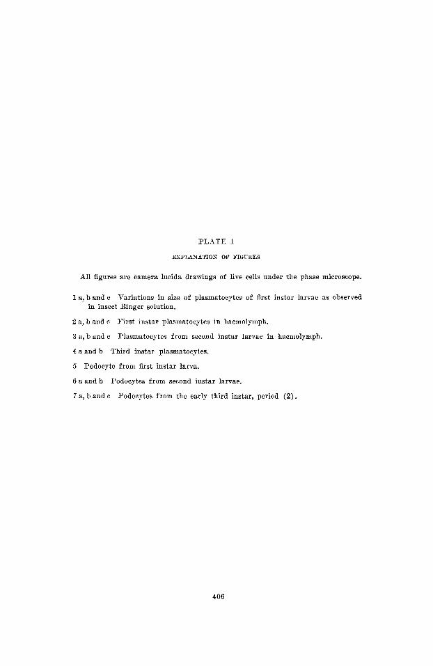
PLATE 1
EXPLANATION OF FIGURES
All figures are camera lucida drawings of live cells under the phase microscope.
1 a, b and c Variations in size of plasmatocytes of first instar larvae as observed in insect Ringer solution.
2 a, b and c
3 a, b and c
4 a and b
5 Podocyte from first instar larva.
6 a and b
7 a, b and c
First instar plasmatocytes in haemolymph.
Plasmatocytes from second instar larvae in haemolymph.
Third instar plasmatocytes.
Podocytes from second instar larvae.
Podocytes from the early third instar, period ( 2 ) .
406

LARVAL BLOOD CELLS OF DROSOPHILA M. T. M. RIZKI
PLATE 1
407

PLATE 2
EXPLANATION O F FIGURES
All figures are camera lucida drawings of live cells under the phase microscope.
8 Podoeyte from third instar, period (4).
9 a and b Podocytes from periods (5) and ( G ) , third instar.
10
11 :I
11 b
1 2
13 a and b Oenocytoid from period (4), third instar.
14 a and b
1 5 a
15 b
16
Splieroidocyte from third instar larva, period (4) .
Oenocytoid diawn from a n intact first instar larva.
Oenocytoid from first instar larva in haemolymph.
Optical section of oenocytoid from the second instar in haeinolymph.
Nematocytes from the third instar, period (1).
Crystalloid cell drawn within the body of an intact first instar larva.
Crystalloid cell from the first instar in a drop of haemolymph.
Crystalloid cell from the second instar.
408

LARVAL BLOOD CELLS O F DROSOPHILA M. T. M. RILKI
P L A T E 2
409

PLATE 3
EXPLANATION OF FIGURES
17, 18, 19 and 20 Photomicrographs of living cells under phase contrast. 17, plas- matocytes and a nematocyte. 18, podocytes from third instar, period (5) . 19, giant podocyte from a larva inmediately after the second moult. 20, oeno- cytoid.
A series of haemocytes in a smear preparation under an oil drop (not air dried). Phase microscope.
2 1
22 and 23 A fixed and stained smear showing plasmatocytes and different stages of spheroidocxtes. Third instar larva. Note the shape of the plasmatocytes and compare with the smears under oil in figure 21.
410

PLATE 3
411