Natural Language Processing in Biomedicine: A Unified System Architecture Overview

1
The Interferon System: an overview. Marco De Andrea°*, Raffaella Ravera°, Daniela Gioia*, Marisa Gariglio* and Santo Landolfo°§
°Department of Public Health and Microbiology, Medical School, University of Turin, Turin, Italy; §Immunogenetics and Experimental Oncology Center, C.N.R., Turin, Italy; and *Department of Medical Sciences, Medical School, University of Eastern Piedmont, Novara, Italy. Correspondence to: Santo Landolfo, MD, Full Professor, Department of Public Health and Microbiology, Medical School, University of Turin, Via Santena 9 bis, 10126 - Torino, Italy. Article category: Review Short running title: An overview on the Interferon system. Abstract
Interferons are a family of related and naturally occurring signal proteins grouped in three major species (alpha, beta and gamma) in accordance to their cellular origin and inducing agents and historically described for their antiviral activity. Upon binding to specific receptors they lead to the activation of signal transduction pathway that activate a broad range of genes, that are now known involved not only in antiviral but also in immunomodulatory and antiproliferative activities. The inhibition of virus growth and/or cell proliferation by interferons are associated with several physiological changes, some of which depending from the activity of specific proteins that are interferon-inducible. This review will attempt to illustrate the history and main properties of the Interferon system, taking a look to the recent discoveries about some interferon-inducible genes. Key words: Interferon, viruses, antiviral state, interferon-inducible genes, Ifi200,
HIN200.

2
History and generalities
Interferons represent one of the so-called natural defences of human organisms against the invasion of external agents such as viruses, microbes or tumour cells.
The notion of interferon is strictly related with the notion of viral interference, a concept which implies that some cells, once infected by a virus, become resistant to subsequent infections caused by the same or by similar viruses. The first observations in this respect were made in the mid-Thirties. Namely, Hoskins’s work on rabbits demonstrated that these animals, after being infected by the herpes simplex virus, were protected against subsequent infections by the same virus1.
A few years later, Findlay and Mac Callum demonstrated the same phenomenon but with a wider spectrum, in that these animals were also resistant to infections caused by viruses which were not antigenically identical2. However, such observations lacked any biological confirmation, since, at that time, the existence of any molecules capable of mediating this activity had not been demonstrated yet. It was only in 1957 that Isaacs and Lindenmann observed that supernatants of cells taken from cell cultures infected by a virus produced a protein that could make other cells resistant to other viruses3. In particular, they made use of chorioid fragments from chicken embryos, infected them with thermo-inactivated influenza virus and saw that these cells could produce a protein that was able to protect other fragments of the same type when subsequently infected by a virus that was no longer an inactivated but a live virus. Therefore, this was the first observation that something that mediated this protection against the viral infection actually existed.
It can therefore be said that the name ‘interferon’ stems from this observation, from this capacity of having a viral interference on the cells. Today’s knowledge of the interferon system is much wider. In particular, it is known that interferon provides protection and is a natural barrier against infections caused by external viruses, bacteria, mitogens and tumour cells.
As of today, interferons are considered cytokines belonging to the group of interleukins and three types of interferon are known: alpha, beta and gamma interferon. Alpha and beta interferons belong to the same type I sub-class, whereas gamma interferon belongs to a separate sub-class or type II. These are functionally correlated proteins that are produced by cells after the latter have been stimulated by external agents. In order to obtain an antiviral effect, however, other proteins must be produced:

3
these are called ‘effector’ proteins. Therefore, interferon is an inducer molecule acting on other proteins which actually trigger an antiviral anr/or antiproliferative mechanism.
To date three key features are known of interferons: there is the historically acknowledged antiviral action, but they show also an immunomodulatory and an antiproliferative action that have been recently studied4,5,6. The fact that, under specific conditions, some interferons can block cells at a given stage of the cell cycle has made the investigation of their antitumour activity become crucial.
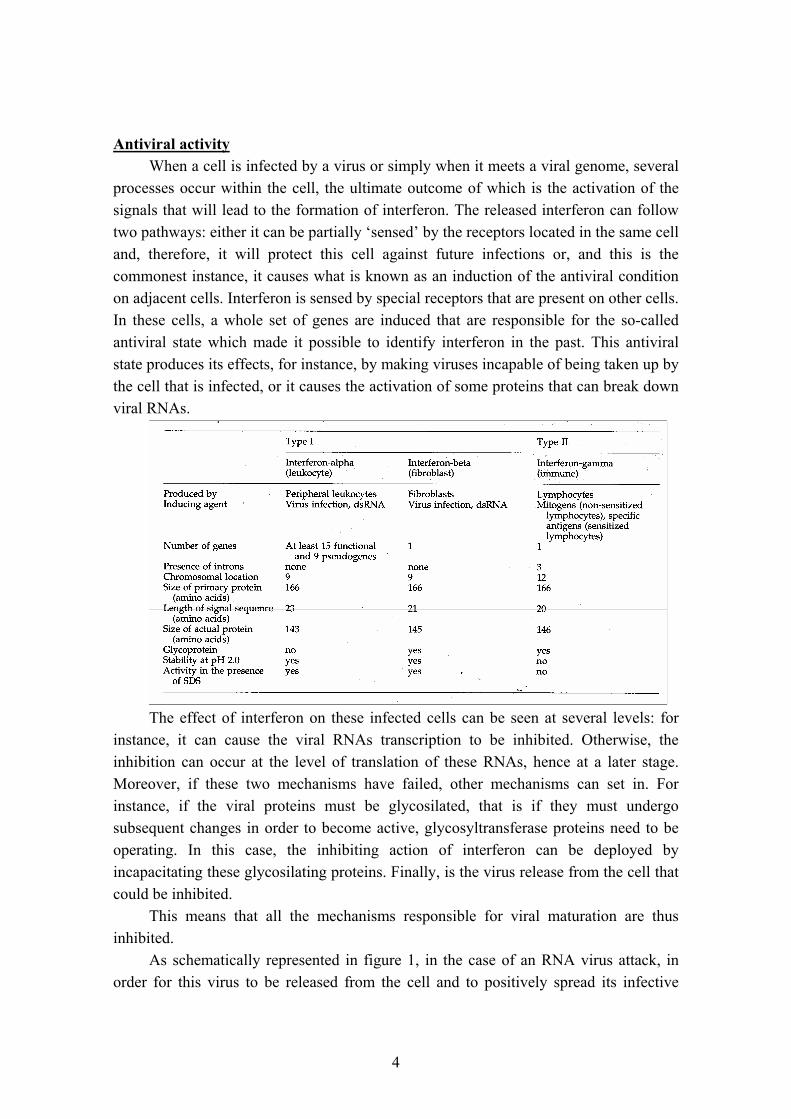
Alpha interferon, which is also known as ‘leukocyte interferon’ due to the cells that produce it, is coded for by not less than 15 genes and 9 pseudogenes docked on human chromosome 9 and murine chromosome 4. It follows that alpha interferon is not unique, but rather a protein family made up of different types of alpha interferon. The main feature is that each of those genes does not contain any introns, i.e. it does not contain any regions that do not code for DNA, and the whole gene codes for alpha interferon. The protein that is thus formed consists of 166 amino acids with no glycosilation sites and stable at a very acidic pH.
The main features of alpha interferon is that it is produced by macrophages and particularly by lymphocytes, especially under the action of external agents such as eukaryotic or tumour cells, or by virus-infected cells, as well as by prokaryotic cells and by mitogens in general. However, the crucial point is that all these inducers trigger the production of alpha interferon by macrophages and lymphocytes.
The first interferon ever discovered is the beta type, which was first observed by Isaacs and Lindenmann. It used to be called ‘fibroblast interferon’ because it is produced by epithelial cells and fibroblasts under the action of foreign nucleic acids like viruses or other types. Also beta interferon is coded by a gene docked on human chromosome 9. In this case, only one gene exists and once again there are no introns, therefore the whole gene codes for the protein. This protein consists of 166 amino acids. In this case, it contains a glycosilation site and it is once again stable at pH 2. Compared to alpha interferon, its homology is as high as 40 to 50%.
Gamma interferon belongs to a separate sub-class, the so-called type II interferon that is named ‘immune interferon’. It is produced especially by lymphocytes sensitised with the help of macrophages and only under the action of external mitogens. Also in this case there is a single gene which, however, is not docked on chromosome 9, but on chromosome 12. In addition, it contains 3 introns, which was not the case with alpha and beta interferons. These and other features cause gamma interferon to be allocated to a separate class which has different characteristics with respect to alpha and beta interferons. As a matter of fact, another peculiar trait of gamma interferon is that it is instable at an acidic pH and these are very important characteristics, especially as far as interferon-based therapy is concerned. Moreover, there is no analogy with alpha and beta interferons.
Properties of different interferon classes are summarized in table 1.
4
Antiviral activity
When a cell is infected by a virus or simply when it meets a viral genome, several processes occur within the cell, the ultimate outcome of which is the activation of the signals that will lead to the formation of interferon. The released interferon can follow two pathways: either it can be partially ‘sensed’ by the receptors located in the same cell and, therefore, it will protect this cell against future infections or, and this is the commonest instance, it causes what is known as an induction of the antiviral condition on adjacent cells. Interferon is sensed by special receptors that are present on other cells. In these cells, a whole set of genes are induced that are responsible for the so-called antiviral state which made it possible to identify interferon in the past. This antiviral state produces its effects, for instance, by making viruses incapable of being taken up by the cell that is infected, or it causes the activation of some proteins that can break down viral RNAs.
The effect of interferon on these infected cells can be seen at several levels: for
instance, it can cause the viral RNAs transcription to be inhibited. Otherwise, the inhibition can occur at the level of translation of these RNAs, hence at a later stage. Moreover, if these two mechanisms have failed, other mechanisms can set in. For instance, if the viral proteins must be glycosilated, that is if they must undergo subsequent changes in order to become active, glycosyltransferase proteins need to be operating. In this case, the inhibiting action of interferon can be deployed by incapacitating these glycosilating proteins. Finally, is the virus release from the cell that could be inhibited.
This means that all the mechanisms responsible for viral maturation are thus inhibited.
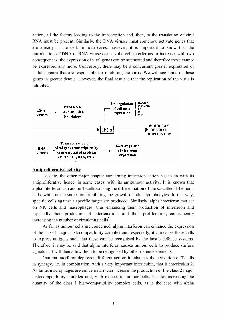
As schematically represented in figure 1, in the case of an RNA virus attack, in order for this virus to be released from the cell and to positively spread its infective
5
action, all the factors leading to the transcription and, then, to the translation of viral RNA must be present. Similarly, the DNA viruses must somehow activate genes that are already in the cell. In both cases, however, it is important to know that the introduction of DNA or RNA viruses causes the cell interferons to increase, with two consequences: the expression of viral genes can be attenuated and therefore these cannot be expressed any more. Conversely, there may be a concurrent greater expression of cellular genes that are responsible for inhibiting the virus. We will see some of these genes in greater details. However, the final result is that the replication of the virus is inhibited.
Antiproliferative activity
To date, the other major chapter concerning interferon action has to do with its antiproliferative hence, in some cases, with its antitumour activity. It is known that alpha interferon can act on T-cells causing the differentiation of the so-called T-helper 1 cells, while at the same time inhibiting the growth of other lymphocytes. In this way, specific cells against a specific target are produced. Similarly, alpha interferon can act on NK cells and macrophages, thus enhancing their production of interferon and especially their production of interleukin 1 and their proliferation, consequently increasing the number of circulating cells7
As far as tumour cells are concerned, alpha interferon can enhance the expression of the class 1 major histocompatibility complex and, especially, it can cause these cells to express antigens such that these can be recognised by the host’s defence systems. Therefore, it may be said that alpha interferon causes tumour cells to produce surface signals that will then allow them to be recognised by other defence elements.
Gamma interferon deploys a different action: it enhances the activation of T-cells in synergy, i.e. in combination, with a very important interleukin, that is interleukin 2. As far as macrophages are concerned, it can increase the production of the class 2 major histocompatibility complex and, with respect to tumour cells, besides increasing the quantity of the class 1 histocompatibility complex cells, as is the case with alpha

6
interferon, it can also decrease the proliferation of tumour cells. This was observed on cultured cells for some types of tumours7.
Interferon-based antitumour therapy has been introduced quite recently. Towards the mid-Eighties, interferons started to be massively used against a large number of tumours. Actually, in this case, the expectations related to the use of interferons have been met but partially. As a matter of fact, a survey was recently published by Jonasch and Haluska7 which lists a number of studies that review the use of interferon through the years. Their conclusion is that interferon is almost always used in combination with other treatments, like chemotherapy, and it is only active against some types of cancer, in particular against chronic myeloid leukaemia or follicular lymphoma. In the case of other tumours and, to a lesser extent, also for these forms, the adverse effects induced in patients by interferon are always rather severe and more in-depth studies still need to be conducted to decide whether it is advisable to use these molecules in antitumour treatments.
Signal transduction
To exert their activities, interferons bind to specific membrane receptors that permit cells sense the external signal of interferon and then transfer it in the interior, so that the cell can produce the so-called interferon-inducible proteins.
As previously described for interferons, receptors too are divided into two classes, namely the receptor for alpha and beta interferon on the one side and the receptor of gamma interferon on the other side. Actually, a receptor is not a single unit, but it consists of two sub-units. Joined together, these two sub-units form the actual interferon receptor. These are transmembrane proteins with an extracytoplasmic domain and an intracytoplasmic domain displaying particular motives that bind to proteins that are key in making it possible for this external signal to be sensed within the cell. There are tyrosine kinase proteins that are particularly important, since, at this stage, they can phosphorylate tyrosine residues; these belong to the so-called JAK proteins. The JAK acronym has a dual meaning: at a time when several proteins that may have this kinase activity were being discovered, an additional protein of this kind was found, so that this acronym would stand for ‘just another kinase’. Another explanation is due to the fact that this molecule contains two very similar kinase domains, but only one of these is active. When the molecule becomes active, these two domains almost face one another, somewhat mimicking the Roman two-faced Janus, hence the name ‘Janus kinase’8.
Other important proteins in this respect are the so-called STAT proteins. These have a dual role in that, as explained by their name, they are signal transducers, so that they can let the external signal get inside and, at the same time, they are also transcription activators, since these proteins, after binding to the receptor, travel until they reach the nucleus where they later trigger the transcription of interferon-inducible genes9.
7
Finally, another group of proteins playing a pivotal role in the interferon signal transduction is composed by the Interferon Regulatory Factors (IRFs). They compose a family of nine structurally related transcription factors whose functional roles, through self-interactions or interactions with other transcription factors, are recently becoming clearer10. In particular, studies from gene knockout mice revealed an involvement in the regulation of host defense, such as innate and adaptive immune responses and oncogenesis11.
Interferon-inducible genes
The number of proteins that are induced by interferons and, therefore, that are transcribed starting from the STAT and/or IRF factors is constantly increasing, also depending on the introduction of new analysis methods, such as the microarray technology.
The best characterised of them are the 2’-5’ oligoadenylate synthetase, the RNA-dependent protein kinase, or PKR, and the Mx proteins. Each of them has peculiar characteristics. For instance, the 2’-5’oligoadenylate synthetase system consists of a set of different enzymes whose ultimate effect is to break down the viral RNA12. The other interferon-inducible protein is the so-called PKR or RNA-dependent protein kinase. Once phosphorylated under the action of interferon or double-stranded RNA, this protein succeeds in triggering several changes within the cell, the outcome of which is, in this case, to inhibit the viral protein synthesis. This happens because of the sequestration of the transcription starting factors that are essential for the viral transcription to occur13. As underlined for the 2’-5’oligoadenylate synthetase, besides having this antiviral action, the PKR has also been shown to have an antiproliferative effect and, in some cases, a tumour-suppressing action14.
Mx proteins are another group of proteins, conserved in different vertebrate species, whose functional characteristics are not well understood yet. They were originally characterized for their anti-viral activity in mouse15 and later in human. In particular, human MxA protein can partially inhibit the action of some viruses, in particular of orthomyxo- and paramyxoviruses16.
8
The interest of our laboratory is currently focused on a particular family of interferon-inducible genes, the murine IfI200 and more recently its human counterpart HIN200 gene family. The name stems from the fact that the nuclear phosphoproteins encoded by these genes contain one or two 200 amino acid domains, defined as a and b. This family is actually coded by a gene cluster docked on chromosome 1, in positions q21-22, both in mouse and in human. The four murine and three human homologous display almost the same characteristics: they are induced by interferon, lipopolysaccharides, DNA or RNA viruses and that they are expressed mostly in haematopoietic tissues. In particular, some of them are induced by interferon in the monocytic macrophage cell system17,18.
Some recent results from our and from other research teams have also demonstrated that these proteins can control the cell cycle and, in particular, the overexpression of cultured cells. As a matter of fact, these cells have shown to be blocked at a precise cell cycle stage, i.e. before the start of DNA synthesis, between phase G1 and phase S19.
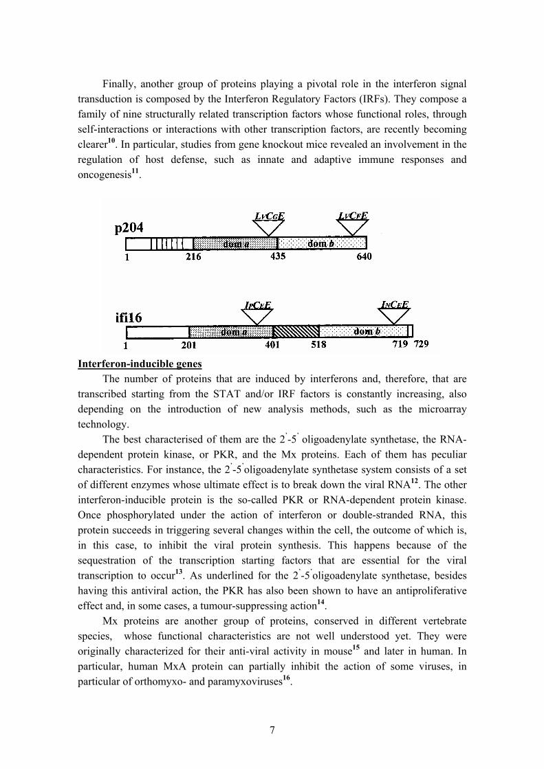
As a paradigm of the family, in figure 2 we schematically represented the more important members of this family: the murine protein p204 and its human counterpart Ifi16. They display both a and b domains and two particularly important sequences that concur to bind a protein that is key in controlling the cell cycle: the Retinoblastoma protein or pRb20.
Recent experiments confirm that p204 is expressed in the haematopoietic tissue like the other proteins of the same family, but its peculiarity is that it is highly inducible in the monocyte and macrophage cell system, besides being expressed in activated endothelial cells and especially in microglial cells, hence in a particular cell population within the midbrain. Moreover, recent immunohistochemical analysis undertaken at our laboratory to check Ifi16 expression seem to demonstrate that this protein is particularly expressed in the endothelial cells of arteries affected by atherosclerotic damage (De Andrea et al., in preparation).
These data, taken together with studies showing an involvement of members of this family in the susceptibility to systemic lupus21,22, prompt us to investigate in more detail the role of human Ifi16 in correlation with several diseases.

9
References 1. Hoskins M. A protective action of neurotropic against viscerotropic yellow fever
virus in Macacus rhesus. Amer. J. Trop. Med. Hyg. 1935; 15: 675-680. 2. Findlay GM, Mac Callum FO. An interference phenomenon in relation to yellow
fever and other viruses. J. Path. Bact. 1937; 44: 405-424. 3. Isaacs A, Lindenmann J. Virus interference. I. The interferon. Proc. Royal Soc.
1957; B 147: 258-267. 4. Pestka S, Langer JA, Zoon KC, Samuel CE. Interferons and their actions. Annu.
Rev. Biochem. 1987; 56: 727-777. 5. DeMaeyer EM, DeMaeyer-Guignard J. Interferons and other regulatory cytokines.
Wiley-Interscience, New York, 1988. 6. Landolfo S, Gribaudo G, Angeretti A, Gariglio M. Mechanisms of viral inhibition
by interferons. Pharmacol. Ther. 1995; 65:415-422. 7. Jonasch E, Haluska FG. Interferon in oncological practice: review of interferon
biology, clinical applications, and toxicities. The Oncologist 2001; 6: 34-55. 8. Duhé RJ, Farrar WL. Structural and mechanistic aspects of Janus kinases: how the
two-faced god wields a double-edged sword. J. Interferon Cytokine Res. 1998; 18:1-15.
9. Schindler C, Darnell JE. Transcriptional responses to polypeptide ligands: the Jak-Stat pathway. Annu. Rev. Biochem. 1995; 64:621-651.
10. Taniguchi T, Ogasawara K, Takaoka A, Tanaka N. IRF family of transcription factors as regulators of host defense. Annu. Rev. Immunol. 2001; 19: 623-655.
11. Sato M, Taniguchi T, Tanaka N. The interferon system and interferon regulatory factor transcription factors – studies from gene knockout mice. Cytokine Growth Factor Rev. 2001; 12: 133-142.
12. Silverman RH. 2-5A dependent RNaseL: a regulated endoribonuclease in the interferon system. In: D’Alessio G. and Riordan JF. (eds.). Ribonuclease: structure and functions. New York: Academic Press, 1997: 515-551.
13. Hovanessian AG. The double-stranded RNA-activated protein kinase induced by interferons: dsRNA-PK. J. Interferon Res. 1989; 9: 641-647.
14. Clemens KL, Elia A. The double-stranded RNA-dependent protein kinase PKR: structure and function. J. Interferon Res. 1997; 17: 503-524.
15. Kolb E, Laine E, Strehler D, Staeheli P. Resistance to influenza virus infection of Mx transgenic mice expression Mx protein under the control of constitutive promoters. J. Virol. 1992; 66: 1709-1716.
16. Ponten A, Sick C, Weeber M, Haller O, Kochs G. Dominant-negative mutants of human MxA protein: domains in the carboxy-terminal moiety are important for oligomerization and antiviral activity. J. Virol. 1997; 71: 2591-2599.
17. Landolfo S, Gariglio M, Gribaudo G, Lembo D. The Ifi200 genes: an emerging family of IFN-inducible genes. Biochemie 1998; 80: 721-728.

10
18. Gariglio M, De Andrea M, Lembo M, Ravotto M, Zappador C, Valente G, Landolfo S. The murine homolog of the HIN200 family, Ifi204, is constitutively expressed in myeloid cells and selectively induced in the monocyte/macrophage lineage. J. Leokocyte Biol. 1998; 64: 608-614.
19. Lembo M, Sacchi C, Zappador C, Bellomo G, Gaboli M, Pandolfi PP, Gariglio M, Landolfo S. Inhibition of cell proliferation by the interferon-inducible 204 gene, a member of the Ifi200 cluster. Oncogene 1998; 16: 1543-1551.
20. Hertel L, Rolle S, De Andrea M, Azzimonti B, Osello R, Gribaudo G, Gariglio M, Landolfo S. The retinoblastoma protein is an essential mediator that links the interferon-inducible 204 gene to cell cycle regulation. Oncogene 2000; 19: 3598-3608.
21. Seelig HP, Ehrfeld H, Renz M. Interferon-gamma-inducible protein p16. A new target of antinuclear antibodies in patients with systemic lupus erythematosus. Arthritis Rheum. 1994; 37: 1672-1683.
22. Rozzo SJ, Allard JD, Choubey D, Vyse TJ, Izui S, Peltz G, Kotzin BL. Evidence for an interferon-inducible gene, Ifi202, in the susceptibility to systemic lupus. Immunity 2001; 15: 435-443.
11
Legends to figures Figure1. Mechanisms of viral replication inhibition by interferons. Figure 2. Homology between murine p204 and human Ifi16 proteins. Triangles represent Retinoblastoma (pRb) binding motifs.

12
Table 1. Properties of human interferons
Copyright © 2022 FDOKUMEN


![[MS-PSSO]: Print Services System Overview - Microsoft](https://static.fdokumen.com/doc/165x107/633d285d42e56a04450a32ad/ms-psso-print-services-system-overview-microsoft.jpg)