SLUG: a new target of lymphoid enhancer factor-1 in human osteoblasts
Upload
independentCategory
view
0download
0
LETTERdoi:10.1038/nature13158
Maternal retinoids control type 3 innate lymphoidcells and set the offspring immunitySerge A. van de Pavert1{*, Manuela Ferreira2*, Rita G. Domingues2, Helder Ribeiro2, Rosalie Molenaar1, Lara Moreira-Santos2,Francisca F. Almeida2, Sales Ibiza2, Ines Barbosa2, Gera Goverse1, Carlos Labao-Almeida2, Cristina Godinho-Silva2, Tanja Konijn1,Dennis Schooneman1, Tom O’Toole1, Mark R. Mizee1, Yasmin Habani1, Esther Haak3, Fabio R. Santori4, Dan R. Littman4,Stefan Schulte-Merker5, Elaine Dzierzak3, J. Pedro Simas2, Reina E. Mebius1* & Henrique Veiga-Fernandes2*
The impact of nutritional status during fetal life on the overall healthof adults has been recognized1; however, dietary effects on the devel-oping immune systemare largely unknown.Developmentof second-ary lymphoidorgans occurs during embryogenesis and is consideredto be developmentally programmed2,3. Secondary lymphoid organformation depends on a subset of type 3 innate lymphoid cells (ILC3)named lymphoid tissue inducer (LTi) cells2–5.Hereweshowthatmousefetal ILC3s are controlled by cell-autonomous retinoic acid (RA) sig-nalling in utero, which pre-sets the immune fitness in adulthood.We found that embryonic lymphoid organs contain ILCprogenitorsthat differentiate locally into mature LTi cells. Local LTi cell differ-entiation was controlled by maternal retinoid intake and fetal RAsignalling acting in a haematopoietic cell-autonomousmanner. RAcontrolled LTi cell maturation upstream of the transcription factorRORct. Accordingly, enforced expression of Rorgt restored matu-ration of LTi cells with impaired RA signalling, whereas RA recep-tors directly regulated the Rorgt locus. Finally, we established thatmaternal levels ofdietary retinoids control the sizeof secondary lym-phoid organs and the efficiency of immune responses in the adultoffspring. Our results reveal a molecular link between maternalnutrients and the formationof immunestructures required for resis-tance to infection in the offspring.Haematopoietic cells that initially colonize secondary lymphoidorgan
(SLO) sites include CD32c-Kit1IL-7Ra2a4b71CD11c1CD42 lym-phoid tissue initiator (LTin) cells and the prototypicalmember of type3 ILCs, LTi cells2–7. Although most LTi cells express CD4, this is a lateevent in LTi differentiation and not all RORct1 LTi cells express thismarker5,6,8,9. Thus, weproposed thatCD32IL-7Ra1a4b71ID21c-Kit1
CD11c2CD42 ILCs (hereafter called ILC4neg cells) receive local cuesgiving rise to ID21RORct1CD41 LTi cells (LTi4) within developingSLOs.Notably, enteric ILC4neg cells includemainly ID21RORct1CD42
LTi cells (LTi0) but also a small fraction of ID21RORct2CD42 pre-cursors with LTi cell potential (hereafter called pre-ILC cells)9. In con-trast, nearly 100%of lymph node ILC4neg cells are LTi0 cells (ExtendedData Fig. 1a, b). Analysis of embryonic day 12.5 (E12.5) guts revealedthat ILC4neg cells are the only appreciable IL-7Ra1 colonizing cells(Fig. 1a, b). Accordingly, non-cyclingmature Sca12LTi4 cells increasedthroughout development, seemingly at the expense of Sca11 ILC4negcells (Fig. 1a–c andExtendedData Fig. 1c). Further evidence that ILC4negcells differentiate locally was provided by organ cultures and trans-plantation of E12.5 intestines. Despite absence of fetal liver output inthese settings, LTi4 cells increased with time at the expense of localILC4neg cells (Fig. 1d, e). Furthermore, in E14.5 Rorgt2/2 embryos,ILC4neg cells were attracted to the intestine and lymph nodes, support-ing initial anlagen colonization by these cells (ExtendedData Fig. 1d, e).
Notably, RA stimulation of E13.5 lymph node cells showed increasedfrequency of LTi4 cells and reduction of ILC4neg cells, indicating thatdifferentiation of LTi4 cells is regulated by RA (Fig. 1f). To confirm theeffect of RA in LTi differentiation in vivo, pregnantmice received aRA-enriched diet starting at E10.5. Supplementation of RA increased theproportion of LTi4 cells in the embryo, to the detriment of ILC4neg cells(Fig. 1f). In agreement with this finding, provision of the RA signallinginhibitor BMS493 to pregnant female mice resulted in a decrease offetal LTi4 cells despite normal frequency of fetal liver progenitors andenteric haematopoietic cells (Fig. 1f and Extended Data Fig. 1f). Con-sequently, despite normal embryo size, BMS493 administration led to areduction in lymph node dimensions and Peyer’s patch developmentalfailure (Fig. 1g–i and ExtendedData Fig. 1g). Collectively, our data indi-cate thatmaternal retinoids control LTi cell differentiationwithindevel-oping SLOs.RA is a vitamin Ametabolite that controls early vertebrate develop-
ment, some immune processes in adulthood, and has been shown tomediate CXCL13 expression in fetal mesenchymal cells10–16. RA bindsto heterodimers formed by the RA receptors (RARs) and retinoid Xreceptors (RXRs), which bind DNARA response elements (RAREs)11.To address putative RA cell-autonomous responses, we assessed RARand RXR expression in E15.5 ILC4neg, LTi4 and LTin cells. RARs andRXRswere predominantly expressedby ILC4neg andLTi4 cells, whereasLTin cells expressed thesemolecules at lower levels (Fig. 2a). RA stimu-lation revealed that only ILC4neg and LTi4 cells respond robustly, asshown by Rarb upregulation (Fig. 2b)16. Together, these data indicatethat impaired SLOdevelopment in BMS493-treatedmicemight be theconsequence of RA signal ablation in LTi cells. To test this hypothesiswe used a lineage-targeted model to block RA signalling. We used amouse line in which a truncated form of the RARa gene was knockedinto the ROSA26 locus preceded by a triple polyadenylation signalflanked by two loxP sites (ROSA26-RARa403). This line was bred toVav-iCre mice that in contrast to other tested Cre lines ensured Creactivity in fetal LTin, ILC4neg andLTi4 cells (ExtendedDataFig. 2a–d)17,18.Despite normal frequencies of fetal liver precursors, and SLO LTin,ILC4neg and LTi0 cells,Vav-iCre/ROSA26-RARa403 embryos (Rarmice)revealed adose-dependent reductionofLTi4 cells (Fig. 2c, d andExtendedData Fig. 3a–f). To assess whether the differentiation potential of ILC4negcells is controlled by RA thresholds, we cultured purified ILC4neg cellsfromRar heterozygous (RarHet), homozygous (RarHom) and wild-typelittermate control mice. Whereas wild-type ILC4neg cells upregulatedpro-inflammatory cytokines and chemokines andgave rise to LTi4 cellsin vitro, these were impaired proportionally to the degree of RA signal-ling abrogation in ILC4neg cells (Fig. 2e, f and Extended Data Fig. 3g).Finally, despite normal frequency of colonizing ILC4neg cells (Fig. 2d),
*These authors contributed equally to this work.
1Department of Molecular Cell Biology and Immunology, VU University Medical Center, van der Boechorststraat 7, 1081BT Amsterdam, The Netherlands. 2Instituto de Medicina Molecular, Faculdade deMedicina de Lisboa, Av. Prof. Egas Moniz, Edifıcio Egas Moniz, 1649-028 Lisboa, Portugal. 3Erasmus Stem Cell Institute, Department of Cell Biology, Erasmus Medical Center, 3000 CA Rotterdam, TheNetherlands. 4Howard Hughes Medical Institute, Molecular Pathogenesis Program, Skirball Institute of Biomolecular Medicine, New York University School of Medicine, New York, New York 10016, USA.5Hubrecht Institute–KNAW (Royal Netherlands Academy of Arts and Sciences) and University Medical Center Utrecht, 3584 CT Utrecht, Netherlands. {Present address: Hubrecht Institute–KNAW (RoyalNetherlands Academy of Arts and Sciences) and University Medical Center Utrecht, 3584 CT Utrecht, Netherlands.
3 A P R I L 2 0 1 4 | V O L 5 0 8 | N A T U R E | 1 2 3
Macmillan Publishers Limited. All rights reserved©2014

0.35
a
29.3
E13.5 E14.5 E15.5
b
***70
***35
12.5
13.5
14.5
15.5
12.5
13.5
14.5
15.5
ILC4neg LTi4
LTi4
CD45+CD3–CD11c–
E12.5
16.910.3
7.917.65
CD
4
Ly6A-GFPCD4
CD45
Ly6A-GFPIL-7R!CD45
E15.5ILC4negLTi4
c
Per
cent
age
of c
ells
0 0
E13.5 E14.5
ILC4n
IL-7R!
27.934.3
Sca1A
rbitr
ary
units
Embryonic day
In vitro
***35 ***30
In vivo
***70
NS
*15
ed f
*8
In vitro
***15
In vivo
**3***
*** ***
Day
0
ILC
4neg
cel
ls (%
)
ILC
4neg
cel
ls (%
)
0
LTi 4
cells
(%)
LTi 4
cells
(%)
0 0
LTi 4/
ILC
4neg
0
Vehicl
e RA
Vehicl
e RA
Vehicl
eBM
S
BMS
DMSO
BMS
DMSO
BMS
DMSO
BMS
DMSO0 0
0 1 2 0 1 2
***
*** *
*
**
Ing
DM
SO
***70
**25
***5
g
h
DM
SO
GFP
iPeyer’s patch Peyer’s patch
Bra Bra
45
Ing
GFP
BM
S
Are
a (1
03 "m
2 )
0
0
Pey
er’s
pat
ches
per
inte
stin
e
0BM
S
Are
a (#
103 "m
2 )
0
Day0 1 2 0 1 2
Figure 1 | Maternal RA controls LTi differentiation. a, Enteric fetal ILC4neg
and LTi4 cells. b, Percentage of enteric fetal ILC4neg and LTi4 cells. E12.5, n5 8;E13.5, n5 3; E14.5, n5 6; E15.5, n5 3. c, Left: Sca1-GFP lymph nodes.Right: E15.5 gut cells. d, e, Cultured or transplanted E12.5 guts. d, Day 0, n5 4;day 1,n5 6; day 2,n5 6. e, Day 0,n5 7; day 1,n5 3; day 2,n5 5. f, Left: E13.5lymph node cells; n5 7. Centre: RA was provided to females. E13.5 lymphnode cells; n5 8. Right: females received BMS493; n5 8. LTi4/ILC4neg cell ratio
is shown. g–i, hCD2-GFP females received BMS493. E17.5 SLOs. g, Brachial(Bra) and inguinal (Ing) lymph nodes; n5 6. h, Arrowheads indicate Peyer’spatch. i, Peyer’s patch number per intestine: DMSO, n5 8; BMS, n5 22.Area: DMSO, n5 30; BMS, n5 25. Scale bars: 50mm (c), 200mm (g, h right),1mm (h, left). Error bars show s.e. *P, 0.05; **P, 0.01; ***P, 0.001.NS, not significant.
8
ILC4n
eg LTi 4
LTin
ILC4neg LTi4 LTinILC
4neg LT
i 4LT
in
ILC4n
eg LTi 4
LTin
ILC4n
eg LTi 4
LTin
ILC4n
eg LTi 4
LTin
ILC4n
eg LTi 4
LTin
Raraa0.03
Rarb
0.12
Rarg
0.12
Rxra
0.08
Rxrb
0.05
Rxrg
Rel
ativ
e un
its
0 0 0 0 0 0
b5
BMS
WT
RABM
S RABM
S RA
16# increaseCD45+CD3–CD11c–
WT
c
Rar
blo
g 2 fo
ld in
crea
se to
DM
SO
0
4# increase
–2
NS NS
WT
22.8
19.4
RarHet
RarHet
RarHom
WTWT RarHet RarHom
WT
Rar
Het
Rar
Hom
RarHom W
T
RarHet
RarHom W
T
RarHet
RarHom
WT
RarHet
RarHom
WT
RarHet
RarHom W
T
RarHet
RarHom W
T
RarHet
RarHom W
T
RarHet
RarHom
10.8
19.1
CD
4
IL-7R!
IL-7R!
11.2
20.7
8.45
75.8
3.31
79.5
1.37
73.3
In vitro differentiation 35
ILC
4neg
cel
ls (%
)
***
**
0.045
LTi 4
/ ILC
4neg
35e
CD
4
***
**
LTi 4
cells
(%)
LTin
cel
ls (%
)
50d
f
0
0
0
Bra Peyer’s patch
VCAM1
Ing
Ing
ND ND ND
Brah
0
Peyer’s patch
g
CD4
Pey
er’s
pat
ch p
er in
test
ine
Pey
er’s
pat
ch a
rea
(#10
3 "m
2 )
6 70**55 20
***
***
***
*** ***
******
0 0
Are
a (#
103 "m
2 )
Are
a (#
103 "m
2 )
0 0
Figure 2 | Cell-autonomous RA controls LTi cells and SLO development.a, RT–PCR of enteric cells. Data represent three independent experiments.b, DMSO, BMS493 or RA stimulation. Data represent three independentexperiments. c, E15.5 enteric ILC subsets. d, LTin, n5 4; ILC4neg, n5 5;LTi4, n5 5. e, E15.5 ILC4neg cells cultured for 6 days. f, ILC4neg cell cultures at
day 6; n5 5. g, Immunostaining of E15.5 embryos and intestines. Developingbrachial (Bra) and inguinal (Ing) lymph nodes and Peyer’s patch are shown.CD4, red; VCAM1, grey. Scale bars: 200mm. h, SLO size; n5 6. Error barsshow s.e. *P, 0.05; **P, 0.01; ***P, 0.001. NS, not significant.ND, not detected.
RESEARCH LETTER
1 2 4 | N A T U R E | V O L 5 0 8 | 3 A P R I L 2 0 1 4
Macmillan Publishers Limited. All rights reserved©2014

haematopoietic cell-autonomous impairment of RAresponses resultedin severely diminished fetal lymph node size and reduced number ofminute Peyer’s patches (Fig. 2g, h andExtendedData Fig. 3h).Our dataindicate that LTi cell differentiation is controlled by cell-autonomousRA signalling in developing SLOs.Previous reports have identified keyLTi cell regulators2.Micemutant
for the transcription factors ID2 and RORct lack LTi cells and do notdevelop SLOs19,20. Runx1, Tox andNotch1were also implicated in LTicell maturation9,21–23. We found that whereas most LTi-related geneswere normally expressed in RarHom and RarHet ILC4neg and LTi4 cells,Runx1was increased andRorgtwas reduced (Fig. 3a andExtendedDataFig. 4a–d). Expression of pro-inflammatory genes was also reduced inRarHom and RarHet ILC4neg and LTi4 cells (Fig. 3a and Extended DataFig. 4b–d). The marked reduction of Rorgt expression suggested thatRA could provide ILC4neg cells with signals leading toRorgt regulation.Accordingly, RA stimulation of ILC4neg cells resulted in Rorgt upre-gulation whereas most other transcription factors were unperturbed,notablyRunx1 (Fig. 3b). In agreement, BMS493 inhibitedRA-inducedRorgt expression, and efficient block of RORct by digoxin preventedRA-induced differentiation of ILC4neg cells into LTi4 cells, while cellviability was unaffected (Fig. 3c and Extended Data Fig. 5a–c). To testfurtherwhetherRA-inducedLTimaturation requiresRORct, wedeter-mined if differentiation of RAR dominant-negative ILC4neg cells isrestored by enforced Rorgt expression. Retroviral transduction of Rorgtrevealed that RARdominant-negative ILC4neg cells restored high levelsof pro-inflammatory genes and reacquired their potential to differen-tiate towards LTi4 cells (Fig. 3d–f). Further evidence thatRAcandirectlyregulate Rorgt expression was provided by computational analysis ofpotential RARE sites and chromatin immunoprecipitation (ChIP) withpan-RAR and RXR antibodies. RA stimulation resulted in increasedbindingofRARandRXRupstreamandwithin theRorgt locus (Fig. 3g, hand ExtendedData Table 1). To analyse the role of these sites we intro-duced the RARE C (25,478 Rorg transcription start site (TSS)), E(21,800 Rorg TSS) and G (21,619 Rorgt TSS) half-sites in a luciferasereporter vector. Mutations in these sites resulted in significant reduc-tion of the regulatory function of these elements as measured by lucif-erase activity (Fig. 3i). Thus, cell-autonomous RA signalling providesLTi cellswith critical differentiation signals via direct regulationofRorgt.
Our data indicate that mature LTi cell numbers regulate the size ofSLOprimordia andmaydetermine lymphoidorgan size in adulthood24.RarHet adult mice had reduced SLOs and lymphocyte numbers whencompared to theirwild-type littermate controls (Fig. 4a, b andExtendedData Fig. 6a, b). In agreement,mice that received a vitamin-A-deficient(VAD)diet throughout life had reduced lymphoidorgan sizewhencom-pared to vitamin-A-control mice (VAC) (Fig. 4c). However, becauseRarHet andVADlymphocytes are continuously exposed to altered levelsof RA signals, it is possible that SLO size might be a consequence ofaltered lymphocyte pools12–15. To clarify this issue, we provided preg-nantmicewithavitamin-A-high (VAH),VADorVACdiet and switchedall diets to the same VAC diet after birth. At 10weeks of age mice thatwere exposed to aVAHdiet exclusively inuterohad larger SLOs,whereasmice exposed to a VAD diet had small SLOs when compared to VACcontrolmice (Fig. 4d).Notably, provisionof variable vitaminAdiet levelsexclusively after birth no longer controlled SLO size (Extended DataFig. 6c, d). Additional evidence that RA determines SLO size in earlylife was provided by transplantation of CD45.1wild-type bonemarrowinto lethally irradiated RarHet (WTRRarHet) or wild-type (WTRWT)CD45.2 littermate control hosts at 2weeks of age (Fig. 4e). Thus, wegenerated mice that pre- and perinatally received low input of RA sig-nals inhaematopoietic cells, but that on transplantationharbour anormalwild-type haematopoietic system. WTRRarHet mice, which receivedlow RA cues in utero, exhibited small SLOs when compared to theirWTRWTcounterparts at 8 weeks after transplantation (Fig. 4f, g andExtended Data Fig. 6e, f). This phenotype also revealed reduced lym-phocyte numbers, albeit normal SLO organization and similar haema-topoietic cell reconstitution (Fig. 4h andExtendedDataFigs 6f and7a–d).In agreement, dendritic cells fromWTRRarHet orWTRWTchimaerashad similar capacity to activate lymphocytes (ExtendedData Fig. 7e, f).Our data indicate that available RA in utero regulates the size of lym-
phocyte pools in the offspring, with possible consequences on their adap-tive immuneresponses.To test thishypothesis,WTRRarHet orWTRWTchimaeraswere infected intranasallywithmurid herpesvirus-4, result-ing in acute lung infection. Analysis of draining intrathoracic lymphnodes revealed reduced expansion but normal frequency of CD81 Tcells specific for the viral epitopesORF61 andORF75c inWTRRarHet
mice (Fig. 4i and Extended Data Fig. 8a–c). Consequently, whereas
a RA
NS
NS
NS
ND
ND ND ND ND
b c
2 32 **1.8
DIG
DIG +
RA RA
**
4# increase
0
8# decreaselog 2
fold
incr
ease
to w
ild ty
pelo
g 2 fo
ld in
crea
seto
em
pty
vect
or
log 2
fold
incr
ease
to D
MS
O
0
–1–4 –4
LTi 4/
ILC
4neg
0
d RarHom transduced with Rorgt
RarHom ILC4neg
Runx1
Runx1
Cxcr5
Klf4Cxcl1
Cxcl2Cxc
l3Tr
ance
Cxcl10
LtbNfkbiz
Tox
Rorc
Rorc
Ccr6Id2 Id2LTa
Il22
Il17a
Il23r
Notch
1
Notch
1
Tnfa
Runx1
Cxcr5
Klf4Cxcl1
Cxcl2Cxc
l3Tr
ance
Cxcl10
LtbNfkbiz
Tox
Rorc
Ccr6Id2 LTa
Il22
Il17a
Il23r
Notch
1
Tnfa
e WT RarHom
92.4 98.3256# increase10
CD
4
4.82 0.72
Empt
y
5.044.60
–2
10
–2
0IL-7R!
Ror
gt
86.093.0
DMSORA
Anti-RAR16
Luciferase activityRA/DMSO0
10*
3.5i
h
g
fFE
TSS (Rorg) TSS (Rorgt)
DCBA GExon 1 Exon 2 Exon 1$
–7,8
37-7
,446
–5,4
78–4
,690
–1,8
00–1
,638
–1,6
19
* **
* *Anti-RXR
020
WTMut
C
E
G
TGAACT
WTMut
GGGTCAAGGGT
WTMut
TCACCTCT
ACGTTA
GGTGGCGGTAG
**
LTi 4
(%)
0
***
****
0GFA B C D EP
erce
ntag
e in
put n
orm
aliz
ed to
IgG
MutAGTGACAC
WT
RarHom
Empt
yR
orgt
Empt
yR
orgt
Figure 3 | RA controls LTi cells via RORct. a, E15.5 ILC4neg cells. Datarepresent three experiments. b, E15.5 ILC4neg cells stimulated with RA. Datarepresent three experiments. c, E13.5 lymph node cells stimulated with RAand digoxin (DIG); n5 16. d–f, ILC4neg cells transduced with pMig.Rorgt-IRES-GFP virus (day 6). d, Data represent three experiments.
e, Cytometry analysis. f, Emergent LTi4 cells. Wild type (WT), n5 5; RarHom,n5 3. g, RARE sites. h, E15.5 ILC4neg cells stimulated with RA. ChIP analysisof five biological replicates. i, Luciferase activity of RARE wild-type andmutated sites. C, n5 6; E, n5 9; G, n5 6. Error bars show s.e. *P, 0.05;**P, 0.01; ***P, 0.001. NS, not significant. ND, not detected.
LETTER RESEARCH
3 A P R I L 2 0 1 4 | V O L 5 0 8 | N A T U R E | 1 2 5
Macmillan Publishers Limited. All rights reserved©2014

WTRWTchimaeras efficiently cleared lytic virus by day 10, high viraltitres were still detected inWTRRarHet chimaeras 14 days after infec-tion (Fig. 4j)25. Thus, a deficit of RA signalswithinhaematopoietic cellsin early life results in small SLOs andpoor capacity to control infection.Defining the requirements that control SLO development is essen-
tial to understand how immunitymay be regulated.We now show thatRA controls LTi cells, regulating LTi pro-inflammatory genes and thefrequency of LTi4 cells (Extended Data Fig. 9). RA operates in a cell-autonomous fashion, via RORct, although additional factors regulateRorgt in ILC3s.SLO development has been considered to be developmentally pro-
grammed; we now show that formation of these structures can be alsocontrolled bydietary signals. Thus, in addition to the established impactof dietary plant-derived chemicals in postnatal immune cells, ourworkreveals dietary retinoids as key regulators of pre-natal ILCswith a lifelongimpact on adult lymphoid organ size26–28. It was previously shown thatcomplete absenceof lymphoidorgans leads to long-life viruspersistence29.Our data reveal that the efficiency of adaptive immune responses toinfection and possibly to other immune insults may be pre-tuned inearly life through dietary signals from maternal origin.We report here that cell-autonomous RA signalling is a key axis for
Rorgt expression and LTi cell differentiation within developing SLOs.Similarly, RAmay also be important after birth in infection and chronicinflammatory diseases30. Lineage-targeted strategies will be central toelucidate the contribution of dietary retinoids in these outcomes.
METHODS SUMMARYMice were maintained at Instituto de Medicina Molecular (IMM) or VU Univer-sityMedical Centre according to national and international guidelines. Bonemarrowcellswere isolated from8-week-oldC57BL/6CD45.1mice and injected intravenouslyinto 2-week-old lethally irradiated CD45.2 ROSA26-RARa403Het (WTRRarHet)orwild-type littermate controls (WTRWT).C57BL/6 femalemice received eithervitamin-A-deficient (VAD), with no vitamin A (vitamin free casein), vitamin-A-high (VAH, 25,000 IUkg21) or vitamin-A-control (VAC, 4,000 IUkg21) diets. Reti-noic acid was provided to pregnant mice from E10.5 until they were euthanized.Quantitative real-time PCR with reverse transcription (RT–PCR) was performedas previously described6,7,10. Computational analysis was performed with TESS.DNA–protein complexes were immunoprecipitated using antibodies againstmousepan-RAR, pan-RXR or control IgG.
Online Content Any additional Methods, ExtendedData display items and SourceData are available in the online version of the paper; references unique to thesesections appear only in the online paper.
Received 6 June 2013; accepted 18 February 2014.Published online 19 March 2014.
1. Gluckman, P. D. & Hanson, M. A. Living with the past: evolution, development, andpatterns of disease. Science 305, 1733–1736 (2004).
2. van de Pavert, S. A. &Mebius, R. E. New insights into the development of lymphoidtissues. Nature Rev. Immunol. 10, 664–674 (2010).
3. Randall, T. D., Carragher, D. M. & Rangel-Moreno, J. Development of secondarylymphoid organs. Annu. Rev. Immunol. 26, 627–650 (2008).
4. Mebius, R. E., Rennert, P. & Weissman, I. L. Developing lymph nodes collectCD41CD3- LTb1 cells that candifferentiate to APC,NKcells, and follicular cells butnot T or B cells. Immunity 7, 493–504 (1997).
RarHetWT
Lifelong dieta b dcPeyer’s patchPLN In utero diet
* ** *Axi Bra Ing
*
Peyer’spatch
**PLN
** ** ** *** ***7 15 3.54 *** *** *** *** *** *** 5 ******NS NS
Lym
ph n
ode
dim
ensi
on (m
m2 )
Pey
er’s
pat
ch d
imen
sion
(mm
2 )
Folli
cles
per
Pey
er’s
pat
ch
0
SLO
siz
e (m
g)
0
* ** ***
e g
Donor:
f
Lym
ph n
ode
dim
ensi
on (m
m2 )
Lym
ph n
ode
dim
ensi
on (m
m2 )
0
4
0Axi Bra Ing
Axi Bra Ing Intra T
Axi Bra Ing
WTRar
Het
RarHet
WT
VACVAH
VADVAC
VAHVAD
VACVAH
VADVAC
VAHVAD
VACVAH
VADRar
Het
***
0
3.5
0
3.5
0 0
Intra T
Pey
er’s
pat
ch d
imen
sion
(mm
2 )
4CD45.1
5 # 106 BM
WT
WT
WT %
WT
WT % WT
WT % WT
Host:CD45.2
1067 8
0
**
ji
B220/Desmin
Axi Ingh
WT %
Rar
Het
WT %
WT
WT %
Rar
Het
WT % RarHet
WT % RarHet
WT % WTWT % RarHet
WT % WTWT % RarHet
Viru
s (p
.f.u.
) per
lung
**
*
Tet+
(OR
F61)
(#10
3 )
Tet+
(OR
F75c
) (#1
03 )
1000 010 147 10 147
d.p.i.10 147
Figure 4 | Retinoid levels in utero determine the offspring resistance toinfection. a, Adult axillary (Axi), brachial (Bra) and inguinal (Ing) peripherallymphnodes (PLN). n5 6. b, Peyer’s patch area and follicle number per Peyer’spatch. Wild type (WT), n5 26; RarHet, n5 23. c, Females received diets thatwere maintained in the offspring. VAC, n5 10; VAH, n5 8; VAD, n5 18.d, Females received variable diets. Their offspring received VAC diet. Lymphnode: VAC, n5 18; VAH, n5 24; VAD, n5 25. Peyer’s patch: VAC, n5 20;VAH, n5 40; VAD, n5 63. e, Transplantation scheme. f, Chimaeric lymph
nodes. Scale bar: 1mm. g, Dimensions of chimaeric lymph nodes. n5 6.h, Chimaeric lymph node structure. Scale bar: 200mm. i, Chimaeras infectedwith murid herpesvirus-4. Tetramer positive CD8 T cells in intrathoraciclymph nodes. j, Virus titres (plaque-forming units (p.f.u.) per lung). 7 days postinfection (d.p.i.): WTRWT, n5 3; WTRRarHet, n5 3. 10 d.p.i.: WTRWT,n5 4; WTRRarHet, n5 3. 14 d.p.i.: WTRWT, n5 8; WTRRarHet, n5 5.Dashed line indicates the detection limit. Error bars show s.e. *P, 0.05;**P, 0.01; ***P, 0.001. NS, not significant.
RESEARCH LETTER
1 2 6 | N A T U R E | V O L 5 0 8 | 3 A P R I L 2 0 1 4
Macmillan Publishers Limited. All rights reserved©2014

5. Eberl, G. et al. An essential function for the nuclear receptor RORct in thegeneration of fetal lymphoid tissue inducer cells. Nature Immunol. 5, 64–73(2004).
6. Veiga-Fernandes,H.et al.Tyrosine kinase receptorRET is a key regulator ofPeyer’sPatch organogenesis. Nature 446, 547–551 (2007).
7. Patel, A. et al.Differential RET signaling pathways drive development of the entericlymphoid and nervous systems. Sci. Signal. 5, ra55 (2012).
8. Cupedo, T.et al.Presumptive lymphnodeorganizersaredifferentially representedin developing mesenteric and peripheral nodes. J. Immunol. 173, 2968–2975(2004).
9. Cherrier,M., Sawa,S.&Eberl,G.Notch, Id2, andRORct sequentially orchestrate thefetal development of lymphoid tissue inducer cells. J. Exp. Med. 209, 729–740(2012).
10. van de Pavert, S. A. et al.Chemokine CXCL13 is essential for lymph node initiationand is induced by retinoic acid and neuronal stimulation. Nature Immunol. 10,1193–1199 (2009).
11. Niederreither, K. & Dolle, P. Retinoic acid in development: towards an integratedview. Nature Rev. Genet. 9, 541–553 (2008).
12. Iwata, M. Retinoic acid production by intestinal dendritic cells and its role in T-celltrafficking. Semin. Immunol. 21, 8–13 (2009).
13. Hall, J. A. et al. Essential role for retinoic acid in the promotion of CD41 T celleffector responses via retinoic acid receptor alpha. Immunity 34, 435–447(2011).
14. Mora, J. R.& vonAndrian,U.H.Role of retinoic acid in the imprinting of gut-homingIgA-secreting cells. Semin. Immunol. 21, 28–35 (2009).
15. Mucida, D. et al. Retinoic acid can directly promote TGF-b-mediated Foxp31Tregcell conversion of naive T cells. Immunity 30, 471–472; Reply 472–473 (2009).
16. Hall, J. A., Grainger, J. R., Spencer, S. P. & Belkaid, Y. The role of retinoic acid intolerance and immunity. Immunity 35, 13–22 (2011).
17. de Boer, J. et al. Transgenic mice with hematopoietic and lymphoid specificexpression of Cre. Eur. J. Immunol. 33, 314–325 (2003).
18. Rosselot, C. et al. Non-cell-autonomous retinoid signaling is crucial for renaldevelopment. Development 137, 283–292 (2010).
19. Sun, Z. et al. Requirement for RORc in thymocyte survival and lymphoid organdevelopment. Science 288, 2369–2373 (2000).
20. Yokota, Y. et al.Development of peripheral lymphoid organs andnatural killer cellsdepends on the helix-loop-helix inhibitor Id2. Nature 397, 702–706 (1999).
21. Aliahmad, P., de la Torre, B. & Kaye, J. Shared dependence on the DNA-bindingfactor TOX for the development of lymphoid tissue-inducer cell and NK celllineages. Nature Immunol. 11, 945–952 (2010).
22. Possot, C. et al.Notch signaling is necessary for adult, but not fetal, development ofRORct1 innate lymphoid cells. Nature Immunol. 12, 949–958 (2011).
23. Tachibana, M. et al. Runx1/Cbfb2 complexes are required for lymphoid tissueinducer cell differentiation at two developmental stages. J. Immunol. 186,1450–1457 (2011).
24. Meier, D. et al. Ectopic lymphoid-organ development occurs through interleukin7-mediated enhanced survival of lymphoid-tissue-inducer cells. Immunity 26,643–654 (2007).
25. Gredmark-Russ, S., Cheung, E. J., Isaacson, M. K., Ploegh, H. L. & Grotenbreg, G. M.TheCD8T-cell response againstmurine gammaherpesvirus68 is directed towarda broad repertoire of epitopes from both early and late antigens. J. Virol. 82,12205–12212 (2008).
26. Kiss, E. A.et al.Natural aryl hydrocarbon receptor ligands control organogenesis ofintestinal lymphoid follicles. Science 334, 1561–1565 (2011).
27. Lee, J. S. et al. AHR drives the development of gut ILC22 cells and postnatallymphoid tissues via pathways dependent on and independent of Notch. NatureImmunol. 13, 144–151 (2011).
28. Qiu, J. et al. The aryl hydrocarbon receptor regulates gut immunity throughmodulation of innate lymphoid cells. Immunity 36, 92–104 (2011).
29. Karrer, U. et al. On the key role of secondary lymphoid organs in antiviral immuneresponses studied in alymphoplastic (aly/aly) and spleenless (Hox112/2) mutantmice. J. Exp. Med. 185, 2157–2170 (1997).
30. Spencer, S. P. et al. Adaptation of innate lymphoid cells to a micronutrientdeficiency promotes type 2 barrier immunity. Science 343, 432–437 (2014).
AcknowledgementsWe thank the imaging, animal and flow cytometry facilities at IMMand UPC for technical assistance; C. Mendelsohn for providing ROSA26-RARa403mice; N. Schmolka, J. G. van Rietschoten, R. E. van Kesteren, T. H. B. Geijtenbeek,S. Gringhuis, E. Keuning, J. Peterson-Maduro, M. G. Roukens, D. D’Astolfo, M. Vermunt,A. Rijerkerk, J. Koning, J. van der Meulen and B. Oliver for technical help; andG. Vilhais-Neto, M. C. Coles and G. Eberl for discussion. M.F., L.M.-S. and R.G.D. weresupported by FCT, Portugal; H.V.-F. by EMBO (1648) and ERC (207057); D.R.L. byNIH(RO1AI080885) and HHMI; M.R.M. by Dutch MS research foundation (MS 12-797);S.A.vd.P. by NGI Breakthrough Horizon (40-41009-98-9077); and R.E.M. by a VICI(918.56.612) and ALW-TOP grant (09.048).
Author Contributions M.F. wrote the manuscript, designed, performed and analysedthe experiments in Figs 1a, b, d, e, g–i, 2a–h, 3a, b, d–i and 4a, b, d–j and ExtendedDataFigs 1a, d–f, 2a–d, 3a–h, 4a–d, 5a–c, 6, 7a–f, 8a–d, 9a, b and 10a–c. S.A.vd.P. wrote themanuscript, designed, performed and analysed the experiments in Figs 1c, f, 3c, g–iand 4c and Extended Data Figs 1b, c, e, 5b, c and 6. R.G.D., H.R., R.M., L.M.-S., F.F.A., S.I.,I.B., G.G., C.L.-A., T.K., D.S., T.O’T., M.R.M., Y.H. and S.S-M. contributed to severalexperiments.C.G.-S. and J.P.S. providedmuridherpesvirus-4. D.R.L. andF.R.SprovidedRorgt2/2 embryos. E.H. and E.D. provided Ly-6A (Sca1)-GFPmice. R.E.M. and H.V.-F.supervised the work, planned the experiments and wrote the manuscript.
Author Information Reprints and permissions information is available atwww.nature.com/reprints. The authors declare no competing financial interests.Readers are welcome to comment on the online version of the paper.Correspondence and requests for materials should be addressed toH.V.-F. ([email protected]).
LETTER RESEARCH
3 A P R I L 2 0 1 4 | V O L 5 0 8 | N A T U R E | 1 2 7
Macmillan Publishers Limited. All rights reserved©2014

METHODSMice. C57BL/6 mice were purchased from Charles River and C57BL/6 CD45.1mice were obtained at IMM. Vav-iCre (ref. 17), hCD2-GFP (ref. 6), Id2-CreERT2(ref. 31), ROSA26-RARa403 (ref. 18), Rorgt-Cre (ref. 32), ROSA26-eYFP (ref. 33),hCD2-Cre (ref. 17), ROSA26-mGFPTomato (ref. 34), Ly-6A (Sca1) GFP (ref. 35),Rorgt2/2 (ref. 19), Id2-GFP (ref. 31), andOT1 Tg Rag12/2 (refs 36, 37) mice werepreviously described. Tamoxifen (2mg per female) was injected daily into Id2-creERT2pregnant females fromE8.5 toE13.5.All animal experimentswere approvedbynational and institutional ethical committees:DireçaoGeral deVeterinaria andInstituto de Medicina Molecular (IMM) ethical committee and Animal Experi-mental Committee,VUUniversity (VUMC). Power analysiswas performed to esti-mate the number of experimental mice.Bonemarrow transplantation.Bonemarrow cells were isolated from8-week-oldC57BL/6 CD45.1 mice and 53 106 cells were injected intravenously into 2-week-old lethally irradiated (1,000rad) CD45.2 ROSA26-RARa403Het (WTRRarHet) orCD45.2 wild-type littermate controls (WTRWT).VitaminAdiets.C57BL/6 femalemice received either vitamin-A-deficient (VAD),with no vitamin A (vitamin free casein), vitamin-A-high (VAH, 25,000 IU kg21)or vitamin-A-control (VAC, 4,000 IU kg21) diet at E8.5 or 2weeks before coitus inFig. 4d. Diets were purchased from MP Biomedical (AIN93M feed, Solon) (seebelow). RA is rapidly degraded under normal light and temperature conditions;thus it is unlikely that bioactive RA is present in normal diets. Therefore, bioactiveRA levels in the differently used diets or normal chow is negligible. Analysis of theadult offspring with lifelong diet (mice kept under VAD, VAH or VAC diet untilanalysis) was performed at 10weeks of age. Analysis of the adult offspring withpre- andperinatal VAHandVADdiet (in utero) was performed at 10weeks of age(all mice were switched to the same VAC diet after birth). Post-birth diet experi-ments (VAD, VAH and VAC) were initiated at 6 weeks of age and analysed at13weeks of age. For adult SLO analysis axillary, brachial, inguinal and intratho-racic lymph nodes and Peyer’s patches were carefully dissected. Dimensions weredetermined using a Zeiss Stereo Lumar V12 microscope with a Zeiss Neolumar S0.8Xobjective andweights determinedwith precision scales (Sartorius). The vitamin-A-deficientdiet had similar composition to the controldiet butwithnoaddedvitaminApalmitate. Vitamin-A-high diet contained 1 g vitaminA palmitate (250,000Ug21)per 10 kg of diet. Thus, 10 kg of vitamin-A-control diet was composed of 4,657 g cornstarch, 1,400 g vitamin free casein, 1,550 g dextrinized corn starch, 1,000 g sucrose,400 g soybean oil, 500 gAlphacel non-nutritive bulk, 350 gAIN-93Mmineralmix,108 g L-cystine, 25 g choline bitartrate, 0.080 g T-butyl hydroquinone, 0.3 g niacin,0.16 g D-calcium pantothenate, 0.07 g pyridoxine HCl, 0.06 g thiamine HCl, 0.06 griboflavin, 0.02 g folic acid, 0.002 g biotin, 0.25 g vitaminB-12 (0.1%Trit), 3 g alphatocopherol powder (250Ug21), 0.16 g vitaminApalmitate (250,000U g21), 0.025 gvitamin D3 (400,000U g21), 0.008 g phylloquinone and 96 g powder sugar.Retinoic acid and BMS493 treatment. For in vivo stimulation, retinoic acid wasprovided to pregnant mice as previously described38. In short, mice were matedovernight and the day of vaginal plug detection was marked as E0.5. Retinoic acidsupplemented food was given from E10.5 until death (Sigma-Aldrich, 250mg g21
chow or vehicle, that is, ethanol). Foodwas stored in the dark at 4 uC and refreshedevery day until death. For in vivo blocking of retinoic acid signalling, pregnantC57BL/6 mice were treated with pan-RAR inverse agonist BMS493 (Tocris Bio-science) (5mgkg21) or vehicle (DMSO) 1:10 in corn oil.Kidney capsule and ex-vivo differentiation assay.E12.5 intestines (CD45.2)weretransplanted under the kidney capsule of anaesthetized 6–7-week-old C57BL/6CD45.1 mice. For explant organ cultures E12.5 intestines were micro-dissectedand cultured as previously described6,39. Intestines were digested with collagenaseD (5mgml21; Roche) and DNase I (0.1mgml21; Roche) and were analysed byflow cytometry.Confocal microscopy. Gut whole-mount analysis was performed as previouslydescribed6,39. For embryo whole-mount analysis, E15.5 mice were fixed with 4%paraformaldehyde and incubated in blocking/permeabilizing buffer (PBS contain-ing 10% fetal bovine serum, 2% BSA and 0.3% Triton X-100). Embryos were incu-bated at 4 uCovernightwithprimaryand secondary antibodies. Sampleswere clearedusing benzyl-alcohol-benzyl-benzoate (BABB) and were imaged in a Zeiss LSM 710(Carl Zeiss) using EC Plan-Neofluar 103/0.30M27 and objective Plan Apochromat20x/0.8M27. Images were processed using Zeiss LSM Image Browser 4.2 software(Carl Zeiss).Immunofluorescence analysis of embryo sections was performed as previously
described10. Briefly, sampleswere formalin-fixed and 8mmcryosectionswere dehy-drated in acetone for 10min. Sections were air-dried for 10min. Cryosections werepost-fixed30minwith1%formalin, pre-incubated for 1 hwithPBS containing10%normal goat serum and 0.2% Triton X-100. Sections were incubated 90min withprimary and secondary antibodies in PBS containing 0.2% Triton X-100 and 2%normal goat serum. Sections were embedded inMowiol withDAPI (Calbiochem)and analysed using Leica SP2 confocal laser scanning microscope.
For immunofluorescence analysis of adult lymph nodes, samples were snapfrozen in isopenthane pre-cooled in liquid nitrogen and kept at 280 uC. Lymphnodes were sectioned (5mm sections), fixed with 4% paraformaldehyde for 10minand incubated in blocking/permeabilizing buffer (PBS containing 10% fetal bovineserum, 2% BSA and 0.3% Triton X-100). Samples were incubated with primaryand secondary antibodies overnight or for 2 h in PBS containing 10% fetal bovineserum, 2% BSA and 0.3% Triton X-100. Sections were mounted in Mowiol withDAPI (Calbiochem). Images were acquired on Zeiss LSM 710 (Carl Zeiss) usingEC Plan-Neofluar 103/0.30 M27 and objective Plan Apochromat 203/0.8 M27.Images were processed using Zeiss LSM Image Browser 4.2 software (Carl Zeiss).Flow cytometry analysis and cell sorting.Embryonic guts and lymph nodeswerecollected,digestedwithcollagenaseD(5mgml21;Roche)orBlenzyme2(0.5mgml21,Roche) and DNase I (0.1mgml21; Roche) in DMEM, 3% FBS for approximately40min at 37 uC under gentle agitation. Fetal liver, adult spleen and lymph nodecell suspensions were obtained using 70mm strainers. Lineage (Lin) was: Ter119,TCRb, CD3e, CD19, NK1.1, CD11c and Gr1. Flow cytometry analysis and cellsorting were performed using FORTESSA, FACSCanto I, FACSAria I and II flowcytometers (BDBiosciences),MoFloXDPorCyanADP flow cytometer (BeckmanCoulter). Data analysis was done using FlowJo software (Tristar). Sorted popula-tions were.95% pure.Cell culture and viral transduction. For in vitro stimulation, lymph node embry-onic cellswere collectedaspreviouslydescribed10.TissuesweredigestedwithBlenzyme2 (0.5mgml21, Roche), DNaseI (0.2mgml21, Roche) in PBS for 15min at 37 uCwhile stirring constantly. Cell suspensions were washed with RPMI (Invitrogen),supplementedwith2%heat-inactivatedFCS, 100Uml21penicillin, and100mgml21
streptomycin. For retinoic acid stimulation experiments, all-trans retinoic acid(Sigma-Aldrich) dissolved at 10mM in 100% ethanol was added at 100 nM aspreviously described10. After 24 h incubation at 37 uC and 5% CO2, cells were iso-lated and analysed by flow cytometry and RT–PCR. Efficiency of RA treatmentwas assessed by expression of the RA target gene Rarb (ref. 16).Digoxin (Sigma-Aldrich) was dissolved in ethanol at 10mMand used at 10mM,
as previously described40. Efficiency of digoxin treatment was assessed by expres-sion of the RORct target genes Il1r1, Il17a, Il23r, Ccr6, Ccl20 and Csf2 (ref. 40).For treatmentof ILC4neg cells invitro, gutsand lymphnodes fromE15.5embryoswere
collected, digestedwith collagenaseD(5mgml21; Roche) andDNaseI (0.1mgml21;Roche) in DMEM, 3% FBS, for approximately 40min at 37 uC with gentle agita-tion. Total cell suspension or flow cytometry sorted cells were starved overnightand stimulatedwithall-trans retinoic acid (Sigma-Aldrich, 1mM)orvehicle (DMSO,0.0015%, Sigma-Aldrich) for 24 h at 37 uC and 5% CO2. Cells were analysed byflow cytometry or quantitative real-time RT–PCR. For cell culture, ILC4neg cellswere sorted from E15.5 guts and lymph nodes and suspended in culture mediumOPTI MEM (Invitrogen) supplemented with 20% FBS, penicillin and streptomycin(respectively 50Uand50mgml21, Invitrogen), sodiumpyruvate (1mM, Invitrogen)and b-mercaptoethanol (50mM, Invitrogen) and recombinant murine RANKligand (rRANKL; 50 ngml21; Peprotech). Cells were seeded into flat-bottom 96-well plates previously coatedwith 3,000-rad-irradiatedOP9 stromal cells for 6 days.E15.5 ILC4neg cells fromRARdominant-negative andwild-type littermate controlswere sorted and transduced with pMig.IRES-GFP retroviral empty vector or con-tainingRorgt in thepresenceof polybrene (0.8mgml21; Sigma-Aldrich). pMig.IRES-Rorgtwas a gift fromD.R. Littman (Addgeneplasmidno. 24069). Transduced cellswere cultured for 6 days. Cultured cells were trypsinized and directly analysed byflow cytometry or FACS sorted and analysed by RT–PCR.QuantitativeRT–PCR.TotalRNAwas extracted usingRNeasymicrokit (Qiagen)or using Trizol (Invitrogen) according to the manufacturer’s protocol. RNA con-centration was determined usingNanodrop Spectrophotometer (Nanodrop Tech-nologies).Quantitative real-timeRT–PCRwasperformedaspreviouslydescribed6,7,10.Listed primers are sense and antisense, respectively. When a nested-sense primerwas used, this is last in the list. Gene sequences were obtained from http://www.ensembl.org. Rara 59-GCATCCAGAAGAACATGGTG-39 and 59-CTCGTTGTTCTGAGCTGTTG-39; Rarb 59-AGCCCACCATCTCCACTTC-39 and 59-CTCGATGGCAAGTGTAGATC-39;Rarg 59-TGCAATGACAAGTCTTCTGG-39 and59-GTTTTTGTCACGGTGACATG-39; Rxra 59-TTCTCTACCCAGGTGAACTC-39 and 59-AGGAGGCCATATTTCCTGAG-39; Rxrb 59-CAAAGACTGTACAGTGGAC-39 and 59-CCTTGGTCACTCTTCTGCTC-39;Rxrg 59-TCTTGGCTCTCCGTATAGAG-39 and 59-CTGCTGACACTGTTGACCAC-39; Hprt1 59-TCCCTGGTTAAGCAGTACAG-39, 59-GCTTTGTATTTGGCTTTTCC-39 and59-GACCTCTCGAAGTGTTGGAT-39.TaqMan specific primers and probes were from Applied Biosystems. TaqMan
GeneExpressionAssayswere the following:GapdhMm99999915_g1;Hprt1Mm00446968_m1; Id2 Mm00711781_m1; Runx1 Mm01213405_m1; Lta Mm00440228_gH; TranceMm00441906_m1; Cxcl10Mm00445235_m1; Cxcr5Mm00432086_m1; Ccr6 Mm99999114_s1; Cxcl3 Mm01701838_m1; Tnfa Mm00443260_g1;Cxcl1Mm04207460_m1;NfkbizMm00600522_m1;Cxcl2Mm00436450_m1;Klf4
RESEARCH LETTER
Macmillan Publishers Limited. All rights reserved©2014

Mm00516104_m1; ToxMm00455231_m1; Il22Mm01226722_g1; Il17aMm00439618_m1; Il23rMm00519943_m1;RorcMm01261022_m1;LtbMm00434774_g1;Notch1 Mm00435249_m1; Il1r1 Mm00434237_m1; Ccl20Mm01268754_m1; andCsf2Mm01290062_m1.Gene expressionwasnormalized toHprt1 andGapdh. Real-time PCR analysis was performed using ABI Prism 7900HT Sequence DetectionSystem or StepOne Real-Time PCR system (Applied Biosystems).Bioinformatics and chromatin immunoprecipitation (ChIP) assay. TESS(http://www.cbil.upenn.edu/tess) web-based tool was used to identify putative RAresponse elements (RARE) in the mouse Rorc locus. Embryonic ILC4neg cells(23 105) were isolated by flow cytometry from E15.5 intestines and PLNs. Cellsuspensions were starved overnight and incubated with all-trans RA (1mM) orDMSO (0.0015%) for 8 h and fixed with 0.8% formaldehyde (EMS sciences). Cellswere lysed and chromosomal DNA–protein complex sonicated to generate DNAfragments ranging from 100 to 300 bp. DNA–protein complexes were immuno-precipitated using 6 mg of rabbit polyclonal antibody againstmouse pan-RAR (M-454, Santa Cruz Biotech, Inc.), 4 mg of rabbit polyclonal antibody against mousepan-RXR (DN197, SantaCruzBiotech, Inc.) or rabbit control IgG (Abcam). Immu-noprecipitateswere uncrosslinked and analysed by quantitative PCR using primerpairs flanking putative RARE sites: A, F-TGAAGCAGCTAGTCACTTCCand R-CAGCTCTCCAGCTTGTATTG; B, F-GAAACTTTATCTGGGGCTGG and R-TGAACTCAGGAAGAGCAGCA; C, F-AACCTGGCACTTCGCACTTAA andR-GAGTGGGCGGACTTCTCAGA; D, F-GAGGCCTCTAAGTACCGCCATTand R-CGCCCTGAATCCTGTCACA; E, F-CAGAGATGACCTAGTCAGTGGAGTACTG and R-ACCCCCCAAAACCCTTGA; F, F-ACCACTGAGCCATCTCTCCTACC and R-TTTTGTGATGTGGGTTCTGGG; G, F-GACAATCTCATCAGAGGAGG and R-GGGCAACCAATGAGTATGTG. Results were normal-ized to input, intensity and control IgG.Luciferase reporter assay. Putative RARE sequences were cloned into pTA-Lucfirefly Luciferase reporter plasmid (Clontech). Corresponding scrambledmutatedsequences were also cloned into the same plasmid. Sequences were cloned in tan-dems (four copies).Wild-type and scrambled sequences (1 copy)were, respectively:C: AACCTGGCACTTCGCACTTAAACCTGTGAACTCTGAGAAGTCCGCCCACTC andAACCTGGCACTTCGCACTTAAACCTGACGTTACTGAGAAGTCCGCCCACTCE; E: CAGAGATGACCTAGTCAGTGGAGTACTGCCACAAACACACTGGGGTCAAGGGTTTTGGGGGGTandCAGAGACACTTGAGTCAGTGGAGTACTGCCACAAACACACTAGGTGGCGGTAGTTTGGGGGGT;G: GACAATCTCATCAGAGGAGGTCACCTCTACTCTTCCATCACATACTCATTGGTTGCCC and GACAATCTCATCAGAGGCCTAGTGACACTCTCTTCCATCACATACTCATTGGTTGCCC.Putativebinding sites andrespective scram-bled sequences areunderlined.To test the activity of these elements, 293T cellsweretransfected with the aforementioned reporter constructs together with expressionvectorsexpressingmurineRARa,RXRa (gift fromG.C.Vilhais-NetoandO.Pourquie)and Renilla-Luciferase using X-tremeGene9 DNATransfection Reagent (Roche).At 48 h cells were starved and 20 h later treatedwithDMSOorRA (1mM) for 24 h,and Luciferase activity wasmeasured in total cell lysates using theDual-LuciferaseReporter Assay System (Promega). Data were normalized for transfection effi-ciency using the ratio between firefly Luciferase activity and Renilla Luciferase.Muridherpesvirus-4 infection.Chimaericmice obtained from transplantationofwild-type bone marrow (CD45.1) into lethally irradiated RarHet (WTRRarHet) orwild-type (WTRWT) littermate control hosts (CD45.2) were used. Mice wereinfected intranasally with 104 p.f.u. of murid herpesvirus-4 (MuHV-4)41,42 underisofluorane anaesthesia 12weeks after transplantation. Intrathoracic lymph nodesand lungs were removed for analysis. Titres of lytic (infectious) virus were deter-mined by plaque assay of freeze-thawed lung tissue homogenates on BHK-21 cells.Plates were incubated for 4 days, then fixed with 4% formal saline and counter-stained with toluidine blue for plaque counting. Frequency and number of virus-specificCD8Tcells (Tetramerpositive) for twoviral epitopes (ORF61andORF75c)were determined in the draining intrathoracic lymph nodes by tetramer staining (agift from H. L. Ploegh) and flow cytometry25.OT1CD8 T-cell activation.Dendritic cells were isolated from single-cell suspen-sions of lymph nodes and spleen fromWTRRarHet orWTRWT chimaeric mice.
Cells were negatively depleted for TCRb, CD19, TER119, Mac1, Gr1, CD3e usingDynaBeadsBiotinBinderkit (LifeTechnologies) andpositively enriched forCD11cusing MACS Cell Separation Reagents (Miltenyi Biotech). OT1 CD8 T cells wereisolated fromperipheral lymphnodes ofOT1TgRag12/2mice byThy1.2 positiveenrichment using MACS Cell Separation Reagents (Miltenyi Biotech). 2.53 104
dendritic cells pre-loaded with 1025mM OVA peptide (Thermo Scientific) werecultured for3 dayswith1.253 105monoclonalOT1CD8Tcells labelledwith2mMCFSE (BioLegend). Expansion ofOT1CD8TcellswasmeasuredbyCFSEdilutionand proliferative index was calculated using FlowJo (TreeStar, Inc.). IntracellularIFN-c levels weremeasured by flow cytometry using IC fixation/permeabilizationkit (eBioscience).Antibody list.Cell suspensions were stained for flow cytometry using: anti-CD45(30-F11); anti-CD11c (N418); anti-CD127 (IL-7Ra; A7R34); anti-Ly-6A/E (Sca1;D7); anti-CD45.1 (A20); anti-CD45.2 (104); anti-CD8a (53-6.7); anti-CD19(eBio1D3);anti-Thy1.2 (53-2.1); anti-NK1.1 (PK136); anti-CD3e (eBio500A2), anti-TER119(TER-119), anti-CD11b (Mi/70), anti-Gr1 (RB6-8C5), anti-IFN-c (XMG1.2), iso-type for IFN-c (eBRG1), anti-a4b7 (DATK32) and anti-MHCII (M5/114.15.2) anti-bodies and streptavidin fluorochromeconjugates fromeBioscience.Anti-CD4(GK1.5)and TCRb (H57-595) antibodies were purchased from Biolegend. Anti-RORct(Q31-378) andRORct isotype (G155-178) were purchased fromBDPharmingen.Anti-GFP (A11008) antibodywaspurchased fromInvitrogen. Intracellular stainingfor flowcytometrywasperformedusing ICfixation/permeabilizationkit (eBioscience).Embryo sections for immunofluorescence analysis were stained with GK1.5 (anti-CD4), MP33 (anti-CD45) and A7R34 (anti-IL-7Ra; provided by S. Nishikawa) puri-fied fromhybridoma cell culture supernatantswith proteinG-Sepharose (Pharmacia).Anti-Ki67 and anti-podoplanin (8.1.1) antibodies were purchased from BD Bio-sciences and Biolegend, respectively. Embryos were whole-mount-stained usingAlexa Fluor 647-conjugated anti-CD4 (YTS191.1) fromAbDSerotec; intestineswerewhole-mount-stained using rat anti-CD106 (VCAM-1, 429MVCAM.A) fromBDBiosciences. Adult lymph node sections were stained using rabbit polyclonal anti-desmin and rat monoclonal anti-B220 (RA3-6B2) from Abcam and eBioscience,respectively. Anti-species-specific Alexa Fluor 594, Alexa Fluor 555, Alexa Fluor488 or Alexa Fluor 647 were used as secondary antibodies (Invitrogen).Statistics.Variancewas analysed using anF-test. Student’s t-testwas performedonhomoscedastic populations and Student’s t-test withWelch correction was appliedon samples with different variances.
31. Rawlins, E. L., Clark, C. P., Xue, Y. & Hogan, B. L. The Id21 distal tip lung epitheliumcontains individual multipotent embryonic progenitor cells. Development 136,3741–3745 (2009).
32. Eberl, G. & Littman, D. R. Thymic origin of intestinal ab T cells revealed by fatemapping of RORct1 cells. Science 305, 248–251 (2004).
33. Srinivas, S. et al. Cre reporter strains produced by targeted insertion of EYFP andECFP into the ROSA26 locus. BMC Dev. Biol. 1, 4 (2001).
34. Muzumdar, M. D., Tasic, B., Miyamichi, K., Li, L. & Luo, L. A global double-fluorescent Cre reporter mouse. Genesis 45, 593–605 (2007).
35. de Bruijn, M. F. et al.Hematopoietic stem cells localize to the endothelial cell layerin the midgestation mouse aorta. Immunity 16, 673–683 (2002).
36. Hogquist, K. A. et al. T cell receptor antagonist peptides induce positive selection.Cell 76, 17–27 (1994).
37. Mombaerts, P. et al. RAG-1-deficient mice have no mature B and T lymphocytes.Cell 68, 869–877 (1992).
38. Niederreither, K. et al. Embryonic retinoic acid synthesis is essential for heartmorphogenesis in the mouse. Development 128, 1019–1031 (2001).
39. Veiga-Fernandes, H., Foster, K., Patel, A., Coles, M. & Kioussis, D. Visualisation oflymphoid organ development. Methods Mol. Biol. 616, 161–179 (2010).
40. Huh, J. R. et al. Digoxin and its derivatives suppress TH17 cell differentiation byantagonizing RORct activity. Nature 472, 486–490 (2011).
41. Sunil-Chandra, N. P., Efstathiou, S., Arno, J. & Nash, A. A. Virological andpathological features ofmice infectedwithmurine gamma-herpesvirus 68. J. Gen.Virol. 73, 2347–2356 (1992).
42. Weck, K. E., Barkon, M. L., Yoo, L. I., Speck, S. H. & Virgin, H. I. Mature B cells arerequired foracute splenic infection, butnot for establishmentof latency,bymurinegammaherpesvirus 68. J. Virol. 70, 6775–6780 (1996).
LETTER RESEARCH
Macmillan Publishers Limited. All rights reserved©2014

Extended Data Figure 1 | Fetal ILCs. a, ILC subsets in fetal gut and lymphnodes (LNs). b, E15.5 intestines and lymph node cells were purified fromId2GFP and wild-typemice. Id2GFP and RORct expression are shown in ILC4neg
(CD32IL-7Ra1a4b71ID21c-Kit1CD11c2CD42) and LTi4 (CD32IL-
7Ra1a4b71ID21c-Kit1CD11c2RORct1CD41) cells. c, E13.5 and E14.5Ly6A-GFP anlagen lymph nodes were stained with GFP, IL-7Ra and Ki67antibodies and analysed by confocal microscopy. d, E14.5 Rorgt2/2mesentericlymph nodes were stained with podoplanin, IL-7Ra and CD45 antibodiesand analysed by confocal microscopy. e, Percentage of E16.5 ILC4neg and LTi4cells gated in CD451CD32CD11c2 determined by flow cytometry in Rorgt1/1
and Rorgt2/2 intestines. Data are representative of three independent
experiments. f, Left: pregnant mice received RAR antagonist BMS493 or thevehicle DMSO from E10.5 until E13.5. The ratio LTi4/ILC4neg cells in the fetalliver was determined at E13.5; n5 8. Right: pregnant mice received BMS493or the vehicle DMSO. Frequency of colonizing haematopoietic cells wasdetermined in E17.5 intestines by flow cytometry; n5 4. g, PregnanthCD2-GFPmice were administered BMS493 or DMSO from E10.5 until E13.5.Embryos were analysed at E17.5; n5 13. Arrowheads show anlagen lymphnodes. Scale bars: 50mm (c, d); 500mm (g). Error bars show s.e. Two-tailedt-test P values are indicated. *P, 0.05; **P, 0.01; ***P, 0.001.NS, not significant.
RESEARCH LETTER
Macmillan Publishers Limited. All rights reserved©2014
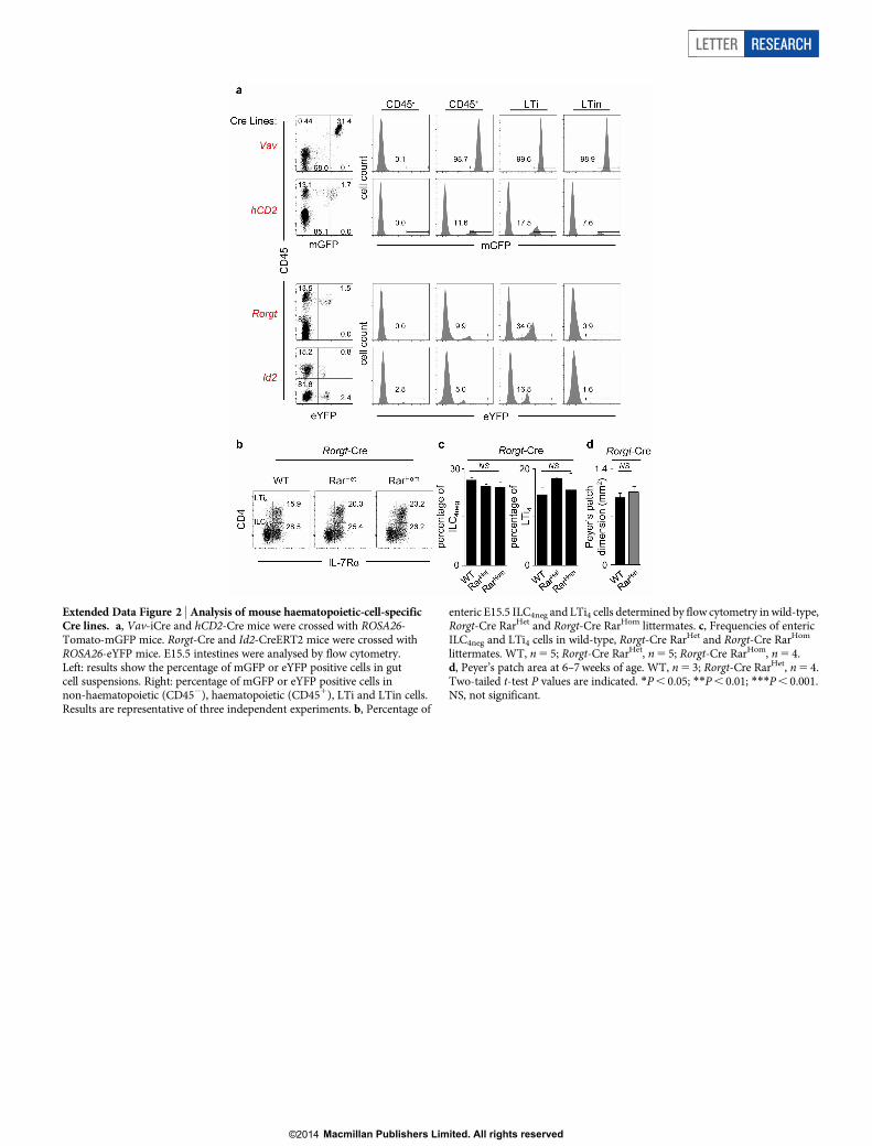
Extended Data Figure 2 | Analysis of mouse haematopoietic-cell-specificCre lines. a, Vav-iCre and hCD2-Cre mice were crossed with ROSA26-Tomato-mGFP mice. Rorgt-Cre and Id2-CreERT2 mice were crossed withROSA26-eYFP mice. E15.5 intestines were analysed by flow cytometry.Left: results show the percentage of mGFP or eYFP positive cells in gutcell suspensions. Right: percentage of mGFP or eYFP positive cells innon-haematopoietic (CD452), haematopoietic (CD451), LTi and LTin cells.Results are representative of three independent experiments. b, Percentage of
enteric E15.5 ILC4neg and LTi4 cells determined by flow cytometry in wild-type,Rorgt-Cre RarHet and Rorgt-Cre RarHom littermates. c, Frequencies of entericILC4neg and LTi4 cells in wild-type, Rorgt-Cre RarHet and Rorgt-Cre RarHom
littermates. WT, n5 5; Rorgt-Cre RarHet, n5 5; Rorgt-Cre RarHom, n5 4.d, Peyer’s patch area at 6–7weeks of age. WT, n5 3; Rorgt-Cre RarHet, n5 4.Two-tailed t-test P values are indicated. *P, 0.05; **P, 0.01; ***P, 0.001.NS, not significant.
LETTER RESEARCH
Macmillan Publishers Limited. All rights reserved©2014

Extended Data Figure 3 | Analysis of Vav-iCre/ROSA26-RARa403 mice.a, E15.5 intestines from wild-type, Vav-iCre RarHet and RarHom mice wereanalysed by flow cytometry. Representative analysis of six independentexperiments is shown. b, E15.5 regions of cervical, brachial and inguinal lymphnode from wild-type, Vav-iCre RarHet and RarHom mice were analysed by flowcytometry. Representative analysis of two independent experiments. c, LTincell percentage and LTi4/ILC4neg cell ratios are shown in E15.5 lymph nodes;n5 4. d, E15.5 fetal livers from wild-type and Vav-iCre RarHet mice wereanalysed by flow cytometry. Results show number of CD451Lin2 (n5 4) andCD452Lin2IL-7Ra1a4b71 progenitors (n5 3). e, Percentage ofCD451CD32CD11c2IL-7Ra1RORct1CD42 (RORct1CD42) andCD451CD32CD11c2IL-7Ra1RORct1CD41 (RORct1CD41) cellsdetermined by flow cytometry in RARHet and wild-type littermate controls in
E15.5 guts and lymph nodes. f, Frequencies of RORct1CD42 andRORct1CD41 cells in mice described in e; WT, n5 9; RarHet, n5 3. g, ILC4neg
cells were purified from E15.5 wild-type intestines by flow cytometry andcultured for 6 days. LTi4 cells raised in vitrowere purified by flow cytometry andquantitative RT–PCR analysis performed. Results show log2 fold increase incomparison to their cultured ILC4neg cell counterparts. Results werenormalized to Hprt1 and Gapdh. h, Left: E15.5 embryos were whole-mountstained for CD4 (red) and imaged by confocal microscopy. Cervical (Cer)lymph nodes are shown. Right: cervical lymph node dimensions are shown.WT, n5 5; RarHet, n5 7; RarHom, n5 6. Scale bar: 50mm. Two-tailed t-testP values are indicated. *P, 0.05; **P, 0.01; ***P, 0.001. NS, notsignificant. ND, not detected.
RESEARCH LETTER
Macmillan Publishers Limited. All rights reserved©2014

Extended Data Figure 4 | Gene expression patterns in ILC4neg and LTi4 cells.a, E15.5 intestines from RarHom and wild-type littermate controls werebrought to suspension and analysed by flow cytometry. Upper panel: RORctexpression. Lower panel: mean fluorescence intensity of RORct expression inILC4neg cells; n5 3. b, ILC4neg cells were purified from E15.5 RarHet andwild-type littermate control intestines and lymph nodes. Quantitative RT–PCRanalysis was performed. Results show log2 fold increase to wild type. Resultswere normalized to Hprt1 and Gapdh. Results from three independentmeasurements are shown. c, d, LTi4 cells were purified from E15.5 RarHet
(c), RarHom (d) and wild-type littermate control intestines and lymph nodes.QuantitativeRT–PCR analysiswas performed. Results show log2 fold increase towild type. Results were normalized to Hprt1 and Gapdh. Data from threeindependent measurements are shown. Two-tailed t-test P values are indicated.*P, 0.05; **P, 0.01; ***P, 0.001. ND, not detected.
LETTER RESEARCH
Macmillan Publishers Limited. All rights reserved©2014

Extended Data Figure 5 | Treatment of ILC4neg and LTi4 cells with digoxin.a, Wild-type ILC4neg cells were FACS purified, starved overnight andstimulatedwithDMSO, BMS493 (100nM), RA (100 nM) andRAplus BMS493(100 nMeach) for 16 h. Results showquantitativeRT–PCRanalysis normalizedto Gapdh; n5 3. b, E13.5 lymph node cell suspensions were cultured withvehicle (ethanol), digoxin, digoxin1RA and RA alone for 24 h. Alive/dead cellratios were determined by flow cytometry and DAPI staining; n5 4. c, ILC4neg
cells were isolated from wild-type E15.5 embryos starved overnight andstimulated for 6 h in the presence of RA (100 nM) or DIG (10mM) 1 RA(100 nM). Results show quantitative RT–PCR analysis of Rorgt and RORctdownstream targets normalized toGapdh. Representative of three independentexperiments. Error bars show s.e. Two-tailed t-test P values are indicated.*P, 0.05; **P, 0.01; ***P, 0.001. NS, not significant.
RESEARCH LETTER
Macmillan Publishers Limited. All rights reserved©2014

Extended Data Figure 6 | Analysis of SLOs from adult mice with variableRA signalling levels. a, Axillary (Axi), brachial (Bra) and inguinal (Ing) lymphnodes from adult RarHet and wild-type littermate controls were analysed.Results show lymph node cell numbers; n5 6. b, Results show Peyer’s patchnumber per intestine from adult RarHet and wild-type littermate controls;n5 6. c, Six-week-old wild-type females received VAC, VAH or VAD (n5 3)diet for 7weeks. Axillary (Axi), brachial (Bra), inguinal (Ing), intrathoracic(IntraT) lymph nodes and Peyer’s patches (PP) dimensions were analysed;n5 3. d, Percentage of CD451CD191 B cells; CD41 and CD81 T cells in
inguinal lymph nodes; n5 3. e, f, Two-week-old CD45.2 RarHet and wild-typelittermate controls were lethally irradiated and transplanted with wild-typeCD45.1 bone marrow cells. Chimaeric mice were analysed 8weeks afterreconstitution. e, Results show Peyer’s patch dimensions and folliclenumber/Peyer’s patch; n5 6. f, Results show number of cells in axillary (Axi),brachial (Bra) inguinal (Ing) and intrathoracic (IntraT) lymph nodes; n5 6.Scale bar: 1mm. Error bars show s.e. Two-tailed t-test P values are indicated.*P, 0.05; **P, 0.01; ***P, 0.001. NS, not significant.
LETTER RESEARCH
Macmillan Publishers Limited. All rights reserved©2014

Extended Data Figure 7 | Analysis of WTRWT and WTRRarHet bonemarrow chimaeras. Two-week-old CD45.2 RarHet and wild-type littermatecontrols were lethally irradiated and transplanted with wild-type CD45.1 bonemarrow cells. Chimaeric mice were analysed 8weeks after reconstitution.a, Reconstitution of donor CD45.1 cells in WTRWT and WTRRarHet
chimaeras in the spleen (n5 4). b, Reconstitution of donor CD45.1 CD4 andCD8T cells inWTRWTandWTRRarHet chimaeras in the spleen.WTRWT,n5 4; WTRRarHet, n5 11. c, Reconstitution of donor CD45.1CD11c1MHCII1 and CD11b1Gr11 myeloid cells in WTRWT andWTRRarHet chimaeras in the spleen; n5 3.d, Reconstitution of donorCD45.1CD11c1MHCII1 and CD11b1Gr11 cells in WTRWT and WTRRarHet
chimaeras in intrathoracic lymph nodes; n5 3. e, Dendritic cells (DCs) werepurified from WTRWT and WTRRarHet chimaeras. Dendritic cells wereloaded with OVA peptide (1025mM) and co-cultured for 3 dayswith CFSE-labelled monoclonal OT1 CD8 T cells. OT1 CD8 T-cellproliferation was analysed by CFSE dilution. Proliferation index is shown.WT,n5 4; RarHet, n5 6. f, Dendritic cells were purified from WTRWT andWTRRarHet chimaeras. Dendritic cells were loaded with OVA peptide(1025mM) and co-cultured for 3 days with OT1 CD8 T cells. Percentage ofIFN-c-producing OT1 CD8 T cells is shown; n5 4. Error bars show s.e.Two-tailed t-test P values are indicated. *P, 0.05; **P, 0.01; ***P, 0.001.NS, not significant.
RESEARCH LETTER
Macmillan Publishers Limited. All rights reserved©2014

Extended Data Figure 8 | Infection ofWTRRarHet orWTRWT chimaeraswith murid herpesvirus-4. a, Intrathoracic lymph node cellularity inWTRWT andWTRRarHet chimaeras at different days post-infection (d.p.i.).WT, n5 5; RarHet, n5 3. b, Percentage of CD81ORF611 (left) and ORF75c1
(right) T cells in intrathoracic lymph nodes at different days post-infection(d.p.i.). WT, n5 5; RarHet, n5 3. c, Percentage of donor CD45.1CD81ORF611 (left) and CD81ORF75c1 (right) T cells in WTRWT andWTRRarHet chimaeras after infection with murid herpesvirus-4. WT, n5 6;RarHet, n5 12. Error bars show s.e. Two-tailed t-test P values are indicated.*P, 0.05; **P, 0.01; ***P, 0.001. NS, not significant.
LETTER RESEARCH
Macmillan Publishers Limited. All rights reserved©2014

Extended Data Figure 9 | Impact of maternal retinoids in ILC3s. Maternaldietary intake of vitamin A is catabolized into bioactive retinoic acid (RA).RA signals control type 3 innate lymphoid cells (ILC3) in the embryo. FetalILC3s include ILC4neg and LTi4 cells. LTi4 cells are Id2
1RORct1, whereas
enteric ILC4neg cells contain a minor subset of Id21RORct2 cells (pre-ILCs).RA signalling operates in a cell-autonomous fashion, via direct regulation ofRorgt, programming innate pro-inflammatory cytokines and chemokines anddifferentiation of LTi4 cells.
RESEARCH LETTER
Macmillan Publishers Limited. All rights reserved©2014

Extended Data Table 1 | Computational analysis of putative RARE sites in the Rorc locus
Computational analysis was performed using TESS (Transcription Element Search System) (http://www.cbil.upenn.edu/tess). TSS (Rorg), ACGGGCCAGGTGCTCCCTCC; TSS (Rorgt),AGAAACACTGGGGGAGAGCTTTG.
LETTER RESEARCH
Macmillan Publishers Limited. All rights reserved©2014
Copyright © 2022 FDOKUMEN