Left inferior parietal dominance in gesture imitation: an fMRI study
13
Neuropsychologia 43 (2005) 1086–1098 Left inferior parietal dominance in gesture imitation: an fMRI study Mark M ¨ uhlau a, ∗ , Joachim Hermsd ¨ orfer c , Georg Goldenberg c , Afra M. Wohlschl¨ ager a, b, c , Florian Castrop a , Robert Stahl a , Michael R ¨ ottinger d , Peter Erhard a , Bernhard Haslinger a , Andres O. Ceballos-Baumann a , Bastian Conrad a , Henning Boecker b a Neurologische Klinik Rechts der Isar, Technische Universit¨ at M ¨ unchen, Munich, Germany b Nuklearmedizinische Klinik Rechts der Isar, Technische Universit¨ at M ¨ unchen, Munich, Germany c Entwicklungsgruppe Klinische Neuropsychologie (EKN), St¨ adtisches Krankenhaus M¨ unchen-Bogenhausen, Munich, Germany d Institut f ¨ ur R ¨ ontgendiagnostik, Technische Universit¨ at M ¨ unchen, Munich, Germany Received 18 January 2004; received in revised form 17 September 2004; accepted 1 October 2004 Abstract The inability to imitate gestures is an essential feature of apraxia. However, discrepancies exist between clinical studies in apraxic patients and neuroimaging findings on imitation. We therefore aimed to investigate: (1) which areas are recruited during imitation under conditions similar to clinical tests for apraxic deficits; (2) whether there are common lateralized areas subserving imitation irrespective of the acting limb side; and also (3) whether there are differences between hand and finger gestures. We used fMRI in 12 healthy, right handed subjects to investigate the imitation of four types of variable gestures that were presented by video clips (16 different finger and 16 different hand gestures with either the right or the left arm). The respective control conditions consisted of stereotyped gestures (only two gestures presented in pseudorandom order). Subtraction analysis of each type of gesture imitation (variable > stereotyped) revealed a bilateral activation pattern including the inferior parietal cortex Brodmann Area (BA 40), the superior parietal cortex, the inferior frontal cortex (opercular region), the prefrontal motor cortex, the lateral occipito-temporal junction, and the cerebellum. These results were supported by statistical conjunction of all four subtraction analyses and by the common analysis of all four types of gesture imitation. The direct comparison of the right and left hemispheric activation revealed a lateralization to the left only of the inferior parietal cortex. Comparisons between different types of gesture imitation yielded no significant results. In conclusion, gesture imitation recruits bilateral fronto-parietal regions, with significant lateralization of only one area, namely the left inferior parietal cortex. These in vivo data indicate left inferior parietal dominance for gesture imitation in right handers, confirming lesion-based theories of apraxia. © 2004 Elsevier Ltd. All rights reserved. Keywords: Apraxia; fMRI; Gestures; Imitation; Parietal cortex 1. Introduction Imitation is a fundamental principle of behavioral learn- ing (Byrne & Russon, 1998; Kymissis & Poulson, 1990) and an important component of non-verbal communication. The inability to imitate is an essential feature of apraxia, a disor- der of skilled movements. Apraxia manifests in tasks like ∗ Corresponding author. Present address: Department of Neurology, Tech- nical University of Munich, M¨ ohlstrasse 28, D-81675 Munich, Germany. Tel.: +49 89 4140 4601; fax: +49 89 4140 4919. E-mail address: [email protected] (M. M ¨ uhlau). performing verbally instructed gestures, pantomiming the use of an object, and imitating actions demonstrated by the examiner (De Renzi, 1990; Heilman & Rothi, 1993; Rothi, Raymer, & Heilman, 1997; Roy & Hall, 1992). Respective errors occur either during known manual actions, such as con- ventional gestures, or meaningless and “novel” gestures (De Renzi, Motti, & Nichelli, 1980; Goldenberg, 1996; Kimura & Archibald, 1974; Roy, Square-Storer, Hogg, & Adams, 1991). It is generally acknowledged that apraxic deficits oc- cur after left hemispheric damage, whereas apraxia resulting from lesions of the right hemisphere remains a controver- sial issue (Alexander, Baker, Naeser, Kaplan, & Palumbo, 0028-3932/$ – see front matter © 2004 Elsevier Ltd. All rights reserved. doi:10.1016/j.neuropsychologia.2004.10.004
-
Upload
independent -
Category
Documents
-
view
1 -
download
0
Transcript of Left inferior parietal dominance in gesture imitation: an fMRI study
Neuropsychologia 43 (2005) 1086–1098
Left inferior parietal dominance in gesture imitation: an fMRI study
Mark Muhlaua, ∗, Joachim Hermsdorferc, Georg Goldenbergc, Afra M. Wohlschlagera, b, c,Florian Castropa, Robert Stahla, Michael Rottingerd, Peter Erharda, Bernhard Haslingera,
Andres O. Ceballos-Baumanna, Bastian Conrada, Henning Boeckerb
a Neurologische Klinik Rechts der Isar, Technische Universit¨at Munchen, Munich, Germanyb Nuklearmedizinische Klinik Rechts der Isar, Technische Universit¨at Munchen, Munich, Germany
c Entwicklungsgruppe Klinische Neuropsychologie (EKN), St¨adtisches Krankenhaus M¨unchen-Bogenhausen, Munich, Germanyd Institut fur Rontgendiagnostik, Technische Universit¨at Munchen, Munich, Germany
Received 18 January 2004; received in revised form 17 September 2004; accepted 1 October 2004
Abstract
The inability to imitate gestures is an essential feature of apraxia. However, discrepancies exist between clinical studies in apraxic patientsa conditionss the actingl ed subjectst rent handg s presentedi tion patterni ion), thep junction ofa ht and lefth of gesturei alio itation inr©
K
1
iaid
nT
they the
econ-
s (
s,oc-
ultingver-bo,
0d
nd neuroimaging findings on imitation. We therefore aimed to investigate: (1) which areas are recruited during imitation underimilar to clinical tests for apraxic deficits; (2) whether there are common lateralized areas subserving imitation irrespective ofimb side; and also (3) whether there are differences between hand and finger gestures. We used fMRI in 12 healthy, right hando investigate the imitation of four types of variable gestures that were presented by video clips (16 different finger and 16 diffeestures with either the right or the left arm). The respective control conditions consisted of stereotyped gestures (only two gesture
n pseudorandom order). Subtraction analysis of each type of gesture imitation (variable > stereotyped) revealed a bilateral activancluding the inferior parietal cortex Brodmann Area (BA 40), the superior parietal cortex, the inferior frontal cortex (opercular regrefrontal motor cortex, the lateral occipito-temporal junction, and the cerebellum. These results were supported by statistical conll four subtraction analyses and by the common analysis of all four types of gesture imitation. The direct comparison of the rigemispheric activation revealed a lateralization to the left only of the inferior parietal cortex. Comparisons between different types
mitation yielded no significant results. In conclusion, gesture imitation recruits bilateral fronto-parietal regions, with significant laterzationf only one area, namely the left inferior parietal cortex. These in vivo data indicate left inferior parietal dominance for gesture imight handers, confirming lesion-based theories of apraxia.
2004 Elsevier Ltd. All rights reserved.
eywords:Apraxia; fMRI; Gestures; Imitation; Parietal cortex
. Introduction
Imitation is a fundamental principle of behavioral learn-ng (Byrne & Russon, 1998; Kymissis & Poulson, 1990) andn important component of non-verbal communication. The
nability to imitate is an essential feature of apraxia, a disor-er of skilled movements. Apraxia manifests in tasks like
∗ Corresponding author. Present address: Department of Neurology, Tech-ical University of Munich, Mohlstrasse 28, D-81675 Munich, Germany.el.: +49 89 4140 4601; fax: +49 89 4140 4919.E-mail address:[email protected] (M. Muhlau).
performing verbally instructed gestures, pantomiminguse of an object, and imitating actions demonstrated bexaminer (De Renzi, 1990; Heilman & Rothi, 1993; Rothi,Raymer, & Heilman, 1997; Roy & Hall, 1992). Respectiverrors occur either during known manual actions, such asventional gestures, or meaningless and “novel” gestureDeRenzi, Motti, & Nichelli, 1980; Goldenberg, 1996; Kimura& Archibald, 1974; Roy, Square-Storer, Hogg, & Adam1991). It is generally acknowledged that apraxic deficitscur after left hemispheric damage, whereas apraxia resfrom lesions of the right hemisphere remains a controsial issue (Alexander, Baker, Naeser, Kaplan, & Palum
028-3932/$ – see front matter © 2004 Elsevier Ltd. All rights reserved.oi:10.1016/j.neuropsychologia.2004.10.004

M. Muhlau et al. / Neuropsychologia 43 (2005) 1086–1098 1087
1992; De Renzi et al., 1980; Goldenberg, 1996,1999; Haaland& Flaherty, 1984; Heath, Roy, Westwood, & Black, 2001;Kertesz & Ferro, 1984; Kimura et al., 1974; Lehmkuhl,Poeck, & Willmes, 1983; Roy et al., 1991). A possible rea-son for this debate is that the specific involvement of cerebralareas critically depends on the detailed characteristics of thegesture under examination.
Indeed, different results have been found for hand gesturesconsisting of different hand positions in relation to the head,and finger gestures defined only by different configurationsof the fingers. While left parietal lesions impaired both, handand finger gestures, right parietal damage impaired primar-ily finger gesture tasks (Goldenberg, 1995; Hermsdorfer etal., 1996). Furthermore, it has been suggested that apraxia onimitation is primarily a deficit of perceptional analysis, sinceerrors also occur when subjects are to match photographs ofgestures (Goldenberg, 1999). This is also supported by PETimaging in healthy subjects, where matching photographsof hand gestures activated the left parietal cortex, whereasmatching photographs of finger gestures activated both pari-etal cortices (Hermsdorfer et al., 2001).
In recent years, in particular, after the discovery of “mir-ror neurons” in the monkey brain (Gallese, Fadiga, Fogassi,& Rizzolatti, 1996; Rizzolatti, Fadiga, Gallese, & Fogassi,1996a), imitation has been investigated intensively in neu-r ther be-c ctiona xper-i rob-a riso e,2 -t weree fur-t ationo pportt di-r mo-t s ona olve-m
,I ni thei ie r,F na om-i bothp thatv holen arer ves-t pro-p 3
Finally, the effect of the acting limb side on imitation isnot completely resolved. This can be attributed to two facts.Firstly, apraxics are mostly paralyzed on the limb side con-tralateral to the damaged hemisphere so that only the ipsile-sional hand can be investigated. Secondly, while several neu-roimaging studies were concerned with active imitation, sub-jects had to imitate with their right hand exclusively (Brasset al., 2001; Decety, Chaminade, Grezes, & Meltzoff, 2002;Iacoboni et al., 1999; Koski et al., 2002; Krams et al., 1998;Tanaka et al., 2001, 2002).
Hence, the purpose of this study was to investigate thecerebral activation that results from more complex imitationsuch that under conditions similar to the clinical testing forapraxic deficits, to analyze the influence of the acting limbside, and to search for differences between the imitation ofhand and finger gestures.
2. Materials and methods
2.1. Subjects
Twelve volunteer subjects (mean age 50, range 41–60years, six females) participated in the study. All subjectswere right-handed, drug-free, and had no history of neu-r meda rit-t inki2 mit-t y ofM
2
ely,t ighto thati s ther
uresws eutralp ent . Sub-j ate itt
ofa l in-v ges-t edw uresw
ner,t tionsr ablya r
oimaging studies. “Mirror neurons” were localized atostral part of the inferior frontal cortex (area F5) andome active both when the monkey performs a given and when it observes a similar action performed by the e
menter. The human homologue of monkey area F5 is pbly Brodmann Area (BA) 44 within the pars operculaf the inferior frontal gyrus (Rizzolatti, Fogassi, & Galles002). Accordingly,Iacoboni et al. (1999)found this area ac
ivated during finger movements, regardless of how theyvoked, and, most importantly, their activation increasedher when the same movement was elicited by the observf an identical movement. These results gave further su
o the “direct matching hypothesis” which postulates theect matching of the observed action onto an internalor representation of that action. However, lesion studiepraxic patients have failed so far to demonstrate an invent of the frontal operculum.With few exceptions (Tanaka & Inui, 2002; Tanaka, Inui
waki, Konishi, & Nakai, 2001), neuroimaging studies omitation investigated simple movements such as liftingndex finger (Brass, Zysset, & von Cramon, 2001; Iacobont al., 1999; Koski et al., 2002; Krams, Rushworth, Deiberackowiak, & Passingham, 1998). In contrast to studies opraxic patients that mostly demonstrated left parietal d
nance, these neuroimaging studies found activation ofarietal cortices at different locations. It seems possibleery simple movements do not lead to activation of the wetwork subserving imitation but also that different areasecruited for copying simple movements. Therefore, inigations of more complex imitative behavior has beenosed (Koski, Iacoboni, Dubeau, Woods, & Mazziotta, 200).
ological or psychiatric disorders. They had been inforbout the purpose of the study before giving their w
en consent, in accordance with the Declaration of Hels000. The study had been approved by the Ethics Com
ee of the Faculty of Medicine at the Technical Universitunich.
.2. General remarks, stimulus material and task
Four types of gesture imitation were investigated; namhe imitation of finger and hand gestures with either the rr the left arm. Exclusively specular imitation was used,
s when the actor moves the left hand, the imitator moveight hand.
Video clips of a person performing meaningless gestere presented one after the other (seeFig. 1). Each cliphowed a meaningless gesture and the return to the nosition (seeFig. 1, upper panels) within two seconds. Th
he freeze image was displayed for another two secondsects were instructed to observe the gesture and to imithoroughly as soon as the image froze.
The clips were taken from colored video recordingsctual movements with the right arm. Stimulus materiaolved 16 different finger gestures and 16 different handures. All clips were mirrored to obtain gestures performith the left arm. During one experiment, none of the gestas displayed more than three times for each side.To enable imitation of hand gestures in the MRI scan
he hand was moved into different positions and orientaelative to the opposite forearm, which rested comfortbove the abdomen (seeFig. 1, left panels). During finge

1088 M. Muhlau et al. / Neuropsychologia 43 (2005) 1086–1098
Fig. 1. Hand and finger gestures. Frames were taken from the video clipsthat were presented during the experiment. For hand (left panels) and finger(right panels) gestures respectively, the neutral position (upper panels), bothstereotyped gestures (middle panels), and three examples of variable gesture(lower panels) are shown.
gestures the hand was held in the centre of the abdomen (seeFig. 1, right panels).
In the experimental condition (variable gesture imitation),subjects imitated gestures that were taken from a set of 16gestures one after the other.
Two control conditions were used. In the first control con-dition (stereotyped gesture imitation), subjects imitated ges-tures one after the other like in the experimental condition;but now, the set of the presented gestures consisted of only
two gestures (seeFig. 1, middle panels). These gestures wererepetitively presented in pseudorandom order, not allowingfor the same gesture more than twice in sequence. The sec-ond control condition (motor rest), consisted of a clip thatshowed the trunk with breathing motions and with the armheld in the resting position (seeFig. 1, upper panels). Underthis condition, subjects were instructed to attend to motionsand not to move.
2.3. Procedure
Each subject underwent four sessions (i.e. imitation of fin-ger and hand gestures with either the right or the left arm).The order of sessions was pseudorandomised over subjects.Each condition (variable and stereotyped gesture imitationas well as motor rest) was presented in form of four blocks(10 different gestures per block, 40 s block duration). A shortinstruction (10 s) preluded every block. Blocks were orderedpseudorandomly not allowing for the same condition in sub-sequent blocks (seeFig. 2).
Before the measurement started, each subject was trainedwith a shorter set of clips outside the scanner. Except forclips that were presented in the control condition of stereo-typed gestures, these clips never reappeared during the MRIscans. During gesture imitation inside the scanner, subjectsv ncew ideoc
2
nerw , thes weeksl aterse er ani t wasi scorefi ed ort r tipst turesw f theh arm.T rat-i s ofd .6%)d s ap-p eeni eeni
2
can-n headc ted
s
iewed the clips by projection onto a mirror. Performaas observed by an investigator and monitored with a vamera.
.4. Performance control
As image definition of the monitoring inside the scanas not good enough to allow an exact error analysisame experiment was repeated outside the scanner 6ater and recorded by a digital video camera. Three rstimated all responses as correct or incorrect. Whenev
mitation was scored as incorrect by at least two raters, interpreted as an error. Raters had been instructed tonger gestures as correct whenever fingers to be extendo be flexed were correct or whenever the correct fingeouched each other, respectively. Imitation of hand gesere only estimated as correct when the end position oand moved was correct in relation to the opposite forehe scores of the three raters coincided in 96.4% of the
ngs (94.4% no error, 2.0% error), in the remaining caseisagreement either two scorers (2.0%) or one scorer (1enoted an error. The non-parametric Wilcoxon test walied to search for differences of error frequencies betw
mitations of hand and finger gestures as well as betwmitations performed with the right and the left arm.
.5. Magnetic resonance imaging
Imaging was performed using a 1.5-T Siemens ser (Magnetom Symphony) with a standard birdcageoil. First, a 3-D structural high-resolution T1-weigh

M. Muhlau et al. / Neuropsychologia 43 (2005) 1086–1098 1089
Fig. 2. Schematic representation of all conditions’ organization.
MRI using an magnetization prepared rapid gradientecho (MPRAGE) sequence was acquired on each subject(sagittal plane; TR, 11.1 ms; TE, 4.3 ms; field of view,224 mm× 256 mm; flip angle, 15◦; number of slice, 160;slice thickness, 1 mm).
Functional imaging was performed using an ultra fast echoplanar gradient echo imaging sequence sensitive to bloodoxygenation level-dependent (BOLD) contrast (TR, 3.100 s;TE, 50 ms; FOV, 192; flip angle, 90◦) with 34 slices ori-ented along the anterior and posterior commissure (obliqueaxial; thickness, 4 mm; gap, 0.4 mm; in-plane resolution,3 mm× 3 mm).
Prior to each run, four images were acquired and discardedto allow for longitudinal magnetization to reach equilibrium.
2.6. Data analysis
SPM 99 software (Wellcome Department of CognitiveNeurology, London, UK), based on the general linear model(Friston, 1996) was applied for data analysis. Calculationswere performed on dual-Pentium 400 PCs running LINUX.
For each subject, images were realigned to the first im-age of the first session to account for head-motion in time.The mean image of all realigned images was used for thedetermination of the individual parameters for the normal-i tem.A aus-s alt icalv
latedo n-m emenp o re-m tiono
Significance was assessed using the delayed box-car ref-erence that was convolved with the haemodynamic responsefunction. In a first level (fixed effects) analysis, we obtainedstatistical parametric maps (SPM) and corresponding con-trast images for each subject from the following categoricalcomparisons:
• Variable > stereotyped gesture imitation (separately foreach type of imitation).
• Stereotyped gesture imitation > motor rest (separately foreach type of imitation).
• Variable hand > finger gestures (separately for each side).• Variable finger > hand gestures (separately for each side).• Variable gestures, right > left arm (separately for finger
and hand gestures).• Variable gestures, left > right arm (separately for finger
and hand gestures).
To allow more generalized inferences, those contrast im-ages were entered into second level (random effects) analysesusing one samplet-tests.
In order to identify regions that are specific to the imitationof hand gestures (compared to the imitation of finger ges-tures), categorical analyses (variable hand > finger gestures)of the right and the left side were statistically conjoined (seeTable 1). The conjunction of the converse categorical analyses
TC
N sts
HFLRV
N
zation into a standard space, the MNI brain-based sysfter normalization, images were smoothed with a Gian filter of 10 mm× 10 mm× 10 mm to increase signo noise ratio and to account for intersubject anatomariability.
For each subject, movement parameters were calcuver time with an IDL routine by subtracting all six realigent parameters of subsequent images. These movarameters were added to the general linear model tove any component that was correlated with this funcf movement estimates.
t
able 1onjunction analyses at the second level
ame of conjunction Conjoined first level contra
and > finger gestures LH > LF∩ RH > RFinger > hand gestures LF > LH∩ RF > RHeft > right LF > RF∩ LH > RHight > left RF > LF∩ RH > LHariable > stereotyped LF: V > S∩ RF: V > S∩ LH:
V > S ∩ RH: V > S
ote: F, finger; H, hand; L, left; R, right; S, stereotyped; V, variable.
1090 M. Muhlau et al. / Neuropsychologia 43 (2005) 1086–1098
was applied to reveal areas that are specific to the imitation offinger gestures (compared to the imitation of hand gestures).Likewise, for the identification of regions that are specificto the imitation performed with the right arm (compared tothe imitation performed with the left arm), categorical anal-yses (right > left) of variable finger and hand gestures werestatistically conjoined (seeTable 1). The conjunction of theconverse categorical analyses was applied to detect areas thatare specific to the imitation performed with the left arm (com-pared to the imitation performed with the right arm). Finally,subtraction analyses of all types of gesture imitation (vari-able > stereotyped) were statistically conjoined in order toreveal areas that are related to all types of gesture imitation(seeTable 1). All conjunctions were performed at the sec-ond level by entering different types of imitation (variable> stereotyped) as covariates in a multiple regression model(without a constant) (Friston, Holmes, Price, Buchel, &Worsley, 1999).
To detect left-right differences in hemispheric activation,we modeled all types of gesture imitation as a single maineffect at the first level. Then, contrast images were left-rightflipped. To compare the original and flipped contrast imagesthe pairedt-test was applied. This procedure resulted in mapsthat, on the left side, indicate areas which are more active onthe left side compared to the corresponding areas of the rights morea areaso thec
r-r tion.O con-t
oldw eryr
con-v Ta-lic
3
3
RI-e cord-i s re-v pedg ges-t (lefts ec ntlyd
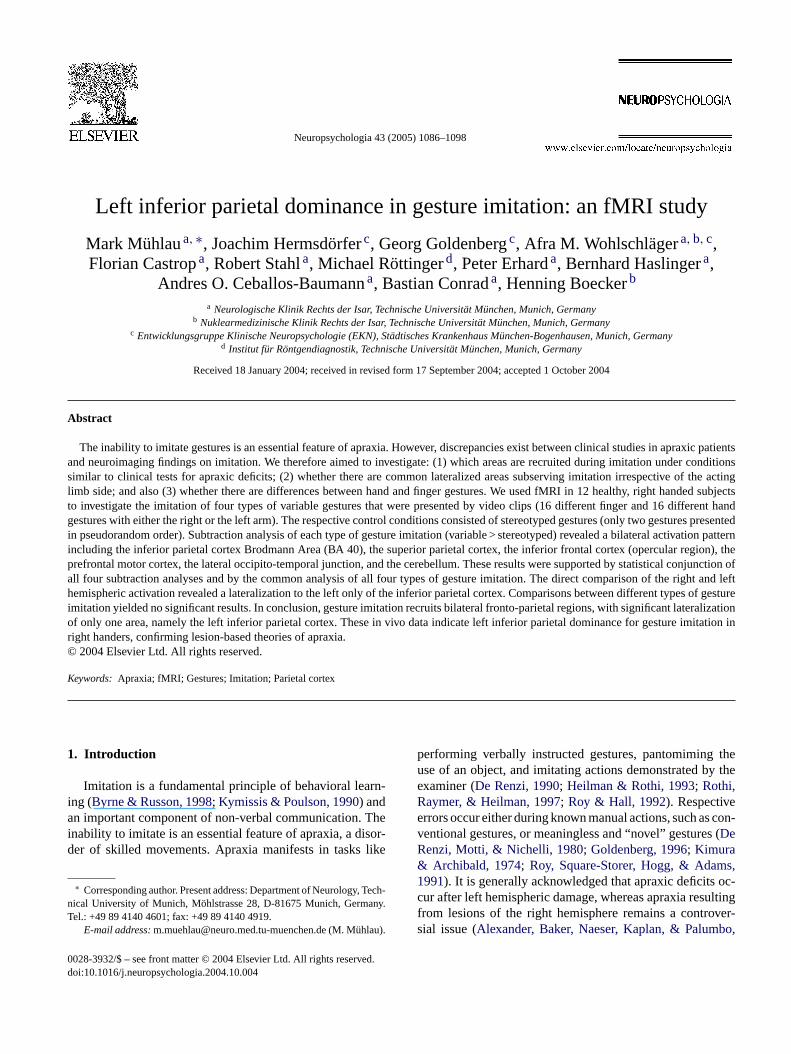
Fig. 3. Performance during imitation of variable gestures. Box plots of thesubjects’ performance during imitation of variable gestures (bold horizontalline, median; rectangles, quartile range; horizontal lines, 1.5× quartile range;◦, *, extreme values). The frequencies of errors are displayed. Individualvalues represent means from four blocks performed under each condition.Note that the frequencies of errors are similar for the right and left arm whilethe imitation of finger gestures resulted in significantly more errors than theimitation of hand gestures.
3.2. MRI results
3.2.1. Stereotyped gesture imitationCompared to motor rest, stereotyped imitation of hand
and finger gestures performed with either the right or leftarm induced activation (second level,P< 0.05, FWE) in thecontralateral primary motor and sensory cortex as well as inthe ipsilateral cerebellum.
3.2.2. Variable gesture imitationWithin each type of gesture imitation (finger and hand
gestures with either the right or the left arm), variable ges-ture imitation induced nearly no significant activation whencompared to stereotyped gesture imitation at a significancethreshold ofP< 0.05 corrected (FWE). After changing thethreshold toq< 0.05 corrected (FDR), all types of imita-tion showed bilateral activation of the inferior parietal cor-tex (BA 40), the superior parietal lobe (precuneus, BA 7),the inferior frontal cortex (opercular region including BA 44on the right and BA 9/44 on the left hemisphere), the pre-frontal motor cortex (BA 6), the lateral occipito-temporaljunction (BA 37/19 including MT/V5), and the cerebellum(lobule VI of the vermis and both hemispheres without ac-tivation within the nuclei) (Dimitrova et al., 2002; Schmah-mann et al., 1999). This activation pattern was also revealedb lltT ofg dau
erica riorp
be-t for
ide. Vice versa on the right side, areas appear that arective on the right side compared to the correspondingf the left side. A region of interest (ROI) was defined byonjunction of all four types of gesture imitation.
A statistical threshold ofP< 0.05 (family wise error coection, FWE) was considered to show significant activanly clusters of activation with an extent exceeding 10
iguous voxels were considered relevant.If there were no clusters surviving this level, the thresh
as changed toq< 0.05, corrected with the false discovate (FDR) (Genovese, Lazar, & Nichols, 2002).
Calculated coordinates of activation peaks wereerted from the MNI brain-based system into theairach brain system (Talairach & Tournoux, 1988) us-ng a non-linear transformation method (http://www.mrc-bu.cam.ac.uk/Imaging/mnispace.html).
. Results
.1. Task performance
Observation of task performance during the Mxperiment showed that subjects performed the task ac
ng to the given instructions. Subsequent error analysiealed not a single error during the imitation of stereotyestures. During the imitation of variable gestures, finger
ures were significantly more difficult than hand gestureside,P= 0.014; right side,P= 0.005; seeFig. 3) whereas thomparison of the acting limb sides did not yield significaifferent error rates (P> 0.1; seeFig. 3).
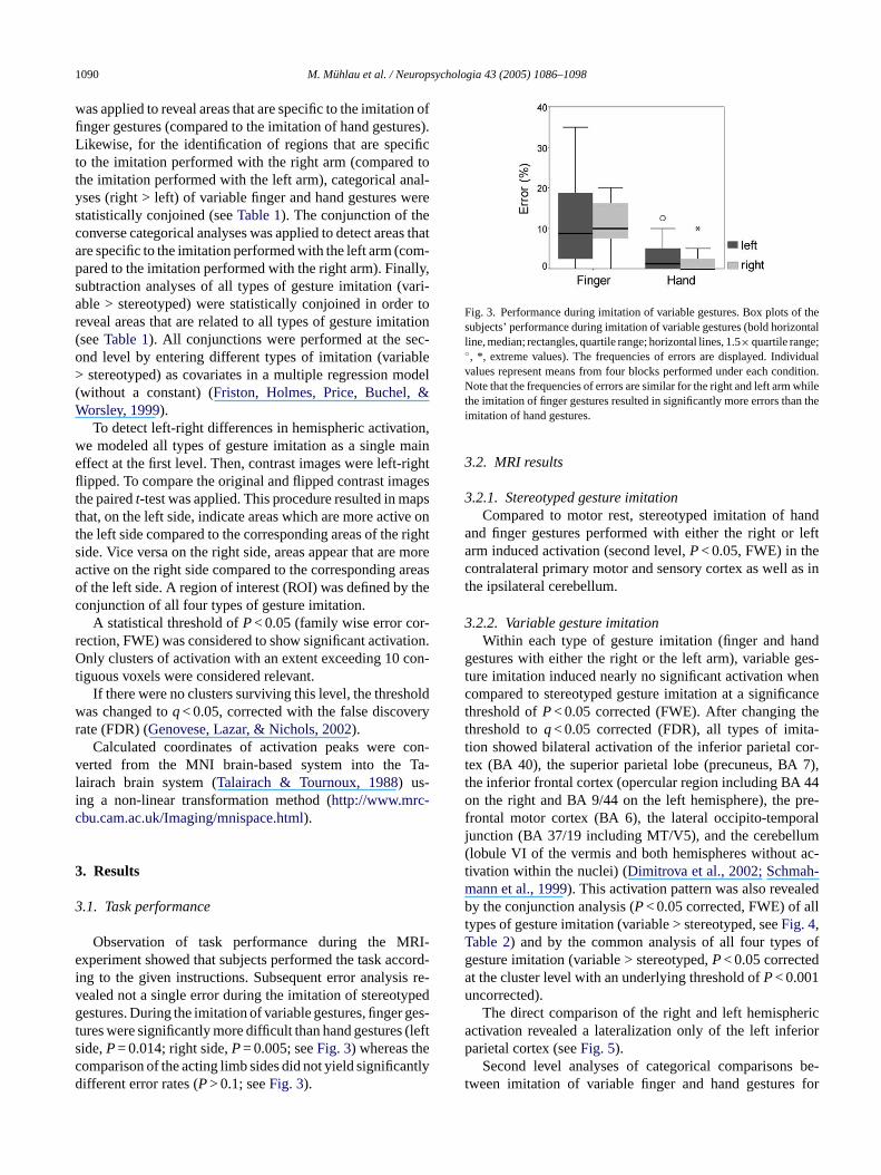
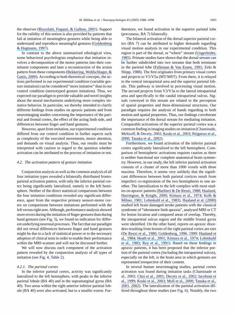
y the conjunction analysis (P< 0.05 corrected, FWE) of aypes of gesture imitation (variable > stereotyped, seeFig. 4,able 2) and by the common analysis of all four typesesture imitation (variable > stereotyped,P< 0.05 correctet the cluster level with an underlying threshold ofP< 0.001ncorrected).
The direct comparison of the right and left hemisphctivation revealed a lateralization only of the left infearietal cortex (seeFig. 5).
Second level analyses of categorical comparisonsween imitation of variable finger and hand gestures
M. Muhlau et al. / Neuropsychologia 43 (2005) 1086–1098 1091
Fig. 4. Conjunction of all types of gesture imitation (P< 0.05, corrected with FWE). Clusters of activation are overlaid onto consecutive axial slices of a T1weighted anatomical magnetic resonance image (right side of image is the right hemisphere). Positions of the slices are indicated in mm relative to the anteriorcommissure. Histograms show parameter estimates for each type of imitation (bars, standard deviation; F, finger; H, hand; L, left; R, right) at the Talairachcoordinates indicated above. The numbers on the upper left corners of the histograms correspond to the first column inTable 2.
both the right and the left side revealed no significantlydifferent activation, even when changing the statisticalthreshold toq< 0.05 (FDR) or P< 0.001 (uncorrected).Significant activation was neither shown on conjunctionanalyses of these comparisons. Likewise, analyses to de-tect areas that are specific to the imitating limb sideyielded no activation difference apart from the contralat-eral primary motor or sensory cortex and the ipsilateralcerebellum.
4. Discussion
4.1. The paradigm
In this study, we have investigated active imitation of vari-able meaningless gestures, thereby adopting tests used forclinical evaluation of apraxic deficits (seeFig. 1) to neu-roimaging. To control visual input, elementary motor output,and basic aspects of imitation, we compared our experimen-
1092 M. Muhlau et al. / Neuropsychologia 43 (2005) 1086–1098
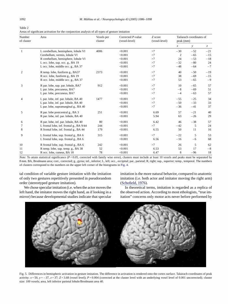
Table 2Areas of significant activation for the conjunction analysis of all types of gesture imitation
Numberof cluster
Area Voxels percluster
CorrectedP-value(voxel-level)
Z score(voxel-level)
Tailarach coordinates ofpeak (mm)
x y z
1 L cerebellum, hemisphere, lobule VI 4006 <0.001 >7 −30 −52 −21Cerebellum, vermis, lobule VI <0.001 >7 2 −65 −15R cerebellum, hemisphere, lobule VI <0.001 >7 24 −53 −18L occ. lobe, sup. occ. g., BA 19 <0.001 >7 −32 −80 24L occ. lobe, middle occ. g., BA 37 <0.001 >7 −48 −64 −7
2 R temp. lobe, fusiform g., BA37 2373 <0.001 >7 40 −50 −19R occ. lobe, fusiform g., BA 19 <0.001 >7 38 −69 −15R occ. lobe, middle occ. g., BA 37 <0.001 >7 53 −65 −9
3 R par. lobe, sup. par. lobule, BA7 912 <0.001 >7 10 −65 57L par. lobe, precuneus, BA7 <0.001 >7 −8 −69 51L par. lobe, precuneus, BA7 <0.001 >7 −4 −63 57
4 L par. lobe, inf. par. lobule, BA 40 1477 <0.001 >7 −55 −25 34L par. lobe, inf. par. lobule, BA 40 <0.001 >7 −50 −33 33L par. lobe, supramarginal g., BA 40 <0.001 >7 −36 −41 37
5 R par. lobe postcentral g., BA 3 251 <0.001 6.69 57 −21 40R par. lobe, inf. par. lobule, BA 40 <0.001 5.94 63 −26 29
6 R par. lobe, inf. par. lobule, BA 40 80 <0.001 6.42 46 −38 577 L frontal lobe, inf. frontal g., BA 9/44 244 <0.001 >7 −42 5 248 R frontal lobe, inf. frontal g., BA 44 179 <0.001 6.55 50 11 16
9 L frontal lobe, sup. frontal g., BA 6 315 <0.001 >7 −22 5 53L frontal lobe, sup. frontal g., BA 6 <0.001 6.56 −16 −6 68
10 R frontal lobe, sup. frontal g., BA 6 242 <0.001 >7 26 5 6211 R temp. lobe, sup. temp. g., BA 38 52 <0.001 6.53 53 17 −812 R occ. lobe, cuneus, BA 18 78 <0.001 6.47 8 −96 18
Note: To attain statistical significance (P< 0.05, corrected with family wise error), clusters must include at least 10 voxels and peaks must be separated by8 mm. BA, Brodmann area; corr., corrected; g., gyrus; inf., inferior; L, left; occ., occipital; par., parietal; R, right; sup., superior; temp., temporal. The numbersof clusters correspond to the numbers on the upper left corner of the histograms inFig. 4.
tal condition of variable gesture imitation with the imitationof only two gestures repetitively presented in pseudorandomorder (stereotyped gesture imitation).
We chose specular imitation (i.e. when the actor moves theleft hand, the imitator moves the right hand, as if looking in amirror) because developmental studies indicate that specular
imitation is the more natural behavior, compared to anatomicimitation (i.e. both actor and imitator moving the right arm)(Schofield, 1976).
In theoretical terms, imitation is regarded as a replica ofthe observed action. According to most ethologists, “true im-itation” concerns only motor acts never before performed by
Fig. 5. Differences in hemispheric activation in gesture imitation. The difference in activation is rendered onto the cortex surface. Talairach coordinates of peakactivity: x= 50,y=−37, z= 37;Z= 3.68 (voxel level);P= 0.004 (corrected at the cluster level with an underlying voxel level of 0.001 uncorrected); clustersize: 100 voxels; area, left inferior parietal lobule/Brodmann area 40.
M. Muhlau et al. / Neuropsychologia 43 (2005) 1086–1098 1093
the observer (Rizzolatti, Fogassi, & Gallese, 2001). Supportfor the validity of this notion is also provided by patients thatfail at imitation of meaningless gestures while being able tounderstand and reproduce meaningful gestures (Goldenberg& Hagmann, 1997).
In contrast to the above summarized ethological view,some behavioral psychologists emphasize that imitation in-volves a decomposition of the motor patterns into their con-stituent components and later a reconstruction of the actionpattern from these components (Bekkering, Wohlschlager, &Gattis, 2000). According to both theoretical concepts, the ac-tions performed in our experimental condition (variable ges-ture imitation) can be considered “more imitative” than in ourcontrol condition (stereotyped gesture imitation). Thus, weexpected our paradigm to provide valuable and novel insightsabout the neural mechanisms underlying more complex im-itative behavior. In particular, we thereby intended to clarifydifferent findings from studies on apraxic patients and fromneuroimaging studies concerning the importance of the pari-etal and frontal cortex, the effect of the acting limb side, anddifferences between finger and hand gestures.
However, apart from imitation, our experimental conditiondiffered from our control condition in further aspects suchas complexity of the executed movements, motor attention,and demands on visual analysis. Thus, our results must bei thera not.
4
of allf to-p or-t mi-s weent fer-e cor-t thel wedm ringh -e digmd turesm sarya ncew r.
tionp s ofi
4tly
l riorp (BA4 ob-u Fur-
thermore, we found activation in the superior parietal lobe(precuneus, BA 7) bilaterally.
The bilateral activation of the dorsal superior parietal cor-tex (BA 7) can be attributed to higher demands regardingvisual motion analysis in our experimental condition. Thisregion is part of the dorsal, or “where” stream (Ungerleider,1985). Primate studies have shown that the dorsal stream canbe further subdivided into two streams that both terminatein the parietal lobe (Felleman & Van Essen, 1991; Zeki &Shipp, 1988). The first originates from primary visual cortexand projects to V5/V5a (MT/MST). From there, it is relayedto the ventral intraparietal area and the superior parietal lob-ule. This pathway is involved in processing visual motion.The second projects from V3/V3a to the lateral intraparietalarea and specifically to the caudal intraparietal sulcus. Sig-nals conveyed in this stream are related to the perceptionof spatial properties and three-dimensional structures. Ourparadigm requires the analysis of hands regarding to bothmotion and spatial properties. Thus, our findings corroboratethe importance of the dorsal stream for mediating imitation.Comparable activations of the superior parietal cortex was acommon finding in imaging studies on imitation (Chaminade,Meltzoff, & Decety, 2002; Koski et al., 2003; Peigneux et al.,2000; Tanaka et al., 2002).
Furthermore, we found activation of the inferior parietalc om-p therei me-t ionc reem nifi-c fromi an-o tud-i ,HM )s sicals CTf reby,t yrusw isor-d rare( tae ina por-t us),e s arer
rtexa ta ni eta al.,2 dif-f
nterpreted with caution in regard to the question whectivation can be attributed to the process of imitation or
.2. The activation pattern of gesture imitation
Conjunction analysis as well as the common analysisour imitation types revealed a bilaterally distributed fronarietal activation pattern, with only the inferior parietal c
ex being significantly lateralized, namely to the left hephere. Neither of the direct statistical comparisons bethe four imitation conditions revealed any significant difnce, apart from the respective primary sensori-motor
ex on comparisons between imitations performed witheft versus right arm. Although, performance analysis sho
ore errors during the imitation of finger gestures than duand gestures (seeFig. 3), we found no indication for differnt underlying neuronal processes. The fact that our paraid not reveal differences between finger and hand gesight be due to a lack of statistical power or to the necesdoption of clinical tests in order to enable their performaithin the MRI-scanner and will not be discussed furtheWe will now discuss each component of the activa
attern revealed by the conjunction analysis of all typemitation (seeFig. 4, Table 2).
.2.1. The parietal cortexIn the inferior parietal cortex, activity was significan
ateralized to the left hemisphere, with peaks in the infearietal lobule (BA 40) and in the supramarginal gyrus0). Two areas within the right anterior inferior parietal lle (BA 40) were also activated, but to a smaller extent.
ortex significantly lateralized to the left hemisphere. Carison of hemispheric activations requires caution as
s neither functional nor complete anatomical brain symry. However, in our study, the left inferior parietal activatonsists of a cluster of more than 1000 voxels with thaxima. Therefore, it seems very unlikely that the sig
ant differences between both parietal cortices resultmperfect co-registration of the two hemispheres to onether. The lateralization to the left complies with most s
es on apraxic patients (Barbieri & De Renzi, 1988; Haalandarrington, & Knight, 2000; Kimura et al., 1974; Kolb &ilner, 1981; Lehmkuhl et al., 1983). Haaland et al. (2000
tudied left brain damaged stroke patients with the clasyndrome of “ideomotor limb apraxia”, analyzed MRI oror lesion location and compared areas of overlap. Thehe intraparietal sulcus region and the middle frontal gere identified. On the other hand, reports on apraxic ders resulting from lesions of the right parietal cortex areDe Renzi et al., 1980; Goldenberg, 1996, 1999; Haaland el., 1984;Heath et al., 2001; Kimura et al., 1974; Lehmkuhlt al., 1983; Roy et al., 1991). Based on these findingspraxic patients, it has been proposed that the inferior
ion of the parietal cortex (including the intraparietal sulcspecially on the left, is the brain area in which gestureepresented irrespective of their content.
In several human neuroimaging studies, parietal coctivation was found during imitation tasks (Chaminade el., 2002; Choi et al., 2001; Decety et al., 2002; Iacobol., 1999; Koski et al., 2003; Moll et al., 2000; Tanaka et001, 2002). The lateralization of the parietal activation
ered throughout these studies (seeFig. 6). Notably, the two

1094 M. Muhlau et al. / Neuropsychologia 43 (2005) 1086–1098
Fig. 6. Summary of parietal activation found in imaging studies on active imitation and pantomiming. Peak activations that were found in the studiesnamed underneath are indicated. Angular symbols refer to experiments with control conditions consisting of motor rest, round symbols refer to experi-ments with control conditions consisting of movements. Color coding: black, complex imitation; red, imitation of simple finger movements; green, pan-
tomiming; blue, imitation of and with active object manipulation.(Moll et al., 2000), (Choi et al., 2001), (Rumiati et al., 2004), (Decety et
al., 2002), (Chaminade et al., 2002), (Grezes, Armony, Rowe, & Passingham, 2003), (Krams et al., 1998), (Iacoboni et al., 1999),
(Koski et al., 2003), (Tanaka et al., 2001), (Tanaka et al., 2002), (Peigneux et al., 2000), present study.
studies (Tanaka et al., 2001, 2002) with paradigms probinggestures more similar to those used in the clinical testing ofapraxic patients found a lateralization to the left inferior pari-etal cortex in most conditions (see black angular symbols inFig. 6). The gestures were presented on photographs and hadto be performed with the right arm exclusively. The experi-mental conditions were compared to motor rest. Therefore,it remained to be resolved whether the activation in the leftparietal cortex originates from complex imitation or whethercopying simple movements alone would also result in suchan activation pattern. Moreover, the lateralization to the lefthemisphere could result from the acting limb side. Now, ourstudy indicates that the activation of the left inferior parietalcortex cannot be attributed to the acting limb side but to thehigher variability and complexity of the gestures presented.
It has to be recognized that the exact location within theparietal cortex differed remarkably throughout the availableimaging studies on active imitation (seeFig. 6). It is impos-sible to explain this variability stringently. This might be dueto the fact that methods, thresholds, as well as experimentaland control conditions differed throughout these studies.
Another two studies investigated cerebral activation dur-ing pantomiming tool-use. Here, tools were presented ver-bally (Moll et al., 2000) or visually (Choi et al., 2001). Thus,both paradigms resembled clinical tests for apraxic deficits.I wasf
ment of the left inferior parietal cortex in both pantomimingtool-use and imitation is comprehensible as both tasks sharecommon components. Both tasks include the need to accessa visual description of the movement, then to assemble thiscomplex movement, and finally to execute it. Correspond-ingly, both tasks are typically disturbed in apraxia.
Moreover, the left parietal cortex is activated by observa-tion of movements (Bonda, Petrides, Ostry, & Evans, 1996;Grafton, Mazziotta, Woods, & Phelps, 1992), motor im-agery (Grafton, Arbib, Fadiga, & Rizzolatti, 1996; Ruby& Decety, 2001), movement preparation (Deiber, Ibanez,Sadato, & Hallett, 1996; Krams et al., 1998; Stephan et al.,1995), and analysis of photographs showing the end posi-tions of meaningless gestures (Hermsdorfer et al., 2001).These functions are primarily devoted to perceptual analy-sis of body movements and postures and thus closely relatedto imitation.
Furthermore, this region has also been related to motorattention and selection (for review seeRushworth, Johansen-Berg, Gobel, & Devlin, 2003). Accordingly, it has also beenproposed that the difficulties, apraxic patients frequently ex-perience when sequencing movements (see alsoHaaland,Elsinger, Mayer, Durgerian, & Rao, 2004; Harrington & Haa-land, 1992), may partly be due to an inability to redirectmovement attention (Rushworth, Nixon, Renowden, Wade,& o-t ac-
nterestingly, in both studies a lateralization to the leftound (see green symbols� and� in Fig. 6). The involve-
Passingham, 1997). Moreover, imaging studies on mor attention and selection found the left parietal cortex
M. Muhlau et al. / Neuropsychologia 43 (2005) 1086–1098 1095
tivated. While motor attention and movement selection areinherent aspects of complex imitation, the critical questionappears whether these factors alone might explain the acti-vation found in our study. However, imaging studies on mo-tor attention and movement selection found the left parietalcortex activated when subjects covertly prepared movements(e.g.Rushworth, Krams, & Passingham, 2001) or switchedintended movements (e.g.Rushworth, Paus, & Sipila, 2001;Schluter, Krams, Rushworth, & Passingham, 2001). Theseparadigms cannot easily be compared with our task that re-quired the spontaneous imitation of observed movementswith a very predictable procedure. Considering further theample evidence for leftward lateralized parietal activation ingesture imitation from clinical studies and from neuroimag-ing studies, we rate the possibility that motor attention andmovement selection alone caused the left inferior parietal ac-tivation in our study unlikely. However, we do not want to ruleout that motor attention and movement selection contributedto the activation. In summary, we attribute the left inferiorparietal dominance demonstrated in our study primarily tothe process of imitation.
4.2.2. The frontal cortexIn the frontal cortex, we found bilateral activation, namely
occurring in the dorsal premotor cortex (BA 6) and the op-e ont
dor-s om-p idedm se-q oveo ,De ss-i s,F as tion,t s re-q the-l sori-m trols xper-i tput.W l re-q ghera didn tiva-t ndi
r re-g efth dur-i ectl osef not
aware of such cases, even in patients with apraxia followingfrontal lesions (Haaland et al., 2000). However, a recent studyon imitation conducted with repetitive transcranial magneticstimulation showed a temporarily degraded imitation capac-ity after the stimulation of either the right or the left Broca’sarea (Heiser, Iacoboni, Maeda, Marcus, & Mazziotta, 2003).
In contrast to lesion studies on apraxia, several imag-ing studies focusing on active imitation have shown recruit-ment of the frontal opercular region (Grezes, Armony, Rowe,& Passingham, 2003; Iacoboni et al., 1999; Koski et al., 2002;Krams et al., 1998; Tanaka et al., 2002). Further imaging stud-ies indicate, that the opercular region of the inferior frontalgyrus, beyond speech control, is recruited during the exe-cution of hand movements (Ehrsson, Fagergren, & Forss-berg, 2001; Schlaug, Knorr, & Seitz, 1994), during mentalimagery of hand movements (Binkofski et al., 2000; Decetyet al., 1994; Grafton et al., 1996), during observation of bodymovements (Buccino et al., 2001), and during object manip-ulation (Binkofski et al., 1999a,b). Notably, object manipu-lation alone yielded activation in the opercular part of BA44 whereas object manipulation and naming of the objectrevealed additional activation in the pars triangularis of theinferior frontal gyrus (BA 45) (Binkofski et al., 1999a). Like-wise activation of only BA 44 during imagery of right fingermovements could be demonstrated by the co-registration off inaf orderf ech-a xperi-m tedf actt ntalo thea thel
4in-
c nsi-b eta ub-j exacte wase s,1 ;Hg ove-m ex-a aredt entoe ort thea
rcular region including BA 44 on the right and BA 9/44he left hemisphere.
Imaging experiments have identified activation of theal premotor cortex during free selection of movements (cared with repetitive movements), during sensory-guovement, new learning (compared with prelearneduences), when subjects await a trigger signal to mr when subjects attend to their performance (Colebatcheiber, Passingham, Friston, & Frackowiak, 1991; Deibert al., 1991; Jenkins, Brooks, Nixon, Frackowiak, & Pa
ngham, 1994; Jueptner et al., 1997; Toni, Schluter, Josephriston, & Passingham, 1999). Specular imitation isensory-guided task. Compared with our control condihe experimental condition consisted of novel postureuiring continuously higher attentional demands. None
ess, the absence of activation differences in primary senotor cortex between the imitation condition and the con
tate (stereotyped gesture imitation) indicate that our emental design was balanced with respect to motor ou
e conclude, therefore, that differences in motor controuired for task performance were responsible for the hictivation in the dorsal premotor cortex. Interestingly, weot disclose any differences in dorsal premotor cortex ac
ion when comparing the more difficult finger with the hamitation task.
Further more, we detected activation of the operculaion including BA 44 on the right and BA 9/44 on the lemisphere. If activation of the frontal opercular region
ng imitation were functionally relevant; one would expesions within this region to result in deficits, similar to thound in apraxia. To the best of our knowledge, we are
MRI data with cytoarchitectonic maps of BA 44 and 45common reference space (Binkofski et al., 2000). There-
ore, it has been suggested that BA 44 mediates higher-orelimb movement control resembling the neuronal mnisms subserving speech as also necessary in our eental condition. In conclusion, our finding of an activa
rontal operculum is in line with imaging findings. The fhat apraxic deficits do not occur after lesions of the froperculum might be due to a bilateral distribution andbility to functionally compensate unilateral damage on
ong term.
.2.3. The occipito-temporal junctionBilaterally, we found the occipito-temporal junction
luding MT/V5 (BA 19/37) activated. This area is respole for visual motion processing (Watson et al., 1993; Zekil., 1991) and was frequently found to be activated when s
ects observed hand movements, irrespectively of thexperimental conditions. In previous studies, activationither bilateral (Decety & Grezes, 1999; Grezes & Coste998) or shifted to the left hemisphere (Decety et al., 1994ermsdorfer et al., 2001; Rizzolatti et al., 1996b). This re-ion is not only activated during the observation of real ments. Its functional implications are more complex. Formple, viewing pictures that suggest movements comp
o viewing mere static pictures also results in a recruitmf V5/MT (Kourtzi & Kanwisher, 2000). Recently,Peigneuxt al. (2000)demonstrated a particular role of V5/MT f
he analysis of static upper limb postures compared tonalysis of intransitive three-dimensional objects.

1096 M. Muhlau et al. / Neuropsychologia 43 (2005) 1086–1098
In our study, the observation of movements with vary-ing content compared to repetitively presented movementsalso resulted in activation of the occipito-temporal junctionincluding V5/MT (BA 19/37). This finding suggests that theinvolvement of the temporo-occipital junction also correlateswith the degree of complexity of the observed biological mo-tion.
4.2.4. The cerebellumThe cerebellar activation in our study included lobule VI
of the vermis and both hemispheres but not the nuclei. Thisfinding is in line with the traditional view that the cerebellumcontrols complex (as opposed to simple) movements prefer-entially. Correspondingly, involvement of both neocerebellarhemispheres and the vermis was reported during learningof new motor sequences as compared to the automatic per-formance of a well trained task (Jueptner & Weiller, 1998).Therefore, the cerebellar activation found in this study maybe sufficiently explained by differences in the motor tasks be-tween the experimental and the control condition. In recentyears, however, evidence has been accumulating that the cere-bellum also plays a role in non-motor tasks, such as mentalimagery (Lotze et al., 1999; Parsons et al., 1995), sensoryprocessing (Gao et al., 1996; Parsons et al., 2000), planning(Dagher, Owen, Boecker, & Brooks, 1999; Kim, Ugurbil, &S -eT ellara
4
ationi iva-t d tot dlesso heres ietalc d byc
A
om-m toH titutf rm
R
A bo,ns of
Allen, G., Buxton, R. B., Wong, E. C., & Courchesne, E. (1997). Atten-tional activation of the cerebellum independent of motor involvement.Science, 275(5308), 1940–1943.
Barbieri, C., & De Renzi, E. (1988). The executive and ideational com-ponents of apraxia.Cortex, 24(4), 535–543.
Bekkering, H., Wohlschlager, A., & Gattis, M. (2000). Imitation ofgestures in children is goal-directed.Q. J. Exp. Psychol. A, 53(1),153–164.
Binkofski, F., Amunts, K., Stephan, K. M., Posse, S., Schormann, T.,Freund, H. J., et al. (2000). Broca’s region subserves imagery ofmotion: A combined cytoarchitectonic and fMRI study.Hum. BrainMapp., 11(4), 273–285.
Binkofski, F., Buccino, G., Posse, S., Seitz, R. J., Rizzolatti, G., & Fre-und, H. (1999). A fronto-parietal circuit for object manipulation inman: Evidence from an fMRI-study.Eur. J. Neurosci., 11(9), 3276–3286.
Binkofski, F., Buccino, G., Stephan, K. M., Rizzolatti, G., Seitz, R. J.,& Freund, H. J. (1999). A parieto-premotor network for object ma-nipulation: Evidence from neuroimaging.Exp. Brain. Res., 128(1–2),210–213.
Bonda, E., Petrides, M., Ostry, D., & Evans, A. (1996). Specific involve-ment of human parietal systems and the amygdala in the perceptionof biological motion.J. Neurosci., 16(11), 3737–3744.
Brass, M., Zysset, S., & von Cramon, D. Y. (2001). The inhibition ofimitative response tendencies.Neuroimage, 14(6), 1416–1423.
Buccino, G., Binkofski, F., Fink, G. R., Fadiga, L., Fogassi, L., Gallese,V., et al. (2001). Action observation activates premotor and parietalareas in a somatotopic manner: An fMRI study.Eur. J. Neurosci.,13(2), 400–404.
Byrne, R. W., & Russon, A. E. (1998). Learning by imitation: A hi-n
C stifyuman
C G.iming
C J., &vol-.
D ingith
D
D an-
D PETtion.
D e per-
D oods,mis-
D bralitron
D ixon,on of
D olb,
trick, 1994), attention (Allen, Buxton, Wong, & Courchsne, 1997), and language (Leiner, Leiner, & Dow, 1993).herefore, it cannot be excluded that some of the cerebctivation is related to the imitation process.
.3. Conclusions
The current imaging study suggests that gesture imitn right-handers results in a bilateral fronto-parietal action pattern, while the inferior parietal cortex is lateralizehe left hemisphere. This activation pattern occurs regarf the acting limb side. Thus, the paradigm employedupports the functional relevance of the left inferior parortex in complex aspects of imitation as also revealelinical studies in apraxic patients.
cknowledgments
The work was supported by grant 8764153 of the Kission fur Klinische Forschung (KKF). We are gratefulelga Grafin von Einsiedel and her colleagues of the Ins
ur Rontgendiagnostik, Technische Universitat Munchen foaking this study possible.
eferences
lexander, M. P., Baker, E., Naeser, M. A., Kaplan, E., & PalumC. (1992). Neuropsychological and neuroanatomical dimensioideomotor apraxia.Brain, 115(1), 87–107.
erarchical approach.Behav. Brain Sci., 21(5), 667–684, Discussio684–721.
haminade, T., Meltzoff, A. N., & Decety, J. (2002). Does the end juthe means? A PET exploration of the mechanisms involved in himitation. Neuroimage, 15(2), 318–328.
hoi, S. H., Na, D. L., Kang, E., Lee, K. M., Lee, S. W., & Na, D.(2001). Functional magnetic resonance imaging during pantomtool-use gestures.Exp. Brain Res., 139(3), 311–317.
olebatch, J. G., Deiber, M. P., Passingham, R. E., Friston, K.Frackowiak, R. S. (1991). Regional cerebral blood flow duringuntary arm and hand movements in human subjects.J. Neurophysiol,65(6), 1392–1401.
agher, A., Owen, A. M., Boecker, H., & Brooks, D. J. (1999). Mappthe network for planning: A correlational PET activation study wthe Tower of London task.Brain, 122(10), 1973–1987.
e Renzi, E. (1990). Apraxia. In F. Boller & J. Grafman (Eds.),Handbookof clinical neuropsychology(pp. 245–263). Amsterdam: Elsevier.
e Renzi, E., Motti, F., & Nichelli, P. (1980). Imitating gestures A qutitative approach to ideomotor apraxia.Arch. Neurol., 37(1), 6–10.
ecety, J., Chaminade, T., Grezes, J., & Meltzoff, A. N. (2002). Aexploration of the neural mechanisms involved in reciprocal imitaNeuroimage, 15(1), 265–272.
ecety, J., & Grezes, J. (1999). Neural mechanisms subserving thception of human actions.Trends Cogn. Sci., 3(5), 172–178.
ecety, J., Perani, D., Jeannerod, M., Bettinardi, V., Tadary, B., WR., et al. (1994). Mapping motor representations with positron esion tomography.Nature, 371(6498), 600–602.
eiber, M. P., Ibanez, V., Sadato, N., & Hallett, M. (1996). Cerestructures participating in motor preparation in humans: A posemission tomography study.J. Neurophysiol., 75(1), 233–247.
eiber, M. P., Passingham, R. E., Colebatch, J. G., Friston, K. J., NP. D., & Frackowiak, R. S. (1991). Cortical areas and the selectimovement: A study with positron emission tomography.Exp. BrainRes., 84(2), 393–402.
imitrova, A., Weber, J., Redies, C., Kindsvater, K., Maschke, M., KF. P., et al. (2002). MRI atlas of the human cerebellar nuclei.Neu-roimage, 17(1), 240–255.
M. Muhlau et al. / Neuropsychologia 43 (2005) 1086–1098 1097
Ehrsson, H. H., Fagergren, E., & Forssberg, H. (2001). Differential fronto-parietal activation depending on force used in a precision grip task:An fMRI study. J. Neurophysiol., 85(6), 2613–2623.
Ehrsson, H. H., Fagergren, A., Jonsson, T., Westling, G., Johansson, R. S.,& Forssberg, H. (2000). Cortical activity in precision versus power-grip tasks: An fMRI study.J. Neurophysiol., 83(1), 528–536.
Felleman, D. J., & Van Essen, D. C. (1991). Distributed hierarchicalprocessing in the primate cerebral cortex.Cereb. Cortex, 1(1), 1–47.
Friston, K. J. (1996). Statistical parametric mapping and other analysesof functional imaging data. In A. W. Toga & J. C. Mazziotta (Eds.),Brain mapping: The methods(pp. 363–386). New York: AcademicPress.
Friston, K. J., Holmes, A. P., Price, C. J., Buchel, C., & Worsley, K.J. (1999). Multisubject fMRI studies and conjunction analyses.Neu-roimage, 10(4), 385–396.
Gallese, V., Fadiga, L., Fogassi, L., & Rizzolatti, G. (1996). Action recog-nition in the premotor cortex.Brain, 119(2), 593–609.
Gao, J. H., Parsons, L. M., Bower, J. M., Xiong, J., Li, J., & Fox, P. T.(1996). Cerebellum implicated in sensory acquisition and discrimina-tion rather than motor control.Science, 272(5261), 545–547.
Genovese, C. R., Lazar, N. A., & Nichols, T. (2002). Thresholding ofstatistical maps in functional neuroimaging using the false discoveryrate.Neuroimage, 15(4), 870–878.
Goldenberg, G. (1995). Imitating gestures and manipulating a manikin:The representation of the human body in ideomotor apraxia.Neu-ropsychologia, 33(1), 63–72.
Goldenberg, G. (1996). Defective imitation of gestures in patients withdamage in the left or right hemispheres.J. Neurol. Neurosurg. Psy-chiatr., 61(2), 176–180.
Goldenberg, G. (1999). Matching and imitation of hand and finger pos-
G gless
G o-ission
G . Hu-
G . Ac-an
G the
H , S.duce.
H xia
H p-
H ithlimb
H ernsges-
H n
Heiser, M., Iacoboni, M., Maeda, F., Marcus, J., & Mazziotta, J. C. (2003).The essential role of Broca’s area in imitation.Eur. J. Neurosci., 17(5),1123–1128.
Hermsdorfer, J., Goldenberg, G., Wachsmuth, C., Conrad, B., Ceballos-Baumann, A. O., Bartenstein, P., et al. (2001). Cortical correlatesof gesture processing: Clues to the cerebral mechanisms underlyingapraxia during the imitation of meaningless gestures.Neuroimage,14(1 Pt 1), 149–161.
Hermsdorfer, J., Mai, N., Spatt, J., Marquardt, C., Veltkamp, R., &Goldenberg, G. (1996). Kinematic analysis of movement imitationin apraxia.Brain, 119(Pt 5), 1575–1586.
Iacoboni, M., Woods, R. P., Brass, M., Bekkering, H., Mazziotta, J. C.,& Rizzolatti, G. (1999). Cortical mechanisms of human imitation.Science, 286(5449), 2526–2528.
Jenkins, I. H., Brooks, D. J., Nixon, P. D., Frackowiak, R. S., & Passing-ham, R. E. (1994). Motor sequence learning: A study with positronemission tomography.J. Neurosci., 14(6), 3775–3790.
Jueptner, M., Stephan, K. M., Frith, C. D., Brooks, D. J., Frackowiak,R. S., & Passingham, R. E. (1997). Anatomy of motor learning. I.Frontal cortex and attention to action.J. Neurophysiol., 77(3), 1313–1324.
Jueptner, M., & Weiller, C. (1998). A review of differences between basalganglia and cerebellar control of movements as revealed by functionalimaging studies.Brain, 121(Pt 8), 1437–1449.
Kertesz, A., & Ferro, J. M. (1984). Lesion size and location in ideomotorapraxia.Brain, 107(3), 921–933.
Kim, S. G., Ugurbil, K., & Strick, P. L. (1994). Activation of a cerebel-lar output nucleus during cognitive processing.Science, 265(5174),949–951.
Kimura, D., & Archibald, Y. (1974). Motor functions of the left hemi-
K fa-
K , J.ve
K , M.mo-
K by.
K ass-ssion
K ing
L andymp-
L an-
L , etring.
M Lima,and
P ster,llum
tures in patients with damage in the left or right hemispheres.Neu-ropsychologia, 37(5), 559–566.
oldenberg, G., & Hagmann, S. (1997). The meaning of meaningestures: A study of visuo-imitative apraxia.Neuropsychologia, 35(3),333–341.
rafton, S. T., Arbib, M. A., Fadiga, L., & Rizzolatti, G. (1996). Lcalization of grasp representations in humans by positron emtomography. 2. Observation compared with imagination.Exp. BrainRes., 112(1), 103–111.
rafton, S. T., Mazziotta, J. C., Woods, R. P., & Phelps, M. E. (1992)man functional anatomy of visually guided finger movements.Brain,115(2), 565–587.
rezes, J., Armony, J. L., Rowe, J., & Passingham, R. E. (2003)tivations related to “mirror” and “canonical” neurones in the humbrain: An fMRI study.Neuroimage, 18(4), 928–937.
rezes, J., & Costes, N. (1998). Top-down effect of strategy onperception of human biological motion: A pet investigation.Cogn.Neuropsychol., 15, 553–582.
aaland, K. Y., Elsinger, C. L., Mayer, A. R., Durgerian, S., & RaoM. (2004). Motor sequence complexity and performing hand prodifferential patterns of hemispheric lateralization.J. Cogn. Neurosci,16(4), 621–636.
aaland, K. Y., & Flaherty, D. (1984). The different types of limb apraerrors made by patients with left vs. right hemisphere damage.BrainCogn., 3(4), 370–384.
aaland, K. Y., Harrington, D. L., & Knight, R. T. (2000). Neural reresentations of skilled movement.Brain, 123(11), 2306–2313.
arrington, D. L., & Haaland, K. Y. (1992). Motor sequencing wleft hemisphere damage. Are some cognitive deficits specific toapraxia?Brain, 115(3), 857–874.
eath, M., Roy, E. A., Westwood, D., & Black, S. E. (2001). Pattof apraxia associated with the production of intransitive limbtures following left and right hemisphere stroke.Brain Cogn., 46(1/2),165–169.
eilman, K. M., & Rothi, L. J. G. (1993). Apraxia. In K. M. Heilma& E. Valenstein (Eds.),Clinical neuropsychology(pp. 141–163). NewYork: Oxford University Press.
sphere.Brain, 97(2), 337–350.olb, B., & Milner, B. (1981). Performance of complex arm and
cial movements after focal brain lesions.Neuropsychologia, 19(4),491–503.
oski, L., Iacoboni, M., Dubeau, M. C., Woods, R. P., & MazziottaC. (2003). Modulation of cortical activity during different imitatibehaviors.J. Neurophysiol., 89(1), 460–471.
oski, L., Wohlschlager, A., Bekkering, H., Woods, R. P., DubeauC., Mazziotta, J. C., et al. (2002). Modulation of motor and pretor activity during imitation of target-directed actions.Cereb. Cortex,12(8), 847–855.
ourtzi, Z., & Kanwisher, N. (2000). Activation in human MT/MSTstatic images with implied motion.J. Cogn. Neurosci., 12(1), 48–55
rams, M., Rushworth, M. F., Deiber, M. P., Frackowiak, R. S., & Pingham, R. E. (1998). The preparation, execution and suppreof copied movements in the human brain.Exp. Brain Res., 120(3),386–398.
ymissis, E., & Poulson, C. L. (1990). The history of imitation in learntheory: The language acquisition process.J. Exp. Anal. Behav., 54(2),113–127.
ehmkuhl, G., Poeck, K., & Willmes, K. (1983). Ideomotor apraxiaaphasia: An examination of types and manifestations of apraxic stoms.Neuropsychologia, 21(3), 199–212.
einer, H. C., Leiner, A. L., & Dow, R. S. (1993). Cognitive and lguage functions of the human cerebellum.Trends Neurosci., 16(11),444–447.
otze, M., Montoya, P., Erb, M., Hulsmann, E., Flor, H., Klose, U.al. (1999). Activation of cortical and cerebellar motor areas duexecuted and imagined hand movements: An fMRI study.J. CognNeurosci., 11(5), 491–501.
oll, J., de Oliveira-Souza, R., Passman, L. J., Cunha, F. C., Souza-F., & Andreiuolo, P. A. (2000). Functional MRI correlates of realimagined tool-use pantomimes.Neurology, 54(6), 1331–1336.
arsons, L. M., Denton, D., Egan, G., McKinley, M., Shade, R., LancaJ., & Fox, P. T. (2000). Neuroimaging evidence implicating cerebein support of sensory/cognitive processes associated with thirst.Proc.Natl. Acad. Sci. U.S.A., 97(5), 2332–2336.
1098 M. Muhlau et al. / Neuropsychologia 43 (2005) 1086–1098
Parsons, L. M., Fox, P. T., Downs, J. H., Glass, T., Hirsch, T. B., Martin,C. C., et al. (1995). Use of implicit motor imagery for visual shapediscrimination as revealed by PET.Nature, 375(6526), 54–58.
Peigneux, P., Salmon, E., van der Linden, M., Garraux, G., Aerts, J.,Delfiore, G., et al. (2000). The role of lateral occipitotemporal junc-tion and area MT/V5 in the visual analysis of upper-limb postures.Neuroimage, 11(6 Pt 1), 644–655.
Rizzolatti, G., Fadiga, L., Gallese, V., & Fogassi, L. (1996). Premotorcortex and the recognition of motor actions.Brain Res. Cogn. BrainRes., 3(2), 131–141.
Rizzolatti, G., Fadiga, L., Matelli, M., Bettinardi, V., Paulesu, E., Perani,D., et al. (1996). Localization of grasp representations in humansby PET. 1. Observation versus execution.Exp. Brain Res., 111(2),246–252.
Rizzolatti, G., Fogassi, L., & Gallese, V. (2001). Neurophysiologicalmechanisms underlying the understanding and imitation of action.Nat. Rev. Neurosci., 2(9), 661–670.
Rizzolatti, G., Fogassi, L., & Gallese, V. (2002). Motor and cognitivefunctions of the ventral premotor cortex.Curr. Opin. Neurobiol., 12(2),149–154.
Rothi, L. J. G., Raymer, A. M., & Heilman, K. M. (1997). Limb praxisassessment. In L. J. G. Rothi & K. M. Heilman (Eds.),Apraxia: Theneuropsychology of action east Sussex(pp. 61–73). UK: PsychologyPress.
Roy, E. A., & Hall, C. (1992). Limb apraxia: A process approach. In D.Elliott & L. Proteau (Eds.),Vision and motor control(pp. 261–282).Amsterdam: Elsevier.
Roy, E. A., Square-Storer, P., Hogg, S., & Adams, S. (1991). Analysisof task demands in apraxia.Int. J. Neurosci., 56(1–4), 177–186.
Ruby, P., & Decety, J. (2001). Effect of subjective perspective taking dur--
R lles,ally
R . T.and
R tten-and.
R ass-tion.
Rushworth, M. F., Paus, T., & Sipila, P. K. (2001). Attention systems andthe organization of the human parietal cortex.J. Neurosci., 21(14),5262–5271.
Schlaug, G., Knorr, U., & Seitz, R. (1994). Inter-subject variability ofcerebral activations in acquiring a motor skill: A study with positronemission tomography.Exp. Brain Res., 98(3), 523–534.
Schluter, N. D., Krams, M., Rushworth, M. F., & Passingham, R. E.(2001). Cerebral dominance for action in the human brain: The se-lection of actions.Neuropsychologia, 39(2), 105–113.
Schmahmann, J. D., Doyon, J., McDonald, D., Holmes, C., Lavoie, K.,Hurwitz, A. S., et al. (1999). Three-dimensional MRI atlas of thehuman cerebellum in proportional stereotaxic space.Neuroimage, 10(3Pt 1), 233–260.
Schofield, W. N. (1976). Hand movements which cross the body midline:Findings relating age differences to handedness.Percept. Mot. Skills,42(2), 643–646.
Stephan, K. M., Fink, G. R., Passingham, R. E., Silbersweig, D., Ceballos-Baumann, A. O., Frith, C. D., et al. (1995). Functional anatomy ofthe mental representation of upper extremity movements in healthysubjects.J. Neurophysiol., 73(1), 373–386.
Talairach, J., & Tournoux, P. (1988).A co-planar stereotaxic atlas of ahuman brain. Stuttgart, Germany: Thieme.
Tanaka, S., & Inui, T. (2002). Cortical involvement for action imitationof hand/arm postures versus finger configurations: An fMRI study.Neuroreport, 13(13), 1599–1602.
Tanaka, S., Inui, T., Iwaki, S., Konishi, J., & Nakai, T. (2001). Neuralsubstrates involved in imitating finger configurations: An fMRI study.Neuroreport, 12(6), 1171–1174.
Toni, I., Schluter, N. D., Josephs, O., Friston, K., & Passingham, R.E. (1999). Signal-, set- and movement-related activity in the human
U cog-ross
:
W , R.Ev-
aphy
Z ons.
Z C.,nal
ing simulation of action: A PET investigation of agency.Nat. Neurosci., 4(5), 546–550.
umiati, R. I., Weiss, P. H., Shallice, T., Ottoboni, G., Noth, J., ZiK., et al. (2004). Neural basis of pantomiming the use of visupresented objects.Neuroimage, 21(4), 1224–1231.
ushworth, M. F., Johansen-Berg, H., Gobel, S. M., & Devlin, J(2003). The left parietal and premotor cortices: Motor attentionselection.Neuroimage, 20(Suppl 1), 89–100.
ushworth, M. F., Krams, M., & Passingham, R. E. (2001). The ational role of the left parietal cortex: The distinct lateralizationlocalization of motor attention in the human brain.J. Cogn. Neurosci,13(5), 698–710.
ushworth, M. F., Nixon, P. D., Renowden, S., Wade, D. T., & Pingham, R. E. (1997). The left parietal cortex and motor attenNeuropsychologia, 35(9), 1261–1273.
brain: An event-related fMRI study.Cereb. Cortex, 9(1), 35–49.ngerleider, L. G. (1985). The corticocortical pathways for object re
nition and spatial perception. In C. Chagas, R. Gattas, & C. G(Eds.), Pattern recognition mechanisms(pp. 21–37). New YorkSpringer.
atson, J. D., Myers, R., Frackowiak, R. S., Hajnal, J. V., WoodsP., Mazziotta, J. C., et al. (1993). Area V5 of the human brain:idence from a combined study using positron emission tomogrand magnetic resonance imaging.Cereb. Cortex, 3(2), 79–94.
eki, S., & Shipp, S. (1988). The functional logic of cortical connectiNature, 335(6188), 311–317.
eki, S., Watson, J. D., Lueck, C. J., Friston, K. J., Kennard,& Frackowiak, R. S. (1991). A direct demonstration of functiospecialization in human visual cortex.J. Neurosci., 11(3), 641–649.