Emulation, imitation, over-imitation and the scope of culture for child and chimpanzee
Upload
independentCategory
view
0download
0
Intact imitation of emotional facial actions in autism spectrumconditions
Clare Pressa,⁎, Daniel Richardsonb, and Geoffrey Birdc
aWellcome Trust Centre for Neuroimaging, Institute of Neurology, University College London, 12Queen Square, London WC1N 3BG, United KingdombCognitive, Perceptual, and Brain Sciences, Division of Psychology and Language Sciences,University College London, 26 Bedford Way, London WC1H 0AP, United KingdomcDivision of Psychological Sciences, Birkbeck College, University of London, Malet Street,London WC1E 7HX, United Kingdom
AbstractIt has been proposed that there is a core impairment in autism spectrum conditions (ASC) to themirror neuron system (MNS): If observed actions cannot be mapped onto the motor commandsrequired for performance, higher order sociocognitive functions that involve understandinganother person's perspective, such as theory of mind, may be impaired. However, evidence ofMNS impairment in ASC is mixed. The present study used an ‘automatic imitation’ paradigm toassess MNS functioning in adults with ASC and matched controls, when observing emotionalfacial actions. Participants performed a pre-specified angry or surprised facial action in response toobserved angry or surprised facial actions, and the speed of their action was measured with motiontracking equipment. Both the ASC and control groups demonstrated automatic imitation of thefacial actions, such that responding was faster when they acted with the same emotionalexpression that they had observed. There was no difference between the two groups in themagnitude of the effect. These findings suggest that previous apparent demonstrations ofimpairments to the MNS in ASC may be driven by a lack of visual attention to the stimuli ormotor sequencing impairments, and therefore that there is, in fact, no MNS impairment in ASC.We discuss these findings with reference to the literature on MNS functioning and imitation inASC, as well as theories of the role of the MNS in sociocognitive functioning in typicaldevelopment.
KeywordsImitation; Autism spectrum conditions; Mirror system; Mirror neuron
1 IntroductionThe ‘Broken Mirrors Hypothesis’ (e.g. Williams, Whiten, Suddendorf, & Perrett, 2001)proposes that the core impairment in individuals with autism spectrum conditions (ASC) isto the ‘mirror neuron system’ (MNS) which maps sensory and motor representations of
© 2010 Elsevier Ltd.⁎Corresponding author. Tel.: +44 0 20 7833 7485. [email protected] document was posted here by permission of the publisher. At the time of deposit, it included all changes made during peerreview, copyediting, and publishing. The U.S. National Library of Medicine is responsible for all links within the document and forincorporating any publisher-supplied amendments or retractions issued subsequently. The published journal article, guaranteed to besuch by Elsevier, is available for free, on ScienceDirect.
Sponsored document fromNeuropsychologia
Published as: Neuropsychologia. 2010 September ; 48(11-3): 3291–3297.
Sponsored Docum
ent Sponsored D
ocument
Sponsored Docum
ent
action, and is hypothesised to reside in ventral premotor and inferior parietal cortices.Evidence of MNS impairments in ASC has been taken to support the hypothesis that theMNS plays a role in higher order sociocognitive functions that require us to understandanother person's perspective, such as action understanding, theory of mind, and empathy(e.g. Gallese & Goldman, 1998; Rizzolatti & Craighero, 2004; Rizzolatti, Fabbri-Destro, &Cattaneo, 2009). It is proposed that an observed action is translated into motor codes that areused for performing that action ourselves. Mental states driving the actions can then bederived on the basis of mental states that drive our own actions. Under this hypothesis, thesocial deficits characteristic of individuals with ASC (American Psychiatric Association,1994) result from damage to the MNS.
Despite the appealing simplicity of the Broken Mirrors Hypothesis, evidence supporting thehypothesis is mixed. Many of the experiments investigating the Broken Mirrors Hypothesishave used imitation tasks, given the evidence that imitation relies on the MNS (e.g. Catmur,Walsh, & Heyes, 2009; Heiser, Iacoboni, Maeda, Marcus, & Mazziotta, 2003; Iacoboni etal., 1999). Although children and adults with ASC perform poorly in a variety of imitationtasks (see Williams, Whiten, & Singh, 2004, for a review) it is not clear whether this is dueto specific impairments in the MNS, or impairments in other systems. Most of the imitationtasks used in studies of ASC make substantial demands on a multitude of systems becausethey assess intentional or ‘voluntary’ imitation. Here, the experimenter asks the participantto copy an action that has many temporal and spatial features, and does not specify exactlywhich features of the action are to be reproduced. For example, many studies instructparticipants simply to ‘do this’ (e.g. Rogers, Hepburn, Stackhouse, & Wehner, 2003).Determining the appropriate action dimensions for imitation, and therefore what constitutessuccessful performance, is accomplished through the interpretation of subtle cues relating tothe social context and the experimenter's mental states. The ability to focus on the selectedaction dimensions, so that performance is not impaired by imitation of task-irrelevant actiondimensions, relies on good theory of mind and understanding of communicative cues, aswell as intact executive function and attentional control. There is evidence of impairment toall of these functions in ASC (Bird, Catmur, Silani, Frith, & Frith, 2006; Frith & Frith, 2006;Klin, Jones, Schultz, Volkmar, & Cohen, 2002; Russell, 1997; Southgate, Gergely, &Csibra, 2008). Therefore, impairments to a multitude of systems could result in poorimitative performance in voluntary imitation tasks in ASC (see Leighton, Bird, Charman, &Heyes, 2008).
Stronger evidence concerning impairments to the MNS in ASC comes from automaticimitation tasks and neurological measures. In tests of automatic imitation, participants arenot asked to imitate modelled movements. Instead, they are required merely to observeactions, either passively or with a simple movement task, while the experimenter measuresinvoluntary muscular responses (passive observation tasks) or involuntary differences inspeed to execute pre-specified actions (simple movement tasks). Both tasks thereforeprovide a measure of the extent to which observing an action is priming its execution, eitherthrough activation of the muscles involved in its execution in a passive observation task, orthrough greater speed to execute an action when it is preceded by observation of the sameaction, relative to a different action. McIntosh, Reichmann-Decker, Winkielman, andWilbarger (2006) used electromyography (EMG) to measure muscular activity in the facewhile adult participants were presented with emotional facial expressions. Compared withtypically developing controls (TD), individuals with ASC showed less expression-compatible muscular activation. That is, when TD controls observed a happy face, theyexhibited greater activity in muscles involved in smiling, and when they observed an angryface, they exhibited more activity in muscles used when frowning. The ASC group showedno such pattern. Beall, Moody, McIntosh, Hepburn, and Reed (2008) observed a similareffect in 7–12-year-old children, and Stel, van den Heuvel, and Smeets (2008) in
Press et al. Page 2
Published as: Neuropsychologia. 2010 September ; 48(11-3): 3291–3297.
Sponsored Docum
ent Sponsored D
ocument
Sponsored Docum
ent
adolescents. Neurological measures have also found that when adults and children with ASCobserve facial actions, the typical cortical activation in motor circuits, such as the inferiorfrontal gyrus, is not seen (Dapretto et al., 2006), or is delayed (Nishitani, Avikainen, & Hari,2004, see also Oberman, Winkielman, & Ramachandran, 2009) relative to TD controls. Aswell as group differences, negative correlations have been reported between the level ofautistic traits identified in reciprocal social interaction in the autism diagnostic observationalschedule-G (‘ADOS’, Lord et al., 2000) and cortical activations in motor circuits whenobserving action (Dapretto et al., 2006; cf. Beall et al., 2008), such that more atypicalreciprocal social interaction is associated with less activity in the MNS. Similar impairmentsin MNS activation have also been reported when those with ASC observe manual actions(Oberman et al., 2005; Theoret et al., 2005; Williams et al., 2006).
However, in contrast with the findings of impairments to the MNS in ASC, several studieshave found evidence that such systems are intact. For example, Bird, Leighton, Press, andHeyes (2007) required adult participants to perform a pre-specified manual action (e.g. opentheir hand) whenever they observed a stimulus hand perform either a hand-opening, or hand-closing action. This generated trials on which the observed stimulus action was compatiblewith the executed action (hand opening) and trials on which it was incompatible (handclosing). The degree to which observation of action primed its execution (‘automaticimitation’) was calculated by subtracting reaction time (RT) on compatible trials from RT onincompatible trials. This study found that those with ASC displayed levels of automaticimitation of the manual actions that were equivalent to, if not higher than, levels in the TDcontrol participants. In addition, Gowen, Stanley, and Miall (2008) required adultparticipants to execute sinusoidal vertical or horizontal arm actions, while watching armactions in the same or opposite dimension. They found that in both the ASC and TD controlgroups, variance was higher in the dimension perpendicular to an executed action (e.g.vertical), when observing actions in this opposite dimension (horizontal) rather than thesame dimension (vertical), suggesting that observing the actions was activatingcorresponding motor codes in both groups. Neurological measurements have also shownthat when participants with ASC observe manual actions, the primary motor cortex isactivated in the same way as in control participants (Avikainen, Kulomaki, & Hari, 1999).Even in voluntary imitation tasks, when the setting is carefully controlled, those with ASChave been sometimes shown to imitate manual actions as accurately as TD controls (e.g.Hamilton, Brindley, & Frith, 2007).
The studies which have and have not demonstrated impaired MNS functioning in ASC tendto differ in two respects. First, the studies finding impairments have tended to use facialactions (but see Oberman et al., 2005; Theoret et al., 2005; Williams et al., 2006), while thestudies which have found no impairments have used manual actions. Second, the majority ofstudies which have found impairments use simple action observation tasks, where actionsare observed and incidental motor activations are recorded (behaviourally or neurologically).In contrast, the studies which have found no impairments have implemented motor tasksdependent on observed actions, and measured the degree to which observing action primesexecution of matching action.
Previous behavioural (Bach, Peatfield, & Tipper, 2007; Bird & Heyes, 2005; Gillmeister,Catmur, Liepelt, Brass, & Heyes, 2008) and neurological (Buccino et al., 2001; Catmur etal., 2008) studies have indicated that the MNS encodes actions in a body part-specific way.For example, Bird and Heyes (2005) found that when participants observed sequences ofactions performed with the fingers, they were subsequently faster to perform thesesequences with their fingers, but not with their thumbs. Many studies have indicated thatthose with ASC do not attend to others’ faces as much as TD controls (e.g. Bird et al., 2006;Klin et al., 2002; Klin, Lin, Gorrindo, Ramsay, & Jones, 2009; Osterling & Dawson, 1994;
Press et al. Page 3
Published as: Neuropsychologia. 2010 September ; 48(11-3): 3291–3297.
Sponsored Docum
ent Sponsored D
ocument
Sponsored Docum
ent
Riby & Hancock, 2009; cf. Bar-Haim, Shulman, Lamy, & Reuveni, 2006; for a review seeBoraston & Blakemore, 2007). Therefore, the lower perceptual input for faces may meanthat the MNS representations do not develop in the same way as in TD controls, and thatthose with ASC have body part-specific impairments in representations of facial actions inthe MNS.
The task through which the MNS is studied may also be of importance because of thepassive nature of the simple action observation tasks. Specifically, if participants are onlyrequired to watch actions, it cannot be assumed that both groups attend to the action stimuliequally, when successful task performance does not require attention to the stimuli. If so,then the individuals with ASC would have exhibited less matching motor activation, even iftheir MNS were intact. Attentional differences are especially likely in these paradigms,given that the passive observation tasks tend to present facial stimuli which, as previouslydiscussed, may be attended to less by those with autism (Bird et al., 2006; Klin et al., 2002,2009; Osterling & Dawson, 1994; Riby & Hancock, 2009; cf. Bar-Haim et al., 2006). Incontrast, if participants are required to make a response to the stimuli then their level ofattention to the stimuli is likely to be higher. The studies that have found no MNSimpairment in ASC require participants to attend to the action stimuli in order to perform thetask. Therefore, it is plausible that previous findings of apparent impaired MNS function inASC may have been caused by reduced attention to social stimuli in ASC.
The present study investigated whether it is the task or the body part that determines whetherMNS impairments are observed in ASC, to gain a better understanding of whether thosewith ASC have any impairment to the MNS and attempt to resolve the mixed findingsreported in this literature. Adult participants with ASC, and age-, gender-, and IQ-matchedcontrol participants, were required to perform a facial motor task dependent on observedfacial actions. Participants saw the upper or lower half of a face. The face first appeared in aneutral posture and after a period the eyebrows would raise or lower (if viewing the upperhalf of the face) or the mouth would open or close (if viewing the lower half), forming halfof surprised and angry expressions, respectively. Participants were required to execute a pre-specified response (e.g. raise their eyebrows) whenever the face moved. This generated trialswhere the response was compatible with the observed movement (eyebrows lifting) andtrials where the response was incompatible (eyebrows lowering). As in our previous study(Bird et al., 2007), the RT on compatible trials was subtracted from the RT on incompatibletrials to obtain a measure of the degree to which the observed action primed its execution(‘automatic imitation’), and therefore, the representation of this action in the MNS.
If those with ASC exhibit impaired automatic imitation of these facial actions, this wouldsuggest that the body part determines whether impairments are observed, and that those withASC have body part-specific impairments to the MNS. This would provide support for theBroken Mirrors Hypothesis. In contrast, if those with ASC exhibit intact automatic imitationof these facial actions, this would suggest that previous demonstrations of impairment maybe actually driven by those with ASC paying less attention to the action stimuli. Thisoutcome, together with previous findings of unimpaired MNS function in ASC, wouldprovide evidence that those with ASC do not have impairments to the MNS, which would beinconsistent with the Broken Mirrors Hypothesis and suggest that the core impairments inASC lie elsewhere.
2 Materials and methods2.1 Participants
Twenty-eight individuals participated in the study; 14 participants with ASC (11 male) and14 TD control participants (12 male). Groups were matched on gender, age (ASC M: 41.1
Press et al. Page 4
Published as: Neuropsychologia. 2010 September ; 48(11-3): 3291–3297.
Sponsored Docum
ent Sponsored D
ocument
Sponsored Docum
ent
years SE: 3.8 years, control M: 38.2 years, SE: 4.1 years), and IQ (ASD M: 114.4 SE: 3.6,control M 117.1 SE: 2.5). Full-scale IQ was measured using the Wechsler Adult IntelligenceScale-3rd UK Edition (Wechsler, 1999). All participants in the ASC group had previouslyreceived a diagnosis from an independent clinician according to standard criteria (see Table1). The ADOS was used in order to characterize the participants. On this measure, sevenparticipants met criteria for autism, six participants met criteria for autism spectrumcondition, and one participant failed to meet criteria (see Section 3). All participants hadnormal or corrected-to-normal vision and were naive with respect to the purpose of theexperiment. The experiment was performed with local ethical committee approval and inaccordance with the ethical standards laid down in the 1964 Declaration of Helsinki.
2.2 StimuliAll stimuli were presented on a computer screen (60 Hz, 400 mm, 96 DPI), in colour on ablack background, and viewing was unrestrained at a distance of approximately 600 mm.Stimuli were taken from the NimStim set (Tottenham et al., 2009). There were four tokens(faces 09F, 12F, 30M, and 33M); two male and two female. We used the surprise (mouthopen) stimuli for mouth opening and eyebrow raising actions, anger (mouth closed) stimulifor mouth closing and eyebrow lowering actions, and the neutral (mouth open) stimuli. Thefour tokens were selected from the NimStim set on the basis of large eyebrow and mouthmovements for the surprised and angry expressions. The stimuli were trimmed such that itwas just facial features that were visible. Either the top or bottom half of the stimuli werepresented, with a fixation cross either between the eyes (upper face stimuli) or centred onthe mouth (lower face stimuli). The different stimulus types can be seen in Fig. 1, using face40F from the NimStim set (our chosen stimuli cannot be published in scientific journals).The stimuli subtended approximately 16.8° of visual angle horizontally and 12.2° vertically.
2.3 Data recording and analysisData were recorded using a Vicon motion tracking system. Markers that were reflective ininfrared were placed in the following positions: one on the inner end of each eyebrow,overlaying the corrugator supercilii muscles and therefore detecting eyebrow movements,one on the chin, and therefore detecting mouth movements, and one on the nose as areference point. The position of each of these sensors was monitored at 360 Hz in X, Y, andZ coordinates, for a 2000 ms period from stimulus movement onset.
The motion tracking data were low pass filtered at 10 Hz. To define a baseline for mouthmovements, the mean and standard deviation of the separation between nose and chinsensors was registered for 100 ms when the participant was not moving at the beginning ofeach trial. To define a baseline for eyebrow movements, the mean of the separationsbetween the nose and each eyebrow sensor was registered. Response onset was defined bythe beginning of the first 50 ms window after the imperative stimulus in which all pointswere more than 10 standard deviations away from the baseline mean, in three-dimensionalspace. Whether the criterion correctly defined movement onset was verified by sight forevery trial performed by each participant by an experimenter who was blind to the trial type.
2.4 ProcedureIn each block of the simple RT automatic imitation task, participants were required to makethe same pre-specified response in every trial, returning after movement to a neutral positionwith the eyebrows relaxed and the mouth slightly open. They were instructed to make thispre-specified response (to open or close their mouth, or raise or lower their eyebrows) asquickly as possible after the face moved. There was one block for each of the four responseaction types. Whether eyebrow or mouth actions, and surprise or anger expressions, were
Press et al. Page 5
Published as: Neuropsychologia. 2010 September ; 48(11-3): 3291–3297.
Sponsored Docum
ent Sponsored D
ocument
Sponsored Docum
ent
executed first, was counterbalanced. Participants were instructed to refrain from movingtheir face in catch trials, when the face did not move.
All trials began with presentation of the neutral warning stimulus. In stimulus trials, this wasreplaced 800, 1000, 1200, or 1400 ms later by onset of the movement stimulus, which wasof 480 ms duration (see Fig. 2). After the imperative stimulus action, the screen went blackfor 3000 ms before the warning stimulus for the next trial appeared. In catch trials, thewarning stimulus remained on the screen for 1980 ms before the 3000 ms inter-trial interval.Each block presented, in random order, 32 stimulus trials and 4 catch trials. There were fourstimulus trials of each type, defined by factorial combination of the stimulus action (raisingor lowering the eyebrows on upper face stimuli, or opening or closing the mouth on lowerface stimuli) and stimulus onset asynchrony (800, 1000, 1200, 1400 ms) variables.
Before testing commenced in each block, participants completed five practice trials (two ofeach appropriate action stimulus and one catch trial) with the response to be used in thatblock.
3 ResultsIncorrect responses (e.g. mouth opening when closing was required, 0.80%) were excludedfrom the analysis, as were all RTs smaller than 100 ms and greater than 1000 ms (0.51%),trials on which the participants failed to execute a response (1.34%), trials on which theprogram did not correctly identify the start of the movement (6.50%), and trials where datafailed to capture (1.34%). On each trial, the stimulus movement was either the same as(compatible) or different from (incompatible) the pre-specified response. RT data are shownin Fig. 3.
RT data were analysed using ANOVA with within-subjects factors of compatibility(compatible and incompatible) and body part (mouth and eyes) and a between subjectsfactor of group (ASC and TD control). This analysis revealed a significant main effect ofbody part (F(1,26) = 8.2, p < 0.01), such that eye responses were faster than mouthresponses. There was also an effect of compatibility (F(1,26) = 43.6, p < 0.001) due to fasterresponses on compatible trials than incompatible trials. There was no group × compatibilityinteraction (F(1,26) = 0.3, p = 0.6), with both the ASC (F(1,13) = 16.4, p = 0.001) and TDcontrol (F(1,13) = 29.1, p < 0.001) groups demonstrating a compatibility effect. There wasalso no body part × compatibility interaction (F(1,26) = 1.9, p = 0.2), no bodypart × group × compatibility interaction (F(1,26) = 0.7, p = 0.4), and no main effect of group(F(1,26) = 1.3, p = 0.3).
Given that one of the ASC participants had only a clinical diagnosis, and failed to meetcriterion on the ADOS, the data were re-analysed without this participant. There was still astrong compatibility effect in the ASC group (F(1,12) = 17.2, p = 0.001), and no sign of acompatibility × group interaction (F(1,25) = 0.1, p = 0.7). In addition, the participant whofailed to meet criterion for ASC on the ADOS displayed one of the lowest automaticimitation effects in the group (4 ms). These data suggest that the equivalent automaticimitation shown by the ASC group was not due to the inclusion of one participant who didnot meet ADOS criteria.
4 DiscussionThe present study found intact automatic imitation of emotional facial actions in individualswith ASC. This finding is consistent with other studies that have found intact automaticimitation of manual actions (e.g. Bird et al., 2007; Gowen et al., 2008), and intact corticalmotor activations when observing manual actions (Avikainen et al., 1999) in individuals
Press et al. Page 6
Published as: Neuropsychologia. 2010 September ; 48(11-3): 3291–3297.
Sponsored Docum
ent Sponsored D
ocument
Sponsored Docum
ent
with ASC. Finding intact automatic imitation of facial actions suggests that there is not aspecific MNS deficit for facial actions in ASC. The findings of intact automatic imitation offacial actions in ASC are inconsistent with findings of apparent impairments to the MNS inASC (e.g. Beall et al., 2008; Dapretto et al., 2006; McIntosh et al., 2006; Nishitani et al.,2004).
We proposed two possible explanations of inconsistencies in the literature concerningpossible MNS impairments in ASC. First, the studies finding impairments have tended touse facial actions, while the studies which have found no impairments have used manualactions. This suggests that there may be a face-specific impairment to representations withinthe MNS in ASC: impaired visual attention to faces (e.g. Boraston & Blakemore, 2007; Klinet al., 2002, 2009; Osterling & Dawson, 1994; Riby & Hancock, 2009; cf. Bar-Haim et al.,2006) may mean that representations of facial actions in the MNS do not develop in thesame way as representations of other actions, resulting in a body part-specific impairment ofthe MNS in ASC. Second, the studies which have found no impairments have implementedmotor tasks dependent on observed actions, and measured the degree to which observingaction primes execution of matching action. In contrast, a large number of studies whichhave reported MNS impairments in ASC have simply required participants to observeactions, while involuntary muscular responses, or cortical activations, are recorded. If theparticipants with ASC did not attend to the actions to the same extent as TD controls in thepassive action observation tasks, they would not have exhibited motor activations, even iftheir MNS were intact. As we found intact automatic imitation of emotional facial actions inASC in the present study when participants had a motor task to perform which wasdependent on observation of the stimuli, it is likely that task demands explain previousmixed findings. If true, this hypothesis suggests that those with ASC do not haveimpairments to the MNS, but apparent impairments in previous studies have resulted from alack of visual attention to social stimuli.
Another explanation of previous findings of apparent impairments to the MNS is that thosewith ASC have problems with motor sequencing. If those with ASC have general problemswith sequencing actions one would expect less mirror activity when observing actions.However, under this account, the reduced mirror activity would not be caused by deficits intranslating observed actions into corresponding motor commands, but instead by a primarydeficit in generating motor sequences. Cattaneo et al. (2007) required children with ASC andTD controls to grasp an object in order to either place it somewhere or to eat it, and recordedthe electromyographic response from the muscles involved in opening the mouth during theentire movement. In TD children the mouth muscle was activated several hundredmilliseconds before they grasped the food in order to eat it, activated more when theygrasped the food, and reaching its peak activation when they opened their mouth. Incontrast, the ASC group only exhibited activation in the mouth muscle just before theyopened their mouth. Similar patterns were seen during observation such that the TD groupexhibited mouth muscle activation when observing early stages of a grasp to eat movement,whereas the ASC group did not. The authors proposed that those with ASC may haveproblems in chaining motor acts. This hypothesis is consistent with the findings ofabnormalities in various motor structures such as the basal ganglia, cerebellum, and parieto-frontal structures in ASC (Brambilla et al., 2003), as well as findings of behavioural motorimpairments (Nayate, Bradshaw, & Rinehart, 2005; Rinehart et al., 2006; Teitelbaum,Teitelbaum, Nye, Fryman, & Maurer, 1998). If those with ASC do have problems chainingmotor acts, then action observation studies may demonstrate motor system impairments onlywhen the actions require longer sequences than in the present study (notably therefore alsorequiring dynamic stimuli, e.g. Cattaneo et al., 2007; Oberman et al., 2005), but theseimpairments would not necessarily be due to a deficit in perception-action matching.
Press et al. Page 7
Published as: Neuropsychologia. 2010 September ; 48(11-3): 3291–3297.
Sponsored Docum
ent Sponsored D
ocument
Sponsored Docum
ent
It has been proposed recently that MNS impairments may only apply to some gesture types,such as meaningless gestures, or emotional gestures (e.g. Hamilton, 2009; although see Birdet al., 2007), but not to mirroring of goal-directed actions. The present study indicates for thefirst time that those with ASC are unimpaired in automatic imitation of emotional facialgestures, therefore demonstrating that they do not have impairments in mirroring emotionalactions. Findings of greater impairments in imitating meaningless gestures relative tomeaningful gestures in ASC appear widespread (Rogers, Bennetto, McEvoy, & Pennington,1996; Royeurs, Van Oost, & Bothuyne, 1998; Williams et al., 2004; see also Theoret et al.,2005). However, there are greater sequencing requirements for meaningless actions (e.g.Press & Heyes, 2008; Rumiati & Tessari, 2002; Tessari & Rumiati, 2004), and therefore,this effect may be driven by motor sequencing problems rather than perception-actionmapping.
It has been hypothesised that the MNS plays a role in higher-level sociocognitivefunctioning (e.g. Gallese & Goldman, 1998; Rizzolatti & Craighero, 2004; Rizzolatti et al.,2009). Under this hypothesis, when we observe another person performing an action, weactivate the motor commands required to perform that action. This process enables theobserver to infer the intentions of the actor, by attributing to the actor the intentions thattypically cause the observed action when produced oneself. Evidence that the MNS isimpaired in ASC, a population where sociocognitive functioning is known to be impaired,has been regarded as support for this hypothesis. If the MNS is unimpaired in ASC, asignificant strand of evidence in support of the role of the MNS in higher-levelsociocognitive functioning is undermined (see also Brass, Schmitt, Spengler, & Gergely,2007). However, it should be noted that it would still be possible that the MNS may beinvolved in higher sociocognitive functions; the social deficits in ASC may be driven byimpairments to alternative mechanisms supporting sociocognitive function.
5 ConclusionThe present study has found evidence of intact automatic imitation of emotional facialactions in ASC. This suggests that those with ASC do not have ‘broken mirrors’, and thatpreviously observed impairments in imitation tasks and MNS activation when observingactions may be driven by impaired visual attention to actions or motor sequencingimpairments.
ReferencesAmerican Psychiatric Association. American Psychiatric Association; Washington, DC: 1994.Avikainen S. Kulomaki T. Hari R. Normal movement reading in Asperger subjects. Neuroreport.
1999; 10:3467–3470. [PubMed: 10619627]Bach P. Peatfield N.A. Tipper S.P. Focusing on body sites: The role of spatial attention in action
perception. Experimental Brain Research. 2007; 178:509–517.Bar-Haim Y. Shulman C. Lamy D. Reuven A. Attention to eyes and mouth in high-functioning
children with autism. Journal of Autism and Developmental Disorders. 2006; 36:131–137.[PubMed: 16402154]
Beall P.M. Moody E.J. McIntosh D.N. Hepburn S.L. Reed C.L. Rapid facial reactions to emotionalfacial expressions in typically developing children and children with autism spectrum disorder.Journal of Experimental Child Psychology. 2008; 101:206–223. [PubMed: 18561942]
Bird G. Catmur C. Silani G. Frith C. Frith U. Attention does not modulate neural responses to socialstimuli in autism spectrum disorders. Neuroimage. 2006; 31:1614–1624. [PubMed: 16616862]
Bird G. Heyes C. Effector-dependent learning by observation of a finger movement sequence. Journalof Experimental Psychology: Human Perception and Performance. 2005; 31:262–275. [PubMed:15826229]
Press et al. Page 8
Published as: Neuropsychologia. 2010 September ; 48(11-3): 3291–3297.
Sponsored Docum
ent Sponsored D
ocument
Sponsored Docum
ent
Bird G. Leighton J. Press C. Heyes C. Intact automatic imitation of human and robot actions in autismspectrum disorders. Proceedings of the Royal Society B: Biological Sciences. 2007; 274:3027–3031.
Boraston Z. Blakemore S.-J. The application of eye-tracking technology in the study of autism. TheJournal of Physiology. 2007; 581:893–898. [PubMed: 17430985]
Brambilla P. Hardan A. di Nemi S.U. Perez J. Soares J.C. Barale F. Brain anatomy and development inautism: Review of structural MRI studies. Brain Research Bulletin. 2003; 61:557–569. [PubMed:14519452]
Brass M. Schmitt R.M. Spengler S. Gergely G. Investigating action understanding: Inferentialprocesses versus action simulation. Current Biology. 2007; 17:2117–2121. [PubMed: 18083518]
Buccino G. Binkofski F. Fink G.R. Fadiga L. Fogassi L. Gallese V. Action observation activatespremotor and parietal areas in a somatotopic manner: An fMRI study. European Journal ofNeuroscience. 2001; 13:400–404. [PubMed: 11168545]
Catmur C. Gillmeister H. Bird G. Liepelt R. Brass M. Heyes C. Through the looking glass: Counter-mirror activation following incompatible sensorimotor learning. European Journal ofNeuroscience. 2008; 28:1208–1215. [PubMed: 18783371]
Catmur C. Walsh V. Heyes C. Associative sequence learning: The role of experience in thedevelopment of imitation and the mirror system. Philosophical Transactions of the Royal Societyof London, Series B, Biological Sciences. 2009; 364:2369–2380.
Cattaneo L. Fabbri-Destro M. Boria S. Pieraccini C. Monti A. Cossu G. Impairment of actions chainsin autism and its possible role in intention understanding. Proceedings of the National Academy ofSciences of the United States of America. 2007; 104:17825–17830. [PubMed: 17965234]
Dapretto M. Davies M.S. Pfeifer J.H. Scott A.A. Sigman M. Bookheimer S.Y. Understandingemotions in others: Mirror neuron dysfunction in children with autism spectrum disorders. NatureNeuroscience. 2006; 9:28–29.
Frith C.D. Frith U. The neural basis of mentalizing. Neuron. 2006; 50:531–534. [PubMed: 16701204]Gallese V. Goldman A. Mirror neurons and the simulation theory of mind-reading. Trends in
Cognitive Sciences. 1998; 2:493–501. [PubMed: 21227300]Gillmeister H. Catmur C. Liepelt R. Brass M. Heyes C. Experience-based priming of body parts: A
study of action imitation. Brain Research. 2008; 1217:157–170. [PubMed: 18502404]Gowen E. Stanley J. Miall R.C. Movement interference in autism-spectrum disorder.
Neuropsychologia. 2008; 46:1060–1068. [PubMed: 18096192]Hamilton, A.F.de.C.; Brindley, U.; Frith, R. Imitation and action understanding in autistic spectrum
disorders: How valid is the hypothesis of a deficit in the mirror neuron system? Neuropsychologia.2007; 45:1859–1868. [PubMed: 17234218]
Hamilton, A.F.de.C. Research review: Goals, intentions and mental states: Challenges for theories ofautism. Journal of Child Psychology and Psychiatry. 2009; 50:881–892. [PubMed: 19508497]
Heiser M. Iacoboni M. Maeda F. Marcus J. Mazziotta J.C. The essential role of Broca's area inimitation. European Journal of Neuroscience. 2003; 17:1123–1128. [PubMed: 12653990]
Iacoboni M. Woods R.P. Brass M. Bekkering H. Mazziotta J.C. Rizzolatti G. Cortical mechanisms ofhuman imitation. Science. 1999; 286:2526–2528. [PubMed: 10617472]
Klin A. Jones W. Schultz R. Volkmar F. Cohen D. Visual fixation patterns during viewing ofnaturalistic social situations as predictors of social competence in individuals with autism.Archives of General Psychiatry. 2002; 59:809–816. [PubMed: 12215080]
Klin A. Lin D.J. Gorrindo P. Ramsay G. Jones W. Two-year-old with autism orient to non-socialcontingencies rather than biological motion. Nature. 2009; 459:257–261. [PubMed: 19329996]
Leighton J. Bird G. Charman T. Heyes C. Weak imitative performance is not due to a functional‘mirroring’ deficit in adults with Autism Spectrum Disorders. Neuropsychologia. 2008; 46:1041–1049. [PubMed: 18177677]
Lord C. Risi S. Lambrecht L. Cook E.J. Levanthal B. DiLavore P.C. The autism diagnosticobservation schedule-generic: A standard measure of social and communication deficits associatedwith the spectrum of autism. Journal of Autism and Developmental Disorders. 2000; 30:205–223.[PubMed: 11055457]
Press et al. Page 9
Published as: Neuropsychologia. 2010 September ; 48(11-3): 3291–3297.
Sponsored Docum
ent Sponsored D
ocument
Sponsored Docum
ent

McIntosh D.N. Reichmann-Decker A. Winkielman P. Wilbarger J.L. When the social mirror breaks:Deficits in automatic, but not voluntary, mimicry of emotional facial expressions in autism.Developmental Science. 2006; 9:295–302. [PubMed: 16669800]
Nayate A. Bradshaw J.L. Rinehart N.J. Autism and Asperger's disorder: Are they movement disordersinvolving the cerebellum and/or basal ganglia? Brain Research Bulletin. 2005; 67:327–334.[PubMed: 16182941]
Nishitani N. Avikainen S. Hari R. Abnormal imitation-related cortical activation sequences inAsperger's Syndrome. Annals of Neurology. 2004; 55:558–562. [PubMed: 15048895]
Oberman L.M. Hubbard E.M. McCleery J.P. Altschuler E.L. Ramachandran V.S. Pineda J.A. EEGevidence for mirror neuron dysfunction in autism spectrum disorders. Cognitive Brain Research.2005; 24:190–198. [PubMed: 15993757]
Oberman L.M. Winkielman P. Ramchandran V.S. Slow echo: Facial EMG evidence for the delay ofspontaneous, but not voluntary, emotional mimicry in children with autism spectrum disorders.Developmental Science. 2009; 12:510–520. [PubMed: 19635079]
Osterling J. Dawson G. Early recognition of children with autism: A study of first birthday homevideotapes. Journal of Autism and Developmental Disorders. 1994; 24:247–257. [PubMed:8050980]
Press C. Heyes C. Stimulus-driven selection of routes to imitation. Experimental Brain Research.2008; 188:147–152.
Riby D.M. Hancock P.J.B. Do faces capture the attention of individuals with Williams syndrome orautism? Evidence from tracking eye movements. Journal of Autism and Developmental Disorders.2009; 39:421–431. [PubMed: 18787936]
Rinehart N.J. Tonge B.J. Iansek R. McGinley J. Brereton A.V. Enticott P.G. Gait function in newlydiagnosed children with autism: Cerebellar and basal ganglia related motor disorder.Developmental Medicine and Child Neurology. 2006; 48:819–824. [PubMed: 16978461]
Rizzolatti G. Craighero L. The mirror-neuron system. Annual Review of Neuroscience. 2004; 27:169–192.
Rizzolatti G. Fabbri-Destro M. Cattaneo L. Mirror neurons and their clinical relevance. Nature ClinicalPractice. Neurology. 2009; 5:24–35.
Rogers S.J. Bennetto L. McEvoy R. Pennington B.F. Imitation and pantomime in high-functioningadolescents with autism spectrum disorders. Child Development. 1996; 67:2060–2073. [PubMed:9022229]
Rogers S.J. Hepburn S.L. Stackhouse T. Wehner E. Imitation performance in toddlers with autism andthose with other developmental disorders. Journal of Child Psychology and Psychiatry, and AlliedDisciplines. 2003; 44:763–781.
Royeurs H. Van Oost P. Bothuyne S. Immediate imitation and joint attention in young children withautism. Developmental Psychopathology. 1998; 10:441–450.
Rumiati R.I. Tessari A. Imitation of novel and well-known actions. The role of short-term memory.Experimental Brain Research. 2002; 142:425–433.
Russell, J. Oxford University Press; New York, NY: 1997.Southgate, V.; Gergely, G.; Csibra, G. Mirror neuron systems: The role of mirroring processes in
social cognition. Pineda, J.A., editor. Humana Press; New York: 2008.Stel M. van den Heuvel C. Smeets R.C. Facial feedback mechanisms in autistic spectrum disorders.
Journal of Autism and Developmental Disorders. 2008; 38:1250–1258. [PubMed: 18293075]Teitelbaum P. Teitelbaum O. Nye J. Fryman J. Maurer R.G. Movement analysis in infancy may be
useful for early diagnosis of autism. Proceedings of the National Academy of Sciences of theUnited States of America. 1998; 95:13982–13987. [PubMed: 9811912]
Tessari A. Rumiati R.I. The strategic control of multiple routes in imitation of actions. Journal ofExperimental Psychology: Human Perception and Performance. 2004; 30:1107–1116. [PubMed:15584818]
Theoret H. Halligan E. Kobayashi M. Fregni F. Tager-Flusberg H. Pascual-Leone A. Impaired motorfacilitation during action observation in individuals with autism spectrum disorder. CurrentBiology. 2005; 15:R84–R85. [PubMed: 15694294]
Press et al. Page 10
Published as: Neuropsychologia. 2010 September ; 48(11-3): 3291–3297.
Sponsored Docum
ent Sponsored D
ocument
Sponsored Docum
ent
Tottenham N. Tanaka J. Leon A.C. McCarry T. Nurse M. Hare T.A. The NimStim set of facialexpressions: Judgments from untrained research participants. Psychiatry Research. 2009; 168:242–249. [PubMed: 19564050]
Wechsler, D. Harcourt assessment. 3rd ed.. Harcourt Assessment; London, UK: 1999.Williams J.H. Waiter G.D. Gilchrist A. Perrett D.I. Murray A.D. Whiten A. Neural mechanisms of
imitation and ‘mirror neuron’ functioning in autistic spectrum disorder. Neuropsychologia. 2006;44:610–621. [PubMed: 16140346]
Williams J.H. Whiten A. Singh T. A systematic review of action imitation in autistic spectrumdisorder. Journal of Autism and Developmental Disorders. 2004; 34:285–299. [PubMed:15264497]
Williams J.H. Whiten A. Suddendorf T. Perrett D.I. Imitation, mirror neurons and autism.Neuroscience and Biobehavioural Reviews. 2001; 25:287–295.
AcknowledgmentsThis research was supported by an Interdisciplinary Postdoctoral Fellowship awarded to CP by the MedicalResearch Council and the Economic and Social Research Council. We are grateful to Jennifer Cook for help withrecruitment and ADOS and IQ testing, and to Cecilia Heyes and Caroline Catmur for comments on an earlierversion of the manuscript.
Press et al. Page 11
Published as: Neuropsychologia. 2010 September ; 48(11-3): 3291–3297.
Sponsored Docum
ent Sponsored D
ocument
Sponsored Docum
ent
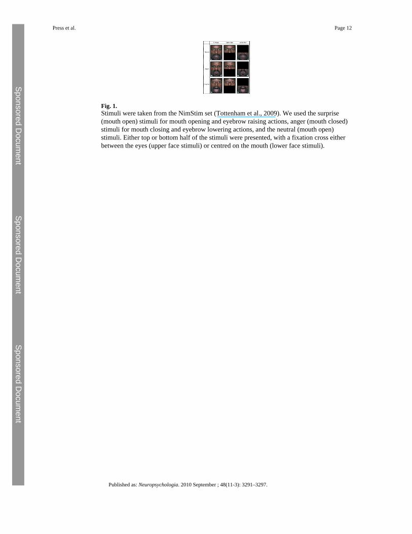
Fig. 1.Stimuli were taken from the NimStim set (Tottenham et al., 2009). We used the surprise(mouth open) stimuli for mouth opening and eyebrow raising actions, anger (mouth closed)stimuli for mouth closing and eyebrow lowering actions, and the neutral (mouth open)stimuli. Either top or bottom half of the stimuli were presented, with a fixation cross eitherbetween the eyes (upper face stimuli) or centred on the mouth (lower face stimuli).
Press et al. Page 12
Published as: Neuropsychologia. 2010 September ; 48(11-3): 3291–3297.
Sponsored Docum
ent Sponsored D
ocument
Sponsored Docum
ent
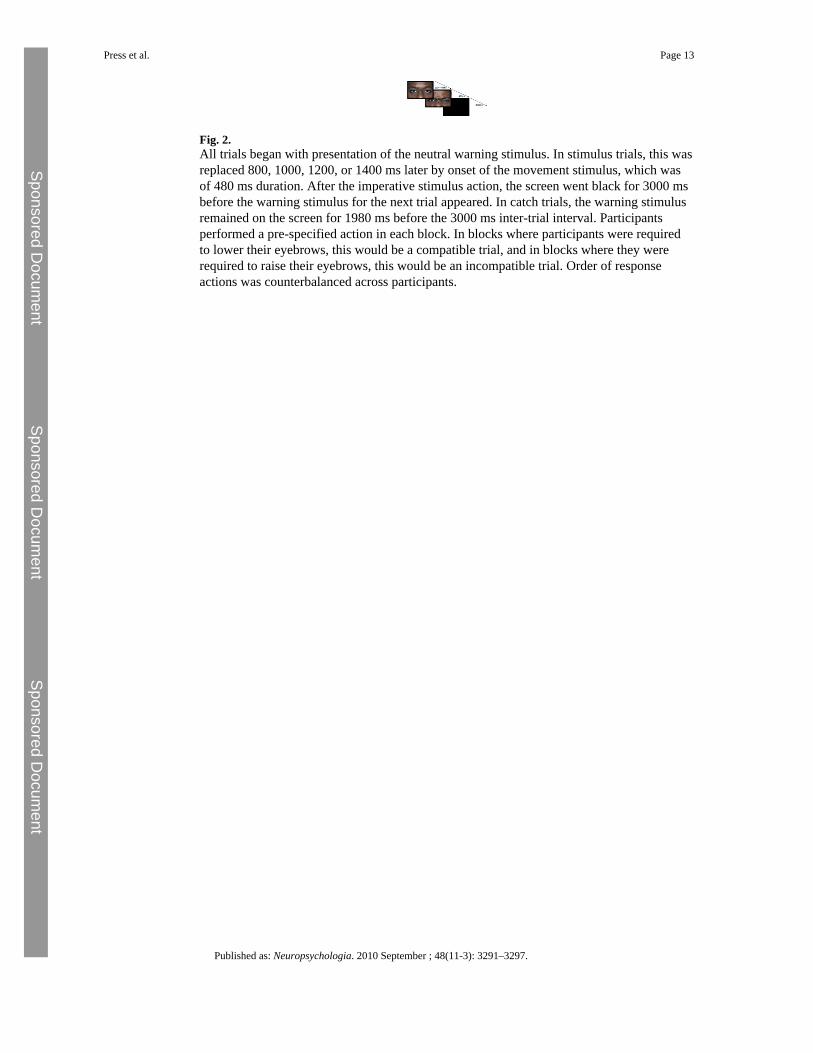
Fig. 2.All trials began with presentation of the neutral warning stimulus. In stimulus trials, this wasreplaced 800, 1000, 1200, or 1400 ms later by onset of the movement stimulus, which wasof 480 ms duration. After the imperative stimulus action, the screen went black for 3000 msbefore the warning stimulus for the next trial appeared. In catch trials, the warning stimulusremained on the screen for 1980 ms before the 3000 ms inter-trial interval. Participantsperformed a pre-specified action in each block. In blocks where participants were requiredto lower their eyebrows, this would be a compatible trial, and in blocks where they wererequired to raise their eyebrows, this would be an incompatible trial. Order of responseactions was counterbalanced across participants.
Press et al. Page 13
Published as: Neuropsychologia. 2010 September ; 48(11-3): 3291–3297.
Sponsored Docum
ent Sponsored D
ocument
Sponsored Docum
ent
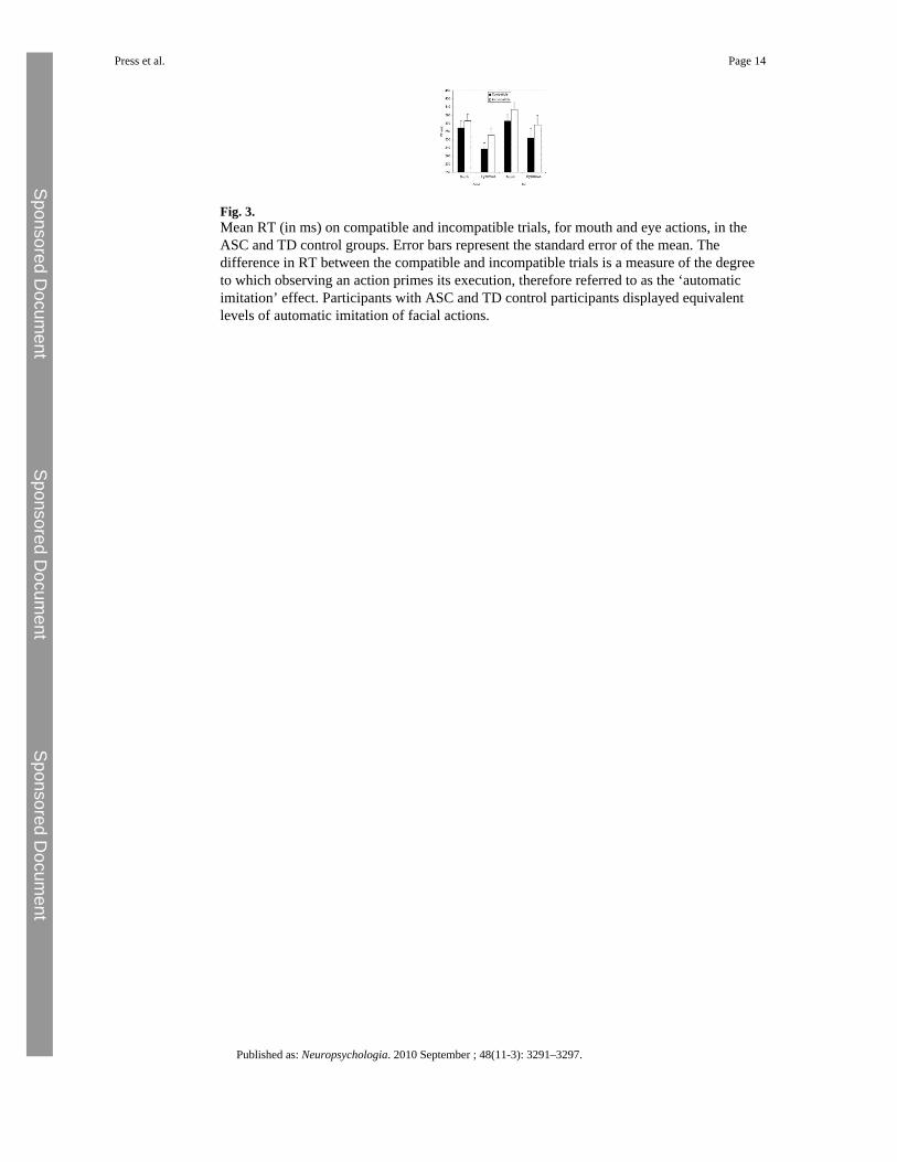
Fig. 3.Mean RT (in ms) on compatible and incompatible trials, for mouth and eye actions, in theASC and TD control groups. Error bars represent the standard error of the mean. Thedifference in RT between the compatible and incompatible trials is a measure of the degreeto which observing an action primes its execution, therefore referred to as the ‘automaticimitation’ effect. Participants with ASC and TD control participants displayed equivalentlevels of automatic imitation of facial actions.
Press et al. Page 14
Published as: Neuropsychologia. 2010 September ; 48(11-3): 3291–3297.
Sponsored Docum
ent Sponsored D
ocument
Sponsored Docum
ent

Sponsored Docum
ent Sponsored D
ocument
Sponsored Docum
ent
Press et al. Page 15
Table 1
Clinical diagnoses and ADOS-G (subscales and total) scores for the ASC group. Clinical diagnosis refers tothe original clinical assessment provided by a psychologist or psychiatrist (A = autism, AS = Asperger'ssyndrome, and ASD = autism spectrum disorder).
Participant Clinical diagnosis ADOS communication ADOS reciprocal social interaction ADOS total score
1 AS 2 5 7
2 ASD 4 6 10
3 AS 2 6 8
4 ASD 2 5 7
5 AS 4 6 10
6 AS 3 4 7
7 Atypical A 3 8 11
8 AS 5 10 15
9 AS 1 2 3
10 AS 5 12 17
11 AS 3 6 9
12 AS 4 5 9
13 AS 4 8 12
14 A 4 10 14
Published as: Neuropsychologia. 2010 September ; 48(11-3): 3291–3297.
Copyright © 2022 FDOKUMEN