Structural and functional consequences of buserelin-induced enteric neuropathy in rat
13
RESEARCH ARTICLE Open Access Structural and functional consequences of buserelin-induced enteric neuropathy in rat Elin Sand 1,2 , Bodil Roth 1 , Bjrn Westrm 3 , Peter Bonn 4 , Eva Ekblad 2 and Bodil Ohlsson 1* Abstract Background: Women treated with gonadotropin-releasing hormone (GnRH) analogs may develop enteric neuropathy and dysmotility. Administration of a GnRH analog to rats leads to similar degenerative neuropathy and ganglioneuritis. The aim of this study on rat was to evaluate the early GnRH-induced enteric neuropathy in terms of distribution of neuronal subpopulations and gastrointestinal (GI) function. Methods: Forty rats were given the GnRH analog buserelin (20 μg, 1 mg/ml) or saline subcutaneously, once daily for 5 days, followed by 3 weeks of recovery, representing one treatment session. Two weeks after the fourth treatment session, the animals were tested for GI transit time and galactose absorption, and fecal weight and fat content was analyzed. After sacrifice, enteric neuronal subpopulations were analyzed. Blood samples were analyzed for zonulin and antibodies against GnRH and luteinizing hormone, and their receptors. Results: Buserelin treatment transiently increased the body weight after 5 and 9 weeks (p < 0.001). Increased estradiol in plasma and thickened uterine muscle layers indicate high estrogen activity. The numbers of both submucous and myenteric neurons were reduced by 27% 61% in ileum and colon. The relative numbers of neurons containing calcitonin gene-related peptide (CGRP), cocaine- and amphetamine-related transcript (CART), galanin, gastrin-releasing peptide (GRP), neuropeptide Y (NPY), nitric oxide synthase (NOS), serotonin, substance P (SP), vasoactive intestinal peptide (VIP) or vesicular acetylcholine transporter (VAchT), and their nerve fiber density, were unchanged after buserelin treatment, but the relative number of submucous neurons containing somatostatin tended to be increased (p = 0.062). The feces weight decreased in buserelin-treated rats (p < 0.01), whereas feces fat content increased (p < 0.05), compared to control rats. Total GI transit time, galactose absorption, zonulin levels in plasma, and antibody titers in serum were unaffected by buserelin treatment. Conclusions: A marked enteric neuronal loss with modest effects on GI function is found after buserelin treatment. Increased feces fat content is suggested an early sign of dysfunction. Keywords: Enteric neuropathy, Enteric subpopulations, Gastrointestinal (GI) tract, Gonadotropin-releasing hormone (GnRH), Luteinizing hormone (LH), Somatostatin Background A subgroup of patients treated with gonadotropin-releasing hormone (GnRH) analogs develops enteric neuropathy with reduced relative number of GnRH-containing neu- rons and dysmotility [1,2], and increased abdominal pain and exacerbation of irritable bowel syndrome (IBS) has been observed in a cohort of GnRH-treated women at follow-up, although no obvious dysmotility was at hand [3]. This knowledge rendered us to set up an experimental rat model to examine the effects on the gastrointestinal (GI) tract of systemic and repeated treatment with the GnRH analog buserelin. About 50% of enteric neurons were lost throughout the GI tract after four treatment periods of buserelin [4,5]. In addition, myenteric ganglia displayed ganglioneuritis [5] and a significant reduction of the relative number of luteinizing hormone (LH) receptor- immunoreactive neurons [4]. In colon, a transient increase in the relative number of vasoactive intestinal peptide (VIP)-immunoreactive myenteric neurons was found after two treatment periods, and increased relative numbers of * Correspondence: [email protected] 1 Department of Clinical Sciences, Division of Internal Medicine Skne University Hospital, Lund University, Inga Marie Nilssons street 32, S-205 02 Malm, Sweden Full list of author information is available at the end of the article 2014 Sand et al.; licensee BioMed Central Ltd. This is an Open Access article distributed under the terms of the Creative Commons Attribution License (http://creativecommons.org/licenses/by/4.0), which permits unrestricted use, distribution, and reproduction in any medium, provided the original work is properly credited. The Creative Commons Public Domain Dedication waiver (http://creativecommons.org/publicdomain/zero/1.0/) applies to the data made available in this article, unless otherwise stated. Sand et al. BMC Gastroenterology 2014, 14:209 http://www.biomedcentral.com/1471-230X/14/209
-
Upload
independent -
Category
Documents
-
view
3 -
download
0
Transcript of Structural and functional consequences of buserelin-induced enteric neuropathy in rat
Sand et al. BMC Gastroenterology 2014, 14:209http://www.biomedcentral.com/1471-230X/14/209
RESEARCH ARTICLE Open Access
Structural and functional consequences ofbuserelin-induced enteric neuropathy in ratElin Sand1,2, Bodil Roth1, Bj?rn Westr?m 3, Peter Bonn4, Eva Ekblad2 and Bodil Ohlsson1*
Abstract
Background: Women treated with gonadotropin-releasing hormone (GnRH) analogs may develop enteric neuropathyand dysmotility. Administration of a GnRH analog to rats leads to similar degenerative neuropathy and ganglioneuritis.The aim of this study on rat was to evaluate the early GnRH-induced enteric neuropathy in terms of distribution ofneuronal subpopulations and gastrointestinal (GI) function.
Methods: Forty rats were given the GnRH analog buserelin (20 μg, 1 mg/ml) or saline subcutaneously, once daily for5 days, followed by 3 weeks of recovery, representing one treatment session. Two weeks after the fourth treatmentsession, the animals were tested for GI transit time and galactose absorption, and fecal weight and fat content wasanalyzed. After sacrifice, enteric neuronal subpopulations were analyzed. Blood samples were analyzed for zonulin andantibodies against GnRH and luteinizing hormone, and their receptors.
Results: Buserelin treatment transiently increased the body weight after 5 and 9 weeks (p < 0.001). Increased estradiolin plasma and thickened uterine muscle layers indicate high estrogen activity. The numbers of both submucous andmyenteric neurons were reduced by 27%? 61% in ileum and colon. The relative numbers of neurons containingcalcitonin gene-related peptide (CGRP), cocaine- and amphetamine-related transcript (CART), galanin, gastrin-releasingpeptide (GRP), neuropeptide Y (NPY), nitric oxide synthase (NOS), serotonin, substance P (SP), vasoactive intestinalpeptide (VIP) or vesicular acetylcholine transporter (VAchT), and their nerve fiber density, were unchangedafter buserelin treatment, but the relative number of submucous neurons containing somatostatin tended tobe increased (p = 0.062). The feces weight decreased in buserelin-treated rats (p < 0.01), whereas feces fat contentincreased (p < 0.05), compared to control rats. Total GI transit time, galactose absorption, zonulin levels in plasma, andantibody titers in serum were unaffected by buserelin treatment.
Conclusions: A marked enteric neuronal loss with modest effects on GI function is found after buserelin treatment.Increased feces fat content is suggested an early sign of dysfunction.
Keywords: Enteric neuropathy, Enteric subpopulations, Gastrointestinal (GI) tract, Gonadotropin-releasing hormone(GnRH), Luteinizing hormone (LH), Somatostatin
BackgroundA subgroup of patients treated with gonadotropin-releasinghormone (GnRH) analogs develops enteric neuropathywith reduced relative number of GnRH-containing neu-rons and dysmotility [1,2], and increased abdominal painand exacerbation of irritable bowel syndrome (IBS) hasbeen observed in a cohort of GnRH-treated women atfollow-up, although no obvious dysmotility was at hand
* Correspondence: [email protected] of Clinical Sciences, Division of Internal Medicine Sk?neUniversity Hospital, Lund University, Inga Marie Nilssons street 32, S-205 02Malm?, SwedenFull list of author information is available at the end of the article
? 2014 Sand et al.; licensee BioMed Central LtdCommons Attribution License (http://creativecreproduction in any medium, provided the orDedication waiver (http://creativecommons.orunless otherwise stated.
[3]. This knowledge rendered us to set up an experimentalrat model to examine the effects on the gastrointestinal(GI) tract of systemic and repeated treatment with theGnRH analog buserelin. About 50% of enteric neuronswere lost throughout the GI tract after four treatmentperiods of buserelin [4,5]. In addition, myenteric gangliadisplayed ganglioneuritis [5] and a significant reduction ofthe relative number of luteinizing hormone (LH) receptor-immunoreactive neurons [4]. In colon, a transient increasein the relative number of vasoactive intestinal peptide(VIP)-immunoreactive myenteric neurons was found aftertwo treatment periods, and increased relative numbers of
. This is an Open Access article distributed under the terms of the Creativeommons.org/licenses/by/4.0), which permits unrestricted use, distribution, andiginal work is properly credited. The Creative Commons Public Domaing/publicdomain/zero/1.0/) applies to the data made available in this article,
Sand et al. BMC Gastroenterology 2014, 14:209 Page 2 of 13http://www.biomedcentral.com/1471-230X/14/209
nitric oxide synthase (NOS)-immunoreactive submucousand myenteric neurons after four treatment periods [4].The enteric nervous system (ENS) contains a plethora
of neurotransmitters, which all participate in the pivotalrole of ENS in controlling and modulating GI motility,secretion, and blood flow. The neurotransmitters NOS, pitu-itary adenylate cyclase-activating peptide (PACAP), purines,somatostatin, and VIP mediate inhibitory transmission, whileexcitatory transmitters represent acetylcholine, calcitoningene-related peptide (CGRP), cocaine- and amphetamine-related transcript (CART), galanin, gastrin-releasingpeptide (GRP), neurokinin A, neuropeptide Y (NPY),serotonin (5-hydroxytryptamine; 5-HT), and substanceP (SP) [6].There is evidence of a high plasticity of the ENS in re-
sponse to injurious events in various experimental models,e.g. axotomy, transplantation, hypertrophy, ischemia/reperfusion, and lipopolysaccharide (LPS) challenge [7-11],as well as in diseases [12,13]. Experimental rat modelsof enteric neuropathy have been described, e.g. neur-opathy after cisplatin-, diabetes-, or fat induction [14-16].Most studies describe pathophysiology and morphologicalchanges in neuropathy, whereas few studies describe anyfunctional consequences on GI motility, nutritional ab-sorption, and intestinal permeability.The aim of the present study was to describe possible
early effects of buserelin-induced enteric neuronal losson subpopulations of neurons and on body weight, circu-lating levels of sex hormones and antibodies, GI transittime, feces weight and fat content, nutrient absorption,and epithelial permeability.
MethodsAnimalsFemale Sprague? Dawley rats (n = 40, 170? 180 g), pur-chased from Charles River, Sulzfeld, Germany, were used.The rats were allowed to acclimatize to the climate- andlight-controlled animal facility for at least 5 days prior toexperimentation. Standard rat chow (4% fat/g) (LactaminR36, Stockholm, Sweden) and water were supplied atall times. The experimental design was approved by theAnimal Ethics Committee, Lund and Malm?, Sweden(M350-12, date of approval: 14.11.12). Animals were usedin accordance with the European Communities CouncilDirective (2010/63/EU) and the Swedish Animal WelfareAct (SFS 1988:534).
Study designTwenty-four rats (n = 24) were given 20 μg (1 mg/ml) ofthe GnRH analog buserelin (Suprefact?, Sanofi-Aventis,Bromma, Sweden) subcutaneously, once daily for 5 days,followed by 3 weeks of recovery, representing one ses-sion of treatment (for details see ref no. [4]). The dosageand administration of buserelin are based on previous
studies which have shown reliable physiological effectsin terms of uterine hypertrophy, without any adverseeffects [4,17]. Control animals (n = 16) received salineinjections. The animals were weighed prior to inclusion inthe study, and weekly in the morning during the study,using an electronic scale. Half of the animals were used toexamine vaginal smears. From the other rats (12 buserelin-treated and eight saline-treated controls), blood sampleswere drawn in the morning before administration of buser-elin or saline in week 1 and 4 of the injection treatment,and at sacrifice. During the 2 weeks after the fourth treat-ment session, GI transit time and galactose absorptionwere studied. Feces were collected during 12 h of fastingand analyzed for weight and fat content. After sacrifice,tissue samples from the stomach, ileum, transverse colon,and the distal part of the uterine horn were collectedand rinsed in saline before fixation and processing forcryo- or paraffin-sectioning and histological evaluation.All methods are described in detail below.
Blood and vaginal samplingTo study possible buserelin effects on the rat estrus cycle,vaginal smears and blood samples for hormone measure-ments were collected. On day 0 and 5 during session 1and 4, vaginal smears were obtained using a cotton tippedapplicator, rotated three times, 2 cm from the vaginal ori-fice. The vaginal smears were placed on microscope slidesand stained with methylene blue and eosin for determin-ation of the phase in the estrus cycle. The classification ofthe different cycles was performed according to estab-lished criteria [18,19]. Briefly, proestrus is characterized bya predominance of nucleated epithelial cells and estrus byanucleated, cornified epithelial cells. Metestrus is charac-terized by an equal portion of nucleated or anucleated epi-thelial cells and leukocytes, while diestrus is characterizedby a predominance of leukocytes. Blood samples werecollected from the tail vein, using Li heparin tubes (BDMicrotainer, New Jersey, USA) and centrifuged at 3000 rcf(1.12 ? R ? (RPM/1000) 2) for 5 min. Sera and plasma wereharvested and stored at −20?C. Samples were collected at9:00 am before the first injection of buserelin or saline(day 0), in the fourth session of injections (day 0 and 5),and at sacrifice. Vaginal smears and blood samples takenon day 0, before the first injection, served as an individualcontrol for each rat.
Tissue preparationThe gut segments and the uteri were opened andembedded, flattened, in filter paper. One portion of eachgut segment was fixed in Stefanini ? s fixative (a mixtureof 2% formaldehyde and 0.2% picric acid in phosphatebuffer, pH 7.2) for 22 h at 4?C, and the other portionand the uteri were fixed in 4% paraformaldehyde in0.1 M phosphate buffer for 22 h at 4?C. Stefanini-fixed
Sand et al. BMC Gastroenterology 2014, 14:209 Page 3 of 13http://www.biomedcentral.com/1471-230X/14/209
specimens were rinsed three times in Tyrode ? s solutioncontaining 10% sucrose, before being orientated andmounted for longitudinal- and cross-sectioning in Tissue-Tek (Sakura, Histolab, Gothenburg, Sweden), frozen ondry ice, and sectioned (10 μm). Paraformaldehyde-fixedspecimens were dehydrated in ethanol, cleared in xylene,orientated for longitudinal- and cross-sectioning, embed-ded in paraffin, and sectioned (5 μm). Sections were proc-essed for immunocytochemistry and histochemistry.
HistochemistryMeasurements of wall layer thickness were performed ondeparaffinized, hydrated, and hematoxylin-eosin-stainedparaffin sections from the uterus by using a computerized,image-analyzing system (Imagescope, Aperio ScanScopeGL SS5082, Vista, CA 92081, USA). The myometrialthicknesses to be measured were indicated manually, andthen measured using a computerized binary cursor. Meanvalues of 6? 10 representative measurements were calcu-lated from each rat.
ImmunocytochemistryFor studies on enteric neuronal survival, antibodies againsthuman neuronal protein HuC/D (HuC/D) were used asthe general neuronal marker. Paraffin sections were depar-affinized, hydrated, and subjected to antigen retrieval byboiling in citrate acid buffer (0.01 M, pH 6) in a microwaveoven (650 W) for 2 ? 7 min. The sections were cooled andwashed in distilled water followed by phosphate-bufferedsaline (PBS)/Triton. Sections were exposed to biotinyl-ated, primary antibodies against HuC/D at 4?C overnight.For visualization of biotinylated HuC/D, a VECTASTAINABC kit containing horseradish peroxidase (HRP) and3,3? -diaminobenzidine tetrahydrochloride (DAB) was used(Vector Laboratories, Inc., CA, USA). HuC/D-immunoreactiveneurons stained dark brown and were counted in submu-cous and myenteric ganglia on longitudinally-cut sectionsusing a computerized, image-analyzing system (Imagescope).The number of HuC/D neurons in colon was counted inscanned sections in a total length of at least 30 mm, cut at6? 9 different depths per region and rat. Synthetic antigensfor testing the specificity of antibodies against HuC/D arenot commercially available. Thus, omission of the primaryantibodies was used as controls. Results are expressed asnumbers of submucous or myenteric neurons, immunore-active to HuC/D, per mm length of GI tract.To study whether the neuronal loss was general or spe-
cific regarding the subpopulations of enteric neurons, therelative numbers of different subpopulations were studiedin colon, as this was the most affected region in the formerstudy [4]. Antibodies against CGRP, CART, galanin, GRP,NPY, NOS, 5-HT, somatostatin, SP, VIP, in combinationwith antibodies against HuC/D, were used on cryo sec-tions. Since vesicular acetylcholine transporter (VAchT)
immunoreactivity is mainly located on nerve fibers, anti-bodies against VAchT alone, and not in combination withHuC/D, were used on cryosections. Details on the anti-bodies are given in Table 1. Absorption controls were per-formed by adding an excess amount of antigen (10? 100 μgof synthetic peptide diluted in antiserum) before exposure.The sites of the antibody-antigen reactions were
visualized by exposure to a mixture of DyLight TM 488-conjugated goat anti-mouse IgG serum and Alexa FluorTM 594-conjugated donkey anti-rabbit IgG serum, orsolely to Texas red donkey anti-goat (all diluted 1:1000;Jackson ImmunoResearch Laboratories, Inc., Novakemi AB,Handen, Sweden), for 1 h in room temperature (RT) andthen mounted in phosphate buffer:glycerol 1:1.HuC/D-immunoreactive neurons also immunoreactive
to NOS, 5-HT or any of the neuropeptides tested, wereevaluated in cross- and longitudinally-cut, whole-wall sec-tions. At least 150 submucous neurons and 250 myentericneurons were counted for each set of double-stains andrat. The results are expressed as the percentage of HuC/D-immunoreactive neurons also immunoreactive to NOS,5-HT or any of the neuropeptides. Since VAchT immuno-reactivity is mainly located on nerve fibers, the relativenumber of VAchT-immunoreactive nerve cell bodiescould not be evaluated. Nerve fiber density was evaluatedon a 0, (+), +, ++, +++ scale, where 0 indicates no fibers,(+) = occasional fibers, + = few fibers, ++ =moderate num-bers of fibers, and +++ = numerous fibers.
Studies on gastrointestinal functionTo measure total GI transit time, the rats fasted overnightby removing their food at 9:00 pm. At 9:00 am the nextday, the rats were given a bolus dose (1 ml) of carbon sus-pension trough gavage administered orally to the stomach(150 mg/ml, Abigo Medical, Gothenburg, Sweden) beforebeing placed in separate cages with free access to food andwater. The rats were continuously and manually moni-tored by staff being present all the time, until the firstcarbon-containing fecal pellets were seen to be excreted.In order to study fecal weight and fecal content [30],
feces were collected and weighed from all rats during the12 h of fasting, prior to the measurement of GI transittime, see above. The fecal samples were kept in open glasstubes and left to dry at RT for 3 months in a fume cup-board. The dry feces were ground in a mortar, transferredto a 20 ml glass vial, and weighed. To extract the fat fromthe feces, 10 ml of dichloromethane (DCM, Chromasolv,Sigma-Aldrich, Stockholm, Sweden) was added. The mix-ture was stirred vigorously for 1 h at RT. The DCM phasewas filtered with a plastic syringe (10 ml), fitted with apolyethylene frit (20 μm, 10 ml, Biotage, Uppsala, Sweden)and a syringe filter (1 μm, Acrodisc glass fiber, Pall, NY,USA), and collected in a pre-weighed glass vessel. The ex-traction procedure was repeated twice for a total of 30 ml

Table 1 Details on antibodies
Raised against Code no. Host Working dilutioncryosections
Supplier References
CART (61 ? 102) H-003-61 Rabbit 1:5000 Phoenix, GmBH, USA Ekblad et al. 2003;Zacharko-Siembida et al. 2014 [20,21]
CGRP 8427 Rabbit 1:5000 Euro-Diagnostica, Sweden Ekblad et al. 1998 [8]
Galanin 8416 Rabbit 1:1000 Euro-Diagnostica, Sweden Ekblad et al. 1988; Ekblad et al. 1998 [8,22]
GRP R-6902 Rabbit 1:640 Ekblad et al. 1988 [22]
Hu proteins(HuC/HuD)
A-2127 Mouse 1:600 Life Technologies, USA Lin et al. 2003 [23]
NOS 9223 Rabbit 1:5000 Euro-Diagnostica, Sweden Ekblad et al. 1994; Kristensson et al. 2007 [24,25]
C-PON* CA-08-300 Rabbit 1:3000 Genosys, UK Ekblad et al. 1988; Kristensson et al. 2007 [22,25]
Serotonin (5-HT) NSER Rabbit 1:1200 Inc. Star Corp, USA Mulder et al. 1997 [26]
Somatostatin 1758 Rabbit 1:3200 Kind gift from prof. J.J. Holst, Denmark Ekblad et al. 1988; Kristensson et al. 2007 [22,25]
Substance P (SP) SP7 Rabbit 1:800 Kind gift from prof. Emson, UK Lindestr?m et al. 2002 [ 27]
VIP 7854 Rabbit 1:2000 Euro-Diagnostica, Sweden Qian B et al. 2001; Kristensson et al. 2007 [25,28]
VAchT AB1578 Get 1:2000 Chemicon, USA Arvidsson U et al., 1997 [29]
*C-terminal flanking peptide of NPY (used for the detection of NPY-containing neurons).
Sand et al. BMC Gastroenterology 2014, 14:209 Page 4 of 13http://www.biomedcentral.com/1471-230X/14/209
of the DCM phase. When executing the last extraction,the solid material and the DMC were transferred to thesyringe. To ensure that as much as possible of the organicsolvent was filtered into the glass vessel, a plunger wasused to squeeze the solid material. The solvent was re-moved by evaporation and the residue (fat content) wasweighed.In order to study the absorptive capacity of galactose
after 12 h of fasting, a bolus dose of 1 ml of 5% galactosedissolved in saline was given via a stomach tube as anoral bolus dose under anesthesia. Blood samples werecollected in Li Heparin tubes from the tail vein using aneoflon catheter, before and 10 min, 30 min, and 90 minafter the bolus dose [31]. Galactose levels in plasma wereanalyzed with a BioVision ? s Galactose Assay Kit (K621-100, BioVision Incorporated, CA, USA) according to themanufacturer ? s protocol. Briefly, galactose is oxidizedusing a galactose probe, an enzyme mix, and HRP, gen-erating a colored product analyzed at the optic densityof 570 nm.
Serum analysesAnalyses of IgM- and IgG antibodies in serum againstGnRH, GnRH receptor (GnRHR), LH, and LH receptor(LHR) were carried out in blood samples collected at sac-rifice by an enzyme-linked immune sorbent assay (ELISA)as described in detail previously [3]. Briefly, the wellsof microtiter plates (456537 Nunc, Roskilde, Denmark)were either coated with human GnRH or N-terminalGnRH-R peptide ((NH2)-ANSASPEQNQNHCSAINNSIPLMQGNLPY) conjugated with ovalbumin (OVA)(Innovagen, Lund, Sweden), 100 ng/well, in an over-night incubation at 4?C. Thereafter, the plastic wells
were blocked with 0.5% bovine serum albumin (BSA)(A-7030, Sigma, St Louis, USA) in PBS (10 mM PO4
3−,137 mM NaCl, and 2.7 mM KCl, pH 7.4) containing0.05% Tween-20 (PBS-T). The dilutions of patient serum(1:400 in 1.0 μg OVA (A-5503, Sigma)/ml 0.5% BSA inPBS-T) or mouse anti-human GnRH antibody (1.11 mg/ml, ab62432, Abcam, Cambridge, MA, USA) in serial dilu-tion (1:2 000? 1:32 000 to construct a standard curve) orrabbit anti-human GnRHR antibody (90217.09, Innovagen)in serial dilution (1:8 000? 1:128 000) were then added tothe plates and incubated for 2 h at RT. After rinsing withPBS-T, deposition of antibodies directed to GnRH orGnRHR was detected using the secondary antibodies rabbitanti-rat IgG-biotin (ABCAM102170, Abcam, Cambridge,MA, USA) or goat anti-rat IgM-biotin (ABCAM97178,Abcam) diluted 1:10 000 in PBS-T.Analysis of antibodies against LH was carried out
with intact, purified, native human LH (MBS537383,MyBiosource, San Diego, CA, USA), 100 ng/well, in PBSor only PBS-T (to provide an internal blank). After over-night incubation at 4?C, the plates were washed threetimes with PBS-T and blocked with 0.5% BSA in PBS-T.Dilutions of serum (1:200) from patients and blooddonors, or rabbit anti-human LH antibody (MBS535386,MyBiosource) in serial dilution (to construct a standardcurve), with BSA in PBS-T were then added to the platesin triplicate (two wells coated with LH and one well coatedwith PBS-T) and incubated for 2 h at RT. The washingprocedure was repeated and deposition of autoantibodiesdirected to LH was detected using secondary antibodies asdescribed above, appropriately diluted in PBS-T.Analysis of antibodies against the LHR was carried out
with the N-terminal LHR peptide ((NH2)-MKQRFSSALQL
Sand et al. BMC Gastroenterology 2014, 14:209 Page 5 of 13http://www.biomedcentral.com/1471-230X/14/209
LKLLLLQPPLPRALC), conjugated with OVA (Innovagen),in 100 mM Carbonate buffer pH 9.2 or only Carbonatebuffer (to provide an internal blank). After overnight incu-bation at 4?C, the plates were washed three times withPBS-T and blocked with 0.5% BSA in PBS-T. Dilutions ofserum (1:200) from patients and blood donors with BSAin PBS-T were then added to the plates in triplicate, twoto LHR and one to Carbonate buffer-coated wells, andincubated for 2 h at RT. The washing procedure wasrepeated and deposition of autoantibodies directed toLHR was detected using secondary antibodies as describedabove, appropriately diluted in PBS-T.The absorbance at 405 nm was measured after 30 min
of incubation at RT. Antibody levels are expressed asarbitrary units (AU) (absorbance values after subtractedbackground levels and multiplied with 1000).
Plasma analysesDue to the small amount of rat plasma, all analyses wereperformed with a single sample. Samples were analyzed ac-cording to the manufacturer ? s instructions, and the result-ing color changes were measured at the optical density of450 nm. Each sample was interpolated from the standardcurve.Follicle-stimulating hormone (FSH)-, 17β estradiol (E2)-,
and LH ELISA kits (MBS720215, MBS162143 andMBS161787, respectively, MyBiosource, San Diego, CA,USA) based on the double antibody sandwich technologywere used in plasma samples collected at study start andduring session 4. Plates were pre-coated with monoclonalantibodies selectively recognizing rat FSH, E2 or LH. Stan-dards of FSH: 2.5, 5.0, 10, 25, and 50 ng/ml; E2: 30, 60,120, 240, and 480 ng/l; and LH: 31.2, 62.5, 125, 250, 500,and 1000 ng/ml (to form a standard curve) or undilutedrat heparin plasma samples were then added to the wellson a microtiter plate (Nunc). A standardized preparationof immune complexes in the form of HRP-conjugatedpolyclonal antibody (FSH), or antibodies labeled withbiotin/streptavidin HRP (E2 and LH), were added.A rat zonulin ELISA kit (MBS753035, MyBiosource)
based on the competitive enzyme immunoassay techniquewas used to study epithelial integrity in plasma harvested atsacrifice. The standards of 25, 50, 100, 250, and 500 ng/ml,undiluted rat heparin plasma, and buffer were incubatedtogether with zonulin HRP conjugate on a plate pre-coatedwith monoclonal anti-zonulin antibodies.
Statistical analysesAll results are presented as medians (interquartile ranges(IQR)), except weight, which is presented as mean ? standarderror of the mean (SEM). Statistical analyses were performedby using the Mann? Whitney U-test. P < 0.05 was consid-ered statistically significant.
ResultsGeneral characteristicsOne rat in each group was terminated because of admin-istration to the lung of the oral galactose solution, leav-ing 11 buserelin-treated (B) rats and seven saline-treatedcontrol (C) rats for the analyses. The body weight gainincreased transiently in buserelin-treated rats in week 5and 9 (p < 0.001; see Additional file 1). These time pointscoincided with the end of the buserelin sessions 2 and 3.The plasma levels of FSH were extremely low or undetect-able in all rats (data not shown). The LH kit was unusableon rat plasma and serum, as no standard curve could beconstructed. The estradiol levels in plasma were in thesame range at start (Day 0, session 1; C = 52 (41? 62) ng/L,B = 55 (46? 58) ng/L), but during session 4, they wereelevated on day 0 and 5 in the buserelin-treated group,reflecting that the hypothalamus-hypophys-gonadal axiswas not suppressed by buserelin treatment in this setting(p < 0.05; for details see Additional file 2). At start (day 0,session 1), the vaginal smears indicated that all rats wereunsynchronized in their estrus cycle (12 buserelin-treatedand eight saline-treated controls). After the first week oftreatment, the saline-treated rats were still unsynchro-nized, while the buserelin-treated rats were all in metes-trus or diestrus. In the fourth week of injection treatment,the saline-treated rats were still unsynchronized on day 0and 5, while the buserelin-treated rats were all in metes-trus on day 0 and in diestrus on day 5 (see Additionalfile 3). The uterine muscle layer was thicker in the groupof buserelin-treated rats compared to saline-treated con-trols (p < 0.05; see Additional file 4).
Neuronal loss and neuronal subpopulationsSignificant reductions in the absolute numbers of sub-mucous neurons were detected in ileum and colon (27%,p < 0.05 and 61%, p < 0.05, respectively). Of the myentericneurons, reduced numbers were noted in the stomach(27%, p < 0.05), ileum (39%, p < 0.01), and colon (31%, p <0.01). The loss of neurons was statistically more pro-nounced in myenteric than in submucous ganglia, andmore pronounced in the colon and ileum than in thestomach (Figure 1; for raw data see Additional file 5).In colon, a small number (fewer than 10%) of submucous
nerve cell bodies were immunoreactive to CGRP, CART,galanin, NPY or SP, while GRP-, NOS-, and VIP immuno-reactivity were found in 10%? 43% of submucous neurons.The relative number of somatostatin-immunoreactive sub-mucous nerve cell bodies was 11% in colon from controls,but showed a tendency to increase to 14% after buserelintreatment (p = 0.062; Figure 2; for raw data see Additionalfile 5). In both controls and buserelin-treated rats, few co-lonic myenteric nerve cell bodies were found to containCGRP-, galanin-, NPY-, SP-, or VIP immunoreactivity,and moderate numbers of neurons were found to contain
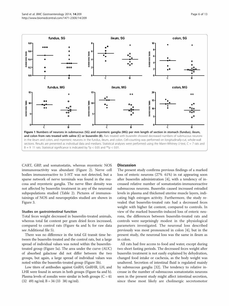
Figure 1 Numbers of neurons in submucous (SG) and myenteric ganglia (MG) per mm length of section in stomach (fundus), ileum,and colon from rats treated with saline (C) or buserelin (B). Rats treated with buserelin showed decreased numbers of submucous neuronsin the ileum and colon, and myenteric neurons in the fundus, ileum, and colon. Cell-counting was performed on longitudinally-cut, whole-wallsections. Results are presented as individual data and medians. Statistical analyses were performed using the Mann-Whitney U-test, C = 7 rats andB = 9 ? 11 rats. Statistical significance is indicated by *p < 0.05 and **p < 0.01.
Sand et al. BMC Gastroenterology 2014, 14:209 Page 6 of 13http://www.biomedcentral.com/1471-230X/14/209
CART, GRP, and somatostatin, whereas myenteric NOSimmunoreactivity was abundant (Figure 2). Nerve cellbodies immunoreactive to 5-HT was not detected, but asparse network of nerve terminals was found in the mu-cosa and myenteric ganglia. The nerve fiber density wasnot affected by buserelin treatment in any of the neuronalsubpopulations studied (Table 2). Pictures of immunos-tainings of NOS and neuropeptides studied are shown inFigure 3.
Studies on gastrointestinal functionTotal feces weight decreased in buserelin-treated animals,whereas total fat content per gram dried feces increased,compared to control rats (Figure 4a and b; for raw datasee Additional file 5).There was no difference in the total GI transit time be-
tween the buserelin-treated and the control rats, but a largespread of individual values was noted within the buserelin-treated group (Figure 5a). The area under the curve (AUC)of absorbed galactose did not differ between the twogroups, but again, a large spread of individual values wasnoted within the buserelin-treated group (Figure 5b).Low titers of antibodies against GnRH, GnRHR, LH, and
LHR were found in serum in both groups (Figure 6a and b).Plasma levels of zonulin were similar in both groups (C = 41(32? 49) ng/ml; B = 34 (33? 38) ng/ml).
DiscussionThe present study confirms previous findings of a markedloss of enteric neurons (27%? 61%) in rat appearing soonafter buserelin administration [4], with a tendency of in-creased relative number of somatostatin-immunoreactivesubmucous neurons. Buserelin caused increased estradiollevels in plasma and thickened uterine muscle layers, indi-cating high estrogen activity. Furthermore, the study re-vealed that buserelin-treated rats had a decreased fecesweight with higher fat content, compared to controls. Inview of the marked buserelin-induced loss of enteric neu-rons, the differences between buserelin-treated rats andcontrols were surprisingly modest in the physiologicalparameters investigated. The neuronal loss describedpreviously was most pronounced in colon [4], but in thepresent study, the neuronal loss was the same in ileum asin colon.All rats had free access to food and water, except during
two short fasting periods. The decreased feces weight afterbuserelin treatment is not easily explained by dehydration,changed food intake or cachexia, as the body weight wasunaltered. Secretion of intestinal fluid is regulated mainlyby submucous ganglia [32]. The tendency to relative in-crease in the number of submucous somatostatin neuronsseen in the present study might affect intestinal secretion,since these most likely are cholinergic secretomotor
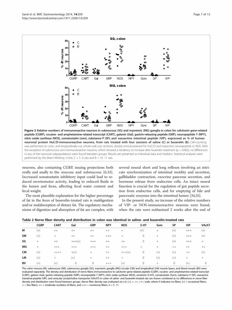
Figure 2 Relative numbers of immunoreactive neurons in submucous (SG) and myenteric (MG) ganglia in colon for calcitonin gene-relatedpeptide (CGRP), cocaine- and amphetamine-related transcript (CART), galanin (Gal), gastrin-releasing peptide (GRP), neuropeptide Y (NPY),nitric oxide synthase (NOS), somatostatin (som), substance P (SP), and vasoactive intestinal peptide (VIP), expressed as % of humanneuronal protein HuC/D-immunoreactive neurons, from rats treated with four sessions of saline (C) or buserelin (B). Cell-countingwas performed on cross- and longitudinally-cut, whole-wall cryo sections, doubly immunostained for HuC/D and respective neuropeptide or NOS. Withthe exception of submucous som-immunoreactive neurons, which showed a tendency to increase after buserelin treatment (p = 0.062), no differencesin any of the neuronal subpopulations were found between groups. Results are presented as individual data and medians. Statistical analyses wereperformed by the Mann-Whitney U-test, C = 5? 6 rats and B = 10? 11 rats.
Sand et al. BMC Gastroenterology 2014, 14:209 Page 7 of 13http://www.biomedcentral.com/1471-230X/14/209
neurons, also containing CGRP, issuing projections bothorally and anally to the mucosa and submucosa [6,33].Increased somatostatin inhibitory input could lead to re-duced secretomotor activity, leading to reduced fluids inthe lumen and feces, affecting fecal water content andfecal weight.The most plausible explanation for the higher percentage
of fat in the feces of buserelin-treated rats is maldigestionand/or malabsorption of dietary fat. The regulatory mecha-nisms of digestion and absorption of fat are complex, with
Table 2 Nerve fiber density and distribution in colon was ide
CGRP CART Gal GRP NPY
M (+) ++ ++ ++ ++
SM (+) + ++ ++ +++
SG + ++ +++(+) +++ ++
MG + +++ +++ +++ ++
CM (+) ++++ +(+) + ++
LM (+) + (+) + ++
BV (+) (+) 0 0 +++
The colon mucosa (M), submucosa (SM), submucous ganglia (SG), myenteric ganglia (Mevaluated separately. The density and distribution of nerve fibers immunoreactive to ca(CART), galanin (Gal), gastrin-releasing peptide (GRP), neuropeptide Y (NPY), nitric oxideintestinal peptide (VIP), and vesicular acetylcholine transporter (VAchT) in colon of salinedensity and distribution were found between groups. Nerve fiber density was evaluated+ = few fibers, ++ = moderate numbers of fibers, and +++ = numerous fibers. n = 5? 11
several neural short and long reflexes involving an intri-cate synchronization of intestinal motility and secretion,gallbladder contraction, exocrine pancreas secretion, andhormone release from endocrine cells. An intact neuralfunction is crucial for the regulation of gut peptide secre-tion from endocrine cells, and for emptying of bile andpancreatic enzymes into the intestinal lumen [34,35].In the present study, no increase of the relative numbers
of VIP- or NOS-immunoreactive neurons were found,when the rats were euthanized 2 weeks after the end of
ntical in saline- and buserelin-treated rats
NOS 5-HT Som SP VIP VAchT
+ (+) + (+) +++ (+)
+ 0 + (+) +++ (+)
++ 0 + (+) +++ +
+++ + + ++ ++ ++
+++(+) 0 (+) (+) ++ (+)
+ 0 (+) (+) + +
(+) 0 + 0 (+) 0
G) circular (CM) and longitudinal (LM) muscle layers, and blood vessels (BV) werelcitonin gene-related peptide (CGRP), cocaine- and amphetamine-related transcriptsynthase (NOS), serotonin (5-HT), somatostatin (Som), substance P (SP), vasoactive- and buserelin-treated rats are shown combined as no differences in nerve fiberon a 0, (+), +, ++, +++ scale, where 0 indicates no fibers, (+) = occasional fibers,.
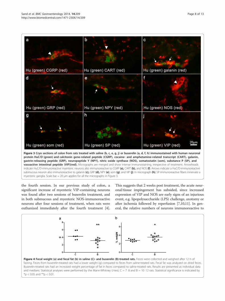
Figure 3 Cryo sections of colon from rats treated with saline (b, c, e, g, j) or buserelin (a, d, f, h) immunostained with human neuronalprotein HuC/D (green) and calcitonin gene-related peptide (CGRP), cocaine- and amphetamine-related transcript (CART), galanin,gastrin-releasing peptide (GRP), neuropeptide Y (NPY), nitric oxide synthase (NOS), somatostatin (som), substance P (SP), andvasoactive intestinal peptide (VIP)(red). Micrographs are merged and show intense immunostaining, irrespective of treatment. Arrowheadsindicate HuC/D-immunoreactive myenteric neurons also immunoreactive to CGRP (a), CART (b), and NOS (f). Arrows indicate a HuC/D-immunoreactivesubmucous neuron also immunoreactive to galanin (c), GRP (d), NPY (e), som (g), and VIP (j). In micrograph (h), SP-immunoreactive fibers innervate amyenteric ganglia. Scale bar = 20 μm applies for all the micrographs in Figure 3.
Sand et al. BMC Gastroenterology 2014, 14:209 Page 8 of 13http://www.biomedcentral.com/1471-230X/14/209
the fourth session. In our previous study of colon, asignificant increase of myenteric VIP-containing neuronswas found after two sessions of buserelin treatment, andin both submucous and myenteric NOS-immunoreactiveneurons after four sessions of treatment, when rats wereeuthanized immediately after the fourth treatment [4].
Figure 4 Fecal weight (a) and fecal fat (b) in saline (C)- and buserelinfasting. Feces from buserelin-treated rats had a lower weight (g) comparedBuserelin-treated rats had an increased weight percentage of fat in feces cand medians. Statistical analyses were performed by the Mann-Whitney U-t*p < 0.05 and **p < 0.01.
This suggests that 2 weeks post treatment, the acute neur-onal/tissue impingement has subsided, since increasedexpression of VIP and NOS are early signs of an injuriousevent, e.g. lipopolysaccharide (LPS) challenge, axotomy orafter ischemia followed by reperfusion [7,10,11]. In gen-eral, the relative numbers of neurons immunoreactive to
(B)-treated rats. Feces were collected and weighed after 12 h ofto feces from saline-treated rats. Fecal fat was analyzed on dried feces.
ompared to saline-treated rats. Results are presented as individual dataest, C = 7? 8 and B = 10? 12 rats. Statistical significance is indicated by
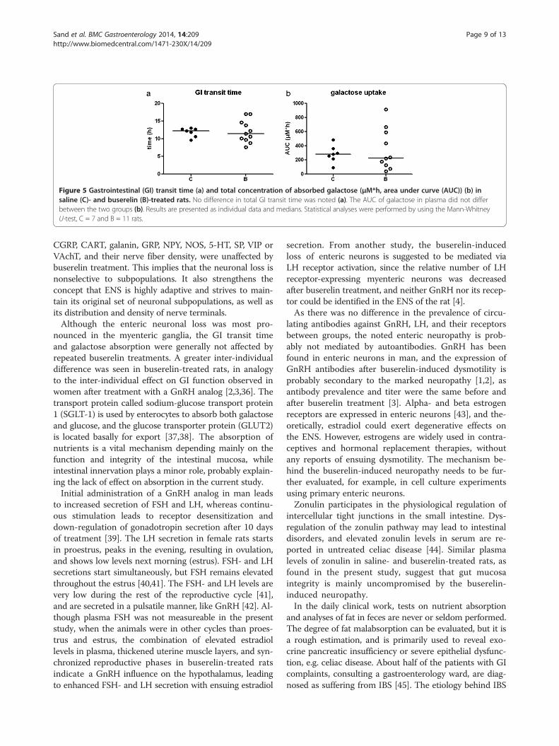
Figure 5 Gastrointestinal (GI) transit time (a) and total concentration of absorbed galactose (μM*h, area under curve (AUC)) (b) insaline (C)- and buserelin (B)-treated rats. No difference in total GI transit time was noted (a). The AUC of galactose in plasma did not differbetween the two groups (b). Results are presented as individual data and medians. Statistical analyses were performed by using the Mann-WhitneyU-test, C = 7 and B = 11 rats.
Sand et al. BMC Gastroenterology 2014, 14:209 Page 9 of 13http://www.biomedcentral.com/1471-230X/14/209
CGRP, CART, galanin, GRP, NPY, NOS, 5-HT, SP, VIP orVAchT, and their nerve fiber density, were unaffected bybuserelin treatment. This implies that the neuronal loss isnonselective to subpopulations. It also strengthens theconcept that ENS is highly adaptive and strives to main-tain its original set of neuronal subpopulations, as well asits distribution and density of nerve terminals.Although the enteric neuronal loss was most pro-
nounced in the myenteric ganglia, the GI transit timeand galactose absorption were generally not affected byrepeated buserelin treatments. A greater inter-individualdifference was seen in buserelin-treated rats, in analogyto the inter-individual effect on GI function observed inwomen after treatment with a GnRH analog [2,3,36]. Thetransport protein called sodium-glucose transport protein1 (SGLT-1) is used by enterocytes to absorb both galactoseand glucose, and the glucose transporter protein (GLUT2)is located basally for export [37,38]. The absorption ofnutrients is a vital mechanism depending mainly on thefunction and integrity of the intestinal mucosa, whileintestinal innervation plays a minor role, probably explain-ing the lack of effect on absorption in the current study.Initial administration of a GnRH analog in man leads
to increased secretion of FSH and LH, whereas continu-ous stimulation leads to receptor desensitization anddown-regulation of gonadotropin secretion after 10 daysof treatment [39]. The LH secretion in female rats startsin proestrus, peaks in the evening, resulting in ovulation,and shows low levels next morning (estrus). FSH- and LHsecretions start simultaneously, but FSH remains elevatedthroughout the estrus [40,41]. The FSH- and LH levels arevery low during the rest of the reproductive cycle [41],and are secreted in a pulsatile manner, like GnRH [42]. Al-though plasma FSH was not measureable in the presentstudy, when the animals were in other cycles than proes-trus and estrus, the combination of elevated estradiollevels in plasma, thickened uterine muscle layers, and syn-chronized reproductive phases in buserelin-treated ratsindicate a GnRH influence on the hypothalamus, leadingto enhanced FSH- and LH secretion with ensuing estradiol
secretion. From another study, the buserelin-inducedloss of enteric neurons is suggested to be mediated viaLH receptor activation, since the relative number of LHreceptor-expressing myenteric neurons was decreasedafter buserelin treatment, and neither GnRH nor its recep-tor could be identified in the ENS of the rat [4].As there was no difference in the prevalence of circu-
lating antibodies against GnRH, LH, and their receptorsbetween groups, the noted enteric neuropathy is prob-ably not mediated by autoantibodies. GnRH has beenfound in enteric neurons in man, and the expression ofGnRH antibodies after buserelin-induced dysmotility isprobably secondary to the marked neuropathy [1,2], asantibody prevalence and titer were the same before andafter buserelin treatment [3]. Alpha- and beta estrogenreceptors are expressed in enteric neurons [43], and the-oretically, estradiol could exert degenerative effects onthe ENS. However, estrogens are widely used in contra-ceptives and hormonal replacement therapies, withoutany reports of ensuing dysmotility. The mechanism be-hind the buserelin-induced neuropathy needs to be fur-ther evaluated, for example, in cell culture experimentsusing primary enteric neurons.Zonulin participates in the physiological regulation of
intercellular tight junctions in the small intestine. Dys-regulation of the zonulin pathway may lead to intestinaldisorders, and elevated zonulin levels in serum are re-ported in untreated celiac disease [44]. Similar plasmalevels of zonulin in saline- and buserelin-treated rats, asfound in the present study, suggest that gut mucosaintegrity is mainly uncompromised by the buserelin-induced neuropathy.In the daily clinical work, tests on nutrient absorption
and analyses of fat in feces are never or seldom performed.The degree of fat malabsorption can be evaluated, but it isa rough estimation, and is primarily used to reveal exo-crine pancreatic insufficiency or severe epithelial dysfunc-tion, e.g. celiac disease. About half of the patients with GIcomplaints, consulting a gastroenterology ward, are diag-nosed as suffering from IBS [45]. The etiology behind IBS

Figure 6 Circulating antibodies. a. Circulating IgG- and IgM antibodies against gonadotropin-releasing hormone (GnRH) and its receptor(GnRHR) in saline (C)- and buserelin (B)-treated rats. Low titers (arbitrary units (AU)) of antibodies were found in both saline- and buserelin-treatedrats, with no differences between the groups. Results are presented as individual data and medians. Statistical analyses were performed by usingthe Mann-Whitney U-test, C = 7 and B = 11 rats. b. Circulating IgG- and IgM antibodies against luteinizing hormone (LH) and its receptor (LHR) insaline (C)- and buserelin (B)-treated rats. Low titers (arbitrary units (AU)) of antibodies were found in both saline- and buserelin-treated rats, withno differences between the groups. Results are presented as individual data and medians. Statistical analyses were performed by using theMann-Whitney U-test, C = 7 and B = 11 rats.
Sand et al. BMC Gastroenterology 2014, 14:209 Page 10 of 13http://www.biomedcentral.com/1471-230X/14/209

Sand et al. BMC Gastroenterology 2014, 14:209 Page 11 of 13http://www.biomedcentral.com/1471-230X/14/209
is uncertain [46], and clinical investigations are unable toreveal objective parameters explaining the dysfunction.Affective disturbances and psychiatric disorders are com-mon, concomitant diagnoses with IBS [45]. Examinationsshow that IBS patients are associated with an enhancedperception of personal vulnerability to illness [47], and theyoften over-report symptoms and suffer from somatizationdisorders [48,49]. Functional magnetic resonance imaging(fMRI) reveals significant differences in the neural process-ing of pain between IBS patients and controls, furtherunderlining the importance of psychological factors in thepathophysiology of visceral hypersensitivity in these pa-tients [50,51]. The results in the present study make thepossibility feasible that severe, unrevealed enteric neuro-degeneration exists in subgroups of IBS patients. Differentetiologies for the GI symptoms may explain the difficultiesto find conclusive explanations in scientific reports.Chronic abdominal complaints may be a stress factor forthe patient, leading to an abnormal central processing ofthe pain [50,51], and thus, psychological and cognitivedysfunctions in IBS may be secondary, rather than causal.A tendency of an increase in the relative number ofsomatostatin-immunoreactive submucous neurons wasfound in our present study. This is interesting since thetransient receptor potential vanilloid 1 (TRPV1), which isinvolved in visceral pain signaling, is up-regulated in IBSpatients where it stimulates increased release of somato-statin and SP [52]. Patients who had undergone severalIVFs with buserelin treatments suffered from more ab-dominal pain and exacerbation of IBS 5 years after thanprior to treatment, although no obvious dysmotility wasfound [3]. As the ENS seems to have a huge reserve offunctional capacity, the treated patients may acquire amodest, but subclinical, enteric neuropathy making themmore vulnerable to complications in, for instance, diabetesmellitus and neurological diseases.
ConclusionA marked enteric neuronal loss with modest effects on GIfunction is found after buserelin treatment. Increased fecesfat content is suggested an early sign of dysfunction.
Additional files
Additional file 1: Body weights. Body weight over time studied on ratstreated with one to four sessions with saline (C, −○) or buserelin (B, −●).One session consists of 5 days of treatment, with one daily subcutaneousinjection of saline or buserelin, followed by 3 weeks recovery. All rats werehealthy and gained weight throughout the experimentation. At the endof the second and third treatments (weeks 5 and 9), buserelin-treated ratsshowed a transient increase in weight compared to saline-treated rats.Results are presented as means and standard error of the mean (SEM)and analyzed by Mann-Whitney U- test, C = 7 and B = 12. Statisticalsignificance is indicated by ***p < 0.001.
Additional file 2: Plasma estradiol levels. Estradiol (E2) plasma levelsin rats, day 0 and 5, of the fourth treatment session with saline (C) or
buserelin (B). The rats receiving buserelin had already high levels ofestradiol, at start of the last treatment session (session 4) comparedto saline-treated rats (p < 0.05). Estradiol levels were still high day 5in buserelin- compared to saline-treated rats (p < 0.05), indicating asustained buserelin-induced high estrogen activity. Results are presentedas medians and analyzed by Mann-Whitney U -test, C = 6 and B = 12.Statistical significance is indicated by *p < 0.05.
Additional file 3: Vaginal smear characterization. Effects of buserelinor saline in individual rats on their estrus cycle. Vaginal smears werecollected day 0 and 5 during session 1 and 4. The classification of thedifferent phases in estrus cycle were performed according to establishedcriteria (Freeman 2006, Maldonao-Devinci 2010): - Proestrus (Pro) ischaracterized by the predominance of nucleated epithelial cells. - Estrus (E)is characterized by the predominance of anucleated cornified cells. -Metestrus (M) is characterized by an equal portion of nucleated andanucleated epithelial cells and leukocytes. - Diestrus (D) is characterized bythe predominance of leukocytes.
Additional file 4: Thickness of uterine muscle layers. Thicknesses ofuterine muscle layer in rats treated with four sessions of saline (C) orbuserelin (B). Buserelin-treated rats had hypertrophic uterine musclelayers compared to saline-treated rats. Results are presented as mediansand analyzed by Mann-Whitney U- test, C = 5 and B = 12. Statisticalsignificance is indicated by p < 0.05, which was considered statisticallysignificant.
Additional file 5: Raw data. The number of enteric neruons insubmucous ganglia (SG) and myenteric ganglia (MG) of fundus, ileum,and colon. The values of feces weight (g) and fat content (% of driedfeces) are given. The number of immunoreactive neurons in submucousganglia (SG) in colon for somatostatin (som), expressed as % of humanneuronal protein HuC/D-immunoreactive neurons, from rats treated withfour sessions of saline (controls) or buserelin. Link to the journalguidelines: http://www.biomedcentral.com.
AbbreviationsCGRP: Calcitonin gene-related peptide; CART: Cocaine- and amphetamine-relatedtranscript (CART); ED: Enteric dysmotility; ENS: Enteric nervous system;E2: 17β estradiol; FSH: Follicle-stimulating hormone; Gal: Galanin;GRP: Gastrin-releasing peptide; GnRH: Gonadotropin-releasing hormone;HuC/D: Human neuronal protein HuC/D; IBS: Irritable bowel syndrome;LH: Luteinizing hormone; MG: Myenteric ganglia; NOS: Nitric oxidesynthases; NPY: Neuropeptide Y; Som: Somatostatin; SG: Submucousganglia; SP: Substance P; VIP: Vasoactive intestinal peptide;VAchT: Vesicular acetylcholine transporter; 5-HT: Serotonin.
Competing interestsThe authors declare that they have no competing interests.
Authors ? contributionsAll authors made substantial contributions to the conception and design ofthe study, participated in the interpretation of the statistical analysis, andwere involved in revising the manuscript critically for important intellectualcontent. ES performed the animal trials, performed some laboratoryexperiments, and made the statistical calculations. BR performed severallaboratory analyses. PB performed some laboratory analyses. BO, ES, andEE wrote the manuscript and financed the study. All authors have readand approved the final manuscript.
AcknowledgementThe study was sponsored by the King Gustaf V and Queen Victoria FreeMason? s Foundation, Development of Region Sk?ne, the Bengt Ihre Foundation,The Royal Physiographic Society in Lund, Ruth and Richard Julin? s foundation,and Dir. Albert P?hlsson ? s foundation. The skillful technical assistance providedby Anna Themner Persson is gratefully acknowledged. Peter H?glund is greatlyacknowledged for statistical help and advices.
Author details1Department of Clinical Sciences, Division of Internal Medicine Sk?neUniversity Hospital, Lund University, Inga Marie Nilssons street 32, S-205 02Malm?, Sweden. 2Department of Experimental Medical Science,
Sand et al. BMC Gastroenterology 2014, 14:209 Page 12 of 13http://www.biomedcentral.com/1471-230X/14/209
Neurogastroenterology Unit, BMC B11, Lund University, 221 84 Lund,Sweden. 3Department of Biology, Functional Biology, Lund University, 221 00Lund, Sweden. 4Department of Medicinal Chemistry, CVMD, AstraZeneca,M?lndal, Sweden.
Received: 10 September 2014 Accepted: 28 November 2014
References1. Ohlsson B, Ver?ss B, Janciauskiene S, Montgomery A, Haglund M, Wallmark A:
Chronic intestinal pseudo-obstruction due to buserelin-induced formationof anti-GnRH antibodies. Gastroenterology 2007, 132:45? 51.
2. Hammar O*, Ohlsson B*, Veress B, Nordin Fredrikson G, Alm R, Montgomery A:Depletion of enteric gonadotropin-releasing hormone is found in a fewpatients suffering from severe gastrointestinal dysmotility. Scand J Gastroenterol2012, 47:1165? 1173. * = Both are first authors.
3. Hammar O, Roth B, Bengtsson M, Mandl T, Ohlsson B: Autoantibodies andgastrointestinal symptoms in infertile women in relation to in vitrofertilization. BMC Pregnancy Childbirth 2013, 13:201.
4. Sand E, Voss U, Hammar O, Nordin Fredrikson G, Alm R, Ohlsson B, Ekblad E:Gonadotropin-releasing hormone analog buserelin causes neuronal lossin rat gastrointestinal tract. Cell Tissue Res 2013, 351:521? 534.
5. Ohlsson B, Sand E, Veress B: Ganglioneuritis is common in rats with entericneuropathy due to buserelin treatment. Regul Pept 2014, 190? 191:43? 45.
6. Furness JB: The Enteric Nervous System. Oxford: Blackwell Publish Ltd; 2006.7. Ekblad E, Mulder H, Sundler F: Vasoactive intestinal peptide expression in
enteric neurons is upregulated by both colchicine and axotomy.Regul Pept 1996, 63:113 ? 121.
8. Ekblad E, Sjuve R, Arner A, Sundler F: Enteric neuronal plasticity and areduced number of interstitial cells of Cajal in hypertrophic rat ileum.Gut 1998, 42:836? 844.
9. Lindestr?m LM, Ekblad E: Structural and neuronal changes in rat ileumafter ischemia with reperfusion. Dig Dis Sci 2004, 49:1212? 1222.
10. Arciszewski MB, Barabasz S, Całka J: Immunohistochemical localization ofgalanin receptors (GAL-R1, GAL-R2, and GAL-R3) on myenteric neuronsfrom the sheep and dog stomach. Ann Anat 2008, 190:360 ? 367.
11. Sand E, Themner-Persson A, Ekblad E: Infiltration of mast cells in ratcolon is a consequences of ischemia/reperfusion. Dig Dis Sci 2008,53:3158 ? 3169.
12. Belai A, Boulos PB, Robson T, Burnstock G: Neurochemical coding in thesmall intestine of patients with Crohn? s disease. Gut 1997, 40:767? 774.
13. Schneider J, Jehle EC, Starlinger MJ, Neunlist M, Michel K, Hoppe S,Schemann M: Neurotransmitter coding of enteric neurons in thesubmucous plexus is changed in non-inflamed rectum of patients withCrohn? s disease. Neurogastroenterol Motil 2001, 13:255 ? 264.
14. Vera G, Castillo M, Cabezos PA, Chiarlone A, Mart?n MI, Gori A, Pasquinelli G,Barbara G, Stanghellini V, Corinaldesi R, De Giorgio R, Abalo R: Entericneuropathy evoked by repeated cisplatin in the rat. Neurogastroenterol Motil2011, 23:370 ? 378.
15. Voss U, Sand E, Olde B, Ekblad E: Enteric neuropathy can be induced byhigh fat diet in vivo and palmitic acid exposure in vitro. PLoS One 2013,8:e81413.
16. Demedts I, Masaoka T, Kindt S, De Hertogh G, Geboes K, Farr? R, VandenBerghe P, Tack J: Gastrointestinal motility changes and myenteric plexusalterations in spontaneously diabetic biobreeding rats. J NeurogastroenterolMotil 2013, 19:161 ? 170.
17. Trindade CR, Camargos AF, Pereira FEL: The effect of buserelin acetate onthe uterus of adult rats: morphological aspects. Clin Exp Obstet Gynecol2008, 3:198 ? 201.
18. Freeman ME: Neuroendocrine Control of the Ovarian Cycle of rat. InKnobil and Neill ?s Physiology of Reproduction. 3rd edition. Edited by Neill JD,Plant TM, Pfaff DW, Challis JRG, de Kretser DM, Richards JS, Wassarman PM.Amsterdam: Elsevier; 2006:2328 ? 2329.
19. Maldonado-Devinccci AM, Alipour KK, Michael LA, Kirstein CL: Repeatedbinge ethanol administration during adolescence enhances voluntarysweetened ethanol intake in young adulthood in male and female rats.Pharmacol Biochem Behav 2010, 96:476 ? 487.
20. Ekblad E, Kuhar M, Wierup N, Sundler F: Cocaine- and amphetamine-regulatedtranscript: distribution and function in rat gastrointestinal tract.Neurogastroenterol Motil 2003, 15:545? 557.
21. Zacharko-Siembida A, Kulik P, Szalak R, Lalak R, Arciszewski MB: Co-expressionpatterns of cocaine- and amphetamine-regulated transcript (CART) withneuropeptides in dorsal root ganglia of the pig. Acta Histochem 2014,116:390? 398.
22. Ekblad E, Ekman R, H?kanson R, Sundler F: Projections of peptide-containingneurons in rat colon. Neurosci 1988, 27:655? 674.
23. Lin Z, Sandgren K, Ekblad E: Increased expression of vasoactive intestinalpeptide in cultured myenteric neurons from rat small intestine.Auton Neurosci 2003, 107:9? 19.
24. Ekblad E, Alm P, Sundler F: Distribution, origin and projections of nitricoxide synthase-containing neurons in gut and pancreas. Neuroscience 1994,63:233? 248.
25. Kristensson E, Themner-Persson A, Ekblad E: Distribution, origin andprojections of nitric oxide synthase-containing neurons in gut andpancreas. Regul Pept 2007, 140:109 ? 116.
26. Mulder H, Ekelund M, Ekblad E, Sundler F: Islet amyloid polypeptide inthe gut and pancreas: localization, ontogeny and gut motility effects.Peptides 1997, 18:771? 783.
27. Lindestr?m LM, Ekblad E: Origins and projections of nerve fibers in ratpyloric sphincter. Auto Neurosci 2002, 97:73 ? 82.
28. Qian BF, Hammarstr?m ML, Danielsson A: Differential expression ofvasoactive intestinal polypeptide receptor 1 and 2 mRNA in murineintestinal T lymphocyte subtypes. J Neuroendocrinol 2001, 13:818? 825.
29. Arvidsson U, Riedl M, Elde R, Meister B: Vesicular acetylcholine transporter(VAChT) protein: a novel and unique marker for cholinergic neurons inthe central and peripheral nervous systems. J Comp Neurol 1997,378:454? 467.
30. Montelius C, Gustafsson K, Westr?m B, Albertsson P?, Emek SC, Rayner M,Erlansson-Albertsson: Chloroplast thylakoids reduce glucose uptakeand decrease intestinal macromolecular permeability. Br J Nutr 2011,106:836 ? 844.
31. Thymann T, Burrin DG, Tappenden KA, Bjornvad CR, Jensen SK, Sangild PT:Formula-feeding reduces lactose digestive capacity in neonatal pigs.Brit J Nutr 2006, 95:1075 ? 1081.
32. Vanner S, Macnaughton WK: Submucosal secretomotor and vasodilatorreflexes. Neurogastroenterol Motil 2004, 16(Suppl 1):39 ? 43.
33. Ekblad E, Winther C, Ekman R, H?kanson R, Sundler F: Projections ofpeptide-containing neurons in rat small intestine. Neuroscience 1987,20:169 ? 188.
34. Ratnayake WM, Galli C: Fat and fatty acid terminology, methods ofanalysis and fat digestion and metabolism: a background review paper.Ann Nutr Metab 2009, 55:8? 43.
35. Mourad FH, Saad? NE: Neural regulation of intestinal nutrient absorption.Prog Neurobiol 2011, 95:149 ? 162.
36. Hammar O, Veress B, Montgomery A, Ohlsson B: Expression of luteinizinghormone receptor in the gastrointestinal tract in patients with andwithout dysmotility. DTI 2012, 6:13? 18.
37. Wright EM, Loo DD, Hirayama BA: Biology of human sodium glucosetransporters. Physiol Rev 2011, 91:733? 794.
38. Cura AJ, Carruthers A: Role of monosaccharide transport proteins incarbohydrate assimilation, distribution, metabolism, and homeostasis.Compr Physiol 2012, 2:863 ? 914.
39. Belchetz PE, Plnt TM, Nakai Y, Keogh EJ, Knobil E: Hypophysialresponses to continuous and intermittent delivery of hypothalamicgonadotropin-releasing hormone. Science 1978, 202:631 ? 633.
40. Roa J, Vigo E, Castellano JM, Navarro VM, Fern?ndez-Fern?ndez R, Casanueva FF,Dieguez C, Aguilar E, Pinilla L, Tena-Sempere M: Hypothalamicexpression of KiSS-1 system and gonadotropin-releasing effectsof kisspeptin in different reproductive states of the female rat.Endocrinology 2006, 147:2864 ? 2878.
41. Salehi MS, Jafarzadeh Shirazi MR, Zamiri MJ, Pazhoohi F, Namavar MR, Niazi A,Ramezani A, Tanideh N, Tamadon A, Zarei A: Hypothalamic expression ofKiSS1 and RFamide-related Peptide-3 mRNAs during the estrous cycle ofrats. Int J Fertil Steril 2013, 6:304? 309.
42. Naor Z: Signalling by G-protein-coupled receptor (GPCR): studies on theGnRH receptor. Front Neuroendocrinol 2009, 30:10 ? 29.
43. Campbell-Thompson M, Reyher KK, Wilkinson LB: Immunolocalization ofestrogen receptor alpha and beta in gastric epithelium and entericneurons. J Endocrinol 2001, 171:65? 73.
44. Fasano A: Zonulin, regulation of tight junctions, and autoimmunediseases. Ann N Y Acad Sci 2012, 1258:25 ? 33.
Sand et al. BMC Gastroenterology 2014, 14:209 Page 13 of 13http://www.biomedcentral.com/1471-230X/14/209
45. Canavan C, West J, Card T: The epidemiology of irritable bowel syndrome.Clin Epidemiol 2014, 6:71? 80.
46. Hughes PA, Zola H, Penttila IA, Blackshaw LA, Andrews JM, Krumbiegel D:Immune activation in irritable bowel syndrome: can neuroimmuneinteractions explain symptoms? Am J Gastroenterol 2013, 108:1066 ? 1074.
47. Crane C, Martin M: Perceived vulnerability to illness in individuals withirritable bowel syndrome. J Psychosom Res 2002, 53:1115 ? 1122.
48. Drossman DA, McKee DC, Sandler RS, Mitchell CM, Cramer EM, Lowman BC,Burger AL: Psychological factors in the irritable bowel syndrome. Amultivariate study of patients and nonpatients with irritable bowelsyndrome. Gastroenterology 1988, 95:701 ? 708.
49. Dorn SD, Palsson OS, Thiwan SI, Kanazawa M, Clark WC, van Tilburg MA,Drossman DA, Scarlett Y, Levy RL, Ringel Y, Crowell MD, Olden KW,Whitehead WE: Increased colonic pain sensitivity in irritable bowelsyndrome is the result of an increased tendency to report pain ratherthan increased neurosensory sensitivity. Gut 2007, 56:1202 ? 1209.
50. Elsenbruch S, Rosenberger C, Enck P, Forsting M, Schedlowski M, Gizewski ER:Affective disturbances modulate the neural processing of visceral painstimuli in irritable bowel syndrome: an fMRI study. Gut 2010, 59:489? 495.
51. Larsson MB, Tillisch K, Craig AD, Engstr?m M, Labus J, Naliboff B, Lundberg P,Str?m M, Mayer EA, Walter SA: Brain responses to visceral stimuli reflectvisceral sensitivity thresholds in patients with irritable bowel syndrome.Gastroenterology 2012, 142:463? 472. e3.
52. Keszthelyi D, Troost FJ, Jonkers DM, Helyes Z, Hamer HM, Ludidi S,Vanhoutvin S, Venema K, Dekker J, Szolcs?nyi J, Masclee AA: Alterationsin mucosal neuropeptides in patients with irritable bowel syndromeand ulcerative colitis in remission: a role in pain symptom generation?Eur J Pain 2013, 9:1299 ? 1306.
doi:10.1186/s12876-014-0209-7Cite this article as: Sand et al.: Structural and functional consequencesof buserelin-induced enteric neuropathy in rat. BMC Gastroenterology2014 14:209.
Submit your next manuscript to BioMed Centraland take full advantage of:
? Convenient online submission
? Thorough peer review
? No space constraints or color ?gure charges
? Immediate publication on acceptance
? Inclusion in PubMed, CAS, Scopus and Google Scholar
? Research which is freely available for redistribution
Submit your manuscript at www.biomedcentral.com/submit