Nonsense mutation in MERTK causes autosomal recessive retinitis pigmentosa in a consanguineous...
11
Nonsense mutation in MERTK causes autosomal recessive retinitis pigmentosa in a consanguineous Pakistani family Amber Shahzadi 1 , S Amer Riazuddin 1 , Shahbaz Ali 1 , David Li 2 , Shaheen N Khan 1 , Tayyab Husnain 1 , Javed Akram 3 , Paul A Sieving 2 , J Fielding Hejtmancik 2 , and Sheikh Riazuddin 1,3 1 National Centre of Excellence in Molecular Biology, University of the Punjab, Lahore, Pakistan 2 Ophthalmic Genetics and Visual Function Branch, National Eye Institute, National Institutes of Health, Bethesda, Maryland, USA 3 Allama Iqbal Medical College, University of Health Sciences, Lahore, Pakistan Abstract Background—Retinitis pigmentosa (RP) is one of the most common ophthalmic disorders affecting one in approximately 5000 people worldwide. A nuclear family was recruited from the Punjab province of Pakistan to study the genetic basis of autosomal recessive RP. Methods—All affected individuals underwent a thorough ophthalmic examination and the disease was characterised based upon results for fundus photographs and electroretinogram recordings. Genomic DNA was extracted from peripheral leucocytes. Exclusion studies were performed with short tandem repeat (STR) markers flanking reported autosomal recessive RP loci. Haplotypes were constructed and results were statistically evaluated. Results—The results of exclusion analyses suggested that family PKRP173 was linked to chromosome 2q harbouring mer tyrosine kinase protooncogene (MERTK), a gene previously associated with autosomal recessive RP. Additional STR markers refined the critical interval and placed it in a 13.4 cM (17 Mb) region flanked by D2S293 proximally and D2S347 distally. Significant logarithm of odds (LOD) scores of 3.2, 3.25 and 3.18 at θ=0 were obtained with markers D2S1896, D2S2269 and D2S160. Sequencing of the coding exons of MERTK identified a mutation, c.718G→T in exon 4, which results in a premature termination of p.E240X that segregates with the disease phenotype in the family. Conclusion—Our results strongly suggest that the nonsense mutation in MERTK, leading to premature termination of the protein, is responsible for RP phenotype in the affected individuals of the Pakistani family. INTRODUCTION Retinitis pigmentosa (RP) is a group of retinopathies involving progressive photoreceptor degeneration. 1 Patients experience night blindness, constriction of peripheral visual field in Correspondence to Dr Sheikh Riazuddin, National Centre of Excellence in Molecular Biology, 87 West Canal Bank Road, Lahore 53700, Pakistan; [email protected]. AS, SAR, JFH and SR contributed equally to this study. Competing interests None. Patient consent Obtained. Ethics approval Institutional Review Board approval was obtained for this study from the Centre of Excellence in Molecular Biology (CEMB) (Lahore, Pakistan) and the National Eye Institute (Bethesda, Maryland, USA). Provenance and peer review Not commissioned; externally peer reviewed. NIH Public Access Author Manuscript Br J Ophthalmol. Author manuscript; available in PMC 2012 July 11. Published in final edited form as: Br J Ophthalmol. 2010 August ; 94(8): 1094–1099. doi:10.1136/bjo.2009.171892. NIH-PA Author Manuscript NIH-PA Author Manuscript NIH-PA Author Manuscript
-
Upload
independent -
Category
Documents
-
view
9 -
download
0
Transcript of Nonsense mutation in MERTK causes autosomal recessive retinitis pigmentosa in a consanguineous...

Nonsense mutation in MERTK causes autosomal recessiveretinitis pigmentosa in a consanguineous Pakistani family
Amber Shahzadi1, S Amer Riazuddin1, Shahbaz Ali1, David Li2, Shaheen N Khan1, TayyabHusnain1, Javed Akram3, Paul A Sieving2, J Fielding Hejtmancik2, and Sheikh Riazuddin1,3
1National Centre of Excellence in Molecular Biology, University of the Punjab, Lahore, Pakistan2Ophthalmic Genetics and Visual Function Branch, National Eye Institute, National Institutes ofHealth, Bethesda, Maryland, USA3Allama Iqbal Medical College, University of Health Sciences, Lahore, Pakistan
AbstractBackground—Retinitis pigmentosa (RP) is one of the most common ophthalmic disordersaffecting one in approximately 5000 people worldwide. A nuclear family was recruited from thePunjab province of Pakistan to study the genetic basis of autosomal recessive RP.
Methods—All affected individuals underwent a thorough ophthalmic examination and thedisease was characterised based upon results for fundus photographs and electroretinogramrecordings. Genomic DNA was extracted from peripheral leucocytes. Exclusion studies wereperformed with short tandem repeat (STR) markers flanking reported autosomal recessive RP loci.Haplotypes were constructed and results were statistically evaluated.
Results—The results of exclusion analyses suggested that family PKRP173 was linked tochromosome 2q harbouring mer tyrosine kinase protooncogene (MERTK), a gene previouslyassociated with autosomal recessive RP. Additional STR markers refined the critical interval andplaced it in a 13.4 cM (17 Mb) region flanked by D2S293 proximally and D2S347 distally.Significant logarithm of odds (LOD) scores of 3.2, 3.25 and 3.18 at θ=0 were obtained withmarkers D2S1896, D2S2269 and D2S160. Sequencing of the coding exons of MERTK identifieda mutation, c.718G→T in exon 4, which results in a premature termination of p.E240X thatsegregates with the disease phenotype in the family.
Conclusion—Our results strongly suggest that the nonsense mutation in MERTK, leading topremature termination of the protein, is responsible for RP phenotype in the affected individuals ofthe Pakistani family.
INTRODUCTIONRetinitis pigmentosa (RP) is a group of retinopathies involving progressive photoreceptordegeneration.1 Patients experience night blindness, constriction of peripheral visual field in
Correspondence to Dr Sheikh Riazuddin, National Centre of Excellence in Molecular Biology, 87 West Canal Bank Road, Lahore53700, Pakistan; [email protected].
AS, SAR, JFH and SR contributed equally to this study.
Competing interests None.
Patient consent Obtained.
Ethics approval Institutional Review Board approval was obtained for this study from the Centre of Excellence in Molecular Biology(CEMB) (Lahore, Pakistan) and the National Eye Institute (Bethesda, Maryland, USA).
Provenance and peer review Not commissioned; externally peer reviewed.
NIH Public AccessAuthor ManuscriptBr J Ophthalmol. Author manuscript; available in PMC 2012 July 11.
Published in final edited form as:Br J Ophthalmol. 2010 August ; 94(8): 1094–1099. doi:10.1136/bjo.2009.171892.
NIH
-PA Author Manuscript
NIH
-PA Author Manuscript
NIH
-PA Author Manuscript

the early stages and complete vision loss at later stage due to melanin-containing bonespicule formation on the fundus. Vascular attenuation and waxy pallor of the optic disc areother characteristic features of RP.2 Epidemiological studies estimate that 50–60% cases ofnon-syndromic familial RP are inherited in an autosomal recessive fashion.3 To date 29genes have been identified with mutations known to cause non syndromic autosomalrecessive RP (http://www.sph.uth.tmc.edu/Retnet). Most of these genes are expressed inphotoreceptors or retinal pigment epithelium and thus are directly involved in photo-transduction cascade or in photoreceptor and retinoid metabolism.4
Mer tyrosine kinase protooncogene (MERTK) belongs to receptor tyrosine kinase family ofcell surface receptors. The gene consist of 19 exons encoding a 999 amino acid proteinexpressed in retinal pigment epithelium (RPE).5MERTK was identified due to thewidespread retinal degeneration resembling RP in a large family harbouring a partialdeletion of the gene.6 After its first association with arRP, three mutations, IVS10-2A→G,R651X and 2070delAGGAC, were identified in patients affected with arRP.7 Subsequentlythree additional novel sequence variations, R722X, R844C and R865W, causing severeretinal dystrophy in a single patient, were reported.5 Recently, a splice donor site mutationin intron 16 has been implicated in rod–cone dystrophy and arRP.89 Altogether sevenmutations in MERTK have been associated with arRP.
Here, we report a consanguineous Pakistani family diagnosed with arRP. Exclusion analyseslocalised the critical interval to chromosome 2q and bi-directional sequencing identified anonsense mutation in MERTK that segregates with disease phenotype in the family. Theseresults strongly suggest that a nonsense mutation in MERTK is responsible for the diseasephenotype in this family.
MATERIALS AND METHODSSeventy-five consanguineous Pakistani families with non-syndromic RP were recruited toparticipate in a collaborative study for identification of retinal disease causing loci betweenthe National Centre of Excellence in Molecular Biology (NCEMB), Lahore, Pakistan andthe National Eye Institute (NEI) Bethesda MD, USA. The family described in this study isfrom the Punjab province of Pakistan. A detailed medical history was obtained byinterviewing family members. All of the ophthalmological examinations were completed ateither the Layton Rahmatulla Benevolent Trust (LRBT) hospital or at NCEMB, Lahore,Pakistan. Fundus photographs were acquired by a camera manufactured by Fuji (Tokyo,Japan). Electroretinogram (ERG) responses were recorded using ERG equipmentmanufactured by LKC (Gaithersburg, Maryland, USA). Scotopic responses were recordedunder dark-adapted conditions using a single bright flash stimulus; photopic responses wererecorded under light-adapted conditions using a 30 Hz flicker stimulus. Blood samples werecollected from affected and unaffected family members. DNA was extracted by a non-organic method as described previously.10
Genotype analysisMultiplex PCR were completed in GeneAmp 9700 PCR System (Applied Biosystems,Foster City, California, USA). Briefly, each reaction was carried out in a 5 μl mixturecontaining 40 ng genomic DNA, various combinations of 10 μM fluorescently labelledprimer pairs, 0.5 μl 10× PCR Buffer (Applied Biosystems), 1 mM dNTP mix, 2.5 mMMgCl2 and 0.2 U of Taq DNA polymerase (Applied Biosystems). Initial denaturation wascarried out for 5 min at 95°C, followed by 10 cycles of 15 s at 94°C, 15 s at 55°C and 30 s at72°C and then 20 cycles of 15 s at 89°C, 15 s at 55°C and 30 s at 72°C. The final extensionwas performed for 10 min at 72°C and followed by a final hold at 4°C. PCR products fromeach DNA sample were pooled and mixed with a loading cocktail containing HD-400 size
Shahzadi et al. Page 2
Br J Ophthalmol. Author manuscript; available in PMC 2012 July 11.
NIH
-PA Author Manuscript
NIH
-PA Author Manuscript
NIH
-PA Author Manuscript

standards (Applied Biosystems). The resulting PCR products were separated in an ABI3100DNA sequencer and analysed by using GENESCAN 4.0 software package (AppliedBiosystems).
Linkage analysisTwo point linkage analyses were performed using the FASTLINK version of MLINK fromthe LINKAGE Program Package.1112 Maximum logarithm of odds (LOD) scores werecalculated. using ILINK. arRP was analysed as a fully penetrant trait with an affected allelefrequency of 0.001. The marker order and distances between the markers were obtainedfrom the Marshfield database (http://research.marshfieldclinic.org/) and the National Centerfor Biotechnology Information chromosome 2 sequence maps(http://www.ncbi.nlm.nih.gov). For the initial genome scan equal allele frequencies wereassumed, while for fine mapping allele frequencies were estimated from 96 unrelated andunaffected individuals from the Punjab province of Pakistan.
Mutation screeningPrimer pairs for individual exons were designed using the primer3 program(http://primer3.sourceforge.net/). The primer sequences and annealing temperatures areavailable upon request. Amplifications were performed in 25 μl reaction containing 50 ng ofgenomic DNA, 400 nM, of each primer, 250 μM dNTPs, 2.5 mM MgCl2 and 0.2 U TaqDNA polymerase in the standard PCR buffer provided by the manufacturer (AppliedBiosystems). PCR amplification consisted of a denaturation step at 96°C for 5 min, followedby 40 cycles, each consisting of 96°C for 45 s followed by 57°C (or primer set specificannealing temperature) for 45 s and at 72°C for 1 min. PCR products were analysed on 2%agarose gel, precipitated and purified by ethanol precipitation. The PCR primers for eachexon were used for bidirectional sequencing using Big Dye Terminator Ready reaction mixaccording to manufacturer instructions (Applied Biosystems). Sequencing products were re-suspended in 10 μl of formamide (Applied Biosystems) and denatured at 95°C for 5 min.Sequencing was performed on an ABI PRISM 3100 Automated sequencer (AppliedBiosystems). Sequencing results were assembled with ABI PRISM sequencing analysissoftware version 3.7 and analysed using SeqScape software (Applied Biosystems).
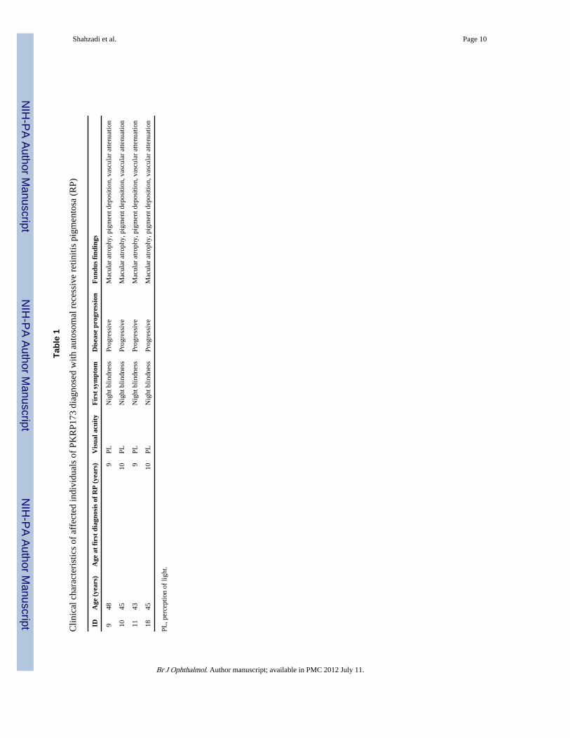
RESULTSPKRP173 was recruited from the Punjab province of Pakistan. A detailed medical andfamily history was obtained by interviewing family members. Three affected and oneunaffected individual underwent detailed ophthalmic examination. According to medicalrecords available to us, all affected individuals started experiencing visual difficulties duringthe first decade of their life; at the time of examination vision was reduced only toperception of light (table 1). Fundus examination revealed marked vascular attenuation, andbony spicules in peripheral regions of the retina combined with atrophic maculopathy withpigment dispersion (figure 1). ERG recordings at 0 dB (scotopic) showed no rod and coneresponse. Isolated cone responses measured at 30 Hz flicker (photopic) were absent (figure2), illustrating an advanced stage of retinopathy encompassing both rod and cone cells of theretina. Retinal attributes and ERG recording in the unaffected individual 12 were completelynormal (figures 1 and 2)
Exclusion studies were performed with closely linked short tandem repeat (STR) markersfor 23 reported arRP loci (data not shown). Haplotypes were constructed to investigatehomozygous regions shared by all the affected individuals of the family. PKRP173 wasfound linked to markers D2S1896, D2S2269 and D2S160 at chromosome 2q14.1 flankingMERTK (figure 3). All the affected individuals were homozygous for D2S1896, D2S2269
Shahzadi et al. Page 3
Br J Ophthalmol. Author manuscript; available in PMC 2012 July 11.
NIH
-PA Author Manuscript
NIH
-PA Author Manuscript
NIH
-PA Author Manuscript
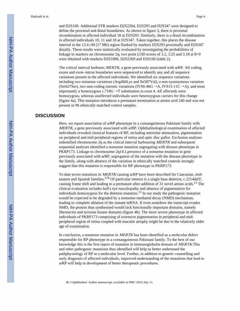
and D2S160. Additional STR markers D2S2264, D2S293 and D2S347 were designed todefine the proximal and distal boundaries. As shown in figure 3, there is proximalrecombination in affected individual 18 at D2S293. Similarly, there is a distal recombinationin affected individuals 10, 11 and 18 at D2S347. Taken together, this places the diseaseinterval in the 13.4 cM (17 Mb) region flanked by markers D2S293 proximally and D2S347distally. These results were statistically evaluated by investigating the probabilities oflinkage to markers on chromosome 2q; two point LOD scores of 3.2, 3.25 and 3.18 at θ=0were obtained with markers D2S1896, D2S2269 and D2S160 (table 2).
The critical interval harbours MERTK, a gene previously associated with arRP. All codingexons and exon–intron boundaries were sequenced to identify any and all sequencevariations present in the affected individuals. We identified six sequence variationsincluding two missense variations (Arg466Lys and Ile587Val), a non-synonymous variation(Ser627Ser), two non-coding intronic variations (IVS6-46G→A, IVS15-11C→A), and mostimportantly a homozygous c.718G→T substitution in exon 4. All affecteds werehomozygous, whereas unaffected individuals were heterozygous carriers for this change(figure 4a). This mutation introduces a premature termination at amino acid 240 and was notpresent in 96 ethnically matched control samples.
DISCUSSIONHere, we report association of arRP phenotype in a consanguineous Pakistani family withMERTK, a gene previously associated with arRP. Ophthalmological examination of affectedindividuals revealed classical features of RP, including arteriolar attenuation, pigmentationon peripheral and mid peripheral regions of retina and optic disc pallor. Exclusion analysesindentified chromosome 2q as the critical interval harbouring MERTK and subsequentsequential analyses identified a nonsense mutation segregating with disease phenotype inPKRP173. Linkage to chromosome 2q14.1,presence of a nonsense mutation in genepreviously associated with arRP, segregation of the mutation with the disease phenotype inthe family, along with absence of the variation in ethnically matched controls stronglysuggest that this mutation is responsible for RP phenotype in PKRP173.
To date seven mutations in MERTK causing arRP have been described for Caucasian, mid-eastern and Spanish families.578 Of particular interest is a single base deletion, c.2214delT,causing frame shift and leading to a premature after-addition of 31 novel amino acids.13 Theclinical evaluation includes bull's eye maculopathy and absence of pigmentation forindividuals homozygous for the deletion mutation.13 In our study the pathogenic mutationwould be expected to be degraded by a nonsense-mediated decay (NMD) mechanism,leading to complete ablation of the mutant mRNA. If even somehow the transcript evadesNMD, the protein thus synthesised would lack functionally important domains, namelyfibronectin and tyrosine kinase domains (figure 4b). The more severe phenotype in affectedindividuals of PKRP173 comprising of extensive pigmentation in peripheral and mid-peripheral region of retina coupled with macular atrophy might be due to the relatively olderage of examination.
In conclusion, a nonsense mutation in MERTK has been identified as a molecular defectresponsible for RP phenotype in a consanguineous Pakistani family. To the best of ourknowledge this is the first report of mutation in immunoglobulin domain of MERTK.Thisand other pathogenic mutations thus identified will help us better understand thepathphysiology of RP at a molecular level. Further, in addition to genetic counselling andearly diagnosis of affected individuals, improved understanding of the mutations that lead toarRP will help in development of better therapeutic procedures.
Shahzadi et al. Page 4
Br J Ophthalmol. Author manuscript; available in PMC 2012 July 11.
NIH
-PA Author Manuscript
NIH
-PA Author Manuscript
NIH
-PA Author Manuscript
AcknowledgmentsThe authors are grateful to all members for their participation in this study. We are also thankful to members of thestaff of Layton Rehmatullah Benevolent Trust (LRBT) hospital for clinical evaluation of affected individuals.
Funding This study was supported, in part by Higher Education Commission and Ministry of Science andTechnology Islamabad, Pakistan.
REFERENCES1. Bird AC. Retinal photoreceptor dystrophies LI. Edward Jackson Memorial Lecture. Am J
Ophthalmol. 1995; 119:543–62. [PubMed: 7733180]
2. Yeung KY, Baum L, Chan WM, et al. Molecular diagnostics for retinitis pigmentosaYEUNG2001Q. Clin Chim Acta. 2001; 313:209–15. [PubMed: 11694261]
3. Hartong DT, Berson EL, Dryja TP. Retinitis pigmentosa. Lancet. 2006; 368:1795–809. [PubMed:17113430]
4. Hims MM, Diager SP, Inglehearn CF. Retinitis pigmentosa: genes, proteins and prospects. DevOphthalmol. 2003; 37:109–25. [PubMed: 12876833]
5. McHenry CL, Liu Y, Feng W, et al. MERTK arginine-844-cysteine in a patient with severe rod—cone dystrophy: loss of mutant protein functions in transfected cells. Invest Ophthalmol Vis Sci.2004; 45:1456–63. [PubMed: 15111602]
6. D'Cruz PM, Yasumura D, Weir J, et al. Mutation of the receptor tyrosine kinase gene Mertk in theretinal dystrophic RCS rat. Hum Mol Genet. 2000; 9:645–51. [PubMed: 10699188]
7. Gal A, Li Y, Thompson DA, et al. Mutations in MERTK, the human orthologue of the RCS ratretinal dystrophy gene, cause retinitis pigmentosa. Nat Genet. 2000; 26:270–1. [PubMed:11062461]
8. Ebermann I, Walger M, Scholl HP, et al. Truncating mutation of the DFNB59 gene causes cochlearhearing impairment and central vestibular dysfunction. Hum Mutat. 2007; 28:571–7. [PubMed:17301963]
9. Brea-Fernandez AJ, Pomares E, Brion MJ, et al. Novel splice donor site mutation in MERTK geneassociated with retinitis pigmentosa. Br J Ophthalmol. 2008; 92:1419–23. [PubMed: 18815424]
10. Kaul H, Riazuddin SA, Yasmeen A, et al. A new locus for autosomal recessive congenital cataractidentified in a Pakistani family. Mol Vis. 2010; 16:240–45. [PubMed: 20161816]
11. Lathrop GM, Lalouel JM. Easy calculations of LOD scores and genetic risks on small computers509. Am J Hum Genet. 1984; 36:460–5. [PubMed: 6585139]
12. Schaffer AA, Gupta SK, Shriram K, et al. Avoiding recomputation in genetic linkage analysis1669. Hum Hered. 1994; 44:225–37. [PubMed: 8056435]
13. Tschernutter M, Jenkins SA, Waseem NH, et al. Clinical characterisation of a family with retinaldystrophy caused by mutation in the Mertk gene. Br J Ophthalmol. 2006; 90:718–23. [PubMed:16714263]
Shahzadi et al. Page 5
Br J Ophthalmol. Author manuscript; available in PMC 2012 July 11.
NIH
-PA Author Manuscript
NIH
-PA Author Manuscript
NIH
-PA Author Manuscript

Figure 1.Fundus photographs of members of family PKRP173. Fundus demonstrating severalfeatures associated with retinitis pigmentosa (RP) including a waxy pallor of the optic disc,attenuated arterioles, atrophy of retinal pigment epithelium (RPE) and peripheral bonespicules. Notably, macular atrophy with pigment clumping in peripheral regions is present.(A) Affected individual 18; (B) affected individual 9; (C) affected individual 10; (D)unaffected individual 12. OD, right eye; OS, left eye.
Shahzadi et al. Page 6
Br J Ophthalmol. Author manuscript; available in PMC 2012 July 11.
NIH
-PA Author Manuscript
NIH
-PA Author Manuscript
NIH
-PA Author Manuscript
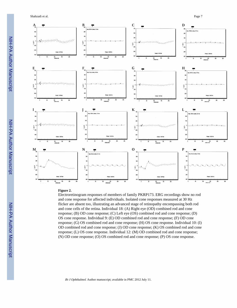
Figure 2.Electroretinogram responses of members of family PKRP173. ERG recordings show no rodand cone response for affected individuals. Isolated cone responses measured at 30 Hzflicker are absent too, illustrating an advanced stage of retinopathy encompassing both rodand cone cells of the retina. Individual 18: (A) Right eye (OD) combined rod and coneresponse; (B) OD cone response; (C) Left eye (OS) combined rod and cone response; (D)OS cone response. Individual 9: (E) OD combined rod and cone response; (F) OD coneresponse; (G) OS combined rod and cone response; (H) OS cone response. Individual 10: (I)OD combined rod and cone response; (J) OD cone response; (K) OS combined rod and coneresponse; (L) OS cone response. Individual 12: (M) OD combined rod and cone response;(N) OD cone response; (O) OS combined rod and cone response; (P) OS cone response.
Shahzadi et al. Page 7
Br J Ophthalmol. Author manuscript; available in PMC 2012 July 11.
NIH
-PA Author Manuscript
NIH
-PA Author Manuscript
NIH
-PA Author Manuscript

Figure 3.Pedigree drawing and haplotype of chromosome 2q markers of family PKRP173. Squaresare males, circles are females, filled symbols are affected individuals, double line betweenindividuals indicates consanguinity and diagonal line through a symbol is deceased familymember. The haplotypes of six adjacent chromosome 2q microsatellite markers are shownwith alleles forming the risk haplotype are shaded black, and alleles not co-segregating withretinitis pigmentosa (RP) are shown in white.
Shahzadi et al. Page 8
Br J Ophthalmol. Author manuscript; available in PMC 2012 July 11.
NIH
-PA Author Manuscript
NIH
-PA Author Manuscript
NIH
-PA Author Manuscript
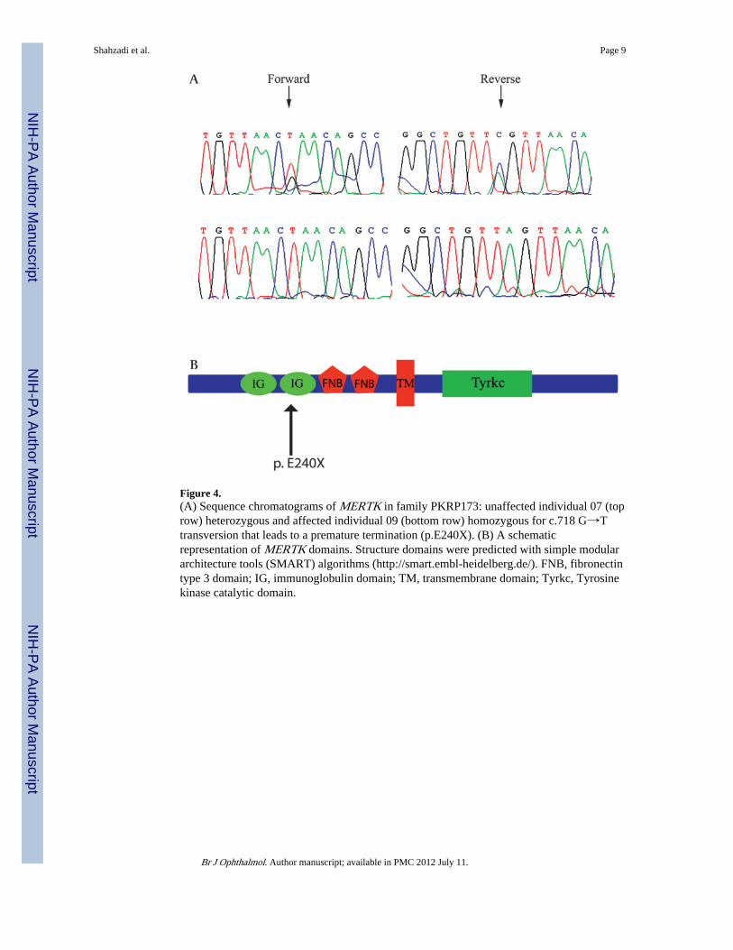
Figure 4.(A) Sequence chromatograms of MERTK in family PKRP173: unaffected individual 07 (toprow) heterozygous and affected individual 09 (bottom row) homozygous for c.718 G→Ttransversion that leads to a premature termination (p.E240X). (B) A schematicrepresentation of MERTK domains. Structure domains were predicted with simple modulararchitecture tools (SMART) algorithms (http://smart.embl-heidelberg.de/). FNB, fibronectintype 3 domain; IG, immunoglobulin domain; TM, transmembrane domain; Tyrkc, Tyrosinekinase catalytic domain.
Shahzadi et al. Page 9
Br J Ophthalmol. Author manuscript; available in PMC 2012 July 11.
NIH
-PA Author Manuscript
NIH
-PA Author Manuscript
NIH
-PA Author Manuscript
NIH
-PA Author Manuscript
NIH
-PA Author Manuscript
NIH
-PA Author Manuscript
Shahzadi et al. Page 10
Tabl
e 1
Clin
ical
cha
ract
eris
tics
of a
ffec
ted
indi
vidu
als
of P
KR
P173
dia
gnos
ed w
ith a
utos
omal
rec
essi
ve r
etin
itis
pigm
ento
sa (
RP)
IDA
ge (
year
s)A
ge a
t fi
rst
diag
nosi
s of
RP
(ye
ars)
Vis
ual a
cuit
yF
irst
sym
ptom
Dis
ease
pro
gres
sion
Fun
dus
find
ings
948
9PL
Nig
ht b
lindn
ess
Prog
ress
ive
Mac
ular
atr
ophy
, pig
men
t dep
ositi
on, v
ascu
lar
atte
nuat
ion
1045
10PL
Nig
ht b
lindn
ess
Prog
ress
ive
Mac
ular
atr
ophy
, pig
men
t dep
ositi
on, v
ascu
lar
atte
nuat
ion
1143
9PL
Nig
ht b
lindn
ess
Prog
ress
ive
Mac
ular
atr
ophy
, pig
men
t dep
ositi
on, v
ascu
lar
atte
nuat
ion
1845
10PL
Nig
ht b
lindn
ess
Prog
ress
ive
Mac
ular
atr
ophy
, pig
men
t dep
ositi
on, v
ascu
lar
atte
nuat
ion
PL, p
erce
ptio
n of
ligh
t.
Br J Ophthalmol. Author manuscript; available in PMC 2012 July 11.

NIH
-PA Author Manuscript
NIH
-PA Author Manuscript
NIH
-PA Author Manuscript
Shahzadi et al. Page 11
Tabl
e 2
Tw
o po
int L
OD
sco
res
of c
hrom
osom
e 2q
mar
kers
for
PK
RP1
73
Mar
ker
cMM
b0
0.01
0.05
0.09
0.1
0.2
0.3
Zm
axθ
max
D2S
2264
114.
410
2.4
– ∞
–0.0
10.
540.
640.
650.
580.
40.
650.
10
D2S
293
118.
110
7.2
– ∞
–0.0
60.
470.
610.
600.
530.
30.
600.
09
D2S
1896
122.
911
2.6
3.20
3.13
2.87
2.61
2.54
1.86
1.18
3.20
0.00
D2S
2269
122.
911
2.9
3.25
3.18
2.91
2.67
2.62
1.96
1.26
3.25
0.00
D2S
160
122.
911
2.9
3.18
3.10
2.84
2.59
2.49
1.85
1.16
3.18
0.00
D2S
347
131.
512
4.2
– ∞
–0.0
10.
540.
640.
650.
580.
40.
650.
10
LO
D, l
ogar
ithm
of
the
odds
.
LO
D s
core
s w
ere
calc
ulat
ed a
t dif
fere
nt θ
val
ues
for
each
mar
ker
with
the
FAST
LIN
K v
ersi
on o
f M
LIN
K f
rom
the
LIN
KA
GE
pro
gram
pac
kage
Max
imum
LO
D s
core
s fo
r ea
ch m
arke
r w
ere
calc
ulat
edus
ing
ILIN
K.
Br J Ophthalmol. Author manuscript; available in PMC 2012 July 11.