Allelic heterogeneity and genetic modifier loci contribute to clinical variation in males with...
11
Allelic Heterogeneity and Genetic Modifier Loci Contribute to Clinical Variation in Males with X-Linked Retinitis Pigmentosa Due to RPGR Mutations Abigail T. Fahim 1 , Sara J. Bowne 1 , Lori S. Sullivan 1 , Kaylie D. Webb 2 , Jessica T. Williams 1 , Dianna K. Wheaton 2,3 , David G. Birch 2,3 , Stephen P. Daiger 1 * 1 Human Genetics Center, School of Public Health, University of Texas Health Science Center at Houston, Houston, Texas, United States of America, 2 Retina Foundation of the Southwest, Dallas, Texas, United States of America, 3 Department of Ophthalmology, University of Texas Southwestern Medical Center, Dallas, Texas, United States of America Abstract Mutations in RPGR account for over 70% of X-linked retinitis pigmentosa (XlRP), characterized by retinal degeneration and eventual blindness. The clinical consequences of RPGR mutations are highly varied, even among individuals with the same mutation: males demonstrate a wide range of clinical severity, and female carriers may or may not be affected. This study describes the phenotypic diversity in a cohort of 98 affected males from 56 families with RPGR mutations, and demonstrates the contribution of genetic factors (i.e., allelic heterogeneity and genetic modifiers) to this diversity. Patients were categorized as grade 1 (mild), 2 (moderate) or 3 (severe) according to specific clinical criteria. Patient DNAs were genotyped for coding SNPs in 4 candidate modifier genes with products known to interact with RPGR protein: RPGRIP1, RPGRIP1L, CEP290, and IQCB1. Family-based association testing was performed using PLINK. A wide range of clinical severity was observed both between and within families. Patients with mutations in exons 1–14 were more severely affected than those with ORF15 mutations, and patients with predicted null alleles were more severely affected than those predicted to make RPGR protein. Two SNPs showed association with severe disease: the minor allele (N) of I393N in IQCB1 (p = 0.044) and the common allele (R) of R744Q in RPGRIP1L (p = 0.049). These data demonstrate that allelic heterogeneity contributes to phenotypic diversity in XlRP and suggest that this may depend on the presence or absence of RPGR protein. In addition, common variants in 2 proteins known to interact with RPGR are associated with severe disease in this cohort. Citation: Fahim AT, Bowne SJ, Sullivan LS, Webb KD, Williams JT, et al. (2011) Allelic Heterogeneity and Genetic Modifier Loci Contribute to Clinical Variation in Males with X-Linked Retinitis Pigmentosa Due to RPGR Mutations. PLoS ONE 6(8): e23021. doi:10.1371/journal.pone.0023021 Editor: Andreas R. Janecke, Innsbruck Medical University, Austria Received February 28, 2011; Accepted July 7, 2011; Published August 12, 2011 Copyright: ß 2011 Fahim et al. This is an open-access article distributed under the terms of the Creative Commons Attribution License, which permits unrestricted use, distribution, and reproduction in any medium, provided the original author and source are credited. Funding: This work was supported by grants from the Foundation Fighting Blindness (www.blindness.org) and grant EY007142 from the Nuclear Energy Institute/National Institutes of Health (www.nei.org) to SPD. The funders had no role in study design, data collection and analysis, decision to publish, or preparation of the manuscript. Competing Interests: The authors have declared that no competing interests exist. * E-mail: [email protected] Introduction Retinitis pigmentosa (RP) is a form of inherited retinal degeneration characterized by progressive loss of photoreceptors. Initially rods are affected and patients present with decreased night vision and mid-peripheral visual field defects. In advanced disease, cones are affected as well and patients progress to macular involvement and eventual blindness. The genetic etiology of RP is exceptionally diverse. Over 40 genes are currently known to cause RP, many displaying allelic heterogeneity, and some demonstrat- ing both dominant and recessive modes of inheritance (RetNet; http://www.sph.uth.tmc.edu/retnet/). Clinical phenotypes are equally diverse in terms of age of onset, clinical severity, rate of progression, degree of cone involvement, and presence of additional sequelae such as macular edema. In particular, X- linked RP (RP3 [MIM 300029]) demonstrates marked variable expressivity among both affected males, with a wide range of severity, and carrier females, who may or may not have clinical symptoms [1,2]. Theories to explain this phenotypic diversity include differences in the disease-causing mutation, genetic modifiers, and environ- mental modifiers. Over 70% of XlRP cases are due to mutations in retinitis pigmentosa GTPase regulator (RPGR [MIM 312610]), which localizes to the connecting cilium in photoreceptors and is thought to play a role in protein transport [3,4,5]. XlRP demonstrates considerable allelic heterogeneity: more than 300 different RPGR mutations have been found to date in families with XlRP (http://rpgr.hgu.mrc.ac.uk). Therefore, clinical variability may be due, in part, to different disease-causing mutations. This prediction is consistent with the remarkable complexity of RPGR splicing, which gives rise to at least 10 different RPGR splice variants in a tissue-specific manner [6,7,8,9]. Therefore the pathologic effect of a particular mutation may depend on its presence or absence in the most prominent retinal splice variants. For example, an estimated 60% of disease-causing mutations in RPGR are found in ORF15, an alternatively spliced exon including exon15 and extending into intron 15 [7]. The ORF15-containing RPGR transcript is abundant in human retina and has been demonstrated as the predominant isoform present in mouse photoreceptor connecting cilia [3,7]. The number of different RPGR protein isoforms expressed in the retina is unclear. One study showed only two isoforms in human retina, one detectable PLoS ONE | www.plosone.org 1 August 2011 | Volume 6 | Issue 8 | e23021
-
Upload
retinafoundation -
Category
Documents
-
view
1 -
download
0
Transcript of Allelic heterogeneity and genetic modifier loci contribute to clinical variation in males with...

Allelic Heterogeneity and Genetic Modifier LociContribute to Clinical Variation in Males with X-LinkedRetinitis Pigmentosa Due to RPGR MutationsAbigail T. Fahim1, Sara J. Bowne1, Lori S. Sullivan1, Kaylie D. Webb2, Jessica T. Williams1, Dianna K.
Wheaton2,3, David G. Birch2,3, Stephen P. Daiger1*
1 Human Genetics Center, School of Public Health, University of Texas Health Science Center at Houston, Houston, Texas, United States of America, 2 Retina Foundation of
the Southwest, Dallas, Texas, United States of America, 3 Department of Ophthalmology, University of Texas Southwestern Medical Center, Dallas, Texas, United States of
America
Abstract
Mutations in RPGR account for over 70% of X-linked retinitis pigmentosa (XlRP), characterized by retinal degeneration andeventual blindness. The clinical consequences of RPGR mutations are highly varied, even among individuals with the samemutation: males demonstrate a wide range of clinical severity, and female carriers may or may not be affected. This studydescribes the phenotypic diversity in a cohort of 98 affected males from 56 families with RPGR mutations, and demonstratesthe contribution of genetic factors (i.e., allelic heterogeneity and genetic modifiers) to this diversity. Patients werecategorized as grade 1 (mild), 2 (moderate) or 3 (severe) according to specific clinical criteria. Patient DNAs were genotypedfor coding SNPs in 4 candidate modifier genes with products known to interact with RPGR protein: RPGRIP1, RPGRIP1L,CEP290, and IQCB1. Family-based association testing was performed using PLINK. A wide range of clinical severity wasobserved both between and within families. Patients with mutations in exons 1–14 were more severely affected than thosewith ORF15 mutations, and patients with predicted null alleles were more severely affected than those predicted to makeRPGR protein. Two SNPs showed association with severe disease: the minor allele (N) of I393N in IQCB1 (p = 0.044) and thecommon allele (R) of R744Q in RPGRIP1L (p = 0.049). These data demonstrate that allelic heterogeneity contributes tophenotypic diversity in XlRP and suggest that this may depend on the presence or absence of RPGR protein. In addition,common variants in 2 proteins known to interact with RPGR are associated with severe disease in this cohort.
Citation: Fahim AT, Bowne SJ, Sullivan LS, Webb KD, Williams JT, et al. (2011) Allelic Heterogeneity and Genetic Modifier Loci Contribute to Clinical Variation inMales with X-Linked Retinitis Pigmentosa Due to RPGR Mutations. PLoS ONE 6(8): e23021. doi:10.1371/journal.pone.0023021
Editor: Andreas R. Janecke, Innsbruck Medical University, Austria
Received February 28, 2011; Accepted July 7, 2011; Published August 12, 2011
Copyright: � 2011 Fahim et al. This is an open-access article distributed under the terms of the Creative Commons Attribution License, which permitsunrestricted use, distribution, and reproduction in any medium, provided the original author and source are credited.
Funding: This work was supported by grants from the Foundation Fighting Blindness (www.blindness.org) and grant EY007142 from the Nuclear EnergyInstitute/National Institutes of Health (www.nei.org) to SPD. The funders had no role in study design, data collection and analysis, decision to publish, orpreparation of the manuscript.
Competing Interests: The authors have declared that no competing interests exist.
* E-mail: [email protected]
Introduction
Retinitis pigmentosa (RP) is a form of inherited retinal
degeneration characterized by progressive loss of photoreceptors.
Initially rods are affected and patients present with decreased night
vision and mid-peripheral visual field defects. In advanced disease,
cones are affected as well and patients progress to macular
involvement and eventual blindness. The genetic etiology of RP is
exceptionally diverse. Over 40 genes are currently known to cause
RP, many displaying allelic heterogeneity, and some demonstrat-
ing both dominant and recessive modes of inheritance (RetNet;
http://www.sph.uth.tmc.edu/retnet/). Clinical phenotypes are
equally diverse in terms of age of onset, clinical severity, rate of
progression, degree of cone involvement, and presence of
additional sequelae such as macular edema. In particular, X-
linked RP (RP3 [MIM 300029]) demonstrates marked variable
expressivity among both affected males, with a wide range of
severity, and carrier females, who may or may not have clinical
symptoms [1,2].
Theories to explain this phenotypic diversity include differences
in the disease-causing mutation, genetic modifiers, and environ-
mental modifiers. Over 70% of XlRP cases are due to mutations in
retinitis pigmentosa GTPase regulator (RPGR [MIM 312610]),
which localizes to the connecting cilium in photoreceptors and is
thought to play a role in protein transport [3,4,5]. XlRP
demonstrates considerable allelic heterogeneity: more than 300
different RPGR mutations have been found to date in families with
XlRP (http://rpgr.hgu.mrc.ac.uk). Therefore, clinical variability
may be due, in part, to different disease-causing mutations.
This prediction is consistent with the remarkable complexity of
RPGR splicing, which gives rise to at least 10 different RPGR splice
variants in a tissue-specific manner [6,7,8,9]. Therefore the
pathologic effect of a particular mutation may depend on its
presence or absence in the most prominent retinal splice variants.
For example, an estimated 60% of disease-causing mutations in
RPGR are found in ORF15, an alternatively spliced exon including
exon15 and extending into intron 15 [7]. The ORF15-containing
RPGR transcript is abundant in human retina and has been
demonstrated as the predominant isoform present in mouse
photoreceptor connecting cilia [3,7]. The number of different
RPGR protein isoforms expressed in the retina is unclear. One
study showed only two isoforms in human retina, one detectable
PLoS ONE | www.plosone.org 1 August 2011 | Volume 6 | Issue 8 | e23021

with a C-terminal antibody specific for the canonical RPGR1–19
(containing exons 1–19), and the other detectable with a C-
terminal exon against the ORF15 alternative exon [10]. Another
study using similarly-placed antibodies showed three different
bands for RPGR 1–19 and five different bands for RPGRORF15,
suggestingting that the various bands could be due to post-
translational modification (i.e., different isotypes) or to alternate
splicing (i.e., different isoforms) [11].
Prior studies have found evidence that different types of RPGR
mutations cause different phenotypes [12,13,14,15,16]. In partic-
ular, patients with mutations in exons 1–14 demonstrated smaller
visual fields than patients with mutations in ORF15 [12]. In
addition, several families with cone-rod dystrophy have been
reported with mutations in RPGR, all of them in ORF15
downstream of ORF15 codon 445 [13,14,15,16]. There is also
evidence that some mutations in RPGR are more likely to be
penetrant in female carriers while other mutations cause disease
only in hemizygous males [1,17,18].
However, allelic heterogeneity alone cannot fully account for
the observed phenotypic variability in XlRP caused by RPGR
mutations. In one report describing two families with the same
disease-causing mutation arising independently, female carriers in
one family had no clinical symptoms while females in the other
family were severely affected [19]. Even members of the same
family can have striking phenotypic differences [20,21]. These
observations strongly suggest the presence of genetic and
environmental modifiers.
This study aimed to categorize the phenotypic diversity in
affected males from 56 families with mutations in RPGR, and to
determine the contribution of genetic factors (i.e. allelic heteroge-
neity and genetic modifiers) to this diversity. Ninety-eight affected
males with 44 different RPGR mutations were included. To
examine genotype-phenotype correlations, mutations were cate-
gorized by the physical location in the gene as well as by the
predicted biologic outcome. A candidate modifier-gene approach
was taken to screen genes meeting the following criteria: 1) the
protein is known to interact with RPGR, 2) the protein has
polymorphic amino acid substitutions, and 3) the gene contains
known retinal disease-causing mutations. Therefore, coding SNPs
in RPGR-interacting protein 1 (RPGRIP1 [MIM 605446]) [22,-
23,24,25], RPGRIP1-like (RPGRIP1L [MIM 610937]) [26,27,
28], centrosomal protein 290 kDa (CEP290 [MIM 610142]) [29,
30,31,32,33,34,35,36], and IQ motif containing B1 (IQCB1 [MIM
609237]; aka nephrocystin-5) [37] were chosen for analysis. This
study demonstrates significant RPGR genotype-phenotype corre-
lations and reports two SNPs in candidate modifier genes
associated with disease severity.
Methods
Patients and clinical assessmentPatients with mutations in RPGR were ascertained from the
Southwest Eye Registry (SER) at the Retina Foundation of the
Southwest (RFSW) [38]. The SER is a database of over 3,000
patients referred to the RFSW for retinal degenerative diseases.
Ninety-eight affected males from 56 families were ascertained.
This study was performed in accordance with the Declaration of
Helsinki and informed consent was obtained from all participants.
The research was approved by the Committees for Protection of
Human Subjects at the University of Texas, Health Science
Center at Houston and the University of Texas Southwestern
Medical Center.
For each patient, manifest refraction and best-corrected visual
acuity were assessed with the NIKON RETINOMAX using the
EVA-ETDRS visual acuity chart. Humphrey visual fields were
obtained using a model 740 Humphrey field analyzer. Program
30-2 was used to determine static parametric thresholds at 74
locations within the central 30 degrees. Frequency domain optical
coherence tomography (fdOCT) retinal scans were obtained using
a Heidelberg Spectralis OCT. Fundus photos were obtained with
a Canon digital camera (CF-60UD) and included a posterior pole
view and additional peripheral views to document bone spicules
and vessel attenuation if present. To assess dark-adaptation, pupils
were maximally dilated using 1.0% cylcopentolate hydrochloride
and 2.5% phenylephrine hydrochloride, followed by 45 minutes of
dark adaptation. The final dark-adapted threshold was determined
using an 11-degree test stimulus located 7 degrees below fixation
on a Goldmann-Weekers dark-adaptometer.
Full-field electroretinograms (ffERG) were obtained according
to ISCEV standards using Burian-Allen electrodes. Signals were
elicited for rod response, mixed rod-cone response, and cone
response, and signals were amplified and computer-averaged.
Multi-focal electroretinograms (mfERG) were obtained using the
Veris system, and responses were recorded from 103 locations
within the central 40 degrees.
Fifty-four of the 98 affected males had 3 or more visits at least 1
year apart. For these individuals, log cone 31 hz flicker ERG
amplitude was plotted as a function of patient age. Linear
regression analysis was used to define a.) the log amplitude at birth
(age 0) and b.) the slope of the best-fit regression line. The
regression line could then be used to determine the predicted
amplitude at age 18, even for those patients who were not tested at
age 18. Predicted amplitude at age 18, therefore, depended on
both the severity at birth (a) and the rate of progression (b). This
analysis assumes a constant rate of progression in log amplitude
versus age, that is, patients are predicted to lose a constant
percentage of amplitude each year. The results of these regression
analyses were consistent with this assumption, as are previous
analyses of ERG longitudinal data [39,40,41].
The 54 patients with multiple visits were characterized as grade
1 (mild), grade 2 (moderate), or grade 3 (severe) primarily on the
basis of the derived cone 31 hz flicker ERG amplitude at age 18,
supplemented by Humphrey visual fields. Supplementary mea-
sures, along with cone ERG amplitude, were also used to
characterize the 44 patients with a single visit. Criteria used to
categorize patients as grade 1, 2 or 3 are summarized in Table 1.
RPGR mutation detectionRPGR mutations were identified or confirmed in-house, in
every family, using our standard RPGR sequencing protocols [42].
Genomic DNA was amplified for 35 cycles with AmpliTaq GoldH360 Master Mix (Applied Biosystems, Foster City, CA) and M13-
tailed primers designed to flank exons 1–19. RPGR ORF15 was
amplified for 40 cycles using AmpliTaq GoldH 360 Master Mix
and the following primers: 59GACTAAACCATAATATCCA-
AATCCA39 and 9GCCAAAATTTACCAGTGCCTCCTAT39.
PCR product was treated with ExoSAPIT (Affymetrix, Santa
Clara, CA) and sequenced using BigDye v1.1 (Applied Biosys-
tems). Exons 1–19 were sequenced in two directions using M13
primers, and ORF15 was sequenced unidirectionally using a set of
7 nested primers. Sequence reactions were purified using BigDye
Xterminator, run on a 3500 Genetic Analyzer., and analyzed
using SeqScape v2.7 (Applied Biosystems). A minimum of one
affected male was tested in each family.
Haplotype analysis of RPGRIP1 and RPGRIP1LDNA samples from hybrid murine-human cell lines monosomic
for either human chromosome 14 or 16 were a gift from Dr. James
Genetic Contribution to Clinical Variation in XLRP
PLoS ONE | www.plosone.org 2 August 2011 | Volume 6 | Issue 8 | e23021

Hixson [43]. All coding SNPs in RPGRIP1 and RPGRIP1L, with
documented minor allele frequency of at least 1.0% in dbSNP or
in the literature [22,26], were sequenced in cell lines monosomic
for human chromosome 14 or 16, respectively. Each SNP was
sequenced in at least 100 cell lines, including 32–38 cell lines
derived from each of three ethnicities: African-Americans (from
Atlanta, GA), European-Americans (from Rochester, MN), and
Mexican-Americans (from Starr County, TX). Exons in RPGRIP1
and RPGRIP1L containing candidate SNPs were amplified from
80 ng of genomic DNA per reaction. PCR was performed using
AmpliTaq Gold 360 (Applied Biosystems, Carlsbad, CA) in a
12.5 uL reaction, including 0.25 uL of GC enhancer (Applied
Biosystems), primers at 0.2 uM each, and bovine serum albumin
at 144 mg/mL. PCR conditions were as follows: 95uC for
15 minutes, 35 cycles of 95uC for 45 seconds, 56uC for
45 seconds, and 72uC for 45 seconds, followed by extension at
72uC for 5 minutes. Two uL of each PCR product was purified in
a 4 uL reaction using ExoSapIt (USB, Cleveland, OH), and
purified product was sequenced using BigDye v1.1 (Applied
Biosystems) in both directions. For each sequencing reaction, 2 uL
of purified PCR product was sequenced in a 5 uL reaction with
0.16 uM sequencing primer. Sequencing reactions were purified
using BigDye Xterminator (Applied Biosystems) according to
manufacturer’s instructions and run on an ABI 3730 DNA
Analyzer (Applied Biosystems). Sequence results were analyzed
using SeqScape v2.6 (Applied Biosystems).
Genotyping candidate modifier loci in patientsBlood samples were collected in EDTA-coated tubes and
centrifuged at 30006g for 10 minutes. The leukocyte-rich buffy
coat interface was separated and stored at 280uC. DNA was later
extracted using the Gentra Puregene blood kit (Qiagen, Valencia,
CA). DNA sequences containing SNPs of interest were amplified
from 37.5 ng genomic DNA per reaction with either AmpliTaq
Gold 360 (Applied Biosystems) and the conditions described
above, or PyroMark PCR Kit (Qiagen).
The PyroMark PCR reactions were performed for subsequent
pyrosequencing, and therefore included two specific primers, one
contained an M13 tail, and the other a universal biotinylated M13
primer, as described by Guo and Milewicz [44]. The final
concentrations of the universal biotinylated M13 primer and the
specific un-tailed primer were 0.2 uM each, and the final
concentration of the specific primer with an M13 tail was
0.01 uM in a 12.5 uL reaction volume. PCR conditions for
PyroMark reactions were as follows: 95uC for 15 minutes, 45
cycles of 94uC for 30 seconds, 52uC for 30 seconds, and 72uC for
30 seconds, followed by extension at 72uC for 10 minutes. Five uL
of PCR product was added to 5 uL water, 2 uL streptavidin-
sepharose beads (GE Healthcare Bio-Sciences AB, Uppsala,
Sweden), and 38 uL PyroMark Binding Buffer (Qiagen), and
vortexed for 30 minutes. PCR product was then captured on a
PyroMark Vacuum Prep Tool (Qiagen) by aspiration according to
manufacturer’s instructions, and washed in 70% ethanol for
5 seconds, Pyromark Denaturation Solution (Qiagen) for 5 sec-
onds, and PyroMark Wash Buffer (Qiagen) for 5 seconds. PCR
product was then released into 15 uL of 0.3 uM sequencing
primer diluted in PyroMark Annealing Buffer (Qiagen), heated at
70uC for 15 minutes, and cooled at room temperature for at least
10 minutes before running on a PSQ HS 96A Instrument
(Qiagen).
Data analysisGenotype-phenotype correlations were analyzed using the
Fisher Exact Test and generalized estimating equation (GEE)
analysis. GEE analysis is a variance-minimization strategy that
tests for significant associations in cohorts that are organized into
clusters, such as families, and accounts for highly correlated
phenotypes within a cluster that may be due to factors other than
the variable being tested [45]. In this case, the ‘‘other factors’’
would be shared genes other than RPGR. GEE analysis accounts
for this by performing a weighted analysis, in which the weight
assigned to each individual is inversely proportional to the degree
of correlation of phenotypes within that individual’s family.
Family-based association testing was performed using PLINK
(http://pngu.mgh.harvard.edu/purcell/plink/) [46]. Specifically,
the DFAM procedure was used, which incorporates TDT using
parent-child trios, sib-TDT using discordant sibships, and
unrelated individuals, which are stratified into clusters and treated
as sibships using the Cochran-Mantel-Haenszel test. In this
analysis, which requires a dichotomous phenotype, patients with
grade 1 (mild) or 3 (severe) RP (as defined above under ‘‘Patients
and Clinical Assessment’’) were compared, while patients with
grade 2 (moderate) RP were excluded. Significance values were
not corrected for multiple comparisons given the limited number
of SNPs tested.
Results
A wide range of clinical consequences was observedboth within and between families
Ninety-eight affected males from 56 families with mutations in
RPGR were enrolled. Clinical work-up included best-corrected
visual acuity, Humphrey visual fields, fdOCT, dark-adapted
threshold, ffERG, mfERG, and fundus exam with photography.
Patients were characterized as having grade 1 (mild), 2 (moderate),
or 3 (severe) disease according to the guidelines in Table 1.
Representative fundus photos, ERGs, and visual fields from each of
these three categories are shown in Figure 1. For 54 of the 98 males,
ERGs from multiple visits were available, and linear regression
analysis was used to derive cone 31 hz flicker ERG amplitude at age
18. This information was used in assigning clinical severity
according to the guidelines in Table 1. Figure 2 shows longitudinal
cone ERG data from individuals with grades 1, 2, and 3 RP. The
cohort included 21 grade 1, 34 grade 2, and 43 grade 3 individuals
as defined by these standards. Of note, there were no unaffected
males with known mutations, consistent with the fact that RPGR
Table 1. Criteria for assigning clinical categories to males with RPGR mutations.
Severity ERG Visual Field
Grade 1 $10 mvolts 30 Hz flicker in teens (1–5 microvolts in 30’s) Good central 30u field sensitivity
Grade 2 1–5 mvolts 30 Hz flicker in teens Marked central field constriction; some sensitivity beyond 20u
Grade 3 ,1 mvolt 30 Hz flicker in teens/20’s Marked central field constriction
doi:10.1371/journal.pone.0023021.t001
Genetic Contribution to Clinical Variation in XLRP
PLoS ONE | www.plosone.org 3 August 2011 | Volume 6 | Issue 8 | e23021

Figure 1. Representative images and studies for grade 1, 2, and 3 RP. Representative fdOCTs, ffERGs, fundus photos, and visual fields (OS)are shown for a male with an RPGR mutation in each of the three clinical categories. Mild = grade 1; Moderate = grade 2; Severe = grade 3.doi:10.1371/journal.pone.0023021.g001
Figure 2. Representative ERGs for grade 1, 2, and 3 RP. Longitudinal cone 31 hz flicker ERGs are shown for a male with an RPGR mutation ineach of the three clinical categories. Mild = grade 1; Moderate = grade 2; Severe = grade 3.doi:10.1371/journal.pone.0023021.g002
Genetic Contribution to Clinical Variation in XLRP
PLoS ONE | www.plosone.org 4 August 2011 | Volume 6 | Issue 8 | e23021

mutations have historically been fully penetrant in the hemizygous
state. Although most pedigrees were small, with only one or two
affected individuals ascertained, some of the larger pedigrees
demonstrated intrafamilial phenotypic variability (Figure 3).
Predicted null alleles are associated with more severedisease while alleles predicted to produce variantproteins are associated with more phenotypic variability
The 56 families carried 44 different mutations in RPGR
(Table 2). Forty-six of the 98 affected males had mutations in
exons 1–14, and 52 had mutations in ORF15. The distribution of
clinical severity differed significantly between these two mutation
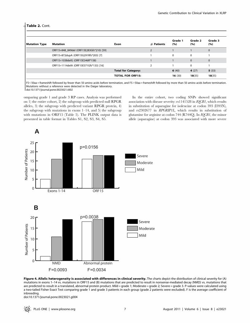
groups (Figure 4A). Patients with mutations in exons 1–14 were
more likely to be severely affected than patients with mutations in
ORF15 (p = 0.016). The group with ORF15 mutations had a
greater degree of phenotypic variability, with patients more evenly
distributed among the 3 clinical categories.
Patients were regrouped according to whether the mutation was
predicted to result in a translated protein product. Premature stop
codons in exons 1–14, or frameshift mutations in exons 1–14 leading
to premature stop codons, are predicted to result in nonsense-
mediated decay of the transcript and are therefore predicted null
alleles. ORF15, by contrast, is the terminal exon of the ORF15
variant transcript, and therefore nonsense mutations and frameshifts
in ORF15 may result in a stable transcript which is translated into a
variant protein product. Missense mutations are also predicted to
result in translated variant proteins. Splice site mutations are
unpredictable in their effects on transcription and translation since
they may eliminate the transcript, may eliminate an exon, may favor
a cryptic splice site, may shift the fraction of alternate transcripts,
and/or may occasionally produce a correctly-spliced transcript. For
this reason, splice-site variants were excluded from the analysis.
Figure 4B shows a striking difference in clinical severity between
these two groups, similar to the difference observed based on
mutation location. Predicted null alleles were associated with more
severe disease, while alleles predicted to result in variant protein
demonstrated more phenotypic variability (p = 0.0038). The
average coefficient of inbreeding (F) was 0.0093 for the group with
predicted null alleles and 0.0034 for the group with predicted
variant protein (Fig. 4B). The increased relatedness in the former
group indicates that shared genetic factors other than the disease-
causing mutation may account for the increased clinical severity. If
only individuals separated by at least 4 meioses are included (sharing
#6.25% of their DNA), then the difference between mutations in
exons 1–14 and ORF15 is suggestive but not significant (p = 0.083),
while the difference between predicted null alleles and predicted
translated protein alleles remains significant (p = 0.049). In addition,
generalized estimating equation (GEE) analysis was used to further
assess the difference in disease severity between patients with
predicted null alleles and patients with predicted translated protein,
and confirmed that the presence of a predicted null RPGR allele is
associated with more severe disease (p = 0.040).
RPGRIP1 and RPGRIP1L protein haplotypesWhen considering polymorphic variation in a protein, the protein
haplotypes may be more biologically relevant than individual amino
acid substitutions. We aimed to determine the common protein
haplotypes in two of our candidate modifiers in order to fulfill two
goals, 1) to determine the degree of polymorphic variation in the
candidate modifier proteins, and 2) to assist in determining phase
when coding SNPs were genotyped in patients.
In order to find common protein haplotypes of RPGRIP1 and
RPGRIP1L, we used DNA from murine-human hybrid cell lines (a
gift from Dr. Jim Hixson) that were monosomic for either human
chromosome 14 or 16, including the RPGRIP1 and RPGRIP1L
genes, respectively [43]. Coding SNPs from dbSNP and the
literature [22,26] were sequenced in at least 100 monosomic cell
lines, including 32–38 lines derived from each of three ethnicities:
African-American, European-American, and Mexican-American
(Tables 3 and 4). Far more variation was found in RPGRIP1, with
14 different polymorphic protein haplotypes and a frequency of
41% for the most common haplotype, compared to only 5 different
protein haplotypes for RPGRIP1L and a frequency of 85% for the
most common. Note that for each protein, the most common
haplotype overall is also the most common haplotype within each
ethnicity. However, there were also striking differences among
ethnicities. In particular, RPGRIP1 haplotype #9 is the second
most common haplotype in European-Americans at 25%, but had
frequencies of only 2.9% and 0% in Mexican-Americans and
African-Americans, respectively.
Modifier SNPsCommon coding SNPs (MAF$2%) in RPGRIP1, RPGRIP1L,
CEP290, and IQCB1 (a.k.a. NPHP5) were sequenced in all 98
affected males as well as 99 available family members. Family-
based association testing was performed using PLINK [46]
Figure 3. A large pedigree with a mutation in RPGR showing intrafamilial phenotypic variability. 1 = grade 1 (mild), 2 = grade 2(moderate), 3 = grade 3 (severe).doi:10.1371/journal.pone.0023021.g003
Genetic Contribution to Clinical Variation in XLRP
PLoS ONE | www.plosone.org 5 August 2011 | Volume 6 | Issue 8 | e23021

Table 2. Mutations in 98 males from 56 families divided by location and mutation type.
Mutation Type Mutation Exon # PatientsGrade 1(%)
Grade 2(%)
Grade 3(%)
Ex1–14 Missense c.194G.A (p.G65D) [16] 3 2 0 1 1
c.296C.T (p.T99I) 4 1 1 0 0
c.617C.T (p.T206I) 6 1 0 0 1
c.815G.A (p.G272D) 8 1 1 0 0
Total for Category: 2 (40) 1 (20) 2 (40)
Ex1–14 Splice Site IVS2-1G.A [52] 3 1 2 0
c.301_IVS4+3del 2 0 1 1
IVS4+3A.G [53] 1 0 0 1
IVS7-1G.A [12] 1 0 1 0
IVS13-8 A.G [54] 1 0 0 1
IVS13-1_1576delGAAACinsAA 1 0 0 1
Total for Category: 1 (12) 4 (44) 4 (44)
Ex1–14 Nonsense c.730A.T (p.K244X) [55] 7 3 0 0 3
c.838_842del (p.L280X) [12] 8 2 0 1 1
c.851C.G (p.S284X) 8 1 0 1 0
c.1126G.T (p.E376X) 10 2 0 0 2
Total for Category: 0 (0) 2(25) 6 (75)
Ex1–14 Frameshift c.101del (p.N34Mfs*34) [55] 2 10 0 2 8
c.219del (p.A74Pfs*11) 3 1 0 1 0
c.356del (p.L119Wfs*14) [56] 5 1 0 1 0
c.372del (p.E125Kfs*8) [53] 5 1 0 0 1
c.629_639del (p.E210Vfs*5) [57] 7 2 0 0 2
c.1092dupA (p.A365Cfs*12) 10 1 0 0 1
c.1243_1244del (p.R415Gfs*37) 10 2 0 2 0
c.1319_1322del (p.C440Ffs*35) 11 2 0 2 0
c.1377_1378del (p.L460Ifs*2) [58] 11 3 1 1 1
c.1662_1665del (p.E555Gfs*14) 14 1 1 0 0
Total for Category: 2 (8) 9 (38) 13 (54)
TOTAL FOR EX1–14: 5(11) 16(35) 25(54)
ORF15 Nonsense ORF15+327A.T (ORF15K109X) [12] 1 0 0 1
ORF15+393G.T (ORF15Glu129X) [57] 2 1 0 1
ORF15+423G.T (ORF15Glu141X) 1 0 1 0
ORF15+459G.T (ORF15G153X) 2 0 0 2
Total for Category: 1 (17) 1 (17) 4 (66)
ORF15 FS ,50aa ORF15+483_484del (ORF15E161Rfs*23) [7] 6 1 1 4
ORF15+587del (ORF15A196Rfs*34) [12] 3 0 3 0
ORF15+652_653del (ORF15E217Gfs*32) [7] 17 6 7 4
ORF15+673_674del (ORF15E224Gfs*25) [7] 2 0 1 1
ORF15+689_692del (ORF15G232fs*2) [7] 2 1 1 0
ORF15+730_743del (ORF15E828Gfs*2) 1 1 0 0
Total for Category: 9 (29) 13 (42) 9 (29)
ORF15 FS.50aa ORF15+185_186insAGAGG (ORF15A62Rfs*52) [57] 1 0 0 1
ORF15+481_484del (ORF15R160Kfs*69) [12] 1 0 0 1
ORF15+499_502del (ORF15K166fs*229) [57] 2 2 0 0
ORF15+521_524del (ORF15G174Kfs*55) 1 0 1 0
ORF15+720del (ORF15E240Rfs*264) 1 0 1 0
ORF15+763_767del (ORF15E254Gfs*238) [12] 2 1 1 0
ORF15 c.764_765del (ORF15E256Gfs*237) [12] 1 0 0 1
Genetic Contribution to Clinical Variation in XLRP
PLoS ONE | www.plosone.org 6 August 2011 | Volume 6 | Issue 8 | e23021
c-
omparing grade 1 and grade 3 RP cases. Analysis was performed
on 1) the entire cohort, 2) the subgroup with predicted null RPGR
alleles, 3) the subgroup with predicted variant RPGR protein, 4)
the subgroup with mutations in exons 1–14, and 5) the subgroup
with mutations in ORF15 (Table 5). The PLINK output data is
presented in table format in Tables S1, S2, S3, S4, S5.
In the entire cohort, two coding SNPs showed significant
association with disease severity: rs1141528 in IQCB1, which results
in substitution of asparagine for isoleucine at codon 393 (I393N),
and rs2302677 in RPGRIP1L, which results in substitution of
glutamine for arginine at codon 744 (R744Q). In IQCB1, the minor
allele (asparagine) at codon 393 was associated with more severe
Mutation Type Mutation Exon # PatientsGrade 1(%)
Grade 2(%)
Grade 3(%)
ORF15+848_849del (ORF15E283Gfs*210) [59] 2 1 1 0
ORF15+872dupA (ORF15G291Rfs*203) [7] 1 0 0 1
ORF15+1038delG (ORF15E346R*158) 1 1 0 0
ORF15+1114delA (ORF15E371Gfs*133) [16] 2 1 0 1
Total for Category: 6 (40) 4 (27) 5 (33)
TOTAL FOR ORF15: 16 (30) 18(35) 18(35)
FS,50aa = frameshift followed by fewer than 50 amino acids before termination, and FS.50aa = frameshift followed by more than 50 amino acids before termination.Mutations without a reference were detected in the Daiger laboratory.doi:10.1371/journal.pone.0023021.t002
Table 2. Cont.
Figure 4. Allelic heterogeneity is associated with differences in clinical severity. The charts depict the distribution of clinical severity for (A)mutations in exons 1–14 vs. mutations in ORF15 and (B) mutations that are predicted to result in nonsense-mediated decay (NMD) vs. mutations thatare predicted to result in a translated, abnormal protein product. Mild = grade 1; Moderate = grade 2; Severe = grade 3. P-values were calculated usinga two-tailed Fisher Exact Test comparing grade 1 and grade 3 patients in each group (grade 2 patients were excluded). F is the average coefficient ofinbreeding.doi:10.1371/journal.pone.0023021.g004
Genetic Contribution to Clinical Variation in XLRP
PLoS ONE | www.plosone.org 7 August 2011 | Volume 6 | Issue 8 | e23021

disease (p = 0.044), while in RPGRIP1L, the common allele
(arginine) at codon 744 was associated with more severe disease
(p = 0.049). In the group with RPGR mutations predicted to result
in a translated protein product, no SNPs showed significant
association with disease severity. However, in the group with
predicted null RPGR alleles, the association between the asparagine
at codon 393 (I393N) in IQCB1 increased in significance (p = 0.030).
Association testing was also performed on groups divided by
mutation location: exons 1–14 vs. ORF15. In the ORF15 group,
which has extensive overlap with the group predicted to result in a
translated protein product, no SNPs showed significant association
with disease severity. In the exons 1–14 group, the minor allele at
I393N in IQCB1 again showed association with disease severity,
increased in significance compared to the total cohort (p = 0.018).
Two SNPs in RPGRIP1 which were not associated with disease
severity in the total cohort showed significant association in the
exons 1–14 group: the minor allele at K192E (p = 0.036) and the
minor allele at E1033Q (0.046).
Discussion
This study aimed to categorize the phenotypic variation in
affected males with XlRP from 52 families carrying mutations in
RPGR, and to determine the contribution of allelic heterogeneity
and genetic modifiers to this variation. We report considerable
variation in clinical severity both between and within families. A
significant genotype-phenotype correlation was observed: indi-
viduals with mutations in exons 1–14 are more likely to have
severe disease than individuals with mutations in ORF15, which
is consistent with previous observations [12]. Furthermore,
individuals with predicted null alleles are also more likely to
have severe disease in comparison to the group predicted to have
variant RPGR proteins, which demonstrates more phenotypic
diversity.
A number of studies support the predication that a premature
termination mutation in other than the final exon of a gene results
in nonsense-mediated decay [47,48]. For example, patients with
the RPGR Q236X nonsense variant have non-detectable RPGR
protein by western blot in lymphoblasts, while control lympho-
blasts demonstrate both RPGR1–19 and RPGRORF15 protein
isoforms [49]. However, categorization of mutant alleles into
predicted nulls vs. predicted translated proteins is based on our
current understanding of nonsense-mediated decay and may not
reflect the biologic presence or absence of protein translation in
every case, nor specifically within the retina. Nonetheless, these
Table 3. Common protein haplotypes of RPGRIP1.
HaplotypeP96Q(ex 3)
K192E(ex 4)
A547S(ex 13)
R598Q(ex 14)
A960P(ex 17)
E1033Q(ex 18)
D1114G(ex 21)
D1150T(ex 21)
G1240E(ex 23)
Overallfreq
AAfreq
EAfreq
MAfreq
1 Pro Lys Ala Arg Ala Glu Asp Asp Gly 0.410 0.485 0.375 0.371
2 Gln Glu Ala Arg Ala Glu Asp Asp Gly 0.150 0.091 0.031 0.314
3 Gln Glu Ala Gln Ala Glu Asp Asp Glu 0.02 0.061 0 0
4 Pro Glu Ala Gln Ala Glu Asp Asp Glu 0.02 0.061 0 0
5 Pro Glu Ala Arg Ala Glu Asp Asp Gly 0.090 0.091 0 0.171
6 Pro Glu Ala Arg Ala Glu Asp Tyr Glu 0.01 0.03 0 0
7 Pro Glu Ser Arg Ala Glu Asp Asp Glu 0.01 0.03 0 0
8 Pro Glu Ser Arg Ala Glu Asp Asp Gly 0.070 0.061 0.125 0.029
9 Pro Glu Ser Arg Ala Gln Asp Asp Gly 0.090 0 0.250 0.029
10 Pro Glu Ala Arg Ala Gln Asp Asp Gly 0.080 0 0.156 0.086
11 Pro Lys Ala Arg Ala Gln Asp Asp Gly 0.02 0 0.063 0
12 Pro Lys Ser Arg Ala Glu Asp Asp Gly 0.01 0.03 0 0
13 Pro Lys Ala Arg Pro Glu Asp Asp Gly 0.01 0.03 0 0
14 Pro Lys Ala Arg Ala Glu Gly Asp Gly 0.01 0.03 0 0
Protein haplotypes from over 100 cell lines monosomic for chromosome 14, and their frequencies. Minor alleles are in bold. Ex = exon, freq = frequency, AA = AfricanAmerican, EA = European American, MA = Mexican American.doi:10.1371/journal.pone.0023021.t003
Table 4. Common protein haplotypes of RPGRIP1L.
HaplotypeA229T(ex 6)
R744Q(ex 16)
G1025S(ex 21)
D1264N(ex 26) Overall frequency AA freq EA freq MA freq
1 Ala Arg Gly Asp 0.851 0.842 0.897 0.811
2 Thr Arg Ser Asp 0.053 0.053 0 0.108
3 Ala Arg Ser Asp 0.035 0.079 0 0.027
4 Ala Arg Ser Asn 0.026 0 0.026 0.054
5 Ala Gln Gly Asp 0.035 0.026 0.077 0
Protein haplotypes from over 100 cell lines monosomic for chromosome 16, and their frequencies. Minor alleles are in bold. Ex = exon, freq = frequency, AA = AfricanAmerican, EA = European American, MA = Mexican American.doi:10.1371/journal.pone.0023021.t004
Genetic Contribution to Clinical Variation in XLRP
PLoS ONE | www.plosone.org 8 August 2011 | Volume 6 | Issue 8 | e23021

findings suggest that expression of variant RPGR protein is
conducive to variable expressivity of disease while the absence of
protein more uniformly causes severe disease.
Candidate modifier loci were chosen on the basis of previously
demonstrated protein-protein interaction with RPGR, known
retinal disease-causing mutations, and known common protein
variation. RPGRIP1 and RPGRIP1L have been shown to interact
with RPGR in yeast 2-hybrid and co-immunoprecipitation
experiments [23,26], and both proteins localize to the photore-
ceptor connecting cilium [24,26]. Mutations in RPGRIP1 cause
Leber congenital amaurosis (LCA6 [MIM 605446]) and cone-rod
dystrophy (CORD13 [MIM 608194]) [22,25], and mutations in
RPGRIP1L cause Joubert syndrome (JBTS7 [MIM 611560]) and
Meckel syndrome (MKS5 [MIM 611561]) [27,28], two syndromic
ciliopathies with associated retinal dystrophy. CEP290 and IQCB1
have shown interaction with RPGR by co-immunoprecipitation
and both localize to the photoreceptor connecting cilium [29,37],
while IQCB1 also localizes to the primary cilia in renal epithelial
cells [37]. Mutations in CEP290 cause Joubert syndrome (JBTS5
[MIM 610188]), Meckel syndrome (MKS4 [MIM 611134]),
Senior Loken syndrome (SLSN6 [MIM 610189]), Leber congen-
ital amaurosis (LCA10 [MIM 611755]) [30,31,32,33,34,35], and
Bardet Biedel syndrome (BBS14 [MIM 209900]) [36], while
mutations in IQCB1 are the most frequent cause of Senior Loken
syndrome (SLSN5 [MIM 609254]) [37], a syndromic disease with
both nephronophthisis and retinitis pigmentosa.
Two coding SNPs in candidate modifier genes showed
marginally significant association with disease severity in our
cohort: the asparagine (N) allele of I393N in IQCB1 and the
arginine (R) allele of R744Q in RPGRIP1L. We hypothesized that
patients with no RPGR protein may be susceptible to different
modifying effects than patients with variant RPGR protein, and
therefore we separated the cohort into patients predicted to have
RPGR protein and those predicted to have no RPGR protein and
recalculated the SNP associations. While no SNPs showed
significant association in the subgroup with predicted RPGR
protein, the N allele of I393N in IQCB1 was associated with
severity in the group predicted to have no RPGR protein, and this
association was more significant in the subgroup than in the total
cohort. Furthermore, this same SNP also showed significant
association with disease severity in the subgroup of patients with
mutations in exons 214.
Polyphen-2 (http://genetics.bwh.harvard.edu/pph2/) [50] pre-
dicts the N allele in I393N to be benign while the R allele in
R744Q is predicted to be probably damaging, with a difference in
position-specific independent count (PSIC) score of 0.94. Amino
acid residue 393 in IQCB1 lies within one of two calmodulin-
binding domains, and IQCB1 has demonstrated interaction with
calmodulin-2 by yeast 2-hybrid experiments and co-immunopre-
cipitation from retinal extracts [37]. Calmodulin has been shown
to modulate the rod cGMP-gated cation channel mediating rod
visual transduction [51]. Amino acid residue 744 in RPGRIP1L
lies between the two C2 domains (PKC conserved region 2 motif)
[28].
The more C-terminal C2 domain of RPGRIP1L is responsible
for binding to nephrocystin-4, and mutations in NPHP4 which
cause Senior Loken Syndrome disrupt this interaction with
RPGRIP1L [27]. Studies have shown that the carboxyl-terminus
of RPGRIP1L is responsible for binding to RPGR, although the
boundaries of the RPGR-binding domain have not been defined
[26]. Given the location of the RPGR-binding domain in
RPGRIP1, which is 29% identical to RPGRIP1L at the amino
acid level and shares the same general domain structure [27], it is
reasonable to postulate that amino acid 744 in RPGRIP1L lies
outside the RPGR-binding domain.
Both modifier loci identified in this study showed marginally
significant association with severity. Future studies should include
genotyping these SNPs in additional males with RPGR mutations to
either confirm or refute these findings, in addition to functional
experiments to investigate a biologic mechanism for the modifying
effect. As with all association studies, it is possible that a coding SNP
showing association is not the true modifier locus, but is in linkage
disequilibrium with the causal SNP. This is especially relevant if the
true modifier is a common non-coding SNP, since we only tested
coding SNPs with MAF$2%. On the other hand, a single rare
variant would not be sufficient to explain the high frequency of
severe and mild cases, and a series of rare variants in aggregate
should not be in disequilibrium with a single common variant.
A common variant in the RPGRIP1L protein, A229T, was
recently reported to be associated with the presence of retinopathy
in a cohort of syndromic ciliopathy patients [26]. The current
study does not demonstrate significant association between the
A229T variant and clinical severity in males with RPGR
mutations. However, our analysis of common RPGRIP1L protein
haplotypes in monosomic cell lines revealed that the A229T
variant occurred on the background of the G1025S variant and
that these 2 loci are in linkage disequilibrium. While G1025S is
frequently present without A229T, the converse was not observed.
Analysis of protein haplotypes is important in our study, because
the protein haplotype is the true ‘‘allele’’ that interacts with RPGR
and therefore has the most biologic relevance when considering
protein-protein interactions. However, association testing of
protein haplotypes did not reveal any significant associations in
our cohort. This may be in part due to sample size: while SNP
Table 5. Two candidate modifier loci are associated with disease severity in the cohort.
Gene: RPGRIP1 RPGRIP1L CEP290 IQCB1
cSNP: P96Q K192E A547S E1033Q A229T R744Q G1025S D1264N K838E L906W I393N C434Y
P value all 0.6224 0.3666 0.6835 0.2943 0.5637 0.0493 0.5227 0.9196 0.6984 NA 0.0438 0.9774
P value protein 0.0768 0.3721 0.2230 0.3018 0.5637 0.2444 0.2383 0.2677 0.3483 NA 1.0000 0.4239
P value null 0.0782 0.0834 0.1195 0.0770 NA 0.4551 0.7288 0.2572 0.1883 NA 0.0298 0.4870
P value ex 1–14 0.0707 0.0358 0.2658 0.0455 0.3173 0.3046 0.6687 0.5909 0.3046 NA 0.0176 0.2353
P value ORF15 0.0768 0.1706 0.0737 0.2528 1.0000 0.2444 0.6749 0.5318 0.3483 NA 1.0000 0.2296
Common coding SNPs in candidate modifier genes were tested for association with disease severity in the total cohort (‘‘all’’ in left column) and in the subgroups withpredicted translated RPGR protein (‘‘protein’’ in left column), with predicted null alleles (‘‘null’’ in left column), with exon 1–14 mutations (‘‘ex 1–14’’ in left column), andwith ORF15 mutations (‘‘ORF15’’ in left column). P-values,0.05 are shown in bold.doi:10.1371/journal.pone.0023021.t005
Genetic Contribution to Clinical Variation in XLRP
PLoS ONE | www.plosone.org 9 August 2011 | Volume 6 | Issue 8 | e23021
analysis is bi-allelic, haplotype analysis is multi-allelic and
therefore the analysis includes fewer patients for each haplotype
‘‘allele.’’
The discovery of modifier genes provides new avenues of
investigation regarding both disease biology and potential
therapeutics. In addition, modifier genes have potential prognostic
utility. While individual genetic modifier loci may have a small
impact on disease severity on their own, the goal is to continue
defining the total genetic contribution to disease in order to build a
more complete prognostic toolset. This includes both the
underlying Mendelian mutations, as well as genetic modifiers
influencing phenotypic expression. This study demonstrates a
contribution from both of these types of variables to phenotypic
heterogeneity.
Supporting Information
Table S1 Output data from PLINK Dfam analysis ofSNP association with disease severity in all grade 1 and3 patients. CHR = chromosome number, SNP = SNP identifier,
A1 = minor allele, A2 = major allele, OBS = number of observed
minor alleles, EXP = number of expected minor alleles,
CHISQ = Chi-squared test statistic, P = asymptotic p-value.
(DOC)
Table S2 Output data from PLINK Dfam analysis ofSNP association with disease severity in grade 1 and 3patients predicted to make a translated RPGR protein.CHR = chromosome number, SNP = SNP identifier, A1 = minor
allele, A2 = major allele, OBS = number of observed minor alleles,
EXP = number of expected minor alleles, CHISQ = Chi-squared
test statistic, P = asymptotic p-value.
(DOCX)
Table S3 Output data from PLINK Dfam analysis ofSNP association with disease severity in grade 1 and 3
patients predicted to have a null RPGR allele.CHR = chromosome number, SNP = SNP identifier, A1 = minor
allele, A2 = major allele, OBS = number of observed minor alleles,
EXP = number of expected minor alleles, CHISQ = Chi-squared
test statistic, P = asymptotic p-value.
(DOCX)
Table S4 Output data from PLINK Dfam analysis ofSNP association with disease severity in grade 1 and 3patients with mutations in RPGR ORF15. CHR = chromo-
some number, SNP = SNP identifier, A1 = minor allele, A2 = ma-
jor allele, OBS = number of observed minor alleles, EXP = num-
ber of expected minor alleles, CHISQ = Chi-squared test statistic,
P = asymptotic p-value.
(DOC)
Table S5 Output data from PLINK Dfam analysis ofSNP association with disease severity in grade 1 and 3patients with mutations in RPGR exons 1–14.CHR = chromosome number, SNP = SNP identifier, A1 = minor
allele, A2 = major allele, OBS = number of observed minor alleles,
EXP = number of expected minor alleles, CHISQ = Chi-squared
test statistic, P = asymptotic p-value.
(DOC)
Acknowledgments
The authors acknowledge Dr. James Hixson for the monosomic cell line
DNA, Martin Klein for creating the image montage, and Hemaxi Patel for
her assistance in visual function testing.
Author Contributions
Conceived and designed the experiments: ATF SJB LSS DKW DGB SPD.
Performed the experiments: ATF SJB. Analyzed the data: ATF LSS JTW
DGB. Contributed reagents/materials/analysis tools: KDW DKW DGB
SPD. Wrote the paper: ATF DKW DGB SPD.
References
1. Souied E, Segues B, Ghazi I, Rozet JM, Chatelin S, et al. (1997) Severe
manifestations in carrier females in X linked retinitis pigmentosa. J Med Genet
34: 793–797.
2. Grover S, Fishman GA, Anderson RJ, Lindeman M (2000) A longitudinal study
of visual function in carriers of X-linked recessive retinitis pigmentosa.
Ophthalmology 107: 386–396.
3. Hong DH, Pawlyk B, Sokolov M, Strissel KJ, Yang J, et al. (2003) RPGR
isoforms in photoreceptor connecting cilia and the transitional zone of motile
cilia. Invest Ophthalmol Vis Sci 44: 2413–2421.
4. Roepman R, Bernoud-Hubac N, Schick DE, Maugeri A, Berger W, et al. (2000)
The retinitis pigmentosa GTPase regulator (RPGR) interacts with novel
transport-like proteins in the outer segments of rod photoreceptors. Hum Mol
Genet 9: 2095–2105.
5. Khanna H, Hurd TW, Lillo C, Shu X, Parapuram SK, et al. (2005) RPGR-
ORF15, which is mutated in retinitis pigmentosa, associates with SMC1, SMC3,
and microtubule transport proteins. J Biol Chem 280: 33580–33587.
6. Kirschner R, Rosenberg T, Schultz-Heienbrok R, Lenzner S, Feil S, et al. (1999)
RPGR transcription studies in mouse and human tissues reveal a retina-specific
isoform that is disrupted in a patient with X-linked retinitis pigmentosa. Hum
Mol Genet 8: 1571–1578.
7. Vervoort R, Lennon A, Bird AC, Tulloch B, Axton R, et al. (2000) Mutational
hot spot within a new RPGR exon in X-linked retinitis pigmentosa. Nat Genet
25: 462–466.
8. Neidhardt J, Glaus E, Barthelmes D, Zeitz C, Fleischhauer J, et al. (2007)
Identification and characterization of a novel RPGR isoform in human retina.
Hum Mutat 28: 797–807.
9. Schmid F, Glaus E, Cremers FP, Kloeckener-Gruissem B, Berger W, et al.
(2010) Mutation- and tissue-specific alterations of RPGR transcripts. Invest
Ophthalmol Vis Sci 51: 1628–1635.
10. Mavlyutov TA, Zhao H, Ferreira PA (2002) Species-specific subcellular
localization of RPGR and RPGRIP isoforms: implications for the phenotypic
variability of congenital retinopathies among species. Hum Mol Genet 11:
1899–1907.
11. He S, Parapuram SK, Hurd TW, Behnam B, Margolis B, et al. (2008) RetinitisPigmentosa GTPase Regulator (RPGR) protein isoforms in mammalian retina:
insights into X-linked Retinitis Pigmentosa and associated ciliopathies. VisionRes 48: 366–376.
12. Sharon D, Sandberg MA, Rabe VW, Stillberger M, Dryja TP, et al. (2003) RP2and RPGR mutations and clinical correlations in patients with X-linked retinitis
pigmentosa. Am J Hum Genet 73: 1131–1146.
13. Demirci FY, Rigatti BW, Wen G, Radak AL, Mah TS, et al. (2002) X-linked
cone-rod dystrophy (locus COD1): identification of mutations in RPGR exonORF15. Am J Hum Genet 70: 1049–1053.
14. Yang Z, Peachey NS, Moshfeghi DM, Thirumalaichary S, Chorich L, et al.
(2002) Mutations in the RPGR gene cause X-linked cone dystrophy. Hum Mol
Genet 11: 605–611.
15. Ebenezer ND, Michaelides M, Jenkins SA, Audo I, Webster AR, et al. (2005)Identification of novel RPGR ORF15 mutations in X-linked progressive cone-
rod dystrophy (XLCORD) families. Invest Ophthalmol Vis Sci 46: 1891–1898.
16. Pelletier V, Jambou M, Delphin N, Zinovieva E, Stum M, et al. (2007)
Comprehensive survey of mutations in RP2 and RPGR in patients affected withdistinct retinal dystrophies: genotype-phenotype correlations and impact on
genetic counseling. Hum Mutat 28: 81–91.
17. Hong DH, Pawlyk BS, Adamian M, Li T (2004) Dominant, gain-of-function
mutant produced by truncation of RPGR. Invest Ophthalmol Vis Sci 45: 36–41.
18. Koenekoop RK, Loyer M, Hand CK, Al Mahdi H, Dembinska O, et al. (2003)
Novel RPGR mutations with distinct retinitis pigmentosa phenotypes in French-Canadian families. Am J Ophthalmol 136: 678–687.
19. Banin E, Mizrahi-Meissonnier L, Neis R, Silverstein S, Magyar I, et al. (2007) A
non-ancestral RPGR missense mutation in families with either recessive or semi-
dominant X-linked retinitis pigmentosa. Am J Med Genet A 143A: 1150–1158.
20. Walia S, Fishman GA, Swaroop A, Branham KE, Lindeman M, et al. (2008)Discordant phenotypes in fraternal twins having an identical mutation in exon
ORF15 of the RPGR gene. Arch Ophthalmol 126: 379–384.
21. Keith CG, Denton MJ, Chen JD (1991) Clinical variability in a family with X-
linked retinal dystrophy and the locus at the RP3 site. Ophthalmic PaediatrGenet 12: 91–98.
Genetic Contribution to Clinical Variation in XLRP
PLoS ONE | www.plosone.org 10 August 2011 | Volume 6 | Issue 8 | e23021

22. Dryja TP, Adams SM, Grimsby JL, McGee TL, Hong DH, et al. (2001) Null
RPGRIP1 alleles in patients with Leber congenital amaurosis. Am J Hum Genet68: 1295–1298.
23. Boylan JP, Wright AF (2000) Identification of a novel protein interacting with
RPGR. Hum Mol Genet 9: 2085–2093.24. Hong DH, Yue G, Adamian M, Li T (2001) Retinitis pigmentosa GTPase
regulator (RPGRr)-interacting protein is stably associated with the photorecep-tor ciliary axoneme and anchors RPGR to the connecting cilium. J Biol Chem
276: 12091–12099.
25. Hameed A, Abid A, Aziz A, Ismail M, Mehdi SQ, et al. (2003) Evidence ofRPGRIP1 gene mutations associated with recessive cone-rod dystrophy. J Med
Genet 40: 616–619.26. Khanna H, Davis EE, Murga-Zamalloa CA, Estrada-Cuzcano A, Lopez I, et al.
(2009) A common allele in RPGRIP1L is a modifier of retinal degeneration inciliopathies. Nat Genet 41: 739–745.
27. Arts HH, Doherty D, van Beersum SE, Parisi MA, Letteboer SJ, et al. (2007)
Mutations in the gene encoding the basal body protein RPGRIP1L, anephrocystin-4 interactor, cause Joubert syndrome. Nat Genet 39: 882–888.
28. Delous M, Baala L, Salomon R, Laclef C, Vierkotten J, et al. (2007) The ciliarygene RPGRIP1L is mutated in cerebello-oculo-renal syndrome (Joubert
syndrome type B) and Meckel syndrome. Nat Genet 39: 875–881.
29. Chang B, Khanna H, Hawes N, Jimeno D, He S, et al. (2006) In-frame deletionin a novel centrosomal/ciliary protein CEP290/NPHP6 perturbs its interaction
with RPGR and results in early-onset retinal degeneration in the rd16 mouse.Hum Mol Genet 15: 1847–1857.
30. Sayer JA, Otto EA, O’Toole JF, Nurnberg G, Kennedy MA, et al. (2006) Thecentrosomal protein nephrocystin-6 is mutated in Joubert syndrome and
activates transcription factor ATF4. Nat Genet 38: 674–681.
31. Valente EM, Silhavy JL, Brancati F, Barrano G, Krishnaswami SR, et al. (2006)Mutations in CEP290, which encodes a centrosomal protein, cause pleiotropic
forms of Joubert syndrome. Nat Genet 38: 623–625.32. Baala L, Audollent S, Martinovic J, Ozilou C, Babron MC, et al. (2007)
Pleiotropic effects of CEP290 (NPHP6) mutations extend to Meckel syndrome.
Am J Hum Genet 81: 170–179.33. Frank V, den Hollander AI, Bruchle NO, Zonneveld MN, Nurnberg G, et al.
(2008) Mutations of the CEP290 gene encoding a centrosomal protein causeMeckel-Gruber syndrome. Hum Mutat 29: 45–52.
34. Brancati F, Barrano G, Silhavy JL, Marsh SE, Travaglini L, et al. (2007)CEP290 mutations are frequently identified in the oculo-renal form of Joubert
syndrome-related disorders. Am J Hum Genet 81: 104–113.
35. den Hollander AI, Koenekoop RK, Yzer S, Lopez I, Arends ML, et al. (2006)Mutations in the CEP290 (NPHP6) gene are a frequent cause of Leber
congenital amaurosis. Am J Hum Genet 79: 556–561.36. Leitch CC, Zaghloul NA, Davis EE, Stoetzel C, Diaz-Font A, et al. (2008)
Hypomorphic mutations in syndromic encephalocele genes are associated with
Bardet-Biedl syndrome. Nat Genet 40: 443–448.37. Otto EA, Loeys B, Khanna H, Hellemans J, Sudbrak R, et al. (2005)
Nephrocystin-5, a ciliary IQ domain protein, is mutated in Senior-Lokensyndrome and interacts with RPGR and calmodulin. Nat Genet 37: 282–288.
38. Wheaton DK, Stier BD, Bowne SJ, Sullivan LS, Daiger SP, et al. (2007) TheSouthwest Eye Registry: a twelve-year evaluation. Invest Ophthalmol Vis Sci 48:
E-Abstract 5494.
39. Berson EL, Sandberg MA, Rosner B, Birch DG, Hanson AH (1985) Naturalcourse of retinitis pigmentosa over a three-year interval. Am J Ophthalmol 99:
240–251.40. Birch DG, Anderson JL, Fish GE (1999) Yearly rates of rod and cone functional
loss in retinitis pigmentosa and cone-rod dystrophy. Ophthalmology 106:
258–268.41. Berson EL (2007) Long-term visual prognoses in patients with retinitis
pigmentosa: the Ludwig von Sallmann lecture. Exp Eye Res 85: 7–14.
42. Sullivan LS, Bowne SJ, Birch DG, Hughbanks-Wheaton D, Heckenlively JR,
et al. (2006) Prevalence of disease-causing mutations in families with autosomal
dominant retinitis pigmentosa: a screen of known genes in 200 families. Invest
Ophthalmol Vis Sci 47: 3052–3064.
43. Shimmin LC, Natarajan S, Ibarguen H, Montasser M, Kim DK, et al. (2007)
Corticotropin releasing hormone (CRH) gene variation: comprehensive
resequencing for variant and molecular haplotype discovery in monosomic
hybrid cell lines. DNA Seq 18: 434–444.
44. Guo DC, Milewicz DM (2003) Methodology for using a universal primer to label
amplified DNA segments for molecular analysis. Biotechnol Lett 25: 2079–2083.
45. Hanley JA, Negassa A, Edwardes MD, Forrester JE (2003) Statistical analysis of
correlated data using generalized estimating equations: an orientation.
Am J Epidemiol 157: 364–375.
46. Purcell S, Neale B, Todd-Brown K, Thomas L, Ferreira MA, et al. (2007)
PLINK: a tool set for whole-genome association and population-based linkage
analyses. Am J Hum Genet 81: 559–575.
47. Frischmeyer PA, Dietz HC (1999) Nonsense-mediated mRNA decay in health
and disease. Hum Mol Genet 8: 1893–1900.
48. Holbrook JA, Neu-Yilik G, Hentze MW, Kulozik AE (2004) Nonsense-mediated
decay approaches the clinic. Nat Genet 36: 801–808.
49. Ferreira PA (2005) Insights into X-linked retinitis pigmentosa type 3, allied
diseases and underlying pathomechanisms. Hum Mol Genet 14 Spec No. 2:
R259–R267.
50. Adzhubei IA, Schmidt S, Peshkin L, Ramensky VE, Gerasimova A, et al. (2010)
A method and server for predicting damaging missense mutations. Nat Methods
7: 248–249.
51. Chen TY, Illing M, Molday LL, Hsu YT, Yau KW, et al. (1994) Subunit 2 (or
beta) of retinal rod cGMP-gated cation channel is a component of the 240-kDa
channel-associated protein and mediates Ca(2+)-calmodulin modulation. Proc
Natl Acad Sci U S A 91: 11757–11761.
52. Demirci FY, Radak AL, Rigatti BW, Mah TS, Gorin MB (2004) A presumed
missense mutation of RPGR causes abnormal RNA splicing with exon skipping.
Am J Ophthalmol 138: 504–505.
53. Buraczynska M, Wu W, Fujita R, Buraczynska K, Phelps E, et al. (1997)
Spectrum of mutations in the RPGR gene that are identified in 20% of families
with X-linked retinitis pigmentosa. Am J Hum Genet 61: 1287–1292.
54. Fujita R, Buraczynska M, Gieser L, Wu W, Forsythe P, et al. (1997) Analysis of
the RPGR gene in 11 pedigrees with the retinitis pigmentosa type 3 genotype:
paucity of mutations in the coding region but splice defects in two families.
Am J Hum Genet 61: 571–580.
55. Guevara-Fujita M, Fahrner S, Buraczynska K, Cook J, Wheaton D, et al. (2001)
Five novel RPGR mutations in families with X-linked retinitis pigmentosa. Hum
Mutat 17: 151.
56. Sharon D, Bruns GA, McGee TL, Sandberg MA, Berson EL, et al. (2000)
X-linked retinitis pigmentosa: mutation spectrum of the RPGR and RP2 genes
and correlation with visual function. Invest Ophthalmol Vis Sci 41: 2712–2721.
57. Breuer DK, Yashar BM, Filippova E, Hiriyanna S, Lyons RH, et al. (2002) A
comprehensive mutation analysis of RP2 and RPGR in a North American
cohort of families with X-linked retinitis pigmentosa. Am J Hum Genet 70:
1545–1554.
58. Zito I, Thiselton DL, Gorin MB, Stout JT, Plant C, et al. (1999) Identification of
novel RPGR (retinitis pigmentosa GTPase regulator) mutations in a subset of
X-linked retinitis pigmentosa families segregating with the RP3 locus. Hum
Genet 105: 57–62.
59. Bader I, Brandau O, Achatz H, Apfelstedt-Sylla E, Hergersberg M, et al. (2003)
X-linked retinitis pigmentosa: RPGR mutations in most families with definite X
linkage and clustering of mutations in a short sequence stretch of exon ORF15.
Invest Ophthalmol Vis Sci 44: 1458–1463.
Genetic Contribution to Clinical Variation in XLRP
PLoS ONE | www.plosone.org 11 August 2011 | Volume 6 | Issue 8 | e23021