Ectromelia virus BTB/kelch proteins, EVM150 and EVM167, interact with cullin-3-based ubiquitin...
33
Ectromelia Virus BTB/kelch Proteins, EVM150 and EVM167, Interact with Cullin-3 Based Ubiquitin Ligases Brianne A. Wilton 1 , Stephanie Campbell 1 , Nicholas Van Buuren 1 , Robyn Garneau 1 , Manabu Furukawa 2 , Yue Xiong 2 , and Michele Barry. 1,* 1Department of Medical Microbiology and Immunology, University of Alberta, Edmonton, Alberta, Canada 2Lineberger Comprehensive Cancer Center, Department of Biochemistry and Biophysics, University of North Carolina at Chapel Hill, NC 27599-7295, USA Abstract Cellular proteins containing BTB and kelch domains have been shown to function as adapters for the recruitment of substrates to cullin-3-based ubiquitin ligases. Poxviruses are the only family of viruses known to encode multiple BTB/kelch proteins, suggesting that poxviruses may modulate the ubiquitin pathway through interaction with cullin-3. Ectromelia virus encodes four BTB/kelch proteins and one BTB-only protein. Here we demonstrate that two of the ectromelia virus encoded BTB/kelch proteins, EVM150 and EVM167, interacted with cullin-3. Similar to cellular BTB proteins, the BTB domain of EVM150 and EVM167 was necessary and sufficient for cullin-3 interaction. During infection, EVM150 and EVM167 localized to discrete cytoplasmic regions, which co-localized with cullin-3. Furthermore, EVM150 and EVM167 co-localized and interacted with conjugated ubiquitin, as demonstrated by confocal microscopy and co-immunoprecipitation. Our findings suggest that the ectromelia virus encoded BTB/kelch proteins, EVM150 and EVM167, interact with cullin-3 potentially functioning to recruit unidentified substrates for ubiquitination. Keywords poxvirus; ectromelia; cullin-3; ubiquitination; BTB/kelch Introduction In order to survive within the host, viruses have evolved an array of sophisticated mechanisms to evade the host anti-viral immune response (Finlay and McFadden, 2006; Tortorella et al., 2000). Typically, viral immune evasion mechanisms are directed towards the deregulation of anti-viral pathways such as apoptosis, antigen presentation, and cytokine activity (Finlay and McFadden, 2006; Tortorella et al., 2000). More recently, however, it has become clear that many viruses also regulate the ubiquitin protein modification pathway in order to ensure their survival (Banks et al., 2003; Shackelford and Pagano, 2005). Protein ubiquitination, which has emerged as a major mechanism for the control of protein degradation, regulates important cellular processes such as cell cycle, DNA repair, signal transduction, transcription, and antigen *Corresponding author. Mailing Address: Department of Medical Microbiology and Immunology, 621 HMRC, University of Alberta, Edmonton, Alberta, Canada, T6G 2S2. Phone: (780)-492-0702, Fax: (780)-492-9828, E-mail: [email protected]. Publisher's Disclaimer: This is a PDF file of an unedited manuscript that has been accepted for publication. As a service to our customers we are providing this early version of the manuscript. The manuscript will undergo copyediting, typesetting, and review of the resulting proof before it is published in its final citable form. Please note that during the production process errors may be discovered which could affect the content, and all legal disclaimers that apply to the journal pertain. NIH Public Access Author Manuscript Virology. Author manuscript; available in PMC 2009 April 25. Published in final edited form as: Virology. 2008 April 25; 374(1): 82–99. NIH-PA Author Manuscript NIH-PA Author Manuscript NIH-PA Author Manuscript
-
Upload
independent -
Category
Documents
-
view
2 -
download
0
Transcript of Ectromelia virus BTB/kelch proteins, EVM150 and EVM167, interact with cullin-3-based ubiquitin...

Ectromelia Virus BTB/kelch Proteins, EVM150 and EVM167,Interact with Cullin-3 Based Ubiquitin Ligases
Brianne A. Wilton1, Stephanie Campbell1, Nicholas Van Buuren1, Robyn Garneau1, ManabuFurukawa2, Yue Xiong2, and Michele Barry.1,*
1Department of Medical Microbiology and Immunology, University of Alberta, Edmonton, Alberta, Canada
2Lineberger Comprehensive Cancer Center, Department of Biochemistry and Biophysics, University of NorthCarolina at Chapel Hill, NC 27599-7295, USA
AbstractCellular proteins containing BTB and kelch domains have been shown to function as adapters forthe recruitment of substrates to cullin-3-based ubiquitin ligases. Poxviruses are the only family ofviruses known to encode multiple BTB/kelch proteins, suggesting that poxviruses may modulate theubiquitin pathway through interaction with cullin-3. Ectromelia virus encodes four BTB/kelchproteins and one BTB-only protein. Here we demonstrate that two of the ectromelia virus encodedBTB/kelch proteins, EVM150 and EVM167, interacted with cullin-3. Similar to cellular BTBproteins, the BTB domain of EVM150 and EVM167 was necessary and sufficient for cullin-3interaction. During infection, EVM150 and EVM167 localized to discrete cytoplasmic regions,which co-localized with cullin-3. Furthermore, EVM150 and EVM167 co-localized and interactedwith conjugated ubiquitin, as demonstrated by confocal microscopy and co-immunoprecipitation.Our findings suggest that the ectromelia virus encoded BTB/kelch proteins, EVM150 and EVM167,interact with cullin-3 potentially functioning to recruit unidentified substrates for ubiquitination.
Keywordspoxvirus; ectromelia; cullin-3; ubiquitination; BTB/kelch
IntroductionIn order to survive within the host, viruses have evolved an array of sophisticated mechanismsto evade the host anti-viral immune response (Finlay and McFadden, 2006; Tortorella et al.,2000). Typically, viral immune evasion mechanisms are directed towards the deregulation ofanti-viral pathways such as apoptosis, antigen presentation, and cytokine activity (Finlay andMcFadden, 2006; Tortorella et al., 2000). More recently, however, it has become clear thatmany viruses also regulate the ubiquitin protein modification pathway in order to ensure theirsurvival (Banks et al., 2003; Shackelford and Pagano, 2005). Protein ubiquitination, which hasemerged as a major mechanism for the control of protein degradation, regulates importantcellular processes such as cell cycle, DNA repair, signal transduction, transcription, and antigen
*Corresponding author. Mailing Address: Department of Medical Microbiology and Immunology, 621 HMRC, University of Alberta,Edmonton, Alberta, Canada, T6G 2S2. Phone: (780)-492-0702, Fax: (780)-492-9828, E-mail: [email protected]'s Disclaimer: This is a PDF file of an unedited manuscript that has been accepted for publication. As a service to our customerswe are providing this early version of the manuscript. The manuscript will undergo copyediting, typesetting, and review of the resultingproof before it is published in its final citable form. Please note that during the production process errors may be discovered which couldaffect the content, and all legal disclaimers that apply to the journal pertain.
NIH Public AccessAuthor ManuscriptVirology. Author manuscript; available in PMC 2009 April 25.
Published in final edited form as:Virology. 2008 April 25; 374(1): 82–99.
NIH
-PA Author Manuscript
NIH
-PA Author Manuscript
NIH
-PA Author Manuscript

presentation (Pickart, 2001; Weissman, 2001). Ubiquitin is an evolutionarily conserved 76amino acid protein that is covalently attached to proteins post-translationally (Pickart, 2001;Weissman, 2001). The addition of ubiquitin to target proteins results in either mono-ubiquitination, leading to the modification of protein function, or poly-ubiquitination, whichtypically targets proteins for degradation via the 26S proteasome (Pickart, 2001; Weissman,2001). The specific addition of ubiquitin to protein substrates is a well-defined, multi-stepprocess that requires a ubiquitin activation enzyme (E1), ubiquitin conjugating enzymes (E2),and ubiquitin ligases (E3) (Pickart, 2001; Weissman, 2001). Once activated, ubiquitin istransferred to a ubiquitin conjugating enzyme, and a ubiquitin ligase catalyses the transfer ofubiquitin from the conjugating enzyme to the substrate protein (Hatakeyama and Nakayama,2003; Huibregtse et al., 1995; Joazeiro and Weissman, 2000). The human genome encodes anabundance of ubiquitin ligases, which function to provide substrate specificity through therecognition of target proteins and the subsequent conjugation of ubiquitin.
The multi-protein ubiquitin ligases include a family of conserved proteins referred to as cullins(Kipreos et al., 1996; Mathias et al., 1996). Cullin-based ubiquitin ligases contain one of sevencullin family members, cullin 1, 2, 3, 4a, 4b, 5 and 7. The cullin protein functions as a molecularscaffold for the RING-finger containing protein, Roc1, which facilitates the transfer ofubiquitin to a substrate protein (Ohta et al., 1999a). Substrate recruitment to cullin-basedubiquitin ligases occurs through specific linker and adapter proteins (Cardozo and Pagano,2004; Petroski and Deshaies, 2005). Recently, proteins containing BTB domains, also knownas Bric-a-Brac, Tramtrack, Broad complex or POZ domains, were shown to function assubstrate specific adapters for the cullin-3 ubiquitin ligase complex (Furukawa et al., 2003;Geyer et al., 2003; Pintard et al., 2003b; Xu et al., 2003). Unlike other members of the cullinfamily which require separate linker and adapter proteins to recruit substrates to the ubiquitinligase complex, cullin-3 substrates are recruited through a single protein containing a BTBdomain as a linker to cullin-3 and a second protein:protein interaction domain, such as Kelchor MATH domain which interact with substrates (Furukawa et al., 2003; Geyer et al., 2003;Pintard et al., 2003b; Xu et al., 2003). The human genome encodes over 200 BTB domain-containing proteins, indicative of a wide range of potential substrate adapters for cullin-3-basedubiquitin ligases (http://www.sanger.ac.uk/Software/Pfam) (Bateman et al., 2000).
The sophisticated mechanisms utilized by viruses to counteract host anti-viral responses havebecome increasingly apparent, particularly when examining virus modulation of the ubiquitinpathway. Recent research has demonstrated that a number of viruses encode their own ubiquitinligases. For example, herpes simplex virus encodes ICP0, a RING-finger containing proteinthat is required for lytic infection and reactivation from latency (Boutell et al., 2002; Everett,2000). Members of the gamma-herpesvirus family regulate the levels of cell surface molecules,such as MHC class I, ICAM-1 and B7, through the expression of a membrane bound ubiquitinligase (Coscoy and Ganem, 2001; Ishido et al., 2000; Lorenzo et al., 2002; Stevenson et al.,2000). In a similar fashion, M153R, encoded by myxoma virus, mediates the degradation ofMHC class I and CD4 molecules (Guerin et al., 2002; Mansouri et al., 2003). Additionally,multiple members of the poxvirus family encode RING-finger containing proteins that arepredicted to function as ubiquitin ligases (Afonso et al., 2000; Huang et al., 2004; Nerenberget al., 2005). We recently demonstrated that the poxviral RING-finger containing protein p28functions as a ubiquitin ligase mediating the formation of polyubiquitin conjugates (Huang etal., 2004; Nerenberg et al., 2005). In addition to encoding ubiquitin ligases, viruses have alsoevolved mechanisms to redirect specific substrates to cellular cullin-based ubiquitin ligases(Barry and Fruh, 2006). For example, the HIV protein Vif promotes APOBEC3G degradationthrough a cullin-5 based ubiquitin ligase (Yu et al., 2003). Similarly, paramyxovirus proteinV prevents interferon signaling by degrading STAT3 through a cullin-4a-based ubiquitinligase, while the adenovirus E4orf6 protein recruits p53 to a cullin-5-based ubiquitin ligaseinducing p53 degradation (Querido et al., 2001; Ulane and Horvath, 2002). In addition,
Wilton et al. Page 2
Virology. Author manuscript; available in PMC 2009 April 25.
NIH
-PA Author Manuscript
NIH
-PA Author Manuscript
NIH
-PA Author Manuscript

members of the poxvirus family encode multiple BTB/kelch proteins, suggesting that like theircellular counterparts the viral BTB/kelch proteins may function as cullin-3 substrate specificadapters (Shchelkunov et al., 2002; Totmenin et al., 2002).
Poxviruses are currently the only viruses known to encode BTB/kelch proteins. Although, thepresence of BTB/kelch domains were first documented in poxviruses more than 10 years ago,the specific function of the poxvirus BTB/kelch proteins is currently undefined (Koonin et al.,1992; Senkevich et al., 1993). Multiple BTB/kelch proteins are present in a wide range ofpoxviruses and genome analysis has revealed the presence of over 100 BTB/kelch proteins,which are subdivided into six BTB/kelch protein families (Ehlers et al., 2002; Shchelkunov etal., 2002). Proteins representing four of the BTB/kelch families are encoded within the genomeof ectromelia virus strain Moscow, the causative agent of mousepox (Chen et al., 2003). Ourstudies have shown that at least two of the ectromelia virus encoded BTB/kelch proteins,EVM150 and EVM167, interact with cullin-3 and that the BTB domain is both necessary andsufficient for cullin-3 interaction. EVM150 and EVM167 co-localize with both cullin-3 andconjugated ubiquitin to discrete punctate regions within the cytoplasm of infected cells andEVM150 and EVM167 co-precipitate with conjugated ubiquitin. Our observations, whichdescribe the first function for EVM150 and EVM167, suggest that the BTB/kelch proteinsfunction by modulating the ubiquitin pathway through an interaction with cullin-3-basedubiquitin ligases.
ResultsEctromelia virus encodes four BTB/kelch proteins and one BTB-only protein
Members of the poxvirus family contain multiple genes encoding proteins with both BTB andkelch domains (Shchelkunov et al., 2002; Totmenin et al., 2002). Although the poxvirus BTB/kelch genes were identified more than 10 years ago, the specific function of these proteinsduring infection remains unknown (Koonin et al., 1992; Senkevich et al., 1993). Ectromeliavirus, the causative agent of mousepox, is predicted to express four BTB/kelch proteins,encoded by EVM018, EVM027, EVM150, and EVM167, and one BTB-only containingprotein, EVM004 (Chen et al., 2003; Esteban and Buller, 2005). All the ectromelia virus BTB-containing genes are located within regions of the genome that typically encode proteinsimportant for modulating the host immune response and cellular signaling (Chen et al.,2003). Although the overall sequence similarity is typically low between BTB family members,BTB domains share a conserved secondary structure composed of a series of alpha helices andbeta sheets (Stogios et al., 2005). Figure 1A shows the five BTB containing proteins encodedby ectromelia virus and the BTB domain of Keap1, a cellular BTB/kelch-containing protein(Stogios et al., 2005). The predicted secondary structure was overlayed onto the sequences andsimilar to Keap1, EVM004, EVM018, EVM027, EVM150, and EVM167 are predicted to forma core BTB fold composed of alpha helices and beta sheets (Stogios et al., 2005) (Fig. 1A).Additionally, EVM018, EVM027, EVM150 and EVM167 also contain a series ofevolutionarily conserved C-terminal kelch repeats of 44 to 56 amino acids in size that arecharacterized by conserved double glycine residues and aromatic residues (Adams et al.,2000). Sequence alignments indicate that EVM018 and EVM167 contain three kelch repeats,while EVM027 and EVM150 contain four and five kelch repeats, respectively (Fig. 1B andFig. 2A).
To determine if the ectromelia virus BTB-containing genes were transcribed during infectionwe examined mRNA levels using RT-PCR. After infecting CV-1 cells with ectromelia virusstrain Moscow, RNA was extracted at the indicated time points and subjected to RT-PCR usingprimers specific for each BTB domain. In all cases, transcripts were detected at 4, 12 and 24hours post infection (Fig. 2B). In the presence of cytosine arabinoside, an inhibitor of DNAsynthesis and, therefore, an inhibitor of poxvirus late gene expression, transcripts were still
Wilton et al. Page 3
Virology. Author manuscript; available in PMC 2009 April 25.
NIH
-PA Author Manuscript
NIH
-PA Author Manuscript
NIH
-PA Author Manuscript

detected for EVM004, EVM018, EVM027, EVM150, and EVM167, indicating that the BTB-containing genes were transcribed early during infection (Fig. 2B). In contrast, transcripts forEVM058, which encodes a homologue to the vaccinia virus I5L gene, a predicted late gene,were not detected in presence of cytosine arabinoside (Fig. 2B).
EVM150 and EVM167 interact with cullin-3Cellular proteins containing both BTB and kelch domains have been shown to function asadapter proteins for cullin-3-based ubiquitin ligases (Furukawa et al., 2003; Geyer et al.,2003; Pintard et al., 2003b; Xu et al., 2003). The BTB domain interacts with cullin-3, whilethe kelch domain specifically recruits proteins to the cullin-3 complex for ubiquitination (Fig.3A). EVM018, EVM027, EVM150, and EVM167 all contain N-terminal BTB domainsfollowed by a series of conserved kelch repeats, suggesting that the ectromelia virus BTB/kelchproteins potentially function as substrate specific adapters for cullin-3-based ubiquitin ligasecomplexes.
To determine whether the ectromelia virus BTB/kelch proteins interact with cullin-3, wegenerated N-terminal EGFP-tagged versions of each full-length gene. HEK293T cells wereco-transfected with pcDNA-Flag-cullin-3 and either pEGFP-EVM018, pEGFP-EVM027,pEGFP-EVM150, pEGFP-EVM167, or pEGFP-FPV039BH1, a cytoplasmic version of thefowlpox virus Bcl-2 protein, as a negative control. Interactions between cullin-3 and the virusencoded BTB/kelch proteins were then assessed by co-immunoprecipitation. Western blottingof the cellular lysates with an anti-Flag antibody indicated that Flag-cullin-3 was expressed inall co-transfected cells (Fig. 3B). Immunoprecipitation and western blotting with an antibodyspecific for EGFP indicated expression of EGFP-FPV039BH1, EGFP-EVM018, EGFP-EVM027, EGFP-EVM150, and EGFP-EVM167 (Fig. 3B). The subsequent western blottingof the immunoprecipitations with an anti-Flag antibody demonstrated a clear interactionbetween Flag-cullin-3 and EVM150 and EVM167 (Fig. 3B). However, no interaction wasobserved between Flag-cullin-3 and EVM018, EVM027 or FPV039BH1 (Fig. 3B). To confirmthe interactions between cullin-3 and EVM150 and EVM167, we performed reciprocalimmunoprecipitations with the anti-Flag antibody (Fig. 3C). Expression of EGFP-EVM150,EGFP-EVM167, and EGFP-FPV039BH1 was detected in the lysates by blotting with anti-EGFP (Fig. 3C). Immunoprecipitation of cell lysates with anti-Flag and western blotting withanti-Flag or anti-EGFP demonstrated precipitation of Flag-tagged cullin-3 and the co-immunoprecipitation of EVM150 and EVM167, confirming the previous observation thatEVM150 and EVM167 interact with cullin-3 (Fig. 3C).
The BTB domain of EVM150 and EVM167 is required for interaction with cullin-3Cellular BTB-containing proteins interact with cullin-3 through the BTB domain (Furukawaet al., 2003; Geyer et al., 2003; Pintard et al., 2003b; Xu et al., 2003). Therefore, to determineif the BTB domains of EVM150 and EVM167 are responsible for binding cullin-3, we assessedthe ability of the BTB domains of EVM150 and EVM167 to interact with cullin-3. Wegenerated EGFP-tagged versions of the BTB domain that included the first 118 amino acidsof EVM150 and the first 123 amino acids of EVM167. HEK293T cells were co-transfectedwith pEGFP-EVM150BTB, pEGFP-EVM167BTB, or pEGFP-FPV039BH1 and pcDNA-Flag-cullin-3. Western blotting of the cellular lysates with anti-Flag antibody indicated thatFlag-cullin-3 was expressed (Fig. 4A). Immunoprecipitation with anti-EGFP precipitatedapproximately equal levels of EGFP-FPV039BH1, EGFP-EVM150BTB, and EGFP-EVM167BTB (Fig. 4A). Cell lysates were subjected to immunoprecipitation with an anti-EGFP antibody and western blotted with anti-Flag to detect Flag-cullin-3. A clear interactionbetween EGFP-EVM150BTB, EGFP-EVM167BTB and Flag-cullin-3, but not EGFP-FPV039BH1, was detected, establishing that the BTB domains were sufficient for interactionwith cullin-3 (Fig. 4A). These observations were confirmed by performing reciprocal
Wilton et al. Page 4
Virology. Author manuscript; available in PMC 2009 April 25.
NIH
-PA Author Manuscript
NIH
-PA Author Manuscript
NIH
-PA Author Manuscript

immunoprecipitations using an anti-Flag antibody, again demonstrating EVM150BTB andEVM167BTB interaction with cullin-3 (Fig. 4B). Furthermore, co-transfection with the EGFP-tagged versions of the kelch domains of EVM150 and EVM167 with Flag-cullin-3demonstrated that EGFP-EVM150kelch and EGFP-EVM167kelch were unable to interact withcullin-3 (Fig. 4C). These studies established that the kelch domains alone are unable to interactwith cullin-3 and that the BTB domains are both necessary and sufficient for interaction withcullin-3 (Fig. 4C).
A highly conserved region in the N-terminus of cullin-1 is important for the interaction withthe linker protein SKP1 (Zheng et al., 2002). Significantly, a similar conserved region incullin-3 has recently been shown to be necessary for interacting with cellular BTB domains(Furukawa et al., 2003). Therefore, to determine if the BTB domains of EVM150 and EVM167interact with the N-terminal region of cullin-3 similar to cellular BTB domains, we transfectedHEK293T cells with either pEGFP-EVM150, pEGFP-EVM167, or pEGFP-FPV039BH1 andco-transfected the cells with either pcDNA-Myc-cullin-3 or pcDNA-Myc-cullin-3ΔN41. Myc-cullin-3ΔN41 is missing 41 amino acids in the N-terminal region of cullin-3 which are requiredto mediate the interaction with cellular BTB-domain containing proteins (Furukawa et al.,2003). Western blotting of cell lysates with anti-Myc established that both Myc-cullin-3 andMyc-cullin-3ΔN41 were expressed in all the co-transfected cells (Fig. 5A), andimmunoprecipitation indicated that all EGFP-tagged proteins were expressed (Fig. 5A).Subsequent western blotting of the immunoprecpitations with an anti-Myc antibodydemonstrated an interaction between Myc-tagged cullin-3 and EGFP-tagged EVM150 andEVM167, supporting our previous observations. No interaction, however, was detectedbetween EVM150 or EVM167 and Myc-cullin3ΔN41 (Fig. 5A). We also performed co-immunoprecipitations with the BTB domains of EVM150 and EVM167 and no interactionbetween Myc-cullin-3ΔN41 and the BTB domain-only constructs of EVM150 and EVM167were detected (Fig. 5B). Reciprocal immunoprecipitations confirmed these results (data notshown), indicating that amino acids 34 to 74 in the N-terminus of cullin-3 are required forinteraction with EVM150 and EVM167 (Fig. 5B). To further illustrate that the BTB-cullin-3interaction was specific for the N-terminal region of cullin-3, we used a second deletionconstruct of cullin-3, Myc-cullin-3ΔRoc1, which is missing amino acids 597 to 615 and unableto interact with the RING-finger protein Roc1 (Furukawa et al., 2003). Co-expression of EGFP-EVM150 or EGFP-EVM167 with Myc-cullin-3ΔRoc1 and co-immunoprecipitationdemonstrated an interaction between cullin-3 and EVM150 and EVM167 irrespective of thepresence of the Roc1 binding site (Furukawa et al., 2003) (Fig. 5C). These data illustrate thatEVM150 and EVM167 interact with the N-terminal region of cullin-3 through the BTBdomain.
EVM150 and EVM167 co-localize with cullin-3 during virus infectionTo further investigate the function of EVM150 and EVM167 we generated recombinantvaccinia viruses that expressed Flag-tagged versions of EVM004, EVM150 and EVM167. Thesubcellular localization of EVM004, the BTB-only containing protein, and EVM150 andEVM167 was examined in HeLa cells during infection. Both Flag-EVM150 and Flag-EVM167showed a subcellular distribution during infection; appearing as discrete punctate regionswithin the cytoplasm (Fig. 6 a–f). In some cells, low levels of Flag-EVM150 and Flag-EVM167were detected in the nucleus (Fig. 6 a–f). In contrast, expression of Flag-EVM004 duringinfection, which naturally encodes only a BTB domain, resulted in cytoplasmic staining patterndistinct from Flag-EVM150 or Flag-EVM167 (Fig. 6 g–i), while no anti-Flag staining wasdetected in VV(Cop) infected cells as expected (Fig. 6 j–l). To demonstrate cells were infectedand to determine if the punctate cytoplasmic expression of EVM150 and EVM167 co-localizedwith cytoplasmic viral replication factories generated during poxvirus infection, cells were co-stained with DAPI, which stains both nuclear DNA and DNA-containing cytoplasmic virus
Wilton et al. Page 5
Virology. Author manuscript; available in PMC 2009 April 25.
NIH
-PA Author Manuscript
NIH
-PA Author Manuscript
NIH
-PA Author Manuscript

factories (Fig. 6). This approach demonstrated no obvious accumulation of EVM150 orEVM167 at the virus factories (Fig. 6 a–f).
Prompted by our previous observations establishing that EVM150 and EVM167 interact withectopically expressed cullin-3 (Fig. 3, 4 and 5), we tested the possibility that ectopicallyexpressed cullin-3 co-localized with EVM150 and EVM167 during infection. HeLa cells wereinfected with the T7 polymerase-expressing vaccinia virus, VVT7lacOI and co-transfectedwith pcDNA-Flag-cullin-3 and either pEGFP, pEGFP-EVM150, or pEGFP-EVM167 all undercontrol of the T7 promoter (Alexander et al., 1992;Fuerst et al., 1986). Cullin-3 expression wasdetected by staining cells with an anti-Flag antibody, and EGFP fluorescence indicated thelocalization of EVM150 and EVM167. HeLa cells infected and expressing EGFP demonstratednuclear and cytoplasmic EGFP expression (Fig. 7A a). Expression of Flag-cullin-3, however,resulted in a punctate cytoplasmic distribution that did not co-localize with EGFP (Fig. 7A band c). HeLa cells infected and co-transfected with pEGFP-EVM150 and pcDNA-Flag-cullin-3 displayed a punctate cytoplasmic staining pattern for EGFP-EVM150 (Fig. 7A d), aspreviously observed for cells infected with VVCop:Flag-EVM150 (Fig. 6). Significantly,EGFP-EVM150 and cullin-3 demonstrated clear co-localization (Fig. 7 d–f), and a similar co-localization between EGFP-EVM167 and cullin-3 was also observed (Fig. 7 g–i). As a control,we performed a similar co-localization experiment using an N-terminal cullin-3 deletionmutant, cullin-3(200–768), that lacked the BTB interaction domain (Fig. 7B) (Wilkins et al.,2004). HeLa cells were infected with VVT7lacOI and co-transfected with pEGFP, pEGFP-EVM150 or pEGFP-EVM167 and pGEMT-Flag-cullin-3(200–768). In contrast to the punctatelocalization pattern observed for cullin-3 (Fig. 7A), localization of Flag-cullin-3(200–768) wasdiffuse throughout the cytoplasm (Fig. 7B b) and did not co-localize with EGFP (Fig. 7B a–c), EGFP-EVM150 (Fig 7B d–f) or EGFP-EVM167 (Fig. 7B g–i) supporting our previous co-immunoprecipitation data and confirming that the interaction between cullin-3 and EVM150and EVM167 requires the N-terminus of cullin-3.
EVM150 and EVM167 interact with endogenous cullin-3 and Roc1 during infectionSince cullin-3 is typically expressed at low levels within the cell we sought to confirm theinteraction between EVM150 and EVM167 with endogenous levels of cullin-3 during virusinfection. HEK293T cells were infected with VV(Cop) or recombinant VVCop:Flag-EVM004,VVCop:Flag-EVM150, or VVCop:Flag-EVM167. Western blotting of cellular lysates withanti-cullin-3 showed the presence of cullin-3 in all samples and the expression of Flag-EVM004, Flag-EVM150 and Flag-EVM167 was detected by blotting with anti-Flag lysates(Fig. 8A). Immunoprecipitation using a cullin-3 specific antibody precipitated cullin-3, asexpected and also precipitated Flag-EVM150 and Flag-EVM167 (Fig. 8A). In contrast, no co-immuoprecipitation of Flag-EVM004 with endogenous cullin-3 was detected, indicating thatEVM150 and EVM167 interacted with endogenous levels of cullin-3 during infection (Fig.8A). This interaction was further supported by the ability to detect EVM150 and EVM167interaction with cullin-3 by a reciprocal immunoprecipitation (Fig. 8B) clearly indicating thatEVM150 and EVM167 associated with cullin-3 based ubiquitin ligases.
To determine if EVM150 and EVM167 interacted with a potentially functional cullin-3 basedubiquitin ligase we analyzed immunoprecipitations for the presence of Roc1, the RING fingerprotein that functions as the ubiquitin ligase and is therefore an essential component of cullin-3based ubiquitin ligases (Furukawa et al., 2002). Cells infected with VV(Cop), VVCop:Flag-EVM004, VVCop:Flag-EVM150 and VVCop:Flag-EVM167 showed no change in theexpression level of Roc1 (Fig. 8C) and significantly, Roc1 co-immunoprecipitated with bothEVM150 and EVM167 indicative of interaction with a functional cullin-3 based ubiquitinligase complex.
Wilton et al. Page 6
Virology. Author manuscript; available in PMC 2009 April 25.
NIH
-PA Author Manuscript
NIH
-PA Author Manuscript
NIH
-PA Author Manuscript

EVM150 and EVM167 associate with conjugated ubiquitinThe observation that EVM150 and EVM167 interacted with endogenous cullin-3 and Roc1 ininfected cells suggested that the poxviral BTB/kelch proteins may form functional cullin-3based ubiquitin ligases resulting in ubiquitination. Given that potential substrates for EVM150and EVM167 are currently unknown we instead sought to determine if EVM150 and EVM167co-immunoprecipitated with conjugated ubiquitin. Cells were infected with VV(Cop) orrecombinant vaccinia viruses expressing Flag-EVM150, Flag-EVM167 or Flag-EVM004, andthe Flag-tagged proteins were immunoprecipitated with anti-Flag antibody and theimmunocomplexes were subjected to western blotting with an antibody (FK2) that recognizesconjugated ubiquitin but not free ubiquitin (Fujimuro et al., 1994) (Fig. 9). This approachdetected the immunoprecipitation of Flag-tagged EVM004, EVM150 and EVM167 anddemonstrated the presence of conjugated ubiquitin in EVM150 and EVM167imunoprecipitations indicating that both EVM150 and EVM167 associated with ubiquitinatedproteins. Notably, VV(Cop) and VV-Flag-EVM004 infected cells showed little co-immunoprecipitation of conjugated ubiquitin (Fig. 9).
The presence of conjugated ubiquitin was confirmed by confocal microscopy. HeLa cells wereinfected with VVT7lacOI and transfected with either pEGFP, pEGFP-EVM004, pEGFP-EVM150, or pEGFP-EVM167 and the presence of conjugated ubiquitin was visualized usingthe FK2 antibody (Fujimuro et al., 1994). Uninfected HeLa cells stained with the FK2 antibodyshowed a diffuse staining pattern of conjugated ubiquitin (Fig. 10 a–d). Upon infection withVVT7lacOI, which encodes four BTB/kelch proteins, a punctate staining pattern indicative ofregions enriched in conjugated ubiquitin was detected (Fig. 10 panel e–h). HeLa cells infectedand transfected with EGFP again showed punctate cytoplasmic regions of conjugated ubiquitin,but no co-localization with EGFP (Fig. 10 panel i–l) and a similar lack of co-localization wasalso seen in cells expressing EVM004 (Fig. 10 panel m–p). Cells infected and transfected withEGFP-tagged EVM150 or EVM167 however, showed regions of obvious co-localization withconjugated ubiquitin (Fig. 9B panel q–x) suggesting that the ectromelia virus BTB/kelchproteins EVM150 and EVM167 support the formation of active cullin-3-based ubiquitin ligasespotentiality recruiting unidentified substrates for ubiquitination.
DiscussionThe efficient degradation of proteins is crucial for normal cellular function, and the majormechanism that regulates protein destruction is the ubiquitin proteasome pathway (Pickart,2001; Weissman, 2001). One family of proteins responsible for the transfer of ubiquitin to thesubstrate protein is the cullin-based ubiquitin ligase family (Petroski and Deshaies, 2005). Byinteracting with a variety of substrate adapters, cullin-based ubiquitin ligases have the capacityto direct the precise ubiquitination of a wide range of proteins (Petroski and Deshaies, 2005).Recently, cellular BTB/kelch proteins have been shown to function as substrate specificadapters for cullin-3 ubiquitin ligases (Furukawa et al., 2003; Geyer et al., 2003; Pintard et al.,2003b; Xu et al., 2003). BTB/kelch proteins were first identified in the genome of poxvirusesmore than 10 years ago; however, a specific function for these viral proteins has not beenestablished (Koonin et al., 1992; Senkevich et al., 1993). Here we show that two of the BTB/kelch proteins encoded by ectromelia virus, EVM150 and EVM167, interact with cullin-3,suggesting that the viral proteins may function similarly to cellular BTB/kelch proteins assubstrate specific adapters for cullin-3 ubiquitin ligases.
Ectromelia virus strain Moscow, the causative agent of mousepox, contains four genes,EVM018, EVM027, EVM150, and EVM167, that are predicted to encode proteins with bothBTB and kelch domains (Chen et al., 2003) (Fig. 1 and 2A). Using a series of transienttransfection and co-immunoprecipitation experiments we found that EVM150 and EVM167reproducibly interacted with cullin-3 and that the BTB domain of EVM150 and EVM167 was
Wilton et al. Page 7
Virology. Author manuscript; available in PMC 2009 April 25.
NIH
-PA Author Manuscript
NIH
-PA Author Manuscript
NIH
-PA Author Manuscript

necessary and sufficient for this interaction (Fig. 3 and 4). Similar to cellular BTB-containingproteins, a conserved domain within the N-terminus of cullin-3 was responsible for interactionwith EVM150 and EVM167, while a C-terminal region of cullin-3 responsible for binding theRING-finger protein Roc1 was not required (Furukawa et al., 2003) (Fig. 5). Furthermore, bothEVM150 and EVM167 co-localized with ectopically expressed cullin-3 during infection, asassessed by confocal microscopy (Fig. 7), and interacted with endogenous cullin-3 and Roc1(Fig. 8).
Confocal microscopy demonstrated that both EVM150 and EVM167 displayed a punctatedistribution in the cytoplasm of infected cells (Fig. 6). In an attempt to discern the role ofcellular structures in the distribution of EVM150 and EVM167 we checked for possible co-localization with lysosomes, early and late endosomes or the Golgi complex and no co-localization was apparent, providing no clue to the unique localization of these proteins (B.Wilton and M. Barry, unpublished data). An examination for the presence of ubiquitinatedproteins during virus infection depicted a similar pattern of localization to EVM150 andEVM167 (Fig. 10). Further analysis demonstrated co-localization with EVM150 and EVM167and conjugated ubiquitin (Fig. 10). BTB domains are known to mediate the formation of proteincomplexes through homo- and hetero-oligomeric interactions (Ahmad et al., 1998;Albagli etal., 1995;Bardwell and Treisman, 1994;Katsani et al., 1999;Li et al., 1999;Muller et al.,1999;Takenaga et al., 2003), and these large complexes have been observed with a number ofcellular BTB/kelch proteins including Kel-8 in C. elegans, the Drosophila KELCH protein,and the promyelocytic leukemia (PML) gene product (Li et al., 1999;Robinson and Cooley,1997;Schaefer and Rongo, 2006). The unique sub-cellular localization of EVM150 andEVM167 combined with the co-localization to the same regions may therefore be the result ofprotein complexes formed through BTB-domain oligomerization and the concordant formationof organized, cullin-3 ligase-containing ubiquitinating centers.
Although EVM150 and EVM167 interact with cullin-3, we were unable to detect an interactionbetween cullin-3 and the other BTB/kelch proteins, EVM018, EVM027, or the BTB-onlyprotein EVM004 (Fig. 3 and 8). The lack of an interaction may be due to the presence of thelarge N-terminal EGFP tag which interferes with the ability of EVM018, EVM027 andEVM004 to interact with cullin-3. Additionally, EVM018 lacks the N-terminal alpha helix andbeta sheet, which could account for the inability of EVM018 to interact with cullin-3 (Fig. 1A).Alternatively, given that BTB domains function in a range of cellular processes other thanubiquitination, it is possible that EVM018, EVM027 and EVM004 have functions independentof cullin-3 interaction and ubiquitination (Ahmad et al., 2003;Bomont et al., 2000;Melnick etal., 2000;Minor et al., 2000;Ziegelbauer et al., 2001).
The co-localization and interaction of EVM150 and EVM167 with cullin-3 strongly suggestedthat, similar to cellular BTB/kelch proteins, these viral proteins function as substrate adaptersfor cullin-3 ubiquitin ligases. Strengthening this idea is the co-immunoprecipitation of Roc1,a necessary component of the cullin-3 ligase complex (Furukawa et al., 2002). In support ofthis, conjugated ubiquitin co-localized with EVM150 and EVM167 and this observation wasconfirmed by the co-immunoprecipitation of EVM150 and EVM167 with conjugated ubiquitin(Fig. 9 and 10). Alternatively, the viral BTB/kelch proteins may function by simplysequestering cullin-3, thereby inhibiting the ubiquitination of cellular proteins that are normallyregulated by cullin-3. For example, sequestration of cullin-1 by HIV encoded Vpu preventsIκB ubiquitination and degradation thereby inhibiting NFκB signaling (Margottin et al.,1998). If EVM150 and EVM167 function by sequestering cullin-3, the co-localization withconjugated ubiquitin may be due to ubiquitination of components of the cullin-3 ubiquitinligase or perhaps EVM150 or EVM167, themselves. Indeed, evidence indicates thatcomponents of cullin ligases are regulated at the level of ubiquitination (Scaglione et al.,2007; Zhou and Howley, 1998). However, since multiple distinct BTB/kelch proteins are
Wilton et al. Page 8
Virology. Author manuscript; available in PMC 2009 April 25.
NIH
-PA Author Manuscript
NIH
-PA Author Manuscript
NIH
-PA Author Manuscript

encoded within poxvirus genomes we speculate that the BTB/kelch proteins are not simplysequestering cullin-3 but may be functioning as cullin-3 specific substrate adapters. EVM150and EVM167 differ within the kelch regions, suggesting the potential for each BTB/kelchprotein to recruit specific substrates to cullin-3-based ubiquitin ligases. Evidence clearlyindicates an important function for the BTB/kelch proteins during poxvirus infection. Forexample, deletion of BTB/kelch genes from the genome of cowpox virus and sheeppox virusdramatically altered virus pathogenicity (Balinsky et al., 2007; Kochneva et al., 2005).Additionally, deletion of the BTB/kelch proteins, C2L, F3L and A55R, from vaccinia virus,resulted in altered pathogenesis following intradermal inoculation of mice (Beard et al.,2006; Froggatt et al., 2007; Pires de Miranda et al., 2003).
To date, only a limited number of targets have been characterized for cullin-3 based ubiquitinligases. These include MEI-1 in C. elegans, which regulates the transition from meiosis tomitosis (Furukawa et al., 2003; Geyer et al., 2003; Pintard et al., 2003a; Pintard et al., 2003b;Xu et al., 2003); ionotropic glutamate receptors in the neurons of C. elegans (Schaefer andRongo, 2006); RhoBTB2, a candidate tumor suppressor deleted in breast and lung cancer(Wilkins et al., 2004); NRF2, a regulator of oxidative stress (Cullinan et al., 2004; Furukawaand Xiong, 2005; Kobayashi et al., 2004; Zhang et al., 2004a); topoisomerase I; and cyclin E(Singer et al., 1999; Zhang et al., 2004b). Although few substrates for cullin-3-based ubiquitinligases have been definitively identified, the majority of BTB containing-proteins examinedthus far interact with cullin-3 (Furukawa et al., 2003; Geyer et al., 2003; Kobayashi et al.,2004; Xu et al., 2003). Combined with the observation that the human genome is predicted toencode over 200 BTB-containing proteins, this raises the possibility that a wide range ofcellular proteins may be specifically targeted to cullin-3 ubiquitin ligases for ubiquitination.Given that EVM150 and EVM167 co-localize with conjugated ubiquitin and interact withcullin-3, we hypothesize that EVM150 and EVM167 may recruit cellular or viral proteins tocullin-3 ubiquitin ligases for degradation. In fact, other viruses have evolved mechanisms tospecifically recruit cellular proteins to cullin-based ubiquitin ligases (Barry and Fruh, 2006).The Vif protein of HIV directs the degradation of APOBEC3G through a cullin-5 basedubiquitin ligase (Bishop et al., 2004; Harris and Liddament, 2004; Yu et al., 2003),paramyxovirus V protein targets STAT proteins for degradation via cullin-4a (Horvath,2004), and adenovirus E4orf6 and E1B55K induce p53 degradation through cullin-5 mediatedubiquitination (Blanchette et al., 2004; Querido et al., 2001). The future identification of anyproteins targeted by the poxvirus BTB/kelch proteins will be highly informative, aiding in thepotential elucidation of cellular anti-viral pathways.
Manipulation of the ubiquitin degradation pathway is emerging as a common theme duringpoxvirus infection. We have recently demonstrated that a RING-finger protein, p28, found insome strains of vaccinia virus, myxoma virus, and ectromelia virus, functions as a bona fideubiquitin ligase (Huang et al., 2004; Nerenberg et al., 2005). To date, p28 substrates have notbeen identified, but deletion of p28 from ectromelia virus results in attenuated virulence,indicating an important role for this protein in pathogenesis (Senkevich et al., 1994). The avianpoxviruses contain an expanded family of genes encoding RING-finger proteins that arepredicted to function as ubiquitin ligases (Afonso et al., 2000; Tulman et al., 2004).Additionally, canarypox and entomopoxviruses encode their own ubiquitin molecules(Bawden et al., 2000; Tulman et al., 2004). Myxoma virus, the causative agent of myxomatosisin rabbits, encodes a RING-finger domain protein, M153R, that reduces surface expression ofMHC class I and CD4 by targeting both for degradation (Guerin et al., 2002; Mansouri et al.,2003). More recently, the myxoma virus MT5 protein, an ankyrin repeat protein that alsocontains an F-box-like domain, has been shown to interact with cullin-1 (Johnston et al.,2005). Numerous other proteins encoded by poxviruses contain F-box-like domains,suggesting that these proteins interact with cullin-1 to modulate the ubiquitin pathway (Afonsoet al., 2006; Mercer et al., 2005). Our data predicts the existence of yet another important
Wilton et al. Page 9
Virology. Author manuscript; available in PMC 2009 April 25.
NIH
-PA Author Manuscript
NIH
-PA Author Manuscript
NIH
-PA Author Manuscript

mechanism that poxviruses have evolved to modulate the ubiquitin pathway through cullin-3-based ubiquitin ligases.
The presence of multiple BTB/kelch proteins encoded in almost every genus of theChordopoxvirinae suggests an evolutionary important function during poxvirus infection.Although the specific biological function of EVM150 and EVM167 remains unknown, theobservation that these proteins interact with cullin-3 and co-localize with cullin-3 is the firstdescribed interaction for these proteins. Given that poxviruses encode multiple discrete BTB/kelch proteins and that EVM150 and EVM167 co-localize with conjugated ubiquitin, wespeculate that cellular or viral proteins are specifically recruited to cullin-3-based ubiquitinligases and targeted for degradation by the 26S proteasome. These observations lay thegroundwork for future studies aimed at identifying and characterizing potential cullin-3substrates targeted by poxviral BTB/kelch proteins that may play a role in cellular anti-viralpathways.
Materials and MethodsCells and viruses
HEK293T, CV-1, HuTK−143B and HeLa cells were obtained from American Type CultureCollection, and grown in Dulbecco’s Modified Eagle Medium (DMEM) supplemented with10% fetal bovine serum (FBS), 50 U of penicillin/ml, 50 μg of streptomycin/ml and 200 μMglutamine (Invitrogen Corporation). Baby green monkey kidney (BGMK) cells were routinelygrown in DMEM supplemented with 10% newborn calf serum, 50 U of penicillin/ml, 50 μgof streptomycin/ml and 200 μM glutamine (Invitrogen Corporation). Recombinant vacciniavirus strain Western Reserve expressing T7 polymerase (VVT7lacOI) was a gift from Dr. B.Moss, National Institute of Allergy and Infectious Diseases (Alexander et al., 1992; Fuerst etal., 1986). VV(Cop) expressing lacZ was a gift from Dr. G. McFadden, University of Florida.Recombinant ectromelia virus strain Moscow (EVM) expressing β-galactosidase was a giftfrom Dr. M. Buller, St. Louis University. All viruses were propagated in BGMK cells andpurified as previously described (Stuart et al., 1991). HeLa or HEK293T cells were infectedat a multiplicity of infection (MOI) of 5 or 10 plaque forming units per cell in 500 μl DMEMat 37°C. One hour after infection, cells were supplemented with DMEM containing 10% FBS.
AntibodiesMouse anti-Flag-M2 and anti-Flag-M2-horseradish peroxidase (HRP) conjugate werepurchased from Sigma-Aldrich Company. Rabbit anti-green fluorescence protein (GFP) andgoat anti-GFP antibodies were gifts from Dr. L. Berthiaume, University of Alberta. Mouseanti-GFP (clone B34) was purchased from Covance Incorporated. Mouse anti-Myc antibody(clone 9E10) was a gift from Dr. T. Hobman, University of Alberta. Rabbit anti-cullin-3 andrabbit anti-Roc1 were generated as previously described (Furukawa et al., 2003; Ohta et al.,1999b). Mouse anti-FK2 was purchased from BIOMOL International. Donkey anti-mouse-and donkey anti-rabbit-horseradish peroxidase-conjugated antibodies were purchased fromJackson Laboratories. Alexa Fluor 546 goat anti-mouse, Alexa Fluor 546 goat anti-rabbit andAlexa Fluor 488 goat anti-mouse were purchased from Invitrogen Corporation.
Alignments and secondary structure predictionProtein alignments for the ectromelia BTB/kelch proteins and the cellular BTB/kelch proteinKeap1 were performed via Clustral W (1.82) (http://www.ebi.ac.uk/clustalW/#) (Chenna etal., 2003). To predict secondary structure, each protein was separately submitted to 3D-PSSMversion 2.6.0 and PYRE version 0.2 (http://www.sbg.bio.ic.ac.uk/~3dpssm/index2.html)(Kelley et al., 2000). Keap 1 was used as a control and verified by comparison to previous
Wilton et al. Page 10
Virology. Author manuscript; available in PMC 2009 April 25.
NIH
-PA Author Manuscript
NIH
-PA Author Manuscript
NIH
-PA Author Manuscript

published data (Stogios et al., 2005). Predicted secondary structures were then assigned withinthe amino acid sequences.
RT-PCRRNA transcripts for EVM004, EVM018, EVM027, EVM150 and EVM167 were analyzed byreverse transcription polymerase chain reaction (RT-PCR). CV-1 cells (1 × 10⁶) were infectedwith ectromelia virus strain Moscow at an MOI 5 in the absence or presence of 80 μg/ml ofcytosine β-D-arabinofuranoside hydrochloride (AraC) (Sigma-Aldrich Company). At 4, 8, 12and 24 hours post infection cells were collected and RNA was extracted with TRIZOL(Invitrogen Corporation) according to the manufacturers’ directions. To exclude contaminatingviral DNA, RNA was treated with 1Unit/μl DNase (Invitrogen Corporation). Reversetranscription was subsequently performed using 1μg of RNA and 200 units of SuperScriptIIreverse transcriptase (Invitrogen Corporation) and Oligo dT (Invitrogen Corporation). The RTreaction was used as template for PCR using primers specific for the BTB domains of EVM004,EVM018, EVM027, EVM150 and EVM167. EVM004 forward primer 5′-(EcoRI)GAATTCTCATGAGTGATTACTATTTT-3′, reverse primer 5′-(BamHI)GGATCCTTAATAATACCTA GAAAATAT-3′; EVM018 forward primer 5′-(EcoRI)GAATTCTCATGGAAAGCGTGATA TTT-3′, reverse 5′-(BamHI)GGATCCTTACATCCTTATACAATTTGTGG-3′; EVM027 forward primer 5′-(BglII)AGATCTCATGCCGATATTTGTCAAT-3′, reverse 5′-(EcoRI)GAATTCTTACTCGACACAATATTCCTTTCT-3′; EVM150 forward primer 5′-(EcoRI)GAATTC TCATGAATAACAGCAGTGAA-3′; reverse 5′-(BamHI)GGATCCTTAATCGATACAGTTTC TAGA-3′; EVM167 forward primer 5′-(EcoRI)GAATTCTCATGGATATTGATGATATT-3′; reverse 5′ (BamHI)GGATCCTTAATATATACAGGTATCATG-3′. To show the inhibition of late geneexpression in the presence of AraC, primers specific to EVM058 (I5L) were used 5′-(EcoRI)GAATTCATGGTGGATGCTATAACC-3′, reverse 5′-(BamHI)GGATCCACTTTTCATTAATAGGGA-3′. As a positive control, β-actin was amplified using primers 5′-GCACCACACCTTCTACAATGAG-3′ and 5′-AAATAGCACAGCCTGGATAGCAAC-3′.
Plasmid constructsFlag- and Myc-tagged cullin-3 constructs and Myc-cullin-3 N41 and Myc-cullin-3 Roc, whichcontain deletions of amino acids 34 to 74 and 597 to 615, respectively have been previouslydescribed (Furukawa et al., 2003). Flag-cullin-3(200–768) was generated by PCR using theforward primer 5′-GTCTATGAAGAAGATTTTGAGGCT-3′ and the reverse primer 5′-TTATGCTACATATGTGTATA-3′ and subcloned into pGEMT (Promega Corporation).EVM004, EVM018, EVM027, EVM150 and EVM167 were amplified by PCR fromectromelia virus DNA using PWO polymerase (Roche Diagnostics Corporation) and genespecific primers. The amplified products were subsequently cloned into pGemT (PromegaCorporation). To generate pGEMT-EVM004, forward primer 5′ (EcoRI)GAATCCTCATGAGTGATTACTATTTT3′ and reverse primer 5′ (BamHI)GGATCCTTAATAATACCTAGAAAATAT3′ were used. To generate pGEMT-EVM018, forward primer 5′-(EcoRI)GAATTCTCATGGAAAGCGTGATATTT-3′ and reverse primer 5′-(BamHI)GGATCCCTATTGTAGGAATTTTTT-3′ were used. To generate pGemT-EVM027, forwardprimer 5′-(BglII)AGATCTCATGCCGATATTTGTCAAT-3′ and reverse primer 5′-(EcoRI)GAATTCTTACCATCTTATCCCATT-3′ were used. To generate pGemT-EVM150, forwardprimer 5′-(EcoRI)GAATTCTCATGAATAACAGCAGTGAA-3′and reverse primer 5′-(BamHI)GGATCCTCAACTACCTATAAAACT-3′ were used. To generate pGemT-EVM167, forward primer 5′-(EcoRI)GAATTCTCATGGATATTGATGATATT-3′ andreverse primer 5′ ′-(BamHI)GGATCCTTAGTAGATGGGTAGTGT-3′ were used. Togenerate N-terminal EGFP-tagged version of EMV004, EVM018, EV0M27, EVM150 andEVM167 each gene was subcloned into pEGFP-C3 (Becton, Dickinson and Company) to
Wilton et al. Page 11
Virology. Author manuscript; available in PMC 2009 April 25.
NIH
-PA Author Manuscript
NIH
-PA Author Manuscript
NIH
-PA Author Manuscript

generate pEGFP-EVM004, pEGFP-EVM018, pEGFP-EVM027, pEGFP-EVM150 andpEGFP-EVM167. To generate EGFP-tagged BTB-only constructs for EVM150 and EVM167the same forward primers described above were used, and reverse primers were generatedbased upon BTB conserved domain alignments generated by NCBI reverse position specific(rsp) BLAST (Marchler-Bauer and Bryant, 2004). Amplification of the BTB-only domainsused the following reverse primers; EVM150BTB reverse 5′-(BamHI)GGATCCTTAATCGATACAGTTTCTAGA-3′; EVM167BTB reverse 5′-(BamHI)GGATCCTTAATATATACAGGTATCATG-3′. Following amplification, the genes werecloned into pGemT and subsequently subcloned into pEGFP-C3 to generate pEGFP-EVM150BTB and pEGFP-EVM167BTB. To generate EGFP-tagged kelch-only constructs forEVM150 and EVM167, the gene specific reverse primers described above were used incombination with forward primers which were created based upon kelch domain alignmentsusing NCBI rspBLAST (Marchler-Bauer and Bryant, 2004). To amplify the kelch regions ofEVM150 and EVM167 the following forward primers were used; EVM150kelch 5′-(EcoRI)GAATTCATGATATCTTCACGTGGATA-3′, and EVM167kelch 5′-(EcoRI)GAATTCATGGAATACAATACCATTTAC-3′. The DNA was amplified, cloned into pGem-T andsubsequently subcloned into pEGFP-C3 to generate pEGFP-EVM150kelch and pEGFP-EVM167kelch. pSC66-Flag-EVM004, pSC66-Flag-EVM150 and pSC66-Flag-EVM167,which places genes under the control of a poxvirus promoter (Davison and Moss, 1990; Mackettet al., 1982), were generated by PCR amplification using primers that incorporated the Flagsequence. To express proteins during VVT7lacOI infection EGFP-tagged constructs wereamplified by PCR using an EGFP forward primer 5′ (KpnI)GGTACCATGGTGAGCAAGGGCGAG-3′ and the gene specific reverse primers forEVM004, EVM150 and EVM167. The amplified DNA was cloned into pGemT (PromegaCorporation) placing the genes under control of the T7 promoter to generate pGEMT-EGFP-EVM004, pGEMT-EGFP-EVM150 and pGEMT-EGFP-EVM167. pEGFP-FPV039BH1encoding a truncated version of the fowlpox virus Bcl-2 homologue was generated aspreviously described (Banadyga et al., 2007). Construction of pSC66-EGFP was previouslydescribed (Wasilenko et al., 2003).
Generation of recombinant virusesTo generate recombinant viruses, CV-1 cells were infected with VV strain Copenhagen(VVCop) and transfected with pSC66-Flag-EVM004, pSC66-Flag-EVM150 or pSC66-Flag-EVM167. Recombinant viruses were selected in HuTK−143B cells in the presence of 25 μg/ml bromodeoxyuridine and plaque purified using 5-bromo-4-chloro-3-indolyl-β-D-galactopyranoside (X-gal). The presence of Flag-tagged EVM004, EVM150 or EVM167 wasanalyzed by western blot.
ImmunoprecipitationsTo determine interaction between the ectromelia virus BTB/kelch proteins and cullin-3,HEK293T cells (1×10⁶) were co-transfected with either 2 μg of DNA of the appropriateconstruct and 3μg pcDNA3-Flag-cullin-3, pcDNA-Myc-cullin-3, pcDNA-Myc-cullin-3ΔRoc1, or Myc-cullin-3 N41 using Lipofectamine 2000 (Invitrogen Corporation). Co-transfected cells were lysed in NP-40 lysis buffer containing 150 mM NaCl, 1% NP-40, 50mM Tris (pH 8.0) and EDTA-free protease inhibitors (Roche Diagnostics Corporation). EGFP-tagged proteins were immunoprecipitated with goat anti-GFP followed by the addition ofprotein G sepharose (GE Healthcare) and co-immunoprecipitation was analyzed by westernblotting. Reciprocal immunoprecipitations were performed using mouse anti-Flag M2 andanalyzed by western blot. To determine if Flag-EVM150, or Flag-EVM167 interacted withendogenous cullin-3 and Roc1 during virus infection, 1×10⁶ HEK293T cells were infectedwith VV(Cop), VV-Flag-EVM004, VV-Flag-EVM150 or VV-Flag-EVM167 at an MOI of 5.Twelve hours post infection cells were lysed in NP-40 lysis buffer and supernatants were
Wilton et al. Page 12
Virology. Author manuscript; available in PMC 2009 April 25.
NIH
-PA Author Manuscript
NIH
-PA Author Manuscript
NIH
-PA Author Manuscript

subjected to immunoprecipitation with rabbit anti-cullin-3 or anti-Flag. To detect ubiquitinatedproteins that co-immunoprecipitate with either Flag-EVM150 or Flag-EVM167,immunoprecipitates were blotted with mouse anti FK-2 (Biomol International) which detectsconjugated ubiquitin but not free ubiquitin (Fujimuro et al., 1994).
Infection/Transfection AssaysTo express proteins during poxvirus infection, cells were infected with a recombinant vacciniavirus expressing T7 polymerase, VVT7lacOI, under the control of the lac repressor (Alexanderet al., 1992; Fuerst et al., 1986). Following infection, cells were transfected with plasmidscontaining genes of interest under the control of a T7 promoter and T7 polymerase expressionwas induced by treating cells with 10mM IPTG. Alternatively, cells were infected with eithervT7lacOI or VV(Cop) and transfected with pSC66 plasmids.
Confocal MicroscopyFor fixed cell confocal microscopy, coverslips were seeded with 2.5 × 10⁵ HeLa cells. Cellswere infected with either VV(Cop), VVCop:Flag-EVM004, VVCop:Flag-EVM150, orVVCop:Flag-EVM167 at an MOI of 5 for 12 hours, fixed with 4% paraformalydehyde andpermeabilized with 1% NP-40. Coverslips were incubated with anti-Flag antibody (1:200) for1 hour followed by staining with Alexa 488 goat anti-mouse. Coverslips were washed withPBS containing 1%FBS and mounted with 4 mg/ml of N-propyl gallate (Sigma Aldrich) in50% glycerol containing 250 μg/ml 4′,6-diamidino-2-phenylindole (DAPI) (InvitrogenCorporation) to visualize nuclei and cytoplasmic viral factories. To determine co-localizationwith Flag-cullin-3, HeLa cells were infected with vT7lacOI for 12 hrs at an MOI of 5 and co-transfected with either 2 μg of pEGFP, pEGFP-EVM150, pEGFP-EVM167 and 3 μg ofpcDNA-Flag-cullin-3 or pGemT-Flag-cullin-3(200–768). Cells were fixed with 4%paraformaldehyde and coverslips were stained with anti-Flag antibody (1:200) for 1 hourfollowed by staining with Alexa 546 goat anti-mouse. Flag-cullin-3 was detected at 543nmand EGFP fluorescence was detected at 488nm. To detect the presence of ubiquitinated proteinsby confocal microscopy, HeLa cells were stained with anti-FK2 (1:500 dilution) (BiomolInternational) in PBS containing 1%FBS and Alexa 546 goat-anti-mouse to visualize FK2localization (Fujimuro et al., 1994). Confocal experiments were performed using a ZeissAxiovert laser scanning confocal microscope.
Western blottingProteins were subjected to SDS-PAGE analysis and transferred to nitrocellulose membranes(Fischer Scientific) using a semi-dry transfer apparatus (Tyler Research Corporation) for 2hours at 420 mA. Membranes were blocked overnight in 5% skim milk containing 0.1%Tween-20, 50 mM Tris (pH7.6) and 150 mM NaCl. EGFP was detected using either rabbitanti-GFP at a dilution of 1:20,000 or mouse anti-GFP at a dilution of 1:5,000. Flag-taggedproteins were detected by HRP-conjugated anti-Flag antibody at a dilution of 1:2,000. Anti-Myc (clone 9E10) was used at a dilution of 1:5,000 to detect Myc-tagged proteins. Anti-cullin-3and anti-Roc1 were used at 1:2000. Ubiquitinated proteins were detected using anti-ubiquitin(FK2) (1:2,000) which detects conjugated ubiquitin and not free ubiquitin (Fujimuro et al.,1994). Membranes were treated with either donkey anti-mouse- or donkey anti-rabbit-HRP-conjugated antibody at 1:10,000. Proteins were visualized with a chemiluminescence detectionsystem (GE Healthcare).
Acknowledgements
We thank Logan Banadyga and John Taylor for critical comments on the manuscript and Klaus Fruh for insightfuldiscussions. Work in the authors’ laboratories is supported by grants to M. B. from the Canadian Institutes of HealthResearch (CIHR), the Howard Hughes Medical Institute (HHMI), and an NIH grant GM067113 to Y.X. S.C. is the
Wilton et al. Page 13
Virology. Author manuscript; available in PMC 2009 April 25.
NIH
-PA Author Manuscript
NIH
-PA Author Manuscript
NIH
-PA Author Manuscript

recipient of a Canada Graduate Scholarship. M.B. is a CIHR New Investigator, an Alberta Heritage Foundation forMedical Research Senior Scholar and a HHMI International Research Scholar.
ReferencesAdams J, Kelso R, Cooley L. The kelch repeat superfamily of proteins: propellers of cell function. Trends
Cell Biol 2000;10(1):17–24. [PubMed: 10603472]Afonso CL, Tulman ER, Delhon G, Lu Z, Viljoen GJ, Wallace DB, Kutish GF, Rock DL. Genome of
crocodilepox virus. J Virol 2006;80(10):4978–91. [PubMed: 16641289]Afonso CL, Tulman ER, Lu Z, Zsak L, Kutish GF, Rock DL. The genome of fowlpox virus. J Virol
2000;74(8):3815–31. [PubMed: 10729156]Ahmad KF, Engel CK, Prive GG. Crystal structure of the BTB domain from PLZF. Proc Natl Acad Sci
U S A 1998;95(21):12123–8. [PubMed: 9770450]Ahmad KF, Melnick A, Lax S, Bouchard D, Liu J, Kiang CL, Mayer S, Takahashi S, Licht JD, Prive
GG. Mechanism of SMRT corepressor recruitment by the BCL6 BTB domain. Mol Cell 2003;12(6):1551–64. [PubMed: 14690607]
Albagli O, Dhordain P, Deweindt C, Lecocq G, Leprince D. The BTB/POZ domain: a new protein-proteininteraction motif common to DNA- and actin-binding proteins. Cell Growth Differ 1995;6(9):1193–8. [PubMed: 8519696]
Alexander WA, Moss B, Fuerst TR. Regulated expression of foreign genes in vaccinia virus under thecontrol of bacteriophage T7 RNA polymerase and the Escherichia coli lac repressor. J Virol 1992;66(5):2934–42. [PubMed: 1560532]
Balinsky CA, Delhon G, Afonso CL, Risatti GR, Borca MV, French RA, Tulman ER, Geary SJ, RockDL. Sheeppox virus kelch-like gene SPPV-019 affects virus virulence. J Virol. 2007
Banadyga L, Gerig J, Stewart T, Barry M. Fowlpox Virus Encodes a Bcl-2 Homologue That ProtectsCells from Apoptotic Death through Interaction with the Pro-Apoptotic Protein Bak. J Virol. 2007
Banks L, Pim D, Thomas M. Viruses and the 26S proteasome: hacking into destruction. Trends BiochemSci 2003;28(8):452–9. [PubMed: 12932734]
Bardwell VJ, Treisman R. The POZ domain: a conserved protein-protein interaction motif. Genes Dev1994;8(14):1664–77. [PubMed: 7958847]
Barry M, Fruh K. Viral modulators of cullin RING ubiquitin ligases: culling the host defense. Sci STKE2006 2006;(335):pe21.
Bateman A, Birney E, Durbin R, Eddy SR, Howe KL, Sonnhammer EL. The Pfam protein familiesdatabase. Nucleic Acids Res 2000;28(1):263–6. [PubMed: 10592242]
Bawden AL, Glassberg KJ, Diggans J, Shaw R, Farmerie W, Moyer RW. Complete genomic sequenceof the Amsacta moorei entomopoxvirus: analysis and comparison with other poxviruses. Virology2000;274(1):120–39. [PubMed: 10936094]
Beard PM, Froggatt GC, Smith GL. Vaccinia virus kelch protein A55 is a 64 kDa intracellular factor thataffects virus-induced cytopathic effect and the outcome of infection in a murine intradermal model.J Gen Virol 2006;87(Pt 6):1521–9. [PubMed: 16690916]
Bishop KN, Holmes RK, Sheehy AM, Malim MH. APOBEC-mediated editing of viral RNA. Science2004;305(5684):645. [PubMed: 15286366]
Blanchette P, Cheng CY, Yan Q, Ketner G, Ornelles DA, Dobner T, Conaway RC, Conaway JW, BrantonPE. Both BC-box motifs of adenovirus protein E4orf6 are required to efficiently assemble an E3ligase complex that degrades p53. Mol Cell Biol 2004;24(21):9619–29. [PubMed: 15485928]
Bomont P, Cavalier L, Blondeau F, Ben Hamida C, Belal S, Tazir M, Demir E, Topaloglu H,Korinthenberg R, Tuysuz B, Landrieu P, Hentati F, Koenig M. The gene encoding gigaxonin, a newmember of the cytoskeletal BTB/kelch repeat family, is mutated in giant axonal neuropathy. NatGenet 2000;26(3):370–4. [PubMed: 11062483]
Boutell C, Sadis S, Everett RD. Herpes simplex virus type 1 immediate-early protein ICP0 and is isolatedRING finger domain act as ubiquitin E3 ligases in vitro. J Virol 2002;76(2):841–50. [PubMed:11752173]
Cardozo T, Pagano M. The SCF ubiquitin ligase: insights into a molecular machine. Nat Rev Mol CellBiol 2004;5(9):739–51. [PubMed: 15340381]
Wilton et al. Page 14
Virology. Author manuscript; available in PMC 2009 April 25.
NIH
-PA Author Manuscript
NIH
-PA Author Manuscript
NIH
-PA Author Manuscript

Chen N, Danila MI, Feng Z, Buller RM, Wang C, Han X, Lefkowitz EJ, Upton C. The genomic sequenceof ectromelia virus, the causative agent of mousepox. Virology 2003;317(1):165–86. [PubMed:14675635]
Chenna R, Sugawara H, Koike T, Lopez R, Gibson TJ, Higgins DG, Thompson JD. Multiple sequencealignment with the Clustal series of programs. Nucleic Acids Res 2003;31(13):3497–500. [PubMed:12824352]
Coscoy L, Ganem D. A viral protein that selectively downregulates ICAM-1 and B7-2 and modulates Tcell costimulation. J Clin Invest 2001;107(12):1599–606. [PubMed: 11413168]
Cullinan SB, Gordan JD, Jin J, Harper JW, Diehl JA. The Keap1-BTB protein is an adaptor that bridgesNrf2 to a Cul3-based E3 ligase: oxidative stress sensing by a Cul3-Keap1 ligase. Mol Cell Biol2004;24(19):8477–86. [PubMed: 15367669]
Davison AJ, Moss B. New vaccinia virus recombination plasmids incorporating a synthetic late promoterfor high level expression of foreign proteins. Nucleic Acids Res 1990;18(14):4285–6. [PubMed:2377486]
Ehlers A, Osborne J, Slack S, Roper RL, Upton C. Poxvirus Orthologous Clusters (POCs). Bioinformatics2002;18(11):1544–5. [PubMed: 12424130]
Esteban DJ, Buller RM. Ectromelia virus: the causative agent of mousepox. J Gen Virol 2005;86(Pt 10):2645–59. [PubMed: 16186218]
Everett RD. ICP0, a regulator of herpes simplex virus during lytic and latent infection. Bioessays 2000;22(8):761–70. [PubMed: 10918307]
Finlay BB, McFadden G. Anti-immunology: evasion of the host immune system by bacterial and viralpathogens. Cell 2006;124(4):767–82. [PubMed: 16497587]
Froggatt GC, Smith GL, Beard PM. Vaccinia virus gene F3L encodes an intracellular protein that affectsthe innate immune response. J Gen Virol 2007;88(Pt 7):1917–21. [PubMed: 17554022]
Fuerst TR, Niles EG, Studier FW, Moss B. Eukaryotic transient-expression system based on recombinantvaccinia virus that synthesizes bacteriophage T7 RNA polymerase. Proc Natl Acad Sci U S A 1986;83(21):8122–6. [PubMed: 3095828]
Fujimuro M, Sawada H, Yokosawa H. Production and characterization of monoclonal antibodies specificto multi-ubiquitin chains of polyubiquitinated proteins. FEBS Lett 1994;349(2):173–80. [PubMed:7519568]
Furukawa M, He YJ, Borchers C, Xiong Y. Targeting of protein ubiquitination by BTB-Cullin 3-Roc1ubiquitin ligases. Nat Cell Biol 2003;5(11):1001–7. [PubMed: 14528312]
Furukawa M, Ohta T, Xiong Y. Activation of UBC5 ubiquitin-conjugating enzyme by the RING fingerof ROC1 and assembly of active ubiquitin ligases by all cullins. J Biol Chem 2002;277(18):15758–65. [PubMed: 11861641]
Furukawa M, Xiong Y. BTB protein Keap1 targets antioxidant transcription factor Nrf2 for ubiquitinationby the Cullin 3-Roc1 ligase. Mol Cell Biol 2005;25(1):162–71. [PubMed: 15601839]
Geyer R, Wee S, Anderson S, Yates J, Wolf DA. BTB/POZ domain proteins are putative substrateadaptors for cullin 3 ubiquitin ligases. Mol Cell 2003;12(3):783–90. [PubMed: 14527422]
Guerin JL, Gelfi J, Boullier S, Delverdier M, Bellanger FA, Bertagnoli S, Drexler I, Sutter G, Messud-Petit F. Myxoma virus leukemia-associated protein is responsible for major histocompatibilitycomplex class I and Fas-CD95 down-regulation and defines scrapins, a new group of surface cellularreceptor abductor proteins. J Virol 2002;76(6):2912–23. [PubMed: 11861858]
Harris RS, Liddament MT. Retroviral restriction by APOBEC proteins. Nat Rev Immunol 2004;4(11):868–77. [PubMed: 15516966]
Hatakeyama S, Nakayama KI. U-box proteins as a new family of ubiquitin ligases. Biochem BiophysRes Commun 2003;302(4):635–45. [PubMed: 12646216]
Horvath CM. Weapons of STAT destruction. Interferon evasion by paramyxovirus V protein. Eur JBiochem 2004;271(23–24):4621–8. [PubMed: 15606749]
Huang J, Huang Q, Zhou X, Shen MM, Yen A, Yu SX, Dong G, Qu K, Huang P, Anderson EM, Daniel-Issakani S, Buller RM, Payan DG, Lu HH. The poxvirus p28 virulence factor is an E3 ubiquitinligase. J Biol Chem 2004;279(52):54110–6. [PubMed: 15496420]
Wilton et al. Page 15
Virology. Author manuscript; available in PMC 2009 April 25.
NIH
-PA Author Manuscript
NIH
-PA Author Manuscript
NIH
-PA Author Manuscript

Huibregtse JM, Scheffner M, Beaudenon S, Howley PM. A family of proteins structurally andfunctionally related to the E6-AP ubiquitin-protein ligase. Proc Natl Acad Sci U S A 1995;92(7):2563–7. [PubMed: 7708685]
Ishido S, Wang C, Lee BS, Cohen GB, Jung JU. Downregulation of major histocompatibility complexclass I molecules by Kaposi’s sarcoma-associated herpesvirus K3 and K5 proteins. J Virol 2000;74(11):5300–9. [PubMed: 10799607]
Joazeiro CA, Weissman AM. RING finger proteins: mediators of ubiquitin ligase activity. Cell 2000;102(5):549–52. [PubMed: 11007473]
Johnston JB, Wang G, Barrett JW, Nazarian SH, Colwill K, Moran M, McFadden G. Myxoma virus M-T5 protects infected cells from the stress of cell cycle arrest through its interaction with host cellcullin-1. J Virol 2005;79(16):10750–63. [PubMed: 16051867]
Katsani KR, Hajibagheri MA, Verrijzer CP. Co-operative DNA binding by GAGA transcription factorrequires the conserved BTB/POZ domain and reorganizes promoter topology. Embo J 1999;18(3):698–708. [PubMed: 9927429]
Kelley LA, MacCallum RM, Sternberg MJ. Enhanced genome annotation using structural profiles in theprogram 3D-PSSM. J Mol Biol 2000;299(2):499–520. [PubMed: 10860755]
Kipreos ET, Lander LE, Wing JP, He WW, Hedgecock EM. cul-1 is required for cell cycle exit in C.elegans and identifies a novel gene family. Cell 1996;85(6):829–39. [PubMed: 8681378]
Kobayashi A, Kang MI, Okawa H, Ohtsuji M, Zenke Y, Chiba T, Igarashi K, Yamamoto M. Oxidativestress sensor Keap1 functions as an adaptor for Cul3-based E3 ligase to regulate proteasomaldegradation of Nrf2. Mol Cell Biol 2004;24(16):7130–9. [PubMed: 15282312]
Kochneva G, Kolosova I, Maksyutova T, Ryabchikova E, Shchelkunov S. Effects of deletions of kelch-like genes on cowpox virus biological properties. Arch Virol 2005;150(9):1857–70. [PubMed:15824883]
Koonin EV, Senkevich TG, Chernos VI. A family of DNA virus genes that consists of fused portions ofunrelated cellular genes. Trends Biochem Sci 1992;17(6):213–4. [PubMed: 1502722]
Li X, Peng H, Schultz DC, Lopez-Guisa JM, Rauscher FJ 3rd, Marmorstein R. Structure-function studiesof the BTB/POZ transcriptional repression domain from the promyelocytic leukemia zinc fingeroncoprotein. Cancer Res 1999;59(20):5275–82. [PubMed: 10537309]
Lorenzo ME, Jung JU, Ploegh HL. Kaposi’s sarcoma-associated herpesvirus K3 utilizes the ubiquitin-proteasome system in routing class major histocompatibility complexes to late endocyticcompartments. J Virol 2002;76(11):5522–31. [PubMed: 11991980]
Mackett M, Smith GL, Moss B. Vaccinia virus: a selectable eukaryotic cloning and expression vector.Proc Natl Acad Sci U S A 1982;79(23):7415–9. [PubMed: 6296831]
Mansouri M, Bartee E, Gouveia K, Hovey Nerenberg BT, Barrett J, Thomas L, Thomas G, McFaddenG, Fruh K. The PHD/LAP-domain protein M153R of myxomavirus is a ubiquitin ligase that inducesthe rapid internalization and lysosomal destruction of CD4. J Virol 2003;77(2):1427–40. [PubMed:12502858]
Marchler-Bauer A, Bryant SH. CD-Search: protein domain annotations on the fly. Nucleic Acids Res2004;32(Web Server issue):W327–31. [PubMed: 15215404]
Margottin F, Bour SP, Durand H, Selig L, Benichou S, Richard V, Thomas D, Strebel K, Benarous R. Anovel human WD protein, h-beta TrCp, that interacts with HIV-1 Vpu connects CD4 to the ERdegradation pathway through an F-box motif. Mol Cell 1998;1(4):565–74. [PubMed: 9660940]
Mathias N, Johnson SL, Winey M, Adams AE, Goetsch L, Pringle JR, Byers B, Goebl MG. Cdc53p actsin concert with Cdc4p and Cdc34p to control the G1-toS-phase transition and identifies a conservedfamily of proteins. Mol Cell Biol 1996;16(12):6634–43. [PubMed: 8943317]
Melnick A, Ahmad KF, Arai S, Polinger A, Ball H, Borden KL, Carlile GW, Prive GG, Licht JD. In-depth mutational analysis of the promyelocytic leukemia zinc finger BTB/POZ domain reveals motifsand residues required for biological and transcriptional functions. Mol Cell Biol 2000;20(17):6550–67. [PubMed: 10938130]
Mercer AA, Fleming SB, Ueda N. F-Box-Like Domains are Present in Most Poxvirus Ankyrin RepeatProteins. Virus Genes 2005;31(2):127–33. [PubMed: 16025237]
Wilton et al. Page 16
Virology. Author manuscript; available in PMC 2009 April 25.
NIH
-PA Author Manuscript
NIH
-PA Author Manuscript
NIH
-PA Author Manuscript

Minor DL, Lin YF, Mobley BC, Avelar A, Jan YN, Jan LY, Berger JM. The polar T1 interface is linkedto conformational changes that open the voltage-gated potassium channel. Cell 2000;102(5):657–70.[PubMed: 11007484]
Muller SA, Sasaki T, Bork P, Wolpensinger B, Schulthess T, Timpl R, Engel A, Engel J. Domainorganization of Mac-2 binding protein and its oligomerization to linear and ring-like structures. JMol Biol 1999;291(4):801–13. [PubMed: 10452890]
Nerenberg BT, Taylor J, Bartee E, Gouveia K, Barry M, Fruh K. The poxviral RING protein p28 is aubiquitin ligase that targets ubiquitin to viral replication factories. J Virol 2005;79(1):597–601.[PubMed: 15596852]
Ohta T, Michel JJ, Schottelius AJ, Xiong Y. ROC1, a homolog of APC11, represents a family of cullinpartners with an associated ubiquitin ligase activity. Mol Cell 1999a;3(4):535–41. [PubMed:10230407]
Ohta T, Michel JJ, Xiong Y. Association with cullin partners protects ROC proteins from proteasome-dependent degradation. Oncogene 1999b;18(48):6758–66. [PubMed: 10597284]
Petroski MD, Deshaies RJ. Function and regulation of cullin-RING ubiquitin ligases. Nat Rev Mol CellBiol 2005;6(1):9–20. [PubMed: 15688063]
Pickart CM. Mechanisms underlying ubiquitination. Annu Rev Biochem 2001;70:503–33. [PubMed:11395416]
Pintard L, Kurz T, Glaser S, Willis JH, Peter M, Bowerman B. Neddylation and deneddylation of CUL-3is required to target MEI-1/Katanin for degradation at the meiosis-to-mitosis transition in C. elegans.Curr Biol 2003a;13(11):911–21. [PubMed: 12781129]
Pintard L, Willis JH, Willems A, Johnson JL, Srayko M, Kurz T, Glaser S, Mains PE, Tyers M, BowermanB, Peter M. The BTB protein MEL-26 is a substrate-specific adaptor of the CUL-3 ubiquitinligase.Nature 2003b;425(6955):311–6. [PubMed: 13679921]
Pires de Miranda M, Reading PC, Tscharke DC, Murphy BJ, Smith GL. The vaccinia virus kelch-likeprotein C2L affects calcium-independent adhesion to the extracellular matrix and inflammation in amurine intradermal model. J Gen Virol 2003;84(Pt 9):2459–71. [PubMed: 12917467]
Querido E, Blanchette P, Yan Q, Kamura T, Morrison M, Boivin D, Kaelin WG, Conaway RC, ConawayJW, Branton PE. Degradation of p53 by adenovirus E4orf6 and E1B55K proteins occurs via a novelmechanism involving a Cullin-containing complex. Genes Dev 2001;15(23):3104–17. [PubMed:11731475]
Robinson DN, Cooley L. Drosophila kelch is an oligomeric ring canal actin organizer. J Cell Biol1997;138(4):799–810. [PubMed: 9265647]
Scaglione KM, Bansal PK, Deffenbaugh AE, Kiss A, Moore JM, Korolev S, Cocklin R, Goebl M,Kitagawa K, Skowyra D. SCF E3-Mediated Autoubiquitination Negatively Regulates Activity ofCdc34 E2 but Plays a Nonessential Role in the Catalytic Cycle In Vitro and In Vivo. Mol Cell Biol2007;27(16):5860–70. [PubMed: 17562869]
Schaefer H, Rongo C. KEL-8 is a substrate receptor for CUL3-dependent ubiquitin ligase that regulatessynaptic glutamate receptor turnover. Mol Biol Cell 2006;17(3):1250–60. [PubMed: 16394099]
Senkevich TG, Koonin EV, Buller RM. A poxvirus protein with a RING zinc finger motif is of crucialimportance for virulence. Virology 1994;198(1):118–28. [PubMed: 8259647]
Senkevich TG, Muravnik GL, Pozdnyakov SG, Chizhikov VE, Ryazankina OI, Shchelkunov SN, KooninEV, Chernos VI. Nucleotide sequence of XhoI O fragment of ectromelia virus DNA revealssignificant differences from vaccinia virus. Virus Res 1993;30(1):73–88. [PubMed: 8266721]
Shackelford J, Pagano JS. Targeting of host-cell ubiquitin pathways by viruses. Essays Biochem2005;41:139–56. [PubMed: 16250903]
Shchelkunov S, Totmenin A, Kolosova I. Species-specific differences in organization of orthopoxviruskelch-like proteins. Virus Genes 2002;24(2):157–62. [PubMed: 12018707]
Singer JD, Gurian-West M, Clurman B, Roberts JM. Cullin-3 targets cyclin E for ubiquitination andcontrols S phase in mammalian cells. Genes Dev 1999;13(18):2375–87. [PubMed: 10500095]
Stevenson PG, Efstathiou S, Doherty PC, Lehner PJ. Inhibition of MHC class I-restricted antigenpresentation by gamma 2-herpesviruses. Proc Natl Acad Sci U S A 2000;97(15):8455–60. [PubMed:10890918]
Wilton et al. Page 17
Virology. Author manuscript; available in PMC 2009 April 25.
NIH
-PA Author Manuscript
NIH
-PA Author Manuscript
NIH
-PA Author Manuscript

Stogios PJ, Downs GS, Jauhal JJ, Nandra SK, Prive GG. Sequence and structural analysis of BTB domainproteins. Genome Biol 2005;6(10):R82. [PubMed: 16207353]
Stuart D, Graham K, Schreiber M, Macaulay C, McFadden G. The target DNA sequence for resolutionof poxvirus replicative intermediates is an active late promoter. J Virol 1991;65(1):61–70. [PubMed:1845909]
Takenaga M, Hatano M, Takamori M, Yamashita Y, Okada S, Kuroda Y, Tokuhisa T. Bcl6-dependenttranscriptional repression by BAZF. Biochem Biophys Res Commun 2003;303(2):600–8. [PubMed:12659862]
Tortorella D, Gewurz BE, Furman MH, Schust DJ, Ploegh HL. Viral subversion of the immune system.Annu Rev Immunol 2000;18:861–926. [PubMed: 10837078]
Totmenin AV, Kolosova IV, Shchelkunov SN. [Orthopoxvirus genes for Kelch-like proteins. I. Analysisof species specific differences by gene structure and organization]. Mol Biol (Mosk) 2002;36(4):610–6. [PubMed: 12173463]
Tulman ER, Afonso CL, Lu Z, Zsak L, Kutish GF, Rock DL. The genome of canarypox virus. J Virol2004;78(1):353–66. [PubMed: 14671117]
Ulane CM, Horvath CM. Paramyxoviruses SV5 and HPIV2 assemble STAT protein ubiquitin ligasecomplexes from cellular components. Virology 2002;304(2):160–6. [PubMed: 12504558]
Wasilenko ST, Stewart TL, Meyers AF, Barry M. Vaccinia virus encodes a previously uncharacterizedmitochondrial-associated inhibitor of apoptosis. Proc Natl Acad Sci U S A 2003;100(24):14345–50.[PubMed: 14610284]
Weissman AM. Themes and variations on ubiquitylation. Nat Rev Mol Cell Biol 2001;2(3):169–78.[PubMed: 11265246]
Wilkins A, Ping Q, Carpenter CL. RhoBTB2 is a substrate of the mammalian Cul3 ubiquitin ligasecomplex. Genes Dev 2004;18(8):856–61. [PubMed: 15107402]
Xu L, Wei Y, Reboul J, Vaglio P, Shin TH, Vidal M, Elledge SJ, Harper JW. BTB proteins are substrate-specific adaptors in an SCF-like modular ubiquitin ligase containing CUL-3. Nature 2003;425(6955):316–21. [PubMed: 13679922]
Yu X, Yu Y, Liu B, Luo K, Kong W, Mao P, Yu XF. Induction of APOBEC3G ubiquitination anddegradation by an HIV-1 Vif-Cul5-SCF complex. Science 2003;302(5647):1056–60. [PubMed:14564014]
Zhang DD, Lo SC, Cross JV, Templeton DJ, Hannink M. Keap1 is a redox-regulated substrate adaptorprotein for a Cul3-dependent ubiquitin ligase complex. Mol Cell Biol 2004a;24(24):10941–53.[PubMed: 15572695]
Zhang HF, Tomida A, Koshimizu R, Ogiso Y, Lei S, Tsuruo T. Cullin 3 promotes proteasomaldegradation of the topoisomerase I-DNA covalent complex. Cancer Res 2004b;64(3):1114–21.[PubMed: 14871846]
Zheng N, Schulman BA, Song L, Miller JJ, Jeffrey PD, Wang P, Chu C, Koepp DM, Elledge SJ, PaganoM, Conaway RC, Conaway JW, Harper JW, Pavletich NP. Structure of the Cul1-Rbx1-Skp1-FboxSkp2 SCF ubiquitin ligase complex. Nature 2002;416(6882):703–9. [PubMed: 11961546]
Zhou P, Howley PM. Ubiquitination and degradation of the substrate recognition subunits of SCFubiquitin-protein ligases. Mol Cell 1998;2(5):571–80. [PubMed: 9844630]
Ziegelbauer J, Shan B, Yager D, Larabell C, Hoffmann B, Tjian R. Transcription factor MIZ-1 is regulatedvia microtubule association. Mol Cell 2001;8(2):339–49. [PubMed: 11545736]
Wilton et al. Page 18
Virology. Author manuscript; available in PMC 2009 April 25.
NIH
-PA Author Manuscript
NIH
-PA Author Manuscript
NIH
-PA Author Manuscript
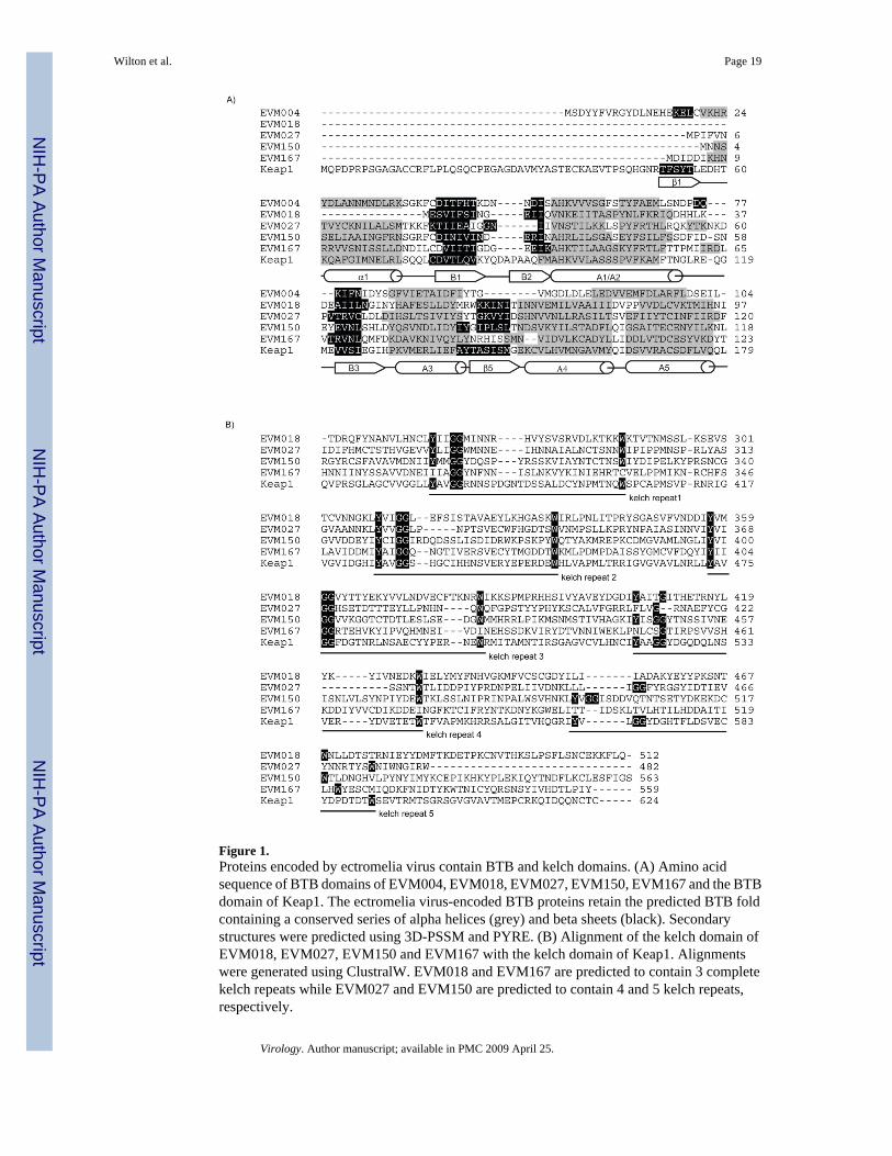
Figure 1.Proteins encoded by ectromelia virus contain BTB and kelch domains. (A) Amino acidsequence of BTB domains of EVM004, EVM018, EVM027, EVM150, EVM167 and the BTBdomain of Keap1. The ectromelia virus-encoded BTB proteins retain the predicted BTB foldcontaining a conserved series of alpha helices (grey) and beta sheets (black). Secondarystructures were predicted using 3D-PSSM and PYRE. (B) Alignment of the kelch domain ofEVM018, EVM027, EVM150 and EVM167 with the kelch domain of Keap1. Alignmentswere generated using ClustralW. EVM018 and EVM167 are predicted to contain 3 completekelch repeats while EVM027 and EVM150 are predicted to contain 4 and 5 kelch repeats,respectively.
Wilton et al. Page 19
Virology. Author manuscript; available in PMC 2009 April 25.
NIH
-PA Author Manuscript
NIH
-PA Author Manuscript
NIH
-PA Author Manuscript

Figure 2.EVM004, EVM018, EVM027, EVM150 and EVM167 are transcribed early during ectromeliavirus infection. (A) Schematic representation of the ectromelia virus BTB/kelch proteinsEVM018, EVM027, EVM150, EVM167 and the BTB-only containing protein EVM004. (B)CV-1 cells were infected with ectromelia virus at an MOI 5 in the presence and absence ofcytosine arabinoside (AraC) and RNA isolated at the indicated times. RNA was subjected toRT-PCR followed by PCR using gene specific primers to detect transcripts for EVM004,EVM018, EVM027, EVM150, EVM167, EVM058 and cellular β-actin.
Wilton et al. Page 20
Virology. Author manuscript; available in PMC 2009 April 25.
NIH
-PA Author Manuscript
NIH
-PA Author Manuscript
NIH
-PA Author Manuscript

Figure 3.EVM150 and EVM167 interact with cullin-3. (A) A schematic representation of cullin-3 basedubiquitin ligase. A single protein containing both BTB and kelch domains serves as thesubstrate specific adaptor for cullin-3 based ubiquitin ligases. The BTB domain interacts withcullin-3 and the kelch domain recruits substrates for ubiquitination. (B) HEK293T cells wereco-transfected with constructs to express EGFP-tagged versions of FPV039BH1, EVM018,EVM027, EVM150, EVM167 and Flag-cullin-3. Lysates were western blotted with anti-Flagto indicate expression of Flag-cullin-3. Immunoprecipitation and western blotting with anantibody specific for EGFP demonstrated expression of FPV039BH1, EVM018, EVM150 andEVM167. Interaction between EVM150 and EVM167 and cullin-3 was demonstrated by
Wilton et al. Page 21
Virology. Author manuscript; available in PMC 2009 April 25.
NIH
-PA Author Manuscript
NIH
-PA Author Manuscript
NIH
-PA Author Manuscript
western blotting the immunoprecipitations with anti-Flag. (C) Reciprocalimmunoprecipitations were performed with anti-Flag to confirm the interaction betweenEVM150, EVM167 and cullin-3. Western blotting of lysates with anti-EGFP demonstratedexpression of FPV039BH1, EVM150 and EVM167. Immunoprecpitation with anti-Flagdemonstrated expression of cullin-3 in the co-transfected samples. Interaction betweenEVM150, EVM167 and cullin-3 was demonstrated by subjecting the immunoprecipitations towestern blotting with anti-EGFP.
Wilton et al. Page 22
Virology. Author manuscript; available in PMC 2009 April 25.
NIH
-PA Author Manuscript
NIH
-PA Author Manuscript
NIH
-PA Author Manuscript

Figure 4.The BTB domain of EVM150 and EVM167 is required for interaction with cullin-3. (A)HEK293T cells were co-transfected with constructs to express EGFP-tagged BTB-onlydomains of EVM150 and EVM167 and co-transfected with Flag-cullin-3 and interactionassessed by co-immunoprecipitation. Westen blotting of lysates with anti-Flag indicatedexpression of Flag-cullin-3. Immunoprecipitation and western blotting with an antibodyspecific for EGFP demonstrated expression of FPV039BH1, and the BTB-only domains ofEVM150 and EVM167. Interaction between the BTB domains of EVM150 and EVM167 andcullin-3 was demonstrated by subjecting the immunoprecipitations to western blotting withanti-Flag. (B) To confirm interaction between the BTB domains of EVM150 and EVM167
Wilton et al. Page 23
Virology. Author manuscript; available in PMC 2009 April 25.
NIH
-PA Author Manuscript
NIH
-PA Author Manuscript
NIH
-PA Author Manuscript

and cullin-3 reciprocal immunoprecipitations were performed with anti-Flag. Expression ofFPV039BH1, EVM150BTB and EVM167BTB was assessed by western blotting lysates withanti-EGFP. Immunoprecpitation with anti-Flag demonstrated expression of cullin-3 in the co-transfected samples. Interaction between the BTB domains of EVM150 and EVM167 withcullin-3 was demonstrated by subjecting the immunoprecipitations to western blotting withanti-EGFP. (C) The kelch domains of EVM 150 and EVM167 do not interact with cullin-3.HEK293T cells were transfected with constructs to express EGFP-tagged kelch-only domainsof EVM150, EVM167 and as a positive control the BTB-only domain of EVM150. Cells wereco-transfected with Flag-cullin-3 and interaction assessed by co-immunoprecipitation. Lysateswere subjected to western blotting with anti-Flag to indicate expression of Flag-cullin-3.Immunoprecipitation and western blotting with an antibody specific for EGFP demonstratedexpression of FPV039BH1, BTB-only domain of EVM150 and the kelch-only domains ofEVM150 and EVM167. Lack of interaction between the kelch domains of EVM150 andEVM167 and cullin-3 was demonstrated by western blotting the immunoprecipitations withanti-Flag.
Wilton et al. Page 24
Virology. Author manuscript; available in PMC 2009 April 25.
NIH
-PA Author Manuscript
NIH
-PA Author Manuscript
NIH
-PA Author Manuscript

Figure 5.A conserved region in the N-terminus of cullin-3 is necessary for EVM150 and EVM167interaction with cullin-3. (A) HEK293T cells were co-transfected with constructs to expressEGFP-tagged full-length versions of EVM150, EVM167 and co-transfected with Myc-cullin-3or Myc-cullin-3ΔN41. Interaction was assessed by co-immunoprecipitation with anti-EGFP.Western blotting of lysates with anti-Myc showed expression of Myc-cullin-3 and Myc-cullin-3ΔN41. Immunoprecipitation and western blotting with an antibody specific for EGFPdemonstrated expression of FPV039BH1, EVM150 and EVM167. Interaction betweenEVM150 and EVM167 and cullin-3 but not Myc-cullin-3ΔN41 was determined by westernblotting the immunoprecipitations with anti-Myc. (B) Expression of the BTB-only domains of
Wilton et al. Page 25
Virology. Author manuscript; available in PMC 2009 April 25.
NIH
-PA Author Manuscript
NIH
-PA Author Manuscript
NIH
-PA Author Manuscript

EVM150 and EVM167 retain the ability to interact with Myc-cullin-3 but not Myc-cullin-3ΔN41. (C) EVM150 and EVM167 interact with both cullin-3 and cullin-3ΔRoc1.HEK293T cells were co-transfected with constructs to express EGFP-tagged full-lengthversions of EVM150, EVM167 and co-transfected with Myc-cullin-3ΔRoc1. As a positivecontrol, EVM150 was co-transfected with Myc-cullin-3. Lysates were subjected to westernblotting with anti-Myc to indicate expression of Myc-cullin-3 and Myc-cullin-3ΔRoc1.Immunoprecipitation and western blotting with an antibody specific for EGFP demonstratedexpression of FPV039BH1, EVM150 and EVM167. Interaction between EVM150 andEVM167 and cullin-3 and Myc-cullin-3ΔRoc1 was demonstrated by subjecting theimmunoprecipitations to western blotting with anti-Myc.
Wilton et al. Page 26
Virology. Author manuscript; available in PMC 2009 April 25.
NIH
-PA Author Manuscript
NIH
-PA Author Manuscript
NIH
-PA Author Manuscript

Figure 6.EVM150 and EVM167 localize to cytoplasmic regions during poxvirus infection. HeLa cellswere infected with VVCop:Flag-EVM150 (a–c), VVCop:Flag-EVM167 (d–f), VVCop:Flag-EVM004 (g–i) or VV(Cop) (j–l) at an MOI of 5. Twelve hours post infection cells were fixedand stained with DAPI and anti-Flag to visualize the viral factories and EVM150, EVM167and EVM004.
Wilton et al. Page 27
Virology. Author manuscript; available in PMC 2009 April 25.
NIH
-PA Author Manuscript
NIH
-PA Author Manuscript
NIH
-PA Author Manuscript

Figure 7.EVM150 and EVM167 co-localize with Flag-cullin-3 during virus infection. A) HeLa cellswere infected with VVT7lacOI (MOI 5) and co-tranfected with pEGFP (a–c), pEGFP-EVM150 (d–f) or pEGFP-EVM167 (g–i) and Flag-cullin-3. Expression was induced by theaddition of IPTG. Twelve hours post-infection, cells were fixed and cullin-3 localizationvisualized by staining with anti-Flag. EGFP fluorescence indicated the localization of EVM150and EVM167. B) HeLa cells were infected with VVT7lacOI (MOI 5) and co-transfected withpEGFP (a–c), pEGFP-EVM150 (d–f) or pEGFP-EVM167 (g–i) and pGEMT-Flag-cullin-3(200–768).
Wilton et al. Page 28
Virology. Author manuscript; available in PMC 2009 April 25.
NIH
-PA Author Manuscript
NIH
-PA Author Manuscript
NIH
-PA Author Manuscript
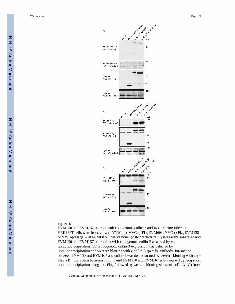
Figure 8.EVM150 and EVM167 interact with endogenous cullin-3 and Roc1 during infection.HEK293T cells were infected with VV(Cop), VVCop:FlagEVM004, VVCop:FlagEVM150or VVCop:Flag167 at an MOI 5. Twelve hours post-infection cell lysates were generated andEVM150 and EVM167 interaction with endogenous cullin-3 assessed by co-immunoprecipitation. (A) Endogneous cullin-3 expression was detected byimmunoprecipitation and western blotting with a cullin-3 specific antibody. Interactionbetween EVM150 and EVM167 and cullin-3 was demonstrated by western blotting with anti-Flag. (B) Interaction between cullin 3 and EVM150 and EVM167 was assessed by reciprocalimmunoprecipitation using anti-Flag followed by western blotting with anti-cullin 3. (C) Roc1
Wilton et al. Page 29
Virology. Author manuscript; available in PMC 2009 April 25.
NIH
-PA Author Manuscript
NIH
-PA Author Manuscript
NIH
-PA Author Manuscript

co-immunoprecipitates with EVM150 and EVM167. Cellular lysates were analyzed byimmunoprecipitaiton with anti-Flag followed by western blotting with anti-Roc1.
Wilton et al. Page 30
Virology. Author manuscript; available in PMC 2009 April 25.
NIH
-PA Author Manuscript
NIH
-PA Author Manuscript
NIH
-PA Author Manuscript

Figure 9.Conjugated ubiquitin co-immunoprecipitates with EVM150 and EVM167. HeLa cells wereinfected with VV(Cop), VVCop:FlagEVM004, VVCop:FlagEVM150 orVVCop:FlagEVM167 at an MOI 5. Twelve hours post infection the cells were subjected toimmunoprecpitation with anti-Flag. Expression of Flag-EVM150 and Flag-EVM167 wasdetected by immunoprecipitation with anti-Flag followed by western blotting with anti-Flag-HRP. Co-immunoprecipiation of EVM150 or EVM167 with conjugated ubiquitin was detectedby immunoprecipitation with anti-Flag followed by western blotting with the anti-ubiquitinantibody (FK2) which detects only conjugated ubiquitin.
Wilton et al. Page 31
Virology. Author manuscript; available in PMC 2009 April 25.
NIH
-PA Author Manuscript
NIH
-PA Author Manuscript
NIH
-PA Author Manuscript

Figure 10.EVM150 and EVM167 co-localize with conjugated ubiquitin. HeLa cells were infected withVVT7lacOI (MOI 5) and transfected with pEGFP, pEGFP-EVM004, pEGFP-EVM150, orpEGFP-EVM167. Expression was induced by treatement with IPTG and twelve hours post-infection cells were fixed and stained with DAPI to visualize nuclei and virus factories andconjugated ubiquitin was detected with an antibody (FK2) that detects conjugated ubiquitin.(A-D) Mock infected HeLa cells display a diffuse staining pattern of conjugated ubiquitin. (E-H) HeLa cells infected with VVT7lacOI demonstrate a punctate localization of conjugatedubiquitin. (I-L) EGFP expression during virus infection does not co-localize with conjugatedubiquitin. (M-P) EGFP-EVM004 does not co-localize with conjugated ubiquitin. (Q-T) EGFP-
Wilton et al. Page 32
Virology. Author manuscript; available in PMC 2009 April 25.
NIH
-PA Author Manuscript
NIH
-PA Author Manuscript
NIH
-PA Author Manuscript

EVM150 co-localizes with conjugated ubiquitin. (U-X) EGFP-EVM167 co-localizes withconjugated ubiquitin.
Wilton et al. Page 33
Virology. Author manuscript; available in PMC 2009 April 25.
NIH
-PA Author Manuscript
NIH
-PA Author Manuscript
NIH
-PA Author Manuscript