Three-dimensional Structures of Fibrillar Sm Proteins: Hfq and Other Sm-like Proteins
Arabidopsis Has Two Redundant Cullin3 Proteins That AreEssential for Embryo Development and That Interact withRBX1 and BTB Proteins to Form Multisubunit E3 UbiquitinLigase Complexes in Vivo W
Pablo Figueroa,a Giuliana Gusmaroli,a Giovanna Serino,b Jessica Habashi,a Ligeng Ma,a,c Yunping Shen,a,c
Suhua Feng,a Magnolia Bostick,d Judy Callis,d Hanjo Hellmann,e and Xing Wang Denga,1
a Department of Molecular, Cellular, and Developmental Biology, Yale University, New Haven, Connecticut 06520-8104b Dipartimento di Genetica e Biologia Molecolare, Universita di Roma ‘‘La Sapienza,’’ 00185 Rome, Italyc Peking–Yale Joint Center of Plant Molecular Genetics and Agrobiotechnology, College of Life Sciences,
Peking University, Beijing 100871, Chinad Section of Biochemistry and Molecular Biology, University of California, Davis, California 95616e Institute for Biology/Applied Genetics, Freie Universitat Berlin, 14195 Berlin, Germany
Cullin-based E3 ubiquitin ligases play important roles in the regulation of diverse developmental processes and
environmental responses in eukaryotic organisms. Recently, it was shown in Schizosaccharomyces pombe, Caenorhabditis
elegans, and mammals that Cullin3 (CUL3) directly associates with RBX1 and BTB domain proteins in vivo to form a new
family of E3 ligases, with the BTB protein subunit functioning in substrate recognition. Here, we demonstrate that
Arabidopsis thaliana has two redundant CUL3 (AtCUL3) genes that are essential for embryo development. Besides
supporting anticipated specific AtCUL3 interactions with the RING protein AtRBX1 and representative Arabidopsis proteins
containing a BTB domain in vitro, we show that AtCUL3 cofractionates and specifically associates with AtRBX1 and
a representative BTB protein in vivo. Similar to the AtCUL1 subunit of the SKP1-CUL1-F-box protein–type E3 ligases, the
AtCUL3 subunit of the BTB-containing E3 ligase complexes is subjected to modification and possible regulation by the
ubiquitin-like protein Related to Ubiquitin in vivo. Together with the presence of large numbers of BTB proteins with diverse
structural features and expression patterns, our data suggest that Arabidopsis has conserved AtCUL3-RBX1-BTB protein
E3 ubiquitin ligases to target diverse protein substrates for degradation by the ubiquitin/proteasome pathway.
INTRODUCTION
It has become increasingly clear that the regulation of many cellu-
lar and developmental processes involves ubiquitin-dependent
degradation of critical proteins (Hershko and Ciechanover, 1998;
Hellmann and Estelle, 2002; Sullivan et al., 2003). Substrates
selected for degradation by this pathway are specifically tagged
by a chain of ubiquitin molecules, which directs them to the 26S
proteasome. Attachment of ubiquitin molecules to a target pro-
tein involves a cascade of three steps, catalyzed by an E1
ubiquitin-activating enzyme, an E2 ubiquitin-conjugating en-
zyme, and an E3 ubiquitin ligase. Typically, the E2 enzyme
associates with an E3 ubiquitin ligase, which is primarily re-
sponsible for recognizing the substrate. As the E3 ligase action
determines the life or death of a protein, it is critical to understand
how these enzymes select their targets.
Ubiquitin E3 ligases come in a large variety of forms, including
multisubunit complexes organized around a cullin (CUL) scaf-
folding protein. The paradigm for this large group of ligases is
based on studies of the SCF (for SKP1-CUL-F-box) complex. A
typical SCF complex consists of a cullin family member, CUL1,
a RING-finger protein RBX1 that interacts with the cullin and
the E2 conjugating enzyme, a SKP1-like adaptor protein, and
an F-box protein that confers the specificity for the recruitment
of the substrate (Schulman et al., 2000; Zheng et al., 2002;
Deshaies, 2003). CUL2 forms a similar group of E3 ligases, in
which an elongin C-SOCS module replaces the SKP1-F-box
protein module (Stebbins et al., 1999). These E3 ligases can
target a range of substrates for protein degradation, as de-
termined by the specificity of their associated F-box or SOCS
protein.
A series of recent studies inCaenorhabditis elegans (Kurz et al.,
2002; Pintard et al., 2003b; Xu et al., 2003), Schizosaccharo-
myces pombe (Geyer et al., 2003), and mammals (Furukawa
et al., 2003; Kobayashi et al., 2004) has shed light on CUL3-
based ubiquitin ligases. The findings include identification of an
in vivo substrate (Kurz et al., 2002; Pintard et al., 2003a) and a
novel type of substrate specificity adaptors for CUL3-based
1 To whom correspondence should be addressed. E-mail [email protected]; fax 203-432-5726.The author responsible for distribution of materials integral to thefindings presented in this article in accordance with the policy describedin the Instructions for Authors (www.plantcell.org) is: Xing Wang Deng([email protected]).W Online version contains Web-only data.Article, publication date, and citation information can be found atwww.plantcell.org/cgi/doi/10.1105/tpc.105.031989.
The Plant Cell, Vol. 17, 1180–1195, April 2005, www.plantcell.org ª 2005 American Society of Plant Biologists

ligases (Geyer et al., 2003; Pintard et al., 2003b; Xu et al., 2003).
The substrate specificity factors associate with CUL3 through
a Bric a brac, Tramtrack, and Broad complex/Pox virus and Zinc
finger (BTB/POZ) domain. Surprisingly, the same BTB protein
was found to also bind the substrate, thus combining in one
protein the functions of the SKP1 adaptor and the F-box subunits.
One of these BTB proteins, MEL-26, binds to the microtubule-
severing protein MEI-1/katanin (Pintard et al., 2003b; Xu et al.,
2003), which is an essential component of the C. elegans meiotic
spindle (Clark-Maguire and Mains, 1994). Although the precise
role of the MEI-1 protein is unknown, it has been found that MEI-1
is degraded at the meiosis-to-mitosis transition and that CUL3
is required for its degradation (Pintard et al., 2003a). Genetic
studies, together with the finding that MEL-26 physically inter-
acts with MEI-1 (Pintard et al., 2003b; Xu et al., 2003), strongly
indicate that a CUL3-based ubiquitin ligase containing MEL-26
might target MEI-1 for degradation. In C. elegans, depletion of
CUL3 by RNA interference causes defects in early embryogen-
esis, resulting in abnormal microfilament and microtubule orga-
nization (Kurz et al., 2002). Drosophila melanogaster CUL3 is
involved in Hedgehog signaling by regulating the stability of the
CUBITUS INTERRUPTUS protein, thus affecting external sen-
sory organ development, pattern formation, and cell growth and
survival (Mistry et al., 2004). The mammalian CUL3 ubiquitin
ligase has been implicated in the turnover of monomeric forms
of cyclin E, and mice lacking cul3 die at embryonic day 6.5
with defects in both embryonic and extraembryonic compart-
ments (Singer et al., 1999; Winston et al., 1999). Plants such
as Arabidopsis thaliana have closely related CUL3 proteins
(Risseeuw et al., 2003). The presence of CUL3-based multi-
subunit protein ligases was implied by a recent report of an in
vitro interaction between Arabidopsis CUL3 and the BTB pro-
tein ETO1, which is involved in ethylene biosynthesis regulation
(Wang et al., 2004).
Here, we show the existence in Arabidopsis of CUL3 (AtCUL3)-
containing multisubunit E3 ubiquitin ligase complexes and dem-
onstrate that AtCUL3 is essential for embryo development.
Furthermore, we show that AtCUL3 interacts in vitro with several
proteins containing a BTB domain, but not with the SKP1-like
proteins ASK1 or ASK2. The domains responsible for this in-
teraction have been identified both in AtCUL3 and in the BTB
domain–containing proteins. In agreement with our in vitro
findings, we further demonstrate that the in vivo AtCUL3 complex
contains the RING finger protein RBX1 as well as a BTB protein
and that the AtCUL3 subunit is subjected to modification by RUB
(for Related to Ubiquitin). Together, our studies confirm that
plants possess a large number of AtCUL3-RBX-BTB E3 ubiquitin
ligase complexes that target proteins for degradation via the
ubiquitin/proteasome pathway.
RESULTS
Arabidopsis Has Two Active AtCUL3 Genes
Analysis of the Arabidopsis genome revealed the presence of at
least 10 predicted cullin-related genes (Risseeuw et al., 2003).
Six of these contain reading frames with apparently intact
canonical N- and C-terminal domains. At least five (AtCUL1,
AtCUL2, AtCUL3A, AtCUL3B, and AtCUL4) of those six genes
are expressed, as indicated by the presence of their correspond-
ing EST clones (Risseeuw et al., 2003). Phylogenetic analysis of
cullins from Arabidopsis and from other eukaryotes indicates
that AtCUL3A and AtCUL3B are two closely related members of
a two-gene family and are clustered in the same clade with CUL3
from Homo sapiens,D.melanogaster,C. elegans, S. pombe, and
Saccharomyces cerevisiae (Risseeuw et al., 2003).
Both of theAtCUL3A andAtCUL3B genes have two exons and
encode proteins of 732 amino acids residues (GenBank acces-
sion numbers CAC87120 and AAG52544, respectively). The
predicted protein sequences for AtCUL3A and AtCUL3B share
88% identity (Figure 1A; see Supplemental Figure 1 online), with
AtCUL3A being slightly more closely related to its human
homolog hCUL3 (52% amino acid identity) (Figure 1A). The
greatest similarity between these proteins is in the C-terminal
region, which contains both the RUB/NEDD8 conjugation site
(del Pozo and Estelle, 1999) and the cullin homology domain
(Figure 1A). This domain represents a conserved module and is
responsible for connecting an E2 ubiquitin-conjugating enzyme
to the ubiquitin ligase (Yu et al., 1998).
AtCUL3A and AtCUL3B Are Both Expressed and Are
Subjected to Posttranslational Modification
As an initial step in studying the biological role of the AtCUL3
proteins, we conducted a database search for Arabidopsis
T-DNA insertion mutant lines. We found one T-DNA line that con-
tains an insertion located within the second exon of AtCUL3A,
417 bp upstream of the STOP codon (Figure 1B). A T-DNA
insertion line for the AtCUL3B gene was also identified, with the
T-DNA inserted 155 bp downstream of the START codon (Figure
1B). Both insertions likely give rise to null mutations, as neither
homozygous line expresses the AtCUL3A or AtCUL3B mRNA,
respectively (Figure 1C). Protein blot analyses of protein extract
from Arabidopsis cul3a mutant seedlings showed a drastic re-
duction in total AtCUL3 protein level with respect to wild-type
plants, whereas no significant differences in overall AtCUL3 level
were observed between the cul3b mutants and wild-type sib-
lings (Figure 1D). Because the anti-CUL3 antibody has similar
affinity for both AtCUL3 proteins (Figure 1E), the results shown
in Figure 1D suggest that AtCUL3A may be predominantly
expressed with respect to AtCUL3B in seedlings.
The presence of two specific bands for CUL3 in the protein blot
analysis of seedling extracts (Figure 1D) suggests that AtCUL3
might be posttranslationally modified by the attachment of
the ubiquitin-like protein RUB (called NEDD8 in mammals and
yeast). In fact, it has been shown that CUL3, like other cullins,
can be covalently modified by the attachment of RUB/NEDD8
(Pintard et al., 2003a). In the case of the SCF complex, the RUB/
NEDD8 modification of CUL1 (rubylation) provides an important
positive regulation of the E3 ligase activity, enhancing E2 re-
cruitment and polyubiquitination of the substrate (Wu et al., 2000;
Kawakami et al., 2001). In the case of CUL3, rubylation and
derubylation have been shown to be required for targeting
MEI-1/katanin for degradation inC.elegans (Pintard et al., 2003a).
To test whether Arabidopsis CUL3 is covalently modified by
RUB, the same protein extracts probed with anti-CUL3 antibody
Arabidopsis CUL3-RBX1-BTB E3 Ubiquitin Ligases 1181

(Figure 1D) were immunoblotted against anti-RUB antibodies.
The results demonstrate that only the upper band is recognized
by anti-RUB antibodies (data not shown). Because both AtCUL3
forms were observed in the cul3a and cul3bmutants, as well as in
wild-type plants, it is possible to conclude that, like AtCUL1, the
AtCUL3A and AtCUL3B proteins are subjected to modification
by RUB.
AtCUL3A and AtCUL3B Are Largely Functionally Redundant
and Are Essential for Embryo Development
Although each insertion gives rise to a null mutation, when grown
under normal growth conditions the single homozygous cul3a
and cul3b insertion mutants are virtually indistinguishable from
their wild-type siblings throughout their entire life cycles (Fig-
ure 1F).
However, no homozygous double mutant plants were recov-
ered, despite multiple attempts using PCR genotyping of 60
segregating progeny obtained from the self-pollination of paren-
tal plants homozygous for the mutation in one of the two AtCUL3
loci and heterozygous for the mutation in the other. To further
investigate the absence of double homozygous cul3a cul3b
mutants, we selected plants that were heterozygous for cul3a
and homozygous for cul3b and performed detailed examinations
with a dissection stereoscope (Figures 2A to 2H) and a differen-
tial interference contrast (DIC) microscope (Figures 2J to 2S).
Siliques and developing seeds, at different developmental
stages after self-pollination, were analyzed. As shown in Figures
2A and 2B, at early developmental stages the fertilized ovules
obtained from the self-pollination of wild-type (Figure 2A) and
CUL3A/cul3a cul3b/cul3b (Figure 2B) parental plants are virtually
indistinguishable from each other, as they all contain develop-
ing embryos at the early globular stage (Figures 2J and 2K).
However, after the transition from the globular to the heart stage
(Figures 2L to 2O), all ovules from wild-type plants uniformly con-
tain heart-stage embryos (data not shown), whereas the ovules
from CUL3A/cul3a cul3b/cul3b plants segregate wild-type–like
ovules with heart-stage embryos (Figures 2L and 2N) and ovules
with embryos arrested at the globular stage (Figure 2M) or
a slightly later stage (Figure 2O). At a later developmental stage,
whereas wild-type siliques uniformly contain green seeds
(Figures 2C and 2E) that undergo the maturation process (Figure
2G), siliques from CUL3A/cul3a cul3b/cul3b plants segregate
both wild-type developing seeds and pale green/yellowish de-
veloping seeds, indicated by yellow arrowheads in Figure 2D.
DIC microscopy of cleared ovules from these siliques demon-
strated that although the green ovules contain mature embryos
(Figure 2R), the pale green ovules contain underdeveloped
embryos arrested at a late globular-to-heart transition (Figure
2Q) or at a heart-like stage (Figure 2S). During the seed matu-
ration and desiccation processes, the wild-type–like green ma-
ture ovules from CUL3A/cul3a cul3b/cul3b plants develop into
brown seeds (Figure 2H), indistinguishable from the wild-type
ovules (Figure 2G), whereas the pale yellowish ovules of the same
plants turned into red shrunken seeds (indicated by red arrow-
heads in Figure 2H), possibly because the absence of a mature
embryo causes the collapse of the seed coat. Similar analyses of
developing siliques obtained from the self-pollination of the
Figure 1. Characterization of Two Redundant AtCUL3 Genes and Their
Mutant Phenotypes.
(A) Schematic comparison of the AtCUL3 proteins with the human
ortholog. The black-boxed regions of the AtCUL3 protein denote the
cullin homology domain. The small black bars toward the C terminus
denote the RUB modification site. The identity percentages of the
homologous regions of the Arabidopsis and human proteins are in-
dicated.
(B) Representation of the AtCUL3A and AtCUL3B genes and the T-DNA
insertion sites, as indicated by black arrows. Both genes have two exons,
denoted E1 and E2.
(C) The AtCUL3A and AtCUL3B mRNAs are not detectable in the cul3a
and cul3b mutants, respectively. RNA gel blot analysis was performed
using a probe specific for the 39 untranslated region of the AtCUL3A gene
(top), whereas RT-PCR was used to examine AtCUL3B expression
(bottom). WT, total RNA from wild-type seedlings; cul3a, total RNA from
the cul3a mutant; cul3b, total RNA from the cul3b mutant. The intensities
of 25S rRNA and EF-1A were used as equal loading controls for RNA gel
blot or RT-PCR, respectively.
(D) Effect of cul3a and cul3b mutations on AtCUL3 protein accumulation.
Protein blot analysis was performed using antibodies against AtCUL3A.
WT, total proteins from wild-type seedlings; cul3a, total proteins from
cul3a mutant seedlings; cul3b, total proteins from cul3b mutant seed-
lings. An anti-RNP6 antibody was used as an equal loading control. For
all samples, 24 mg of total protein from 6-d-old seedlings was used.
(E) Anti-CUL3 antibody has similar affinity for the AtCUL3A and AtCUL3B
proteins. MBP-AtCUL3A (lane 1) and MBP-AtCUL3B (lane 2) were
expressed in E. coli, and the protein extracts were examined by im-
munoblot analysis using anti-CUL3 and anti-MBP antibodies.
(F) Both the cul3a and cul3b mutant plants have no obvious change from
the wild-type (WT) phenotype. Plants at top are 5 weeks old, and those at
bottom are 3 weeks old.
1182 The Plant Cell

reciprocal cul3a/cul3a CUL3B/cul3b double mutant gave results
identical to those in Figure 2 for the self-pollination of CUL3A/
cul3a cul3b/cul3b double mutant plants (data not shown).
As shown in Table 1, stereoscopic analyses of a large number
of developing siliques from the self-pollination of both CUL3A/
cul3a cul3b/cul3b and cul3a/cul3a CUL3B/cul3b parental plants
indicate that the frequency of pale green/yellowish ovules that
turned into red shrunken seeds corresponds to ;16.5 and 17%
of the total developing seeds, respectively. Because these
defective seeds have not been observed in either of the single
Figure 2. Loss of Function in Both Arabidopsis CUL3 Genes Causes Arrest in Embryogenesis.
(A) and (B) Stereomicroscopic images of siliques obtained from self-pollinated wild-type (WT [A]) or CUL3A/cul3a cul3b/cul3b (B) parental plants at an
early developmental stage, when wild-type embryos are at the transition between the globular and heart stages.
(C) and (D) Stereomicroscopic images of siliques obtained from self-pollinated wild-type (C) or CUL3A/cul3a cul3b/cul3b (D) parental plants at a later
developmental stage, when wild-type embryos are at the mature stage. Yellow arrowheads indicate pale green/yellow ovules containing arrested
embryos. Red arrowheads indicate red shrunken ovules.
(E) to (H) Stereomicroscopic images of siliques obtained from self-pollinated wild-type ([E] and [G]) or CUL3A/cul3a cul3b/cul3b ([F] and [H]) parental
plants during the onset of seed maturation. Red arrowheads in (F) and (H) indicate the pale green/yellow ovules (described in [D]) that turned into
shrunken seeds.
(I) Scheme of Arabidopsis embryo development, adapted from Laux et al. (2004). Red bars indicate the stages of arrest of Arabidopsis cul1 and cul3
mutant embryos.
(J) to (S) DIC images of cleared ovules obtained from the self-pollination of CUL3A/cul3a cul3b/cul3b parental plants. At an early developmental stage
(early globular; [J] and [K]), all of the embryos are uniformly developed. Starting from the transition between the late globular and torpedo stages, the
cul3a cul3b embryos arrest at late globular/transition stages ([M], [O], and [Q]), whereas the sibling embryos ([L], [N], and [P]) from the same siliques
proceed from globular, to heart, to torpedo, to mature embryos. Occasionally, cul3a cul3b embryos were arrested at a heart-like stage (S).
a, axis; c, cotyledons; eg, early globular-stage embryo; eh, early heart-stage embryo; hl, heart-like embryo; lg, late globular-stage embryo; lh, late heart-
stage embryo; s, suspensor; t, transition-stage embryo; to, torpedo-stage embryo.
Arabidopsis CUL3-RBX1-BTB E3 Ubiquitin Ligases 1183
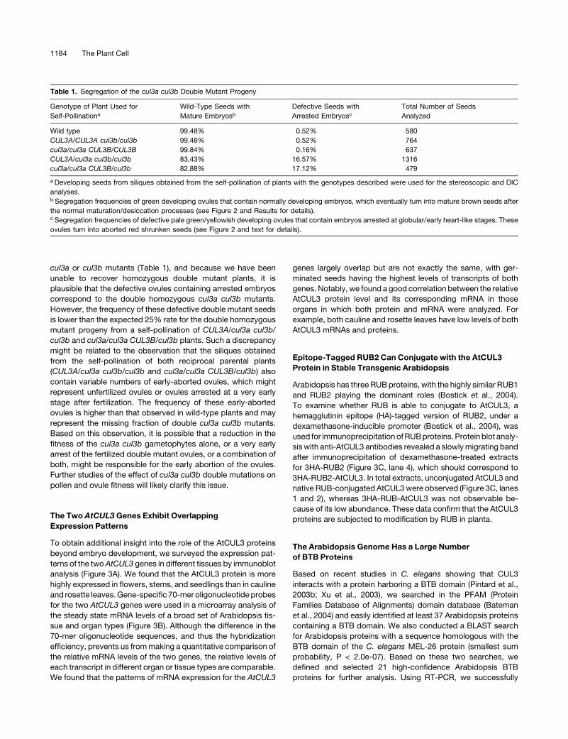
cul3a or cul3b mutants (Table 1), and because we have been
unable to recover homozygous double mutant plants, it is
plausible that the defective ovules containing arrested embryos
correspond to the double homozygous cul3a cul3b mutants.
However, the frequency of these defective double mutant seeds
is lower than the expected 25% rate for the double homozygous
mutant progeny from a self-pollination of CUL3A/cul3a cul3b/
cul3b and cul3a/cul3a CUL3B/cul3b plants. Such a discrepancy
might be related to the observation that the siliques obtained
from the self-pollination of both reciprocal parental plants
(CUL3A/cul3a cul3b/cul3b and cul3a/cul3a CUL3B/cul3b) also
contain variable numbers of early-aborted ovules, which might
represent unfertilized ovules or ovules arrested at a very early
stage after fertilization. The frequency of these early-aborted
ovules is higher than that observed in wild-type plants and may
represent the missing fraction of double cul3a cul3b mutants.
Based on this observation, it is possible that a reduction in the
fitness of the cul3a cul3b gametophytes alone, or a very early
arrest of the fertilized double mutant ovules, or a combination of
both, might be responsible for the early abortion of the ovules.
Further studies of the effect of cul3a cul3b double mutations on
pollen and ovule fitness will likely clarify this issue.
The Two AtCUL3 Genes Exhibit Overlapping
Expression Patterns
To obtain additional insight into the role of the AtCUL3 proteins
beyond embryo development, we surveyed the expression pat-
terns of the twoAtCUL3 genes in different tissues by immunoblot
analysis (Figure 3A). We found that the AtCUL3 protein is more
highly expressed in flowers, stems, and seedlings than in cauline
and rosette leaves. Gene-specific 70-mer oligonucleotide probes
for the two AtCUL3 genes were used in a microarray analysis of
the steady state mRNA levels of a broad set of Arabidopsis tis-
sue and organ types (Figure 3B). Although the difference in the
70-mer oligonucleotide sequences, and thus the hybridization
efficiency, prevents us from making a quantitative comparison of
the relative mRNA levels of the two genes, the relative levels of
each transcript in different organ or tissue types are comparable.
We found that the patterns of mRNA expression for the AtCUL3
genes largely overlap but are not exactly the same, with ger-
minated seeds having the highest levels of transcripts of both
genes. Notably, we found a good correlation between the relative
AtCUL3 protein level and its corresponding mRNA in those
organs in which both protein and mRNA were analyzed. For
example, both cauline and rosette leaves have low levels of both
AtCUL3 mRNAs and proteins.
Epitope-Tagged RUB2 Can Conjugate with the AtCUL3
Protein in Stable Transgenic Arabidopsis
Arabidopsis has three RUB proteins, with the highly similar RUB1
and RUB2 playing the dominant roles (Bostick et al., 2004).
To examine whether RUB is able to conjugate to AtCUL3, a
hemagglutinin epitope (HA)-tagged version of RUB2, under a
dexamethasone-inducible promoter (Bostick et al., 2004), was
used for immunoprecipitation of RUB proteins. Protein blot analy-
sis with anti-AtCUL3 antibodies revealed a slowly migrating band
after immunoprecipitation of dexamethasone-treated extracts
for 3HA-RUB2 (Figure 3C, lane 4), which should correspond to
3HA-RUB2-AtCUL3. In total extracts, unconjugated AtCUL3 and
native RUB-conjugated AtCUL3 were observed (Figure 3C, lanes
1 and 2), whereas 3HA-RUB-AtCUL3 was not observable be-
cause of its low abundance. These data confirm that the AtCUL3
proteins are subjected to modification by RUB in planta.
The Arabidopsis Genome Has a Large Number
of BTB Proteins
Based on recent studies in C. elegans showing that CUL3
interacts with a protein harboring a BTB domain (Pintard et al.,
2003b; Xu et al., 2003), we searched in the PFAM (Protein
Families Database of Alignments) domain database (Bateman
et al., 2004) and easily identified at least 37 Arabidopsis proteins
containing a BTB domain. We also conducted a BLAST search
for Arabidopsis proteins with a sequence homologous with the
BTB domain of the C. elegans MEL-26 protein (smallest sum
probability, P < 2.0e-07). Based on these two searches, we
defined and selected 21 high-confidence Arabidopsis BTB
proteins for further analysis. Using RT-PCR, we successfully
Table 1. Segregation of the cul3a cul3b Double Mutant Progeny
Genotype of Plant Used for
Self-Pollinationa
Wild-Type Seeds with
Mature Embryosb
Defective Seeds with
Arrested Embryosc
Total Number of Seeds
Analyzed
Wild type 99.48% 0.52% 580
CUL3A/CUL3A cul3b/cul3b 99.48% 0.52% 764
cul3a/cul3a CUL3B/CUL3B 99.84% 0.16% 637
CUL3A/cul3a cul3b/cul3b 83.43% 16.57% 1316
cul3a/cul3a CUL3B/cul3b 82.88% 17.12% 479
a Developing seeds from siliques obtained from the self-pollination of plants with the genotypes described were used for the stereoscopic and DIC
analyses.b Segregation frequencies of green developing ovules that contain normally developing embryos, which eventually turn into mature brown seeds after
the normal maturation/desiccation processes (see Figure 2 and Results for details).c Segregation frequencies of defective pale green/yellowish developing ovules that contain embryos arrested at globular/early heart-like stages. These
ovules turn into aborted red shrunken seeds (see Figure 2 and text for details).
1184 The Plant Cell

cloned full-coding cDNA for 11 of those Arabidopsis BTB genes.
Phylogenetic analysis of the BTB domain of the 11 proteins,
together with C. elegans MEL-26, revealed the presence of three
main branches, with the BTB domain encoded by At1g21780
being the most similar to the BTB domain of MEL-26, with 35%
sequence identity between the two (Figure 4A; see Supplemental
Figure 2 online).
AtCUL3A Interacts in Vitro with the BTB Proteins but Not
with the SKP1-Like Proteins ASK1 and ASK2
To examine a possible interaction between AtCUL3 and the
Arabidopsis BTB proteins, the 11 cloned BTB proteins were
subjected to an in vitro pull-down assay with the recombinant
Maltose Binding Protein (MBP)-AtCUL3A protein. Briefly, the
purified MBP-AtCUL3A was incubated separately with each
individual in vitro–translated BTB protein, and its affinity for
a given BTB protein was examined after an extensive washing
procedure. The results shown in Figure 4B demonstrate that all of
the 11 Arabidopsis BTB proteins tested to date were able to
interact with MBP-AtCUL3A in the in vitro pull-down assay. As
expected, on the basis of previous findings in other eukaryotic
organisms, Arabidopsis RBX1 was able to interact with MBP-
AtCUL3A in the same pull-down assay, whereas, interestingly,
the SKP1-like Arabidopsis proteins ASK1 and ASK2 were not.
Based on the highest sequence homology in the BTB domains
between At1g21780 and MEL-26, and on the positive interaction
observed in our pull-down assay, At1g21780 was selected for
further detailed structural and functional analysis.
The BTB Protein At1g21780 Is Capable of Dimerization
A yeast two-hybrid assay was used to further verify the in-
teraction between the At1g21780 protein and AtCUL3A (Figure
5A). In agreement with the results obtained from the in vitro
pull-down assay, the yeast two-hybrid assay confirmed that
AtCUL3A interacts with the At1g21780 BTB protein but not with
the SKP1-like ASK1 or ASK2 protein. A control yeast two-
hybrid assay also showed that AtCUL1 was unable to interact
with At1g21780, further supporting the notion that the interac-
tion between At1g21780 and AtCUL3A is specific and does not
apply to the entire cullin family.
Interestingly, the yeast two-hybrid assay also revealed that
At1g21780 is able to interact with itself (Figure 5A). This obser-
vation is consistent with the report that BTB proteins are capable
of homodimerization and of heterodimerization with other mem-
bers of the same family (Bardwell and Treisman, 1994; Pintard
et al., 2004).
Figure 3. Protein and mRNA Expression Patterns of the AtCUL3 Genes
and Posttranslational Modification of AtCUL3 Proteins.
(A) The AtCUL3 protein is differentially expressed in Arabidopsis tissues.
Total soluble protein extracts from flowers (lane 1), cauline leaves (lane
2), stems (lane 3), rosette leaves (lane 4), and light-grown seedlings (lane
5) were examined by protein gel blot analysis using anti-AtCUL3 and
anti-AtCUL1 antibodies. An anti-RPT5 antibody was used as a sample
loading control.
(B) Expression profiles of the two AtCUL3 genes in different Arabidopsis
tissues/organs derived from a genome-profiling study using a 70-mer
oligonucleotide microarray (Ma et al., 2005). The relative expression
levels of each tissue/organ considered, shown with normalized intensity,
are represented by black bars. Bar 1, cauline leaf; bar 2, rosette leaf; bar
3, pistil, 1 d after pollination; bar 4, pistil, 1 d before pollination; bar 5,
silique, 3 d after pollination; bar 6, silique, 8 d after pollination; bar 7,
stem; bar 8, sepal; bar 9, stamen; bar 10, petal; bar 11, germinated seed;
bar 12, root in dark; bar 13, root in white light; bar 14, hypocotyl in dark;
bar 15, hypocotyl in white light; bar 16, cotyledon in dark; bar 17,
cotyledon in white light.
(C) The AtCUL3 protein is subjected to modification by RUB2 in vivo.
Total protein extracts (T) from 3HA-RUB2 transgenic seedlings with (þ)
or without (�) dexamethasone (DEX) induction were subjected to
immunoprecipitation (IP) with anti-HA antibodies. Protein gel blot anal-
ysis was subsequently performed using anti-AtCUL3 antibodies.
Arabidopsis CUL3-RBX1-BTB E3 Ubiquitin Ligases 1185

The N-Terminal Region of AtCUL3 and the BTB Domain
of At1g21780 Are Critical for the AtCUL3–BTB
Protein Interaction
A comparative analysis of CUL3 proteins from several organisms
revealed the presence of highly conserved N-terminal elements
corresponding to the H2 helix of CUL1 (Zheng et al., 2002;
Xu et al., 2003). Indeed, mutagenesis analyses indicated that
residues in this predicted helix region are important for the
interaction between the C. elegans CUL3 and MEL-26 proteins
(Xu et al., 2003). To examine the involvement of these elements
in BTB protein binding in Arabidopsis, we generated a mutant
version of AtCUL3A in the H2 region (S48A/F49A) and tested it
for interaction with At1g21780 using a yeast two-hybrid assay
(Figure 5B). The results clearly demonstrate that the mutation in
the H2 helix of AtCUL3A completely abolishes its ability to
interact with At1g21780 (Figure 5D). We were also interested
in defining the effect of RUB modification on the AtCUL3A–
At1g21780 interaction. Therefore, we generated an AtCUL3A
K676R mutation that abolished the RUB modification of AtCUL3A
(Figure 5B). It is interesting that the AtCUL3A K676R mutant
protein, which is not RUB-modified in yeast (Figure 5E, lane 2), is
still able to interact with At1g21780 at a wild-type level (Figure
5D). This result indicates that the RUB modification of AtCUL3A
is not required for and does not affect the AtCUL3A interaction
with At1g21780 in the yeast assay. Previous structural analyses
indicate that SKP1 and elongin C adopt a/b-structures similar to
the BTB domain of human promyelocytic leukemia zinc finger
(Schulman et al., 2000). In the case of SKP1, several residues
located in b-strand 3 (S3) and in a-helix 5 (H5) contact CUL1
(Zheng et al., 2002). In addition, recent studies have shown that
analogous residues in the MEL-26 BTB domain are important for
the interaction between MEL-26 and CUL3 (Xu et al., 2003).
Therefore, we generated mutations in analogous residues of the
BTB domain (residues 151 to 259) of At1g21780 (Figure 5C),
which are conserved in several Arabidopsis BTB proteins (see
Supplemental Figure 2 online), and tested their ability to interact
with AtCUL3A by yeast two-hybrid assay. Some mutations in the
predicted S3 (I204R) or H5 (Y242A) abolished the interaction of
the BTB protein with AtCUL3A (Figure 5D), whereas other
mutations in S3 (D206A) or H5 (I244R) reduced the interaction
by three times with respect to wild-type At1g21780 (Figure 5D).
Clearly, these data suggest that analogous structural elements
in the SKP1, MEL-26, and At1g21780 proteins may be used to
interact with a common interface located in the N terminus of
distinct cullin proteins. On the other hand, those same point
mutations in the BTB domain did not significantly affect the
self-interaction of the BTB protein (Figure 5D), suggesting
that distinct interacting surfaces are responsible for its self-
interaction and for its interaction with AtCUL3.
The BTB Domain of At1g21780 May Not Be Sufficient to
Interact with AtCUL3A
As our yeast two-hybrid results suggested that several residues
of the BTB domain of At1g21780 are critical for the interaction
with AtCUL3 (Figure 5D), we further tested whether the BTB
domain itself is sufficient to interact with AtCUL3A. Several
Figure 4. Phylogenetic Comparison of 11 Cloned BTB Protein Genes
and Their in Vitro Binding to AtCUL3.
(A) Phylogenetic analysis of the BTB domains in several Arabidopsis
proteins. Sequence data for the proteins used in the phylogenetic
analysis were taken from the EMBL/GenBank databases using the
following accession numbers: At1g21780 (AAM65090), At5g19000
(AAM97116), At3g06190 (AAF30312), At2g39760 (NP_030522),
At4g37610 (AAN15363), At1g01640 (AAF78396), At5g48510
(NP_199662), At3g43700 (CAB83071), At1g05690 (AAQ87006),
At3g48360 (AAN31824), At5g63160 (AAR24650), and C. elegans MEL-26
(NP_492449). Sequence alignment of the BTB protein families was
conducted using ClustalX (version 1.83) software (Thompson et al.,
1997). The Gonnet protein weight matrices (Gonnet 40, 80, 120, 160,
250, and 350 matrices [Gonnet et al., 1992]) were selected, and the gap-
opening and extension parameters were 10 and 0.2, respectively. Dis-
tance (mean standard distance) phylogenetic analysis was performed in
PAUP 4.0 (score 3.41; Swofford, 2000). Bootstrap support (Felsenstein,
1985) for nodes was estimated using 1000 replicates under mean stan-
dard distance.
(B) AtCUL3A interacts in vitro with several members of the Arabidopsis
BTB family but not with SKP1/ASK1 or SKP1/ASK2. An in vitro pull-down
assay was performed using purified MBP-AtCUL3A and in vitro–trans-
lated Arabidopsis BTB proteins. Lane T, total in vitro–translated BTB
protein; lane W, the last wash before elution; lane E, the protein eluted.
1186 The Plant Cell

truncated versions of At1g21780 (Figure 6A) were generated,
and their ability to interact with AtCUL3A was analyzed using in
vitro pull-down assays. To verify the specificity of this in vitro
assay for interaction between AtCUL3A and At1g21780, two
different negative controls were included. First, an in vitro trans-
lated full-length At1g21780 was used in the in vitro binding assay
with MBP alone (Figure 6B, left). Second, an in vitro–translated
Luciferase was used in the in vitro binding assay with MBP-
CUL3A (Figure 6B, right). In both cases, no detectable interac-
tions were observed, confirming the specificity of the results
described below.
The deletion of 62 residues at the C-terminal region of
At1g21780 (truncation T1, Figure 6C) did not affect the binding
to MBP-AtCUL3A. Two other deletions of 50 and 100 residues at
the N-terminal region of At1g21780, in addition to the 62 residues
at the C terminus, also did not affect the binding to AtCUL3A
(truncations T2 and T3, Figure 6C). Nevertheless, a third trun-
cated version of At1g21780, lacking 124 residues at the N
terminus and 62 residues at the C terminus but still containing
the entire BTB region, did not bind to MBP-AtCUL3A (truncation
T4, Figure 6C). Similarly, a truncated version of At1g21780,
lacking 145 residues at the N terminus but containing the
complete C-terminal region, did not bind to MBP-AtCUL3A
(truncation T5, Figure 6C). The binding results obtained with
truncations T4 and T5 suggest that the BTB domain of
At1g21780 is not sufficient to confer a detectable interaction
with AtCUL3A in our assay. Furthermore, comparison of the
binding results for truncations T3 and T4 suggests that a region
between residues 100 and 124 of At1g21780 is important for the
interaction between this BTB protein and AtCUL3A.
The AtCUL3, RBX1, and BTB Proteins Likely Form E3
Ubiquitin Ligase Complexes in Vivo
To test whether AtCUL3 forms the expected E3 ubiquitin ligase
complex in vivo, we performed a gel filtration analysis of protein
extracts obtained from tandem affinity purification (TAP)-AtRBX1
and At1g21780-TAP transgenic lines (see Methods). Protein blot
analysis with anti-AtCUL3 antibodies showed that AtCUL3 is
present in a broad range of size fractions, from the monomeric
form to high molecular mass complex(es) (Figure 7, top gel, first
row). The gel filtration profile of AtCUL3 is somewhat similar
to that of AtCUL1 (Figure 7, top gel, second row), which forms
well-characterized SCF complexes. Based on the fact that
the AtCUL3A protein is predominantly expressed with respect
to AtCUL3B in the same tissue (Figure 1D), we conclude that
AtCUL3A can form high molecular mass complexes in vivo. The
Figure 5. Interaction of the Arabidopsis BTB Protein At1g21780 with
Itself and with AtCUL3A and Their Structural Requirements by Yeast
Two-Hybrid Assay.
(A) The prey contains the DNA binding domain, and the bait contains the
activation domain. The degree of interaction is shown by b-galactosi-
dase activity (black bars). Numbers on the x axis are average values of at
least six independent transformants, and error bars represent SD.
(B) Scheme of the AtCUL3 protein, with the regions of site-directed
mutagenesis highlighted. The cullin homology (CH) domain at the C
terminus of the AtCUL3 protein region are shown in black. WT, wild type.
(C) Scheme of the At1g21780 BTB protein, with the region where site-
directed mutagenesis was performed. The BTB domain in the C terminus
region is shown in black.
(D) Yeast two-hybrid assay of the effect of point mutations on the BTB
protein and AtCUL3A interaction.
(E) AtCUL3A K676R is not modified by RUB/NEDD8 in yeast. AtCUL3A
and AtCUL3A K676R were expressed in yeast, and protein extracts were
examined by immunoblot analysis using anti-AtCUL3 antibody.
Arabidopsis CUL3-RBX1-BTB E3 Ubiquitin Ligases 1187

gel filtration profile of At1g21780 with a TAP tag (detected using
anti-myc antibodies) is broad and overlaps with that of AtCUL3A
(Figure 7, top gel, third row, fractions 8 to 14). Meanwhile, the gel
filtration profile of TAP-RBX1 protein from TAP-AtRBX1 trans-
genic seedlings (Figure 7, bottom) overlaps well with the corre-
sponding fractions of AtCUL3A and At1g21780-TAP proteins
(fractions 8 to 14), in agreement with the idea that the three
proteins can form a multisubunit complex in vivo.
To test whether AtCUL3 interacts with RBX1 in vivo, coimmu-
noprecipitation experiments of the two proteins were performed.
After immunoprecipitation of Arabidopsis seedling extracts with
anti-AtCUL3 antibodies, the blot was probed with either anti-
AtCUL3 or anti-RBX1 antibodies. As shown in Figure 8A, RBX1
coimmunoprecipitatedwellwithAtCUL3 (lane 3), whereas the con-
trol TATA binding protein (TBP) did not (lane 3, bottom). This assay
suggests that AtCUL3 associates specifically with RBX1 in vivo.
To verify the interaction between AtCUL3 and At1g21780, the
protein extract from At1g21780-TAP transgenic plants was used
for coimmunoprecipitation analysis. The results, shown in Figure
8B, demonstrate that the IgG beads can specifically pull down
At1g21780-TAP and coimmunoprecipitate AtCUL3 but not the
control protein TBP. The same beads cannot pull down AtCUL3
in either wild-type or TAP-green fluorescent protein (GFP)
seedling extracts, suggesting that AtCUL3 specifically associ-
ates with the At1g21780 BTB protein in vivo.
Arabidopsis BTB Proteins Are a Diverse Group of Proteins
with Distinct Expression Patterns
Careful analysis of the domain structure of the Arabidopsis BTB
proteins using the PFAM domain database revealed a broad
arrangement of different additional domains present in Arabi-
dopsis BTB proteins. A comparison of the domain structures in
the 11 cloned BTB proteins is shown in Figure 9A. In addition
to the Meprin and TRAF-Homology (MATH) domain and the
TAZ zinc finger domain shown in Figure 9A, ankyrin repeats,
armadillo/b-catenin–like repeats, and BSD (for found in BTF2-
like transcription factors, synapse-associated, and DOS2-like
proteins) domains are present in other BTB proteins (data
not shown).
To gain a quick glimpse into the possible function of the BTB
proteins under study, the expression of the BTB genes at the
mRNA level was examined by DNA microarray analysis (Figure
9B) (Ma et al., 2005). All of the BTB genes show distinct organ or
tissue expression profiles and also are not identical to those of
theAtCUL3 genes (Figure 3B). For example, the At1g21780 gene
shows higher relative levels in petal, stamen, and hypocotyl of
the dark-grown seedling.
DISCUSSION
In mice, a null mutation in cul3 results in early embryonic lethality
(Singer et al., 1999), whereas depletion of CUL3 by RNA in-
terference in C. elegans causes defects in early embryogenesis,
resulting in abnormal microfilament and microtubule organiza-
tion in spindle positioning and failed cytokinesis (Kurz et al.,
2002). Therefore, CUL3 is important for early embryo develop-
ment in animals. Here, we show that the Arabidopsis cul3 double
mutant arrests at the transition between the globular and heart
stages, demonstrating that AtCUL3A and AtCUL3B together
play redundant functions that are essential for plant embryogen-
esis. In fact, although we were not able to see any obvious
phenotype in either of the single cul3a or cul3b null mutants
(Figure 1F), mutations in both the AtCUL3A and AtCUL3B genes
result in the failure to produce viable double homozygous
progeny. This observation establishes an essential role for
AtCUL3 in plant development. DIC analyses of a large number
Figure 6. The BTB Domain of At1g21780 Is Not Sufficient for the
Interaction with AtCUL3A.
(A) Scheme of the truncations of At1g21780 used in the in vitro binding
assay. FL, full-length At1g21780 protein.
(B) An in vitro pull-down assay was performed using purified MBP-
AtCUL3A or MBP and in vitro–translated full-length At1g21780 (left) or
purified MBP-AtCUL3A and Luciferase proteins (right). Lane T, total in
vitro–translated protein; lane W, the last wash before elution; lane E, the
protein eluted.
(C) An in vitro pull-down assay was performed using purified MBP-
AtCUL3A and in vitro–translated truncated versions of At1g21780. T1 to
T5 correspond to the truncated version of the At1g21780 protein
represented in (A).
Figure 7. AtCUL3 Protein Fractionation Profile in Vivo.
Gel filtration fractions of protein extracts from 6-d-old light-grown
At1g21780-TAP (top) or TAP-RBX1 (bottom) seedlings were subjected
to protein blot analysis with anti-CUL3, anti-CUL1, and anti-myc anti-
bodies. Lanes 1 through 23 contain the fractions from the gel filtration
elution after the void volume was achieved. The numbers below the
arrows indicate the peak positions of the molecular mass markers in
kilodaltons. Lane T, total protein extract.
1188 The Plant Cell

of developing seeds indicate that the majority of the cul3 double
mutant embryos were arrested between the late globular and
transition stages (Figures 2M, 2O, and 2Q), even though we
observed a minority of embryos arrested at a heart-like stage
(Figure 2S). However, the embryo lethality in the double null
mutant of AtCUL3 occurs considerably later than the embryo
defect caused by a null mutation in AtCUL1, which impairs the
very first cell division of the fertilized zygotes (Shen et al., 2002).
Based both on the higher level of AtCUL3 protein in flowers and in
light-grown seedlings (Figure 3A) and on the ubiquitous expres-
sion patterns of the two AtCUL3 genes, it is plausible that
AtCUL3 plays important roles in other developmental processes
beyond embryo development.
Our data collectively are consistent with the possibility that in
Arabidopsis, AtCUL3 forms multisubunit E3 ubiquitin ligase
complexes containing the common core component RBX1 and
the substrate recognition subunit BTB protein(s). First, similar to
AtCUL1, AtCUL3 is present in monomeric as well as in com-
plexed forms in vivo. Second, AtCUL3, At1g21780, and RBX1
have similar gel filtration profiles. Third, AtCUL3 interacts directly
with RBX1 in vitro and associates specifically with RBX1 in vivo.
Two recent publications also independently demonstrated these
interactions through in vitro binding assays or in yeast (Dieterle
et al., 2005; Weber et al., 2005). Fourth, AtCUL3 is subjected to
the familiar RUB modification in vivo, like the well-characterized
AtCUL1 subunit of the SCF complex. Fifth, AtCUL3 directly
interacts with 11 distinct Arabidopsis BTB proteins tested in
vitro, and in one sample case, specific in vivo association of
AtCUL3 and a BTB protein (At1g21780) was demonstrated.
Because the Arabidopsis genome encodes a large number of
BTB proteins, it is likely that the core components AtCUL3 and
RBX1 associate with different individual BTB proteins, resulting
in the formation of distinct combinations of CUL3-BTB-RBX E3
ubiquitin ligase complexes potentially involved in the recognition
and degradation of multiple substrates. Based on their wide-
spread expression profiles, these E3 ligases could be critical for
a large number of events during development throughout the
plant life cycle.
The Arabidopsis BTB proteins, as in other organisms, have
broad diversity in their protein domain structure. Our initial
analysis identified six subgroups based on additional shared
domains or motifs: BTB proteins that contain a MATH domain
(Uren and Vaux, 1996), BTB proteins that contain a BTB domain
as the only recognizable domain (Ahmad et al., 1998), BTB
proteins that contain an ankyrin repeat (Kohl et al., 2003), BTB
proteins that contain a TAZ zinc finger domain (De Guzman et al.,
2000), BTB proteins that contain an armadillo/b-catenin–like
repeat (Huber et al., 1997), and BTB proteins that contain a BSD
domain (Doerks et al., 2002).
One of the best-characterized BTB proteins is the C. elegans
MEL-26 protein, which contains a MATH domain in the
N-terminal region and a BTB domain in the C-terminal region.
All six Arabidopsis BTB proteins that contain both domains
are clustered in the same branch of the phylogenetic tree for
Arabidopsis BTB proteins (Figure 4A). Based on the observation
made in C. elegans, we can speculate that the MATH domain in
those Arabidopsis BTB proteins could be responsible for medi-
ating their interaction with the substrates. On the other hand,
several other characterized BTB proteins, such as KIAA1309,
KIAA1354, and KEAP1 from human and BTB1 and BTB2 from S.
pombe, contain six kelch repeats and an ankyrin repeat, re-
spectively (Furukawa et al., 2003; Geyer et al., 2003; Cullinan
et al., 2004; Kobayashi et al., 2004). It was speculated that those
kelch or ankyrin repeats might be responsible for recognizing the
substrates. Thus, parts of the BTB protein other than the BTB
domain, with or without known structural features, may be
responsible for recognizing the substrates, whereas the BTB
domain itself is responsible for the interaction with AtCUL3 and
for the possible dimerization with other BTB-containing proteins.
However, the results shown in Figure 6 suggest that, in the case
of At1g21780, at least another upstream motif is required for the
binding to AtCUL3A. The great variety of the modules present in
BTB proteins, beyond the BTB domain itself, would confer a vast
diversity on the substrates controlled by those AtCUL3-based E3
ubiquitin ligases (Dieterle et al., 2005; Weber et al., 2005).
It is interesting that some Arabidopsis BTB proteins do not
seem to contain any other known modules or features (Figure
9A). At least three general possibilities exist to explain how BTB
domain proteins recognize their substrates: the BTB protein
interacts with the substrate using a small structural feature not
yet recognized; the BTB protein forms a heterodimer with a BTB
protein with known protein-interacting domains, such as MATH,
Figure 8. AtCUL3 Is Specifically Associated with AtRBX1 and the BTB
Protein.
(A) AtCUL3 associates with AtRBX1 in vivo. Lane 1, total protein extract;
lane 2, extract after immunoprecipitation with the preimmune antibodies;
lane 3, extract after immunoprecipitation with specific antibodies against
AtCUL3. The blot was probed with antibodies against AtCUL3, AtRBX1,
and TBP proteins (control for the specificity).
(B) The At1g21780 BTB protein coimmunoprecipitates with AtCUL3 in
vivo. Protein extracts from 6-d-old wild-type (WT) or transgenic seedlings
were immunoprecipitated with IgG-coupled Sepharose. Protein blot
analysis was subsequently performed using anti-AtCUL3A and anti-
TBP antibodies. Odd-numbered lanes represent total proteins, and
even-numbered lanes represent immunoprecipitated products with IgG
beads. Lanes 1 and 2, protein extract from wild-type seedlings; lanes 3
and 4, protein extracts from TAP-GFP line; lanes 5 and 6, protein extracts
from the At1g21780-TAP transgenic line.
Arabidopsis CUL3-RBX1-BTB E3 Ubiquitin Ligases 1189

ankyrin, TAZ zinc finger, armadillo, or BSD domains, to recognize
the substrate; and the BTB protein is the substrate itself. In
support of this last possibility, the human RhoBTB2 protein has
been shown to be a substrate of the CUL3 E3 ubiquitin ligase
(Wilkins et al., 2004). This is reminiscent of the ubiquitination and
subsequent degradation of the F-box protein SKP2 by the CUL1-
based SCF-type E3 ubiquitin ligase (Wirbelauer et al., 2000),
except that SKP2 can form part of an SCF ligase that targets
other substrates for ubiquitination.
In the literature, several BTB proteins have been characterized
in plants: NPH3 and RPT2, signal transducers of the phototropic
response (Motchoulski and Liscum, 1999; Sakai et al., 2000);
Figure 9. Representative BTB Protein Domain Structures and Organ-Specific Expression Profiles.
(A) Schematic comparison of the domain structures of selected Arabidopsis BTB proteins. The protein sequences of several Arabidopsis BTB proteins
were analyzed using the PFAM domain database. The black boxes denote the BTB domains, the light gray boxes denote the MATH domains, and the
dark gray boxes denote the TAZ domains.
(B) Expression profile of six selected BTB genes in different Arabidopsis tissues/organs derived from a genome-profiling study using a 70-mer
oligonucleotide microarray (Ma et al., 2005). For each tissue/organ under consideration, the relative expression levels of the BTB genes are shown, with
the normalized intensities represented by black bars. Bar 1, cauline leaf; bar 2, rosette leaf; bar 3, pistil, 1 d after pollination; bar 4, pistil, 1 d before
pollination; bar 5, silique, 3 d after pollination; bar 6, silique, 8 d after pollination; bar 7, stem; bar 8, sepal; bar 9, stamen; bar 10, petal; bar 11,
germinated seed; bar 12, root in dark; bar 13, root in white light; bar 14, hypocotyl in dark; bar 15, hypocotyl in white light; bar 16, cotyledon in dark; bar
17, cotyledon in white light.
1190 The Plant Cell

NPR1, which is important in systemic acquired resistance (Cao
et al., 1997); ETO1, which regulates ethylene production (Wang
et al., 2004); AtBT1-5, a calmodulin binding protein (Du and
Poovaiah, 2004); GMPOZ, which is involved in gibberellic acid–
responsive gene expression in aleurone (Woodger et al., 2004);
BOP1, which is required for leaf morphogenesis (Ha et al., 2004);
and ARIA, a novel abscisic acid signaling component (Kim et al.,
2004). However, to date, direct in vitro interaction with AtCUL3
has been reported only for ETO1 (Wang et al., 2004). The re-
ported interaction between ETO1 and AtCUL3A by in vitro
pull-down assay supports the notion that the ETO1-AtCUL3A-
RBX complex could be involved in the regulation of ethylene
biosynthesis (Wang et al., 2004). The facts that RUB2 is able to
modify AtCUL3 (Figure 3C) and that silencing of RUB expression
resulted in the overproduction of ethylene and a constitutive
partial triple response (Bostick et al., 2004) support this idea.
The observation that AtCUL3 also forms similar E3 ubiquitin
ligase complexes with RBX1 and BTB proteins sets the stage for
addressing many exciting questions. Those questions include,
but are not limited to, the following. What are the substrates
recognized by each of the BTB proteins in the AtCUL3 E3
ligases? What are their physiological and developmental roles?
How is the activity of this group of AtCUL3-based ubiquitin
ligases regulated?
METHODS
Plant Materials and Growth Conditions
We obtained the T-DNA insertion line for Arabidopsis thaliana CUL3A
from the Salk collection (SALK_050756; Columbia ecotype) (Alonso et al.,
2003) and the T-DNA insertion line for AtCUL3B from screening the
Wisconsin collection (Wassilewskija ecotype) (Sussman et al., 2000).
Plants homozygous for the T-DNA insertion were identified by PCR-
based genotyping. F1 plants from cul3a and cul3b plants were self-
crossed. F2 plants were then genotyped by PCR, and the siliques from
CUL3A/cul3a cul3b/cul3b plants were analyzed. 3HA-RUB1 and 3HA-
RUB2 transgenic lines were described previously (Bostick et al., 2004).
The wild-type Arabidopsis plants used in this study were of the Columbia-0
ecotype. To grow Arabidopsis seedlings, seeds were surface sterilized,
put on MS plates (Invitrogen, Carlsbad, CA) containing 1% sucrose, and
cold-treated at 48C for 3 to 5 d before being placed in a continuous white
light growth chamber (150 mmol�m�2�s�1) or in darkness. To obtain adult
plants, 7- to 9-d-old light-grown seedlings were transferred to soil and
grown in a standard long-day (16 h of light/8 h of dark) growth room.
For the microarray experiments, we used wild-type Arabidopsis tissues
or organs as described below. The germinating seeds were collected
after the seeds were planted on agar plates containing growth medium
plus 1% sucrose and grown under continuous white light (150 mmol�m�2�s�1) for 48 h. The Arabidopsis seedlings used in this microarray
study were 6 d old. The cotyledon, hypocotyl, and root were collected
from the same seedling. Rosette leaves were collected from 3-week-old
plants grown under continuous white light. The adult Arabidopsis plant
was grown under continuous white light (250 mmol�m�2�s�1). Cauline
leaves and stems were collected from 4-week-old plants. Floral organs
were collected on the day of flowering. Siliques were collected 3 or 8 d
after pollination. Suspension culture cells were prepared starting from
seeds as described by Mathur and Koncz (1998). The cultured cells used
for RNA isolation were collected at the logarithmic growth phase.
Whole-Mount Preparation of Ovules
Siliques at different developmental stages were harvested, dissected
using a stereoscope, and fixed overnight in an ethanol/acetic acid (8:1)
solution at 48C. Fixed siliques were cleared in a derivative of Hoyer’s
solution (cloral hydrate/glycerol/water, 80:13:30 g) for 24 to 48 h at 48C.
Cleared ovules were further removed from their siliques in a drop of the
same clearing solution, whole-mounted, and observed using a Zeiss
Axiophot Microscope (Zeiss, Jena, Germany) equipped with Nomarsky
optics (DIC). Digital images were captured using Axiovision 3.0 software
(Zeiss).
Oligonucleotide Microarray
The 70-mer oligonucleotide set of the Arabidopsis genome was designed
and synthesized by Qiagen/Operon (http://oligos.qiagen.com/arrays/
omad.php) based on the Arabidopsis genome information available as
of February 20, 2002. The Arabidopsis 26K 70-mer oligomer microarray
contains 26,090 individual oligomers that represent 25,676 unique
locus identifiers (genes) of Arabidopsis (Ma et al., 2005). The 70-mer
sequences whose expression data are reported in this article are as
follows: At1g26830 (AtCUL3A), 59-TTCCGTTCTTACTACCTTGGGACA-
CATACCGGTAGAAGATTGTCCTGGCAAACAAACATGGGCACAG-
CAG-39; At1g69670 (AtCUL3B), 59-AGGCAACTGAAATCCCCACCCCAGA-
CCTAAAGCGTTGCTTGCAGTCAATGGCGTGTGTAAAAGGTAAAAA-39;
At1g21780, 59-TTCAAAAGCATGTTCCACCACGACCTCATGGAAAAGG-
AATCATCCACAATCCACATAGACGACATGTCGA-39; At3g48360,59-CGT-
AGTAGATAACAACAAAAAGTCAATGACTGCGGAAAAGTCAGAGCCTT-
GTAAGGCGTTTTCCACGTGT-39; At3g06190, 59-AAAACTCTGTGAAG-
GAATCAGCATAAATACGGTAGCAACCACCTTGGCTCTTGCAGAGCA-
GCATCATTGT-39; At2g39760, 59-AACTTGATGTTGATAATGTGGCAAC-
AACACTTGCGTTGGCTGAACAGCACCAATTCTTACAGCTCAAAGC-39;
At4g37610, 59-AAAGCTACGAGAGATTCATGGGATCGTATGTTCGAC-
GAAGCTCATGGAGCTGATGTTTTGATTCATACTG-39; and At5g63160,
59-TCATCGAGAAGCCGAGGAAGATTCACGGCGGATCATCGAAGAA-
AGTTATTAAGATTCTCGGTGTTCCATG-39.
RNA Isolation, Probe Labeling, and Hybridization in
Microarray Analysis
The expression profiles of the 2 CUL3 genes and 11 BTB domain–
containing genes in Arabidopsis were extracted from a recent whole-
genome microarray-profiling study (Ma et al., 2005). Details of the
materials and methods can be found in the original publication. In brief,
total RNA was extracted from 17 Arabidopsis organs: light-grown
cotyledon, dark-grown cotyledon, light-grown hypocotyl, dark-grown
hypocotyl, rosette leaf, cauline leaf, stem, sepal, petal, stamen, pistil 1 d
before pollination, pistil 1 d after pollination, silique 3 d after pollination,
silique 8 d after pollination, germinated seed, light-grown root, and dark-
grown root. Total RNA (50 mg) was first labeled with aminoallyl-dUTP
(Sigma-Aldrich, St. Louis, MO) by direct incorporation of aminoallyl-dUTP
during reverse transcription, as described previously (Ma et al., 2002).
The purified probe was further labeled with fluorescent dye by conjugat-
ing aminoallyl-dUTP and monofunctional Cy-3 or Cy-5 (Amersham
Biosciences, Piscataway, NJ). The dye-labeled probe was purified from
the unincorporated dye molecules by washing three times through
a Microcon YM-30 filter (Millipore, Bedford, MA). The purified labeled
probes from each specific organ versus cultured cells as common control
pairs or dark versus light organ pairs were combined to hybridize the
microarray slide for 12 to 16 h at 428C (Ma et al., 2002). Except for petal
(two replicates) and pistil 1 d after pollination (four replicates), there were
three biological replicates used for all organ types, with one quality data
set from each replicate. In this genome-profiling study, all 17 organ
Arabidopsis CUL3-RBX1-BTB E3 Ubiquitin Ligases 1191
sample probes were labeled with the same Cy-3 dye and were directly
comparable after normalization (see below).
Microarray Data Processing and Normalization
Spot intensities on microarray slides after hybridization were scanned
with an Axon GenePix 4000B scanner (Foster City, CA) set at the same
voltage and quantified using Axon GenePix Pro 3.0 image-analysis
software. The net intensities for each channel were measured using the
GenePix Pro 3.0 median of intensity. Because the 70-mer oligomer
microarray slides are highly uniform and reproducible from slide to slide,
and all of the organ mRNAs were labeled with the same Cy-3 dye, a simple
two-step normalization based on the median intensities of each micro-
array slide was performed for the entire data sets of 17 organs—namely,
normalization of the replicates from the same organ type followed by
normalization among all organs. For each specific gene (spot), the mean
of the normalized intensities of the same spots from all replicates for
a given organ was considered representative of the relative expression
level of the gene in that organ. This is shown as bar graphs in Figures 3B
and 9. It should be noted that because of the differences in the 70-mer
oligonucleotide sequences and thus the hybridization efficiency for
distinct genes, only the relative levels of each given transcript in different
organs are comparable. Therefore, quantitative comparison of the
relative mRNA levels of the different genes is not reliable.
Nucleic Acid Preparation and mRNA Gel Blot or RT-PCR Analysis
Total RNA was extracted from whole seedlings using the RNeasy Plant
Mini Kit (Qiagen, Valencia, CA). Poly(A)þ RNA was enriched with Oligotex
according to the manufacturer’s protocol (Qiagen). RNA gel blot analysis
was performed by standard procedures under stringent conditions with32P-labeled probe. The AtCUL3A probe was a 167-bp DNA fragment
obtained by PCR amplification from the 39 untranslated region. The probe
was radiolabeled using the Rediprime kit (Amersham Biosciences).
Cloning of Arabidopsis BTB cDNAs
To obtain the full-length open reading frame of AtBTB cDNAs, first-strand
cDNA synthesis was performed on 1 mg of poly(A)þ RNA with oligo(dT)
according to the instructions of the Superscript Preamplification System
(Invitrogen). PCR amplifications were performed on one-tenth of the
cDNA using Herculase DNA polymerase (Stratagene, La Jolla, CA) and
specific primers. PCR products were purified by agarose gel electropho-
resis, recombined with the pCR-Blunt II-TOPO plasmid (Invitrogen), and
sequenced. The cDNA sequences are identical to those of the spliced
products predicted by the genomic accessions.
In Vitro Binding Assays
The AtBTB cDNAs and truncated versions of At1g21780 cDNA were
subcloned into the pTNT plasmid (Promega, Madison, WI) under the
control of the T7 promoter. In vitro–translated AtBTBs were produced
with the TNT-Quick Coupled transcription/translation system (Promega)
using 35S-Met (Amersham Bioscience). The AtCUL3A coding region was
subcloned downstream of the MBP coding sequence of plasmid pMAL2c
(New England Biolabs, Beverly, MA). The fusion protein was produced in
BL21 Codon Plus Escherichia coli (Stratagene) and purified using
amylose–Sepharose 4B (New England Biolabs). The protein (5 mg) was
incubated with in vitro–translated AtBTBs in 500 mL of binding buffer
(50 mM Tris-Cl, pH 7.5, 150 mM NaCl, 5 mM MgCl2, and 0.2% Nonidet
P-40). After 45 min at room temperature, 20 mL of amylose resin was
added to the reaction and incubated for an additional 45 min. After several
washes with binding buffer, bound proteins were resuspended in
Laemmli buffer and separated by SDS–PAGE. The gel was then
exposed for fluorography.
Protein Blot Analysis and Antibodies
The N-terminal region of AtCUL3A (333 N-terminal residues) was ex-
pressed in the vector pET28a (Novagen, San Diego, CA) and the E. coli
strain BL21(DE3)lysS (Novagen). The protein (insoluble fraction) was
electrophoresed by SDS-PAGE and electroeluted. Rabbits were injected
with the purified protein, and the antibodies were affinity-purified using
the purified Cullin3A protein attached to a polyvinylidene difluoride
membrane. For detection, the ECL Plus protein gel blotting detection
system (Amersham Biosciences) was used.
For direct immunoblot analysis of plant extracts, Arabidopsis seedlings
or tissues were homogenized in lysis buffer: 50 mM Tris-HCl, pH 7.5,
150 mM NaCl, 10 mM MgCl2, 50 mM NaF, 10 mM Na3VO4, 60 mM
b-glycerolphosphate, 0.1% Nonidet P-40, 1 mM phenylmethylsulfonyl
fluoride, and 13 complete protease inhibitor (Roche, Basel, Switzerland).
The extracts were centrifuged twice at 13,000g for 10 min each at 48C,
and the protein concentration in the supernatant was determined by
Bradford assay (Bio-Rad, Hercules, CA). Protein samples were boiled in
sample buffer, run on SDS-PAGE gels (10%), and blotted onto polyviny-
lidene difluoride membranes (Millipore). The blots were probed with
different primary antibodies. Other primary antibodies used in this study
include anti-AtCUL1 (Schwechheimer et al., 2001), anti-RPT5 (Kwok et al.,
1999), anti-RPN6 (Kwok et al., 1999), anti-AtRBX1 (Schwechheimer et al.,
2002), and anti-TBP (Schwechheimer et al., 2001).
In Vivo Coimmunoprecipitation Experiments
Coimmunoprecipitation experiments were performed as described pre-
viously (Yanagawa et al., 2004) with minor modifications. The lysis/
binding/washing buffer was 50 mM Tris-HCl, pH 7.5, 150 mM NaCl,
10 mM MgCl2, 50 mM NaF, 10 mM Na3VO4, 60 mM b-glycerolphosphate,
0.1% Nonidet P-40, 1 mM phenylmethylsulfonyl fluoride, and 13 com-
plete protease inhibitor (Roche). After immunoprecipitation with IgG-
coupled Sepharose (Amersham Biosciences), the proteins were eluted by
cleavage of the IgG binding domain with 3C protease (Amersham
Biosciences). The eluted proteins were concentrated using Strataclean
resin (Stratagene).
Gel Filtration Chromatography
For gel filtration chromatography, 6-d-old seedlings were homogenized in
lysis buffer (see above). The extract was centrifuged at 13,000g for 10 min
at 48C and subsequently filtered though a 0.2-mm syringe filter. Total
soluble protein was fractionated through a 25-mL Superdex-6 HR fast
protein liquid chromatography column (Amersham Biosciences) with the
lysis buffer. After a 7-mL void volume was achieved, consecutive
fractions of 0.5 mL each were collected. Proteins were concentrated
using Strataclean resin (Stratagene).
Yeast Two-Hybrid Assay
The QuickChange II XL site-directed mutagenesis kit (Stratagene) was
used to create AtCUL3A(K676R), AtCUL3A(S48A, F49A), and At1g21780
(I204R, D206A, Y242A, I244R) constructs. The full-length cDNA clones for
AtCUL3A, AtCUL1, At1g21780, and the mutated versions of AtCUL3A
and At1g21780 were cloned as translational fusions to the LexA DNA
binding domain (in pEG202; Origene Technologies, Rockville, MD).
At1g21780, ASK1, ASK2, and mutated versions of At1g21780 were
cloned fused to the transcription activation domain (in pJG4-5; Origene
1192 The Plant Cell

Technologies). All of the LexA fusion constructs were transformed into
yeast strain EGY48 (Invitrogen). The activation domain fusion constructs
were transformed into yeast strain L40 (Invitrogen), which contains
a b-galactosidase reporter gene. The transformants were selected and
mated (Bendixen et al., 1994), and the resulting diploids were placed
in medium selective for the diploids and containing X-Gal as a substrate
for detection of the interaction. Selected pair-wise interactions were
analyzed further by liquid assay as described previously (Serino et al.,
1999).
At1g21780-TAP and RBX1-TAP Transgenic Plants
Full-length open reading frames of Arabidopsis At1g21780 and AtRBX1
were cloned via Gateway reactions (Invitrogen) into a plant binary vector
with a C-terminal modified TAP tag fusion and a N-terminal modified TAP
tag fusion, respectively (Rubio et al., 2005). The transgenes were in-
troduced individually into wild-type Arabidopsis plants by the floral dip
infiltration method using Agrobacterium tumefaciens strain GV3101
(pMP90). Transgenic plants were selected with gentamycin (200 mg/
mL; Sigma-Aldrich).
BTB Protein Phylogenetic Analysis
Arabidopsis proteins containing a BTB domain were searched in the
PFAM protein families and domains database (Bateman et al., 2004).
Sequence alignment (;110 amino acid residues of the BTB domain) of
the BTB protein families was conducted using ClustalX software (version
1.83) (Thompson et al., 1997). The Gonnet protein weight matrices
(Gonnet 40, 80, 120, 160, 250, and 350 matrices [Gonnet et al., 1992])
were selected, and the gap-opening and extension parameters were 10
and 0.2, respectively. Distance (mean standard distance) phylogenetic
analysis was performed in PAUP 4.0 (score 3.41; Swofford, 2000).
Bootstrap support (Felsenstein, 1985) for nodes was estimated using
1000 replicates under mean standard distance.
Sequence data for the proteins used in the phylogenetic analysis were
taken from the EMBL/GenBank databases, with the following accession
numbers (Arabidopsis Genome Initiative locus number with GenBank
accession number in parentheses): At1g21780 (AAM65090), At5g19000
(AAM97116), At3g06190 (AAF30312), At2g39760 (NP_030522),
At4g37610 (AAN15363), At1g01640 (AAF78396), At5g48510
(NP_199662), At3g43700 (CAB83071), At1g05690 (AAQ87006),
At3g48360 (AAN31824), At5g63160 (AAR24650), C. elegans MEL-26
(NP_492449), AtCUL3A (CAC87120), AtCUL3B (AAG52544), and human
CUL3 (NP_003581).
ACKNOWLEDGMENTS
We are grateful to Vicente Rubio for advice on the TAP coimmunopre-
cipitation experiments and to Lena Hileman for help with the phyloge-
netic analysis. We also thank the Salk Institute Genomic Analyses
Laboratory for providing the sequence-indexed Arabidopsis T-DNA
insertion line. This research was supported by grants from the National
Science Foundation (MCB-0077217 and NSF 2010 MCB-0115870)
to X.W.D. P.F. was initially supported by a fellowship from the Pew
Latin American Program in the Biomedical Sciences, and G.G. was
initially supported by a fellowship from the Universita degli Studi di
Milano.
Received February 17, 2005; accepted February 19, 2005.
REFERENCES
Ahmad, K.F., Engel, C.K., and Prive, G.G. (1998). Crystal structure of
the BTB domain from PLZF. Proc. Natl. Acad. Sci. USA 95, 12123–
12128.
Alonso, J.M., et al. (2003). Genome-wide insertional mutagenesis of
Arabidopsis thaliana. Science 301, 653–657.
Bardwell, V.J., and Treisman, R. (1994). The POZ domain: A conserved
protein-protein interaction motif. Genes Dev. 8, 1664–1677.
Bateman, A., et al. (2004). The Pfam protein families database. Nucleic
Acids Res. 32, D138–D141.
Bendixen, C., Gangloff, S., and Rothstein, R. (1994). A yeast mating-
selection scheme for detection of protein–protein interactions. Nucleic
Acids Res. 22, 1778–1779.
Bostick, M., Lochhead, S., Honda, A., Palmer, S., and Callis, J.
(2004). Related to Ubiquitin 1 and 2 are redundant and essential and
regulate vegetative growth, auxin signaling, and ethylene production
in Arabidopsis. Plant Cell 16, 2418–2432.
Cao, H., Glazebrook, J., Clarke, J.D., Volko, S., and Dong, X. (1997).
The Arabidopsis NPR1 gene that controls systemic acquired resis-
tance encodes a novel protein containing ankyrin repeats. Cell 88,
57–63.
Clark-Maguire, S., and Mains, P.E. (1994). mei-1, a gene required for
meiotic spindle formation in Caenorhabditis elegans, is a member of
a family of ATPases. Genetics 136, 533–546.
Cullinan, S.B., Gordan, J.D., Jin, J., Harper, J.W., and Diehl, J.A.
(2004). The Keap1-BTB protein is an adaptor that bridges Nrf2 to
a Cul3-based E3 ligase: Oxidative stress sensing by a Cul3-Keap1
ligase. Mol. Cell. Biol. 24, 8477–8486.
De Guzman, R.N., Liu, H.Y., Martinez-Yamout, M., Dyson, H.J., and
Wright, P.E. (2000). Solution structure of the TAZ2 (CH3) domain of
the transcriptional adaptor protein CBP. J. Mol. Biol. 303, 243–253.
del Pozo, J.C., and Estelle, M. (1999). The Arabidopsis cullin AtCUL1 is
modified by the ubiquitin-related protein RUB1. Proc. Natl. Acad. Sci.
USA 96, 15342–15347.
Deshaies, R.J. (2003). SCF and Cullin/Ring H2-based ubiquitin ligases.
Annu. Rev. Cell Dev. Biol. 15, 435–467.
Dieterle, M., Thomann, A., Renou, J.-P., Parmentier, Y., Cognat, V.,
Lemonnier, G., Muller, R., Shen, W.-H., Kretsch, T., and
Genschick, P. (2005). Molecular and functional characterization of
Arabidopsis Cullin 3A. Plant J. 41, 386–399.
Doerks, T., Huber, S., Buchner, E., and Bork, P. (2002). BSD: A novel
domain in transcription factors and synapse-associated proteins.
Trends Biochem. Sci. 27, 168–170.
Du, L., and Poovaiah, B.W. (2004). A novel family of Ca2þ/calmodulin-
binding proteins involved in transcriptional regulation: Interaction with
fsh/Ring3 class transcription activators. Plant Mol. Biol. 54, 549–569.
Felsenstein, J. (1985). Confidence limits on phylogenies: An approach
using the bootstrap. Evolution 39, 783–791.
Furukawa, M., He, Y.J., Borchers, C., and Xiong, Y. (2003). Targeting
of protein ubiquitination by BTB–Cullin 3–Roc1 ubiquitin ligases. Nat.
Cell Biol. 5, 1001–1007.
Geyer, R., Wee, S., Anderson, S., Yates, J., and Wolf, D.A. (2003).
BTB/POZ domain proteins are putative substrate adaptors for cullin 3
ubiquitin ligases. Mol. Cell 12, 783–790.
Gonnet, G.H., Cohen, M.A., and Benner, S.A. (1992). Exhaustive
matching of the entire protein sequence database. Science 256,
1443–1445.
Ha, C.M., Jun, J.H., Nam, H.G., and Fletcher, J.C. (2004). BLADE-ON-
PETIOLE1 encodes a BTB/POZ domain protein required for leaf
morphogenesis in Arabidopsis thaliana. Plant Cell Physiol. 45, 1361–
1370.
Arabidopsis CUL3-RBX1-BTB E3 Ubiquitin Ligases 1193
Hellmann, H., and Estelle, M. (2002). Plant development: Regulation by
protein degradation. Science 297, 793–797.
Hershko, A., and Ciechanover, A. (1998). The ubiquitin system. Annu.
Rev. Biochem. 67, 425–479.
Huber, A.H., Nelson, W.J., and Weis, W.I. (1997). Three-dimensional
structure of the armadillo repeat region of beta-catenin. Cell 90,
871–882.
Kawakami, T., Chiba, T., Suzuki, T., Iwai, K., Yamanaka, K., Minato,
N., Suzuki, H., Shimbara, N., Hidaka, Y., Osaka, F., Omata, M., and
Tanaka, K. (2001). NEDD8 recruits E2-ubiquitin to SCF E3 ligase.
EMBO J. 20, 4003–4012.
Kim, S., Choi, H., Ryu, H.-J., Park, J.H., Kim, M.D., and Kim, S.Y.
(2004). ARIA, an Arabidopsis arm repeat protein interacting with
a transcriptional regulator of abscisic acid-responsive gene expres-
sion, is a novel abscisic acid signaling component. Plant Physiol. 136,
3639–3648.
Kobayashi, A., Kang, M.I., Okawa, H., Ohtsuji, M., Zenke, Y., Chiba,
T., and Yamamoto, I.K. (2004). Oxidative stress sensor Keap1
functions as an adaptor for Cul3-based E3 ligase to regulate protea-
somal degradation of Nrf2. Mol. Cell. Biol. 24, 7130–7139.
Kohl, A., Binz, H.K., Forrer, P., Stumpp, M.T., Pluckthun, A., and
Grutter, M.G. (2003). Designed to be stable: Crystal structure of
a consensus ankyrin repeat protein. Proc. Natl. Acad. Sci. USA 100,
1700–1705.
Kurz, T., Pintard, L., Willis, J.H., Hamill, D.R., Gonczy, P., Peter, M.,
and Bowerman, B. (2002). Cytoskeletal regulation by the Nedd8
ubiquitin-like protein modification pathway. Science 295, 1294–1298.
Kwok, S., Staub, J., and Deng, X.W. (1999). Characterization of two
subunits of Arabidopsis 19S proteasome regulatory complex and its
possible interaction with the COP9 complex. J. Mol. Biol. 285, 85–95.
Laux, T., Wurschum, T., and Breuninger, H. (2004). Genetic regulation
of embryonic pattern formation. Plant Cell 16 (suppl.), S190–S202.
Ma, L., Gao, Y., Qu, L., Chen, Z., Li, J., Zhao, H., and Deng, X.W.
(2002). Genomic evidence for COP1 as a repressor of light-regulated
gene expression and development in Arabidopsis. Plant Cell 14,
2383–2398.
Ma, L., Sun, N., Liu, X., Jiao, Y., Zhao, H., and Deng, X.W. (2005).
Organ-specific genome expression atlas during Arabidopsis devel-
opment. Plant Physiol., in press.
Mathur, J., and Koncz, C. (1998). Establishment and maintenance of
cell suspension cultures. In Methods in Molecular Biology, Arabidop-
sis Protocols, J. Martinez-Zapater and J. Salinas, eds (New York:
Humana Press), pp. 27–30.
Mistry, H., Wilson, B.A., Roberts, I.J.H., O’Kane, C.J., and Skeath,
J.B. (2004). Cullin-3 regulates pattern formation, external sensory
organ development and cell survival during Drosophila development.
Mech. Dev. 121, 1495–1507.
Motchoulski, A., and Liscum, E. (1999). Arabidopsis NPH3: A NPH1
photoreceptor-interacting protein essential for phototropism. Science
286, 961–964.
Pintard, L., Kurz, T., Glaser, S., Willis, J.H., Peter, M., and
Bowerman, B. (2003a). Neddylation and deneddylation of CUL-3 is
required to target MEI-1/Katanin for degradation at the meiosis-to-
mitosis transition in C. elegans. Curr. Biol. 13, 911–921.
Pintard, L., Willems, A., and Peter, M. (2004). Cullin-based ubiquitin
ligases: Cul3-BTB complexes join the family. EMBO J. 23, 1681–1687.
Pintard, L., et al. (2003b). The BTB protein MEL-26 is a substrate-
specific adaptor of the CUL-3 ubiquitin-ligase. Nature 425, 311–316.
Risseeuw, E.P., Daskalchuk, T.E., Banks, T.W., Liu, E., Cotelesage,
J., Hellmann, H., Estelle, M., Somers, D.E., and Crosby, W.L.
(2003). Protein interaction analysis of SCF ubiquitin E3 ligase subunits
from Arabidopsis. Plant J. 34, 753–767.
Rubio, V., Shen, Y., Saijo, Y., Liu, Y., Gusmaroli, G., Dinesh-Kumar,
S.P., and Deng, X.W. (2005). An alternative tandem affinity purifica-
tion strategy applied to Arabidopsis protein complex isolation. Plant J.
41, 767–778.
Sakai, T., Wada, T., Ishiguro, S., and Okada, K. (2000). RPT2. A signal
transducer of the phototropic response in Arabidopsis. Plant Cell 12,
225–236.
Schulman, B.A., Carrano, A.C., Jeffrey, P.D., Bowen, Z., Kinnucan,
E.R., Finnin, M.S., Elledge, S.J., Harper, J.W., Pagano, M., and
Pavletich, N.P. (2000). Insights into SCF ubiquitin ligases from the
structure of the Skp1-Skp2 complex. Nature 408, 381–386.
Schwechheimer, C., Serino, G., Callis, J., Crosby, W.L., Lyapina, S.,
Deshaies, R.J., Gray, W.M., Estelle, M., and Deng, X.W. (2001).
Interactions of the COP9 signalosome with the E3 ubiquitin ligase
SCFTIRI in mediating auxin response. Science 292, 1379–1382.
Schwechheimer, C., Serino, G., and Deng, X.W. (2002). The COP9
signalosome and AXR1 are required for multiple E3 ubiquitin ligase-
mediated processes in Arabidopsis thaliana. Plant Cell 14, 2553–
2563.
Serino, G., Tsuge, T., Kwok, S., Matsui, M., Wei, N., and Deng, X.W.
(1999). Arabidopsis cop8 and fus4 mutations define the same gene
that encodes subunit 4 of the COP9 signalosome. Plant Cell 11, 1967–
1980.
Shen, W.H., Parmentier, Y., Hellmann, H., Lechner, E., Dong, A.,
Masson, J., Granier, F., Lepiniec, L., Estelle, M., and Genschik, P.
(2002). Null mutation of AtCUL1 causes arrest in early embryogenesis
in Arabidopsis. Mol. Biol. Cell 13, 1916–1928.
Singer, J.D., Gurian-West, M., Clurman, B., and Roberts, J.M. (1999).
Cullin-3 targets cyclin E for ubiquitination and controls S phase in
mammalian cells. Genes Dev. 13, 2375–2387.
Stebbins, C.E., Kaelin, W.G., and Pavletich, N.P. (1999). Structure of
the VHL-ElonginC-ElonginB complex: Implications for VHL tumor
suppressor function. Science 284, 455–461.
Sullivan, J.A., Shirasu, K., and Deng, X.W. (2003). The diverse roles of
ubiquitin and the 26S proteasome in the life of plants. Nat. Rev.
Genet. 4, 948–958.
Sussman, M.R., Amasino, R.M., Young, J.C., Krysan, P.J., and
Austin-Phillips, S. (2000). The Arabidopsis knockout facility at the
University of Wisconsin-Madison. Plant Physiol. 124, 1465–1467.
Swofford, D.L. (2000). PAUP*: Phylogenetic Analysis Using Parsimony
(and Other Methods). (Sunderland, MA: Sinauer Associates).
Thompson, J.D., Gibson, T.J., Plewniak, F., Jeanmougin, F., and
Higgins, D.G. (1997). The CLUSTAL X Windows interface: Flexible
strategies for multiple sequence alignment aided by quality analysis
tools. Nucleic Acids Res. 24, 4876–4882.
Uren, A.G., and Vaux, D.L. (1996). TRAF proteins and meprins share
a conserved domain. Trends Biochem. Sci. 21, 244–245.
Wang, K.L., Yoshida, H., Lurin, C., and Ecker, J.R. (2004). Regulation
of ethylene gas biosynthesis by the Arabidopsis ETO1 protein. Nature
428, 945–950.
Weber, H., Bernhardt, A., Dieterle, M., Hano, P., Mutlu, A., Estelle,
M., Genschik, P., and Hellmann, H. (2005). Arabidopsis AtCUL3a
and AtCUL3b form complexes with members of the BTB/POZ-MATH
protein family. Plant Physiol. 137, 83–93.
Wilkins, A., Ping, Q., and Carpenter, C.L. (2004). RhoBTB2 is a sub-
strate of the mammalian Cul3 ubiquitin ligase complex. Genes Dev.
18, 856–861.
Winston, J.T., Chu, C., and Harper, J.W. (1999). Culprits in the
degradation of cyclin E apprehended. Genes Dev. 13, 2751–2757.
Wirbelauer, C., Sutterluty, H., Blondel, M., Gstaiger, M., Peter, M.,
Reymond, F., and Krek, W. (2000). The F-box protein Skp2 is
a ubiquitylation target of a Cul1-based core ubiquitin ligase complex:
Evidence for a role of Cul1 in the suppression of Skp2 expression in
quiescent fibroblasts. EMBO J. 19, 5362–5375.
1194 The Plant Cell

Woodger, F.J., Jacobsen, J.V., and Gubler, F. (2004). GMPOZ, a
BTB/POZ domain nuclear protein, is a regulator of hormone re-
sponsive gene expression in barley aleurone. Plant Cell Physiol. 45,
945–950.
Wu, K., Chen, A., and Pan, Z.Q. (2000). Conjugation of Nedd8 to CUL1
enhances the ability of the ROC1–CUL1 complex to promote ubiquitin
polymerization. J. Biol. Chem. 275, 32317–32324.
Xu, L., Wei, Y., Reboul, J., Vaglio, P., Shin, T.H., Vidal, M., Elledge,
S.J., and Harper, J.W. (2003). BTB proteins are substrate-specific
adaptors in an SCF-like modular ubiquitin ligase containing CUL-3.
Nature 425, 316–321.
Yanagawa, Y., Sullivan, J.A., Komatsu, S., Gusmaroli, G., Suzuki, G.,
Yin, J., Ishibashi, T., Saijo, Y., Rubio, V., Kimura, S., Wang, J., and
Deng, X.W. (2004). Arabidopsis COP10 forms a complex with DDB1
and DET1 in vivo and enhances the activity of ubiquitin conjugating
enzymes. Genes Dev. 18, 2172–2181.
Yu, H., Peters, J.M., King, R.W., Page, A.M., Hieter, P., and
Kirschner, M.W. (1998). Identification of a cullin homology region in
a subunit of the anaphase-promoting complex. Science 279, 1219–
1222.
Zheng, N., et al. (2002). Structure of the Cul1-Rbx1-Skp1-F boxSkp2
SCF ubiquitin ligase complex. Nature 416, 703–709.
Arabidopsis CUL3-RBX1-BTB E3 Ubiquitin Ligases 1195
Copyright © 2022 FDOKUMEN