Three-dimensional Structures of Fibrillar Sm Proteins: Hfq and Other Sm-like Proteins
11
Three-dimensional Structures of Fibrillar Sm Proteins: Hfq and Other Sm-like Proteins Ve ´ ronique Arluison 1 * †, Cameron Mura 2 †, Maria Romero Guzma ´n 3 Jean Liquier 3 , Olivier Pellegrini 1 , Mari Gingery 4 , Philippe Re ´gnier 1 and Sergio Marco 5 1 Institut de Biologie Physico- Chimique, CNRS UPR 9073 conventionne ´e avec l’universite ´ Paris 7, 13 rue P. et M. Curie 75005 Paris, France 2 Department of Chemistry and Biochemistry and Center for Theoretical Biological Physics University of California, San Diego; La Jolla, CA 92093-0365 USA 3 Laboratoire BioMoCeTi UMR CNRS 7033-Universite ´ Paris 13, UFR Me ´decine Sante ´ Biologie humaine, 74 rue Marcel Cachin, 93017 Bobigny, France 4 UCLA-DOE Institute for Genomics and Proteomics Los Angeles, CA 90095-1570 USA 5 Institut Curie CNRS UMR 168, 11rue P. et M. Curie 75005 Paris, France Hfq is a nucleic acid-binding protein that functions as a global regulator of gene expression by virtue of its interactions with several small, non-coding RNA species. Originally identified as an Escherichia coli host factor required for RNA phage Qb replication, Hfq is now known to post-transcriptionally regulate bacterial gene expression by modulating both mRNA stability and translational activity. Recently shown to be a member of the diverse Sm protein family, Hfq adopts the OB-like fold typical of other Sm and Sm-like (Lsm) proteins, and also assembles into toroidal homo-oligomers that bind single-stranded RNA. Similarities between the structures, functions, and evolution of Sm/Lsm proteins and Hfq are continually being discovered, and we now report an additional, unexpected biophysical property that is shared by Hfq and other Sm proteins: E. coli Hfq polymerizes into well- ordered fibres whose morphologies closely resemble those found for Sm-like archaeal proteins (SmAPs). However, the hierarchical assembly of these fibres is dissimilar: whereas SmAPs polymerize into polar tubes (and striated bundles of such tubes) by head-to-tail stacking of individual homo- heptamers, helical Hfq fibres are formed by cylindrical slab-like layers that consist of 36 subunits arranged as a hexamer of Hfq homo-hexamers (i.e. protofilaments in a 6!6 arrangement). The different fibrillar ultrastructures formed by Hfq and SmAP are presented and examined herein, with the overall goal of elucidating another similarity amongst the diverse members of the Sm protein family. q 2005 Elsevier Ltd. All rights reserved. Keywords: RNA-binding protein; Sm-like (Lsm) protein; helical fibre; electron microscopy; FTIR *Corresponding author Introduction Hfq is a phylogenetically conserved RNA-binding protein that was recently found to function as a key post-transcriptional regulator of bacterial gene expression. Discovered as an Escherichia coli host factor required for activity of RNA bacteriophage Qb replicase, 1 the highly abundant Hfq protein (z90 mM intracellularly) 2 coordinates several aspects of bacterial RNA metabolism (e.g. mRNA decay pathways), with its function typically being of a passive rather than active/catalytic nature (e.g. binding poly(A) tails and protecting mRNA from RNase E digestion 3 ). Hfq also has been found to be a nucleoid-associated protein, thus suggesting a possible role in DNA packaging. 4,5 The regulatory activities of Hfq are frequently mediated by small, non-coding RNAs (sRNA), which can be considered as bacterial analogs of the diverse families of microRNAs that were recently uncovered as crucial regulatory elements in eukaryotic gene expression. 6 0022-2836/$ - see front matter q 2005 Elsevier Ltd. All rights reserved. † V. A. and C. M. contributed equally to this work. Abbreviations used: ATR, attenuated total reflectance; FT, Fourier transform; IR, infrared; EM, electron microscopy; Mth, Methanobacterium thermautotrophicum; S(f), power spectrum; SmAP, Sm-like archaeal protein; Lsm, Sm-like protein; sRNA, small (non-coding) RNA; ssRNA, single-stranded RNA; b , aggregated b-sheet. E-mail address of the corresponding author: [email protected] doi:10.1016/j.jmb.2005.11.010 J. Mol. Biol. (2006) 356, 86–96
-
Upload
independent -
Category
Documents
-
view
1 -
download
0
Transcript of Three-dimensional Structures of Fibrillar Sm Proteins: Hfq and Other Sm-like Proteins

doi:10.1016/j.jmb.2005.11.010 J. Mol. Biol. (2006) 356, 86–96
Three-dimensional Structures of Fibrillar Sm Proteins:Hfq and Other Sm-like Proteins
Veronique Arluison1*†, Cameron Mura2†, Maria Romero Guzman3
Jean Liquier3, Olivier Pellegrini1, Mari Gingery4, Philippe Regnier1
and Sergio Marco5
1Institut de Biologie Physico-Chimique, CNRS UPR 9073conventionnee avec l’universiteParis 7, 13 rue P. et M. Curie75005 Paris, France
2Department of Chemistry andBiochemistry and Center forTheoretical Biological PhysicsUniversity of California, SanDiego; La Jolla, CA 92093-0365USA
3Laboratoire BioMoCeTiUMR CNRS 7033-UniversiteParis 13, UFR Medecine SanteBiologie humaine, 74 rue MarcelCachin, 93017 Bobigny, France
4UCLA-DOE Institute forGenomics and ProteomicsLos Angeles, CA 90095-1570USA
5Institut Curie CNRS UMR168, 11 rue P. et M. Curie75005 Paris, France
0022-2836/$ - see front matter q 2005 E
† V. A. and C. M. contributed equAbbreviations used: ATR, attenua
FT, Fourier transform; IR, infrared;microscopy; Mth, MethanobacteriumS(f), power spectrum; SmAP, Sm-likLsm, Sm-like protein; sRNA, smallssRNA, single-stranded RNA; b , a
E-mail address of the [email protected]
Hfq is a nucleic acid-binding protein that functions as a global regulator ofgene expression by virtue of its interactions with several small, non-codingRNA species. Originally identified as an Escherichia coli host factor requiredfor RNA phage Qb replication, Hfq is now known to post-transcriptionallyregulate bacterial gene expression by modulating both mRNA stability andtranslational activity. Recently shown to be a member of the diverse Smprotein family, Hfq adopts the OB-like fold typical of other Sm and Sm-like(Lsm) proteins, and also assembles into toroidal homo-oligomers that bindsingle-stranded RNA. Similarities between the structures, functions, andevolution of Sm/Lsm proteins and Hfq are continually being discovered,and we now report an additional, unexpected biophysical property that isshared by Hfq and other Sm proteins: E. coli Hfq polymerizes into well-ordered fibres whose morphologies closely resemble those found forSm-like archaeal proteins (SmAPs). However, the hierarchical assembly ofthese fibres is dissimilar: whereas SmAPs polymerize into polar tubes (andstriated bundles of such tubes) by head-to-tail stacking of individual homo-heptamers, helical Hfq fibres are formed by cylindrical slab-like layers thatconsist of 36 subunits arranged as a hexamer of Hfq homo-hexamers(i.e. protofilaments in a 6!6 arrangement). The different fibrillarultrastructures formed by Hfq and SmAP are presented and examinedherein, with the overall goal of elucidating another similarity amongst thediverse members of the Sm protein family.
q 2005 Elsevier Ltd. All rights reserved.
Keywords: RNA-binding protein; Sm-like (Lsm) protein; helical fibre;electron microscopy; FTIR
*Corresponding authorIntroduction
Hfq is a phylogenetically conserved RNA-bindingprotein that was recently found to function as akey post-transcriptional regulator of bacterial gene
lsevier Ltd. All rights reserve
ally to this work.ted total reflectance;
EM, electronthermautotrophicum;e archaeal protein;(non-coding) RNA;ggregated b-sheet.ing author:
expression. Discovered as an Escherichia coli hostfactor required for activity of RNA bacteriophageQb replicase,1 the highly abundant Hfq protein(z90 mM intracellularly)2 coordinates severalaspects of bacterial RNA metabolism (e.g. mRNAdecay pathways), with its function typically beingof a passive rather than active/catalytic nature(e.g. binding poly(A) tails and protecting mRNAfrom RNase E digestion3). Hfq also has been foundto be a nucleoid-associated protein, thus suggestinga possible role in DNA packaging.4,5 The regulatoryactivities of Hfq are frequently mediated by small,non-coding RNAs (sRNA), which can be consideredas bacterial analogs of the diverse families ofmicroRNAs that were recently uncovered as crucialregulatory elements in eukaryotic gene expression.6
d.

Polymerization of Fibrillar Sm Proteins 87
The pleiotropic effects of Hfq upon bacterial geneexpression are illustrated by the fact that disruptionof the E. coli hfq gene affects cell viability innumerous ways, including heightened UV sensi-tivity and decreased growth rates.7 These dele-terious phenotypes are linked to the role of Hfq inmodifying the gene expression patterns of bacteriasubjected to environmental stresses. In particular,Hfq is required for the regulatory function of varioussRNA “riboregulators” (such as DsrA, OxyS, andRprA) that mediate the production of an RNApolymerase subunit (ss) specific to stationary-phasesurvival by virtue of base-pairing between thesRNAs and the corresponding ss transcript (i.e.rpoS mRNA).8 Indeed, this demonstrates the generalprinciple that the global/downstream effects of Hfqare often achieved via interactions with sRNAs.Other examples of protein pathways regulated byHfq-mediated coupling of a regulatory sRNA to aparticular target RNA (typically an mRNA such asrpoS) include the combinations OxyS/Hfq/fhlA,{DsrA, OxyS, RprA}/Hfq/rpoS (ss), and Spot42/Hfq/galK, where {braces} indicate a set of multiplepossible mRNA or sRNA binding partners, mRNAtargets are italicized, and regulatory sRNAs areunderlined (reviewed by Masse et al.9); note that Hfqmay also operate by directly interacting withupstream regulatory elements of mRNA, as in thecase of the 5 0 untranslated region of ompA mRNA.10
Hfq fulfils these sRNA-related functions byvarious mechanisms. For example, Hfq can alterthe stability of mRNA transcripts, either secondarystructural elements within those transcripts11 or the
contacts are mediated by residues lying on both faces of thethe equatorial rim of the SmAP1 heptamer participate in intethe 1JRI structure (broken green lines) or between the pair of S(see also Figure 4(e) and (f)). For the sake of clarity, a single Sstyle of a ribbon cartoon, and equatorial planes are drawn as
degradation of the transcripts themselves,3 and canalso increase or decrease the translational activityassociated with certain mRNAs, possibly viadirect interactions with 30 S ribosomalproteins.12 Likewise, Hfq also modulates sRNAstabilities.13 Despite the wealth of recentinformation about the roles of Hfq in bacterialphysiology, many details of the molecular mechan-isms underlying its biological activities are unclear.
Current molecular-level understanding of Hfqactivity stems from the recent finding that it is anSm-like protein. Other members of the phylogene-tically well-conserved (but functionally diverse) Smfamily include the Sm-like archaeal proteins(SmAPs) and the canonical eukaryotic Sm andLsm proteins that function in such tasks as pre-mRNA splicing,14 mRNA decapping and decay,15
and telomere maintenance.16 The relationshipbetween Hfq and Sm proteins was first suggestedby weak sequence similarities between theN-terminal regions of the z80–120 residue Hfqand Sm proteins, was further corroborated byphylogenetic, biophysical, fold recognition, andhomology modelling studies of E. coli Hfq,17–20
and has been definitively established by crystalstructures of Hfq hexamers from Staphylococcusaureus,21 E. coli,22 and Pseudomonas aeruginosa.23 Aswith Sm proteins, Hfq monomers adopt an OB-likefold consisting of five strongly bent, antiparallelb-strands capped by an N-terminal a-helix, andindividual protomers assemble into cyclic quater-nary structures perforated by a cationic pore(Figure 1). Whereas non-bacterial Sm proteins
Figure 1. Tertiary and quaternarystructures of Hfq and other Sm-likeproteins. Nearly perpendicularviews of the 3D structures ofhexameric (a) E. coli Hfq proteinand (b) heptameric Sm-likearchaeal protein Mth SmAP1 areillustrated as tubular splines thattrace the Ca backbones (monomericsubunits are delimited by brokengrey lines). In these renditions,residue-specific tube diametersvary inversely with the minimumdistance between a given residueand all of its nearby oligomericneighbors within the 3D structuresof the Hfq fibres and SmAP tubes(likewise, color is graded fromblue/white/red). Thus, besidesproviding an overview of thesimilarities and differencesbetween the tertiary and quater-nary structures formed by Hfq andSmAP monomers and oligomers,this Figure also indicates thatthe majority of inter-oligomeric
disc-shaped Hfq hexamer. In addition, residues formingr-heptamer contacts, either within the asymmetric unit ofmAP tubes that associate along the crystallographic ðc axismAP1 monomer is illustrated in (b) in the more commonfilled (a) hexagons or (b) heptagons.

88 Polymerization of Fibrillar Sm Proteins
form toroidal heptamers and hexamers (homomericor heteromeric), all known Hfq structures assembleas disc-shaped homohexamers.
Besides revealing a eubacterial branch of the Smprotein family, identification of Hfq as a member ofthis family implies that additional features may beconserved between Hfq and other Sm proteins.Emerging similarities in terms of sequence, struc-ture, and function include (i) the aforementionedidentity of the Hfq and Sm folds, (ii) the conservedproperty of assembling into characteristic ring-shaped oligomers, and (iii) the fact that Hfqpreferentially binds to adenine-rich and uridine-rich single-stranded RNA (e.g. the UAU4 tract justupstream of the Shine–Dalgarno sequence in ompAmRNA24). As with oligouridine binding sites for Smproteins, these Hfq recognition sites are onlyweakly sequence-specific, and are generally flankedby at least one stem–loop secondary structuralelement. Apart from minor variations, the generalmode of ssRNA-binding within the central cationicpore appears to be conserved between the S. aureusHfq hexamer21 and SmAP heptamers.22,25 Unliketraditional Sm proteins, Hfq also binds poly(A)RNA,3 probably by presenting different bindingsites for the various classes of RNA (mRNA, sRNA,poly(A)) with which it interacts.26 The wide varietyof Hfq/ssRNA interactions and diversity of itsRNA-related activities suggest that the primarybiochemical function of Hfq may be as an RNAchaperone. Consistent with this are the surprisingdiscovery of an ATPase activity for E. coli Hfq,12 thepotential of Hfq to unfold local regions of RNA,11,24
and the requirement of Hfq for the activities of abroad range of sRNAs. Similar functionality as ageneric, single-stranded nucleic acid-binding pro-tein has been conjectured for the ancestral SmAPs.27
The work reported here further explores simi-larities and differences between Hfq and other Smproteins by describing an unexpected biophysicalproperty shared between these proteins: E. coli Hfqpolymerizes into well-ordered fibres that closelyresemble those found for SmAPs.27 However, Hfqand SmAP fibres are assembled in differentmanners; whereas SmAPs polymerize into polartubes via head-to-tail stacking of homo-heptamers,helical Hfq fibres are formed by slab-like layers thatconsist of 36 monomers hierarchically arranged asa hexamer of homo-hexamers. Combined withexisting Hfq and SmAP crystal structures, examin-ation of these fibrillar ultrastructures by infrared(IR) spectroscopy and electron microscopy (EM)-based image analysis has enabled construction ofthe 3D structural models presented herein.
Results
FTIR spectroscopy of hexameric Hfq revealsfibrillar aggregates rich in b-sheet content
Biophysical properties of the Hfq hexamer shownin Figure 1 have been characterized by a variety of
means, most recently including vibrational spec-troscopy.28 As a continuation of such efforts, thesecondary structural content of the full-lengthE. coli Hfq hexamer was probed in vitro byattenuated total reflectance-Fourier transforminfrared (ATR-FTIR) spectroscopy, and a represen-tative example of these spectra over the absorptionrange most sensitive to protein secondary structuralfeatures (i.e. the conformationally sensitive amide Iregion) is shown in Figure 2 (spectra were recordedin 2H2O so as to alleviate interference fromoverlapping H2O bands). The broad absorptionband in the amide I region lies at roughly1730)1530 cmK1, and arises from the numerousabsorption bands associated with individualsecondary structural components, as well asvibrational coupling between such components.Thus, further analysis of Hfq IR spectra necessitatedresolution of this broad band into its variouscomponents. Prior to doing so by spectral deconvo-lution, an initial estimate of the number and relativecontributions of secondary structural componentswas made by computing the second derivativespectrum (not shown); peak positions were sub-sequently assigned to different types of secondarystructural elements as indicated in the legend toFigure 2 (see also Table 1).29–31
Detailed analysis of Hfq spectra proceeded byresolution enhancement via Fourier self-deconvolu-tion,32 thereby producing a set of component peaks.Iterative least-squares curve-fitting was used tomaximize the agreement between the amide Iabsorption region of the observed spectrum andthe overall spectrum computed from these fittedGaussian peaks, the summation being restricted tothose components with maxima lying within theamide I region. A first round of iterations wasperformed based on the initial assignment ofsecondary structural elements to frequency ranges(Figure 2) in order to minimize the root-mean-square error. Minimum and maximum wavenumbers were then fixed for these ranges and asecond iterative round was used to optimize peakintensities (i.e. agreement between the calculatedand overall observed spectrum); eight distinctbands were identified in this second stage. Thefitted peaks centered at 1556 cmK1 and 1585 cmK1
were included in the curve-fitting but not inevaluating the relative secondary structuralcontents because they arise from side-chain absorp-tion and are not part of the amide I region.33,34 Thefraction of a particular secondary structural elementwas calculated as the integrated intensity (area) ofthat peak, normalized by the overall peak area inthe amide I region. Table 1 lists the peak positions,their assignments and estimated secondary struc-tural contents. The strong peak at 1636 cmK1
confirms the antiparallel b-sheet content of theprotein. We note that the fractional secondarystructural contents determined at these relativelyhigh (fibrous) concentrations are consistent with the34(G2)% b-strand and 10(G1)% a-helix valuesreported by Arluison et al.28 for Hfq at lower

Figure 2. Secondary structure of fibrillar Hfq by FTIR spectroscopy. An ATR-FTIR spectrum of fibrillar Hfq is shown,as measured experimentally (dotted line; recorded in 2H2O) and as computed from the sum of components determinedby least-squares curve-fitting (continuous line). The spectrum is centered near the amide I region (z1650 cmK1), andpositions of individual absorption bands corresponding to various secondary structural elements are indicated: a-helix(a), 1660–1650 cmK1; antiparallel b-sheet (be), 1636–1630 cmK1 and 1685–1675 cmK1; b-turns (b ), 1675–1670 cmK1;irregular/random coil (§), 1648–1640 cmK1; and aggregated b-sheet (b ), 1628–1605 cmK1 (see Table 1 for peak positions,assignments, and estimated secondary structure contents). Note the excellent agreement between experimental andcomputed curves, and that the predominant feature of this spectral region is the intense shoulder at 1609 cmK1 that ischaracteristic of aggregated b-sheets found in other fibrillar proteins.
Polymerization of Fibrillar Sm Proteins 89
concentrations (i.e. solution samples devoid offibres). Both the initial qualitative procedure andthis more detailed deconvolution approachrevealed the predominant feature of the amide Iregion to be an intense band centered at 1609 cmK1
(b , Figure 2), consistent with the spectral featuresof aggregated b-sheet structures that are typicallyfound in fibrillar proteins.29,35,36
A 3D model for helical Hfq fibres based on EMand image analysis
Discovery of the substantial b-aggregated signa-ture in the FTIR spectra of soluble E. coli Hfqspurred further investigation of these samples byelectron microscopy. The (unexpected) polymeri-zation of Hfq into bundles of well-ordered,unbranched fibres is shown in the transmissionelectron micrograph in Figure 3. The fact that bothHfq and SmAP fibres formed under solutionconditions not too dissimilar from physiologicalconditions provided additional motivation forinvestigating these fibrillar ultrastructures (asdetailed in Materials and Methods, no denaturants,chaotropic agents, or otherwise harsh treatmentswere required for fibrogenesis). Negatively stainedsamples reproducibly displayed similarly orderedand unbranched structures, and such fibrillarmorphologies are presumably a general feature ofHfq polymerization (see Discussion). Because oftheir highly regular appearance, image analysis
techniques were used to further investigate theultrastructure of these fibres and to provideconstraints for construction of an atomicallydetailed 3D structural model.
Analysis of the Hfq EM images proceeded bycalculation of their diffraction patterns (i.e. Fouriertransformation to a power spectrum), and revealedthat these Hfq fibres possess helical symmetry(Figure 3). Transforms of the EM images displayroughly equivalent resolutions in the equatorial andmeridional directions (Figure 3(b)), and the slightlyhigher resolution along the meridian (i.e. parallel withthe helical fibre axis) probably results from greaterintra-fibrillar order compared to lateral (inter-fibrillar) packing of the helical fibres. A diffractionresolution of 23 A was estimated via a spectralsignal-to-noise criterion,37 and Figure 3(c) illus-trates an isodensity surface of the Hfq fibre model(Figure 3(d) and (e)) after filtering at this resolution.Further image analysis proceeded by establishedmethods of image alignment, clustering-basedclassification of subimages (as done in single-particle analysis of cryo-EM images), and class-averaging, as detailed in Materials and Methodsand the legend to Figure 3. Combined with thestandard theory for diffraction by helical struc-tures,38,39 these data revealed the helical pitch (P)to be 245 A and the corresponding vertical spacingbetween consecutive helical subunits (h) to be 40 A(Figure 3(b)). Assuming an integral number ofsubunits per helical turn, the number of hexameric

Table 1. Analysis of hexameric and fibrillar Hfq by FTIR spectroscopy
Absorption bands (cmK1) Secondary structure component Hfq oligomeric state (%)
Monoments a Hexameric Fibrillar
1670 b-turn (b ) 7 7 81650-1659 a-helix (a) 14 11 91640-1641 irregular/random coil ( ) 46 48 111636-1640, 1680 antiparallel b-sheet (be) 33 34 341609 aggregated b-sheet (b ) ND ND 38
ATR-FTIR spectra of both fibrillar and hexameric Hfq samples were analyzed by the deconvolution and curve-fitting proceduredescribed in Method, with secondary structural elements assigned to particular frequency bands within the amide I absorption regionas given in the first two columns. Narrow ranges (or single peaks) of absorption maxima are listed. and fractions of correspondingsecondary structures are provided as percentages in the last two columns (ND, not detectable). The major difference between hexamericand fibrillar states is an increase in the spectral component characteristic of aggregated b-sheets (1609 cmK1 in the fiber, with anattendant decrease in the percentage of irregular/random coil component (see also Figure 1).
a Calculated from the 3D structure of E. coli Hfq (1HK9) and listed as the average value over the six monomers in the a.u.
90 Polymerization of Fibrillar Sm Proteins
Hfq subunits per helical pitch repeat (NZP/h) isz36. Other computed parameters of the averagedmajor class image include a helical diameter of170 A (see also Figure 3(a)).
Putative 3D models for Hfq fibres were initiallybuilt by trial-and-error so as to be consistent withthese helical parameters. The final model wasconstructed by rigid-body transformation of theE. coli Hfq hexamer crystal structure and sub-sequent refinement by energy minimization, andis illustrated in Figure 3(d) and (e). This structureoptimizes the agreement between computed modeltransforms (Figure 3(b), right) and the helicaldiffraction patterns computed for the original EMimages (Figure 3(b), left), and consists of Hfqhexamers stacked at a tilt angle of 37.78 with respectto the fibre axis (corresponding to an h of z40.5 A,for which Pz240 A). Alternative fibre models, suchas one in which individual layers consist of a perfect6-fold rotationally symmetric arrangement of Hfqhexamers, were not as consistent with the EM andimage analysis data. The helical pitch is indicated asa dotted yellow line in the coarse-grained represen-tation of Figure 3(d), and the packing of Hfqhexamers along the length of the fibre is illustratedin the axial view of Figure 3(e). A quantitative viewof the interactions between Hfq hexamers withinthe fibre can be obtained by considering thedistribution of distances of interatomic contactsbetween a given hexamer and its six nearestneighbors, and such a representation (Figure 1(a))reaffirms that most of the residues that mediateinter-hexamer contacts lie along both faces of thedisc-shaped hexamer.
A 3D model for tubular SmAP fibres based onEM, image analysis, and crystallography
The polymerization of SmAPs into well-orderedfibres, and the further association of these fibres intothe striated bundles shown in Figure 4(a), werediscovered by transmission EMs, as reported.27
Analysis of SmAP EM images proceeded in asimilar manner as described above for Hfq.Diffraction patterns obtained by Fourier transform-
ation of fibre images revealed strong peaks on anapparently first-order layer-line, with a correspondingreal-space repeat length of 42 A (Figure 4(b)). Incontrast to the Hfq fibres, a plausible starting modelfor these SmAP fibres already exists in the form of a2.8 A resolution crystal structure: of the variouscrystal habits of Mth SmAP1, heptamers arearranged in tubes within the crystal lattice of thisorthorhombic form (P212121; PDB code 1JRI) via thehead-to-tail stacking of disc-shaped heptamers.
The prominent feature of the Fourier-transformeddiffraction pattern resulting from such a stackedarrangement of heptamers into tubes (Figure 4(c)) isan z42 A spacing between the equator and the firstintense layer-line (Figure 4(d)), which correspondsclosely to the z40 A height of most SmAPheptamers. Transforms of the observed(Figure 4(b)) and modelled (Figure 4(d)) fibrillarlattices are in qualitative agreement, although moredetailed features in the layer-lines of the crystallattice-derived model are absent from the experi-mental diffraction patterns (such additional finestructure in the equatorial layer-line may corre-spond to, e.g., interference fringes arising fromlateral packing of SmAP tubes). As done for the 3Dmodel of Hfq fibres, interactions between hepta-meric subunits of the polymer were analyzed bysurveying interatomic contacts between a givenSmAP1 heptamer and the 14 neighbors thatsurround it in the quasi-hexagonal crystal latticeof Figure 4(e). Quantification of heptamer/heptamercontact distances within a single SmAP1 tube aswell as between tubes is represented in Figure 1(b),thus illustrating such features as the edge–edgecontacts between the two SmAP1 heptamers of asingle asymmetric unit of 1JRI (broken green linesin Figure 1(b)). As with Hfq, most of the short-rangecontacts are mediated by facial residues.
Discussion and Conclusions
We initially detected the in vitro polymerizationof full-length E. coli Hfq into soluble aggregates byvirtue of characteristic features in its FTIR spectra.

Figure 3. Structural model offibrillar Hfq from EM and imageanalysis. (a) Transmission EMs ofE. coli Hfq reveal bundlesof regular, unbranched fibres.(b) Fibre images were analyzed byalignment and clustering, and aver-aged power spectra of a singlewindowed subimage from themajor class of observed fibres(hS(f)Obsi) are shown in the leftpanel. Note the good agreementbetween this hS(f)Obsi and the trans-form derived from resolution-fil-tered back-projections of the bestcandidate 3D structural model ((b),right panel). Putative models suchas those shown in (c) were basedupon layer-line spacings in thesespectra, as indicated in (b). Twoviews of a refined, atomicallydetailed 3D model for Hfq fibresare shown in (d) and (e). Hfqhexamers are illustrated as hexago-nal plates, with the color rampedfrom blue (nearest)/green(furthest). In order to convey theoverall architecture of individuallayers and inter-layer packingwithin each fibre, various hexamersfrom the 12th and 13th layers arerendered as solvent-accessible sur-face areas or as ribbon cartoons. Thedotted yellow line in (e) spans adistance of z245 A, and presum-ably corresponds to the stronglayer-line spacing in (b).
Polymerization of Fibrillar Sm Proteins 91
This was possible because of the general principlethat the highest amplitude normal modes ofvibration of protein backbones are sensitive tolocal conformational and structural features (e.g.hydrogen bonding strength and directionality ofamide groups), making these transitions useful assensitive gauges of such properties as secondarystructural composition and orientation.39 As withother molecules, transitions between ground statevibrational levels of proteins occur in the IRregion, and the amide I absorption band (lyingat roughly 1730)1530 cmK1) is particularlyuseful in resolving different types of proteinsecondary structures, including a-helices, paralleland antiparallel b-sheets, b-turns, and irregular/random coil regions (Table 1). Moreover, higher-order structures formed from interactions betweensecondary structural motifs (e.g. intermolecular b/b-strand contacts) often can be distinguished bytheir unique IR spectral features, which ultimatelyarise from vibrational couplings between subunitsof an oligomer that are non-existent in themonomeric state (b regions can be distinguisheddue to slight differences between inter- andintramolecular b/b-strand contacts in terms ofhydrogen bonding strength and the typical number
of residues involved in such interactions35,36,40,41).As an example of this, IR spectra of specimenscontaining aggregated b-sheets typically exhibit acharacteristic component centered around1609 cmK1.
Analysis of the amide I absorption band ofsoluble Hfq by spectral deconvolution and curve-fitting reveals that the higher-frequency shoulder ofthe overall peak arises from such an aggregatedb-sheet component (Figure 2). Quantification of thesecondary structural components (Table 1) showsthat the same overall structure is maintained in thefibrillar state, leading us to conclude that solubleHfq self-assembles into fibres without disruption ofsecondary structure. This property is in contrast tothe likely mechanism underlying the formation ofother b-rich polymers (e.g. amyloid fibrils), whichmay self-assemble via large conformational changesin the constituent proteins. Indeed, it has becomeclear that a common feature of the assemblypathways for many b-rich protein fibrils andaggregates involves conversion of a helical segmentof the monomeric protein into a b-strand that thenhelps mediate the self-association of monomers intoa highly ordered, regular structure,42 althoughother proteins predisposed to form b-rich fibres
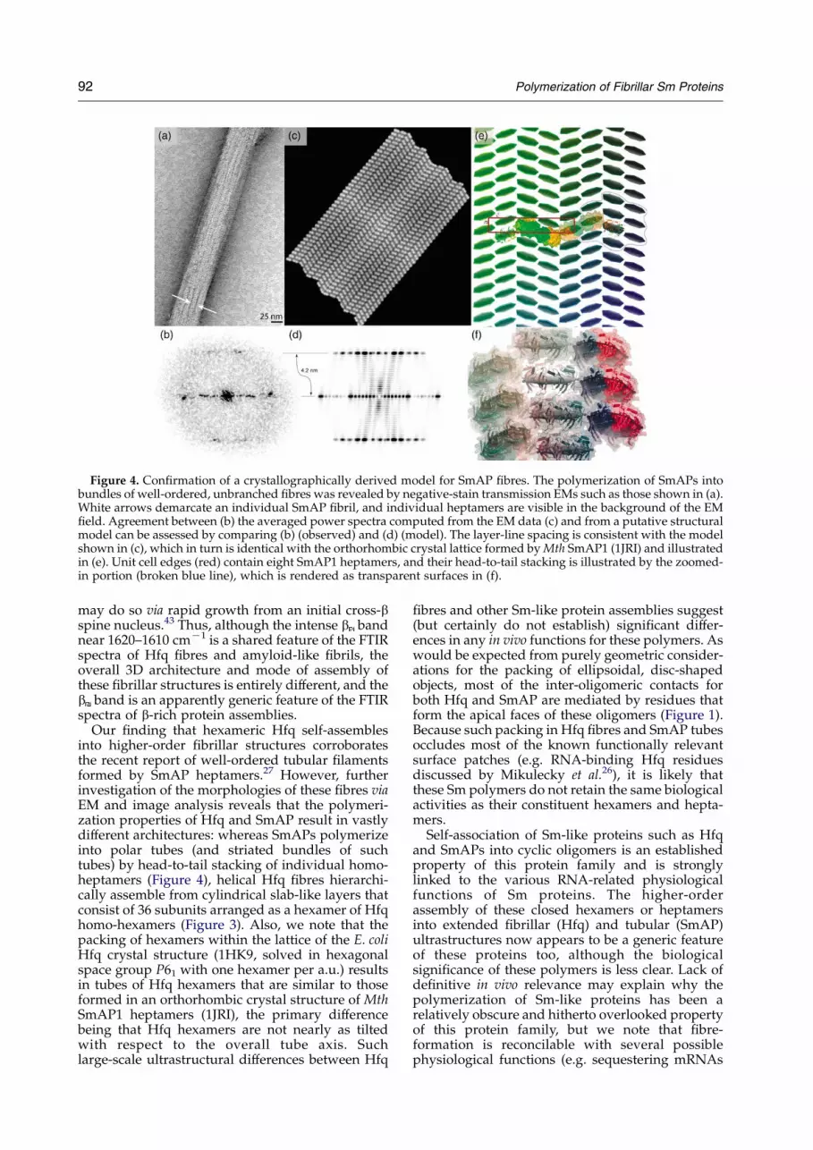
Figure 4. Confirmation of a crystallographically derived model for SmAP fibres. The polymerization of SmAPs intobundles of well-ordered, unbranched fibres was revealed by negative-stain transmission EMs such as those shown in (a).White arrows demarcate an individual SmAP fibril, and individual heptamers are visible in the background of the EMfield. Agreement between (b) the averaged power spectra computed from the EM data (c) and from a putative structuralmodel can be assessed by comparing (b) (observed) and (d) (model). The layer-line spacing is consistent with the modelshown in (c), which in turn is identical with the orthorhombic crystal lattice formed by Mth SmAP1 (1JRI) and illustratedin (e). Unit cell edges (red) contain eight SmAP1 heptamers, and their head-to-tail stacking is illustrated by the zoomed-in portion (broken blue line), which is rendered as transparent surfaces in (f).
92 Polymerization of Fibrillar Sm Proteins
may do so via rapid growth from an initial cross-bspine nucleus.43 Thus, although the intense b bandnear 1620–1610 cmK1 is a shared feature of the FTIRspectra of Hfq fibres and amyloid-like fibrils, theoverall 3D architecture and mode of assembly ofthese fibrillar structures is entirely different, and theb band is an apparently generic feature of the FTIRspectra of b-rich protein assemblies.
Our finding that hexameric Hfq self-assemblesinto higher-order fibrillar structures corroboratesthe recent report of well-ordered tubular filamentsformed by SmAP heptamers.27 However, furtherinvestigation of the morphologies of these fibres viaEM and image analysis reveals that the polymeri-zation properties of Hfq and SmAP result in vastlydifferent architectures: whereas SmAPs polymerizeinto polar tubes (and striated bundles of suchtubes) by head-to-tail stacking of individual homo-heptamers (Figure 4), helical Hfq fibres hierarchi-cally assemble from cylindrical slab-like layers thatconsist of 36 subunits arranged as a hexamer of Hfqhomo-hexamers (Figure 3). Also, we note that thepacking of hexamers within the lattice of the E. coliHfq crystal structure (1HK9, solved in hexagonalspace group P61 with one hexamer per a.u.) resultsin tubes of Hfq hexamers that are similar to thoseformed in an orthorhombic crystal structure of MthSmAP1 heptamers (1JRI), the primary differencebeing that Hfq hexamers are not nearly as tiltedwith respect to the overall tube axis. Suchlarge-scale ultrastructural differences between Hfq
fibres and other Sm-like protein assemblies suggest(but certainly do not establish) significant differ-ences in any in vivo functions for these polymers. Aswould be expected from purely geometric consider-ations for the packing of ellipsoidal, disc-shapedobjects, most of the inter-oligomeric contacts forboth Hfq and SmAP are mediated by residues thatform the apical faces of these oligomers (Figure 1).Because such packing in Hfq fibres and SmAP tubesoccludes most of the known functionally relevantsurface patches (e.g. RNA-binding Hfq residuesdiscussed by Mikulecky et al.26), it is likely thatthese Sm polymers do not retain the same biologicalactivities as their constituent hexamers and hepta-mers.
Self-association of Sm-like proteins such as Hfqand SmAPs into cyclic oligomers is an establishedproperty of this protein family and is stronglylinked to the various RNA-related physiologicalfunctions of Sm proteins. The higher-orderassembly of these closed hexamers or heptamersinto extended fibrillar (Hfq) and tubular (SmAP)ultrastructures now appears to be a generic featureof these proteins too, although the biologicalsignificance of these polymers is less clear. Lack ofdefinitive in vivo relevance may explain why thepolymerization of Sm-like proteins has been arelatively obscure and hitherto overlooked propertyof this protein family, but we note that fibre-formation is reconcilable with several possiblephysiological functions (e.g. sequestering mRNAs

Polymerization of Fibrillar Sm Proteins 93
or the store of Hfq itself, creating high localdensities of cognate RNAs, etc.). In this regard, wealso note that: (i) fibre formation in our experimentsis compatible with known intracellular concen-trations of some Sm-like proteins (e.g. z10–20 mMhexamer for E. coli Hfq;44); and (ii) polymerizationoccurs in the absence of RNA, and is therefore anintrinsic feature of these proteins (not an RNA-induced artefact). We have found that higher-orderHfq oligomers (possibly fibrillar) are capableof RNA-binding as assayed by electrophoreticmobility shift assays (EMSA) (unpublished dataand Supplementary Figure 1), so any physiologicalroles for Hfq and Sm polymers are probably linkedto their RNA-binding properties in vivo. Also inconnection with putative in vivo functions, it isknown that nucleoid-associated Hfq may influenceDNA packaging by polymerizing onto regions ofDNA rich in A-tracts.5 Finally, we note that Sm-likeproteins are not the sole example of RNA-bindingproteins that polymerize into fibres; similarbiochemical (RNA-binding) and biophysical(polymerization) properties have been reportedfor the HIV Rev protein,45–47 which is thought toform helical polymers that function in coating viralnucleic acids. Further elucidation of questionsregarding the in vivo existence of Hfq and Smfibres, their potential biological activities, andmechanistic aspects of their polymerization (e.g.kinetics, reversibility) will significantly advance ourpreliminary state of knowledge about these higher-order assemblies of Hfq and Sm-like proteins.
Materials and Methods
Unless specified otherwise, all reagents werepurchased from Sigma or Merck-Biochemicals.
Preparation of Hfq and SmAP fibres
E. coli Hfq was over-expressed as described,28 andpurified by affinity chromatography as follows: cellsfrom post-induction cultures were resuspended in 10 mlof a buffer containing 20 mM Tris–HCl (pH 7.8), 0.5 MNaCl, 10% (v/v) glycerol, 0.1% (v/v) Triton X-100, 10 mlDNase I (10 mg/ml), and a protease inhibitor cocktail(EDTA-free Complete mini, Roche Diagnostic) at 4 8C.The suspension was passed through a French press(1200 bar, 20,000 psi) and lysed cells were cleared bycentrifugation at 15,000g for 30 min. The supernatant washeated at 80 8C for 15 min, followed by centrifugation at15,000g for 30 min. Imidazole–HCl (pH 7.8) was added toa final concentration of 1 mM, and the resulting solutionwas applied to a 1 ml Ni2C-NTA column (Qiagen). Theresin was sequentially washed with approximately 15column volumes of (i) 20 mM Tris–HCl (pH 7.8), 0.3 MNaCl, 20 mM imidazole, and then (ii) 50 mM sodiumphosphate (pH 6.0), 0.3 M NaCl. The Hfq protein wasthen eluted in a buffer containing 20 mM Tris–HCl(pH 7.8), 0.3 M NaCl, 0.5 M imidazole. Hfq-containingfractions were dialyzed against poly(A) buffer (50 mMTris–HCl (pH 7.5), 1 mM EDTA, 5% (v/v) glycerol)supplemented with 50 mM NH4Cl. After centrifugation,the supernatant was applied to a 5 ml poly(A) Sepharose
column (Amersham Biosciences), and the resin waswashed with a poly(A) rinse buffer (poly(A) bufferC1.0 M NH4Cl); Hfq was eluted in a gradient of therinse buffer supplemented with 8.0 M urea. Hfq-contain-ing fractions were then dialyzed against poly(A) buffercontaining 50 mM NH4Cl and 0.1% (v/v) Triton X-100,and the protein was stored at 4 8C (typically at 1–2 mg/ml). Fibrous Hfq was produced by dialysis of proteinsamples (at a concentration of z5 mM hexamer) into abuffer consisting of 5 mM Tris–HCl (pH 8), 5 mM NaCl,and 0.005% (w/v) dodecyl-b-D-maltoside (buffer HfqIR),followed by lyophilization and re-suspension in water.
The expression and purification of Methanobacteriumthermautotrophicum strain DH (Mth) SmAP1 also has beendescribed in detail;27 fibrous Mth SmAP1 was obtained inthe course of preparing 0.5 mg/ml (z9 mM heptamer)protein samples in 10 mM Tris (pH 7.5), 60 mM NaCl forelectron microscopy on carbon-coated parlodion film.Hfq was found to be soluble at concentrations of at least30 mg/ml (0.4 mM hexamer) in most buffers, and,similarly, Mth SmAP1 could be concentrated to greaterthan 50 mg/ml (0.8 mM heptamer) without visibleprecipitation. Finally, we note that solution conditionsfor Hfq and SmAP fibrogenesis were not optimized interms of yield or fibre quality/homogeneity.
Infrared spectroscopy
Fourier transform IR (FTIR) spectra of Hfq wererecorded with a Perkin-Elmer system 2000 infraredspectrophotometer equipped with a multiple-reflectionattenuated total reflectance (ATR) system. Samples of Hfqprotein that had been dialyzed into buffer HfqIR werelyophilized and then dissolved in 2H2O (!99.8% purity;from Euriso-Top CEA) at a protein concentration of15 mg/ml. An 18 ml sample of the protein solution wasdeposited under dry air on the ZnSe crystal of the ATRdevice (12 internal reflections) and covered with anotherZnSe plate. Twenty-five scans were typically accumulatedat a nominal resolution of 2 cmK1, and buffer HfqIRspectra recorded under conditions identical with those ofthe protein sample were automatically subtracted. Datatreatment consisted of two-point baseline correction,Fourier self-deconvolution, and second derivativecalculation, and was performed with the Perkin-ElmerSpectrum software. Iterative least-squares curve-fittingsof spectral components (modelled as Gaussian lineshapes) were performed using Grams (Galactic Software).The method used to estimate the secondary structurecontent of Hfq was similar to that described,48 in whichthe fraction of a particular secondary structural element istaken as the sum of integrated intensities of thecorresponding Gaussian components (i.e. bands withmaxima falling within the frequency range assigned tothe structural element of interest) divided by the total areaof all bands over the entire frequency range. Absorptionbands were assigned as described by Krimm et al.40 andBarth & Zscherp.41 In brief, the band centered at1653 cmK1 is due to the amide I vibrational mode(essentially the symmetric )C]O/ stretching ofbackbone carbonyl amides), while the band centered at1550 cmK1 is attributed to amide II vibrations (essentiallya deformation mode resulting from a combination ofamide N–H in-plane bending and CKN stretchingmotions). The fine structures of both bands are sensitiveto local protein conformation, with the amide I bandbeing most frequently used in determining proteinsecondary structure content.

94 Polymerization of Fibrillar Sm Proteins
Electron microscopy and image analysis
Aliquots of Hfq that had been analyzed by FTIRspectroscopy in order to confirm the presence ofabsorption bands characteristic of aggregated b-sheets(z1610 cmK1; see Table 1) were diluted 100-fold,adsorbed to 300-mesh carbon-coated grids, and thenstained with 1% (w/v) uranyl acetate. Samples werevisualized in a Philips CM120 transmission electronmicroscope (Philips; Eindhoven, Netherlands) operatingat an accelerating voltage of 120 kV. Twenty images wererecorded at a nominal magnification of 60,000! andaverage defocus of K1300 nm by using a 1024 byte GatanssCCD. A total of 122 fibre-containing subimages (each of256!256 pixels) were windowed from the original 1024!1024 dataset, and discrete, clearly defined fibrous speci-mens from each image were masked and registered to ahorizontal reference before computing their Fouriertransforms (FTs). Power spectral images (S(f)) werecalculated from the complex modulus of these trans-forms, and such images were then rotationally alignedand classified into groups via an unsupervised self-organizing mapping algorithm.49 In order to increasesignal intensities of Bragg reflections, an averaged powerspectrum hS(f)i was computed from the power spectra of87 single images belonging to the major class, as proposedby Watts et al.45 The helical pitch (P) and inter-subunitspacing (h) were then calculated from hS(f)i via thestandard theory for diffraction from a helical (one-dimensional) point lattice.38,50 An improved, averagedimage was computed from sub-images of fibres corre-sponding to individual major-class images after rotationaland translational alignment, and final helical parameterswere determined from this final image. This averagedimage was also used to estimate a helical radius (r) ofz8.5 nm and a resolution value of 2.3 nm by using aspectral signal-to-noise criterion.37 All steps of this single-particle image analysis procedure were performed withinthe Xmipp software package.51
Candidate 3D models of Hfq fibres were generated bypositioning the X-ray crystallographic structure ofhexameric E. coli Hfq (PDB code 1HK922) into 3Dlocations, which increased the correlation betweenpower spectra calculated from projection images of themodel (S(f)Model) and the averaged experimentallyobserved spectra (hS(f)Obsi). More specifically, positioningof Hfq hexamers and calculation of synthetic EMprojection images (using the Spider software52) wasfollowed by filtering of the synthetic images to theresolution limit computed from the experimental EMimages. Computed power spectra of these filtered imagesðSðf ÞfiltModelÞ were then compared to corresponding experi-mental spectra (S(f)obs)), and the 3D model was manuallyrefined by rigid-body transformations of the hexameruntil more optimal agreement was obtained (i.e. mini-mization of the difference between S(f)obs and Sðf ÞfiltModelsubject to the constraint of consistency with the helicalparameters P, h, and r). This heuristic procedure wasiteratively applied until only minimal model improve-ment was attained.
† http://pymol.sourceforge.net
Model refinement and analysis
A final stage of model refinement included grossgeometric/stereochemical correction of the crystallo-graphically derived fibre structure (e.g. de-bumpingatomic clashes), followed by more careful potentialenergy minimization to relax Hfq monomers into a
conformation more likely to be adopted in the fibrillarstate. The energy of the model was reduced in vacuo withrespect to the Amber FF03 forcefield (12 A cut-off for non-bonded interactions) over the course of O5000 steps ofsteepest descent (earlier cycles) and conjugate gradient(later cycles) minimization in Amber v8.0.53 The startingstructure consisted of three layers of the fibre (corres-ponding to 6!6!3Z108 independent 1HK9 monomers),and a final multi-layered fibre model was built bysuperimposing the relaxed/minimized middle layerupon adjacent layers in both directions along the helicalaxis (rather than imposing one-dimensional periodicboundary conditions along the fibre axis). Implicit in thismethod is the assumption that only interactions within G1layer are energetically significant (layers are separated byO50 A, so this is likely to be a valid approach).
A 3D model for SmAP fibres was derived by twoapproaches: (i) using the quasi-hexagonal packing of MthSmAP1 heptamers in the P212121 crystal form describedby Mura et al.,27 and (ii) by applying the same EM andimage analysis-based approach described above for Hfq.The only difference in the case of SmAP1 was that startingimages used to construct this model were extracted fromthe electron micrographs of Mth SmAP1 described byMura et al.27 by digitization using a LeafScan 45 linearCCD scanner with a 25 mm window spacing.
The molecular modelling toolkit54 and Python scriptswere utilized for coordinate transformations and manip-ulations involving large numbers of subunits (e.g. a singlefibre layer of 36 Hfq monomers), and the programsPymol† and VMD55 were utilized along with theirrespective Python and Tcl application programminginterfaces in order to compute and analyze the crystallattices and intermolecular contact distances used increating Figure 1. Pymol was also used to generate allFigures involving illustrations of protein structures(i.e. Figures 1, 3 and 4).
Acknowledgements
We gratefully acknowledge the CNRS (UPR 9073)and University Denis Diderot-Paris 7 for financialsupport, as well as funding from an Alfred P. Sloan/DOE postdoctoral fellowship (C.M.) and a Conacytdoctoral fellowship (M.R.G.). We also thank theInstitut Curie (Paris) for providing EM and imageanalysis facilities, Dr Claudine Mayer (U. Paris 6)for her assistance in constructing models of Hfqfibres, M. Folichon (IBPC) for EMSA work, andProfessor Eliane Taillandier (U. Paris 13) forconstant interest in this work. Drs Linda Columbus(Scripps), David Eisenberg (UCLA), and AndyMcCammon (UCSD) are thanked for reviewingthe manuscript.
Supplementary Data
Supplementary data associated with this articlecan be found, in the online version, at doi:10.1016/j.jmb.2005.11.010

Polymerization of Fibrillar Sm Proteins 95
References
1. Franze de Fernandez, M. T., Hayward, W. S. &August, J. T. (1972). Bacterial proteins required forreplication of phage Qb ribonucleic acid. J. Biol. Chem.247, 821–824.
2. Valentin-Hansen, P., Eriksen, M. & Udesen, C. (2004).The bacterial Sm-like protein Hfq: a key player inRNA transactions. Mol. Microbiol. 51, 1525–1533.
3. Folichon, M., Arluison, V., Pellegrini, O., Huntzinger, E.,Regnier, P. & Hajnsdorf, E. (2003). The poly(A)binding protein Hfq protects RNA from RNase Eand exoribonucleolytic degradation. Nucl. Acids Res.31, 7302–7310.
4. Azam, T. A. & Ishihama, A. (1999). Twelve species ofthe nucleoid-associated protein from Escherichia coli.J. Biol. Chem. 274, 33105–33113.
5. Tolstorukov, M. Y., Virnik, K. M., Adhya, S. &Zhurkin, V. B. (2005). A-tract clusters may facilitateDNA packaging in bacterial nucleoid. Nucl. Acids Res,33, 3907–3918.
6. Storz, G. (2002). An expanding universe of noncodingRNAs. Science, 296, 1260–1263.
7. Tsui, H.-C. T., Leung, H.-C. E. & Winkler, M. E. (1994).Characterization of broadly pleiotropic phenotypescaused by an hfq insertion mutation in Escherichia coliK-12. Mol. Microbiol. 13, 35–49.
8. Hengge-Aronis, R. (2002). Signal transduction andregulatory mechanisms involved in control of thesigma(S) (RpoS) subunit of RNA polymerase.Microbiol. Mol. Biol. Rev. 66, 373–395.
9. Masse, E., Majdalani, N. & Gottesman, S. (2003).Regulatory roles for small RNA in bacteria. Curr.Opin. Microbiol. 6, 120–124.
10. Vytvytska, O., Jakobsen, J. S., Balcunate, G., Andersen,J. S., Baccarini, M., von Gabain, A. & Host-factor, I.(1998). Hfq, binds to Escherichia coli ompA mRNA in agrowth rate-dependent fashion and regulates itsstability. Proc. Natl Acad. Sci. USA, 95, 14118–14123.
11. Geissmann, T. A. & Touati, D. (2004). Hfq, a newchaperoning role: binding to messenger RNA deter-mines access for small RNA regulator. EMBO J. 23,396–405.
12. Sukhodolets, M. V. & Garges, S. (2003). Interaction ofEscherichia coli RNA polymerase with the ribosomalprotein S1 and the Sm-like ATPase Hfq. Biochemistry,42, 8022–8034.
13. Moll, I., Afonyushkin, T., Vytvytska, O., Kaberdin, V. R.& Blasi, U. (2003). Coincident Hfq binding and RNaseE cleavage sites on mRNA and small regulatoryRNAs. RNA, 9, 1308–1314.
14. Will, C. & Luhrmann, R. (2001). SpliceosomalUsnRNP biogenesis, structure and function. Curr.Opin. Cell Biol. 13, 290–301.
15. Tharun, S., He, W., Mayes, A. E., Lennertz, P.,Beggs, J. D. & Parker, R. (2000). Yeast Sm-like proteinsfunction in mRNA decapping and decay. Nature, 404,515–518.
16. Seto, A. G., Zaug, A. J., Sobel, S. G., Wolin, S. L. &Cech, T. R. (1999). Saccharomyces cerevisiae telomer-ase is an Sm small nuclear ribonucleoprotein particle.Nature, 401, 177–180.
17. Arluison, V., Derreumaux, P., Allemand, F.,Folichon, M., Hajnsdorf, E. & Regnier, P. (2002).Structural modelling of the Sm-like protein Hfqfrom Escherichia coli. J. Mol. Biol. 320, 705–712.
18. Sun, X., Zhulin, I. & Wartell, R. M. (2002). Predictedstructure and phyletic distribution of the RNA-binding protein Hfq. Nucl. Acids Res. 30, 3662–3671.
19. Moller, T., Franch, T., Hojrup, P., Keene, D. R.,Bachinger, H. P., Brennan, R. G. & Valentin-Hansen,P. (2002). Hfq. A bacterial Sm-like protein thatmediates RNA–RNA interaction. Mol. Cell. 9, 23–30.
20. Zhang, A., Wassarman, K. M., Ortega, J., Steven, A. C.& Storz, G. (2002). The Sm-like Hfq protein increasesOxyS RNA interaction with target mRNAs. Mol. Cell.9, 11–22.
21. Schumacher, M. A., Pearson, R. F., Moller, T., Valentin-Hansen, P. & Brennan, R. G. (2002). Structures of thepleiotropic translational regulator Hfq and an Hfq-RNA complex: a bacterial Sm-like protein. EMBO J.21, 3546–3556.
22. Sauter, C., Basquin, J. & Suck, D. (2003). Sm-likeproteins in Eubacteria: the crystal structure of the Hfqprotein from Escherichia coli. Nucl. Acids Res. 31,4091–4098.
23. Nikulin, A., Stolboushkina, E., Perederina, A.,Vassilieva, I., Blaesi, U., Moll, I. et al. (2005). Structureof Pseudomonas aeruginosa Hfq protein. Acta Crystallog.sect. D, 61, 141–146.
24. Moll, I., Leitsch, D., Steinhauser, T. & Blasi, U. (2003).RNA chaperone activity of the Sm-like Hfq protein.EMBO Rep. 4, 284–289.
25. Toro, I., Thore, S., Mayer, C., Basquin, J., Seraphin, B.& Suck, D. (2001). RNA binding in an Sm coredomain: X-ray structure and functional analysis of anarchaeal Sm protein complex. EMBO J. 20, 2293–2303.
26. Mikulecky, P. J., Kaw, M. K., Brescia, C. C.,Takach, J. C., Sledjeski, D. D. & Feig, A. L. (2004).Escherichia coli Hfq has distinct interaction surfaces forDsrA, rpoS and poly(A) RNAs. Nature Struct. Mol.Biol. 11, 1206–1214.
27. Mura, C., Kozhukhovsky, A., Gingery, M., Phillips, M.& Eisenberg, D. (2003). The oligomerization andligand-binding properties of Sm-like archaeal pro-teins (SmAPs). Protein Sci. 12, 832–847.
28. Arluison, V., Folichon, M., Marco, S., Derreumaux, P.,Pellegrini, O., Seguin, J. et al. (2004). The C-terminaldomain of Escherichia coli Hfq increases the stability ofthe hexamer. Eur. J. Biochem. 271, 1258–1265.
29. Jackson, M. & Mantsch, H. H. (1995). The use andmisuse of FTIR spectroscopy in the determination ofprotein structure. Crit. Rev. Biochem. Mol. Biol. 30,95–120.
30. Tatulian, S. A. (2003). Attenuated total reflectionFourier transform infrared spectroscopy: a methodof choice for studying membrane proteins and lipids.Biochemistry, 42, 11898–11907.
31. Fandrich, M. & Dobson, C. M. (2002). The behaviourof polyamino acids reveals an inverse side chain effectin amyloid structure formation. EMBO J. 21,5682–5690.
32. Arrondo, J. L., Muga, A., Castresana, J. & Goni, F. M.(1993). Quantitative studies of the structure ofproteins in solution by Fourier-transform infraredspectroscopy. Prog. Biophys. Mol. Biol. 59, 23–56.
33. Chirgadze, Y. N., Fedorov, O. V. & Trushina, N. P.(1975). Estimation of amino acid residue side-chainabsorption in the infrared spectra of protein solutionsin heavy water. Biopolymers, 14, 679–694.
34. Bendit, E. G. (1967). Infrared absorption of tyrosineside chains in proteins. Biopolymers, 5, 4525–4533.
35. Kubelka, J. & Keiderling, T. A. (2001). Differentiationof beta-sheet-forming structures: ab initio-basedsimulations of IR absorption and vibrational CD formodel peptide and protein beta-sheets. J. Am. Chem.Soc. 123, 12048–12058.

96 Polymerization of Fibrillar Sm Proteins
36. Kubelka, J. & Keiderling, T. A. (2001). The anomalousinfrared amide I intensity distribution in (13)Cisotopically labeled peptide beta-sheets comes fromextended, multiple-stranded structures: an ab initiostudy. J. Am. Chem. Soc. 123, 6142–6150.
37. Unser, M., Trus, B. L. & Steven, A. C. (1987). A newresolution criterion based on spectral signal-to-noiseratios. Ultramicroscopy, 23, 39–51.
38. Klug, A., Crick, F. H. C. & Wyckoff, H. W. (1958).Diffraction by helical structures. Acta Crystallog. 11,199–213.
39. Cantor, C. R. & Schimmel, P. R. (1980). BiophysicalChemistry, Freeman Co., San Francisco, CA.
40. Krimm, S. & Bandekar, J. (1986). Vibrational spec-troscopy and conformation of peptides, polypeptides,and proteins. Advan. Protein Chem. 38, 181–364.
41. Barth, A. & Zscherp, C. (2002). What vibrations tell usabout proteins. Quart. Rev. Biophys. 35, 369–430.
42. Ohnishi, S. & Takano, K. (2004). Amyloid fibrils fromthe viewpoint of protein folding. Cell Mol. Life Sci. 61,511–524.
43. Nelson, R., Sawaya, M. R., Balbirnie, M., Madsen, A. O.,Riekel, C., Grothe, R. & Eisenberg, D. (2005). Structureof the cross-beta spine of amyloid-like fibrils. Nature,435, 773–778.
44. Kajitani, M., Kato, A., Wada, A., Inokuchi, Y. &Ishihama, A. (1994). Regulation of the Escherichia colihfq gene encoding the host factor for phage Q beta.J. Bacteriol. 176, 531–534.
45. Watts, N. R., Misra, M., Wingfield, P. T., Stahl, S. J.,Cheng, N., Trus, B. L. et al. (1998). Three-dimensionalstructure of HIV-1 Rev protein filaments. J. Struct. Biol.121, 41–52.
46. Jain, C. & Belasco, J. G. (2001). Structural model for thecooperative assembly of HIV-1 Rev multimers on theRRE as deduced from analysis of assembly-defectivemutants. Mol. Cell. 7, 603–614.
47. Blanco, F. J., Hess, S., Pannell, L. K., Rizzo, N. W. &Tycko, R. (2001). Solid-state NMR data support ahelix-loop-helix structural model for the N-terminalhalf of HIV-1 Rev in fibrillar form. J. Mol. Biol. 313,845–859.
48. Cabiaux, V., Brasseur, R., Wattiez, R., Falmagne, P.,Ruysschaert, J. M. & Goormaghtigh, E. (1989).Secondary structure of diphtheria toxin and itsfragments interacting with acidic liposomes studiedby polarized infrared spectroscopy. J. Biol. Chem. 264,4928–4938.
49. Marabini, R. & Carazo, J. M. (1994). Pattern recog-nition and classification of images of biologicalmacromolecules using artificial neural networks.Biophys. J. 66, 1804–1814.
50. Sherwood, D. (1976). Crystals, X ray and Proteins,Longman Group, London.
51. Marabini, R., Masegosa, I. M., San Martin, C.,Marco, S., Fernandez, J. J., de la Fraga, L. G. et al.(1996). Xmipp: an image processing package forelectron microscopy. J. Struct. Biol. 116, 237–240.
52. Frank, J., Radermacher, M., Penczek, P., Zhu, J., Li, Y.,Ladjadj, M. & Leith, A. (1996). SPIDER and WEBprocessing and visualization of images in 3D electronmicroscopy and related fields. J. Struct. Biol. 116,190–199.
53. Case, D. A., Darden, T. A., Cheatham, T. E., III,Simmerling, C. L., Wang, J., Duke, R. E. et al.(2004). AMBER v8, University of California, SanFrancisco.
54. Hinsen, K. (2000). The molecular modelling toolkit: anew approach to molecular simulations. J. Comput.Chem. 21, 79–85.
55. Humphrey, W., Dalke, A. & Schulten, K. (1996).VMD–visual molecular dynamics. J. Mol. Graph. 14,33–38.
Edited by I. B. Holland
(Received 26 August 2005; received in revised form 2 November 2005; accepted 3 November 2005)Available online 22 November 2005