The BTB–zinc finger transcriptional regulator PLZF controls the development of invariant natural...
23
The BTB-zinc finger transcriptional regulator, PLZF, controls the development of iNKT cell effector functions Damian Kovalovsky 1,8 , Olisambu U. Uche 3,8 , Sonia Eladad 1,8 , Robin M. Hobbs 2,6 , Woelsung Yi 1 , Eric Alonzo 4 , Kevin Chua 1 , Maggie Eidson 5 , Hye-Jung Kim 1 , Jin S. Im 7 , Pier Paolo Pandolfi 2,6 , and Derek B. Sant'Angelo 1,3,4 1Immunology Program, Sloan-Kettering Institute, Memorial Sloan-Kettering Cancer Center, New York, NY 10065, USA 2Cancer Biology and Genetics Program, Sloan-Kettering Institute, Memorial Sloan-Kettering Cancer Center, New York, NY 10065, USA 3Weill Graduate School of Medical Sciences of Cornell University, New York, NY 10065, USA 4Louis V. Gerstner Jr. Graduate School of Biomedical Sciences, Memorial Sloan-Kettering Cancer Center New York, NY 10065, USA 5Department of Pediatrics Memorial Sloan-Kettering Cancer Center New York, NY 10065 USA 7Department of Microbiology and Immunology, Albert Einstein College of Medicine, Bronx, NY 10461, USA Abstract Invariant Natural Killer T cell (iNK Tcell) have an innate immunity-like rapidity of response and the capacity to modulate effector functions of other cells. We show that the BTB-ZF transcriptional regulator, PLZF, is specifically expressed in iNKT cells. iNKT cells develop in the absence of PLZF, but lack many innate T cell features. PLZF deficient iNKT cells accumulate in the lymph nodes rather than in the liver and do not have an activated phenotype or express NK markers. PLZF deficient iNKT cells do not secrete high levels of IL-4 and IFNγ upon activation, however some cells produce either IL-4 or IFNγ, but not both. PLZF, therefore, is an iNKT cell specific transcription factor that is necessary for full functionality. Nearly all hematopoietic cells mature in the bone marrow. In contrast, multipotent progenitor T cells leave the bone marrow and home to the thymus, where signals from stromal cells are required for commitment to the T lineage 1 . Once directed into the T lineage, the cells undergo a rigorous selection process that eliminates more than 95% of the candidate T cells. Full maturation requires the expression of a T cell receptor (TCR) that binds self-peptide:self-MHC complexes with sufficient avidity. At some point during development, T cells are directed into one of several distinct T cell lineages such as CD4 single positive “helper” cells, CD8 single positive “killer” cells or CD4 + CD25 + regulatory cells. Commitment to these various lineages defines the specialized functions of the cell, which is critical since each cell type plays an essential and distinct role for host defense. The genes responsible for directing multipotent T cell progenitors into the various lineages are largely unknown 2 . Correspondence and requests for materials should be addressed to D.B.S (Email: [email protected]). 6 Current address: Cancer Genetics Program, Beth Israel Deaconess Cancer Center and Department of Medicine, Beth Israel Deaconess Medical Center, Harvard Medical School, Boston, MA 02115, USA 8 These authors contributed equally to this work NIH Public Access Author Manuscript Nat Immunol. Author manuscript; available in PMC 2009 March 30. Published in final edited form as: Nat Immunol. 2008 September ; 9(9): 1055–1064. doi:10.1038/ni.1641. NIH-PA Author Manuscript NIH-PA Author Manuscript NIH-PA Author Manuscript
-
Upload
independent -
Category
Documents
-
view
2 -
download
0
Transcript of The BTB–zinc finger transcriptional regulator PLZF controls the development of invariant natural...

The BTB-zinc finger transcriptional regulator, PLZF, controls thedevelopment of iNKT cell effector functions
Damian Kovalovsky1,8, Olisambu U. Uche3,8, Sonia Eladad1,8, Robin M. Hobbs2,6,Woelsung Yi1, Eric Alonzo4, Kevin Chua1, Maggie Eidson5, Hye-Jung Kim1, Jin S. Im7, PierPaolo Pandolfi2,6, and Derek B. Sant'Angelo1,3,4
1Immunology Program, Sloan-Kettering Institute, Memorial Sloan-Kettering Cancer Center, New York, NY10065, USA
2Cancer Biology and Genetics Program, Sloan-Kettering Institute, Memorial Sloan-Kettering Cancer Center,New York, NY 10065, USA
3Weill Graduate School of Medical Sciences of Cornell University, New York, NY 10065, USA
4Louis V. Gerstner Jr. Graduate School of Biomedical Sciences, Memorial Sloan-Kettering Cancer CenterNew York, NY 10065, USA
5Department of Pediatrics Memorial Sloan-Kettering Cancer Center New York, NY 10065 USA
7Department of Microbiology and Immunology, Albert Einstein College of Medicine, Bronx, NY 10461, USA
AbstractInvariant Natural Killer T cell (iNK Tcell) have an innate immunity-like rapidity of response and thecapacity to modulate effector functions of other cells. We show that the BTB-ZF transcriptionalregulator, PLZF, is specifically expressed in iNKT cells. iNKT cells develop in the absence of PLZF,but lack many innate T cell features. PLZF deficient iNKT cells accumulate in the lymph nodes ratherthan in the liver and do not have an activated phenotype or express NK markers. PLZF deficientiNKT cells do not secrete high levels of IL-4 and IFNγ upon activation, however some cells produceeither IL-4 or IFNγ, but not both. PLZF, therefore, is an iNKT cell specific transcription factor thatis necessary for full functionality.
Nearly all hematopoietic cells mature in the bone marrow. In contrast, multipotent progenitorT cells leave the bone marrow and home to the thymus, where signals from stromal cells arerequired for commitment to the T lineage1. Once directed into the T lineage, the cells undergoa rigorous selection process that eliminates more than 95% of the candidate T cells. Fullmaturation requires the expression of a T cell receptor (TCR) that binds self-peptide:self-MHCcomplexes with sufficient avidity. At some point during development, T cells are directed intoone of several distinct T cell lineages such as CD4 single positive “helper” cells, CD8 singlepositive “killer” cells or CD4+CD25+ regulatory cells. Commitment to these various lineagesdefines the specialized functions of the cell, which is critical since each cell type plays anessential and distinct role for host defense. The genes responsible for directing multipotent Tcell progenitors into the various lineages are largely unknown 2.
Correspondence and requests for materials should be addressed to D.B.S (Email: [email protected]).6Current address: Cancer Genetics Program, Beth Israel Deaconess Cancer Center and Department of Medicine, Beth Israel DeaconessMedical Center, Harvard Medical School, Boston, MA 02115, USA8These authors contributed equally to this work
NIH Public AccessAuthor ManuscriptNat Immunol. Author manuscript; available in PMC 2009 March 30.
Published in final edited form as:Nat Immunol. 2008 September ; 9(9): 1055–1064. doi:10.1038/ni.1641.
NIH
-PA Author Manuscript
NIH
-PA Author Manuscript
NIH
-PA Author Manuscript

Among the various lineages of T cells, invariant Natural Killer T cells (iNKT cells) have severalunique phenotypic traits such as the expression of receptors typically associated with NaturalKiller cells (NK cells), the constitutive expression of activation markers and extremelyrestricted TCR diversity3. iNKT cells express an identical TCRα chain and most use aTCRβ chain that utilizes the Vβ8.2 gene segment. This TCR confers specificity to the non-MHC encoded self-molecule, CD1d, which binds and presents glycolipids rather than thetypical peptide cargo presented by conventional MHC molecules.
iNKT cells are also functionally distinct. Of particular interest is their ability to secrete largequantities of a variety of cytokines only minutes after activation via the TCR3. The rapidresponse of these cells, the conserved nature of the TCR and their indirect ability to modulatethe function of many different cell types of the immune system has led to the appreciation thatiNKT cells lay at a functional cusp between the innate and adaptive immune systems4. Thebroad range of cytokines released by iNKT cells results in their potential to regulate seeminglyopposing immune responses. For example, iNKT cells have been shown to enhance immuneresponses against tumors, but they have also been shown to prevent autoimmunity bydiminishing self-reactive T cell responses5.
The expression of a TCR specific for the CD1d ligand loaded with an appropriate glycolipidis the only unique identifier of iNKT cells. This highly conserved TCR also allows for thedirect detection of iNKT cells by the use of a tetramerized version of the CD1d molecule loadedwith a glycolipid, referred to as α- galactosylceramide (α-GalCer)6. This reagent allows forthe unambiguous detection of iNKT cells, which has made these cells much more amenableto genetic studies than the extremely diverse conventional T cell populations. As a result,several genes that influence iNKT cell development have been identified. Loss of any one ofa handful of genes, such as T-bet or CSF-2, impacts events late in development and typicallyresults in altered iNKT cell function7. Loss of Fyn expression dramatically affectsdevelopment, presumably due to a decrease in signaling via the TCR or other receptors, forexample SLAM8-10. Indeed, loss of the SLAM family adapter protein, SAP, also eliminatesiNKT cell development11-13. The Runx1 transcription factor has also been shown to berequired for iNKT cell development14. Loss of Runx1 expression, however, has severe andpleiotropic affects on thymocyte development. Indeed, none of the genes that have beenidentified to affect iNKT cell development are specifically expressed in iNKT cells.
The BTB/POZ-ZF [Broad complex, Tramtrack, Bric-à-brac (BTB) or poxvirus and zinc finger(POZ)-zinc finger] protein family of transcription factors has been found to control a widevariety of biological processes15. These BTB-zinc finger (BTB-ZF) family members aredefined by the presence of an N-terminal protein-protein interaction domain (BTB/POZ) andC-terminal C2H2 zinc finger domains. Several members of this gene family have been shownto have transcriptional repressor activity. BTB-ZF genes have also been shown to controlseveral fundamental aspects of immune system function. For example, BCL6 controls thegerminal center reaction that is required for affinity maturation of B cells16. In T cells, it hasbeen shown that Th-POK is the master regulator of the CD4 versus CD8 lineage commitmentstep in the thymus17, 18,which is fundamental for T cell function. And, most recently, the Tversus B cell fate decision was also shown to be controlled by a BTB-ZF gene called LRF19.Here we report that PLZF, also a member of the BTB-ZF gene family, is necessary for thedevelopment of iNKT cells.
ResultsAmong lymphoid cells, PLZF expression is highly restricted to iNKT cells
Recognizing the increasing number of BTB-ZF finger genes involved in the function of theimmune system, we screened various thymocyte populations for the expression of several
Kovalovsky et al. Page 2
Nat Immunol. Author manuscript; available in PMC 2009 March 30.
NIH
-PA Author Manuscript
NIH
-PA Author Manuscript
NIH
-PA Author Manuscript

different family members. Many of the BTB-ZF genes we analyzed were expressed in allthymic subpopulations (data not shown). One, however, the promyelocytic leukemia zincfinger gene (PLZF)20, was highly expressed in iNKT cells, as compared to conventional Tcells. The expression of PLZF in iNKT cells was apparent in thymocytes and also inlymphocytes isolated from the liver of mice (Fig. 1A). To further analyze PLZF expressionpatterns, we used a new monoclonal antibody directed against the hinge region of the protein.CD1d:αGalCer tetramers6 were used to specifically identify iNKT cells (Fig. 1B, left).Intracellular staining for PLZF in iNKT cells and conventional T cells clearly showed that thetranscription factor was expressed at variable levels in thymic iNKT cells and at moderatelevels in liver iNKT cells (Fig. 1B, right). PLZF was not detected, however, in conventionalthymocytes or conventional liver T cells (Fig. 1B). T cells from a PLZF deficient mouse21(see below) were used to confirm the specificity of the antibody (Fig. 1B).
Peripheral blood lymphocytes from humans were collected and examined by FACS for PLZFexpression. iNKT cells were identified with the 6B11 monoclonal antibody, which recognizesa unique determinant within the TCR CDR3 region, in combination with an antibody againstCD3 (Fig. 1C). PLZF was detected by intracellular staining with our anti-mouse antibody,which crossreacts with the human protein. Human iNKT cells clearly express PLZF, whileCD3+ T cells express little or no PLZF (Fig. 1C).
iNKT cell development is typically divided into three stages22, which are primarily identifiedby the cell surface upregulation of CD44, followed by NK1.1 (Fig. 1D). Nearly all stage 1 cellsexpress high levels of PLZF. The extremely rare PLZF negative events (∼0.003%) seen amongthe stage 1 cells could not definitively be determined to be real iNKT cell events since stainingwith empty tetramer resulted in the detection of similar events (data not shown). Consistentwith the finding that PLZF is downregulated in other cell types during differentiation, includingCD34+ human stem cells23, 24, we find that the transcription factor is downregulated in theiNKT cells found within the more mature stage 2 and 3 populations (Fig. 1D). PLZF expressionat stage 3 is equivalent to expression in mature, liver iNKT cells.
T-bet deficient iNKT cells are blocked at stage 2 of development25 (Fig. 1E). These geneticallyblocked iNKT cells did not downregulate PLZF to the mature stage 3 levels. The levels ofPLZF in stage 1 and 2, however, were similar to wild type cells (Fig. 1E). Therefore, regulationof PLZF expression is tightly linked to the stage of iNKT cell development.
Young mice have a much greater number of immature, developing iNKT cells than adultmice9. Therefore, we examined 10-13 days old mice to determine the expression of PLZFduring early subsets of cells typically not detectable in adult mice. CD44neg, HSAhi iNKT cells,which are thought to represent the most immature iNKT cell population in the thymus, clearlyexpress high levels of PLZF (Fig. 1F). As the developing iNKT cells begin to express CD44(Stage 1 CD44lo), HSA starts to be downregulated, while PLZF remains high (Fig. 1F). Asseen in adult mice (Fig. 1D), PLZF expression continued to downregulate as the cells mature.
Lack of PLZF expression does not affect lymphocyte developmentTo further examine the expression of PLZF in the lymphoid compartment, we examined Bcells and NK cells by FACS. PLZF was not detected in either of these cell types (Fig. 2A).PLZF was also not detected in eosinophils, neutrophils or macrophages (data not shown).
PLZF deficient mice were previously shown to have disrupted patterning of the hindlimbs21 and a progressive loss of spermatogonia26. Development of the hematopoietic system,however, was not overtly perturbed in PLZF deficient mice (Fig. 2B). For example, normalpercentages of all the thymic subpopulations were found, although the size of the thymus fromPLZF deficient mice was consistently decreased by ∼25%. Nonetheless, normal thymic
Kovalovsky et al. Page 3
Nat Immunol. Author manuscript; available in PMC 2009 March 30.
NIH
-PA Author Manuscript
NIH
-PA Author Manuscript
NIH
-PA Author Manuscript

structure was observed (Fig. 2C). Also, thymocytes expressing an MHC class II restricted TCRtransgene developed normally in the absence of PLZF (data not shown). Lymph node T cellsalso appeared normal in the PLZF deficient mice (Fig. 2B) as well as in the spleen (data notshown). DX5+ T cells, which largely represent non-CD1d restricted NKT cells27, NK cellsand γδ T cells also appeared normal (Fig. 2D). Finally, no obvious differences were seen inthe B cell compartment (data not shown). Therefore, consistent with the restricted expressionpattern of PLZF, the bulk of the lymphoid compartment is intact. The alteration in thymiccellularity appears to be secondary to any affects that PLZF may have on T cell developmentand potentially is simply a consequence of the stress caused by the malformed hind limbs.
Altered iNKT cell development in PLZF deficient miceAlthough conventional T cell development was not perturbed in the absence of PLZF, thefrequency of CD1d tetramer positive iNKT cells in the thymus was dramatically reduced (∼9fold) as compared to wild type mice (Fig. 3A). The specificity of the FACS staining of theiNKT cells is made clear by comparison to CD1d deficient mice28, which are devoid of iNKTcells due to the lack of the selecting ligand and by staining with empty CD1d tetramer (datanot shown). The detection of some tetramer positive cells was important for several reasons.First, it proved that the invariant TCRα chain can be generated, which is different than thesituation in RORγ-T deficient mice14. Generation of the iNKT TCRα chain was also confirmedby rt-PCR followed by DNA sequencing (data not shown). Secondly, the iNKT cells in thePLZF deficient mice must have undergone positive selection. Random V(D)J recombinationof the TCR locus is estimated to produce at least 1015 different TCR combinations29.Therefore, the frequency of 1 tetramer positive cell per ∼2500 thymocytes (∼0.04%) that wefind in the PLZF deficient mice (Fig. 3A) must be a consequence of positive selection thatresults in the enrichment of thymocytes expressing this TCR. Thirdly, the presence of tetramerpositive cells suggested that we could directly examine the phenotype and function of iNKTcells in the absence of PLZF.
CD44, which is an activation marker for conventional T cells, is upregulated duringdevelopment of all iNKT cells in the thymus and is constitutively expressed by all matureiNKT. The tetramer positive iNKT cells found in the thymus of PLZF deficient mice, however,had not upregulated CD44 (Fig. 3B). Nearly half of the PLZF deficient iNKT cells hadupregulated CD69, which, like CD44, is a marker of T cell activation that is expressed by mostmature iNKT cells. In the thymus, CD69 also serves as a marker for thymocytes that haveproductively interacted with self-MHC molecules and, therefore, upregulation of CD69suggests that the PLZF deficient iNKT cells have been signaled through the TCR. Therequirement for PLZF is intrinsic to iNKT cells since PLZF deficient bone marrow transferredinto irradiated C57Bl/6 hosts resulted in the development of few CD44lo, iNKT cells (Fig. S1).Transfer of bone marrow from wild type littermates into C57Bl/6 hosts resulted in the expectedfrequencies of iNKT cells that had fully matured to the CD44hiCD69+ stage. Additionally,FACS analysis showed that CD1d expression is not altered in PLZF deficient mice (Fig. S2).
iNKT cells are known to proliferate during the early stages (HSAlo, CD44lo)of thymicdevelopment. This expansion appears to be subsequent to positive selection30. Division ofiNKT cells in wild type and PLZF deficient mice was tested by analyzing BrdU incorporationinto iNKT cells. Young mice were injected with BrdU and sacrificed sixteen hours later. iNKTcells were stained with CD44, CD69, CD1d tetramer, CD3 and then permeabilzed and stainedwith anti-BrdU. Incorporation of BrdU into CD4−CD8− thymocytes was tested as a positivecontrol (data not shown). The percentage of BrdU positive iNKT cells in the absence of PLZFwas found to be similar to wild type (Fig. 3C). Similar results were obtained when either totaliNKT cells were compared or when just the CD44lo cells were compared.
Kovalovsky et al. Page 4
Nat Immunol. Author manuscript; available in PMC 2009 March 30.
NIH
-PA Author Manuscript
NIH
-PA Author Manuscript
NIH
-PA Author Manuscript
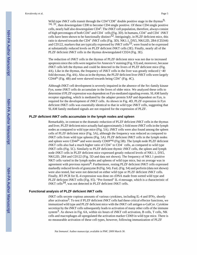
Wild type iNKT cells transit through the CD4+CD8+ double positive stage in the thymus9,14, 31, then downregulate CD8 to become CD4 single positive. Of these CD4 single positivecells, nearly half also downregulate CD43. The iNKT cell population, therefore, always consistof high percentages of both CD4+ and CD4− cells (Fig. 3D). In humans, CD4+ and CD4− iNKTcells have been shown to be functionally distinct32. Intriguingly, in PLZF deficient mice, thisratio is skewed towards the CD4+ iNKT cells (Fig. 3D). NK1.1, DX5, NKG2D, 2B4 (CD244)and CD122, markers that are typically expressed by iNKT cells33, were found to be expressedat substantially reduced levels on PLZF deficient iNKT cells (3E). Finally, nearly all of thePLZF deficient iNKT cells in the thymus downregulated CD24 (Fig. 3E).
The reduction of iNKT cells in the thymus of PLZF deficient mice was not due to increasedapoptosis since the cells were negative for Annexin V staining (Fig. S3) and, moreover, becauseiNKT cells left the thymus and could be detected in the livers of PLZF deficient mice (Fig.4A). Like in the thymus, the frequency of iNKT cells in the liver was greatly reduced (∼40fold decrease, Fig. 4A). Also as in the thymus, the PLZF deficient liver iNKT cells were largelyCD44lo (Fig. 4B) and were skewed towards being CD4+ (Fig. 4C).
Although iNKT cell development is severely impaired in the absence of the Src-family kinaseFyn, some iNKT cells do accumulate in the livers of older mice. We analyzed these cells todetermine if PLZF expression was dependent on Fyn-mediated signaling events. SLAM familyreceptor signaling, which is mediated by the adapter protein SAP and dependent on Fyn, isrequired for the development of iNKT cells. As shown in Fig. 4D, PLZF expression in Fyndeficient iNKT cells was essentially identical to that in wild type iNKT cells, suggesting thatSLAM family mediated signals are not required for the expression of PLZF.
PLZF deficient iNKT cells accumulate in the lymph nodes and spleenRemarkably, in contrast to the dramatic reduction of PLZF deficient iNKT cells in the thymusand liver, PLZF deficient mice actually had approximately 2-fold more iNKT cells in the lymphnodes as compared to wild type mice (Fig. 5A). iNKT cells were also found among the spleencells of PLZF deficient mice (Fig. 5A), although the frequency was reduced as compared toiNKT cells from wild type spleens (Fig. 5A). PLZF deficient iNKT cells in the lymph nodesand spleen were CD44lo and were mostly CD69neg (Fig.5B). The lymph node PLZF deficientiNKT cells also had a much higher ratio of CD4+ to CD4− cells, as compared to wild typeiNKT cells (Fig. 5C). Similarly to PLZF deficient thymic iNKT cells, the spleen and lymphnode iNKT cells in PLZF deficient mice expressed greatly reduced levels of NK1.1, DX5,NKG2D, 2B4 and CD122 (Fig. 5D and data not shown). The frequency of NK1.1 positiveNKT cells varied in the lymph nodes and spleens of wild type mice, but on average was inagreement with previous reports6. Furthermore, resting PLZF deficient iNKT cells expressedmarkedly reduced levels of granzyme B (Fig. S4). FasL (Fig. S4) and perforin (data not shown)were also tested, but were not detected on either wild type or PLZF deficient iNKT cells.Finally, RT-PCR for IL-4 expression was done on cDNA made from sorted wild type andPLZF deficient iNKT cells (Fig. S5). “Pre-formed” IL-4 message, which is a characteristic ofiNKT cells34, was not detected in PLZF deficient iNKT cells.
Functional analysis of PLZF deficient iNKT cellsiNKT cells secrete copious amounts of various cytokines, including IL-4 and IFNγ, shortlyafter activation3. To test if PLZF deficient iNKT cells had these critical effector functions, weimmunized wild type and PLZF deficient mice with the iNKT cell antigen α-GalCer. Cytokinesecretion by the iNKT cells subsequently leads to activation of many other cells of the immunesystem3. As shown in Fig. 6A, within six hours of iNKT cell activation, B cells, T cells, NKcells and macrophages all upregulated the activation marker CD69 in wild type mice. There isno measurable activation of these cell types, however, following immunization of PLZF
Kovalovsky et al. Page 5
Nat Immunol. Author manuscript; available in PMC 2009 March 30.
NIH
-PA Author Manuscript
NIH
-PA Author Manuscript
NIH
-PA Author Manuscript

deficient mice, similar to the negative control, CD1d deficient mice. IL-2, IL-4 and IFNγ wereeasily detectable in the serum of wild type animals following immunization, however, thesecytokines could not be detected in the PLZF deficient or the control CD1d deficient mice (Fig.6B).
The failure to detect cytokines in immunized PLZF deficient mice might be due to the decreasedfrequency of iNKT cells. Therefore, to further test iNKT cell function, tetramer positive iNKTcells were sorted from the spleen and lymph nodes of wild type and PLZF deficient mice.Sorted cells were activated with PMA/ionomycin for a total of five hours; Brefeldin A wasadded for the final four hours of the culture. The activated cells were then analyzed for cytokineproduction by intracellular staining followed by FACS. As expected a large percentage of wildtype iNKT cells produced cytokine under these conditions; with approximately half of theactivated cells expressing both IL-4 and IFN (Fig. 7A). Remarkably, far fewer PLZF deficientiNKT cells expressed cytokine (Fig. 7A). Of particular interest was the complete loss of thecapacity to simultaneously produce both IL-4 and IFNγ. CD4 T cells from wild type and PLZFdeficient mice produced some IFNγ and little IL-4 (Fig. 7A).
To further analyze the functionality of PZLF deficient iNKT cells, sorted cells were activatedwith plate bound anti-CD3 and soluble anti-CD28 in the presence of IL-2 and IL-15. Theaddition of IL-15 was required for the in vitro culture of the wild type iNKT cells. As a controland for comparison, wild type and PLZF deficient CD4 T cells were also collected and grownin culture under the same conditions. Supernatants were removed after twenty-four hours andreplaced with fresh media. Five days after the initial activation the T cells were reactivatedwith plate bound anti-CD3 and soluble anti-CD28. Supernatants were again collected aftertwenty-four hours.
The supernatants from the primary and secondary activations were analyzed by with BDBiosciences Cytokine Bead Arrays for the presence of a variety of cytokines. As suggested bythe experiments in Fig. 6 and Fig. 7A, PLZF deficient iNKT cells produced little IL-4 andIFNγ upon primary activation (Fig. 7B). Failure to produce cytokines was not a general defectin the cells, however, since upon secondary activation, PLZF deficient iNKT cells producedrobust amounts of the two cytokines (Fig. 7B). The limited capacity of PLZF deficient iNKTcells to produce IL-4 and IFNγ upon primary activation was in stark contrast to wild type iNKTcells (Fig. 7C). As a comparison, the capacity of sorted CD4 T cells from wild type and PLZFdeficient CD4 T cells is also shown (Fig. 7D). Wild type iNKT cells also produced more IL-4and IFNγ upon secondary activation than PLZF deficient iNKT cells (Fig. 7D).
iNKT cells are known to produce multiple cytokines upon primary activation including IL-3,IL-5, IL-9, IL-10, IL-12p70, IL-13, GMCSF and TNF5. Therefore, we further testedsupernatants for the presence of these cytokines (Fig. 8). Wild type iNKT cells producedsignificantly more IL-13, GMCSF, IL-10 and IL-3 and TNF than PLZF deficient iNKT cells,upon primary activation. IL-5, IL-9 and IL-12p70 were not detected under these activatingconditions. Sorted CD4 T cells from wild type and PLZF deficient cells were included ascontrols.
Secondary activation of the cells showed that the PLZF deficient iNKT cells were capable ofproducing all of these cytokines (Fig. 8). Wild type iNKT cells produced significantly moreIL-13 and IL-10 upon secondary activation. More TNF was produced by the wild type cellsthan was produced by the PLZF deficient iNKT cells, as well, but this difference did not reachstatistical significance. The results of the secondary activation of wild type and PLZF deficientCD4 T cells are included as a control.
Kovalovsky et al. Page 6
Nat Immunol. Author manuscript; available in PMC 2009 March 30.
NIH
-PA Author Manuscript
NIH
-PA Author Manuscript
NIH
-PA Author Manuscript

DiscussionWe show that in the absence of the BTB-ZF gene, PLZF, iNKT cells develop, but fail to acquiremany of the salient features characteristic to these cells. In particular, the frequency of PLZFdeficient iNKT cells is reduced, the cells do not express high levels of cell surface markerssuch as NK1.1, DX5, 2B4, CD122 and NKG2D and the cells fail to make numerous cytokinesupon primary activation. PLZF deficient cells also expressed reduced levels of Granzyme Band do not have the pre-formed IL-4 mRNA that is characteristic of wild type iNKT cells.Furthermore, PLZF deficient iNKT cells accumulate in the lymph nodes and spleen rather thanin the thymus and liver like wild type iNKT cells. Finally, among cells of the immune system,PLZF expression appears to be largely limited to iNKT cells. Together, these data show thatPLZF is a critical regulator of iNKT cell differentiation. PLZF is also expressed in humaniNKT cells where this highly conserved protein is likely to have a similar function.
PLZF is likely not involved in positive selection of iNKT cells since PLZF deficient mice dodevelop substantial numbers of T cells expressing the canonical TCR. PLZF is, however,expressed at high levels even in HSAhi, CD44neg iNKT cells, which are the most immaturecells that have been described30. Therefore, PLZF expression must occur immediately afteror perhaps even during positive selection of iNKT cells.
It is not clear why there is a reduced number of iNKT cells in the thymus and liver and spleenof PLZF deficient mice. The decreased frequency of iNKT cells is not directly a consequenceof altered proliferative capacity in the thymus since an equal percentage of the PLZF deficientiNKT cells were labeled with BrdU over a sixteen-hour period. TCRβ thymocytes at the doublenegative stage of development have been shown to undergo a ∼500-fold expansion within fourdays35. Each TCRβ clone divides, on average, more than three times a day. It is speculated,however, that different clones may expand with different kinetics. Therefore, it is possible thatwild type iNKT cells, like some double negative thymocytes, are undergoing more rounds ofdivision than the PLZF deficient iNKT cells. It is not possible to make conclusions about thetotal number of cell divisions, since BrudU labeling does not differentiate between cells thathave undergone one round or several rounds of proliferation.
There is no indication that PLZF deficient iNKT cells are dying. The cells in the thymus arenot annexin V positive and clearly can migrate from the thymus and populate peripheral tissues.Apoptotic thymocytes are rapidly cleared from the thymus, however, so increased cell deathremains a formal possibility. It is also possible that PLZF deficient iNKT cells simply do notaccumulate in the thymus and liver because they fail to express chemokine receptors necessaryfor their retention in these tissues. Furthermore, it is known that NK1.1 negative iNKT cellsmore readily emigrate from the thymus than NK1.1 positive iNKT cells36. Since PLZFdeficient iNKT cells do not upregulate NK1.1 it is possible that these cells more efficientlyleave the thymus.
It is not known why PLZF is specifically expressed in iNKT cells. It is tempting to speculatethat the engagement of SLAM family receptor signals via Fyn results in the expression of PLZFsince the SLAM family adapter protein, SAP is required for iNKT cell development13, 37.Furthermore, the SLAM family receptors Slamf1 and Slamf6 were recently shown to be criticalfor iNKT cell development38. SAP is thought to signal via the SRC family protein tyrosinekinase Fyn and, in the absence of Fyn, there is a dramatic reduction in the frequency of iNKTcells8, 10. PLZF is, however, expressed at wild type levels in Fyn deficient iNKT cells.Preliminary data also shows that the rare CD1d tetramer positive cells found in the thymus ofSAP deficient mice express PLZF (data not shown). These data would suggest that SLAMmediated signaling is not essential for PLZF expression. Alternatively, the few iNKT cells that
Kovalovsky et al. Page 7
Nat Immunol. Author manuscript; available in PMC 2009 March 30.
NIH
-PA Author Manuscript
NIH
-PA Author Manuscript
NIH
-PA Author Manuscript

develop in these SAP and Fyn deficient mice might be “escapees” that have managed to expressPLZF without appropriate signaling events.
Several genes have been shown to impact the development and/or function of iNKT cells. Forexample, T-Bet deficient mice have a profound block at stage 2 of iNKT celldevelopment25. RORγt deficient mice are devoid of iNKT cells as a result of a failure toproduce the invariant Vα14 TCR due to the short lifespan of CD4+CD8+ thymocytes14. Thissame report showed that Runx1 is necessary for the very early stages of iNKT cell development.GMCSF (CSF-2) has also been shown to be important for iNKT cells. iNKT cells in GMCSFdeficient mice develop normally, but fail to secrete cytokines due to a defect in secretoryvesicles39. It was recently shown that mice deficient in the Tec family tyrosine kinase, ITK,have defects in iNKT cell development40, 41. The defects in iNKT cell function and phenotype,while seemingly related, were far milder than the changes in PLZF deficient iNKT cells. CD4and CD8 T cells in IKT deficient mice have also been shown to have innate-likefeatures42-46. However, the innate-like T cells in ITK deficient mice do not appear to expressPLZF (data not shown).
BTB-ZF proteins have typically been found to be transcriptional repressors. For example,BCL6 represses p53 expression in germinal center B cells presumably to prevent the apoptoticcell death that the double stranded DNA breaks associated with affinity maturation shouldcause47. A second example is LRF, which is thought to suppress Notch mediated signals inhematopoietic stem cells, thereby promoting T cell over B cell commitment19. Gene targetsof PLZF include HoxD gene during limb bud formation48 and kit during spermatogonialdifferentiation49. Neither of these genes, however, has been reported to have a function iniNKT cells. Cell line based studies also suggest that Pbx150,VLA-451, c-myc52 and BID53are transcriptionally repressed by expression of PLZF. Preliminary gene array data shows thatexpression of these genes is not altered in PLZF deficient iNKT cells. Expression of Th-POK17, another BTB-ZF gene known to be expressed in iNKT cells, is also not affected bythe loss of PLZF expression (data not shown). Perhaps PLZF suppresses unknown suppressorsof transcription factors required for cytokine production such as T-Bet54 or RORγt55. Indeedboth T-Bet and RORγt have been shown to be essential for iNKT cell development14, 25.
Finally it is interesting to note that PLZF deficient iNKT cells do not have an activatedphenotype. It has been generally accepted that iNKT cells are not tolerant to self and that thisis likely the reason that the cells maintain an activated phenotype7, 56. Reactivity to self hasbeen proposed to be a consequence of the unique and highly restricted TCRs that are expressedby iNKT cells. The failure of PLZF deficient iNKT cells to maintain the activated phenotypestrongly implies that high affinity interactions of the TCR with self-antigens do not play a rolein maintaining the activated phenotype of iNKT cells. Rather, it would appear, that expressionof PLZF controls this aspect of iNKT cell biology.
Overall, our findings offer fundamental insight into the mechanisms of iNKT cell development.This work also highlights the fundamental role that BTB-ZF genes play in immune systemdevelopment and function.
MethodsMice
PLZF deficient mice have been previously described21. PLZF deficient mice were generatedin the 129 ES cells. The resulting mice have been backcrossed to the C57Bl/6 for fourgenerations. T-bet deficient mice were purchased from The Jackson Labs, Bar Harbor, ME.All animal work was approved and done in compliance with MSKCC's IACUC guidelines.
Kovalovsky et al. Page 8
Nat Immunol. Author manuscript; available in PMC 2009 March 30.
NIH
-PA Author Manuscript
NIH
-PA Author Manuscript
NIH
-PA Author Manuscript
Flow cytometry and cell sortingCells were prepared and analyzed by standard methods. Antibodies were obtained from BDBiosciences. CD1d:α-GalCer tetramers were prepared according to published protocols57.CD1d:PBS57 tetramers, used for some experiments, were obtained from the NIH TetramerCore Facility. The Armenian hamster anti-PLZF monoclonal antibody was generated byMSKCC's Monoclonal Antibody Core Facility using standard approaches by immunizinganimals with a combination of peptides covering the amino, carboxyl and hinge regions of theprotein. During FACS analysis, dead cells were excluded from analysis by the use of the DNAspecific dye, DAPI. Cell doublets were excluded by comparing side scatter width to forwardscatter area. For iNKT cell analysis, irrelevant cells that were MHC class II positive and CD8positive were excluded from analysis when possible. Samples were collected on either a DakoCyAn ADP or a BD LSR II. Cell sorts were done by MSKCC's Flow Cytometry Core Facilityon a Dako MoFlo, a BD FACS Vantage or a BD FACS ARIA. Post sort analysis was done forall samples to confirm that cell purity was greater than 97%. BrdU labeling of T cells anddetection by FACS was done as previously described31.
In Vitro T cell activationT cells were activated with 5 μg/ml anti-CD3 and 5 μg/ml anti-CD28 in round bottom 96-wellplates in the presence of IL-2 and IL-15. Secreted cytokines were detected with BD Biosciencescytokine bead arrays.
Histology6 μm serial sections were cut and prepared by standard methods. Sections were stained withDec-205 biotin and UEA-1 – Fitc, followed by streptavidin-PE.
StatisticsStatistical validation of cell number and frequency comparisons (Figs. 3, 4 and 5) was donewith unpaired, two-tailed Student's t-test. The use t-tests for samples of this size (approximately5-10 samples per group) requires an assumption of normality and homogeneity ofvariance58. These assumptions were valid for these samples. Statistical validation of cytokinedata (Figs. 7 and 8) was done with the Mann-Whitney U test, which is a nonparametricalternative to the Student's t-test that is more robust for small sample sizes (4-5 repeats of eachexperiment). One important difference is that this test does not assume a Gaussian distributionof the data. A second independent test (not shown) was use to confirm the validity of the results.The log value of each data point was calculated prior to applying unpaired, two-tailed Student'st-test. Transforming the data in this way enhances the validity of the t-test since the data takeon a more Gaussian distribution. All differences determined to be significant or not significantwith the Mann-Whitney U-test were confirmed by this second, independent test. The validityof these tests was discussed and confirmed with MSKCC's Biostatistics Department.
Supplementary MaterialRefer to Web version on PubMed Central for supplementary material.
AcknowledgementsThe authors gratefully acknowledge T. Martillotti for technical assistance; L. Denzin, J. Chaudhuri, E. Pamer, C.-H.Chang for critical reading of the manuscript and excellent advice. We also thank MSKCC's Monoclonal Antibody andFlow Cytometry Core Facilities. This work was partially supported by NIH grant AI059739 and funds from MSKCC.D.K was supported by NIH T32 training grant CA009149.
Kovalovsky et al. Page 9
Nat Immunol. Author manuscript; available in PMC 2009 March 30.
NIH
-PA Author Manuscript
NIH
-PA Author Manuscript
NIH
-PA Author Manuscript

References1. Petrie HT, Zuniga-Pflucker JC. Zoned out: functional mapping of stromal signaling microenvironments
in the thymus. Annu Rev Immunol 2007;25:649–679. [PubMed: 17291187]2. Rothenberg EV, Anderson MK. Elements of transcription factor network design for T-lineage
specification. Dev Biol 2002;246:29–44. [PubMed: 12027432]3. Bendelac A, Savage PB, Teyton L. The Biology of NKT Cells. Annu Rev Immunol 2007;25:297–336.
[PubMed: 17150027]4. Bendelac A, Medzhitov R. Adjuvants of immunity: harnessing innate immunity to promote adaptive
immunity. J Exp Med 2002;195:F19–23. [PubMed: 11877490]5. Kronenberg M. Toward an understanding of NKT cell biology: progress and paradoxes. Annu Rev
Immunol 2005;23:877–900. [PubMed: 15771592]6. Matsuda JL, et al. Tracking the response of natural killer T cells to a glycolipid antigen using CD1d
tetramers. J Exp Med 2000;192:741–754. [PubMed: 10974039]7. Kronenberg M, Engel I. On the road: progress in finding the unique pathway of invariant NKT cell
differentiation. Curr Opin Immunol 2007;19:186–193. [PubMed: 17303398]8. Gadue P, Morton N, Stein PL. The Src family tyrosine kinase Fyn regulates natural killer T cell
development. J Exp Med 1999;190:1189–1196. [PubMed: 10523617]9. Dao T, et al. Development of CD1d-restricted NKT cells in the mouse thymus. Eur J Immunol
2004;34:3542–3552. [PubMed: 15549774]10. Eberl G, Lowin-Kropf B, MacDonald HR. Cutting edge: NKT cell development is selectively
impaired in Fyn- deficient mice. J Immunol 1999;163:4091–4094. [PubMed: 10510341]11. Borowski C, Bendelac A. Signaling for NKT cell development: the SAP-FynT connection. J Exp
Med 2005;201:833–836. [PubMed: 15781574]12. Chung B, Aoukaty A, Dutz J, Terhorst C, Tan R. Signaling lymphocytic activation molecule-
associated protein controls NKT cell functions. J Immunol 2005;174:3153–3157. [PubMed:15749842]
13. Pasquier B, et al. Defective NKT cell development in mice and humans lacking the adapter SAP, theX-linked lymphoproliferative syndrome gene product. J Exp Med 2005;201:695–701. [PubMed:15738056]
14. Egawa T, et al. Genetic evidence supporting selection of the Valpha14i NKT cell lineage from double-positive thymocyte precursors. Immunity 2005;22:705–716. [PubMed: 15963785]
15. Kelly KF, Daniel JM. POZ for effect--POZ-ZF transcription factors in cancer and development.Trends Cell Biol 2006;16:578–587. [PubMed: 16996269]
16. Dent AL, Shaffer AL, Yu X, Allman D, Staudt LM. Control of inflammation, cytokine expression,and germinal center formation by BCL-6. Science 1997;276:589–592. [PubMed: 9110977]
17. He X, et al. The zinc finger transcription factor Th-POK regulates CD4 versus CD8 T-cell lineagecommitment. Nature 2005;433:826–833. [PubMed: 15729333]
18. Sun G, et al. The zinc finger protein cKrox directs CD4 lineage differentiation during intrathymic Tcell positive selection. Nat Immunol 2005;6:373–381. [PubMed: 15750595]
19. Maeda T, et al. Regulation of B versus T lymphoid lineage fate decision by the proto-oncogene LRF.Science 2007;316:860–866. [PubMed: 17495164]
20. McConnell MJ, Licht JD. The PLZF gene of t (11;17)-associated APL. Curr Top Microbiol Immunol2007;313:31–48. [PubMed: 17217037]
21. Barna M, Hawe N, Niswander L, Pandolfi PP. Plzf regulates limb and axial skeletal patterning. NatGenet 2000;25:166–172. [PubMed: 10835630]
22. Matsuda JL, Gapin L. Developmental program of mouse Valpha14i NKT cells. Curr Opin Immunol2005;17:122–130. [PubMed: 15766670]
23. Zelent A, Guidez F, Melnick A, Waxman S, Licht JD. Translocations of the RARalpha gene in acutepromyelocytic leukemia. Oncogene 2001;20:7186–7203. [PubMed: 11704847]
24. Koken MH, et al. Leukemia-associated retinoic acid receptor alpha fusion partners, PML and PLZF,heterodimerize and colocalize to nuclear bodies. Proc Natl Acad Sci U S A 1997;94:10255–10260.[PubMed: 9294197]
Kovalovsky et al. Page 10
Nat Immunol. Author manuscript; available in PMC 2009 March 30.
NIH
-PA Author Manuscript
NIH
-PA Author Manuscript
NIH
-PA Author Manuscript

25. Townsend MJ, et al. T-bet regulates the terminal maturation and homeostasis of NK and Valpha14iNKT cells. Immunity 2004;20:477–494. [PubMed: 15084276]
26. Costoya JA, et al. Essential role of Plzf in maintenance of spermatogonial stem cells. Nat Genet2004;36:653–659. [PubMed: 15156143]
27. Pellicci DG, et al. DX5/CD49b-positive T cells are not synonymous with CD1d-dependent NKT cells.J Immunol 2005;175:4416–4425. [PubMed: 16177083]
28. Mendiratta SK, et al. CD1d1 mutant mice are deficient in natural T cells that promptly produce IL-4.Immunity 1997;6:469–477. [PubMed: 9133426]
29. Slansky JE. Antigen-specific T cells: analyses of the needles in the haystack. PLoS Biol 2003;1:E78.[PubMed: 14691549]
30. Benlagha K, Wei DG, Veiga J, Teyton L, Bendelac A. Characterization of the early stages of thymicNKT cell development. J Exp Med 2005;202:485–492. [PubMed: 16087715]
31. Gapin L, Matsuda JL, Surh CD, Kronenberg M. NKT cells derive from double-positive thymocytesthat are positively selected by CD1d. Nat Immunol 2001;2:971–978. [PubMed: 11550008]
32. Gumperz JE, Miyake S, Yamamura T, Brenner MB. Functionally distinct subsets of CD1d-restrictednatural killer T cells revealed by CD1d tetramer staining. J Exp Med 2002;195:625–636. [PubMed:11877485]
33. Hammond KJ, et al. NKT cells are phenotypically and functionally diverse. Eur J Immunol1999;29:3768–3781. [PubMed: 10556834]
34. Stetson DB, et al. Constitutive cytokine mRNAs mark natural killer (NK) and NK T cells poised forrapid effector function. J Exp Med 2003;198:1069–1076. [PubMed: 14530376]
35. Falk I, Potocnik AJ, Barthlott T, Levelt CN, Eichmann K. Immature T cells in peripheral lymphoidorgans of recombinase-activating gene-1/-2-deficient mice. Thymus dependence and responsivenessto anti-CD3 epsilon antibody. J Immunol 1996;156:1362–1368. [PubMed: 8568235]
36. Pellicci DG, et al. A natural killer T (NKT) cell developmental pathway iInvolving a thymus-dependent NK1.1(−)CD4(+) CD1d-dependent precursor stage. J Exp Med 2002;195:835–844.[PubMed: 11927628]
37. Nichols KE, et al. Regulation of NKT cell development by SAP, the protein defective in XLP. NatMed 2005;11:340–345. [PubMed: 15711562]
38. Griewank K, et al. Homotypic interactions mediated by Slamf1 and Slamf6 receptors control NKTcell lineage development. Immunity 2007;27:751–762. [PubMed: 18031695]
39. Bezbradica JS, et al. Granulocyte-macrophage colony-stimulating factor regulates effectordifferentiation of invariant natural killer T cells during thymic ontogeny. Immunity 2006;25:487–497. [PubMed: 16949316]
40. Felices M, Berg LJ. The tec kinases itk and rlk regulate NKT cell maturation, cytokine production,and survival. J Immunol 2008;180:3007–3018. [PubMed: 18292523]
41. Au-Yeung BB, Fowell DJ. A key role for Itk in both IFN gamma and IL-4 production by NKT cells.J Immunol 2007;179:111–119. [PubMed: 17579028]
42. Broussard C, et al. Altered development of CD8+ T cell lineages in mice deficient for the Tec kinasesItk and Rlk. Immunity 2006;25:93–104. [PubMed: 16860760]
43. Horai R, et al. Requirements for selection of conventional and innate T lymphocyte lineages.Immunity 2007;27:775–785. [PubMed: 18031697]
44. Hu J, Sahu N, Walsh E, August A. Memory phenotype CD8+ T cells with innate function selectivelydevelop in the absence of active Itk. Eur J Immunol 2007;37:2892–2899. [PubMed: 17724684]
45. Berg LJ. Signalling through TEC kinases regulates conventional versus innate CD8(+) T-celldevelopment. Nat Rev Immunol 2007;7:479–485. [PubMed: 17479128]
46. Hu J, August A. Naive and innate memory phenotype CD4+ T cells have different requirements foractive Itk for their development. J Immunol 2008;180:6544–6552. [PubMed: 18453573]
47. Phan RT, Dalla-Favera R. The BCL6 proto-oncogene suppresses p53 expression in germinal-centreB cells. Nature 2004;432:635–639. [PubMed: 15577913]
48. Barna M, et al. Plzf mediates transcriptional repression of HoxD gene expression through chromatinremodeling. Dev Cell 2002;3:499–510. [PubMed: 12408802]
Kovalovsky et al. Page 11
Nat Immunol. Author manuscript; available in PMC 2009 March 30.
NIH
-PA Author Manuscript
NIH
-PA Author Manuscript
NIH
-PA Author Manuscript

49. Filipponi D, et al. Repression of kit expression by Plzf in germ cells. Mol Cell Biol 2007;27:6770–6781. [PubMed: 17664282]
50. Shiraishi K, et al. Pre-B-cell leukemia transcription factor 1 is a major target of promyelocyticleukemia zinc-finger-mediated melanoma cell growth suppression. Oncogene 2007;26:339–348.[PubMed: 16862184]
51. Quaranta MT, et al. PLZF-mediated control on VLA-4 expression in normal and leukemic myeloidcells. Oncogene 2006;25:399–408. [PubMed: 16158049]
52. McConnell MJ, et al. Growth suppression by acute promyelocytic leukemia-associated protein PLZFis mediated by repression of c-myc expression. Mol Cell Biol 2003;23:9375–9388. [PubMed:14645547]
53. Parrado A, et al. The promyelocytic leukemia zinc finger protein down-regulates apoptosis andexpression of the proapoptotic BID protein in lymphocytes. Proc Natl Acad Sci U S A2004;101:1898–1903. [PubMed: 14769944]
54. Szabo SJ, et al. A novel transcription factor, T-bet, directs Th1 lineage commitment. Cell2000;100:655–669. [PubMed: 10761931]
55. Ivanov II, et al. The orphan nuclear receptor RORgammat directs the differentiation program ofproinflammatory IL-17+ T helper cells. Cell 2006;126:1121–1133. [PubMed: 16990136]
56. Kronenberg M, Rudensky A. Regulation of immunity by self-reactive T cells. Nature 2005;435:598–604. [PubMed: 15931212]
57. Yu KO, et al. Production and characterization of monoclonal antibodies against complexes of theNKT cell ligand alpha-galactosylceramide bound to mouse CD1d. J Immunol Methods 2007;323:11–23. [PubMed: 17442335]
58. Lamb TJ, Graham AL, Petrie A. T testing the immune system. Immunity 2008;28:288–292. [PubMed:18341999]
Kovalovsky et al. Page 12
Nat Immunol. Author manuscript; available in PMC 2009 March 30.
NIH
-PA Author Manuscript
NIH
-PA Author Manuscript
NIH
-PA Author Manuscript
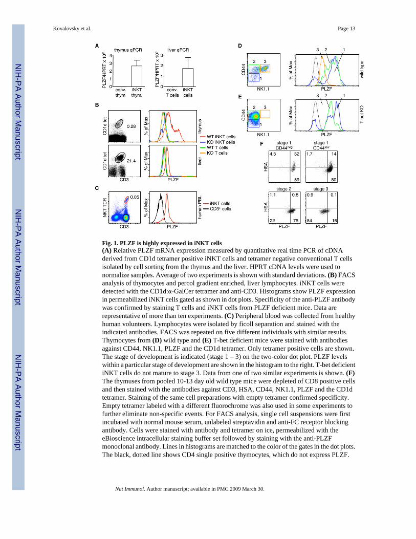
Fig. 1. PLZF is highly expressed in iNKT cells(A) Relative PLZF mRNA expression measured by quantitative real time PCR of cDNAderived from CD1d tetramer positive iNKT cells and tetramer negative conventional T cellsisolated by cell sorting from the thymus and the liver. HPRT cDNA levels were used tonormalize samples. Average of two experiments is shown with standard deviations. (B) FACSanalysis of thymocytes and percol gradient enriched, liver lymphocytes. iNKT cells weredetected with the CD1d:α-GalCer tetramer and anti-CD3. Histograms show PLZF expressionin permeabilized iNKT cells gated as shown in dot plots. Specificity of the anti-PLZF antibodywas confirmed by staining T cells and iNKT cells from PLZF deficient mice. Data arerepresentative of more than ten experiments. (C) Peripheral blood was collected from healthyhuman volunteers. Lymphocytes were isolated by ficoll separation and stained with theindicated antibodies. FACS was repeated on five different individuals with similar results.Thymocytes from (D) wild type and (E) T-bet deficient mice were stained with antibodiesagainst CD44, NK1.1, PLZF and the CD1d tetramer. Only tetramer positive cells are shown.The stage of development is indicated (stage 1 – 3) on the two-color dot plot. PLZF levelswithin a particular stage of development are shown in the histogram to the right. T-bet deficientiNKT cells do not mature to stage 3. Data from one of two similar experiments is shown. (F)The thymuses from pooled 10-13 day old wild type mice were depleted of CD8 positive cellsand then stained with the antibodies against CD3, HSA, CD44, NK1.1, PLZF and the CD1dtetramer. Staining of the same cell preparations with empty tetramer confirmed specificity.Empty tetramer labeled with a different fluorochrome was also used in some experiments tofurther eliminate non-specific events. For FACS analysis, single cell suspensions were firstincubated with normal mouse serum, unlabeled streptavidin and anti-FC receptor blockingantibody. Cells were stained with antibody and tetramer on ice, permeabilized with theeBioscience intracellular staining buffer set followed by staining with the anti-PLZFmonoclonal antibody. Lines in histograms are matched to the color of the gates in the dot plots.The black, dotted line shows CD4 single positive thymocytes, which do not express PLZF.
Kovalovsky et al. Page 13
Nat Immunol. Author manuscript; available in PMC 2009 March 30.
NIH
-PA Author Manuscript
NIH
-PA Author Manuscript
NIH
-PA Author Manuscript

Fig. 2. PLZF expression is limited to iNKT cells and does not affect the bulk of lymphocytedevelopment(A) Intracellular staining for PLZF expression in B cells and NK cells was done and comparedto the same cell types from PLZF deficient mice. Expression of PLZF in iNKT cells is shownas a positive control. (B) Single cell suspensions of thymus and lymph node cells were stainedwith the indicated monoclonal antibodies. The percentage and phenotype of the majorsubpopulations of thymocytes and lymphocytes were similar in wild type and PLZF deficientmice, based on staining with CD4, CD8 and TCR. Subtle variations in thymocyte numberswere not reproducible. Only TCR+ lymph node cells are shown. Experiment was repeatedapproximately 10 times. (C) Frozen thymus sections were stained with Dec205 and withUEA-1 to identify the thymic cortex and medulla. No structural changes to the organ wereobserved. (D) The frequency of NK cells, DX5+ T cells, and γδ T cells is not affected by theloss of PLZF expression. Data are representative of three independent experiments.
Kovalovsky et al. Page 14
Nat Immunol. Author manuscript; available in PMC 2009 March 30.
NIH
-PA Author Manuscript
NIH
-PA Author Manuscript
NIH
-PA Author Manuscript

Fig. 3. Altered iNKT cell development in PLZF deficient miceThymocytes from the indicated mouse lines were stained with CD1d:α-GalCer tetramers andantibodies against CD3, CD44, CD69, CD4 and CD8. (A) PLZF deficient mice have anapproximately 9-fold decrease in the number of iNKT cells. Numbers indicate the percentageof cells within the gate. The frequency of iNKT cells in 13 wild type and 14 PLZF deficientmice is shown in the scatter plot. Analysis was done by determining the percentage of tetramerpositive cells among the CD3hi thymocytes. Percentages were used rather than absolutenumbers to normalize for differences in the total cellularity of the thymus due to the age or sexof the mice. P values are indicated in the figure. (B) iNKT cells from PLZF deficient mice donot upregulate CD44. Both WT and KO iNKT cells upregulate CD69. (C) BrdU incorporationinto iNKT cells in the thymus 16-hours after injection into 10-13 day old mice. Both total iNKTcells and only CD44lo iNKT cells are shown. Experiment was repeated twice. (D) iNKT cellsfrom PLZF deficient mice are skewed towards CD4+. Only iNKT cells are shown in the FACSplot. Numbers indicate the percentage of cells in the region. The increase in the percentage ofCD4+ iNKT cells and the corresponding decrease in the percentage of CD4− iNKTs in the inPLZF deficient mice in 8 wild type and 9 PLZF deficient mice are shown as a scatter plots.
Kovalovsky et al. Page 15
Nat Immunol. Author manuscript; available in PMC 2009 March 30.
NIH
-PA Author Manuscript
NIH
-PA Author Manuscript
NIH
-PA Author Manuscript

Data set comparisons and P values are indicated in the figure. (E) PLZF deficient iNKT cellshave reduced expression of many cell surface markers typical of iNKT cells such as NK1.1,DX5, NKG2D, 2B4 (CD244) and CD122. Change in expression of each marker was confirmedin at least three independent experiments. P values were derived from two-tailed, unpairedStudent's t-test. Choices of statistical tests are explained in the methods section.
Kovalovsky et al. Page 16
Nat Immunol. Author manuscript; available in PMC 2009 March 30.
NIH
-PA Author Manuscript
NIH
-PA Author Manuscript
NIH
-PA Author Manuscript

Fig. 4. Reduced frequency and altered phenotype of liver iNKT cells in PLZF deficient mice(A) Percoll enriched liver lymphocytes were stained and analyzed as above. PLZF deficientmice had approximately 40-fold fewer iNKT cells. The percentage of iNKT cells amongCD3+ cells was used to compare 10 WT and 10 KO mice. Percentages were used rather thanabsolute numbers to correct for differences in extraction of lymphocytes and potentialvariations of other cell populations. The P values are indicated in the figure. (B) PLZF deficientliver iNKT cells were mostly CD44lo and CD69lo as compared to wild type iNKT cells. (C)As in the thymus, PLZF deficient iNKT cells are skewed towards expressing CD4. Data from5 WT and 5 KO mice are shown in the scatter plot along with P values. (D) Liver iNKT cellsharvested from older (∼6 months) Fyn deficient mice expressed wild type levels of PLZF. This
Kovalovsky et al. Page 17
Nat Immunol. Author manuscript; available in PMC 2009 March 30.
NIH
-PA Author Manuscript
NIH
-PA Author Manuscript
NIH
-PA Author Manuscript

finding was confirmed in three independent experiments. P values were derived from two-tailed, unpaired Student's t-test. Choices of statistical tests are explained in the methods section.
Kovalovsky et al. Page 18
Nat Immunol. Author manuscript; available in PMC 2009 March 30.
NIH
-PA Author Manuscript
NIH
-PA Author Manuscript
NIH
-PA Author Manuscript

Fig. 5. PLZF deficient iNKTs accumulate in the lymph nodes and spleenLymph node (axillary, brachial, inguinal and mesenteric) and spleen cells from the indicatedmouse lines were stained with CD1d:α-GalCer tetramers and antibodies against CD3, CD44,CD69, CD4 and CD8. (A) PLZF deficient mice have approximately 2-fold more lymph nodeiNKT cells and approximately 5-fold fewer spleen iNKT cells. Numbers indicate thepercentage of cells within the gate. The frequency of iNKT cells among lymph node cells from6 wild type and 8 PLZF deficient mice and spleen cells from 8 wild type and 10 PLZF deficientmice are shown in the scatter plots. Analysis was done by determining the percentage oftetramer positive cells among the total CD3+ T cells. Percentages were used rather than absolutenumbers to normalize for differences in the total cellularity of the tissues due to the age of themice. Cellularity of the lymph nodes and the spleens from age matched WT and PLZF KOmice were, however, nearly identical. Data set comparisons and P values are indicated in thefigure. (B) iNKT cells from the lymph nodes and spleens of PLZF deficient mice do notupregulate CD44 and tend to be CD69lo. Data is representative of at least five independentexperiments. (C) Lymph node PLZF deficient iNKT cells are skewed towards expressing CD4.The increase in the percentage of CD4+ iNKT cells and the corresponding decrease in thepercentage of CD4− iNKTs in the lymph nodes and spleens from wild type and PLZF deficientmice are shown in the scatter plots. P values are shown in the figure. (D) PLZF deficient iNKTcells have markedly reduced expression of several markers typically found on wild type iNKTcells from the spleen. P values were derived from two-tailed, unpaired Student's t-test. Choicesof statistical tests are explained in the methods section.
Kovalovsky et al. Page 19
Nat Immunol. Author manuscript; available in PMC 2009 March 30.
NIH
-PA Author Manuscript
NIH
-PA Author Manuscript
NIH
-PA Author Manuscript

Fig. 6. PLZF deficient iNKT cells do not rapidly produce cytokines(A) Mice (wild type, PLZF deficient, CD1d deficient) were I.P. immunized with 4 gof α-GalCer. Six hours later the spleen was removed, dissociated and stained with the indicatedantibodies. Activation of the indicated cell type was measured by upregulation of CD69. (B)Sera were collected from the immunized mice six hours after immunization, diluted 10-foldand ELISAs were done to detect the cytokines IL-4, IFNγ and IL-2. These cytokines were notdetected (n.d.) in the PLZF deficient mice or in the CD1d deficient mice. Experiments weredone with three animals per group and were repeated twice with nearly identical results. ELISAdata represent the means, with standard deviations for the two independent experiments.
Kovalovsky et al. Page 20
Nat Immunol. Author manuscript; available in PMC 2009 March 30.
NIH
-PA Author Manuscript
NIH
-PA Author Manuscript
NIH
-PA Author Manuscript

Fig. 7. PLZF deficient iNKT present an altered cytokine secretion pattern(A) Cells were collected by cell sorting and then activated with 50 ng/ml PMA and 500 ng/mlionomycin for a total of five hours. Brefeldin A was added for the last four hours of theincubation. Cells were then fixed and permeabilized with BD's perm/fix reagents.Permeabilized cells were then stained with antibodies against IL-4 and IFNγ. (B) Single cellsuspensions of combined lymph node and spleen cells were depleted of CD8 and MHC classII expressing cells and then stained with the CD1d tetramer and an antibody against TCRCβ. Tetramer+,TCR Cβ+ cells then were collected by FACS. Approximately 1×10⁴ of each Tcell type was activated with 5 μg/ml plate bound anti-CD3 and 5 μg/ml soluble anti-CD28 inthe presence of IL-2 and IL-15. Supernatants from the primary activation were collected after
Kovalovsky et al. Page 21
Nat Immunol. Author manuscript; available in PMC 2009 March 30.
NIH
-PA Author Manuscript
NIH
-PA Author Manuscript
NIH
-PA Author Manuscript

24 hours. Cells were then fed with fresh media containing IL-2 and IL-15. After a total of fivedays, supernatant was removed and discarded. The cells, in fresh media, were reactivated withplate bound anti-CD3 and soluble anti-CD28. Supernatants were again collected after 24 hours.Supernatants were analyzed for the presence of IL-4 and IFNγ by the use of BD BiosciencesCytokine Bead Arrays. Supernatants were collected and analyzed as described above fromsorted T cells that were (C) activated and then (D) reactivated as described above. Each T celltype was set up in duplicate or triplicate depending upon the number of cells collected. Repeatsfrom each experiment were averaged. A total of five independent experiments were done.Means and standard deviations for the five experiments are shown. The value for the PLZFdeficient iNKTs (KO iNKT) was compared to the other three cell types. P values are markedas * <0.05; ** <0.005. P values were derived using a two-tailed, unparied Mann-Whitney U-test. Choices of statistical tests are explained in the methods section.
Kovalovsky et al. Page 22
Nat Immunol. Author manuscript; available in PMC 2009 March 30.
NIH
-PA Author Manuscript
NIH
-PA Author Manuscript
NIH
-PA Author Manuscript

Fig. 8. Expression of Multiple cytokines are affected by the loss of PLZF expressionSupernatants generated and collected as for Fig. 7 were analyzed for TNF, IL-13, GMCSF,IL-3, and IL-10 by BD Biosciences Cytokine Bead Arrays. Supernatants were also tested forIL-5, IL-9 and IL-12p70, but these cytokines were not detected under these conditions. Fiveindependent experiments were done. Triplicates from each experiment were averaged andconsidered as a single data point. Means and standard deviations for the five experiments areshown. The value for the PLZF deficient iNKTs (KO iNKT) was compared to the other threecell types. P values are marked as * <0.05; ** <0.005. P values were derived using a two-tailed,unparied Mann-Whitney U-test. Choices of statistical tests are explained in the methodssection.
Kovalovsky et al. Page 23
Nat Immunol. Author manuscript; available in PMC 2009 March 30.
NIH
-PA Author Manuscript
NIH
-PA Author Manuscript
NIH
-PA Author Manuscript