Structural and functional adaptations to extreme temperatures in psychrophilic, mesophilic, and...
9
Structural and Functional Adaptations to Extreme Temperatures in Psychrophilic, Mesophilic, and Thermophilic DNA Ligases* Received for publication, May 16, 2003 Published, JBC Papers in Press, July 10, 2003, DOI 10.1074/jbc.M305142200 Daphne ´ Georlette‡, Benjamin Damien§, Vinciane Blaise‡, Eric Depiereux§, Vladimir N. Uversky ¶, Charles Gerday‡, and Georges Feller‡** From the ‡Laboratory of Biochemistry, Institute of Chemistry B6, University of Lie `ge, B-4000 Lie `ge, Belgium, the §Unite ´ de Biologie Mole ´culaire, De ´partement de Biologie, Faculte ´s Universitaires Notre-Dame de la Paix, B-5000 Namur, Belgium, the ¶Institute for Biological Instrumentation, Russian Academy of Sciences, Pushchino, Moscow Region 142290, Russia, and the Department of Chemistry and Biochemistry, University of California, Santa Cruz, California 95064 Psychrophiles, host of permanently cold habitats, dis- play metabolic fluxes comparable to those exhibited by mesophilic organisms at moderate temperatures. These organisms have evolved by producing, among other pe- culiarities, cold-active enzymes that have the properties to cope with the reduction of chemical reaction rates induced by low temperatures. The emerging picture suggests that these enzymes display a high catalytic efficiency at low temperatures through an improved flexibility of the structural components involved in the catalytic cycle, whereas other protein regions, if not implicated in catalysis, may be even more rigid than their mesophilic counterparts. In return, the increased flexibility leads to a decreased stability of psychrophilic enzymes. In order to gain further advances in the anal- ysis of the activity/flexibility/stability concept, psychro- philic, mesophilic, and thermophilic DNA ligases have been compared by three-dimensional-modeling studies, as well as regards their activity, surface hydrophobicity, structural permeability, conformational stabilities, and irreversible thermal unfolding. These data show that the cold-adapted DNA ligase is characterized by an in- creased activity at low and moderate temperatures, an overall destabilization of the molecular edifice, espe- cially at the active site, and a high conformational flex- ibility. The opposite trend is observed in the mesophilic and thermophilic counterparts, the latter being charac- terized by a reduced low temperature activity, high sta- bility and reduced flexibility. These results strongly sug- gest a complex relationship between activity, flexibility and stability. In addition, they also indicate that in cold- adapted enzymes, the driving force for denaturation is a large entropy change. The temperature range in which biological activity has been detected extends from 20 °C, the temperature recorded in the brine veins of Arctic or Antarctic sea ice (1), to 113 °C, the temperature at which the archae Pyrolobus fumarii is still able to grow (2). Although numerous investigations have been car- ried out on thermophilic microorganisms and on their molecu- lar components, especially enzymes, the efforts devoted to cold- adapted microorganisms have been comparatively limited despite their tremendous biotechnological (1, 3–5) and funda- mental (1, 6 – 8) applications. Indeed, the biochemical and physiological bases of cold adaptation, which include, for exam- ple, regulation of gene expression by low temperatures, mem- brane adaptation in relation with the homeoviscosity concept, and the activity/stability relationships sustaining the catalytic efficiency of cold-adapted enzymes, are still poorly understood. In permanent cold habitats, low temperatures have con- strained psychrophiles to develop among other peculiarities enzymatic tools allowing metabolic rates compatible to life that are close to those of temperate organisms. Thermal compensa- tion in these enzymes is reached, in most cases, through a high catalytic efficiency at low and moderate temperatures (for re- view, see Ref. 9 and 10). The emerging picture is that this increased catalytic efficiency is attributed to an increase of the plasticity or flexibility of appropriate parts of the molecular structure in order to compensate for the lower thermal energy provided by the low temperature habitat. This plasticity would enable a good complementarity with the substrate at a low energy cost, thus explaining the high specific activity of psy- chrophilic enzymes. In return, this flexibility would be respon- sible for the weak thermal stability of cold-adapted enzymes. This relationship between activity, flexibility and stability con- stitutes a hot topic and represents a central issue in the adap- tation of proteins to various environments. Moreover, it is believed that all proteins evolve through a balanced compro- mise between these features, i.e. structural rigidity allowing the retention of a specific three-dimensional-conformation at the physiological temperature and in contrast flexibility, al- lowing the protein to perform its catalytic function. In the context of temperature adaptation of enzymes, it is assumed that high temperatures require stable protein structure and activity, whereas high enzyme activity is mandatory at low temperatures. While the common trait of a low conformational stability in cold-adapted enzymes has been demonstrated (see Ref. 11 for review), the stability/flexibility relationship is still controver- sial since some authors consider that the instability of psychro- philic enzymes is due to a random genetic drift (12). Moreover, if the decreased stability of cold-adapted enzymes is well doc- umented, there is however, no direct experimental evidence of an increased flexibility. Besides, controversial results were ob- tained when the flexibility of a few psychrophilic enzymes was investigated by measuring hydrogen-deuterieum exchange rates. In the case of 3-isopropylmalate dehydrogenase (13), while the psychrophilic and mesophilic enzymes were found * This work was supported by the European Union (Grant CT970131), the Re ´gion Wallonne (Grants Bioval 981/3860, Bioval 981/ 3848, Initiative 114705), the FNRS Belgium (Grant 2.4515.00), and the Institut Polaire Franc ¸ais. The costs of publication of this article were defrayed in part by the payment of page charges. This article must therefore be hereby marked “advertisement” in accordance with 18 U.S.C. Section 1734 solely to indicate this fact. ** To whom correspondence should be addressed: Laboratory of Bio- chemistry, Institute of Chemistry B6, University of Lie `ge, B-4000 Lie `ge, Belgium. Tel.: 32-4-366-33-43; Fax: 32-4-366-33-64; E-mail: [email protected]. THE JOURNAL OF BIOLOGICAL CHEMISTRY Vol. 278, No. 39, Issue of September 26, pp. 37015–37023, 2003 © 2003 by The American Society for Biochemistry and Molecular Biology, Inc. Printed in U.S.A. This paper is available on line at http://www.jbc.org 37015 by on April 14, 2008 www.jbc.org Downloaded from
-
Upload
independent -
Category
Documents
-
view
8 -
download
0
Transcript of Structural and functional adaptations to extreme temperatures in psychrophilic, mesophilic, and...

Structural and Functional Adaptations to Extreme Temperaturesin Psychrophilic, Mesophilic, and Thermophilic DNA Ligases*
Received for publication, May 16, 2003Published, JBC Papers in Press, July 10, 2003, DOI 10.1074/jbc.M305142200
Daphne Georlette‡, Benjamin Damien§, Vinciane Blaise‡, Eric Depiereux§,Vladimir N. Uversky¶�, Charles Gerday‡, and Georges Feller‡**
From the ‡Laboratory of Biochemistry, Institute of Chemistry B6, University of Liege, B-4000 Liege, Belgium, the §Unitede Biologie Moleculaire, Departement de Biologie, Facultes Universitaires Notre-Dame de la Paix, B-5000 Namur,Belgium, the ¶Institute for Biological Instrumentation, Russian Academy of Sciences, Pushchino, Moscow Region 142290,Russia, and the �Department of Chemistry and Biochemistry, University of California, Santa Cruz, California 95064
Psychrophiles, host of permanently cold habitats, dis-play metabolic fluxes comparable to those exhibited bymesophilic organisms at moderate temperatures. Theseorganisms have evolved by producing, among other pe-culiarities, cold-active enzymes that have the propertiesto cope with the reduction of chemical reaction ratesinduced by low temperatures. The emerging picturesuggests that these enzymes display a high catalyticefficiency at low temperatures through an improvedflexibility of the structural components involved in thecatalytic cycle, whereas other protein regions, if notimplicated in catalysis, may be even more rigid thantheir mesophilic counterparts. In return, the increasedflexibility leads to a decreased stability of psychrophilicenzymes. In order to gain further advances in the anal-ysis of the activity/flexibility/stability concept, psychro-philic, mesophilic, and thermophilic DNA ligases havebeen compared by three-dimensional-modeling studies,as well as regards their activity, surface hydrophobicity,structural permeability, conformational stabilities, andirreversible thermal unfolding. These data show thatthe cold-adapted DNA ligase is characterized by an in-creased activity at low and moderate temperatures, anoverall destabilization of the molecular edifice, espe-cially at the active site, and a high conformational flex-ibility. The opposite trend is observed in the mesophilicand thermophilic counterparts, the latter being charac-terized by a reduced low temperature activity, high sta-bility and reduced flexibility. These results strongly sug-gest a complex relationship between activity, flexibilityand stability. In addition, they also indicate that in cold-adapted enzymes, the driving force for denaturation is alarge entropy change.
The temperature range in which biological activity has beendetected extends from �20 °C, the temperature recorded in thebrine veins of Arctic or Antarctic sea ice (1), to 113 °C, thetemperature at which the archae Pyrolobus fumarii is still ableto grow (2). Although numerous investigations have been car-
ried out on thermophilic microorganisms and on their molecu-lar components, especially enzymes, the efforts devoted to cold-adapted microorganisms have been comparatively limiteddespite their tremendous biotechnological (1, 3–5) and funda-mental (1, 6–8) applications. Indeed, the biochemical andphysiological bases of cold adaptation, which include, for exam-ple, regulation of gene expression by low temperatures, mem-brane adaptation in relation with the homeoviscosity concept,and the activity/stability relationships sustaining the catalyticefficiency of cold-adapted enzymes, are still poorly understood.
In permanent cold habitats, low temperatures have con-strained psychrophiles to develop among other peculiaritiesenzymatic tools allowing metabolic rates compatible to life thatare close to those of temperate organisms. Thermal compensa-tion in these enzymes is reached, in most cases, through a highcatalytic efficiency at low and moderate temperatures (for re-view, see Ref. 9 and 10). The emerging picture is that thisincreased catalytic efficiency is attributed to an increase of theplasticity or flexibility of appropriate parts of the molecularstructure in order to compensate for the lower thermal energyprovided by the low temperature habitat. This plasticity wouldenable a good complementarity with the substrate at a lowenergy cost, thus explaining the high specific activity of psy-chrophilic enzymes. In return, this flexibility would be respon-sible for the weak thermal stability of cold-adapted enzymes.This relationship between activity, flexibility and stability con-stitutes a hot topic and represents a central issue in the adap-tation of proteins to various environments. Moreover, it isbelieved that all proteins evolve through a balanced compro-mise between these features, i.e. structural rigidity allowingthe retention of a specific three-dimensional-conformation atthe physiological temperature and in contrast flexibility, al-lowing the protein to perform its catalytic function. In thecontext of temperature adaptation of enzymes, it is assumedthat high temperatures require stable protein structure andactivity, whereas high enzyme activity is mandatory at lowtemperatures.
While the common trait of a low conformational stability incold-adapted enzymes has been demonstrated (see Ref. 11 forreview), the stability/flexibility relationship is still controver-sial since some authors consider that the instability of psychro-philic enzymes is due to a random genetic drift (12). Moreover,if the decreased stability of cold-adapted enzymes is well doc-umented, there is however, no direct experimental evidence ofan increased flexibility. Besides, controversial results were ob-tained when the flexibility of a few psychrophilic enzymes wasinvestigated by measuring hydrogen-deuterieum exchangerates. In the case of 3-isopropylmalate dehydrogenase (13),while the psychrophilic and mesophilic enzymes were found
* This work was supported by the European Union (GrantCT970131), the Region Wallonne (Grants Bioval 981/3860, Bioval 981/3848, Initiative 114705), the FNRS Belgium (Grant 2.4515.00), and theInstitut Polaire Francais. The costs of publication of this article weredefrayed in part by the payment of page charges. This article musttherefore be hereby marked “advertisement” in accordance with 18U.S.C. Section 1734 solely to indicate this fact.
** To whom correspondence should be addressed: Laboratory of Bio-chemistry, Institute of Chemistry B6, University of Liege, B-4000Liege, Belgium. Tel.: 32-4-366-33-43; Fax: 32-4-366-33-64; E-mail:[email protected].
THE JOURNAL OF BIOLOGICAL CHEMISTRY Vol. 278, No. 39, Issue of September 26, pp. 37015–37023, 2003© 2003 by The American Society for Biochemistry and Molecular Biology, Inc. Printed in U.S.A.
This paper is available on line at http://www.jbc.org 37015
by on April 14, 2008
ww
w.jbc.org
Dow
nloaded from

more flexible than the thermophilic counterpart, the psychro-phile was however more rigid than the mesophile. Neverthe-less, in this case, the technique suffered from the disadvantageof being a measure of the accessibility of deeply buried resi-dues, and thus did not detect local flexibility, in particular thatassociated with the active site which is generally quite acces-sible. Using a similar technique, Fields (14) showed that whilea psychrophilic and a mesophilic lactate dehydrogenases hadsimilar flexibility at 2 °C, the global flexibility of the psychro-phile was significantly larger at 23 °C. In addition to the lack ofdata about the flexibility of cold-adapted enzymes, informationon the thermodynamics of inactivation and unfolding is alsomissing and the few available reports are controversial. In-deed, Siddiqui et al. (15) proposed that the thermolability ofpsychrophilic enzymes is due to enthalpic effects, while others(16–18) found this to be entropically driven. Further studiesare thus required to resolve whether an unfavorable entropic orenthalpic contribution determines the irreversible unfoldingand inactivation of these enzymes.
In attempt to elucidate how stability, catalytic activity, andconformational flexibility are connected in psychrophilic en-zymes, we have investigated three structurally homologousNAD�-dependent DNA ligases. The psychrophilic DNA ligasefrom the Antarctic bacterium Pseudoalteromonas haloplanktis(Phlig)1 has been overexpressed and characterized (19), high-lighting an increased catalytic efficiency as well as an in-creased thermolability of the enzyme. Cold adaptation of Phligis believed to be due to a decreased level of arginine and prolineresidues, as well as an overall destabilization of its N-terminaldomain (19). The mesophilic reference chosen is the DNA ligaseform Escherichia coli (Eclig) (20) and the thermophilic homo-logue is the Thermus scotoductus DNA ligase (Tslig) (21). Thethree enzymes are of a similar size, share all the propertiescommon to NAD�-dependent DNA ligases, but are adapted todifferent extremes of the temperature scale (19), therefore con-stituting an adequate series of homologous enzymes for tem-perature adaptation studies. In the present work, the overalldestabilization of Phlig is examined with the help of three-dimensional modeling and investigation of solvent-exposed hy-drophobic clusters. In addition, the intricate relationship be-tween activity, flexibility and stability in Phlig is analyzed bycomparison of its temperature dependent activity, conforma-tional flexibility, and thermal and chemical stabilities with itsmesophilic and thermophilic counterpart Eclig and Tslig,respectively.
EXPERIMENTAL PROCEDURES
Three-dimensional Modeling—The target sequences for modeling arethe NAD�-dependent DNA ligases from P. haloplanktis, E. coli, andT. scotoductus. The best template for the three proteins was 1DGS.A,the homologue NAD�-DNA ligase of T. filiformis (22). The modeling,minimizations and molecular dynamics procedures were described else-where.2 The equilibrium temperatures chosen, known as the optimalorganism temperature, were 277, 310, and 354 K, for P. haloplanktis,E. coli, and T. scotoductus, respectively.
Protein Preparation and Assays—The recombinant cold-adaptedNAD�-dependent DNA ligase (Phlig) was overexpressed at 18 °C inE. coli BL21(DE3) as previously described (19), except that the growthwas performed in TB medium (12 g/liter tryptone, 24 g/liter yeastextract, 4 ml/liter glycerol, 12.54 g/liter K2HPO4, 2.32 g/liter KH2PO4,pH 7) containing 50 �g ml�1 kanamycin, and that induction was per-formed when OD600 reached �4. The cold His-tagged enzyme was then
purified by Ni2�-Hitrap Chelating followed by thrombin digestion, sec-ond Ni2�-Hitrap Chelating to separate untagged protein from the un-digested ligase, and Mono Q HR 5/5, as described previously (19).Plasmid encoding E. coli DNA ligase was a generous gift from V.Sriskanda and S. Shuman (20). The pET-EcoLIG plasmid (20) wasdigested with NdeI and BamHI and the insert cloned into pET23aplasmid, leading to the overexpression of an untagged protein. Therecombinant Eclig was overexpressed at 37 °C in E. coli BL21(DE3) aspreviously described (20), except that the growth was performed inTB-ampicillin medium containing 100 �g ml�1 ampicillin, and thatinduction was performed when OD600 reached �3. The mesophilic pro-tein was purified according to a protocol established for T. scotoductusDNA ligase (Tslig) (23), except that the heatings treatments werereplaced by a DEAE-agarose column chromatography. Plasmid encod-ing Tslig was a kind gift from Z. O. Jonsson and G. Eggertsson (Rey-kjavik, Iceland) (21). The recombinant wild-type Tslig was overex-pressed and purified by two successive heatings (65 and 80 °C), followedby Hi-Trap Heparin and Mono Q HR chromatographic steps, as de-scribed (23). Protein concentration was determined with the CoomassieProtein Assay Reagent (Pierce), using bovine serum albumin as stand-ard. The final yield was �100 mg, �250 mg, and �55 mg per liter ofculture for Phlig, Eclig, and Tslig respectively. N-terminal sequencingconfirmed the integrity of recombinant proteins. Enzyme activity wasdetermined using a fluorimetric assay, as described previously (19).
Adenylation Assays—In order to compare DNA ligases in a similaradenylation state, enzyme stock solutions were adenylated by addingan excess of �-NAD� (24). The ratio [NAD�]/[DNA ligase] was �80. Theadenylated mixtures were then incubated at 10 °C (Phlig), 25 °C (Eclig),and 65 °C (Tslig) for 30 min and then cooled rapidly on ice. Proteinsolutions were dialyzed against appropriate buffer before experiments.
Fluorescence Measurements—Both intrinsic and ANS fluorescencemeasurements were recorded on an Aminco SLM 8100 spectrofluorim-eter. Thermal denaturation (scan rate 1 °C/min) of the DNA ligases wasmonitored by recording the fluorescence intensity change at 330 nm,using a protein concentration of 50 �g/ml (0.66 �M) in 20 mM phosphatesodium, 50 mM NaCl, pH 7.6, with excitation at 280 nm (2-nm bandpass) and emission at 330 nm (4-nm band pass). Data were normalizedusing the pre- and post-transition baseline slopes as described (25) andwere fit according to a three-state model (26).
ANS fluorescence measurements were performed at 25 °C except forPhlig (15 °C), using a protein concentration of 25 �g/ml (0.33 �M) in 20mM phosphate sodium, 50 mM NaCl, pH 7.6, with excitation at 390 nm(2-nm band pass), and emission (4-nm band pass) spectra recorded from420 to 600 nm (scan speed 350 nm min�1). The fluorescence spectrawere corrected for the background fluorescence of ANS. The ratio[ANS]/[ligase] was �200 using �350 nm � 4950 M�1 cm�1 for ANS.Thermal unfolding of samples containing ANS was performed at a scanrate of 1 °C/min. In this case, the recorded fluorescence spectra werecorrected for the background fluorescence of ANS at the correspondingtemperature.
Dynamic Quenching of Fluorescence—The conformational resiliencesof Phlig, Eclig, and Tslig were characterized by acrylamide-inducedfluorescence quenching. Samples were prepared in 20 mM sodium phos-phate buffer, 50 mM NaCl, pH 7.6, and the protein concentrations (�50�g/ml) were adjusted to provide an optical density at the excitationwavelength less than 0.1. Aliquots of a 1.2 M acrylamide stock solutionwere consecutively added to 1 ml protein solution in order to increaseacrylamide concentration by �5 mM steps. Experiments were per-formed on an Aminco SLM 8100 spectrofluorimeter at 10 and 25 °C,using excitation at 280 nm with fluorescence emission set at 330 nm(excitation and emission slit widths were 1 and 4, respectively) and thefluorescence intensities were recorded for 30 s. Data, that are the meanof three experiments, were corrected for the dilution effects and for theabsorptive screening caused by acrylamide (�280 nm � 4.3 M�1 cm�1 foracrylamide). Quenching data were plotted as the ratio of fluorescence inthe absence of quencher (F0) to the intensity in the presence of quencher(F) against quencher concentration. The resulting data were fit todynamic parameters according to the Stern-Volmer relation shown inEquation 1,
F0/F � 1 � KSV[Q] (Eq. 1)
where KSV is the Stern-Volmer quenching constant and [Q] thequencher concentration (27).
Differential Scanning Calorimetry (DSC)—Measurements were per-formed using a MicroCal MCS-DSC instrument as detailed (28). Sam-ples were dialyzed overnight against 30 mM MOPS, 50 mM KCl, pH 7.6.In order to decrease aggregation, a non-detergent sulfobetaine (3-(1-
1 The abbreviations used are: Phlig; P. haloplanktis DNA ligase; FI,fluorescence intensity; DSC, differential scanning calorimetry; Eclig,E. coli DNA ligase; Tslig, T. scotoductus DNA ligase; ANS, 8-anilino-1-naphthalene sulfonic acid; MOPS, 4-morpholinepropanesulfonic acid.
2 D. Georlette, V. Blaise, F. Bouillenne, B. Damien, S. H.Thorbjarnardottir, E. Depiereux, C. Gerday, V. N. Uversky, and G.Feller, submitted manuscript.
Extremophilic DNA Ligases37016
by on April 14, 2008
ww
w.jbc.org
Dow
nloaded from

pyridinio)-1-propane sulfonate) was added prior to DSC experiment(29), to a final concentration of 0.5 M for Phlig and Eclig, and 0.75 M forTslig. Protein concentration was determined with the Coomassie Pro-tein Assay Reagent (Pierce), and was �4 mg/ml except for Phlig (�7mg/ml). Thermograms were analyzed according to a non-two-statemodel in which Tm, �Hcal, and �Heff of individual transitions are fittedindependently using the MicroCal Origin software (version 2.9). Themagnitude and source of the errors in the Tm and enthalpies valueshave been discussed elsewhere (30). Fitting standard errors on a seriesof three DSC measurements made under the same conditions in thepresent study were found to be � 0.1 K on Tm and �4% on bothenthalpies. All scans were found to be irreversible under the experi-mental conditions used for these studies. Kinetically driven unfoldingwas recorded at two different scan rates (0.4 and 1 K min�1 for Phlig,and 1 and 2 K min�1 for Eclig), and the rate constant kdenat wascalculated from the relation shown in Equation 2 (31),
k � vCp/(�Hcal � Q) (Eq. 2)
where v represents the scan rate (K min�1), Cp the excess heat capacityat a given temperature, �Hcal the total heat of the process, and Q theheat evolved at the given temperature.
GdmCl-induced Unfolding—Approximately 25 �g/ml (A280 nm � 0.1)protein samples in 50 mM phosphate sodium, 50 mM NaCl, pH 7.6, wereincubated overnight at 25 °C (18 °C for the cold-adapted Phlig), indefined guanidine hydrochoride (GdmCl) concentrations, and unfoldingwas monitored by intrinsic fluorescence analysis. The pH was checkedto ensure a constant value throughout the whole transition, and thedenaturant concentration was determined from refractive index meas-urements (25) using a R5000 hand refractometer from Atago. Relativefluorescence was measured on an Aminco SLM 8100 spectrofluorimeterat 25 °C, except for Phlig (18 °C), at an excitation wavelength of 280 nm(2-nm band pass) and at an emission wavelength of 330 nm (4-nm band
FIG. 1. Domain structure of Ph, Ec, and Ts DNA ligases. A, NAD�-dependent DNA ligases are depicted as a linear assortment of conservedstructural domains (Ia, adenylation, OB (oligomer-binding) fold, Zn-binding, HhH (helix-hairpin-helix), and BRCT (BRCA1 C-terminus) in fourdomains D1–D4. B–D, schematic view of the three-dimensional structure of Phlig (B), Eclig (C), and Tslig (D). Tryptophan (W) residues are locatedas gray filled structures. Phlig, Eclig, and Tslig contain 3, 4, and 6 W, respectively. The covalently bound AMP moiety is located as a white filledstructure. E, multiple sequence alignment (ClustalW) of NAD�-dependent DNA ligases. Every 10th residue is marked with an asterisk symbol.Conserved residues in the four DNA ligases are boxed in black. The solid circle symbol (● ) indicates the AMP-binding lysine (K) residue of bacterialDNA ligases. Domain Ia is shown with a thin continuous line; Adenylation domain is shown with a dotted line ( . . . ); OB-fold domain is shown withlong dashed line (—); Zn-binding domain is shown with a “dash-dot-dot” line; HhH domain is shown with a short dashed line (– –), and BRCTdomain is shown with a thick continuous line. Ph, P. haloplanktis DNA ligase; Ec, E. coli DNA ligase; Ts, T. scotoductus DNA ligase; Tf, T. filiformisDNA ligase.
Extremophilic DNA Ligases 37017
by on April 14, 2008
ww
w.jbc.org
Dow
nloaded from

pass), except Tslig (�em 333 nm). Unfolding of the three DNA ligasesPhlig, Eclig, and Tslig was fully reversible since they regained nativeconformation following renaturation after complete denaturation in 7 M
GdmCl. Fluorescence spectroscopy data were normalized using the pre-and post-transition baseline slopes as described (25). The transition
curves obtained were analyzed using a three-state model (N7I7U)(26).
Determination of Activation Parameters—Thermodynamic parame-ters of activation were calculated as described (32), using Equations3–5,
FIG. 2. ANS fluorescence intensity. A, ANS fluorescence spectra ofPhlig, Eclig, and Tslig in the native state. B, temperature dependence ofANS binding by Phlig (Œ) and Eclig (● ), measured by the intensity ofANS fluorescence at 480 nm (�ex 390 nm).
FIG. 3. Thermodependence of activity (A) and denaturation asrecorded by fluorescence emission (B). Compared with Eclig (● )and Tslig (f), the cold-adapted Phlig (Œ) is characterized by: (i) a shiftof the optimal activity toward low temperatures, (ii) an increased spe-cific activity at low and moderate temperatures, and (iii) a low stabilityof its active site.
TABLE IComposition and surface accessibility of the active site (adenylation domain) of Phlig, Eclig, and Tslig
Phlig Eclig Tslig
CompositionNumber of amino acids (aa) forming the active site 248 248 246Number of positively charged aa (basic) 37 (15%) 36 (14%) 42 (17%)Number of negatively charged aa (acidic) 35 (14%) 33 (13%) 39 (15%)Number of neutral aa 52 (21%) 45 (18%) 36 (14%)Number of nonpolar aa 124 (50%) 134 (54%) 129 (52%)
Surface accessibility (Å2)Recalculated accessible surface 14375 13575 13888Surface of positively charged aa (basic) 4339 (30%) 4329 (31%) 5629 (40%)Surface of negatively charged aa (acidic) 2579 (18%) 2510 (18%) 3220 (23%)Surface of neutral aa 2911 (20%) 2519 (18%) 1312 (9%)Surface of nonpolar aa 4546 (32%) 4217 (31%) 3727 (26%)
TABLE IIComposition of the accessible surface of Phlig, Eclig, and Tslig
Phlig Eclig Tslig
Surface accessibility (Å2)Recalculated accessible surface 36183 36085 36626Neutral accessible surface 3254 (9%) 2362 (7%) 2269 (6%)Hydrophobic accessible surface 7994 (22%) 6468 (18%) 6270 (17%)Hydrophilic accessible surface 24935 (69%) 27255 (75%) 28087 (77%)
positively charged 9202 (25%) 11800 (33%) 13129 (36%)negatively charged 7184 (20%) 9187 (25%) 8834 (24%)
Extremophilic DNA Ligases37018
by on April 14, 2008
ww
w.jbc.org
Dow
nloaded from

�G# � RT � �lnkBTh
�lnk� (Eq. 3)
�H# � Ea � RT (Eq. 4)
�S# � ��H# � �G#/T (Eq. 5)
where kB is the Boltzmann constant (1.3805 10�23 J K�1), h the Planckconstant (6.6256 10�34 J s), k (s�1) the rate constant at temperature T(K), Ea the activation energy of the reaction, and R (8.314 J mol�1 K�1)the gas constant.
RESULTS
Molecular Model of Adenylated Phlig, Eclig, and Tslig—Theamino acid sequences of Phlig, Eclig, and Tslig show 45, 45, and87% identity with T. filiformis (Tflig) DNA ligase, allowing usto build three-dimensional models from the known x-ray struc-ture (Fig. 1, B–D). The final models of Phlig, Eclig, and Tsligcan be superimposed, showing a strong conservation of theactive site architecture, of the main secondary structures, andof the overall fold. These monomeric enzymes are folded intofour discrete domains characterizing NAD�-dependent DNAligases (22) (Fig. 1, A and E), i.e. Domain 1 (containing Ia andadenylation subdomains), Domain 2 (OB-fold domain), Domain3 (Zn-finger and HhH subdomains), and Domain 4 (BRCT orBRCA C-terminal-related domain). As shown for Tf DNA ligase(22), the circular arrangement of the four domains leads to ahole sufficiently large to hold a double-stranded DNA.
Sequence alignment reveals that the psychrophilic DNA li-gase contains all the conserved boxes that have previously beendescribed in eubacterial DNA ligases (Fig. 1E). Moreover, therecent determination of the structure of Tflig (22), as well asthe investigation of Domain Ia of Eclig (33) have pointed outessential residues implicated in catalysis, that appear to beconserved among NAD�-dependent DNA ligases adapted todifferent temperatures. To further characterize the active siteof the psychrophilic enzyme, the number as well as the surfaceof positively charged, negatively charged, neutral, and nonpo-lar amino acids of the adenylation domain were determined(Table I). The main trend is a decrease in the number andmainly in the accessible surface of neutral side chains formPhlig to Eclig and Tslig. This is accompanied by a decrease ofthe accessible surface of nonpolar side chains and an increaseof the accessible charged surfaces. As a result, the active site ofthe psychrophilic ligase is characterized by an excess of hydro-
FIG. 4. Stern-Volmer plots of fluorescence quenching by acryl-amide. Fluorescence quenching at 10 °C (● ) and 25 °C (E) of Phlig (A),Eclig (B) and Tslig (C). S.D. was within 2% of the experimental values.The variation of fluorescence quenching between 10 and 25 °C is ob-tained by subtracting the regression lines of Stern-Volmer plots atindividual temperatures (D). According to Equation 1, the quenchingconstant Ksv corresponding to the plot slope are 4.87(�0.07),3.37(�0.04), and 6.95(�0.10) M�1 at 10 °C and 7.32(�0.12), 4.32(�0.06),and 7.59(�0.08) M�1 at 25 °C for Phlig, Eclig, and Tslig, respectively.
FIG. 5. Thermal unfolding of DNA ligases recorded by DSC.Phlig is characterized by a lower Tmax (top of the transition) and �Hcal(area under the transition) than Eclig and Tslig. In addition, Phligdisplays one calorimetric domain, whereas Eclig and Tslig are charac-terized by two and three calorimetric domains, respectively (deconvo-lutions are in short dashed lines). All thermograms are baseline sub-tracted and normalized for protein concentrations.
Extremophilic DNA Ligases 37019
by on April 14, 2008
ww
w.jbc.org
Dow
nloaded from

phobic surfaces and reduced charged surfaces when comparedwith the adenylation domain of the thermophilic enzyme. Asimilar trend is observed when the whole molecule is analyzed(Table II). As shown in Table II, the psychrophilic enzymedisplays an increased exposure of hydrophobic residues to thesolvent, whereas the thermophilic DNA ligase shows an in-creased hydrophilic accessible surface area, including an in-crease of the charged accessible surface. Such discrepanciesprobably lead to improved electrostatic interactions in the ther-mophilic ligase that are likely to stabilize the enzyme at hightemperatures, whereas the excess of hydrophobic surfaces inPhlig represents an entropy-driven destabilizing factor.
ANS Fluorescence—It is well known that the interaction ofhydrophobic fluorescent probes such as 8-anilino-1-naphtale-nesulfonic acid (ANS) with the exposed hydrophobic sites onthe surface of native protein results in a considerable increaseof the dye fluorescence intensity and a blue shift of its fluores-cence spectrum (34, 35). As the three-dimensional modelingpredicted an increased exposure of hydrophobic residues to thesolvent in Phlig, the binding of ANS was investigated in thethree adenylated DNA ligases.
As seen in Fig. 2A, no significant binding of ANS can beobserved with the native adenylated Tslig. In the case of Eclig,a slight binding is detected, suggesting the exposure of somehydrophobic patches at the surface of the native enzyme. Bycontrast, strong enhancement of ANS fluorescence is recordedfor Phlig, underlining a significant population of solvent-acces-sible nonpolar clusters in the native enzyme. Changes in ANSfluorescence were also monitored during thermal denaturation.In the case of Phlig (Fig. 2B), a slight increase of ANS bindingis observed when increasing the temperature, which reaches amaximum around 33 °C (corresponding to a maximal surfaceexposition of hydrophobic clusters), and then decreases to anintensity lower than that of the native enzyme. Eclig displays asimilar behavior (Fig. 2B), except that the increase in ANSfluorescence is more pronounced when increasing the temper-ature, reaches a maximum around 55 °C and then decreases athigher temperatures, but still exhibits significant ANS fluo-rescence, at a level significantly higher than that of thenative enzyme. Such result suggests that Eclig is more proneto form aggregates at high temperatures. Such aggregatesare still able to bind ANS, leading to the observed ANS signalat high temperatures. The high stability of Tslig (see below)did not allow monitoring ANS changes occurring upon thermaldenaturation.
Thermodependence of Activity—The thermodependence ofactivity of Phlig, Eclig, and Tslig is shown in Fig. 3A. It can beseen that the maximal activity of Phlig (16 °C) is shifted towardlow temperatures (42 °C for Eclig), reflecting its weak stability.Denaturation of the DNA substrate at high temperatures didnot allow the determination of the optimal temperature forTslig activity, but our results indicate that this value is higher
than 55 °C. This highlights a clear link between optimal activ-ity acquisition and thermal adaptation. It should be noted inFig. 3A that Phlig exhibits significantly higher reaction rates(kcat) at low and moderate temperatures as compared withEclig and Tslig. Comparison of the temperature dependence ofactivity (Fig. 3A) and thermal unfolding recorded by fluores-cence emission (Fig. 3B) also reveals an interesting behavior ofPhlig. Indeed, the maximal activity of Eclig closely correspondsto the beginning of the unfolding transition, therefore explain-ing the loss of activity at higher temperatures. By contrast, themaximal activity of Phlig is reached 10 °C before unfolding andthe enzyme is inactivated at the beginning of the transition.This suggests an extremely labile active site or catalytic inter-mediates in the cold-adapted enzyme.
Fluorescence Quenching—The conformational flexibility ofthe three DNA ligases was probed by dynamic fluorescencequenching, using acrylamide as quencher. Fig. 4 shows theStern-Volmer plots for Phlig (Fig. 4A), Eclig (Fig. 4B), and Tslig(Fig. 4C) at 10 and 25 °C and clearly demonstrates a higheraccessibility of tryptophan (W) residues in Phlig as the temper-ature increases. Absolute values of the Stern-Volmer quench-ing constant (Ksv) can be compared only if the number andlocation of W residues in the structures are identical, as well astheir environment. This condition is not met for the threerelated DNA ligases and thus, the variation of the Ksv withtemperature becomes the relevant parameter. Indeed, asshown in Fig. 1B, Phlig contains 3 tryptophan residues; Trp-
FIG. 6. Irreversible thermal unfolding as monitored by DSC. A,DSC profiles recorded at scan rates of 0.4 and 1 K min�1 for thecold-adapted Phlig and 1 and 2 K min�1 for the mesophilic Eclig. B,Arrhenius plots for the irreversible denaturation of Phlig (triangles,continuous line) and Eclig (circles, short dashed line). For both enzymes,symbols include experiments performed at the two different scan rates.
TABLE IIIThermodynamic parameters of heat-induced unfolding of
Phlig, Eclig and Tslig
Enzyme Tmaxa Transitions Tm
c �Hcal �Hcal
°C nb °C kJ mol�1 kJ mol�1
Phlig 32.7 1 32.7 193 193Eclig 53.9 2 51.6 615 1059
54.1 444Tslig 95.2 3 91.8 301 1729
101.3 95.6 1009101.4 419
a Tmax corresponds to the temperature at the top of the peak.b n refers to the number of calorimetric domains identified by decon-
volution of DSC thermograms.c Tm is the melting point of the unfolding transition.
Extremophilic DNA Ligases37020
by on April 14, 2008
ww
w.jbc.org
Dow
nloaded from

249 is rather buried, whereas Trp-309 and Trp-583 are rela-tively and fully accessible to the solvent, respectively. Ecligcontains an additional tryptophan, Trp-110. This tryptophan istotally buried, whereas Trp-309 is rather buried and Trp-249and Trp-583 are relatively accessible to the solvent (Fig. 1C).Finally, Tslig contains 6 tryptophan residues; Trp-135, Trp-246, Trp-274, and Trp-405 are rather buried, whereas Trp-298and Trp-375 are relatively accessible to the solvent (Fig. 1D).Fig. 4D illustrates the difference curves (25 °C � 10 °C) for theStern-Volmer plots. The higher slope observed for Phlig isindicative of a greater effect of temperature on the acrylamidequenching of the protein fluorescence. This indicates a greaterpermeability of this enzyme to acrylamide in a temperaturerange where the native state prevails.
Thermal Stability of DNA Ligases—Thermal unfolding ofPhlig, Eclig, and Tslig was monitored by differential scanningcalorimetry (DSC). A non-detergent sulfobetaine was addedprior to DSC experiments (see “Experimental Procedures”), inorder to limit aggregation (29) that distorts the calorimetrictraces and impairs deconvolution processes. Fig. 5 illustratesthe range of stabilities encountered within the DNA ligasefamily, and Table III provides the thermodynamic parametersassociated with thermal unfolding recorded by microcalorim-etry. As indicated by the large differences in transition tem-perature Tmax, the psychrophilic enzyme is by far the leaststable when compared with its mesophilic and thermophillichomologues. In addition, the lowest calorimetric enthalpy, rep-resenting the total heat absorbed during unfolding, is recordedfor Phlig, underlining its weak thermostability. Deconvolutionof the excess heat capacity (Cp) functions revealed one, two, andthree subsequent transitions for Phlig, Eclig, and Tslig, respec-tively (Fig. 5 and Table III), pointing out the presence in theenzymes of an increased number of domains of distinct stabilityin the order psychrophile 3 mesophile 3 thermophile, alsocorresponding to a decrease of unfolding cooperativity.
The absence of heat absorption effects on rescanning of de-natured samples indicates that thermal unfolding of the threeenzymes studied is irreversible. The deviation from a two-satemodel was further confirmed by the fact that the �Hcal/�Heff
ratio for the three enzymes exceeds unity (not shown). Theirreversible conformational unfolding of DNA ligases is alsohighlighted by the scan-rate dependence of their apparent Tmax
(Fig. 6A), revealing that the thermal denaturation of theseproteins is kinetically controlled. Analysis of the thermody-namic parameters of activation for the denaturation process(Fig. 6B and Table IV) indicates that, as expected, the dena-turation rate is highest for Phlig (higher kdenat) and corre-spondingly, the energy barrier, �G#, is lowest. However, Ea,�H# and T�S# are all highest for the psychrophile, indicatingthat the lower thermostability of the cold-adapted DNA ligaseis due to an unfavorable entropic contribution. The complexunfolding pattern of Tslig (Fig. 5) precludes such analysis of thekinetically driven denaturation.
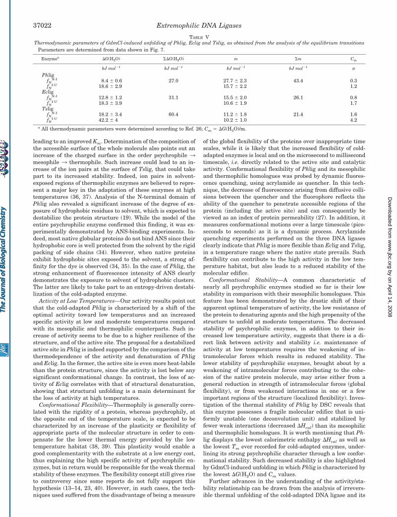
Chemical Stability of DNA Ligases—The stability of the Ph-lig, Eclig, and Tslig was further investigated by GdmCl-in-duced unfolding monitored by intrinsic fluorescence. As shownin Fig. 7, these enzymes unfold according to a three-statepathway, involving a stable intermediate between the nativeand unfolded states. For the three enzymes, unfolding occurs atdistinct denaturant concentrations, with a direct relation be-tween the unfolding transition and the above-mentioned ther-mal stability. Reversibility of unfolding allowed the determina-tion of the thermodynamic parameters (Table V), i.e. �G(H2O)(conformational stability in the absence of denaturant), m(slope of the transition, representing the unfolding cooperativ-ity), and Cm (concentration of GdmCl at the transition mid-point). Compared with Eclig and Tslig, the psychrophilic Phligexhibits the lowest �G(H2O) and Cm values, indicating its weakstability. In addition, the cold-adapted enzyme possesses thehighest m value, reflecting the highest unfolding cooperativityduring chemical unfolding.
DISCUSSION
Protein Structure and Active Site Destabilization—Thethree-dimensional modeling of the three extremophilic DNAligases Phlig, Eclig, and Tslig extend previous observationsmade for the N-terminal domain of Phlig (19) and providefurther evidence for the structural determinants implicated inthe adaptation to low temperatures of Phlig. Our results sug-gest that the active site of the cold-enzyme is destabilized by anexcess of hydrophobic surfaces and contains a decreased num-ber of charged residues compared with its thermophilic coun-terpart. These findings are in perfect agreement with the ki-netic parameters determined for both enzymes (19). Indeed,kcat is believed to be the most important adaptive parameter forPhlig hence compensating for the effect of low temperatures onthe catalytic rate, while for Tslig, the adaptive parameter isexpected to be Km for nicked DNA, which is prone to melting athigh temperatures. In the latter, the increase of positivelycharged amino acids within the active site is likely to facilitatethe interaction with the oppositely charged DNA substrate,
FIG. 7. Equilibrium unfolding of Phlig (Œ), Eclig (● ), and Tslig(f) as recorded by fluorescence emission. Data were analyzedaccording to a three-state reaction. The solid lines represent the bestfits of the data analyzed according to a three-state reaction.
TABLE IVThermodynamic parameters for the irreversible unfolding
Ea �G# �H# T�S#
kJ mol�1 kJ mol�1 kJ mol�1 kJ mol�1
For kdenat � 3.4 10�5 s�1 TPhlig 300.2°K 424.7 93.5 422.2 328.7Eclig 314.5°K 164.7 98.1 162.1 64.0
For T � 308.2°K kdenatPhlig 2.7 10�2 s�1 424.7 84.8 422.1 337.3Eclig 9.3 10�5 s�1 164.7 99.4 162.1 62.7
Extremophilic DNA Ligases 37021
by on April 14, 2008
ww
w.jbc.org
Dow
nloaded from
leading to an improved Km. Determination of the composition ofthe accessible surface of the whole molecule also points out anincrease of the charged surface in the order psychrophile 3mesophile 3 thermophile. Such increase could lead to an in-crease of the ion pairs at the surface of Tslig, that could takepart to its increased stability. Indeed, ion pairs in solvent-exposed regions of thermophilic enzymes are believed to repre-sent a major key in the adaptation of these enzymes at hightemperatures (36, 37). Analysis of the N-terminal domain ofPhlig also revealed a significant increase of the degree of ex-posure of hydrophobic residues to solvent, which is expected todestabilize the protein structure (19). While the model of theentire psychrophilic enzyme confirmed this finding, it was ex-perimentally demonstrated by ANS-binding experiments. In-deed, most native globular proteins do not bind ANS since theirhydrophobic core is well protected from the solvent by the rigidpacking of side chains (34). However, when native proteinsexhibit hydrophobic sites exposed to the solvent, a strong af-finity for the dye is observed (34, 35). In the case of Phlig, thestrong enhancement of fluorescence intensity of ANS clearlydemonstrates the exposure to solvent of hydrophobic clusters.The latter are likely to take part to an entropy-driven destabi-lization of the cold-adapted enzyme.
Activity at Low Temperatures—Our activity results point outthat the cold-adapted Phlig is characterized by a shift of theoptimal activity toward low temperatures and an increasedspecific activity at low and moderate temperatures comparedwith its mesophilic and thermophilic counterparts. Such in-crease of activity seems to be due to a higher resilience of thestructure, and of the active site. The proposal for a destabilizedactive site in Phlig is indeed supported by the comparison of thethermodependence of the activity and denaturation of Phligand Eclig. In the former, the active site is even more heat-labilethan the protein structure, since the activity is lost below anysignificant conformational change. In contrast, the loss of ac-tivity of Eclig correlates with that of structural denaturation,showing that structural unfolding is a main determinant forthe loss of activity at high temperatures.
Conformational Flexibility—Thermophily is generally corre-lated with the rigidity of a protein, whereas psychrophily, atthe opposite end of the temperature scale, is expected to becharacterized by an increase of the plasticity or flexibility ofappropriate parts of the molecular structure in order to com-pensate for the lower thermal energy provided by the lowtemperature habitat (38, 39). This plasticity would enable agood complementarity with the substrate at a low energy cost,thus explaining the high specific activity of psychrophilic en-zymes, but in return would be responsible for the weak thermalstability of these enzymes. The flexibility concept still gives riseto controversy since some reports do not fully support thishypothesis (13–14, 23, 40). However, in such cases, the tech-niques used suffered from the disadvantage of being a measure
of the global flexibility of the proteins over inappropriate timescales, while it is likely that the increased flexibility of cold-adapted enzymes is local and on the microsecond to millisecondtimescale, i.e. directly related to the active site and catalyticactivity. Conformational flexibility of Phlig and its mesophilicand thermophilic homologues was probed by dynamic fluores-cence quenching, using acrylamide as quencher. In this tech-nique, the decrease of fluorescence arising from diffusive colli-sions between the quencher and the fluorophore reflects theability of the quencher to penetrate accessible regions of theprotein (including the active site) and can consequently beviewed as an index of protein permeability (27). In addition, itmeasures conformational motions over a large timescale (pico-seconds to seconds) as it is a dynamic process. Acrylamidequenching experiments performed on the three DNA ligasesclearly indicate that Phlig is more flexible than Eclig and Tslig,in a temperature range where the native state prevails. Suchflexibility can contribute to the high activity in the low tem-perature habitat, but also leads to a reduced stability of themolecular edifice.
Conformational Stability—A common characteristic ofnearly all psychrophilic enzymes studied so far is their lowstability in comparison with their mesophilic homologues. Thisfeature has been demonstrated by the drastic shift of theirapparent optimal temperature of activity, the low resistance ofthe protein to denaturing agents and the high propensity of thestructure to unfold at moderate temperatures. The decreasedstability of psychrophilic enzymes, in addition to their in-creased low temperature activity, suggests that there is a di-rect link between activity and stability i.e. maintenance ofactivity at low temperatures requires the weakening of in-tramolecular forces which results in reduced stability. Thelower stability of psychrophilic enzymes, brought about by aweakening of intramolecular forces contributing to the cohe-sion of the native protein molecule, may arise either from ageneral reduction in strength of intramolecular forces (globalflexibility), or from weakened interactions in one or a fewimportant regions of the structure (localized flexibility). Inves-tigation of the thermal stability of Phlig by DSC reveals thatthis enzyme possesses a fragile molecular edifice that is uni-formly unstable (one deconvolution unit) and stabilized byfewer weak interactions (decreased �Hcal) than its mesophilicand thermophilic homologues. It is worth mentioning that Ph-lig displays the lowest calorimetric enthalpy �Hcal as well asthe lowest Tm ever recorded for cold-adapted enzymes, under-lining its strong psychrophilic character through a low confor-mational stability. Such decreased stability is also highlightedby GdmCl-induced unfolding in which Phlig is characterized bythe lowest �G(H2O) and Cm values.
Further advances in the understanding of the activity/sta-bility relationship can be drawn from the analysis of irrevers-ible thermal unfolding of the cold-adapted DNA ligase and its
TABLE VThermodynamic parameters of GdmCl-induced unfolding of Phlig, Eclig and Tslig, as obtained from the analysis of the equilibrium transitions
Parameters are determined from data shown in Fig. 7.
Enzymea �G(H2O) �G(H2O) m m Cm
kJ mol�1 kJ mol�1 kJ mol�1 kJ mol�1M
PhligfN
N-I 8.4 � 0.6 27.0 27.7 � 2.3 43.4 0.3fN
I-U 18.6 � 2.9 15.7 � 2.2 1.2Eclig
fNN-I 12.8 � 1.2 31.1 15.5 � 2.0 26.1 0.8
fNI-U 18.3 � 3.9 10.6 � 1.9 1.7
TsligfN
N-I 18.2 � 3.4 60.4 11.2 � 1.8 21.4 1.6fN
I-U 42.2 � 4 10.2 � 1.0 4.2a All thermodynamic parameters were determined according to Ref. 26; Cm � �G(H2O)/m.
Extremophilic DNA Ligases37022
by on April 14, 2008
ww
w.jbc.org
Dow
nloaded from

mesophilic counterpart. Our results show an increase in freeenergies of activation �G# from Phlig to Eclig, reflecting thatthe same denaturation rate kdenat is reached at increasingtemperatures (see also Table IV). The small differences in both�G# values arise however from large differences in the enthal-pic and entropic contributions. Indeed, the low �G# value re-corded for Phlig corresponds to the largest �H# and T�S#
contributions, and conversely for Eclig. Such trend has alreadybeen noticed for a few psychrophilic enzymes (17, 18) and thusseems to be a common property of psychrophilic enzymes.
The low kinetic barrier (�G#) recorded for the psychrophilicenzyme allows in fact unfolding at a high rate, leading to asymmetrical and relatively narrow denaturation peak in mi-crocalorimetry (Fig. 5), and consequently to high activationenergy Ea and high �H#. This reflects a high cooperativity ofunfolding for the psychrophilic enzyme, that probably origi-nates from the lower number of interactions required to disruptthe active conformation. The large entropic contribution no-ticed in Phlig suggests that its transition state is more disor-dered than that of its mesophilic counterpart, probably due tothe fact that at any specific temperature, more interactions arebroken into the psychrophile. The entropy loss associated withhydration of nonpolar groups upon denaturation can also con-tribute significantly to the differences in entropic contributionbetween Phlig and Eclig. Indeed, in the case of the mesophilicenzyme and more particularly in the thermophilic counterpart,an increased proportion of nonpolar groups are exposed uponunfolding, increasing the order of water molecules and there-fore decreasing the entropy change relative to Phlig. All to-gether, our results perfectly fit those obtained for the icefishglutamate dehydrogenase (16), a cold-adapted -amylase (18)and xylanase (17), demonstrating that thermolability of psy-chrophilic enzymes is entropically driven. The contradictoryresult obtained by Siddiqui et al. (15) with the elongation factor2 from a psychrotolerant archae is likely to arise from anunusual broader DSC profile of the cold enzyme (even thoughassociated with a lower Tm) than that of the thermophilichomologue.
Concluding Remarks—Until recently, the activity/flexibility/stability relationship within psychrophilic enzymes was stillappreciated with caution. However, the results obtained for thethree DNA ligases adapted to different thermal habitats clearlyestablish a link between activity, flexibility, and stability. Thecold-adapted DNA ligase is characterized by a high activity atlow temperatures, a high flexibility and a low stability espe-cially at the active site. These results are in perfect agreementwith those recently obtained from studies performed on ex-tremophilic -amylases (18) and xylanases (17). Therefore, theemerging picture suggests that psychrophilic enzymes arecharacterized by increased catalytic efficiency attributed to anincrease of the flexibility of appropriate parts of the molecularstructure, in order to compensate for the lower thermal energyprovided by the low temperature habitat. In return, this flexi-bility would be responsible for the weak thermal and chemicalstabilities of cold-adapted enzymes.
Acknowledgments—We thank N. Gerardin and R. Marchand for theirskilful technical assistance. We also thank F. Biemar, T. Collins,S. D.’Amico, and M. Vanhove for helpful assistance and discussions.
REFERENCES
1. Deming, J. W. (2002) Curr. Opin. Microbiol. 5, 301–3092. Blochl, E., Rachel, R., Burggraf, S., Hafenbradl, D., Jannasch, H. W., and
Stetter, K. O. (1997) Extremophiles 1, 14–213. Gerday, C., Aittaleb, M., Arpigny, J. L., Baise, E., Chessa, J. P., Garsoux, G.,
Petrescu, I., and Feller, G. (1997) Biochim. Biophys. Acta 1342, 119–1314. Russell, N. J. (1998) Adv. Biochem. Eng. Biotechnol. 61, 1–215. Demirjian, D. C., Moris-Varas, F., and Cassidy, C. S. (2001) Curr. Opin. Chem.
Biol. 5, 144–1516. Levy, M., and Miller, S. L. (1998) Proc. Natl. Acad. Sci. U. S. A. 95, 7933–79387. Wintrode, P. L., Miyazaki, K., and Arnold, F. H. (2000) J. Biol. Chem. 275,
31635–316408. Kumar, S., Tsai, C. J., and Nussinov, R. (2002) Biochemistry 41, 5359–53749. Smalas, A. O., Leiros, H. K., Os, V., and Willassen, N. P. (2000) Biotechnol.
Annu. Rev. 6, 1–5710. Cavicchioli, R., Siddiqui, K. S., Andrews, D., and Sowers, K. R. (2002) Curr.
Opin. Biotechnol. 13, 253–26111. D’Amico, S., Claverie, P., Collins, T., Georlette, D., Gratia, E., Hoyoux, A.,
Meuwis, M. A., Feller, G., and Gerday, C. (2002) Philos. Trans. R. Soc.Lond. B Biol. Sci. 357, 917–925
12. Miyazaki, K., Wintrode, P. L., Grayling, R. A., Rubingh, D. N., and Arnold,F. H. (2000) J. Mol. Biol. 297, 1015–1026
13. Svingor, A., Kardos, J., Hajdu, I., Nemeth, A., and Zavodszky, P. (2001) J. Biol.Chem. 276, 28121–28125
14. Fields, P. A. (2001) Comp. Biochem. Physiol. A Mol. Integr. Physiol. 129,417–431
15. Siddiqui, K. S., Cavicchioli, R., and Thomas, T. (2002) Extremophiles 6,143–150
16. Ciardiello, M. A., Camardella, L., Carratore, V., and di Prisco, G. (2000)Biochim. Biophys. Acta 1543, 11–23
17. Collins, T., Meuwis, M. A., Gerday, C., and Feller, G. (2003) J. Mol. Biol. 328,419–428
18. D’Amico, S., Gerday, C., and Feller, G. (2003) J. Biol. Chem. 278, 7891–789619. Georlette, D., Jonsson, Z. O., Van Petegem, F., Chessa, J.-P., Van Beeumen, J.,
Hubscher, U., and Gerday, C. (2000) Eur. J. Biochem. 267, 3502–351220. Sriskanda, V., Schwer, B., Ho, C. K., and Shuman, S. (1999) Nucleic Acids Res.
27, 3953–396321. Thorbjarnardottir, S. H., Jonsson, Z. O., Andresson, O. S., Kristjansson, J. K.,
Eggertsson, G., and Palsdottir, A. (1995) Gene (Amst.) 161, 1–622. Lee, J. Y., Chang, C., Song, H. K., Moon, J., Yang, J. K., Kim, H. K., Kwon,
S. T., and Suh, S. W. (2000) EMBO J. 19, 1119–112923. Hernandez, G., Jenney, F. E., Jr., Adams, M. W., and LeMaster, D. M. (2000)
Proc. Natl. Acad. Sci. U. S. A. 97, 3166–317024. Timson, D. J., and Wigley, D. B. (1999) J. Mol. Biol. 285, 73–8325. Pace, C. N. (1986) Methods Enzymol. 131, 266–28026. Vanhove, M., Guillaume, G., Ledent, P., Richards, J. H., Pain, R. H., and Frere,
J. M. (1997) Biochem. J. 321, 413–41727. Lakowicz, J. (1983) Principles of Fluorescence Spectroscopy, pp. 257–301,
Plenum Press, New York28. Feller, G., d’Amico, D., and Gerday, C. (1999) Biochemistry 38, 4613–461929. Goldberg, M. E., Expert-Bezançon, N., Vuillard, L., and Rabilloud, T. (1995)
Fold. Des. 1, 21–2730. Matouschek, A., Matthews, J. M., Johnson, C. M., and Fersht, A. R. (1994)
Protein Eng. 7, 1089–109531. Sanchez-Ruiz, J. M., Lopez-Lacomba, J. L., Cortijo, M., and Mateo, P. L. (1988)
Biochemistry 27, 1648–165232. Lonhienne, T., Gerday, C., and Feller, G. (2000) Biochim. Biophys. Acta 1543,
1–1033. Sriskanda, V., and Shuman, S. (2002) J. Biol. Chem. 277, 9695–970034. Stryer, L. (1965) J. Mol. Biol. 13, 482–49535. Semisotnov, G. V., Rodionova, N. A., Razgulyaev, O. I., Uversky, V. N., Gripas,
A. F., and Gilmanshin, R. I. (1991) Biopolymers 31, 119–12836. Kumar, S., and Nussinov, R. (2001) Proteins 43, 433–45437. Chakravarty, S., and Varadarajan, R. (2002) Biochemistry 41, 8152–816138. Hochachka, P. W., and Somero, G. N. (1984) Biochemical Adaptations, pp.
355–449, Princeton University Press, Princeton39. Fields, P. A., and Somero, G. N. (1998) Proc. Natl. Acad. Sci. U. S. A. 95,
11476–1148140. Fitter, J., and Heberle, J. (2000) Biophys. J. 79, 1629–1936
Extremophilic DNA Ligases 37023
by on April 14, 2008
ww
w.jbc.org
Dow
nloaded from