Development of Morphological Diversity of Dendrites in< i> Drosophila by the BTB-Zinc Finger Protein...
14
Neuron, Vol. 43, 809–822, September 16, 2004, Copyright 2004 by Cell Press Development of Morphological Diversity of Dendrites in Drosophila by the BTB-Zinc Finger Protein Abrupt spikes, and coincidence detection (Mainen and Sejnow- ski, 1996; Vetter et al., 2001; Schaefer et al., 2003). There- fore, the proper function of the nervous system relies not only on correct targeting of axons or dendrites, but also on development of class-specific dendritic pat- Kaoru Sugimura, 1,2 Daisuke Satoh, 1,2 Patricia Estes, 3,5 Stephen Crews, 3 and Tadashi Uemura 1,4.6, * 1 Laboratory of Molecular Genetics The Institute for Virus Research terns. However, genetic programs underlying dendritic 2 Graduate School of Science diversity are not yet well understood. Kyoto University Dendritic arborization (da) neurons in the Drosophila Kyoto 606-8507 peripheral nervous system (PNS) provide an excellent Japan model system to tackle this question in vivo. The embry- 3 Biochemistry and Biophysics onic PNS organizes a stereotyped pattern of identified School of Medicine sensory neurons, and da neurons constitute a subfamily The University of North Carolina at Chapel Hill of multidendritic (md) neurons (Figure 1A; Bodmer and Chapel Hill, North Carolina 27599 Jan, 1987; Jan and Jan, 1993; Campos-Ortega and 4 Core Research for Evolutional Science Hartenstein, 1997). da neurons grow two-dimensional and Technology (CREST) dendrites underneath the epidermis during late embry- Japan Science and Technology onic, larval, and pupal stages (Bodmer and Jan, 1987; Kawaguchi, Saitama 332-0012 Williams and Truman, 2004). On the basis of images of Japan single da neurons in mature larva, the 15 da neurons in each abdominal hemisegment are classified into four categories, classes I–IV, in order of increasing territory Summary size and/or arbor complexity (Figure 1A; Grueber et al., 2002). Class I neurons are characterized by formation Morphological diversity of dendrites contributes to of simple comb-like small dendritic trees (Figure 1B), specialized functions of individual neurons. In the whereas class IV develop far more complicated and present study, we examined the molecular basis that expansive arbors (Figure 1C; Grueber et al., 2002; Sugi- generates distinct morphological classes of Drosoph- mura et al., 2003). ila dendritic arborization (da) neurons. da neurons are Class-specific transgenic GFP markers have made it classified into classes I to IV in order of increasing feasible to undertake time-lapse analysis of da neurons territory size and/or branching complexity. We found from dendritic birth till maturation, and such analyses that Abrupt (Ab), a BTB-zinc finger protein, is ex- have provided us insight into the cellular basis of class- pressed selectively in class I cells. Misexpression of specific dendritic morphogenesis (Grueber et al., 2003b; ab in neurons of other classes directed them to take Sugimura et al., 2003). Class I neurons and class IV the appearance of cells with smaller and/or less elabo- neurons employ distinct strategies of dendritic emer- rated arbors. Loss of ab functions in class I neurons gence from the cell body and branching, which contrib- resulted in malformation of their typical comb-like ute to differences in their basic arbor patterns. In con- arbor patterns and generation of supernumerary trast to class I neurons, class IV neurons continue to branch terminals. Together with the results of monitor- elaborate higher-order branches throughout larval ing dendritic dynamics of ab-misexpressing cells or stages. In addition to these distinct developmental dy- ab mutant ones, all of the data suggested that Ab namics, class IV cells are characterized by their class- endows characteristics of dendritic morphogenesis of specific mutual avoidance of both isoneuronal and hetero- the class I neurons. neuronal dendritic branches, and this allows complete but minimal overlapping innervation of the body wall (de- Introduction scribed later in Figure 4; Sugimura et al., 2003; Grueber et al., 2003b). On the other hand, class I and class II neurons do not show such inhibitory dendrodendritic Morphologies of dendritic trees are highly variable from interaction to shape the territorial boundary between one neuronal type to another, and this diversity contrib- the same class of neurons, and dendritic branches of utes to differential processing and computation of syn- class III neurons avoid each other only at their short aptic or sensory inputs (Scott and Luo, 2001; Masland, terminal extensions (Grueber et al., 2003b). 2001; Whitford et al., 2002; Ha ¨ usser and Mel, 2003; Wong Then what are molecular mechanisms that make den- and Ghosh, 2002; Jan and Jan, 2003; Grueber and Jan, dritic behaviors of each class distinct? Our previous 2004). For example, dendritic geometry plays key roles enhancer trap screening aimed at hunting for either pan- in shaping intrinsic firing patterns, both back and for- da or subset markers and then looking for trapped genes ward propagation of action potentials and dendritic that might be expressed in a particular class(es), with the expectation of roles of those genes in class-specific *Correspondence: [email protected] dendrite morphogeneis (Sugimura et al., 2003). Here we 5 Present address: Department of Genetics, North Carolina State report that one of our class I markers trapped abrupt University, North Carolina 27695. (ab) and that the Ab protein was selectively expressed 6 Present address: Graduate School of Biostudies, Kyoto University, Kyoto 606-8502, Japan. in class I da neurons. The expression profile of Ab was
Transcript of Development of Morphological Diversity of Dendrites in< i> Drosophila by the BTB-Zinc Finger Protein...
Neuron, Vol. 43, 809–822, September 16, 2004, Copyright 2004 by Cell Press
Development of Morphological Diversityof Dendrites in Drosophilaby the BTB-Zinc Finger Protein Abrupt
spikes, and coincidence detection (Mainen and Sejnow-ski, 1996; Vetter et al., 2001; Schaefer et al., 2003). There-fore, the proper function of the nervous system reliesnot only on correct targeting of axons or dendrites, butalso on development of class-specific dendritic pat-
Kaoru Sugimura,1,2 Daisuke Satoh,1,2
Patricia Estes,3,5 Stephen Crews,3
and Tadashi Uemura1,4.6,*1Laboratory of Molecular GeneticsThe Institute for Virus Research
terns. However, genetic programs underlying dendritic2 Graduate School of Sciencediversity are not yet well understood.Kyoto University
Dendritic arborization (da) neurons in the DrosophilaKyoto 606-8507peripheral nervous system (PNS) provide an excellentJapanmodel system to tackle this question in vivo. The embry-3 Biochemistry and Biophysicsonic PNS organizes a stereotyped pattern of identifiedSchool of Medicinesensory neurons, and da neurons constitute a subfamilyThe University of North Carolina at Chapel Hillof multidendritic (md) neurons (Figure 1A; Bodmer andChapel Hill, North Carolina 27599Jan, 1987; Jan and Jan, 1993; Campos-Ortega and4 Core Research for Evolutional ScienceHartenstein, 1997). da neurons grow two-dimensionaland Technology (CREST)dendrites underneath the epidermis during late embry-Japan Science and Technologyonic, larval, and pupal stages (Bodmer and Jan, 1987;Kawaguchi, Saitama 332-0012Williams and Truman, 2004). On the basis of images ofJapansingle da neurons in mature larva, the 15 da neurons ineach abdominal hemisegment are classified into fourcategories, classes I–IV, in order of increasing territorySummarysize and/or arbor complexity (Figure 1A; Grueber et al.,2002). Class I neurons are characterized by formationMorphological diversity of dendrites contributes toof simple comb-like small dendritic trees (Figure 1B),specialized functions of individual neurons. In thewhereas class IV develop far more complicated andpresent study, we examined the molecular basis thatexpansive arbors (Figure 1C; Grueber et al., 2002; Sugi-generates distinct morphological classes of Drosoph-mura et al., 2003).ila dendritic arborization (da) neurons. da neurons are
Class-specific transgenic GFP markers have made itclassified into classes I to IV in order of increasingfeasible to undertake time-lapse analysis of da neuronsterritory size and/or branching complexity. We foundfrom dendritic birth till maturation, and such analyses
that Abrupt (Ab), a BTB-zinc finger protein, is ex-have provided us insight into the cellular basis of class-
pressed selectively in class I cells. Misexpression of specific dendritic morphogenesis (Grueber et al., 2003b;ab in neurons of other classes directed them to take Sugimura et al., 2003). Class I neurons and class IVthe appearance of cells with smaller and/or less elabo- neurons employ distinct strategies of dendritic emer-rated arbors. Loss of ab functions in class I neurons gence from the cell body and branching, which contrib-resulted in malformation of their typical comb-like ute to differences in their basic arbor patterns. In con-arbor patterns and generation of supernumerary trast to class I neurons, class IV neurons continue tobranch terminals. Together with the results of monitor- elaborate higher-order branches throughout larvaling dendritic dynamics of ab-misexpressing cells or stages. In addition to these distinct developmental dy-ab mutant ones, all of the data suggested that Ab namics, class IV cells are characterized by their class-endows characteristics of dendritic morphogenesis of specific mutual avoidance of both isoneuronal and hetero-the class I neurons. neuronal dendritic branches, and this allows complete but
minimal overlapping innervation of the body wall (de-Introduction scribed later in Figure 4; Sugimura et al., 2003; Grueber
et al., 2003b). On the other hand, class I and class IIneurons do not show such inhibitory dendrodendriticMorphologies of dendritic trees are highly variable frominteraction to shape the territorial boundary betweenone neuronal type to another, and this diversity contrib-the same class of neurons, and dendritic branches ofutes to differential processing and computation of syn-class III neurons avoid each other only at their shortaptic or sensory inputs (Scott and Luo, 2001; Masland,terminal extensions (Grueber et al., 2003b).2001; Whitford et al., 2002; Hausser and Mel, 2003; Wong
Then what are molecular mechanisms that make den-and Ghosh, 2002; Jan and Jan, 2003; Grueber and Jan,dritic behaviors of each class distinct? Our previous2004). For example, dendritic geometry plays key rolesenhancer trap screening aimed at hunting for either pan-in shaping intrinsic firing patterns, both back and for-da or subset markers and then looking for trapped genesward propagation of action potentials and dendriticthat might be expressed in a particular class(es), withthe expectation of roles of those genes in class-specific
*Correspondence: [email protected] morphogeneis (Sugimura et al., 2003). Here we5 Present address: Department of Genetics, North Carolina Statereport that one of our class I markers trapped abruptUniversity, North Carolina 27695.(ab) and that the Ab protein was selectively expressed6 Present address: Graduate School of Biostudies, Kyoto University,
Kyoto 606-8502, Japan. in class I da neurons. The expression profile of Ab was

Neuron810
Figure 1. Distinct Morphological Classes ofda Neurons and Selective ab Expression inClass I
(A) A diagram of positions of multidendritic(md) neurons in an abdominal hemisegmentof the Drosophila embryonic and larval PNS.Diamonds represent individual dendritic ar-borization (da) neurons and triangles repre-sent other types of md neurons. Besides allda neurons, only two bipolar dendrite neu-rons (dbd and vbd) and dmd1 that appear insubsequent figures are indicated for simplic-ity. d, l, v�, and v represent dorsal, lateral,ventral prime, and ventral clusters, respec-tively. Each class of the da neurons is differ-ently colored. Black lines represent fasciclesof axons that extend from the neuronal cellbodies. In this and all subsequent figures, an-terior is to the left and dorsal is at the top.(B and C) Morphologies of a class I neuron(ddaE) and the class IV neuron (ddaC) in thedorsal cluster at 74–78 hr after egg laying(AEL). Genotypes were IG1-1/mCD8::GFP (B)and NP7028 mCD8::GFP/mCD8::GFP (C).(D–O) Expression patterns of Abrupt (Ab) pro-tein in dorsal (D–I and M–O) and ventral(J–L) clusters.(D–L) Stage 16 embryo of a class I markerline (D–F, NP2225/mCD8::GFP) or that of apan-da marker line (G–L, IG1-2 mCD8::GFP)was doubly labeled for GFP (D, G, J, andgreen in F, I, and L) and for Ab (E, H, K, andmagenta in F, I, and L). Merged images shownin (F), (I), and (L). Among da neurons, Ab wasexpressed highly and in a selectable mannerin all of the three class I neurons, ddaD andddaE (arrows in D, E, G, and H) and vpda(right arrow in J and K). The Ab expressionlevel was also high in vbd (left arrow in J andK). We did not detect significant signals inother neurons in the abdominal segment.Outside the nervous system, Ab was pro-duced by epidermal cells (asterisks in E andN) and muscles (bracket in H).
(M–O) Stage 13 embryo of a panneuronal marker line, elav-GAL4 mCD8::GFP, was stained for GFP (M and green in O) and Ab (N and magentain O). Two neurons in the dorsal cluster were weakly stained at this stage, whereas other neurons including those indicated by the arrowheadin (M) (possibly one of external sensory neurons) were not.Scale bars equal 50 �m for (B) and (C) and 10 �m for (D)–(O).
roughly complementary to that of Cut, a homeodomain simple and small dendrites with a comb-like shape,which are characteristic of class I da neurons. We furtherprotein. The level of Cut is below the limit of detection
in class I neurons, whereas it is either moderate or strong addressed whether or not ab and cut influenced eachother at the level of gene expression and at the level ofin class II to IV neurons. Most importantly, those different
levels of Cut regulate dendrite growth as well as class- dendritic morphology as a final read-out.specific terminal branching (Grueber et al., 2003a).
We addressed whether Ab might be required for class ResultsI dendrite morphogenesis or not and whether the abgene must be silent in class II-IV cells for their normal Selective Expression of abrupt
in Class I da Neuronsdendrite development. Loss of ab function in class Ineurons caused deformation of the characteristic comb- One of our class I marker lines is NP2225, which labels
all of the three class I da neurons, but no other dalike shape of their dendritic arbors, produced excessbranch terminals of all of three cells examined, and in- neurons, in the abdominal hemisegment (Figures 1D and
1F; Sugimura et al., 2003). In the genome of NP2225, acreased in the territory size of one cell. Misexpressionof ab influenced dendritic designs of neurons of class P element was inserted into the first intron of the abrupt
(ab) gene (see details in Experimental Procedures). abII to IV, which do not express ab normally; that is, ab-misexpressing neurons of class II to IV generated less encodes a BTB domain-zinc finger protein and is re-
quired for neuromuscular connectivity in the embryo (Huexpansive dendritic trees, and those of class III and IVhad fewer branch terminals compared with control cells. et al., 1995). By using an antibody to Ab protein, we
detected immunoreactivity in the nuclei of the threeThese data showed that Ab directed morphologically
Development of Morphological Diversity of Dendrite811
class I da neurons, but expression in the other classes of S1). In our subsequent analysis, we focused mostly onthe dorsal cluster in abdominal segments 2–6; this wasda neurons was below the limit of our detection (Figures
1D–1O). Outside the nervous system, Ab was expressed partly because the dorsal cluster has all classes of daneurons and partly because axons extending from dor-in epidermal cells and muscles (see asterisks in Figures
1E and 1N and bracket in Figure 1H, respectively; Hu sally located neurons sometimes make images of theventral, ventral-prime, and lateral clusters difficult toet al., 1995). Immunoreactivity per nucleus was compa-
rable between the class I da neurons and epidermal interpret (Figure 1A).cells, and muscle nuclei were stained less brightlyaround 13 hr after egg laying (AEL) (stage 16), when Dendritic Arbors of Class IV Neurons Becamedendritogenesis was initiated (Figures 1E, 1H, and 1K). Downsized and Less ElaboratedThe selective expression within the da neuron classes by ab Expressionpersisted at third instar larval stages (data not shown). To visualize how ab expression affected dendritic mor-At earlier stages such as 9–10 hr AEL (stage 13) when phologies of individual classes, we performed single-all da neurons are born, a small subset of peripheral cell analysis first on ddaC (class IV), which should haveneurons was stained, but very faintly compared with formed the most expansive and the most highlyepidermal cells (Figures 1M–1O), and those positive neu- branched arbor in the dorsal cluster (Figure 1C). To ex-rons were possibly class I neurons. tract pattern information about ddaC, we ablated all
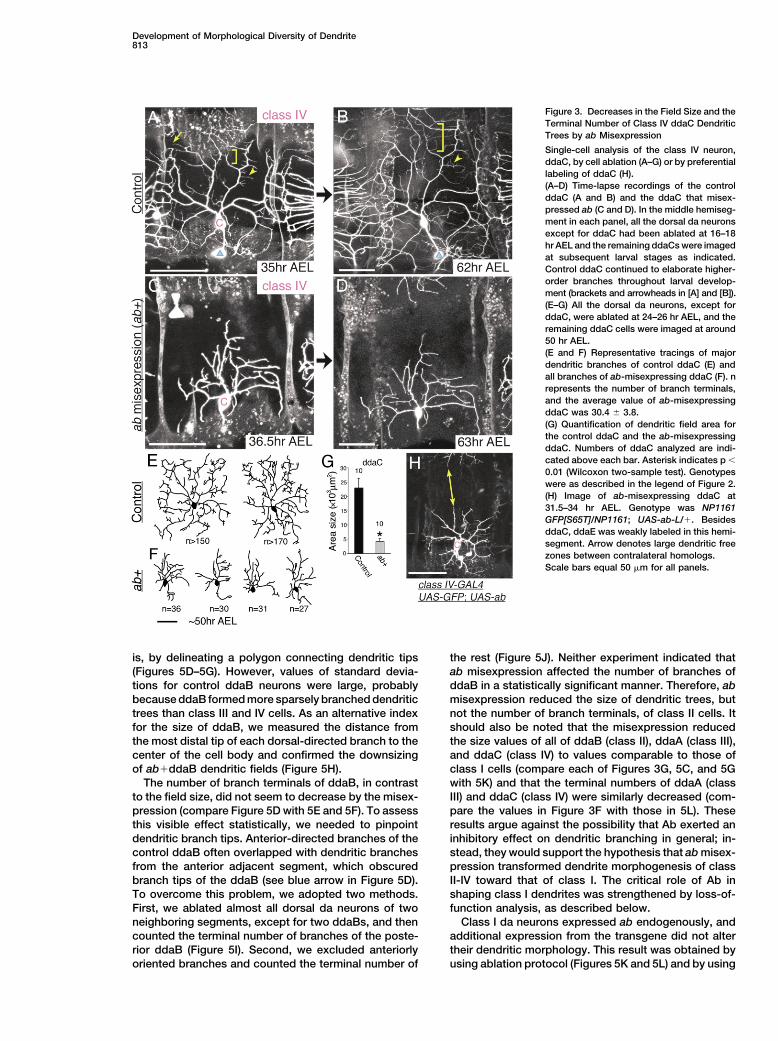
dorsal da neurons except for ddaC at embryonic or earlylarval stages and then tracked dendritic developmentEctopic Expression of ab Reduced the Sizeof the remaining ddaC at subsequent stages.of Overall Dendritic Trees
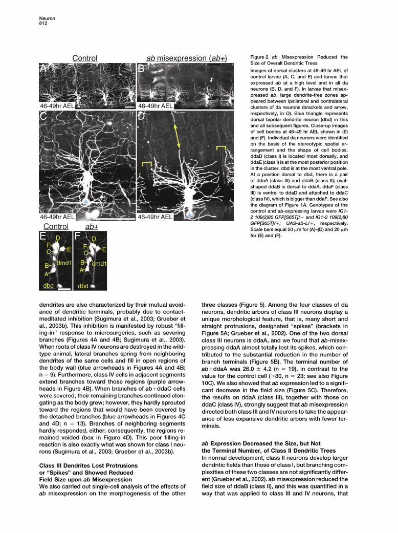
When compared with the control ddaC, ab-misex-To address whether the class I-selective expression ofpressing ddaC (ab�ddaC) at the same age showed aab plays roles in class-specific dendrite patterning orreduced size of its dendritic field and exhibited a visiblynot, we took two complementary approaches: one wasless complex morphology (compare Figures 3A with 3C;to study effects of misexpression of ab on morphogene-3B with 3D; and 3E with 3F). Branch terminals of ab�-sis of class II-IV neuron dendrites, which innervate largerddaC did not reach far enough to meet those of adjacentregions of the body wall than class I dendrites. The othercells. Furthermore, ab�ddaC did not elaborate higher-was to observe dendritic arbors of class I neurons, whichorder fine branches in contrast to the wild-type cell (seeexpress ab normally, in ab loss-of-function mutant cells.arrow and bracket in Figures 3A and 3B). These pheno-We first present the results of our misexpression experi-types of the downsizing and the decrease in the terminalments (Figures 2–5).number were consolidated by our semiquantitative anal-We drove ab expression in all da neurons in a postmi-ysis (p � 0.01, Wilcoxon two-sample test; Figure 3G).totic manner by using pan-da GAL4 lines, and this pan-When normalized to total dendritic length, the branchingda expression caused dramatic alteration of overall pat-index (a measure of the number of branch terminals perterns of dendritic arbors (Figures 2A–2D). The body walldendritic length) of ab�ddaC was smaller than that ofof the control larva is entirely covered with dendriticthe control ddaC, and this decrease was statisticallytrees of da neurons from 30–35 hr AEL onward (Figuressignificant (p � 0.01, Wilcoxon two-sample test). This2A and 2C; Gao et al., 2000; Grueber et al., 2003b; Sugi-result supported that ab misexpression in ddaC mademura et al., 2003). Although the larvae that expressedits dendritic arbor less complicated. In addition to theab in the pan-da fashion kept expanding their body wallsabove single-cell analysis with the help of ablation, wetill late second instar larval stages, large dendrite-freeused a GAL4 line that drove expression of both ab andzones appeared around segmental boundaries and dor-GFP preferentially in class IV neurons and obtained qual-sal midlines, which lie midway between ipsilateral anditatively similar results (Figure 3H).contralateral dorsal clusters (Figures 2B and 2D). In nor-
mal development, those zones are covered by dendriticbranches of class II-IV neurons that form larger dendritic Dendritic Dynamics of ab-Expressing Class IV
Neurons Resembled Those of Class Ifields than class I cells; and class IV branches contributemost to this filling (Grueber et al., 2002). We imaged cell It has been shown that the wild-type ddaC (class IV)
continues branch formation throughout larval develop-bodies of the GFP-labeled neurons and confirmed thatab ectopic expression did not alter the total number ment (brackets and arrowheads of Figures 3A and 3B;
n � 12; Sugimura et al., 2003); however, our time-lapseor the stereotypic arrangement of da neurons in eachcluster (Figures 2E and 2F). These results suggest the analysis showed that the ab�ddaC almost halted
branching and stabilized its overall dendritic shape bypossibility that ab expression reduced the size of den-dritic trees of class II-IV. midlarval stages (compare Figure 3C with 3D; n � 11).
This temporal mode of ab�ddaC resembled what hasThis apparent downsizing effect occurred with com-plete penetrance (n � 150 dorsal clusters examined), been shown for class I neurons during normal develop-
ment (Sugimura et al., 2003). In contrast to the arrest ofirrespective of the expression of either of the two slightlydistinct forms of Ab protein (Supplemental Figure S1 at higher-order branch formation, ab expression did not
retard elongation of preexisting branches per se. Thishttp://www.neuron.org/cgi/content/full/43/6/809/DC1),and in clusters other than the dorsal one as well (yellow was shown by the fact that dendrites of ab�ddaC kept
growing in a coordinated fashion with expansion of thearrow in Figure 5B). Ab consists of a BTB domain andzinc fingers, and the expression of a truncated form body wall (compare scale bar of Figure 3C with that
of 3D).that had only one of the two motifs did not result inmorphologically visible effects (Supplemental Figure In addition to their continuous branching, class IV
Neuron812
Figure 2. ab Misexpression Reduced theSize of Overall Dendritic Trees
Images of dorsal clusters at 46–49 hr AEL ofcontrol larvae (A, C, and E) and larvae thatexpressed ab at a high level and in all daneurons (B, D, and F). In larvae that misex-pressed ab, large dendrite-free zones ap-peared between ipsilateral and contralateralclusters of da neurons (brackets and arrow,respectively, in D). Blue triangle representsdorsal bipolar dendrite neuron (dbd) in thisand all subsequent figures. Close-up imagesof cell bodies at 46–49 hr AEL shown in (E)and (F). Individual da neurons were identifiedon the basis of the stereotypic spatial ar-rangement and the shape of cell bodies.ddaD (class I) is located most dorsally, andddaE (class I) is at the most posterior positionin the cluster. dbd is at the most ventral pole.At a position dorsal to dbd, there is a pairof ddaA (class III) and ddaB (class II); oval-shaped ddaB is dorsal to ddaA. ddaF (classIII) is ventral to ddaD and attached to ddaC(class IV), which is bigger than ddaF. See alsothe diagram of Figure 1A. Genotypes of thecontrol and ab-expressing larvae were IG1-2 109(2)80 GFP[S65T]/� and IG1-2 109(2)80GFP[S65T]/�; UAS-ab-L/�, respectively.Scale bars equal 50 �m for (A)–(D) and 20 �mfor (E) and (F).
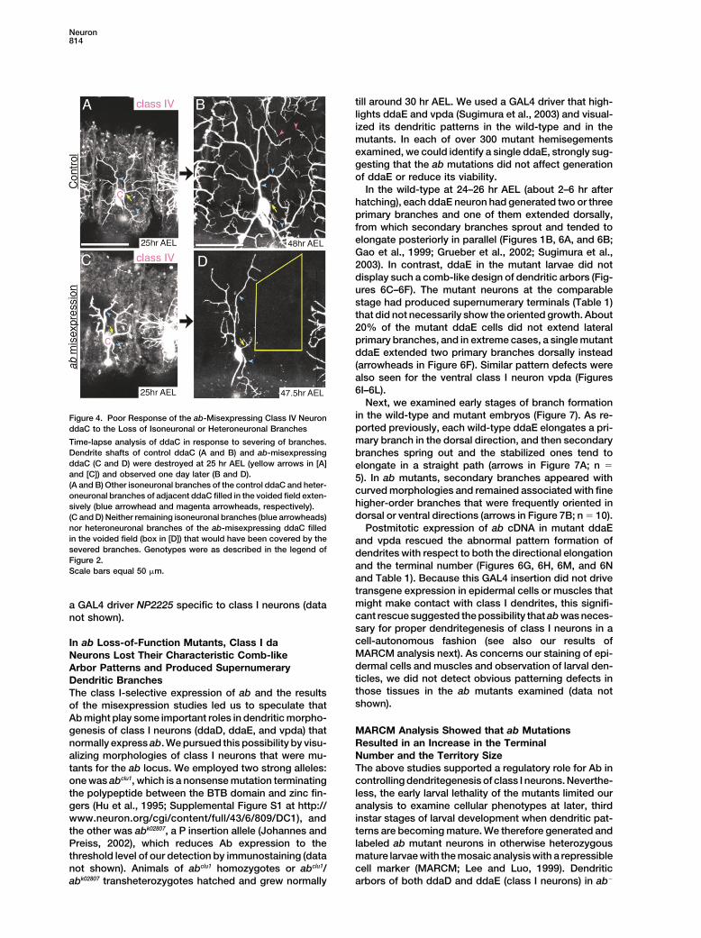
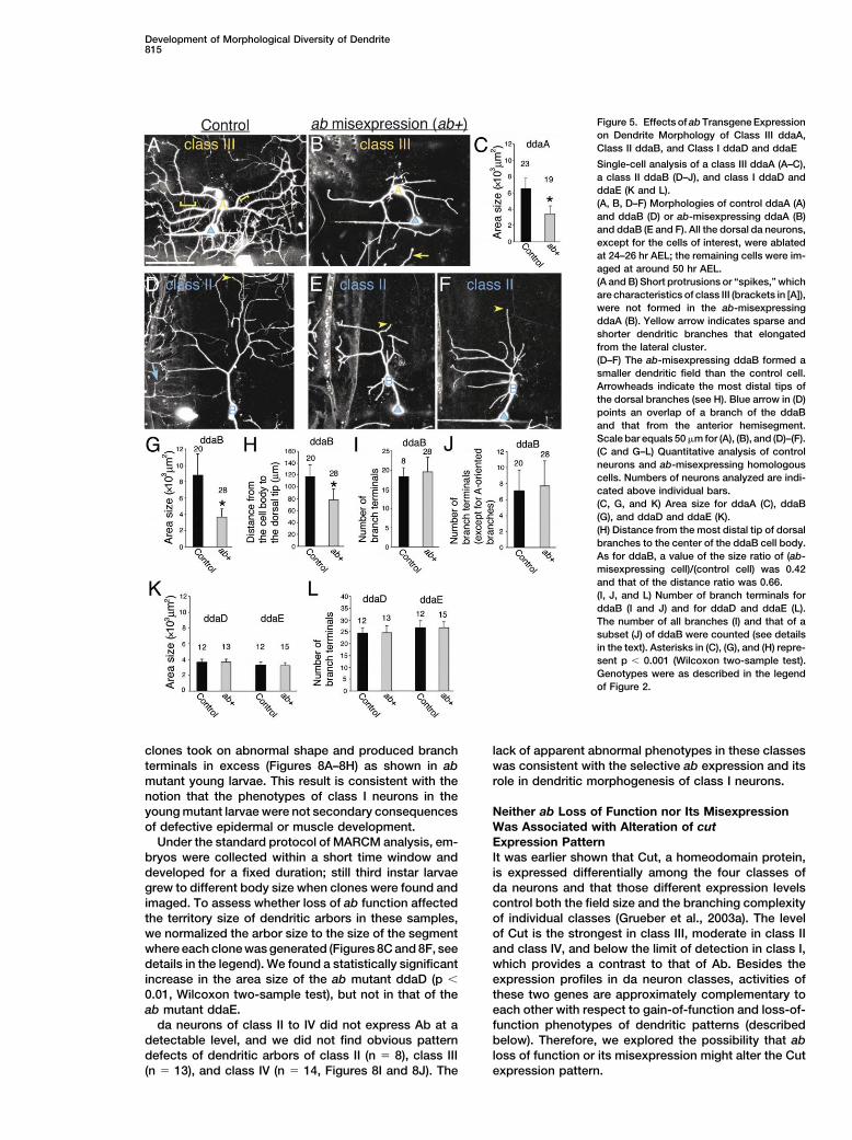
dendrites are also characterized by their mutual avoid- three classes (Figure 5). Among the four classes of daance of dendritic terminals, probably due to contact- neurons, dendritic arbors of class III neurons display ameditated inhibition (Sugimura et al., 2003; Grueber et unique morphological feature, that is, many short andal., 2003b). This inhibition is manifested by robust “fill- straight protrusions, designated “spikes” (brackets ining-in” response to microsurgeries, such as severing Figure 5A; Grueber et al., 2002). One of the two dorsalbranches (Figures 4A and 4B; Sugimura et al., 2003). class III neurons is ddaA, and we found that ab-misex-When roots of class IV neurons are destroyed in the wild- pressing ddaA almost totally lost its spikes, which con-type animal, lateral branches spring from neighboring tributed to the substantial reduction in the number ofdendrites of the same cells and fill in open regions of branch terminals (Figure 5B). The terminal number ofthe body wall (blue arrowheads in Figures 4A and 4B; ab�ddaA was 26.0 � 4.2 (n � 19), in contrast to then � 9). Furthermore, class IV cells in adjacent segments value for the control cell (�80, n � 23; see also Figureextend branches toward those regions (purple arrow- 10C). We also showed that ab expression led to a signifi-heads in Figure 4B). When branches of ab�ddaC cells cant decrease in the field size (Figure 5C). Therefore,were severed, their remaining branches continued elon- the results on ddaA (class III), together with those ongating as the body grew; however, they hardly sprouted ddaC (class IV), strongly suggest that ab misexpressiontoward the regions that would have been covered by directed both class III and IV neurons to take the appear-the detached branches (blue arrowheads in Figures 4C ance of less expansive dendritic arbors with fewer ter-and 4D; n � 13). Branches of neighboring segments minals.hardly responded, either; consequently, the regions re-mained voided (box in Figure 4D). This poor filling-in
ab Expression Decreased the Size, but Notreaction is also exactly what was shown for class I neu-the Terminal Number, of Class II Dendritic Treesrons (Sugimura et al., 2003; Grueber et al., 2003b).In normal development, class II neurons develop largerdendritic fields than those of class I, but branching com-Class III Dendrites Lost Protrusionsplexities of these two classes are not significantly differ-or “Spikes” and Showed Reducedent (Grueber et al., 2002). ab misexpression reduced theField Size upon ab Misexpressionfield size of ddaB (class II), and this was quantified in aWe also carried out single-cell analysis of the effects of
ab misexpression on the morphogenesis of the other way that was applied to class III and IV neurons, that
Development of Morphological Diversity of Dendrite813
Figure 3. Decreases in the Field Size and theTerminal Number of Class IV ddaC DendriticTrees by ab Misexpression
Single-cell analysis of the class IV neuron,ddaC, by cell ablation (A–G) or by preferentiallabeling of ddaC (H).(A–D) Time-lapse recordings of the controlddaC (A and B) and the ddaC that misex-pressed ab (C and D). In the middle hemiseg-ment in each panel, all the dorsal da neuronsexcept for ddaC had been ablated at 16–18hr AEL and the remaining ddaCs were imagedat subsequent larval stages as indicated.Control ddaC continued to elaborate higher-order branches throughout larval develop-ment (brackets and arrowheads in [A] and [B]).(E–G) All the dorsal da neurons, except forddaC, were ablated at 24–26 hr AEL, and theremaining ddaC cells were imaged at around50 hr AEL.(E and F) Representative tracings of majordendritic branches of control ddaC (E) andall branches of ab-misexpressing ddaC (F). nrepresents the number of branch terminals,and the average value of ab-misexpressingddaC was 30.4 � 3.8.(G) Quantification of dendritic field area forthe control ddaC and the ab-misexpressingddaC. Numbers of ddaC analyzed are indi-cated above each bar. Asterisk indicates p �
0.01 (Wilcoxon two-sample test). Genotypeswere as described in the legend of Figure 2.(H) Image of ab-misexpressing ddaC at31.5–34 hr AEL. Genotype was NP1161GFP[S65T]/NP1161; UAS-ab-L/�. BesidesddaC, ddaE was weakly labeled in this hemi-segment. Arrow denotes large dendritic freezones between contralateral homologs.Scale bars equal 50 �m for all panels.
is, by delineating a polygon connecting dendritic tips the rest (Figure 5J). Neither experiment indicated thatab misexpression affected the number of branches of(Figures 5D–5G). However, values of standard devia-
tions for control ddaB neurons were large, probably ddaB in a statistically significant manner. Therefore, abmisexpression reduced the size of dendritic trees, butbecause ddaB formed more sparsely branched dendritic
trees than class III and IV cells. As an alternative index not the number of branch terminals, of class II cells. Itshould also be noted that the misexpression reducedfor the size of ddaB, we measured the distance from
the most distal tip of each dorsal-directed branch to the the size values of all of ddaB (class II), ddaA (class III),and ddaC (class IV) to values comparable to those ofcenter of the cell body and confirmed the downsizing
of ab�ddaB dendritic fields (Figure 5H). class I cells (compare each of Figures 3G, 5C, and 5Gwith 5K) and that the terminal numbers of ddaA (classThe number of branch terminals of ddaB, in contrast
to the field size, did not seem to decrease by the misex- III) and ddaC (class IV) were similarly decreased (com-pare the values in Figure 3F with those in 5L). Thesepression (compare Figure 5D with 5E and 5F). To assess
this visible effect statistically, we needed to pinpoint results argue against the possibility that Ab exerted aninhibitory effect on dendritic branching in general; in-dendritic branch tips. Anterior-directed branches of the
control ddaB often overlapped with dendritic branches stead, they would support the hypothesis that ab misex-pression transformed dendrite morphogenesis of classfrom the anterior adjacent segment, which obscured
branch tips of the ddaB (see blue arrow in Figure 5D). II-IV toward that of class I. The critical role of Ab inshaping class I dendrites was strengthened by loss-of-To overcome this problem, we adopted two methods.
First, we ablated almost all dorsal da neurons of two function analysis, as described below.Class I da neurons expressed ab endogenously, andneighboring segments, except for two ddaBs, and then
counted the terminal number of branches of the poste- additional expression from the transgene did not altertheir dendritic morphology. This result was obtained byrior ddaB (Figure 5I). Second, we excluded anteriorly
oriented branches and counted the terminal number of using ablation protocol (Figures 5K and 5L) and by using
Neuron814
till around 30 hr AEL. We used a GAL4 driver that high-lights ddaE and vpda (Sugimura et al., 2003) and visual-ized its dendritic patterns in the wild-type and in themutants. In each of over 300 mutant hemisegementsexamined, we could identify a single ddaE, strongly sug-gesting that the ab mutations did not affect generationof ddaE or reduce its viability.
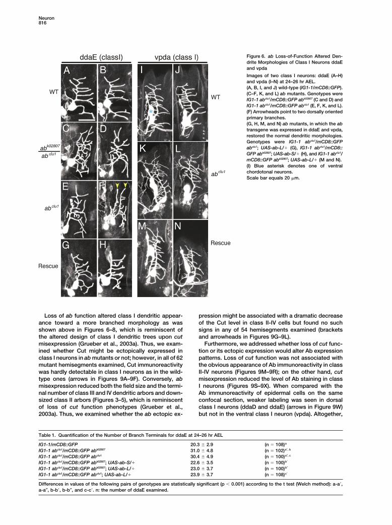
In the wild-type at 24–26 hr AEL (about 2–6 hr afterhatching), each ddaE neuron had generated two or threeprimary branches and one of them extended dorsally,from which secondary branches sprout and tended toelongate posteriorly in parallel (Figures 1B, 6A, and 6B;Gao et al., 1999; Grueber et al., 2002; Sugimura et al.,2003). In contrast, ddaE in the mutant larvae did notdisplay such a comb-like design of dendritic arbors (Fig-ures 6C–6F). The mutant neurons at the comparablestage had produced supernumerary terminals (Table 1)that did not necessarily show the oriented growth. About20% of the mutant ddaE cells did not extend lateralprimary branches, and in extreme cases, a single mutantddaE extended two primary branches dorsally instead(arrowheads in Figure 6F). Similar pattern defects werealso seen for the ventral class I neuron vpda (Figures6I–6L).
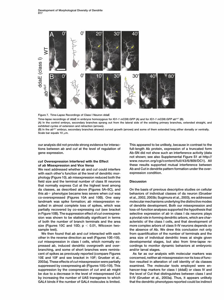
Next, we examined early stages of branch formationin the wild-type and mutant embryos (Figure 7). As re-Figure 4. Poor Response of the ab-Misexpressing Class IV Neuronported previously, each wild-type ddaE elongates a pri-ddaC to the Loss of Isoneuronal or Heteroneuronal Branchesmary branch in the dorsal direction, and then secondaryTime-lapse analysis of ddaC in response to severing of branches.
Dendrite shafts of control ddaC (A and B) and ab-misexpressing branches spring out and the stabilized ones tend toddaC (C and D) were destroyed at 25 hr AEL (yellow arrows in [A] elongate in a straight path (arrows in Figure 7A; n �and [C]) and observed one day later (B and D). 5). In ab mutants, secondary branches appeared with(A and B) Other isoneuronal branches of the control ddaC and heter-
curved morphologies and remained associated with fineoneuronal branches of adjacent ddaC filled in the voided field exten-higher-order branches that were frequently oriented insively (blue arrowhead and magenta arrowheads, respectively).dorsal or ventral directions (arrows in Figure 7B; n � 10).(C and D) Neither remaining isoneuronal branches (blue arrowheads)
nor heteroneuronal branches of the ab-misexpressing ddaC filled Postmitotic expression of ab cDNA in mutant ddaEin the voided field (box in [D]) that would have been covered by the and vpda rescued the abnormal pattern formation ofsevered branches. Genotypes were as described in the legend of dendrites with respect to both the directional elongationFigure 2.
and the terminal number (Figures 6G, 6H, 6M, and 6NScale bars equal 50 �m.and Table 1). Because this GAL4 insertion did not drivetransgene expression in epidermal cells or muscles thatmight make contact with class I dendrites, this signifi-a GAL4 driver NP2225 specific to class I neurons (datacant rescue suggested the possibility that ab was neces-not shown).sary for proper dendritegenesis of class I neurons in acell-autonomous fashion (see also our results ofIn ab Loss-of-Function Mutants, Class I daMARCM analysis next). As concerns our staining of epi-Neurons Lost Their Characteristic Comb-likedermal cells and muscles and observation of larval den-Arbor Patterns and Produced Supernumeraryticles, we did not detect obvious patterning defects inDendritic Branchesthose tissues in the ab mutants examined (data notThe class I-selective expression of ab and the resultsshown).of the misexpression studies led us to speculate that
Ab might play some important roles in dendritic morpho-genesis of class I neurons (ddaD, ddaE, and vpda) that MARCM Analysis Showed that ab Mutations
Resulted in an Increase in the Terminalnormally express ab. We pursued this possibility by visu-alizing morphologies of class I neurons that were mu- Number and the Territory Size
The above studies supported a regulatory role for Ab intants for the ab locus. We employed two strong alleles:one was abclu1, which is a nonsense mutation terminating controlling dendritegenesis of class I neurons. Neverthe-
less, the early larval lethality of the mutants limited ourthe polypeptide between the BTB domain and zinc fin-gers (Hu et al., 1995; Supplemental Figure S1 at http:// analysis to examine cellular phenotypes at later, third
instar stages of larval development when dendritic pat-www.neuron.org/cgi/content/full/43/6/809/DC1), andthe other was abk02807, a P insertion allele (Johannes and terns are becoming mature. We therefore generated and
labeled ab mutant neurons in otherwise heterozygousPreiss, 2002), which reduces Ab expression to thethreshold level of our detection by immunostaining (data mature larvae with the mosaic analysis with a repressible
cell marker (MARCM; Lee and Luo, 1999). Dendriticnot shown). Animals of abclu1 homozygotes or abclu1/abk02807 transheterozygotes hatched and grew normally arbors of both ddaD and ddaE (class I neurons) in ab�
Development of Morphological Diversity of Dendrite815
Figure 5. Effects of ab Transgene Expressionon Dendrite Morphology of Class III ddaA,Class II ddaB, and Class I ddaD and ddaE
Single-cell analysis of a class III ddaA (A–C),a class II ddaB (D–J), and class I ddaD andddaE (K and L).(A, B, D–F) Morphologies of control ddaA (A)and ddaB (D) or ab-misexpressing ddaA (B)and ddaB (E and F). All the dorsal da neurons,except for the cells of interest, were ablatedat 24–26 hr AEL; the remaining cells were im-aged at around 50 hr AEL.(A and B) Short protrusions or “spikes,” whichare characteristics of class III (brackets in [A]),were not formed in the ab-misexpressingddaA (B). Yellow arrow indicates sparse andshorter dendritic branches that elongatedfrom the lateral cluster.(D–F) The ab-misexpressing ddaB formed asmaller dendritic field than the control cell.Arrowheads indicate the most distal tips ofthe dorsal branches (see H). Blue arrow in (D)points an overlap of a branch of the ddaBand that from the anterior hemisegment.Scale bar equals 50 �m for (A), (B), and (D)–(F).(C and G–L) Quantitative analysis of controlneurons and ab-misexpressing homologouscells. Numbers of neurons analyzed are indi-cated above individual bars.(C, G, and K) Area size for ddaA (C), ddaB(G), and ddaD and ddaE (K).(H) Distance from the most distal tip of dorsalbranches to the center of the ddaB cell body.As for ddaB, a value of the size ratio of (ab-misexpressing cell)/(control cell) was 0.42and that of the distance ratio was 0.66.(I, J, and L) Number of branch terminals forddaB (I and J) and for ddaD and ddaE (L).The number of all branches (I) and that of asubset (J) of ddaB were counted (see detailsin the text). Asterisks in (C), (G), and (H) repre-sent p � 0.001 (Wilcoxon two-sample test).Genotypes were as described in the legendof Figure 2.
clones took on abnormal shape and produced branch lack of apparent abnormal phenotypes in these classeswas consistent with the selective ab expression and itsterminals in excess (Figures 8A–8H) as shown in ab
mutant young larvae. This result is consistent with the role in dendritic morphogenesis of class I neurons.notion that the phenotypes of class I neurons in theyoung mutant larvae were not secondary consequences Neither ab Loss of Function nor Its Misexpression
Was Associated with Alteration of cutof defective epidermal or muscle development.Under the standard protocol of MARCM analysis, em- Expression Pattern
It was earlier shown that Cut, a homeodomain protein,bryos were collected within a short time window anddeveloped for a fixed duration; still third instar larvae is expressed differentially among the four classes of
da neurons and that those different expression levelsgrew to different body size when clones were found andimaged. To assess whether loss of ab function affected control both the field size and the branching complexity
of individual classes (Grueber et al., 2003a). The levelthe territory size of dendritic arbors in these samples,we normalized the arbor size to the size of the segment of Cut is the strongest in class III, moderate in class II
and class IV, and below the limit of detection in class I,where each clone was generated (Figures 8C and 8F, seedetails in the legend). We found a statistically significant which provides a contrast to that of Ab. Besides the
expression profiles in da neuron classes, activities ofincrease in the area size of the ab mutant ddaD (p �0.01, Wilcoxon two-sample test), but not in that of the these two genes are approximately complementary to
each other with respect to gain-of-function and loss-of-ab mutant ddaE.da neurons of class II to IV did not express Ab at a function phenotypes of dendritic patterns (described
below). Therefore, we explored the possibility that abdetectable level, and we did not find obvious patterndefects of dendritic arbors of class II (n � 8), class III loss of function or its misexpression might alter the Cut
expression pattern.(n � 13), and class IV (n � 14, Figures 8I and 8J). The
Neuron816
Figure 6. ab Loss-of-Function Altered Den-drite Morphologies of Class I Neurons ddaEand vpda
Images of two class I neurons: ddaE (A–H)and vpda (I–N) at 24–26 hr AEL.(A, B, I, and J) wild-type (IG1-1/mCD8::GFP).(C–F, K, and L) ab mutants. Genotypes wereIG1-1 abclu1/mCD8::GFP abk02807 (C and D) andIG1-1 abclu1/mCD8::GFP abclu1 (E, F, K, and L).(F) Arrowheads point to two dorsally orientedprimary branches.(G, H, M, and N) ab mutants, in which the abtransgene was expressed in ddaE and vpda,restored the normal dendritic morphologies.Genotypes were IG1-1 abclu1/mCD8::GFPabclu1; UAS-ab-L/� (G), IG1-1 abclu1/mCD8::GFP abk02807; UAS-ab-S/� (H), and IG1-1 abclu1/mCD8::GFP abk02807; UAS-ab-L/� (M and N).(I) Blue asterisk denotes one of ventralchordotonal neurons.Scale bar equals 20 �m.
Loss of ab function altered class I dendritic appear- pression might be associated with a dramatic decreaseof the Cut level in class II-IV cells but found no suchance toward a more branched morphology as was
shown above in Figures 6–8, which is reminiscent of signs in any of 54 hemisegments examined (bracketsand arrowheads in Figures 9G–9L).the altered design of class I dendritic trees upon cut
misexpression (Grueber et al., 2003a). Thus, we exam- Furthermore, we addressed whether loss of cut func-tion or its ectopic expression would alter Ab expressionined whether Cut might be ectopically expressed in
class I neurons in ab mutants or not; however, in all of 62 patterns. Loss of cut function was not associated withthe obvious appearance of Ab immunoreactivity in classmutant hemisegments examined, Cut immunoreactivity
was hardly detectable in class I neurons as in the wild- II-IV neurons (Figures 9M–9R); on the other hand, cutmisexpression reduced the level of Ab staining in classtype ones (arrows in Figures 9A–9F). Conversely, ab
misexpression reduced both the field size and the termi- I neurons (Figures 9S–9X). When compared with theAb immunoreactivity of epidermal cells on the samenal number of class III and IV dendritic arbors and down-
sized class II arbors (Figures 3–5), which is reminiscent confocal section, weaker labeling was seen in dorsalclass I neurons (ddaD and ddaE) (arrows in Figure 9W)of loss of cut function phenotypes (Grueber et al.,
2003a). Thus, we examined whether the ab ectopic ex- but not in the ventral class I neuron (vpda). Altogether,
Table 1. Quantification of the Number of Branch Terminals for ddaE at 24–26 hr AEL
IG1-1/mCD8::GFP 20.3 � 2.9 (n � 108)a
IG1-1 abclu1/mCD8::GFP abk02807 31.0 � 4.8 (n � 102)a�, b
IG1-1 abclu1/mCD8::GFP abclu1 30.4 � 4.9 (n � 100)a″, c
IG1-1 abclu1/mCD8::GFP abk02807; UAS-ab-S/� 22.6 � 3.5 (n � 100)b�
IG1-1 abclu1/mCD8::GFP abk02807; UAS-ab-L/� 23.0 � 3.7 (n � 100)b″
IG1-1 abclu1/mCD8::GFP abclu1; UAS-ab-L/� 23.9 � 3.7 (n � 108)c�
Differences in values of the following pairs of genotypes are statistically significant (p � 0.001) according to the t test (Welch method): a-a�,a-a″, b-b�, b-b″, and c-c�. n: the number of ddaE examined.
Development of Morphological Diversity of Dendrite817
Figure 7. Time-Lapse Recordings of Class I Neuron ddaE
Time-lapse recordings of ddaE in embryos homozygous for IG1-1 mCD8::GFP (A) and for IG1-1 mCD8::GFP abclu1 (B).(A) In the control embryo, secondary branches sprang out from the lateral side of the existing primary branches, extended straight, andexhibited cycles of extension and retraction (arrows).(B) In the abclu1 embryo, secondary branches showed curved growth (arrows) and some of them extended long either dorsally or ventrally.Scale bar equals 10 �m.
our analysis did not provide strong evidence for interac- This appeared to be unlikely, because in contrast to thefull-length Ab protein, expression of a truncated formtions between ab and cut at the level of regulation of
gene expression. Ab-SN did not show such an interference activity (datanot shown; see also Supplemental Figure S1 at http://www.neuron.org/cgi/content/full/43/6/809/DC1). Allcut Overexpression Interfered with the Effectthese results supported mutual interference betweenof ab Misexpression and Vice VersaAb and Cut in dendrite pattern formation under the over-We next addressed whether ab and cut could interfereexpression condition.with each other’s function at the level of dendritic mor-
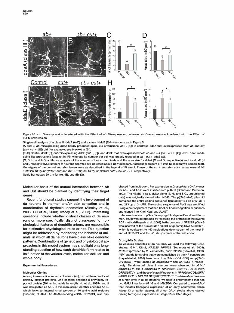
phology (Figure 10). ab misexpression reduced both thefield size and the terminal number of class III neurons Discussionthat normally express Cut at the highest level amongda classes, as described above (Figures 5A–5C), and On the basis of previous descriptive studies on cellular
behaviors of individual classes of da neuron (Grueberthis ab� phenotype became less severe when cut wasco-overexpressed (Figures 10A and 10B). One easy et al., 2002, 2003b; Sugimura et al., 2003), we addressed
molecular mechanisms underlying the distinctive modeslandmark was spike formation; ab misexpression re-sulted in almost complete loss of spikes, which was of dendrite development. Both our misexpression and
loss-of-function analyses supported the hypothesis thatpartially recovered by co-expressing cut (see bracketin Figure 10B). The suppression effect of cut overexpres- selective expression of ab in class I da neurons plays
a pivotal role in forming dendritic arbors, which are char-sion was shown to be statistically significant in termsof both the number of branch terminals and the area acteristic of the class I cells, and that development of
more complex arbors of class II-IV neurons depends onsize (Figures 10C and 10D; p � 0.01, Wilcoxon two-sample test). the absence of Ab. We drew this conclusion not only
from quantification of the number of terminals and theWe then found that ab and cut interacted with eachother in the reverse direction as well (Figures 10E–10I). area size of individual dendritic trees at single given
developmental stages, but also from time-lapse re-cut misexpression in class I cells, which normally ex-pressed ab, induced dendritic overgrowth and over- cordings to monitor dynamic behaviors at embryonic
and/or larval stages.branching, and some of short branches were reminis-cent of spikes as previously reported (compare Figures As far as our analysis with molecular markers was
concerned, neither ab misexpression nor its loss of func-10E and 10F and see bracket in 10F; Grueber et al.,2003a). These effects of cut misexpression were partially tion resulted in alteration of cell identity of da classes
examined. The molecular tools employed were en-suppressed by coexpressing ab (Figures 10G–10I). Thissuppression by the coexpression of cut and ab might hancer-trap markers for class I (ddaE) or class IV and
the level of Cut that distinguishes between class I andbe due to a decrease in the level of misexpressed Cutby increasing the number of UAS transgenes to which II-IV (Grueber et al., 2003a). Thus, it appears unlikely
that the dendritic phenotypes reported could be indirectGAL4 binds if the number of GAL4 molecules is limited.

Neuron818
Figure 8. MARCM Analysis of ab MutantClones
(A, B, D, E, G–J) MARCM clones of ddaD (Aand B), ddaE (D, E, G, and H), and ddaC (Iand J). Wild-type clones (A, D, G, and I),abk02807 clones (B, E, and H), and abclu1 clone(J). (B and E) abk02807 class I clones formeddeformed comb-like arbors that should haveconsisted of laterally oriented secondarybranches.(C and F) Numbers of branch terminals andnormalized territory size of individual arborswere plotted. Blue diamonds represent wtclones; yellow triangles indicate ab mutantclones. To normalize the arbor size in larvaeof different body size, the size of each poly-gon connecting dendritic tips was divided bythe square of the distance between the cellbody and the landmark denticle (relative unit,RU). The increase in both the terminal numberand the arbor size was statistically significantfor ddaD and increase in the terminal number,for ddaE (p � 0.01, Wilcoxon two-sampletest).(G and H) Tracings of wild-type ddaE clone (G)and abk02807 clones (H) that were normalized interms of body size. Bars, 100 �m for (A), (B),(D), (E), (I), and (J).
consequences of cell identity alteration; we would prefer with the notion that Ab endows every feature of class Idendritic patterning.an alternative hypothesis that Ab is more immediately
involved in regulating dendritic morphology through Although our manipulations of ab misexpression causedsevere and reproducible phenotypes, they did not nec-transcriptional regulation of its target genes.
ab misexpression decreased the number of branch essarily provide evidence for almost complete morpho-logical transformation from classes II-IV into class I neu-terminals of class III and IV neurons, and conversely, ab
mutant class I cells produced supernumerary branches. rons. Class II, III, or IV neurons that had misexpressedab was morphologically recognized as such, in termsOne possible interpretation of these results would be
that Ab may negatively regulate dendritic branching in a of the number and the direction of dendritic shafts thatgrew out of each cell body and the branching patternbroad range of neuronal types. However, this hypothesis
would be difficult to reconcile with our following find- within the region proximal to the soma. Previous time-lapse recordings showed that da neurons of class I andings: in normal development, the territory size of class
I dendrites is smaller than that of class II ones, while the class IV use distinct strategies from the very beginningof dendritic birth from the soma that contribute to differ-dendritic trees of both classes are similarly complicated
(Grueber et al., 2002). ab misexpression in class II neu- ences in their basic arbor patterns (Sugimura et al.,2003). The partial alteration of the arbor patterns by abrons reduced the territory size, but the terminal number
did not change significantly. Furthermore, ab misex- misexpression might be due to a late onset and/or alow level of ab transgene expression obtained by usingpression in any da neuron of class II-IV in the dorsal
cluster reduced the size and/or the terminal number to the available postmitotic drivers.In contrast to the formation of supernumerary branchvalues that were comparable to those of class I neurons
of the same cluster. The easiest interpretation of these terminals of class I neurons in the ab mutants, the samecells did not show obvious expansion of the arbor sizeresults would be that the misexpression morphologically
altered class II-IV dendrites toward that of class I. It compared with the control cells; this could be due tothe possibility that the expansion, if any, was too smallshould be also noted that dendritic patterns are defined
not only by the numerical parameters such as the termi- to be detected at the early larval stage when differencesin the field size of class I and that of other classes werenal number or the territory size, but also by other proper-
ties such as the comb-like design of class I, spike protru- subtle compared with those at late larval stages. Weperformed MARCM analysis to explore phenotypes atsion of class III, and mutual avoidance of class IV. Effects
of ab loss of function or misexpression were consistent late larval stages and showed that ddaD increased in

Development of Morphological Diversity of Dendrite819
its arbor size, but that another class I neuron examined,ddaE, did not. This variation could be explained by per-durance of the wild-type protein in each mutant cell(Lee and Luo, 1999). Alternatively, dysfunction of Ab-dependent mechanisms might not have been sufficientfor expansion of the territorial field, and an additionalmechanism, which works in classes II-IV neurons in nor-mal development, may be required.
The Ab protein has two zinc fingers of the C2H2 class,which is one of the most common types of DNA bindingdomains; in addition, a BTB/POZ domain is found at theN terminus of the fingers (Hu et al., 1995). The BTB/POZdomain is an evolutionarily conserved protein-proteininteraction domain, and BTB/POZ domains of severalzinc finger proteins, such as PLZF and Tramtrack, havebeen shown to be responsible for transcriptional regula-tion (Zollman et al., 1994; Wong and Privalsky, 1998).Recent studies have discovered several putative tran-scriptional factors of other families that regulate mor-phological heterogeneity of dendrites including branchingcomplexity, field size, and targeting specificity in differ-ent model systems, suggesting that transcriptional reg-ulation is a common mechanism to generate morpholog-ical diversity of dendrites (Moore et al., 2002; Grueberet al., 2003a; Komiyama et al., 2003; Grueber and Jan,2004). Therefore, it is likely that class-specific profilesof gene expression controlled by these factors are re-sponsible for distinctive dendritic morphogenesis. Tar-get genes of these transcriptional regulators in the con-text of dendritic pattern formation have not yet beenfound, and their future identification should give us de-tailed pictures of the molecular machineries at work.
Ab and Cut provided striking contrasts to each otherin terms of class-dependent levels of immunoreactivityand, furthermore, gain-of-function and loss-of-functionphenotypes of dendritic morphology (Grueber et al.,2003a, and this study). We found that neither ab lossof function nor its misexpression was associated withFigure 9. ab Mutations or ab Ectopic Expression Did Not Alter Cut
Expression Pattern alteration of cut expression, which did not provide evi-dence for a simple epistatic or mutually dependent rela-(A–F) The wild-type (A–C) and abclu1 homozygous embryos (D–F)
were stained for a panneuronal marker Elav (A and D, green in C tionship between the two genes at the level of geneand F) and for Cut (B and E, magenta in C and F). In the ab mutant, expression. It could be that selective expression of AbCut immunoreactivity was hardly detected in class I neurons as in and that of Cut are operated by a mechanism that isthe wild-type (arrows in A, C, D and F). The same result was obtained
separate, at least partially. When the two putative tran-for abk02807 homozygotes (data not shown).scription factors were examined at the level of dendritic(G–L) Control embryo (elav-GAL4 UAS-mCD8::GFP/� or Y; [G]–[I])
and the one that misexpressed ab in all neurons (elav-GAL4 UAS- morphology as a final read-out, we found that they couldmCD8::GFP/� or Y; UAS-ab-L/�; [J]–[L]) were stained for GFP (G interfere with each other’s function upon overexpres-and J, green in I and L) and for Cut (H and K, magenta in I and L). sion, which argues against a simple epistatic relation-ab misexpression did not result in a dramatic decrease in the level ship between them. A couple of possibilities could ex-of Cut staining or obvious alteration of the number of Cut-positive
plain this mutual interaction. For example, target genescells in the cluster. Compared with the pan-da drivers that wereof Ab and those of Cut may be partially overlapped, andused in ectopic expression in Figures 2–5, elav-GAL4, which was
used in this experiment, initiates transgene expression earlier in the interference may be due to competitions betweenpostmitotic neurons. Embryos of elav-GAL4 UAS-mCD8::GFP/� or the two for cis regulatory elements of the same targetY; UAS-ab-L/� did not hatch, so they could not be used for analysis gene. Alternatively, Ab’s targets and those of Cut mayof larval dendritic development. operate on cytoskeletal reorganization in different ways.(M–R) The wild-type (M–O) and ctdb3 hemizygous embryos (P–R) werestained for Elav (M and P, green in O and R) and Ab (N and Q,magenta in O and R). No other neurons were labeled as brightly asclass I neurons in the wild-type and in the ct mutant (arrows). The
arrows. (W) Two dorsal class I neurons showed a decrease in thesame result was obtained for ctc145 hemizygotes (data not shown).(S–X) Control embryo (S–U) and one that misexpressed ct in all level of Ab immunoreactivity when compared with that of epidermal
cells in the same confocal image.neurons (V–X) were stained for GFP (S and V, green in U and X) andfor Ab (T and W, magenta in U and X). Genotypes were elav-GAL4 Throughout this figure, images of every pair of the control and the
mutant or the ab misexpressing embryo were acquired and pro-UAS-mCD8::GFP/� or Y (S–U) and elav-GAL4 UAS-mCD8::GFP/�or Y; UAS- cut5/� (V–X). Nuclei of class I neurons are indicated with cessed with identical parameters. Scale bar equals 10 �m.
Neuron820
Figure 10. cut Overexpression Interfered with the Effect of ab Misexpression, whereas ab Overexpression Interfered with the Effect ofcut Misexpression
Single-cell analysis of a class III ddaA (A–D) and a class I ddaE (E–I) was done as in Figure 5.(A and B) ab-misexpressing ddaA hardly produced spike-like protrusions (ab�, [A]); in contrast, ddaA that overexpressed both ab and cut(ab� cut�, [B]) did (for example, see bracket in [B]).(E–G) Control ddaE (E), cut-misexpressing ddaE (cut�, [F]), and ddaE that overexpressed both ab and cut (ab� cut�, [G]). cut� ddaE madespike-like protrusions (bracket in [F]), whereas its number per cell was greatly reduced in ab� cut� ddaE (G).(C, D, H, and I) Quantitative analysis of the number of branch terminals and the area size for ddaA (C and D, respectively) and for ddaE (Hand I, respectively). Numbers of neurons analyzed are indicated above individual bars. Asterisks represent p � 0.01 (Wilcoxon two-sample test).Genotypes of the control and ab� larvae were as described in the legend of Figure 2. Those of the cut� and ab� cut� larvae were IG1-2109(2)80 GFP[S65T]/UAS-cut5 and IG1-2 109(2)80 GFP[S65T]/UAS-cut5; UAS-ab-S/�, respectively.Scale bar equals 50 �m for (A), (B), and (E)–(G).
chased from Invitrogen. For expression in Drosophila, cDNA clonesMolecular basis of the mutual interaction between Abfor Ab-L and Ab-S were inserted into pUAST (Brand and Perrimon,and Cut should be clarified by identifying their target1993). The NBab7-1 ab-L cDNA clone (S. Hu and S.C., unpublishedgenes.data) was originally cloned into pNB40. The p[UAS-ab-L] plasmid
Recent functional studies support the involvement of contained the entire coding sequence flanked by 164 bp of 5�-UTRda neurons in thermo- and/or pain sensation and in and 213 bp of 3�-UTR. The coding sequence of Ab-S was amplified
using a pair of primers that had XhoI or XbaI recognition sequencescoordination of rhythmic locomotion (Ainsley et al.,and cloned into XhoI-XbaI-cut pUAST.2003; Liu et al., 2003; Tracey et al., 2003). Interesting
An insertion site of pGawB carrying GAL4 gene (Brand and Perri-questions include whether distinct classes of da neu-mon, 1993) was determined by following the protocol of the inverserons or, more specifically, distinct class-specific mor-PCR method (Hayashi et al., 2002). In the genome of NP2225, pGawB
phological features of dendritic arbors, are responsible was inserted at the nucleotide 153,801 of genomic DNA AE003631,for distinctive physiological roles or not. This question which is equivalent to 463 nucleotides downstream of the most 5�
end of RE25924 and to �21 kb upstream of the first codon.might be addressed by monitoring the behavior of ani-mals, in which all da neurons have class I-like dendritic
Drosophila Strainspatterns. Combinations of genetic and physiological ap-To visualize dendrites of da neurons, we used the following GAL4proaches in this model system may shed light on a long-strains: IG1-1, IG1-2, NP2225, NP7028 (Sugimura et al., 2003),
standing question of how each dendritic form relates to NP1161 (provided by M. Yamamoto), and 109(2)80 (Gao et al., 1999).its function at the various levels, molecular, cellular, and “NP” stands for strains that were established by the NP consortium
(Hayashi et al., 2002). Insertions of p[UAS- mCD8::GFP] and p[UAS-whole body.GFP(S65T)] were labeled as mCD8::GFP and GFP[S65T], respec-tively. Dendrites of class I neurons were observed in IG1-1/Experimental ProceduresmCD8::GFP, IG1-1 mCD8::GFP, NP2225/mCD8::GFP, or NP2225GFP[S65T]/�; and those of class IV neurons, in NP7028 mCD8::GFP/Molecular Cloning
Among known splice variants of abrupt (ab), two of them produced mCD8::GFP or NP1161 GFP[S65T]/NP1161. To drive ab expressionat a high level in all da neurons, we used a chromosome that haspartially distinct proteins. One of them encodes a previously re-
ported protein (904 amino acids in length; Hu et al., 1995), and it two GAL4 insertions (IG1-2 and 109(2)80). Compared to elav-GAL4that initiates transgene expression at an early postmitotic phasewas designated as Ab-L in this manuscript. Another encodes Ab-S,
which lacks an internal small portion of 10 amino acid residues (stage 13 or earlier stages), all of our GAL4 stocks above starteddriving tarnsgene expression at stage 15 or later stages.(356–367) of Ab-L. An Ab-S-encoding cDNA, RE25924, was pur-
Development of Morphological Diversity of Dendrite821
abclu1 is a nonsense mutation that terminates the polypeptide at Foundation (Japan) for the Promotion of Science; and from theNational Science Foundation to S.C.residue 531 of Ab-L (Vactor et al., 1993; Hu et al., 1995; Supplemental
Figure S1 at http://www.neuron.org/cgi/content/full/43/6/809/DC1).abk02807 is a P insertion allele (Johannes and Preiss, 2002). Each of Received: April 23, 2004the ab alleles was recombined with FRT40A for MARCM analysis, Revised: July 15, 2004and we mated the following pairs: females of elav-GAL4 UAS- Accepted: August 10, 2004mCD8::GFP hsFLP; tub-Gal80 FRT40A to males of FRT40A/FRT40A Published: September 15, 2004to generate control ab� clones and to males of ab FRT40A/CyO togenerate ab mutant clones. To image clones, we mounted each Referenceslarva in 90% glycerol in PBS on a slide between spacers made ofvinyl tapes. Other strains used were null mutants of cut (ctdb3 and Ainsley, J.A., Pettus, J.M., Bosenko, D., Gerstein, C.E., Zinkevich,ctc145; Blochlinger et al., 1988) and UAS-cut5 (Hardiman et al., 2002; N., Anderson, M.G., Adams, C.M., Welsh, M.J., and Johnson, W.A.remobilized by C. Micchelli). All fly embryos and larvae were grown (2003). Enhanced locomotion caused by loss of the Drosophila DEG/at 25C. ENaC protein Pickpocket1. Curr. Biol. 13, 1557–1563.
Blochlinger, K., Bodmer, R., Jack, J., Jan, L.Y., and Jan, Y.N. (1988).Image Collection of Dendritic Trees Primary structure and expression of a product from cut, a locusEffects of ab overexpression on dendritic morphogenesis were visu- involved in specifying sensory organ identity in Drosophila. Naturealized at the single-cell level as follows: In each experiment, a stage 333, 629–635.17 embryo or an anesthetized larva at 24–26 hr AEL was mounted, Blochlinger, K., Bodmer, R., Jan, L.Y., and Jan, Y.N. (1990). Patternsand individual da neurons in the dorsal cluster were identified under of expression of cut, a protein required for external sensory organfluorescence microscope (Olympus) on the basis of their stereotypic development in wild-type and cut mutant Drosophila embryos.arrangement and the shape of cell body (see details in the legend Genes Dev. 4, 1322–1331.of Figures 2E and 2F). All neurons, except for a single da neuron of
Bodmer, R., and Jan, Y.N. (1987). Morpohological differentiation ofinterest and dorsal bipolar neuron, were ablated by using Micropointthe embryonic peripheral neurons in Drosophila. Rouxs Arch. Dev.(Photonics Instruments). We selected dorsal clusters of abdominalBiol. 196, 69–77.segments 2–6 for this experiment. Following the ablations, we keptBrand, A.H., and Perrimon, N. (1993). Targeted gene expression aswatching the embryos, and as soon as they hatched, individuala means of altering cell fates and generating dominant phenotypes.larvae were returned to yeasted apple plates with forceps, gentlyDevelopment 118, 401–415.cleaned of oil, and allowed to develop. Those larvae were anesthe-
tized at subsequent stages and images of their dendritic trees were Campos-Ortega, J.A., and Hartenstein, V. (1997). The Embryoniccollected under a confocal microscope (BioRad). Details of the Development of Drosophila melanogaster (Berlin: Springer).above and other procedures (branch severing of da neurons and Gao, F.B., Brenman, J.E., Jan, L.Y., and Jan, Y.N. (1999). Genestime-lapse analysis) were as described (Sugimura et al., 2003; see regulating dendritic outgrowth, branching, and routing in Drosoph-also Grueber et al., 2003b). ila. Genes Dev. 13, 2549–2561.
Gao, F.B., Kohwi, M., Brenman, J.E., Jan, L.Y., and Jan, Y.N. (2000).Quantification Control of dendritic field formation in Drosophila: the roles of fla-Quantitative or semiquantitative analysis was performed essentially mingo and competition between homologous neurons. Neuronas previously described (Grueber et al., 2002). Each dendritic field 28, 91–101.was delineated by a polygon connecting dendritic tips, and its size Grueber, W.B., and Jan, Y.N. (2004). Dendritic development: lessonswas calculated by using Laser Sharpe (BioRad). Total length of from Drosophila and related branches. Curr. Opin. Neurobiol. 14,dendritic branches was measured by using Axiocam ver 3.1 (Zeiss). 74–82.Dendritic branches and their tips of ddaA, ddaB, and ddaC were
Grueber, W.B., Jan, L.Y., and Jan, Y.N. (2002). Tiling of the Drosoph-identified by monitoring individual Z-series files when necessary,ila epidermis by multidendritic sensory neurons. Developmentand the tip number per cell was counted (for ddaB, see details in129, 2867–2878.Results). As regards control ddaA and ddaC, it was sometimesGrueber, W.B., Jan, L.Y., and Jan, Y.N. (2003a). Different levels ofdifficult to pinpoint the very ends of dendritic tips at segmentalthe homeodomain protein cut regulate distinct dendrite branchingboundaries, so we measured the length of each branch between apatterns of Drosophila multidendritic neurons. Cell 112, 805–818.branching point and a distal-most position that we could identify.
Thus, the territory size and the number of dendritic branches were Grueber, W.B., Ye, B., Moore, A.W., Jan, L.Y., and Jan, Y.N. (2003b).underestimated for the control cells; nevertheless, ab ectopic ex- Dendrites of distinct classes of Drosophila sensory neurons showpression caused statistically significant differences (Figures 3G and different capacities for homotypic repulsion. Curr. Biol. 13, 618–626.5C). Data are presented as means � SD. Hardiman, K.E., Brewster, R., Khan, S.M., Deo, M., and Bodmer, R.
(2002). The bereft gene, a potential target of the neural selector genecut, contributes to bristle morphogenesis. Genetics 161, 231–247.Immunochemistry
Whole-mount embryos and dissected larvae were stained according Hausser, M., and Mel, B. (2003). Dendrites: bug or feature? Curr.to standard protocols with the following primary antibodies: rabbit Opin. Neurobiol. 13, 372–383.anti-Abrupt (Hu et al., 1995), mouse anti-Cut (Blochlinger et al., 1990; Hayashi, S., Ito, K., Sado, Y., Taniguchi, M., Akimoto, A., Takeuchi,2B10 from Developmental Studies Hybridoma Bank at the University H., Aigaki, T., Matsuzaki, F., Nakagoshi, H., Tanimura, T., et al. (2002).of Iwoa), rat anti-Elav (7E8A10; Developmental Studies Hybridoma GETDB, a database compiling expression patterns and molecularBank), mouse anti-GFP (Sigma), and rabbit anti-GFP (Molecular locations of a collection of gal4 enhancer traps. Genesis 34, 58–61.Probes).
Hu, S., Fambrough, D., Atashi, J.R., Goodman, C.S., and Crews,S.T. (1995). The Drosophila abrupt gene encodes a BTB-zinc finger
Acknowledgments regulatory protein that controls the specificity of neuromuscularconnections. Genes Dev. 9, 2936–2948.
We thank Yuh Nung Jan, Wes Grueber, Susan Younger, RolfJan, Y.N., and Jan, L.Y. (1993). The peripheral nervous system. In The
Bodmer, the Bloomington Stock Center, and Drosophila GeneticDevelopment of Drosophila melanogaster, M. Bate and A. Martinez-
Resource Center (Kyoto) for Drosophila stocks, and Kazuo Emoto,Arias, eds. (Cold Spring Harbor, NY: Cold Spring Harbor Laboratory
Toshihiko Fujimori, Yasuyuki Shima, and Kei Ito for technical advicePress), pp. 1207–1244.
and encouragement. We are also grateful to Developmental StudiesJan, Y.N., and Jan, L.Y. (2003). The control of dendrite development.Hybridoma Bank for antibodies. This work was supported by grantsNeuron 40, 229–242.to T.U. from Core Research for Evolutional Science and Technology;
from the Ministry of Science, Culture, and Education; from Toray Johannes, B., and Preiss, A. (2002). Wing vein formation in Drosoph-
Neuron822
ila melanogaster: hairless is involved in the cross-talk betweenNotch and EGF signaling pathways. Mech. Dev. 115, 3–14.
Komiyama, T., Johnson, W.A., Luo, L., and Jefferis, G.S. (2003).From lineage to wiring specificity. POU domain transcription factorscontrol precise connections of Drosophila olfactory projection neu-rons. Cell 112, 157–167.
Lee, T., and Luo, L. (1999). Mosaic analysis with a repressible cellmarker for studies of gene function in neuronal morphogenesis.Neuron 22, 451–461.
Liu, L., Yermolaieva, O., Johnson, W.A., Abboud, F.M., and Welsh,M.J. (2003). Identification and function of thermosensory neuronsin Drosophila larvae. Nat. Neurosci. 6, 267–273.
Mainen, Z.F., and Sejnowski, T.J. (1996). Influence of dendritic struc-ture on firing pattern in model neocortical neurons. Nature 382,363–366.
Masland, R.H. (2001). Neuronal diversity in the retina. Curr. Opin.Neurobiol. 11, 431–436.
Moore, A.W., Jan, L.Y., and Jan, Y.N. (2002). hamlet, a binary geneticswitch between single- and multiple-dendrite neuron morphology.Science 297, 1355–1358.
Schaefer, A.T., Larkum, M.E., Sakmann, B., and Roth, A. (2003).Coincidence detection in pyramidal neurons is tuned by their den-dritic branching pattern. J. Neurophysiol. 89, 3143–3154.
Scott, E.K., and Luo, L. (2001). How do dendrites take their shape?Nat. Neurosci. 4, 359–365.
Sugimura, K., Yamamoto, M., Niwa, R., Satoh, D., Goto, S., Tani-guchi, M., Hayashi, S., and Uemura, T. (2003). Distinct develop-mental modes and lesion-induced reactions of dendrites of twoclasses of Drosophila sensory neurons. J. Neurosci. 23, 3752–3760.
Tracey, W.D., Jr., Wilson, R.I., Laurent, G., and Benzer, S. (2003).painless, a Drosophila gene essential for nociception. Cell 113,261–273.
Vactor, D.V., Sink, H., Fambrough, D., Tsoo, R., and Goodman, C.S.(1993). Genes that control neuromuscular specificity in Drosophila.Cell 73, 1137–1153.
Vetter, P., Roth, A., and Hausser, M. (2001). Propagation of actionpotentials in dendrites depends on dendritic morphology. J. Neuro-physiol. 85, 926–937.
Whitford, K.L., Dijkhuizen, P., Polleux, F., and Ghosh, A. (2002).Molecular control of cortical dendrite development. Annu. Rev. Neu-rosci. 25, 127–149.
Williams, D.W., and Truman, J.W. (2004). Mechanisms of dendriticelaboration of sensory neurons in Drosophila: insights from in vivotime lapse. J. Neurosci. 24, 1541–1550.
Wong, R.O., and Ghosh, A. (2002). Activity-dependent regulation ofdendritic growth and patterning. Nat. Rev. Neurosci. 3, 803–812.
Wong, C.W., and Privalsky, M.L. (1998). Components of the SMRTcorepressor complex exhibit distinctive interactions with the POZdomain oncoproteins PLZF, PLZF-RARalpha, and BCL-6. J. Biol.Chem. 273, 27695–27702.
Zollman, S., Godt, D., Prive, G.G., Couderc, J.L., and Laski, F.A.(1994). The BTB domain, found primarily in zinc finger proteins,defines an evolutionarily conserved family that includes several de-velopmentally regulated genes in Drosophila. Proc. Natl. Acad. Sci.USA 91, 10717–10721.