Crystal structure of the proteasomal deubiquitylation module Rpn8-Rpn11
17
Crystal structure of the proteasomal deubiquitylation module Rpn8-Rpn11 Ganesh Ramnath Pathare a , István Nagy a , Pawel Sled z a , Daniel J. Anderson b , Han-Jie Zhou b , Els Pardon c,d , Jan Steyaert c,d , Friedrich Förster a , Andreas Bracher e,1 , and Wolfgang Baumeister a,1 a Department of Molecular Structural Biology, Max Planck Institute of Biochemistry, 82152 Martinsried, Germany; b Cleave Biosciences, Burlingame, CA 94010; c Structural Biology Brussels, Vrije Universiteit Brussel, 1050 Brussels, Belgium; d Structural Biology Research Center, Vlaams Instituut voor Biotechnologie, 1050 Brussels, Belgium; and e Department of Cellular Biochemistry, Max Planck Institute of Biochemistry, 82152 Martinsried, Germany Contributed by Wolfgang Baumeister, January 15, 2014 (sent for review December 20, 2013) The ATP-dependent degradation of polyubiquitylated proteins by the 26S proteasome is essential for the maintenance of proteome stability and the regulation of a plethora of cellular processes. Degradation of substrates is preceded by the removal of poly- ubiquitin moieties through the isopeptidase activity of the sub- unit Rpn11. Here we describe three crystal structures of the heterodimer of the Mpr1–Pad1–N-terminal domains of Rpn8 and Rpn11, crystallized as a fusion protein in complex with a nanobody. This fusion protein exhibits modest deubiquitylation activity to- ward a model substrate. Full activation requires incorporation of Rpn11 into the 26S proteasome and is dependent on ATP hydro- lysis, suggesting that substrate processing and polyubiquitin removal are coupled. Based on our structures, we propose that premature activation is prevented by the combined effects of low intrinsic ubiquitin affinity, an insertion segment acting as a physical barrier across the substrate access channel, and a con- formationally unstable catalytic loop in Rpn11. The docking of the structure into the proteasome EM density revealed contacts of Rpn11 with ATPase subunits, which likely stabilize the active con- formation and boost the affinity for the proximal ubiquitin moi- ety. The narrow space around the Rpn11 active site at the entrance to the ATPase ring pore is likely to prevent erroneous deubiquity- lation of folded proteins. Mpr1 | POH1 | PSMD7 | PSMD14 | JAMM protease I n eukaryotes, the ubiquitin (Ub) proteasome system (UPS) is responsible for the regulated degradation of proteins (1–5). The UPS plays a key role in the maintenance of protein ho- meostasis by removing misfolded or damaged proteins, which could impair cellular functions, and by removing proteins whose functions are no longer needed. Consequently, the UPS is criti- cally involved in numerous cellular processes, including cell cycle progression, apoptosis, and DNA damage repair, and malfunc- tions of the system often result in disease. The 26S proteasome executes the degradation of substrates that are marked for destruction by the covalent attachment of polyubiquitin chains. It is a molecular machine of 2.5 MDa comprising two subcomplexes, the 20S core particle (CP) and one or two 19S regulatory particles (RPs), which associate with the ends of the cylinder-shaped CP (6–8). The recognition and recruitment of polyubiquitylated substrates, their deubiquityla- tion, ATP-dependent unfolding, and translocation into the core particle take place in the RP. The structurally and mechanisti- cally well-characterized CP houses the proteolytic activities and sequesters them from the environment, thereby avoiding collat- eral damage (9). The RPs attach to the outer α-rings of the CP, which control access to the proteolytic chamber formed by the inner β-subunit rings (10). Recently, the molecular architecture of the 26S holo- complex was established using cryo-EM–based approaches (11, 12), and a pseudoatomic model of the holocomplex was put forward (13). The RP is formed by two subcomplexes, known as the base and the lid, which assemble independently (12, 14). The base contains the hetero-hexameric AAA-ATPase ring (Rpt1– Rpt6), which drives the conformational changes required for substrate processing, including unfolding and translocation into the CP (15, 16). The base also contains the largest RP non- ATPase subunits, Rpn1 and Rpn2, and the Ub receptor Rpn13. The second resident Ub receptor, Rpn10, is not part of either the base or the lid; it binds only to the assembled 26S proteasome and is positioned close to the ATPase module. The lid scaffold is composed of the Rpn3, Rpn5, Rpn6, Rpn7, Rpn8, Rpn9, Rpn11, and Rpn12 subunits (14). These subunits can be grouped according to their domain structures. Rpn3, Rpn5, Rpn6, Rpn7, Rpn9, and Rpn12 each comprise an N-terminal helix repeat segment, a proteasome-COP9/signalosome-eIF3 (PCI) module, and a long helix at the C terminus (8). The Rpn8 and Rpn11 subunits each consist of an Mpr1–Pad1–N-terminal (MPN) domain, followed by long C-terminal helices (Fig. 1A). The PCI subunits form a horseshoe-shaped structure and the MPN domains form a heterodimer, which are connected by a large helical bundle, to which all subunits contribute (13, 17, 18). Each of these eight subunits has paralogs in the COP9/sig- nalosome (CSN) and the elongation initiation factor 3 (eIF3), which likely adopt a similar architecture (18–21). The lid strengthens the interaction between the CP and RP (17) and deubiquitylates substrates before their processing by the AAA-ATPase module and the CP. Cleavage of polyubiquitin Significance The 26S proteasome is a multiprotein complex that degrades proteins marked for destruction by the covalent attachment of polyubiquitin chains. Proteasome activity is essential for the removal of damaged, potentially toxic proteins and for the regulation of numerous cellular processes. Multiple crystal struc- tures of the Rpn8-Rpn11 heterodimer, which is responsible for the removal of polyubiquitin tags before substrate degrada- tion in the proteasome, provide insight into how substrate unfolding and isopeptide bond cleavage might be coupled, and how premature activation of this module is prevented. Its accurate function ensures timely degradation of substrates and, ultimately, the replenishment of the limited cellular pool of free ubiquitin. Author contributions: G.R.P., I.N., H.-J.Z., F.F., A.B., and W.B. designed research; G.R.P., I.N., P. S., D.J.A., and A.B. performed research; E.P. and J.S. contributed new reagents/ analytic tools; G.R.P., I.N., P. S., D.J.A., F.F., and A.B. analyzed data; and G.R.P., F.F., A.B., and W.B. wrote the paper. Conflict of interest statement: D.J.A. and H.Z. are full-time employees of Cleave Biosciences. Freely available online through the PNAS open access option. Data deposition: The atomic coordinates and structure factors have been deposited in the Protein Data Bank, www.pdb.org (PDB ID codes 4OCL, 4OCM, and 4OCN). 1 To whom correspondence may be addressed. E-mail: [email protected] or [email protected]. This article contains supporting information online at www.pnas.org/lookup/suppl/doi:10. 1073/pnas.1400546111/-/DCSupplemental. www.pnas.org/cgi/doi/10.1073/pnas.1400546111 PNAS Early Edition | 1 of 6 BIOPHYSICS AND COMPUTATIONAL BIOLOGY
-
Upload
independent -
Category
Documents
-
view
0 -
download
0
Transcript of Crystal structure of the proteasomal deubiquitylation module Rpn8-Rpn11
Crystal structure of the proteasomal deubiquitylationmodule Rpn8-Rpn11Ganesh Ramnath Patharea, István Nagya, Paweł �Sled�za, Daniel J. Andersonb, Han-Jie Zhoub, Els Pardonc,d,Jan Steyaertc,d, Friedrich Förstera, Andreas Brachere,1, and Wolfgang Baumeistera,1
aDepartment of Molecular Structural Biology, Max Planck Institute of Biochemistry, 82152 Martinsried, Germany; bCleave Biosciences, Burlingame, CA 94010;cStructural Biology Brussels, Vrije Universiteit Brussel, 1050 Brussels, Belgium; dStructural Biology Research Center, Vlaams Instituut voor Biotechnologie, 1050Brussels, Belgium; and eDepartment of Cellular Biochemistry, Max Planck Institute of Biochemistry, 82152 Martinsried, Germany
Contributed by Wolfgang Baumeister, January 15, 2014 (sent for review December 20, 2013)
The ATP-dependent degradation of polyubiquitylated proteins bythe 26S proteasome is essential for the maintenance of proteomestability and the regulation of a plethora of cellular processes.Degradation of substrates is preceded by the removal of poly-ubiquitin moieties through the isopeptidase activity of the sub-unit Rpn11. Here we describe three crystal structures of theheterodimer of the Mpr1–Pad1–N-terminal domains of Rpn8 andRpn11, crystallized as a fusion protein in complexwith a nanobody.This fusion protein exhibits modest deubiquitylation activity to-ward a model substrate. Full activation requires incorporation ofRpn11 into the 26S proteasome and is dependent on ATP hydro-lysis, suggesting that substrate processing and polyubiquitinremoval are coupled. Based on our structures, we propose thatpremature activation is prevented by the combined effects oflow intrinsic ubiquitin affinity, an insertion segment acting asa physical barrier across the substrate access channel, and a con-formationally unstable catalytic loop in Rpn11. The docking of thestructure into the proteasome EM density revealed contacts ofRpn11 with ATPase subunits, which likely stabilize the active con-formation and boost the affinity for the proximal ubiquitin moi-ety. The narrow space around the Rpn11 active site at the entranceto the ATPase ring pore is likely to prevent erroneous deubiquity-lation of folded proteins.
Mpr1 | POH1 | PSMD7 | PSMD14 | JAMM protease
In eukaryotes, the ubiquitin (Ub) proteasome system (UPS) isresponsible for the regulated degradation of proteins (1–5).
The UPS plays a key role in the maintenance of protein ho-meostasis by removing misfolded or damaged proteins, whichcould impair cellular functions, and by removing proteins whosefunctions are no longer needed. Consequently, the UPS is criti-cally involved in numerous cellular processes, including cell cycleprogression, apoptosis, and DNA damage repair, and malfunc-tions of the system often result in disease.The 26S proteasome executes the degradation of substrates
that are marked for destruction by the covalent attachment ofpolyubiquitin chains. It is a molecular machine of 2.5 MDacomprising two subcomplexes, the 20S core particle (CP) andone or two 19S regulatory particles (RPs), which associate withthe ends of the cylinder-shaped CP (6–8). The recognition andrecruitment of polyubiquitylated substrates, their deubiquityla-tion, ATP-dependent unfolding, and translocation into the coreparticle take place in the RP. The structurally and mechanisti-cally well-characterized CP houses the proteolytic activities andsequesters them from the environment, thereby avoiding collat-eral damage (9).The RPs attach to the outer α-rings of the CP, which control
access to the proteolytic chamber formed by the inner β-subunitrings (10). Recently, the molecular architecture of the 26S holo-complex was established using cryo-EM–based approaches (11,12), and a pseudoatomic model of the holocomplex was putforward (13). The RP is formed by two subcomplexes, known asthe base and the lid, which assemble independently (12, 14). The
base contains the hetero-hexameric AAA-ATPase ring (Rpt1–Rpt6), which drives the conformational changes required forsubstrate processing, including unfolding and translocation intothe CP (15, 16). The base also contains the largest RP non-ATPase subunits, Rpn1 and Rpn2, and the Ub receptor Rpn13.The second resident Ub receptor, Rpn10, is not part of either thebase or the lid; it binds only to the assembled 26S proteasomeand is positioned close to the ATPase module.The lid scaffold is composed of the Rpn3, Rpn5, Rpn6, Rpn7,
Rpn8, Rpn9, Rpn11, and Rpn12 subunits (14). These subunitscan be grouped according to their domain structures. Rpn3,Rpn5, Rpn6, Rpn7, Rpn9, and Rpn12 each comprise an N-terminalhelix repeat segment, a proteasome-COP9/signalosome-eIF3(PCI) module, and a long helix at the C terminus (8). The Rpn8and Rpn11 subunits each consist of an Mpr1–Pad1–N-terminal(MPN) domain, followed by long C-terminal helices (Fig. 1A).The PCI subunits form a horseshoe-shaped structure and theMPN domains form a heterodimer, which are connected bya large helical bundle, to which all subunits contribute (13, 17,18). Each of these eight subunits has paralogs in the COP9/sig-nalosome (CSN) and the elongation initiation factor 3 (eIF3),which likely adopt a similar architecture (18–21).The lid strengthens the interaction between the CP and RP
(17) and deubiquitylates substrates before their processing by theAAA-ATPase module and the CP. Cleavage of polyubiquitin
Significance
The 26S proteasome is a multiprotein complex that degradesproteins marked for destruction by the covalent attachment ofpolyubiquitin chains. Proteasome activity is essential for theremoval of damaged, potentially toxic proteins and for theregulation of numerous cellular processes. Multiple crystal struc-tures of the Rpn8-Rpn11 heterodimer, which is responsible forthe removal of polyubiquitin tags before substrate degrada-tion in the proteasome, provide insight into how substrateunfolding and isopeptide bond cleavage might be coupled,and how premature activation of this module is prevented. Itsaccurate function ensures timely degradation of substratesand, ultimately, the replenishment of the limited cellular poolof free ubiquitin.
Author contributions: G.R.P., I.N., H.-J.Z., F.F., A.B., and W.B. designed research; G.R.P.,I.N., P.�S., D.J.A., and A.B. performed research; E.P. and J.S. contributed new reagents/analytic tools; G.R.P., I.N., P.�S., D.J.A., F.F., and A.B. analyzed data; and G.R.P., F.F., A.B.,and W.B. wrote the paper.
Conflict of interest statement: D.J.A. and H.Z. are full-time employees of CleaveBiosciences.
Freely available online through the PNAS open access option.
Data deposition: The atomic coordinates and structure factors have been deposited in theProtein Data Bank, www.pdb.org (PDB ID codes 4OCL, 4OCM, and 4OCN).1To whom correspondence may be addressed. E-mail: [email protected] [email protected].
This article contains supporting information online at www.pnas.org/lookup/suppl/doi:10.1073/pnas.1400546111/-/DCSupplemental.
www.pnas.org/cgi/doi/10.1073/pnas.1400546111 PNAS Early Edition | 1 of 6
BIOPH
YSICSAND
COMPU
TATIONALBIOLO
GY
chains from the substrate enables recycling of Ub into the cel-lular pool, and the removal of the unfolding-resistant Ub moi-eties promotes translocation of substrates. The MPN domain ofRpn11 contains the catalytic site for deubiquitylation (22, 23).Rpn11 belongs to the JAB1/MPN/Mov34 metalloenzyme (JAMM)family of metalloproteases, which provide the isopeptidase ac-tivities in the proteasome, CSN, and exo-deubiquitylating enzymes(DUBs), such as associated molecule with the SH3 domain ofSTAM-like protein (AMSH-LP). The signature motif for thisfamily is a conserved glutamate upstream of a zinc-coordinatingcatalytic loop, H(S/T)HX7SXXD, first revealed in the structureof an archaeal homolog, AfJAMM (24). The substrate-bindingmode of JAMM DUBs was clarified by the crystal structure ofAMSH-LP in complex with Lys63-linked diubiquitin (25). Theother proteasomal MPN subunit, Rpn8, is catalytically inactive;it does not contain the JAMM motif and appears to have mainlya supporting role for Rpn11. Isolated Rpn11 is catalytically in-active, as is the isolated lid (22). Rpn11 is activated upon integra-tion into the 26S holocomplex and is dependent on ATP hydrolysis(23). The 26S proteasome was recently shown to undergo large-scale conformational changes from a substrate-accepting confor-mation to a substrate-engaged conformation that may be criticalfor Rpn11 function (15, 26), but the mechanistic basis for theregulation of Rpn11 remains unclear. Loss-of-function mutantsof the JAMM motif cause stalling of substrates above the mouthof the ATPase module and lead to clogging of the 26S protea-some (23, 26).Inhibitors of human Rpn11 (hRpn11, also known as POH1)
have been proposed as potential antitumor agents working up-stream of the β5 proteolytic subunits in the UPS. The β5 sub-units have been clinically validated by the approval of bortezomib
and carilfzomib for the treatment of hematologic malignancies.siRNA and mutagenesis studies show that expression of the zinccatalytic domain of hRpn11 is essential for cell survival (27).Inhibition of hRpn11 in combination with EGFR inhibition hasbeen suggested to be beneficial in the treatment of nonsmall celllung cancer (28). Overexpression of hRpn11 in cancer cells hasbeen linked to their tumor escape from cytotoxic agents (29). Thus,hRpn11 is an attractive target for pharmacologic intervention ofthe UPS.Here we present three crystal structures of the catalytically
active Rpn8/Rpn11 MPN heterodimer from Saccharomyces cer-evisiae, revealing the details of the Rpn11 active site and themode of interaction with other subunits. Not all structures showproper active site geometry, hinting at possible mechanismspreventing activation outside of the proteasome complex. Theaccess path for the C-terminal peptide of the substrate-bound Ubis blocked by a highly conserved insertion specific to Rpn11.Fitting of the Rpn8-Rpn11 crystal structure into the cryo-EMdensity of both the substrate-accepting and substrate-engagedproteasome revealed how the subcomplex is situated betweenbase and PCI domain subunits, which involves long insertionsunique to Rpn11 and Rpn8. Contacts to the coiled coils and theoligosaccharide-binding fold (OB) domain ring of the AAAsubunits appear to control active site geometry and proper accessof the isopeptide bond segment. In the substrate-engaged pro-teasome, the catalytic center becomes situated just above the mawof the ATPase ring.
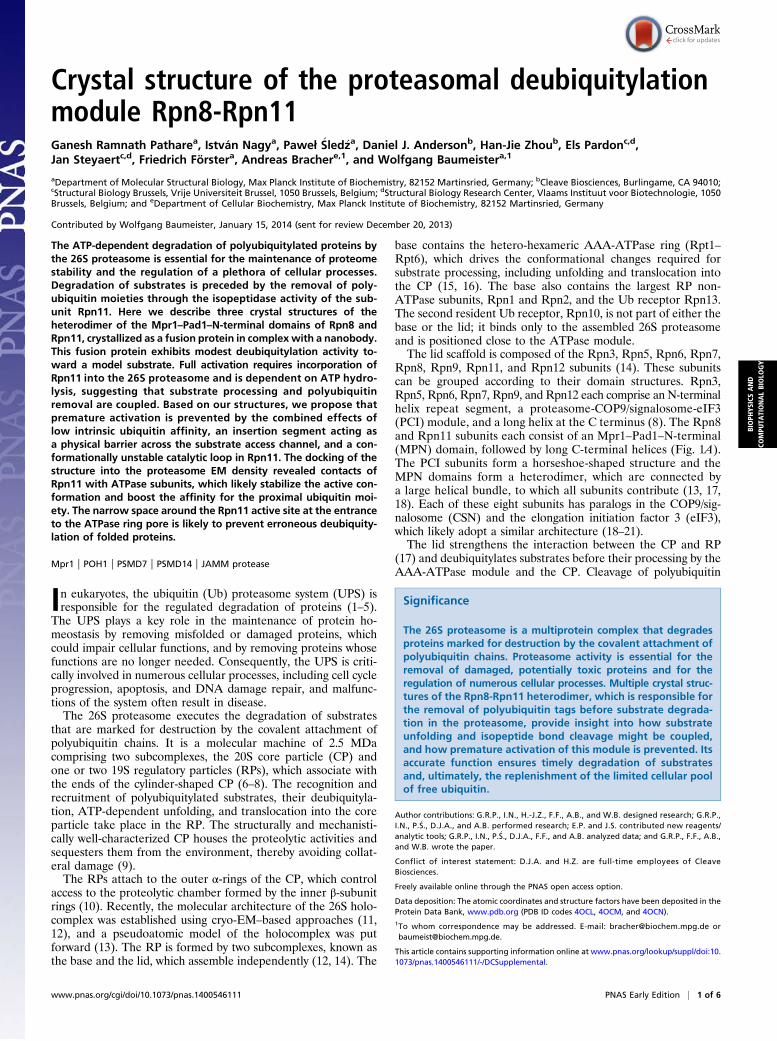
Results and DiscussionStructure Determination of the Rpn8-Rpn11 Core Complex. The MPNdomains of Rpn8 and Rpn11 were expressed as a fusion proteinwith a 9-aa-long connecting linker (Fig. 1A). The domain limitswere selected based on limited proteolysis experiments withproteinase K (residues 1–175 of Rpn8 and residues 1–219 ofRpn11), and the domains were fused to ensure stability andformation of a stoichiometric complex. Notably, this fusion con-struct, designated Rpn8-Rpn11, cleaves a model substrate, a lineartetraubiquitin (Ub4)-peptide fusion protein, indicating that thisconstruct samples the catalytically active conformation. It requiresa 7,000-fold higher concentration than the complete proteasome,however (Fig. 1B).The Rpn8-Rpn11 crystals suitable for structure determination
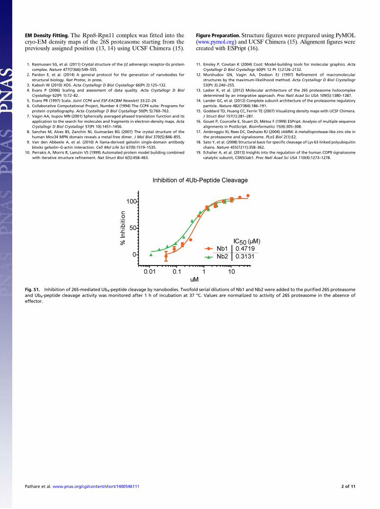
were grown with the aid of a tailored nanobody (variable domainof camelid heavy chain-only antibodies). Rpn8-Rpn11–specificnanobodies were selected from llama antisera raised againstpurified yeast 26S proteasome. The successful nanobody Nb1inhibited the deubiquitylation activity of 26 proteasome in a con-centration-dependent manner (Fig. S1).
Crystal Lattices.We obtained three crystal forms, here designatedIa, Ib and II, of the Rpn8-Rpn11 fusion protein complex with thenanobody (Table S1 and Fig. S2 A and B). Crystal forms Ia andIb are closely related. All crystal forms contain two Rpn8-Rpn11-nanobody complexes per asymmetric unit. The backbones in thecore regions of the complex subunits are very similar, yieldingrmsd values between 0.304 and 0.916 Å (Fig. S2 C and D); onlyhelix α4 of Rpn8 is displaced in one copy of crystal form II (Fig.S2C). No density could be assigned to any of the linker regionsbetween the Rpn8 and Rpn11 MPN domains.A major difference between crystal forms I and II is the
presence of bound Zn in the former. In crystal form II, a crystalcontact between the two copies of Rpn11 distorts the geometryof the catalytic loop, displacing the Zn-coordinating residueHis109 from the Zn-binding site (Fig. S2F). Thus, crystal form IIappears to be incompatible with Zn binding. Thus, we focus oncrystal form Ia, diffracting to the highest resolution (2.0 Å).
Fig. 1. Biochemical activity of the Rpn8-Rpn11 fusion protein. (A) Domainstructures of Rpn8, Rpn11 and the fusion protein. (B) Ub4 cleavage activity of26S proteasome, WT Rpn8-Rpn11 and Rpn8-Rpn11 (E48Q). Cleavage of la-beled peptide from Ub4 was detected by the change in fluorescence polar-ization after 1hr incubation at 37 °C at the indicated concentrations. Valuesare normalized tomaximum cleavage activity of 26S proteasome. The used 26Sproteasome preparation contained only trace amounts of the DUB Ubp6.
2 of 6 | www.pnas.org/cgi/doi/10.1073/pnas.1400546111 Pathare et al.
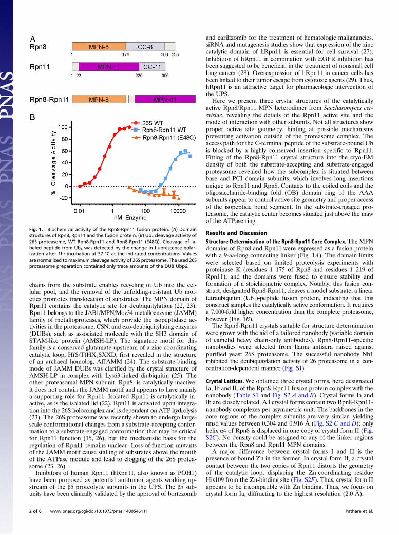
Structure of the Rpn8-Rpn11 Complex. The Rpn8-Rpn11 core com-plex structure exhibits pseudo-twofold symmetry (Fig. 2).Each protomer assumes a MPN domain fold and consists of fourα-helices, α1–α4, flanking a circular β-sheet of seven β-strands,βA–βG (Figs. S2E and S3). The topology of the β-sheet is βA-βC-βB-βD-βE-βF-βG. The long, twisted β-strand βG makes contactswith both βA and βF. Rpn8 and Rpn11 contact each other viatwo pseudosymmetrical interfaces, a coiled coil between helicesα2 and a four-helix bundle of helices α1 and α4 (Fig. 2). The C-terminal α4 helices are associated mainly with the opposingsubunit (Fig. 2), causing the domain swapping first observed inthe crystal structure of human Rpn8/Mov34 (30) and anticipatedin the pseudoatomic models of the 26S proteasome (11, 12).Two regions connecting βC-α2 and βF-βG are variable in MPN
domain sequences, designated here as insertion 1 and insertion 2(Fig. 2) (24, 25, 30–32). Insertion 1 of Rpn8 forms a β-hairpin ontop of the MPN domain. The corresponding region in Rpn11forms a poorly ordered loop adjacent to the active site, as dis-cussed in more detail below. The insertion 2 regions of Rpn8 andRpn11 protrude from the opposite ends of the pseudo-twofoldsymmetric subcomplex (Fig. 2). Insertion 2 of Rpn8 assumes anelongated β-hairpin structure in crystal form II. The hairpinsfrom two Rpn8 molecules align to create a mixed β-sheet contactin this crystal lattice (Fig. S2B). In the form I crystal structures,the tips of the β-hairpin are disordered, suggesting that this re-gion is stably structured only in the presence of a suitable in-teraction partner. The much longer insertion 2 in Rpn11 forms
a helical protrusion with a disordered tip in crystal forms Ia andIb. The helices from two adjacent Rpn11 molecules form anti-parallel three-helix bundles (Figs. S2A and S3). In crystal formII, both corresponding segments are disordered, suggesting thatinsertion 2 of Rpn11 stably folds only in appropriate environ-ments, in line with secondary structure predictions.
Role of the Nanobody in Crystal Formation. The nanobody contactsan area that involves β-strands βB, βC, and βG and α4 in Rpn11and a section of helix α1 in Rpn8, thereby establishing additionalcontacts between the proteins and rigidifying the complex (Fig.2). This contact area forms a depression on the surface of theRpn8-Rpn11 complex, providing a concave binding site for theCDR3 loop of the nanobody. Furthermore, in all crystal lattices,the nanobodies contribute important crystal contacts to adjacentRpn8 molecules. Thus, a combination of both effects might ex-plain why the nanobody is required for the successful crystalli-zation of Rpn8-Rpn11.
Active Site of Rpn11. The active site of MPN domain metal-loproteases is located between the N-terminal end of helix α3and the adjacent β-strands βB and βD (31, 33, 34). Clear densityfor the catalytic zinc was identified between the sidechains ofHis109, His111, and Asp122 (Fig. 3A and Fig. S4A). Thesesidechains, together with a water molecule, form a slightly dis-torted tetrahedral coordination shell around the metal ion. Thiscore structure is almost identical to that of the DUB AMSH-LP(25, 33, 34), for which both apo and substrate-bound crystalstructures have been characterized at high-resolution (Fig. 3Band Fig. S4E). In the AMSH-LP cocrystal structure, the iso-peptide bond carbonyl group was positioned directly on top ofthe metal site (Fig. 3B) (34), strongly suggesting that this con-formation of Rpn8-Rpn11 represents a catalytically active state.The residue Glu48 at the beginning of β-strand βB corre-
sponds to residue Glu292 of AMSH-LP, which is essential forAMSH-LP activity (25, 33). Mutating this residue to glutamineabolished Ub4 cleavage activity in Rpn8-Rpn11 (Fig. 1B). Glu48is positioned for activation of the attacking water molecule andprotonation of the isopeptide amide group. With the location ofGlu48, His109, and His111 in two adjacent β-strands, the re-spective geometry is largely fixed (Fig. 3A and Fig. S4A). Theconformation of the catalytic loop (residues 109–122) is stabi-lized by an extended hydrogen bond network in Rpn11 (Fig. 3Aand Fig. S4A), which is also observed in the apo crystal structuresof AMSH-LP (25), the Rpn11 paralog Csn5 (31), and the ar-chaeal Rpn11 homolog AfJAMM (24) (Fig. S4 D–F). Hydrogenbond contacts between the carbonyl group of Gly115 and theimidazole ring of His111 and between the amide group of Ser119and the carboxyl group of Asp122 orient the coordinating side-chains toward Zn and establish the proper polarity. The imid-azole ring of His111 is further buttressed by the sidechains of thecatalytic loop residues Phe114 and Trp117 (Fig. 3A and Fig.S4A). The orientation of the indole group of Trp117 is stabilizedby a hydrogen bond to the carbonyl group of Phe114. In addition,the carboxyl group of Asp142 links the amide groups of Gly115and Ile144. These two interactions are conserved in Csn5 as well(31, 32) (Fig. S4F). Asp142 and Ile144 belong to the highly conservedloop connection between βE and βF in Rpn11. The respective loopis much shorter in AMSH-LP. Other important hydrogen bondcontacts with the backbone are formed by the JAMM motif resi-dues Ser110 and Ser119. The former extends the β-sheet contactsbetween βB and βD, and the latter stabilizes the N-terminal part ofhelix α3 and buttresses Asp122.Alternative conformations of the catalytic loop were found in
one of the two copies of Rpn11 in crystal forms Ia and Ib each(Fig. 3C and Fig. S4 B and C). Both conformers are character-ized by a wider separation of the His111 imidazole ring from Zn(2.9 Å vs. 2.1 Å), whereas His109 and Asp122 remain virtually
Fig. 2. Crystal structure of the MPN domain fusion protein of Rpn8 andRpn11 with the attached nanobody. The composite structure of Rpn8 (chainA) from crystal form II superposed on the Rpn8-Rpn11–nanobody complex(chains D, E, and F) from crystal form Ia is shown in side and bottom views.The Rpn8 and Rpn11 units are indicated in purple and brown, respectively;the nanobody is represented in silver. Disordered segments are indicated bydotted lines. Helices are represented by cylinders; the catalytic Zn ion, bya green sphere. The unique insertions into the canonical MPN structure areindicated.
Pathare et al. PNAS Early Edition | 3 of 6
BIOPH
YSICSAND
COMPU
TATIONALBIOLO
GY
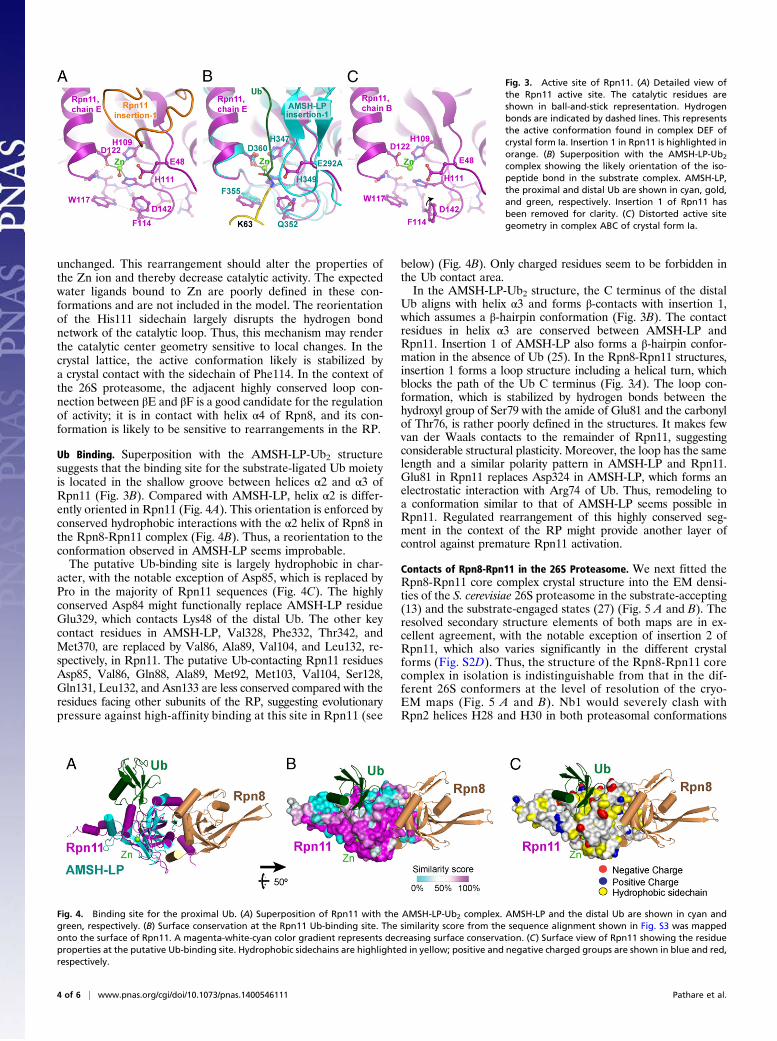
unchanged. This rearrangement should alter the properties ofthe Zn ion and thereby decrease catalytic activity. The expectedwater ligands bound to Zn are poorly defined in these con-formations and are not included in the model. The reorientationof the His111 sidechain largely disrupts the hydrogen bondnetwork of the catalytic loop. Thus, this mechanism may renderthe catalytic center geometry sensitive to local changes. In thecrystal lattice, the active conformation likely is stabilized bya crystal contact with the sidechain of Phe114. In the context ofthe 26S proteasome, the adjacent highly conserved loop con-nection between βE and βF is a good candidate for the regulationof activity; it is in contact with helix α4 of Rpn8, and its con-formation is likely to be sensitive to rearrangements in the RP.
Ub Binding. Superposition with the AMSH-LP-Ub2 structuresuggests that the binding site for the substrate-ligated Ub moietyis located in the shallow groove between helices α2 and α3 ofRpn11 (Fig. 3B). Compared with AMSH-LP, helix α2 is differ-ently oriented in Rpn11 (Fig. 4A). This orientation is enforced byconserved hydrophobic interactions with the α2 helix of Rpn8 inthe Rpn8-Rpn11 complex (Fig. 4B). Thus, a reorientation to theconformation observed in AMSH-LP seems improbable.The putative Ub-binding site is largely hydrophobic in char-
acter, with the notable exception of Asp85, which is replaced byPro in the majority of Rpn11 sequences (Fig. 4C). The highlyconserved Asp84 might functionally replace AMSH-LP residueGlu329, which contacts Lys48 of the distal Ub. The other keycontact residues in AMSH-LP, Val328, Phe332, Thr342, andMet370, are replaced by Val86, Ala89, Val104, and Leu132, re-spectively, in Rpn11. The putative Ub-contacting Rpn11 residuesAsp85, Val86, Gln88, Ala89, Met92, Met103, Val104, Ser128,Gln131, Leu132, and Asn133 are less conserved compared with theresidues facing other subunits of the RP, suggesting evolutionarypressure against high-affinity binding at this site in Rpn11 (see
below) (Fig. 4B). Only charged residues seem to be forbidden inthe Ub contact area.In the AMSH-LP-Ub2 structure, the C terminus of the distal
Ub aligns with helix α3 and forms β-contacts with insertion 1,which assumes a β-hairpin conformation (Fig. 3B). The contactresidues in helix α3 are conserved between AMSH-LP andRpn11. Insertion 1 of AMSH-LP also forms a β-hairpin confor-mation in the absence of Ub (25). In the Rpn8-Rpn11 structures,insertion 1 forms a loop structure including a helical turn, whichblocks the path of the Ub C terminus (Fig. 3A). The loop con-formation, which is stabilized by hydrogen bonds between thehydroxyl group of Ser79 with the amide of Glu81 and the carbonylof Thr76, is rather poorly defined in the structures. It makes fewvan der Waals contacts to the remainder of Rpn11, suggestingconsiderable structural plasticity. Moreover, the loop has the samelength and a similar polarity pattern in AMSH-LP and Rpn11.Glu81 in Rpn11 replaces Asp324 in AMSH-LP, which forms anelectrostatic interaction with Arg74 of Ub. Thus, remodeling toa conformation similar to that of AMSH-LP seems possible inRpn11. Regulated rearrangement of this highly conserved seg-ment in the context of the RP might provide another layer ofcontrol against premature Rpn11 activation.
Contacts of Rpn8-Rpn11 in the 26S Proteasome. We next fitted theRpn8-Rpn11 core complex crystal structure into the EM densi-ties of the S. cerevisiae 26S proteasome in the substrate-accepting(13) and the substrate-engaged states (27) (Fig. 5 A and B). Theresolved secondary structure elements of both maps are in ex-cellent agreement, with the notable exception of insertion 2 ofRpn11, which also varies significantly in the different crystalforms (Fig. S2D). Thus, the structure of the Rpn8-Rpn11 corecomplex in isolation is indistinguishable from that in the dif-ferent 26S conformers at the level of resolution of the cryo-EM maps (Fig. 5 A and B). Nb1 would severely clash withRpn2 helices H28 and H30 in both proteasomal conformations
Fig. 3. Active site of Rpn11. (A) Detailed view ofthe Rpn11 active site. The catalytic residues areshown in ball-and-stick representation. Hydrogenbonds are indicated by dashed lines. This representsthe active conformation found in complex DEF ofcrystal form Ia. Insertion 1 in Rpn11 is highlighted inorange. (B) Superposition with the AMSH-LP-Ub2
complex showing the likely orientation of the iso-peptide bond in the substrate complex. AMSH-LP,the proximal and distal Ub are shown in cyan, gold,and green, respectively. Insertion 1 of Rpn11 hasbeen removed for clarity. (C) Distorted active sitegeometry in complex ABC of crystal form Ia.
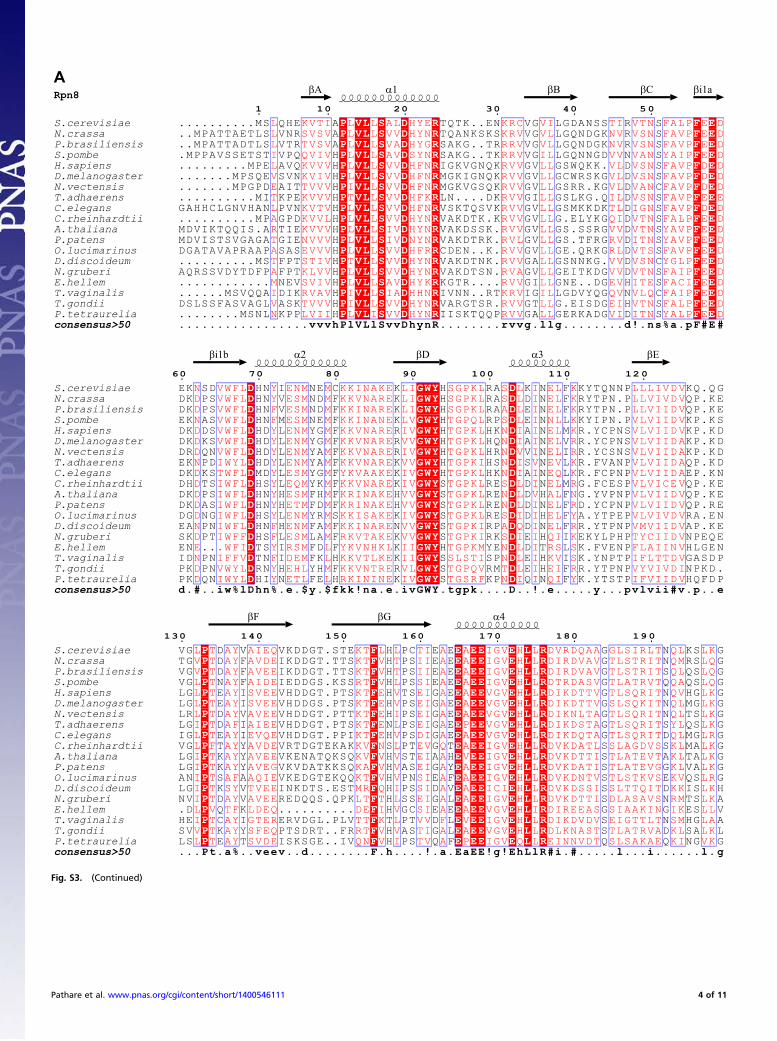
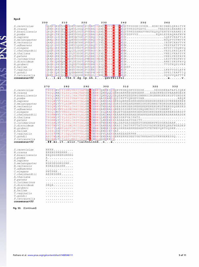
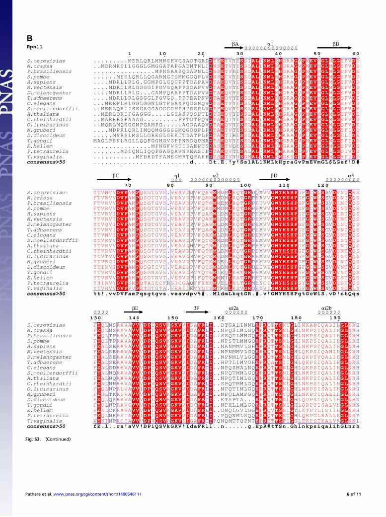
Fig. 4. Binding site for the proximal Ub. (A) Superposition of Rpn11 with the AMSH-LP-Ub2 complex. AMSH-LP and the distal Ub are shown in cyan andgreen, respectively. (B) Surface conservation at the Rpn11 Ub-binding site. The similarity score from the sequence alignment shown in Fig. S3 was mappedonto the surface of Rpn11. A magenta-white-cyan color gradient represents decreasing surface conservation. (C) Surface view of Rpn11 showing the residueproperties at the putative Ub-binding site. Hydrophobic sidechains are highlighted in yellow; positive and negative charged groups are shown in blue and red,respectively.
4 of 6 | www.pnas.org/cgi/doi/10.1073/pnas.1400546111 Pathare et al.
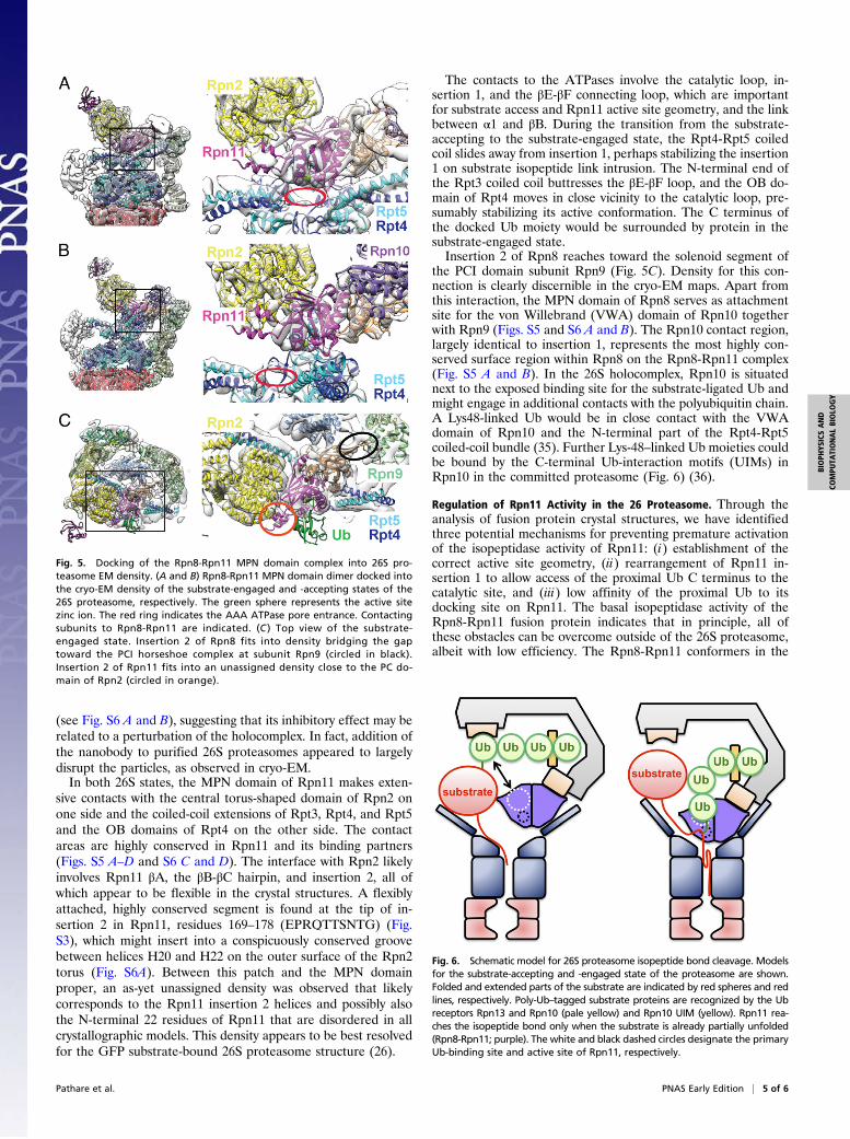
(see Fig. S6 A and B), suggesting that its inhibitory effect may berelated to a perturbation of the holocomplex. In fact, addition ofthe nanobody to purified 26S proteasomes appeared to largelydisrupt the particles, as observed in cryo-EM.In both 26S states, the MPN domain of Rpn11 makes exten-
sive contacts with the central torus-shaped domain of Rpn2 onone side and the coiled-coil extensions of Rpt3, Rpt4, and Rpt5and the OB domains of Rpt4 on the other side. The contactareas are highly conserved in Rpn11 and its binding partners(Figs. S5 A–D and S6 C and D). The interface with Rpn2 likelyinvolves Rpn11 βA, the βB-βC hairpin, and insertion 2, all ofwhich appear to be flexible in the crystal structures. A flexiblyattached, highly conserved segment is found at the tip of in-sertion 2 in Rpn11, residues 169–178 (EPRQTTSNTG) (Fig.S3), which might insert into a conspicuously conserved groovebetween helices H20 and H22 on the outer surface of the Rpn2torus (Fig. S6A). Between this patch and the MPN domainproper, an as-yet unassigned density was observed that likelycorresponds to the Rpn11 insertion 2 helices and possibly alsothe N-terminal 22 residues of Rpn11 that are disordered in allcrystallographic models. This density appears to be best resolvedfor the GFP substrate-bound 26S proteasome structure (26).
The contacts to the ATPases involve the catalytic loop, in-sertion 1, and the βE-βF connecting loop, which are importantfor substrate access and Rpn11 active site geometry, and the linkbetween α1 and βB. During the transition from the substrate-accepting to the substrate-engaged state, the Rpt4-Rpt5 coiledcoil slides away from insertion 1, perhaps stabilizing the insertion1 on substrate isopeptide link intrusion. The N-terminal end ofthe Rpt3 coiled coil buttresses the βE-βF loop, and the OB do-main of Rpt4 moves in close vicinity to the catalytic loop, pre-sumably stabilizing its active conformation. The C terminus ofthe docked Ub moiety would be surrounded by protein in thesubstrate-engaged state.Insertion 2 of Rpn8 reaches toward the solenoid segment of
the PCI domain subunit Rpn9 (Fig. 5C). Density for this con-nection is clearly discernible in the cryo-EM maps. Apart fromthis interaction, the MPN domain of Rpn8 serves as attachmentsite for the von Willebrand (VWA) domain of Rpn10 togetherwith Rpn9 (Figs. S5 and S6 A and B). The Rpn10 contact region,largely identical to insertion 1, represents the most highly con-served surface region within Rpn8 on the Rpn8-Rpn11 complex(Fig. S5 A and B). In the 26S holocomplex, Rpn10 is situatednext to the exposed binding site for the substrate-ligated Ub andmight engage in additional contacts with the polyubiquitin chain.A Lys48-linked Ub would be in close contact with the VWAdomain of Rpn10 and the N-terminal part of the Rpt4-Rpt5coiled-coil bundle (35). Further Lys-48–linked Ub moieties couldbe bound by the C-terminal Ub-interaction motifs (UIMs) inRpn10 in the committed proteasome (Fig. 6) (36).
Regulation of Rpn11 Activity in the 26 Proteasome. Through theanalysis of fusion protein crystal structures, we have identifiedthree potential mechanisms for preventing premature activationof the isopeptidase activity of Rpn11: (i) establishment of thecorrect active site geometry, (ii) rearrangement of Rpn11 in-sertion 1 to allow access of the proximal Ub C terminus to thecatalytic site, and (iii) low affinity of the proximal Ub to itsdocking site on Rpn11. The basal isopeptidase activity of theRpn8-Rpn11 fusion protein indicates that in principle, all ofthese obstacles can be overcome outside of the 26S proteasome,albeit with low efficiency. The Rpn8-Rpn11 conformers in the
Fig. 5. Docking of the Rpn8-Rpn11 MPN domain complex into 26S pro-teasome EM density. (A and B) Rpn8-Rpn11 MPN domain dimer docked intothe cryo-EM density of the substrate-engaged and -accepting states of the26S proteasome, respectively. The green sphere represents the active sitezinc ion. The red ring indicates the AAA ATPase pore entrance. Contactingsubunits to Rpn8-Rpn11 are indicated. (C ) Top view of the substrate-engaged state. Insertion 2 of Rpn8 fits into density bridging the gaptoward the PCI horseshoe complex at subunit Rpn9 (circled in black).Insertion 2 of Rpn11 fits into an unassigned density close to the PC do-main of Rpn2 (circled in orange).
Fig. 6. Schematic model for 26S proteasome isopeptide bond cleavage. Modelsfor the substrate-accepting and -engaged state of the proteasome are shown.Folded and extended parts of the substrate are indicated by red spheres and redlines, respectively. Poly-Ub–tagged substrate proteins are recognized by the Ubreceptors Rpn13 and Rpn10 (pale yellow) and Rpn10 UIM (yellow). Rpn11 rea-ches the isopeptide bond only when the substrate is already partially unfolded(Rpn8-Rpn11; purple). The white and black dashed circles designate the primaryUb-binding site and active site of Rpn11, respectively.
Pathare et al. PNAS Early Edition | 5 of 6
BIOPH
YSICSAND
COMPU
TATIONALBIOLO
GY

crystal structures show that proper active site geometry is ac-cessible, but not stable. Similarly, insertion 1 of Rpn11 blocks theaccess path for Ub in our structures; however, this elementappears to be mobile, as suggested by the high B-factors anddisorder in several conformers, and thus it should rearrangeeasily once the Ub C terminus enters the access path.The affinity of the Rpn8-Rpn11 fusion protein for Ub appears
to be modest at best (Fig. 1B). Simultaneous contacts with bothsites should allow efficient substrate binding only in the closepresence of Ub receptors, particularly the Rpn10 UIM motif, inthe assembled proteasome. In support of this idea, a 26S com-plex without Rpn10 and Rpn13 demonstrated greatly reducedUb4 cleavage activity (Fig. S8). Another mechanism of pre-venting cleavage of noncommitted substrates is limited access forbulky folded domains to the narrow surroundings of the Rpn11active site in both 26S conformations.Full activation of Rpn11 is presumably realized by contacts
with the coiled coils and the OB ring of the AAA subunits, whichhave been proposed to have chaperone activity (37) and fur-thermore could stabilize the active conformation in Rpn11 andthereby increase the affinity for the primary Ub by opening thebinding site for the C-terminal tail. The strong sequence con-servation of the involved elements suggests tight coevolution ofa defined functional interface (Figs. S5 A–D and S6 C and D).Therefore, the 26S proteasome might have an extended “com-posite” deubiquitylase active site, converting the access groove forthe C-terminal end of the Ub chain in AMSH-LP into a channel,which allows exact control of substrate orientation; this is neces-sary because the sequences flanking Ub acceptor sites are variable
in proteasomal substrates. Only the structure of polyubiquitylatedsubstrate bound to Rpn11 in the context of a stalled proteasome willreveal the molecular mechanism of deubiquitylation in full detail.
Materials and MethodsThe experimental procedures are described in detail in SI Materials andMethods. In brief, Rpn8-Rpn11 from S. cerevisiae was expressed as a His6 tagfusion protein including a tobacco etch virus (TEV) protease site in E. coliBL21 (DE3) cells and purified by Ni-affinity chromatography, TEV cleavage,and Superose-12 size exclusion chromatography. Crystals were grown using50 mM MES pH 6.0, 200 mM Ca acetate, and 22% (wt/vol) PEG-3350 or with50 mM MES pH 6.0, 100 mM MgCl2, and 21% (wt/vol) PEG-3350. The Rpn8-Rpn11-nanobody crystal structure was solved by molecular replacement. Theisopeptidase activity assay was performed with a fluorogenic Ub4 fusionprotein, with reaction progress monitored by fluorescence polarization. Thenanobody was selected and produced following standard procedures (38).
ACKNOWLEDGMENTS. We thank Yousuf Mohiuddin Mohammed, AndréMourão, Günter Pfeifer, Oana Mihalache, Sándor Varga, Ágnes Hubert, JanSchuller, Antje Aufderheide, Andreas Schweitzer, Pia Unverdorben, and Ro-man Körner (Max Planck Insitute of Biochemistry); Alison Lundquist (VrijeUniversiteit Brussels); Ethan Emberley, Brajesh Kumar, and David J. Wustrow(Cleave Bisciences); the Joint Structural Biology Group group at the ElectronSynchrotron Radiation Facility Grenoble; and staff at Swiss Light SourceX10SA Villigen, the MPIB Crystallization Facility, and the MPIB Core Facilityfor their excellent support. Our research is supported by funding from theDeutsche Forschungsgemeinschaft Excellence Cluster Center for IntegratedProtein Science Munich and SFB-1035/Project A01 (to W.B.), Instruct (part ofthe European Strategy Forum on Research Infrastructures and supported bynational member subscriptions) through a Research and Development PilotProject grant (to J.S., I.N., and P.�S.), as well as Graduiertenkolleg 1721 (to F.F.)and European Molecular Biology Organization (to P.�S.).
1. Finley D (2009) Recognition and processing of ubiquitin-protein conjugates by theproteasome. Annu Rev Biochem 78:477–513.
2. Goldberg AL (2003) Protein degradation and protection against misfolded or dam-aged proteins. Nature 426(6968):895–899.
3. Varshavsky A (2012) The ubiquitin system, an immense realm. Annu Rev Biochem81:167–176.
4. Buchberger A, Bukau B, Sommer T (2010) Protein quality control in the cytosol andthe endoplasmic reticulum: Brothers in arms. Mol Cell 40(2):238–252.
5. Hershko A, Ciechanover A, Varshavsky A (2000) Basic Medical Research Award: Theubiquitin system. Nat Med 6(10):1073–1081.
6. Tanaka K (2009) The proteasome: Overview of structure and functions. Proc Jpn Acad,Ser B, Phys Biol Sci 85(1):12–36.
7. Voges D, Zwickl P, Baumeister W (1999) The 26S proteasome: A molecular machinedesigned for controlled proteolysis. Annu Rev Biochem 68:1015–1068.
8. Förster F, Unverdorben P, Sled�z P, Baumeister W (2013) Unveiling the long-held se-crets of the 26S proteasome. Structure 21(9):1551–1562.
9. Baumeister W, Walz J, Zühl F, Seemüller E (1998) The proteasome: Paradigm of a self-compartmentalizing protease. Cell 92(3):367–380.
10. Peters JM, Cejka Z, Harris JR, Kleinschmidt JA, Baumeister W (1993) Structural featuresof the 26 S proteasome complex. J Mol Biol 234(4):932–937.
11. Lasker K, et al. (2012) Molecular architecture of the 26S proteasome holocomplexdetermined by an integrative approach. Proc Natl Acad Sci USA 109(5):1380–1387.
12. Lander GC, et al. (2012) Complete subunit architecture of the proteasome regulatoryparticle. Nature 482(7384):186–191.
13. Beck F, et al. (2012) Near-atomic resolution structural model of the yeast 26S pro-teasome. Proc Natl Acad Sci USA 109(37):14870–14875.
14. Glickman MH, et al. (1998) A subcomplex of the proteasome regulatory particle re-quired for ubiquitin-conjugate degradation and related to the COP9-signalosomeand eIF3. Cell 94(5):615–623.
15. �Sled�z P, et al. (2013) Structure of the 26S proteasome with ATP-γS bound providesinsights into the mechanism of nucleotide-dependent substrate translocation. ProcNatl Acad Sci USA 110(18):7264–7269.
16. Beckwith R, Estrin E, Worden EJ, Martin A (2013) Reconstitution of the 26S protea-some reveals functional asymmetries in its AAA+ unfoldase. Nat Struct Mol Biol20(10):1164–1172.
17. Pathare GR, et al. (2012) The proteasomal subunit Rpn6 is a molecular clamp holdingthe core and regulatory subcomplexes together. Proc Natl Acad Sci USA 109(1):149–154.
18. Estrin E, Lopez-Blanco JR, Chacón P, Martin A (2013) Formation of an intricate helicalbundle dictates the assembly of the 26S proteasome lid. Structure 21(9):1624–1635.
19. Enchev RI, Schreiber A, Beuron F, Morris EP (2010) Structural insights into the COP9signalosome and its common architecture with the 26S proteasome lid and eIF3.Structure 18(4):518–527.
20. Sun C, et al. (2011) Functional reconstitution of human eukaryotic translation initia-tion factor 3 (eIF3). Proc Natl Acad Sci USA 108(51):20473–20478.
21. Querol-Audi J, et al. (2013) Architecture of human translation initiation factor 3.Structure 21(6):920–928.
22. Yao T, Cohen RE (2002) A cryptic protease couples deubiquitination and degradationby the proteasome. Nature 419(6905):403–407.
23. Verma R, et al. (2002) Role of Rpn11 metalloprotease in deubiquitination and deg-radation by the 26S proteasome. Science 298(5593):611–615.
24. Ambroggio XI, Rees DC, Deshaies RJ (2004) JAMM: A metalloprotease-like zinc site inthe proteasome and signalosome. PLoS Biol 2(1):E2.
25. Sato Y, et al. (2008) Structural basis for specific cleavage of Lys 63-linked polyubiquitinchains. Nature 455(7211):358–362.
26. Matyskiela ME, Lander GC, Martin A (2013) Conformational switching of the 26Sproteasome enables substrate degradation. Nat Struct Mol Biol 20(7):781–788.
27. Gallery M, et al. (2007) The JAMMmotif of human deubiquitinase Poh1 is essential forcell viability. Mol Cancer Ther 6(1):262–268.
28. Bivona TG, et al. (2011) FAS and NF-κB signalling modulate dependence of lungcancers on mutant EGFR. Nature 471(7339):523–526.
29. Spataro V, Simmen K, Realini CA (2002) The essential 26S proteasome subunit Rpn11confers multidrug resistance to mammalian cells. Anticancer Res 22(6C):3905–3909.
30. Sanches M, Alves BS, Zanchin NI, Guimarães BG (2007) The crystal structure of thehuman Mov34 MPN domain reveals a metal-free dimer. J Mol Biol 370(5):846–855.
31. Echalier A, et al. (2013) Insights into the regulation of the human COP9 signalosomecatalytic subunit, CSN5/Jab1. Proc Natl Acad Sci USA 110(4):1273–1278.
32. Zhang H, et al. (2012) The crystal structure of the MPN domain from the COP9 sig-nalosome subunit CSN6. FEBS Lett 586(8):1147–1153.
33. Davies CW, Paul LN, Kim MI, Das C (2011) Structural and thermodynamic comparisonof the catalytic domain of AMSH and AMSH-LP: Nearly identical fold but differentstability. J Mol Biol 413(2):416–429.
34. Kikuchi K, Ishii N, Asao H, Sugamura K (2003) Identification of AMSH-LP containinga Jab1/MPN domain metalloenzyme motif. Biochem Biophys Res Commun 306(3):637–643.
35. Rani N, Aichem A, Schmidtke G, Kreft SG, Groettrup M (2012) FAT10 and NUB1L bindto the VWA domain of Rpn10 and Rpn1 to enable proteasome-mediated proteolysis.Nat Commun 3:749.
36. Riedinger C, et al. (2010) Structure of Rpn10 and its interactions with polyubiquitinchains and the proteasome subunit Rpn12. J Biol Chem 285(44):33992–34003.
37. Djuranovic S, et al. (2009) Structure and activity of the N-terminal substrate recog-nition domains in proteasomal ATPases. Mol Cell 34(5):580–590.
38. Pardon E, et al. (2014) A general protocol for the generation of nanobodies forstructural biology. Nat Protoc, in press.
6 of 6 | www.pnas.org/cgi/doi/10.1073/pnas.1400546111 Pathare et al.
Supporting InformationPathare et al. 10.1073/pnas.1400546111SI Materials and MethodsPurification of the Rpn8-Rpn11 Fusion Protein. The Rpn8-Rpn11expression construct consisted of an N-terminal His8-tag, RAARprotein, a tobacco etch virus (TEV) protease cleavage site, res-idues 1–176 of Rpn8, a nine-residue linker (GSGGSGGSG), andresidues 1–220 of Rpn11. The Saccharomyces cerevisiae subunitDNA sequences were codon-optimized for Escherichia coli ex-pression. The resulting sequence was cloned between the NcoIand XhoI sites of a modified pRSFDuet DNA (Merck Millipore).To express the fusion protein, E. coli BL21 (DE3) was trans-
formed with the plasmid and grown overnight in a shaking cul-ture on LB medium containing 50 μg mL−1 kanamycin at 37 °C.At OD600 = 0.8, protein expression was induced with 0.5 mMisopropyl β-D-1-thiogalactopyranoside, and the temperature waslowered to 25 °C for 12 h. Subsequently, cells were harvested bysedimentation at 4,000 × g, washed with sterile water, re-suspended with 2.5 mL of lysis buffer (50 mM Tris pH 8.0, 10mM imidazole, 150 mM NaCl, and 1 mg mL−1 lysozyme) pergram, and incubated on ice for 1 h. Then 100 ppm Benzonase(Sigma-Aldrich) was added, followed by ultrasonication on ice. Celldebris was removed by ultracentrifugation at 28,000 × g at 4 °Cfor 30 min. His8-tagged Rpn8-Rpn11 was purified on Ni-nitrilotri-acetic acid (NTA) fast-flow beads (Qiagen) according to the sup-plier’s recommendations. Fractions containing His8-Rpn8-Rpn11were merged, His8-TEV protease was added, and the mixture wasincubated for 12 h at 4 °C in a dialysis chamber equilibrating against25 mM Tris·HCl pH 7.5. Uncleaved protein, the affinity tag, andHis8-TEV protease were removed by Ni-affinity purification usingNi-NTA fast-flow beads (Qiagen), as described above. The un-bound protein was subjected to a Superose-12 column (GEHealthcare) equilibrated with 10 mM Tris pH 7.5, 150 mM NaCl,and 90 mM KCl. Rpn8-Rpn11–containing fractions were mergedand concentrated.
Cloning and Purification of Rpn8-Rpn11–Targeted Nanobodies. Theuse of protein complexes for animal immunization (1) was im-plemented to obtain nanobodies against epitopes of 26S pro-teasome subunits in their biologically relevant conformation. Forthis, a llama was immunized six times with 100 μg of intact 26Sproteasomes of S. cerevisiae essentially as described by Pardonet al. (2). Peripheral blood lymphocytes were extracted, and theirRNA was purified and converted into cDNA via RT-PCR. ThecDNA was cloned into phage-display vector pMESy4 containinga C-terminal 6X His tag followed by the CaptureSelect C-tag.Several nanobodies that bind to the Rpn8-Rpn11 subcomplexwere identified by biopanning on the unmodified Rpn8-Rpn11heterodimer immobilized using an anti-streptavidin monoclonalantibody that was solid-phase–coated on the ELISA plate. An-tigen-bound phages were recovered by proteolysis with trypsin.After each round of selection, ELISA was performed on peri-plasmic extracts of 48 individual colonies to screen for Rpn8-Rpn11–specific nanobodies. Nb1, which was used to solve thestructure of the Rpn8-Rpn11 complex, was selected after oneround of biopanning.Phagemids (pMESy4) encoding for specific nanobodies were
transformed into E. coli WK6 Su− cells for periplasmic expres-sion. Protein expression was carried out as described for theexpression of His8-Rpn8-Rpn11 in E. coli BL21 (DE3) cells. Thecell pellet was resuspended in 15 mL of hypotonic TES buffer(200 mM Tris pH 8.0, 0.5 mM EDTA, and 500 mM sucrose) pergram and stirred for 1 h on ice. Subsequently, twice the volumeof TES/4 (TES buffer diluted four times) was added, followed by
45 min of incubation on ice. Cell debris was sedimented bycentrifugation at 28,000 × g for 30 min. The supernatant wasapplied to Ni-NTA (Qiagen) and incubated for several min at 4 °C.The column was washed with 20 mM Tris pH 7.4 buffer containing20 mM imidazole and 150 mM NaCl. Bound nanobody protein waseluted with 20 mM Tris pH 7.4, 500 mM imidazole, and 150 mMNaCl and then further purified on Superose-12 equilibrated with10 mM Tris pH 7.5, 150 mM NaCl, and 90 mM KCl.Nanobodies were tested for inhibition of the 26S proteasome
holoenzyme complex using a deubiquitylation assay. The deu-biquitylation-inhibitory nanobodies were used for further ex-periments.
Tetraubiquitin Cleavage Activity Assays.A fusion protein containingfour ubiquitin (Ub) copies followed by the sequence MQIFV-KTSQSSCVDKLAALEHHHHHH was expressed and purifiedfrom a single transcript in E. coli and then labeled with OregonGreen 488 using maleimide labeling. Then 5 nM tetraubiquitin(Ub4) peptide was combined with purified 26S proteasome fromS. cerevisiae, Rpn8-Rpn11 fusion protein, or mutant forms of thesemacromolecules in 50 mM Tris·HCl pH 7.5, 0.01% Nonidet P-40,50 μM MgCl2, 50 mM ATP, and 1 mM DTT. The reaction wasincubated at 37 °C for the indicated times, and fluorescence po-larization was determined.
Crystallization. For formation of the Rpn8-Rpn11 fusion protein–nanobody complex, the nanobody Nb1 and the Rpn8-Rpn11 fusionproteinweremixed at a 2:1 ratio, and the stoichiometric complex waspurified by size exclusion chromatography on Superose-12 equili-brated with 10 mM Tris pH 7.5, 150 mM NaCl, and 90 mM KCl.Crystals of the Rpn8-Rpn11–nanobody complex were grown by
the sitting drop vapor diffusionmethod at 4 °C or 18 °C, equilibratingequal volumes of 10mgmL−1 protein and precipitant against a largevolume of precipitant. Crystal forms Ia and Ib grew with precipitantcontaining 50 mM Mes pH 6.0, 200 mM Ca-acetate, and 22%(wt/vol) PEG-3350. Crystal form II was obtained with a precipitantcontaining 50 mM MES pH 6.0, 100 mM MgCl2, and 21% (wt/vol)PEG-3350. For cryoprotection, crystals were transferred stepwiseinto an otherwise unmodified precipitant solution containing15% (vol/vol) glycerol before being flash-frozen in liquid nitrogen.
Structure Determination. Diffraction data were collected at theElectron Synchrotron Radiation Facility (ESRF) in Grenoble,France, and Swiss Light Source (SLS) in Switzerland. The datawere processed with XDS (3) and transferred into the CCP4format using Pointless (4), Scala (5), and Truncate as im-plemented in the CCP4 interface (6). The structure of crystalform Ib was solved by molecular replacement with MOLREP (7)using the Rpn8 homodimer (8) and 2X1P nanobody (9) struc-tures as search models for the Mpr1–Pad1–N-terminal (MPN)domain dimers and the nanobody, respectively. Starting from thisinitial phase information, a greatly improved model was auto-matically built by ArpWarp 7.2 (10). Subsequently, the model wasimproved by iterative cycles of manual model building in Coot(11) and refinement with Refmac5 (12). The remaining crystalforms were solved by molecular replacement with the crystalform Ia model. The presence of Zn in crystal form Ib was con-firmed by X-ray fluorescence.
Site-Directed Mutagenesis. Site-directed mutations in Rpn11 wereintroduced with the QuikChange Site-Directed Mutagenesis Kit(Stratagene) using pRSF-Rpn8-Rpn11 as a template.
Pathare et al. www.pnas.org/cgi/content/short/1400546111 1 of 11
EM Density Fitting. The Rpn8-Rpn11 complex was fitted into thecryo-EM density maps of the 26S proteasome starting from thepreviously assigned position (13, 14) using UCSF Chimera (15).
Figure Preparation. Structure figures were prepared using PyMOL(www.pymol.org) and UCSF Chimera (15). Alignment figures werecreated with ESPript (16).
1. Rasmussen SG, et al. (2011) Crystal structure of the β2 adrenergic receptor-Gs proteincomplex. Nature 477(7366):549–555.
2. Pardon E, et al. (2014) A general protocol for the generation of nanobodies forstructural biology. Nat Protoc, in press.
3. Kabsch W (2010) XDS. Acta Crystallogr D Biol Crystallogr 66(Pt 2):125–132.4. Evans P (2006) Scaling and assessment of data quality. Acta Crystallogr D Biol
Crystallogr 62(Pt 1):72–82.5. Evans PR (1997) Scala. Joint CCP4 and ESF-EACBM Newslett 33:22–24.6. Collaborative Computational Project, Number 4 (1994) The CCP4 suite: Programs for
protein crystallography. Acta Crystallogr D Biol Crystallogr 50(Pt 5):760–763.7. Vagin AA, Isupov MN (2001) Spherically averaged phased translation function and its
application to the search for molecules and fragments in electron-density maps. ActaCrystallogr D Biol Crystallogr 57(Pt 10):1451–1456.
8. Sanches M, Alves BS, Zanchin NI, Guimarães BG (2007) The crystal structure of thehuman Mov34 MPN domain reveals a metal-free dimer. J Mol Biol 370(5):846–855.
9. Van den Abbeele A, et al. (2010) A llama-derived gelsolin single-domain antibodyblocks gelsolin–G-actin interaction. Cell Mol Life Sci 67(9):1519–1535.
10. Perrakis A, Morris R, Lamzin VS (1999) Automated protein model building combinedwith iterative structure refinement. Nat Struct Biol 6(5):458–463.
11. Emsley P, Cowtan K (2004) Coot: Model-building tools for molecular graphics. ActaCrystallogr D Biol Crystallogr 60(Pt 12 Pt 1):2126–2132.
12. Murshudov GN, Vagin AA, Dodson EJ (1997) Refinement of macromolecularstructures by the maximum-likelihood method. Acta Crystallogr D Biol Crystallogr53(Pt 3):240–255.
13. Lasker K, et al. (2012) Molecular architecture of the 26S proteasome holocomplexdetermined by an integrative approach. Proc Natl Acad Sci USA 109(5):1380–1387.
14. Lander GC, et al. (2012) Complete subunit architecture of the proteasome regulatoryparticle. Nature 482(7384):186–191.
15. Goddard TD, Huang CC, Ferrin TE (2007) Visualizing density maps with UCSF Chimera.J Struct Biol 157(1):281–287.
16. Gouet P, Courcelle E, Stuart DI, Métoz F (1999) ESPript: Analysis of multiple sequencealignments in PostScript. Bioinformatics 15(4):305–308.
17. Ambroggio XI, Rees DC, Deshaies RJ (2004) JAMM: A metalloprotease-like zinc site inthe proteasome and signalosome. PLoS Biol 2(1):E2.
18. Sato Y, et al. (2008) Structural basis for specific cleavage of Lys 63-linked polyubiquitinchains. Nature 455(7211):358–362.
19. Echalier A, et al. (2013) Insights into the regulation of the human COP9 signalosomecatalytic subunit, CSN5/Jab1. Proc Natl Acad Sci USA 110(4):1273–1278.
Fig. S1. Inhibition of 26S-mediated Ub4-peptide cleavage by nanobodies. Twofold serial dilutions of Nb1 and Nb2 were added to the purified 26S proteasomeand Ub4-peptide cleavage activity was monitored after 1 h of incubation at 37 °C. Values are normalized to activity of 26S proteasome in the absence ofeffector.
Pathare et al. www.pnas.org/cgi/content/short/1400546111 2 of 11
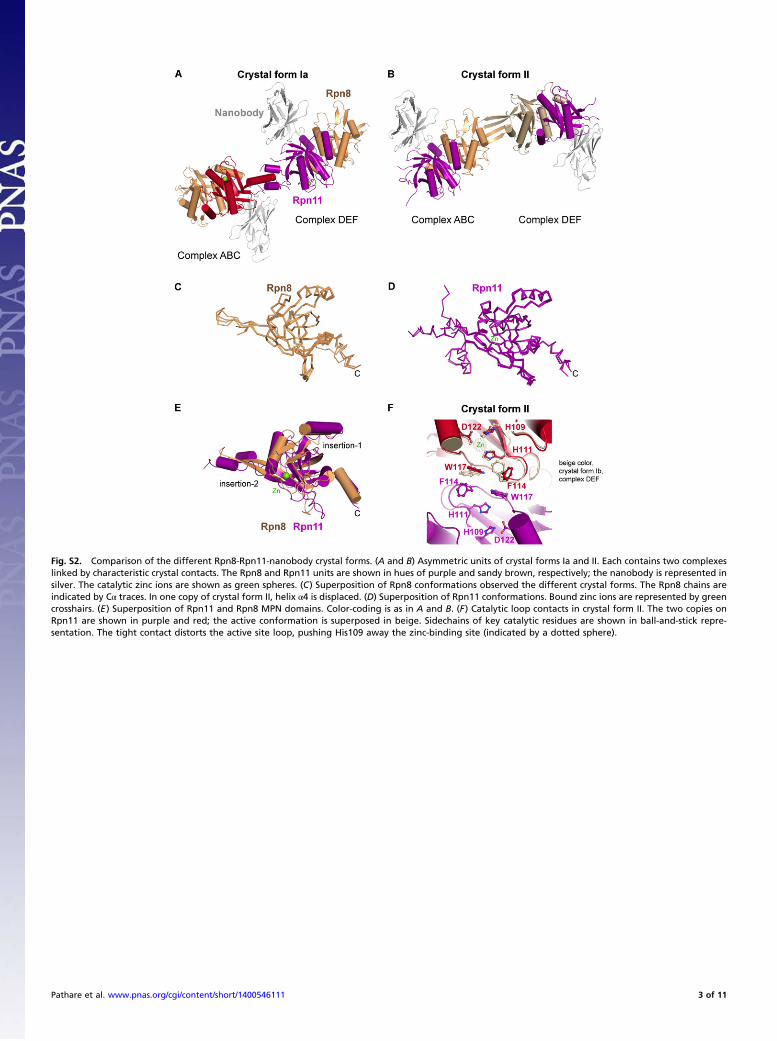
Fig. S2. Comparison of the different Rpn8-Rpn11-nanobody crystal forms. (A and B) Asymmetric units of crystal forms Ia and II. Each contains two complexeslinked by characteristic crystal contacts. The Rpn8 and Rpn11 units are shown in hues of purple and sandy brown, respectively; the nanobody is represented insilver. The catalytic zinc ions are shown as green spheres. (C) Superposition of Rpn8 conformations observed the different crystal forms. The Rpn8 chains areindicated by Cα traces. In one copy of crystal form II, helix α4 is displaced. (D) Superposition of Rpn11 conformations. Bound zinc ions are represented by greencrosshairs. (E) Superposition of Rpn11 and Rpn8 MPN domains. Color-coding is as in A and B. (F) Catalytic loop contacts in crystal form II. The two copies onRpn11 are shown in purple and red; the active conformation is superposed in beige. Sidechains of key catalytic residues are shown in ball-and-stick repre-sentation. The tight contact distorts the active site loop, pushing His109 away the zinc-binding site (indicated by a dotted sphere).
Pathare et al. www.pnas.org/cgi/content/short/1400546111 3 of 11
1 10 20 30 40 50
S.cerevisiae
Rpn8
P VL S D R F E L KVTI L L AL HYE KRC VI TI VTN FALP E D..........MS QHE A TQTK..EN VG LGDANSS R SN.crassa P VL S D R F E L SVSV L L VV HYN KRV VL NV VSN FAVP E D..MPATTAETLS VNR A TQANKSKS VG LGQNDGK R SP.brasiliensis P VL S D R F E L TVSV L L VA HYG RRV VL NV VSN FAVP E D..MPATTADTLS VTR A SAKG..TR VG LGQNDGK R SS.pombe P VL S D R F E I QVIV L L AV SYN RRV IL VV VAN YAIP E D.MPPAVSSETST VPQ H SAKG..TK VG LGQNNGD N SH.sapiens P VL S D R F E L KVVV L L VV HFN RVV LL VL VSN FAVP D D.........MPE AVQ H IGKVGNQK GV GSWQKK. D SD.melanogaster P VL S D R F E V KVIV L L VV HFN RVV LL VL VSN FAVP D D.......MPSQE SVN H MGKIGNQK GV GCWRSKG D SN.vectensis P VL S D R F E E TVVV I L VV HFN RVV LL VL VAN FAVP D D.......MPGPD AIT H MGKVGSQK GV GSRR.KG D CT.adhaerens P VL S D R F E T KVVV T L VV HFK RVV LL IL VSN FAVP E E..........MI KPE H LN....DK GI GSLKG.Q D SC.elegans P VL S D R F E L KVTV L L VV HFN RVV LL TL IGN FAVP D DGAHHCLGNVHAN PVN H VSKTQSVK GV GSMKKDK D SC.rheinhardtii P VL S D R F E A KVVL L L VV HYN RVV LL QI VTN FALP E D..........MP GPD H VAKDTK.K GV G.ELYKG D SA.thaliana P VL S D R F E R KVVV L L IV HYN RVV LL VV VTN YAVP E DMDVIKTQQIS.A TIE H VAKDSSK. GV GS.SSRG D SP.patens P VL S D R F E T NVVV L L IV NYN RVL LL RV ITN YAVP E DMDVISTSVGAGA GIE H VAKDTRK. GV GS.TFRG D SO.lucimarinus P VL S D R F E A EVVV L L VV HFR RVV LL RL VTS FAVP E DDGATAVAPRAAP SAS H CDEN..K. GV GE.QRKG D SD.discoideum P VL S D R F E T STIV T L VV HYN RVV LL VV VSN YGLP E D..........MS FPT H VAKDTNK. GA GSNNKG. D CN.gruberi P VL S D R F E A KLVV L L VV HYN RVA LL VV VTN FAIP E DAQRSSVDYTDFP FPT H VAKDTSN. GV GEITKDG D SE.hellem P VL S D R F E M SVIV L L AV HYK RVV LL EV ITE FACI E D............ NEV H KGTR.... GI GNE..DG H ST.vaginalis P VL S D R F E I RVAV I L IA HHN KRV IL QV VLQ FAIP E D......MSVQQA DIK H IVNN..RT IG LGDVYQG N CT.gondii P VL S D R F E V TVVV I L VV HYN RVV LL EI VTN FALP E DDSLSSFASVAGL ASK H VARGTSR. GT G.EISDG H SP.tetraurelia P VL S D R F E N LVII L I VV HYN RVV LL VI ITN YALP E D........MSNL KPP H IISKTQQP GA GERKADG D Sconsensus>50 .................vvvhPlVLlSvvDhynR........rvvg.llg........d!.ns%a.pF#E #
60 70 80 90 100 110 120
S.cerevisiae D GWY D N VWFL Y ENM M KKINAKE LI SGPKL S L I EL K LLLIVD VEK SD HN I NE C K H RA K N F KYTQNNP KQ.Q GN.crassa D GWY D D VWFL Y ESM M KKVNARE LI SGPKL S L I EL K LLVIVD VDK PS HN V ND F K H RA D N F RYTPN.P QP.K EP.brasiliensis D GWY D D VWFL F ESM M KKINARE LI SGPKL A L I EL K LLVIID VDK PS HN V ND F K H RA E N F RYTPN.P QP.K ES.pombe D GWY D N VWFL F ESM M KKINANE LV TGPQL S L I NL K VLVIID VEK AS HN M NE F K H RP E N L KYIPN.P KP.K SH.sapiens D GWY D D VWFL Y ENM M KKVNARE IV TGPKL N I I EL K VLVIID VDK DS HD L YG F R H HK A N M R.YCPNS KP.K DD.melanogaster D GWY D D VWFL Y ENM M KKVNARE VV TGPKL N I I EL R VLVIID ADK KS HD L YG F R H HQ A N V R.YCPNS KP.K DN.vectensis D GWY D D VWFL Y ENM M KKVNARE IV TGPKL N V I EL R VLVIID ADR QN HD L YA F R H HR V N I R.YCSNS KP.K DT.adhaerens D GWY D N IWYL Y ENM M KKVNARE VV TGPKI N I V EV K VLVIID AEK PD HD L YA F K H HS S N L R.FVANP QP.K DC.elegans D GWY D D TWFL Y ESM M YKVAAKE IV TGPKL N I I EQ K VLVIID ADK KS MD L YG F K H HK A N L R.FCPNP EP.K NC.rheinhardtii D GWY D D IWFL Y EQM M KKVNARE IV TGPKL S L I EL R VLVICE VDH TS HS L YK F K S RE D N M G.FCESP QP.K EA.thaliana D GWY D D IWFL Y ESM M KRINAKE VV TGPKL N L V AL N VLVIID VDK PS HN H FH F H S RE D H F G.YVPNP QP.K EP.patens D GWY D D IWFL Y ETM M KRINAKE VV TGPKL N L I EL R VLVIID VDK AS HN H FD F H S RE D N F D.YCPNP QP.R EO.lucimarinus D GWY D D IWFL Y ENM M KKISAKE IV TGPKL S I I EL Y VLVIVD VDG NG HS L YR S K S RE D H F A.YTPEP RA.E ND.discoideum D GWY D N IWFL F ENM M KKINARE VV TGPKI A Q I EL R VMVIID VEA PN HN H FA F N S RP D N F R.YTPNP AP.K EN.gruberi D GWY D D IWFF F ESM M RKVTAKE VV TGPKI S I I QI K TYCIID VSK PT HS L LA F K S RK E H I EKYLPHP NPEQ EE.hellem D GWY D E .WFI Y RSM L YKVNHKL II TGPKM N L I RS S FLAIIN VEN .. TS I FD F K H YE D T L K.FVENP HLGE NT.vaginalis D GWY D N IFFV F DEM L KKVTLKE II SLSTI N L I KV S IFLTTD VID PN TN I FK H K S SP E H I K.YNPTP GASD PT.gondii D GWY D D VWYL Y EHL M KKVNTRE VL TGPQV T L I EI R VYVIVD IPK PN RN H YH F R S RM E H F R.YTPNP NPKD .P.tetraurelia D GWY D D IWYL Y ETL L RKININE IV TGSRF N I I QI Y IFVIID VPK QN HI N FE H K S KP Q N F K.YTSTP HQFD Pconsensus>50 d.#..iw%lDhn%.e.$y.$fkk!na.e.ivGWY.tgpk....D..!.e.....y...pvlvii#v.p.. e
130 140 150 160 170 180 19 0
S.cerevisiae P F E EE E L R L T AY AIEQ KT L L TI A A IGV H L DVRDQAA GLSIRLT QL LKVG D V VKDDGT.STE H PC E E G N KS GN.crassa P F E EE E L R V T AY AVDE KT V T II A A IGV H L DIRDVAV TLSTRIT QM LQTG D F IKDDGT.TTS H PS E E G N RS GP.brasiliensis P F E EE E L R V T AY AVEE KT V T II A A IGV H L DIRDVAV TLSTRIT QL LQVG D F IKDDGT.TTS H PS E E G S QS GS.pombe P F E EE E L R L T AY AIDE RT V L SI A A IGV H L DTRDASV TLATRVT QA LQVG N F IEDDGS.KSS H PS E E G Q QS GH.sapiens P F E EE E L R L T AY SVEE KT E V EI A A VGV H L DIKDTTV TLSQRIT QV LKLG E I VHDDGT.PTS H TS G E G N HG GD.melanogaster P F E EE E L R L T AY SVEE KT E V EI A A VGV H L DIKDTTV SLSQKIT QL LKLG E I VHDDGS.PTS H PS G E G N MG GN.vectensis P F E EE E L R L T AY AVEE KT E I EI A A VGV H L DIKNLTA TLSQRIT QL LKLR D V VHDDGT.PTT H PS G E G N TS GT.adhaerens P F E EE E L R I T AF AIEE KT E L EI A P VGV H L DIKDSTA TLSQRIT YL LKLG D I VHDDGT.PTS N PS G E G S QS GC.elegans P F E EE E L R L T AY EVQE KT E V DI A A VGV H L DIKDQTA TLSQRIT QL LRIG E I VHDDGT.PPI H PS G E G D MG GC.rheinhardtii P F E EE E L R L F AY AVDE KV N L EV Q A IGV H L DVKDATL SLAGDVS KL LKVG T Y VRTDGTEKAK S PT G T S S MA GA.thaliana P F E EE E L R I T AY AVEE KV V V EI A V IGV H L DVKDTTI TLATEVT KL LKLG K Y VKENATQKSQ H ST A H S A TA GP.patens P F E EE E L R I T AY AVEG KA V V EI A A IGV H L DVKDATI TLATEVG KL LKLG K Y VKVDATKKSQ H AS G Y S G VA GO.lucimarinus P F E EE E L R I T AF AQIE KT V V SI A A IGV H L DVKDNTV TLSTKVS KV LRAN S A VKEDGTEKQQ H PN E F S E QS GD.discoideum P F E EE E L R I T SY TVEE MR Q I SI A A ICI H L DVKDSSI SLTTQIT KK LKLG K V INKDTS.EST H PS D V S D IS HN.gruberi P F E EE E L R I T AY AVEE LT T L EI A A IGV H L DVKDTTI DLASAVS RM LKNV D V REDQQS.QPK H SS G L S N TS AE.hellem P F E EE E L R L V TF LDEQ DE I V SI A A VGV H I DIREEAS SIAAKIN IK LL.D Q K .......... H GC E E G G ES VT.vaginalis P F E EE E L R I T AY GTER TT K L VV F V IGV H L DIKDVDV EIGTTLT SM LAHE C I ERVDGL.PLV T PT D L S N HG AT.gondii P F E EE E L R V T AY SFEQ RT V V TI A A VGV H L DLKNAST TLATRVA KL LKSV K Y PTSDRT..FR H AS G L S D SA LP.tetraurelia P F E EE E L R L T AY SVDE QN V I TV A P IGV Q L EINNVDT SLSAKAE KI VKLS E T ISKSGE..IV H PS Q F Q Q NG Gconsensus>50 ...Pt.a%..veev..d........F.h....!.a.EaEE!g!EhLlR#i.#.....l...i......l. g
βA α1 βB βC βi1a
βi1b α2 βD α3 βE
βF βG α4
A
Fig. S3. (Continued)
Pathare et al. www.pnas.org/cgi/content/short/1400546111 4 of 11
200 210 220 230 240 250 26 0
S.cerevisiae Y Q N PL KL V L V L N IL L DVF LL L QS KD VE DK INKE PI HT GK N GTPDDDEIDVEN..HDRINISNNLQKALTV KN.crassa Y Q N PL RL I L V L N IL L DVF LL L HH RD QA QK LDGQ PV HA GN N STPKSGPGATG......TNADSELNHAMSI KP.brasiliensis Y Q N PL RL I L V L N IL L DVF LL L HL RD GQ QK LDGD PV HA GN N STPKSSANAPVNGTEQSQTENTEVARAMSI KS.pombe Y Q N PL RL I L V L N IL L SVF LL I GQ TE AD RK VDGQ PI HA AE N FSGPVVS.........EQALESEAQRAFNV NH.sapiens Y Q N PL KL I L V L N II L DVF LL V NS LD RS EK ATGK PI HQ YQ D S...................LQEFVKAFYL KD.melanogaster Y Q N PL QL I L V M N IV L DIF LL I NA RD KQ QR GDSK PI HQ YQ D T...................NDQFTGTMYV KN.vectensis Y Q N PL RL I L V L N II L DVF LL L HA GD RD EK ATKK PV HT YE N D...................IEDFCKAVTV KT.adhaerens Y Q N PA EL I L V L N TI L EIN LL V QL DG TK DK IQGK PL HQ YT N N...................VEEFAKTFTV KC.elegans Y Q N PL QL I L I L N VI V EVL LL V QS ES EK HD VRGT PV HH YY D T...................HPDYIVSQNV QC.rheinhardtii Y Q N PL RL I L V L N VI V EIF LL M QG GE SE QL LDGK PT HD NI N N...................VESLSKSLAV KA.thaliana Y Q N PL RL I L V L N IL L DVF LL L DA RE RS DL IEGK PL HE YH N N...................VNELVKAFSV KP.patens Y Q N PL RL I L V L N IL L DVF LL L EA KE HA EL VEGK PL HE YH N N...................VHELVKSFAV KO.lucimarinus Y Q N PL RL I M V L N IM L DAF LL L KA EE KS DK VDGS PM HE GH N N...................LEDYVKGFNV SD.discoideum Y Q N PL NL M L V L N II I DII LS L LT QE QH KL CDGT PP HQ GY N N...................ANEISKSFAV QN.gruberi Y Q N PL RL V L V M N IL L DVF LL L RG SE NT SQ STGK PV HN YL G H...................TTDTKQSLVK KE.hellem Y Q N PY VL I L V I S II C EII SI L KG AE RS DD INGG AP QE NL K EKP........................... .T.vaginalis Y Q N PL RL I L V L E II I SIF LL L EH KA SD ND IEGK PI NE GV N T...................LKETVDSLAT KT.gondii Y Q N PL KI I L A I N IM I DIF LL L IG QE YA QD ANKK VA PN YT D S...................DPELIEAFTI QP.tetraurelia Y Q N PM KI I L I V N II L EIL YL L NK AQ QQ TL QQGK KP QL NN N G...................SQDVVQAFTT Kconsensus>50 l...l.ei..Y$d.v.dg.lp.nh.i....QdvfNllPnl......................e......v .
270 280 290 300 310 320 33 0
S.cerevisiae LTNDEL IYISNLVRSIIAFD I NKI K QE MV D E QN KI QRVKDKQSKVSDDSE.....SESGDKEATAPLIQR KN.crassa LTNDQL IYLSSLIRAITAFH I NKI R QE MA D E QN QQ ENDAKKKEGENGEKK...EGADKKEGSPAAANGES KP.brasiliensis LTNDQL IYLSSLIRAITAFH I NKI R QE MA D D QN QQ EQEAKKEEEKNGSMNNSINGNNNGKKSKSPTGNDS KS.pombe LSNDQL IYISSIVRAVIALH L SLA K EQ MS D D AS AM QDIKPTVQNGEVSAN.................AEQ KH.sapiens LTNDQM VYLASLIRSVVALH I NKI R EK VV N N AN DA KEGQEKEESKKDRKEDKEKDKDKEKSDVKKEEKKE KD.melanogaster LTNDQM VYLASMVRSIIALH I NKL R EE LV N N AN DA GKSDSKEAKEKNKDSKDKDNKETKDKDGKKAEEKA DN.vectensis LTNDQM VYVASLIRSIIALH I NKV R EK LV N S TN DA DEASKKDEKSKEKEKEKEKEKDKDTKEKDSQGKSK DT.adhaerens LTNDQM MYLASMIRAIIALH I NKE R EL LV N N AN NA NEESSSDKPDKSDNQGMYRSIFYSHRNFI...... .C.elegans LTNDQL VYMGSLVRSVVALH I NKI Q EK MC N D SL KA EQETGEAEKKKDEKDKKDKKDEKKDEKKEKDSKSS TC.rheinhardtii LSNDMM IYVASMVRSILALH I NKE L EK HV K D GR WA EAAKKEKEKAEDKAKKEAKEKEEKEKADAKAKADG KA.thaliana LTNDMM IYLSSLIRSVIALH I NKL K EK LV N N LN EH AEDSKPVAIPATS...................... .P.patens LTNDMM IYLSSLIRSVIALH I NKI K ER LV N N LN EH IADAASASVSAITV..................... .O.lucimarinus LTNDAM VYLSSLIRSVIALH I NKA K ER LV D N TN ER ALDAPGASDAEKDTDKENEKPKDSGKADAAK.... .D.discoideum LNNDTM IYLSSMIRSIIALH I NKT R EK SV N I AN EA KADIINSTPPTTATSPSVADKGKEKEQNAFNGADK PN.gruberi LTNDTY MYLGSVIRCIVSLH I NKI R EA LA D N EM EI KQSKKEAEKKEEAEKTSTETKEVQKTSQDKK.... .E.hellem LLDENL CYVSVLAKTVVALN R NRL G TS SD D R EN IE AS................................. .T.vaginalis LADDET IFISQLCRSVVSLH V TRH L KE FM D N PP AE KAKEEAEKPKN........................ .T.gondii LANDTM LYLGSVVRSVLALH I NKV K SE LN N N EN RA EKKEKKEDEETGETKKEAGTDTKKGEKESSQ.... .P.tetraurelia LNNDNM IYLASLMRSIIAYH I NQA E VK LT N N QQ KK ................................... .consensus>50 .##.ml.i%..slir.!ialhnLinnk..n...e.................................... .
S.cerevisiae NKKN..........N.crassa EKENSPKEKKK...P.brasiliensis EEQDGSKEKGKKKGS.pombe A.............H.sapiens K.............D.melanogaster KGKDEGGKGSRK..N.vectensis KDKASDASKK....T.adhaerens ..............C.elegans PNTPKK........C.rheinhardtii AEDKDGKK......A.thaliana ..............P.patens ..............O.lucimarinus ..............D.discoideum SKQA..........N.gruberi ..............E.hellem ..............T.vaginalis ..............T.gondii ..............P.tetraurelia ..............consensus>50 ..............
Rpn8
Fig. S3. (Continued)
Pathare et al. www.pnas.org/cgi/content/short/1400546111 5 of 11
1 10 20 30 40 50 6 0
S.cerevisiae D E S AL KML H G P EV GL LG D TK TV I SI L K GRA V M M M EFV D.........MERLQRLMMNSKVGSADTGRD YN.crassa D E S AL KML H G P EV GL LG D NS TV I SL L R GRA V M M M EFV D..MDRMRSLLGGGLGMGGATAPGADNTNLI YP.brasiliensis D E S AL KML H G P EV GL LG D NS TV I SL L R GRA V M M M DFV E................MPNSAASQDAPNLI HS.pombe D E S AL KML H G P EV GL LG D NS CV I SL L R GRH T M M M EFV D......MESLQRLLQGARMGTGMMGDQPLV YH.sapiens D E S AL KML H G P EV GL LG D TA QV I SL L K GRA V M M M EFV D....MDRLLRLG.GGMPGLGQGPPTDAPAV YN.vectensis D E S AL KML H G P EV GL LG D TA QV I SL L K GRA V M M M EFV D....MDRILRLGSGGIPGVGQAPPSDAPVV YD.melanogaster D E S AL KML H G P EV GL LG D TA QV I SL L K GRA V M M M EFV D....MDRLLRLG...GAMPQAAPPTDAPVV YT.adhaerens D E S AL KML H G P EV GL LG D TA QV I SL L R GRA V M M M QFV E....MDRLLRLGSGGLPGVGQ.PPPEAPNV YC.elegans D E S AL KML H G P EV GL LG D TS TV I SL L K GRA V M M M EFV D...MERFLRLGGLGGNLGTFGANPQDSNQV YS.moellendorffii D E S AL KML H G P EV GL LG D TS QV I SL L K GRA V M M M DFV E..MERLQRIISSGAGGAGGGGMPHPDSPLV YA.thaliana D E S AL KML H G P EV GL LG D TS QV I SL L K GRA V M M M EFV E..MERLQRIFGAGGG....LGHASPDSPTL YC.rheinhardtii D E S AL KML H G P EV GL LG D TA QI I SL L K GRA V M M M EFV D..MARHRSPAAAG.........PPTDTPQV YO.lucimarinus D E S AL KML H G P EV GL LG D TA QV I SL L K GRA V M M M QFV E..MQRLMQSGGMPGAMPG.....AGDAAQV YN.gruberi D E S AL KML H G P EV GL LG D TA TV I SL L K GRA V M M M EFI D....MDPRLQRLIMQQMGGGGGMQGDQPLP TD.discoideum D E S AL KML H G P EV GL LG D TA TI I SL L Q ARA V L M M ELI E.....MNRSLMSLLGREGLGEKITDATPLP HT.gondii D E S AL KML H G P EV GL LG D TS QV I SL L K GRA V M M M EFI DMAGLPSNLRGLLQQFGGMGVGPPNRDQPMA YE.hellem D E S AL KML H G P EV GL LG D AS TV I SL L K GRA I L M M EFV E...............MFNGFVGTDDAEPTS QP.tetraurelia D E S AL KML H G P EV GL LG D TA QV I AL I K ARA I F M L DIV D.......MDIQNLIQQFGAGQAVNPEASIP TT.vaginalis D E S AL KML H G P EV GL LG D TS TV V GM L K GKS I L C M RFI D...........MFDHDTFAMEGMRTQPAHF Yconsensus>50 .........................d....Dt.E.!y!SslALlKMLkHgraGvPmEVmGL$LGef!D #
70 80 90 100 110 120
S.cerevisiae DVF P M L GR GWYHSHP G W S D QYTVNVV AM QSGTGVS VEAVD VFQAK M M KQT Q VV GF C L V VNT K S . D D D M SN.crassa DVF P M L GR GWYHSHP G W S D QFTVRVV AM QSGTGVS VEAVD VFQMN M M RQT E VV GF C L V INT Q S . P D P A SP.brasiliensis DVF P M L GR GWYHSHP G W S D QFTVRVV AM QSGTGVS VEAVD VFQTK M M RQT E VV GF C L V INT Q S . P E P T SS.pombe DVF P M L GR GWYHSHP G W S D QFTVRVV AM QSGTGVS VEAVD VFQKN M M KQT E VV GF C L V INT Q S . P D P M SH.sapiens DVF P M L GR GWYHSHP G W S D QYTVRVI AM QSGTGVS VEAVD VFQAK L M KQT E VV GF C L V INT Q S . P D P M GN.vectensis DVF P M L GR GWYHSHP G W S D QYTVRVI AM QSGTGVS VEAVD VFQAK L M KQT E VV GF C L V INT Q S . P D P M GD.melanogaster DVF P M L GR GWYHSHP G W S D QYTVQVI AM QTGTGVS VEAVD VFQAK L M KQT E VV GF C L V INT Q S . P D P M GT.adhaerens DVF P M L GR GWYHSHP G W S D QYTVRVI AM QSGTGVS VEAVD VFQAR L M KQT E VV GF C L V VNT Q S . P D P M GC.elegans DVF P M L GR GWYHSHP G W S D QYTVNVI AM QSGTGVS VEAVD VFQAK L M KQT E VV GF C L V INT Q S . P D P M GS.moellendorffii DVF P M L GR GWYHSHP G W S D QYTVRVV AM QSGTGVS VEAVD VFQTK M M KQT E VV GF C L V INT Q S . P D S M GA.thaliana DVF P M L GR GWYHSHP G W S D QYTVRVV AM QSGTGVS VEAVD VFQTN L M KQT E VV GF C L V INT Q S . H D P M GC.rheinhardtii DVF P M L GR GWYHSHP G W S D QYTVRVV AM QSGTGVS VEAVD VFQTK L M KQV E VV GF C L V INT Q S . P D P M GO.lucimarinus DVF P M L GR GWYHSHP G W S D QYTVTVV AM QSGTGVS VEAVD VFQTK L M KQT E VV GF C L V INT Q S . P D E M GN.gruberi DVF P M L GR GWYHSHP G W S D QYTVRCI AM QSGTGVS VEAVD VFQTK L L KQT E VV GF C L V INT Q S . P E P M SD.discoideum DVF P M L GR GWYHSHP G W S D QYTIRVI AM QSGTSVS VEAID VFQTK L M KQT E VI GF C L V VNT Q S . P D D I ST.gondii DVF P M L GR GWYHSHP G W S D QYTVRVV SM QSGNSVS VEAVD VYQTE L Q KRT E VV GF C F T VNT Q S . P E P M GE.hellem DVF P M L GR GWYHSHP G W S D QYTVKVV AM QSGTNVT VESVD IFQTE M I KAT E VV GF C L V IST Q S . P N H T TP.tetraurelia DVF P M L GR GWYHSHP G W S D QYHIRVY SM QTASECF GICGA FFNKK V L NLT E CI SY C L V INT Q S R Q E M N ST.vaginalis DVF P M L GR GWYHSHP G W S D QYTVHVV PV STGTGTA VEAID VYQIS T M KSV E VI GF V L V INQ L Y . E K Q D Nconsensus>50 %t!.vvDVFamPqsgtgvs.veavdpv%#..MldmLkqtGR.#.v!GWYHSHPg%GcWlS.vD!ntQq s
130 140 150 160 170 180 19 0
S.cerevisiae E VV DP QSV GKV I FR I E R TS G G LF QL RAVA V I K V DA P QT NT LNKANIQALIH NR NS L ..DTGALINNL L HN.crassa E VV DP QSV GKV I FR I E R TS G G LF QL RAVA I I K V DA P QT NL LNKPSIQALIH NR NS L ..NPQSLMLGQ H HP.brasiliensis E VV DP QSV GKV I FR I E R TS G G LF QL RAVA V I K V DA P QT NL LNKPSIQALIH NR TP L ..SSQTLMMGQ H HS.pombe E VV DP QSV GKV I FR I E R TS G G LF QL RAVA V I K V DA P QT NL INKPSIQALIH GR TP L ..NPSTLMMGQ H HH.sapiens E VV DP QSV GKV I FR I E R TS G G LF AL RAVA V I K V DA P QT NL LNKPSIQALIH NR SE L ..NANMMVLGH H HN.vectensis E VV DP QSV GKV I FR I E R TS G G LF AL RAVA V I K V DA P QT NL LQKPSIQAMIH NR SE L ..NPNMMVLGQ H HD.melanogaster E VV DP QSV GKV I FR I E R TS G G LF AL RAVA V I K V DA P QT NL LQKPSVQALIH NR SE L ..NPNMLVLGQ H HT.adhaerens E VV DP QSV GKV I FR I E R TS G G LF AL RAVA V I K V DA P QA II LQKPSIQALIH NR SE L ..HPTLIMVGH H SC.elegans E VV DP QSV GKV I FR I E R TS G G LF AL RAVA V I K V DA P QT NL LQKPSIQALIH NR SD T ..NPQSMALNQ H HS.moellendorffii E VV DP QSV GKV I FR I E R TS G G LF AL RAVA V I K V DA P QT NL LNKPSIQALIH NR NQ L ..NPQTMMLGQ H HA.thaliana E VV DP QSV GKV I FR I E R TS G G LF AL RAVA V I K V DA P QT NL LNKPSIQALIH NR NQ S ..NPQTIMLGQ H HC.rheinhardtii E VV DP QSV GKV I FR I E R TS G G LF AL RAVA V V K V DA P QT NL LNKPSIQALIH NR NN L ..SPQTMMLGQ H HO.lucimarinus E VV DP QSV GKV I FR I E R TS G G LF QL RLVA I V R V DA P QT NL LNKPSISALIH NR NP L ..NPQTIMLGQ H HN.gruberi E VV DP QSV GKV I FR I E R TS G G LF SL RSVA V I K V DA P QS VQ LNKPTIQALIH NR TK T ..NPQLAMFGQ H HD.discoideum E VV DP QSV GKV I FR I E R TS G G LF QL RAVA V L R V DA P QI NL LQDPSIQALIH NR QS T ..KTSPTA... H NT.gondii E VV DP QSV GKV I FR I E R TS G G LF QL RAVG V I K V DC L QT NI LQRPTISALVH NR NP L ..NPHLLMLGQ H NE.hellem E VV DP QSV GKV I FR I E R TS G G LF KL RAVA V I K V DA P QV NI LKTPTLISIIH NK CK L ..DNQLGVLGG Y HP.tetraurelia E VV DP QSV GKV I FR I E R TS G G LY QL KSIA I I R V DA P QT NT LQKPGLEALLR NR NK L ..PQQNMLSQQ H YT.vaginalis E VV DP QSV GKV I FR I E R TS G G LW KI RCIA V V R I GA P ET FI LEKPSIKALVR NK NP C PQNQMTFQPNT S Lconsensus>50 fE.l..ra!aVV!DPiQSVkGKV!IdaFRlI..n......g.EpR#tTSn.GhlnkpsiqalihGLnr h
βA α1 βB
βC η1 α2 βD α3
βE βF αi2a αi2b
Rpn11B
Fig. S3. (Continued)
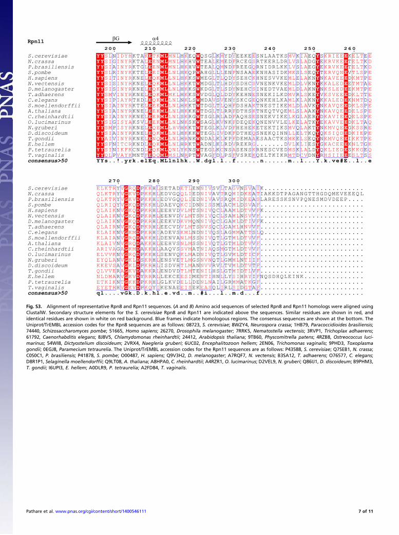
Pathare et al. www.pnas.org/cgi/content/short/1400546111 6 of 11
200 210 220 230 240 250 26 0
S.cerevisiae YY E ML W Y E SL IDYH K TK MNL K L M Y E MV IA KRIEE ELT E N KTA H EQ QSG K YD EEKE SNLAATKS K EQ S K EN.crassa YY E ML W Y E SI INYR L EN MNL K L M F S LV LA KRVKE ELT D G KTA H HV TEA E ED RCEG RTKERLDR S DG E T KP.brasiliensis YY E ML W Y E SI INYR L EN MNL K L M F Q LV LA KRVKE ELT D A KTG H HV TEA Q ND REEG RNIDRLKK S EG E T KS.pombe YY E ML W Y E SL INYK L EI LNL K L L F E MK LS ERVQN TLS E R KTE H QP AHG L EN NSAA KNHASIDK S EQ T V PH.sapiens YY E ML W Y E SI INYR L QK LNL K L L Y K ML LA KAVEE KMT E T KNE H KS MEG T QD SEHC HNESVVKE E KN N D PN.vectensis YY E ML W Y E SI INYR L QK LNL K L L Y S ML LV KALED TMT E S KNE H KT MDG T HD SDHC VNENKVKE D KN K E AD.melanogaster YY E ML W Y E SI INYR L QK LNL K L L Y S ML LA KSLED KMT E S KNE H KS KDG T SD NEHC INEDTVAE D KN N E PT.adhaerens YY E ML W Y E SM INYR L QK MKL K L L Y E MV LS KSVEE KLT E V KNE H KS VDG Q DD KEHN SNEKILKD R KE V D TC.elegans YY E ML W Y E SI IAYR L QK LNL K V V Y E ML LA KALED NMT Q P THD N LS MDA S EN SKCG QNKEHLKA K KN K K DS.moellendorffii YY E ML W Y E SI INYR L EK LNL K L L F K ML LA KAVQE KLS E A KTE H KT TDG T QH DSHA TNESTIKE D VK N D PA.thaliana YY E ML W Y E SI INYR L EK LNL K L L F K ML LA KAVQE ELS E A KNE H KK TDG T RR DTHS TNEQTVQE S AK N D PC.rheinhardtii YY E ML W Y E SI INYR L ER LNL K L L F E LK LA KAVIE ELS E A KNE S RG TSG R AD AQHS SNEKVIKE G ER D Q PO.lucimarinus YY E ML W Y E SI ISYA L EK LNL K L V F K LK LA KAVVE KLT Q G KSV N SK SAG K NK DEQE QNENVVLE E TK E D AN.gruberi YY E ML W Y E SM ISYR L QE LHV K L L F K MV LA KMVQE KSS E P KNE H KT TEG K VD HEHE ETEKTIKS Q KT N G RD.discoideum YY E ML W Y E SI INYR L QK LNL K L V F Q LL LT KSIQD KIE E A KNE H KK TEG I DK DTHE SNEKQINN E KQ Q D PT.gondii YY E ML W Y E AI INYR L NQ LNL R L L F A MK LS KMVQE KKT E V KNE H NK NDA K KP DEMA ESAACTKS E EQ N I PE.hellem YY E ML W Y E SF ITCR L QK LNL R L L V G VL LI KACEE NLT K N KND H KT ADN K RD REKR .......D K ES G K GP.tetraurelia YY E ML W Y E SI IKFK L QK QNL K L C A K MS LA KLIED KKG Q N CND Y NS TEG K NS SENS RNESCVED K LD Q S ET.vaginalis YY E ML W Y E QL VAYK F QQ MSL R F L F K MT CV HSIIE SLT S P MNT N PT VAG D PS VSRE QELTKIKR D DN R E Sconsensus>50 YYs..!.yrk.elEq.MLlnlhk..W.dgl.l..f......n......m..l...Y.k.ve#E..l.. e
270 280 290 300
S.cerevisiae G DELKTR V RQ KK L ETL NIV L GV SV I Y P H SETAD EN SV TA N A K........................ .N.crassa G DQLKTR V KV KK L QLI NIV T MI KE T Y P H EDVGQ ED AV RQ D A IAKKDTPAGANGTTHGDQMEVEEEQ LP.brasiliensis G DQLKTR V KV KK I QLI NIV S MI KE S Y P H EDVGQ ED AV RQ D A LARESSKSNVPQNESMDVDEEP... .S.pombe G DQLRIQ V KQ KK L KCI NIS L ML SV F Y P H DAEVQ DN SM AC D A ......................... .H.sapiens G DQLAIK V KQ KR L VLM NIV L ML TV F N P H EEHVD TS QC AA D V K........................ .N.vectensis G DQLAIK V KQ KR L VLM NIV L ML SV F N P H EENVD TS QC SA N V ......................... .D.melanogaster G DQLAIK V KQ KR L KVM NIV L ML TI F N P H EEKVD QN QC GA D V K........................ .T.adhaerens G DQLAIR V KQ KR L VLM NVS L ML NV F N P H EECVD TS QC GA H V ......................... .C.elegans G DELAIK V KM KR I KML NIV L MM TT L N P H ADEVS ND QS AG A S Q........................ .S.moellendorffii G DKLAIA V RQ KK L NLM NIV L ML TV F N A H DENVA SS QT GT D V ......................... .A.thaliana G DKLAIV V RQ KK L NLM NIV L ML TV F N A H EEHVS SS QT GT D V ......................... .C.rheinhardtii G DARIVA A KM KK L SVM NIA M ML TV F G A H AAQVS AT QS GT D V ......................... .O.lucimarinus G DELVVK V RQ KK L KLM NIV L ML TI F N P H SENVQ AD QT GV D C ......................... .N.gruberi G DEYQLA V KL KR L TLM NVN L ML TI F N S H ENSVE GS QT GM N I ......................... .D.discoideum G DKKEVS V KL KR L TLM NVV L ML TV F A P H ISDVH AN RV TV D T ......................... .T.gondii G DQLVVE A KV KK L TLM NIL L MI TL F R A R ENDVD TE HS GT D V ......................... .E.hellem G DNLDMA V KV RR L ESI NTI L SI RY F R Y R LEKCE ME HN LY H I NQSDHQLEINK.............. .P.tetraurelia G DETKIK T KK KR L ELL NLN L MM TK F N P H GLKVD DE AI GR A G ......................... .T.vaginalis G DEYETR V KV KQ I EIS EAS I SI GT F H P Y KENAE EK QL RL D A ......................... .consensus>50 ql....vGk.D.k.hl.e.vd..m..#i...l..m.d...f......................... .
βG α4Rpn11
Fig. S3. Alignment of representative Rpn8 and Rpn11 sequences. (A and B) Amino acid sequences of selected Rpn8 and Rpn11 homologs were aligned usingClustalW. Secondary structure elements for the S. cerevisiae Rpn8 and Rpn11 are indicated above the sequences. Similar residues are shown in red, andidentical residues are shown in white on red background. Blue frames indicate homologous regions. The consensus sequences are shown at the bottom. TheUniprot/TrEMBL accession codes for the Rpn8 sequences are as follows: 08723, S. cerevisiae; 8WZY4, Neurospora crassa; 1HB79, Paracoccidioides brasiliensis;74440, Schizosaccharomyces pombe; 51665, Homo sapiens; 26270, Drosophila melanogaster; 7RRK5, Nematostella vectensis; 3RVP1, Trichoplax adhaerens;61792, Caenorhabditis elegans; 8J8V5, Chlamydomonas rheinhardtii; 24412, Arabidopsis thaliana; 9TB60, Physcomitrella patens; 4RZB8, Ostreococcus luci-marinus; 54WI8, Dictyostelium discoideum; 2VRX4, Naegleria gruberi; 6UC02, Encephalitozoon hellem; 2EN06, Trichomonas vaginalis; 9PHD3, Toxoplasmagondii; 0EGJ8, Paramecium tetraurelia. The Uniprot/TrEMBL accession codes for the Rpn11 sequences are as follows: P43588, S. cerevisiae; Q7SEB1, N. crassa;C0S0C1, P. brasiliensis; P41878, S. pombe; O00487, H. sapiens; Q9V3H2, D. melanogaster; A7RQF7, N. vectensis; B3SA12, T. adhaerens; O76577, C. elegans;D8R1P1, Selaginella moellendorffii; Q9LT08, A. thaliana; A8HPA0, C. rheinhardtii; A4RZR1, O. lucimarinus; D2VEL9, N. gruberi; Q86IJ1, D. discoideum; B9PHM3,T. gondii; I6UPI3, E. hellem; A0DLR9, P. tetraurelia; A2FD84, T. vaginalis.
Pathare et al. www.pnas.org/cgi/content/short/1400546111 7 of 11
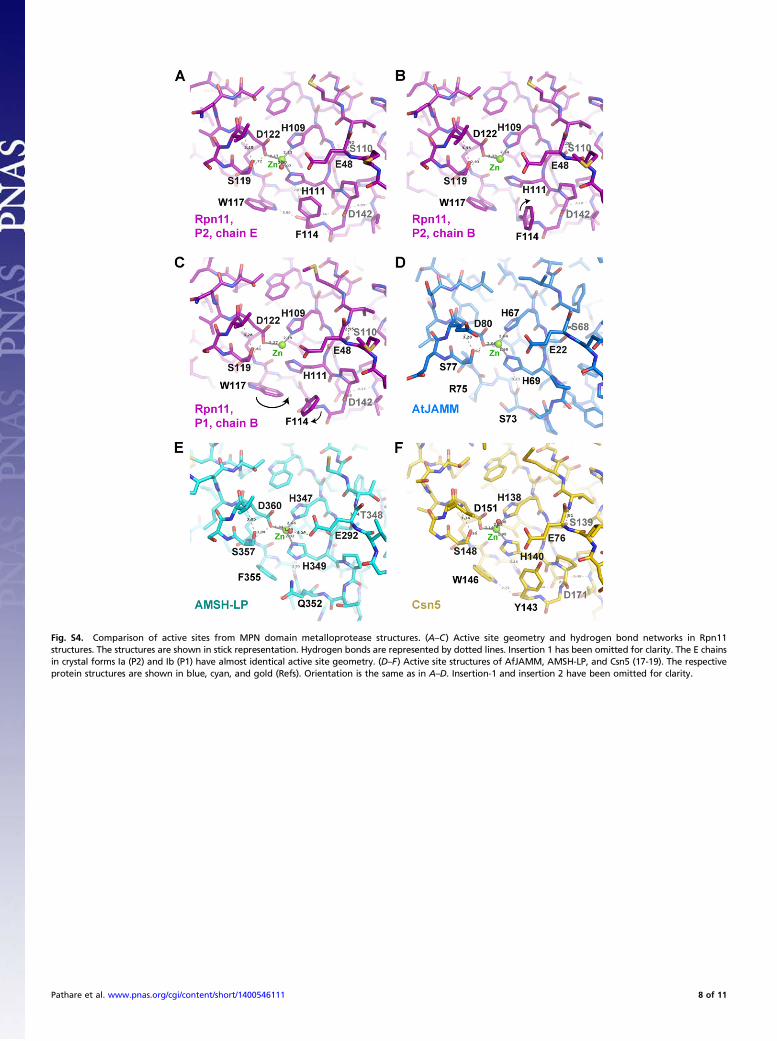
Fig. S4. Comparison of active sites from MPN domain metalloprotease structures. (A–C) Active site geometry and hydrogen bond networks in Rpn11structures. The structures are shown in stick representation. Hydrogen bonds are represented by dotted lines. Insertion 1 has been omitted for clarity. The E chainsin crystal forms Ia (P2) and Ib (P1) have almost identical active site geometry. (D–F) Active site structures of AfJAMM, AMSH-LP, and Csn5 (17-19). The respectiveprotein structures are shown in blue, cyan, and gold (Refs). Orientation is the same as in A–D. Insertion-1 and insertion 2 have been omitted for clarity.
Pathare et al. www.pnas.org/cgi/content/short/1400546111 8 of 11
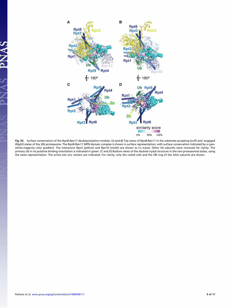
Fig. S5. Surface conservation of the Rpn8-Rpn11 deubiquitylation module. (A and B) Top views of Rpn8-Rpn11 in the substrate-accepting (Left) and -engaged(Right) states of the 26S proteasome. The Rpn8-Rpn11 MPN domain complex is shown in surface representation, with surface conservation indicated by a cyan-white-magenta color gradient. The interactors Rpn2 (yellow) and Rpn10 (violet) are shown as Cα traces. Other lid subunits were removed for clarity. Theprimary Ub in its putative binding orientation is indicated in green. (C and D) Bottom views of the docked crystal structure in the two proteasomal states, usingthe same representation. The active site zinc centers are indicated. For clarity, only the coiled coils and the OB ring of the AAA subunits are shown.
Pathare et al. www.pnas.org/cgi/content/short/1400546111 9 of 11
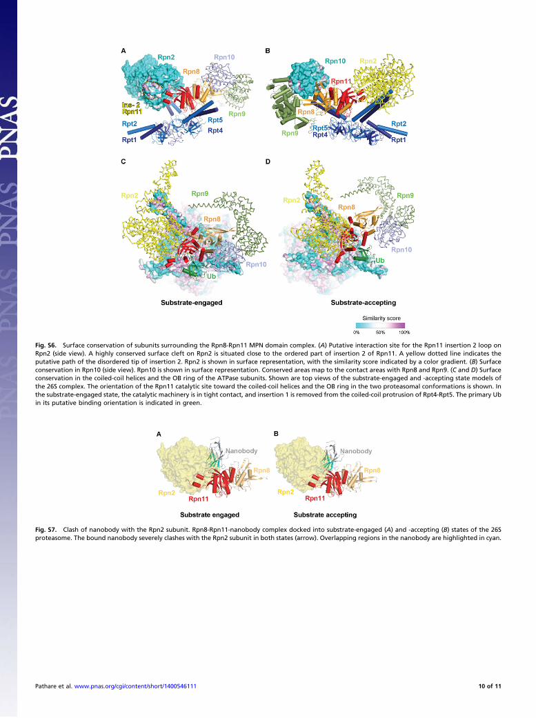
Fig. S6. Surface conservation of subunits surrounding the Rpn8-Rpn11 MPN domain complex. (A) Putative interaction site for the Rpn11 insertion 2 loop onRpn2 (side view). A highly conserved surface cleft on Rpn2 is situated close to the ordered part of insertion 2 of Rpn11. A yellow dotted line indicates theputative path of the disordered tip of insertion 2. Rpn2 is shown in surface representation, with the similarity score indicated by a color gradient. (B) Surfaceconservation in Rpn10 (side view). Rpn10 is shown in surface representation. Conserved areas map to the contact areas with Rpn8 and Rpn9. (C and D) Surfaceconservation in the coiled-coil helices and the OB ring of the ATPase subunits. Shown are top views of the substrate-engaged and -accepting state models ofthe 26S complex. The orientation of the Rpn11 catalytic site toward the coiled-coil helices and the OB ring in the two proteasomal conformations is shown. Inthe substrate-engaged state, the catalytic machinery is in tight contact, and insertion 1 is removed from the coiled-coil protrusion of Rpt4-Rpt5. The primary Ubin its putative binding orientation is indicated in green.
Fig. S7. Clash of nanobody with the Rpn2 subunit. Rpn8-Rpn11-nanobody complex docked into substrate-engaged (A) and -accepting (B) states of the 26Sproteasome. The bound nanobody severely clashes with the Rpn2 subunit in both states (arrow). Overlapping regions in the nanobody are highlighted in cyan.
Pathare et al. www.pnas.org/cgi/content/short/1400546111 10 of 11
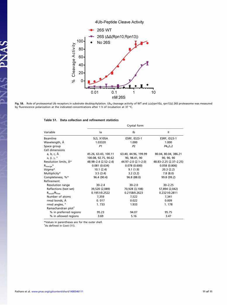
Fig. S8. Role of proteasomal Ub receptors in substrate deubiquitylation. Ub4 cleavage activity of WT and ΔΔ(rpn10Δ, rpn13Δ) 26S proteasome was measuredby fluorescence polarization at the indicated concentrations after 1 h of incubation at 37 °C.
Table S1. Data collection and refinement statistics
Crystal form
Variable Ia Ib II
Beamline SLS, X10SA ESRF, ID23-1 ESRF, ID23-1Wavelength, Å 1.03320 1.000 1.000Space group P1 P2 P43212Cell dimensionsa, b, c, Å 45.26, 63.43, 100.11 63.40, 44.96, 199.99 80.04, 80.04, 386.21α, β, γ, ° 100.08, 92.75, 90.62 90, 98.41, 90 90, 90, 90
Resolution limits, Å* 48.98–2.4 (2.52–2.4) 44.97–2.0 (2.1–2.0) 48.83–2.25 (2.37–2.25)Rmerge* 0.081 (0.634) 0.078 (0.600) 0.058 (0.806)I/sigma* 10.1 (2.4) 9.1 (1.9) 20.3 (2.2)Multiplicity* 3.5 (3.4) 3.2 (3.2) 7.8 (8.0)Completeness, %* 96.4 (90.4) 96.8 (88.0) 99.8 (99.2)RefinementResolution range 30–2.4 30–2.0 30–2.25Reflections (test set) 39,520 (2,089) 70,928 (3,108) 57,894 (2,042)Rwork/Rfree 0.1951/0.2522 0.2158/0.2623 0.2321/0.2811Number of atoms 7,359 7,522 7,341rmsd bonds, Å 0. 017 0.022 0.009rmsd angles, ° 1. 733 1.933 1. 178Ramachandran plot†
% in preferred regions 95.23 94.07 95.75% in allowed regions 3.69 5.16 3.47
*Values in parentheses are for the outer shell.†As defined in Coot (11).
Pathare et al. www.pnas.org/cgi/content/short/1400546111 11 of 11