Massive cellular disruption occurs during early imbibition of Cuphea seeds containing crystallized...
12
Planta (2006) 224:1415–1426 DOI 10.1007/s00425-006-0310-4 123 ORIGINAL ARTICLE Massive cellular disruption occurs during early imbibition of Cuphea seeds containing crystallized triacylglycerols Gayle M. Volk · Jennifer Crane · Ann M. Caspersen · Lisa M. Hill · Candice Gardner · Christina Walters Received: 18 January 2006 / Accepted: 18 April 2006 / Published online: 9 June 2006 © Springer-Verlag 2006 Abstract The transition from anhydrobiotic to hydrated state occurs during early imbibition of seeds and is lethal if lipid reserves in seeds are crystalline. Low temperatures crystallize lipids during seed stor- age. We examine the nature of cellular damage observed in seeds of Cuphea wrightii and C. lanceolata that diVer in triacylglycerol composition and phase behavior. Intracellular structure, observed using trans- mission electron microscopy, is profoundly and irre- versibly perturbed if seeds with crystalline triacylglycerols are imbibed brieXy. A brief heat treat- ment that melts triacylglycerols before imbibition pre- vents the loss of cell integrity; however, residual eVects of cold treatments in C. wrightii cells are reXected by the apparent coalescence of protein and oil bodies. The timing and temperature dependence of cellular changes suggest that damage arises via a physical mechanism, perhaps as a result of shifts in hydrophobic and hydrophilic interactions when triacylglycerols undergo phase changes. Stabilizers of oil body struc- ture such as oleosins that rely on a balance of physical forces may become ineVective when triacylglycerols crystallize. Recent observations linking poor oil body stability and poor seed storage behavior are potentially explained by the phase behavior of the storage lipids. These Wndings directly impact the feasibility of pre- serving genetic resources from some tropical and sub- tropical species. Keywords Cuphea · DiVerential scanning calorimetry · Imbibitional damage · Intermediate seeds · Liposome · Recalcitrant seeds Abbreviations DSC DiVerential scanning calorimetry GRIN Germplasm resources information network TEM Transmission electron microscopy Introduction Water interactions with crystallized storage lipids are lethal to seeds (Crane et al. 2003, 2006) and may cause a syndrome known as intermediate storage behavior (Ellis et al. 1990). Seeds exhibiting this storage behavior sur- vive drying to very low water contents, but do not survive low temperature storage or the combined eVects of drying and cooling. Seeds in this category usually orig- inate from tropical or subtropical regions and often have storage lipids that crystallize above ¡10°C and melt at temperatures above 25°C, respectively. A simple heat exposure that melts the triacylglycerols before seeds imbibe eliminates evidence of damage, and allows seeds to germinate normally (Crane et al. 2003, 2006). This publication is dedicated in memory of Dr. Vincent Franceschi, a mentor and friend. Mention of trade names or commercial products in this article is solely for the purpose of providing speciWc information and does not imply recommendation or endorsement by the US Department of Agriculture. G. M. Volk · J. Crane · A. M. Caspersen · L. M. Hill · C. Walters (&) USDA-ARS National Center for Genetic Resources Preservation, 1111 S. Mason Street, Fort Collins, CO 80521, USA e-mail: [email protected] C. Gardner USDA-ARS North Central Regional Plant Introduction Station, Ames, IA, USA
-
Upload
independent -
Category
Documents
-
view
0 -
download
0
Transcript of Massive cellular disruption occurs during early imbibition of Cuphea seeds containing crystallized...
Planta (2006) 224:1415–1426
DOI 10.1007/s00425-006-0310-4ORIGINAL ARTICLE
Massive cellular disruption occurs during early imbibition of Cuphea seeds containing crystallized triacylglycerols
Gayle M. Volk · Jennifer Crane · Ann M. Caspersen · Lisa M. Hill · Candice Gardner · Christina Walters
Received: 18 January 2006 / Accepted: 18 April 2006 / Published online: 9 June 2006© Springer-Verlag 2006
Abstract The transition from anhydrobiotic tohydrated state occurs during early imbibition of seedsand is lethal if lipid reserves in seeds are crystalline.Low temperatures crystallize lipids during seed stor-age. We examine the nature of cellular damageobserved in seeds of Cuphea wrightii and C. lanceolatathat diVer in triacylglycerol composition and phasebehavior. Intracellular structure, observed using trans-mission electron microscopy, is profoundly and irre-versibly perturbed if seeds with crystallinetriacylglycerols are imbibed brieXy. A brief heat treat-ment that melts triacylglycerols before imbibition pre-vents the loss of cell integrity; however, residual eVectsof cold treatments in C. wrightii cells are reXected bythe apparent coalescence of protein and oil bodies. Thetiming and temperature dependence of cellularchanges suggest that damage arises via a physicalmechanism, perhaps as a result of shifts in hydrophobicand hydrophilic interactions when triacylglycerols
undergo phase changes. Stabilizers of oil body struc-ture such as oleosins that rely on a balance of physicalforces may become ineVective when triacylglycerolscrystallize. Recent observations linking poor oil bodystability and poor seed storage behavior are potentiallyexplained by the phase behavior of the storage lipids.These Wndings directly impact the feasibility of pre-serving genetic resources from some tropical and sub-tropical species.
Keywords Cuphea · DiVerential scanning calorimetry · Imbibitional damage · Intermediate seeds · Liposome · Recalcitrant seeds
AbbreviationsDSC DiVerential scanning calorimetryGRIN Germplasm resources information networkTEM Transmission electron microscopy
Introduction
Water interactions with crystallized storage lipids arelethal to seeds (Crane et al. 2003, 2006) and may cause asyndrome known as intermediate storage behavior (Elliset al. 1990). Seeds exhibiting this storage behavior sur-vive drying to very low water contents, but do notsurvive low temperature storage or the combined eVectsof drying and cooling. Seeds in this category usually orig-inate from tropical or subtropical regions and often havestorage lipids that crystallize above ¡10°C and melt attemperatures above 25°C, respectively. A simple heatexposure that melts the triacylglycerols before seedsimbibe eliminates evidence of damage, and allows seedsto germinate normally (Crane et al. 2003, 2006).
This publication is dedicated in memory of Dr. Vincent Franceschi, a mentor and friend. Mention of trade names or commercial products in this article is solely for the purpose of providing speciWc information and does not imply recommendation or endorsement by the US Department of Agriculture.
G. M. Volk · J. Crane · A. M. Caspersen · L. M. Hill · C. Walters (&)USDA-ARS National Center for Genetic Resources Preservation, 1111 S. Mason Street, Fort Collins, CO 80521, USAe-mail: [email protected]
C. GardnerUSDA-ARS North Central Regional Plant Introduction Station, Ames, IA, USA
123

1416 Planta (2006) 224:1415–1426
The reason why water contacting crystallized triacyl-glycerols is lethal to seeds is not known. Crystallizationand melting of storage lipids are completely reversiblein dry seeds, as is evident from the routine storage ofmany crop species (so-called orthodox seeds) at liquidnitrogen temperatures (< ¡135°C; Stanwood and Bass1981; Walters et al. 2004). We have used principlesdeveloped from the imbibitional chilling injury litera-ture (Crowe et al. 1989; Hoekstra et al. 2001; Sacandéet al. 2001; Golovina and Hoekstra 2003) and from thefreeze-desiccation damage literature (Crowe andCrowe 1992; Steponkus et al. 1995) to hypothesizeabout the mechanisms of damage induced when seedswith crystallized triacylglycerols are imbibed (Craneet al. 2003, 2006). These hypotheses may be explainedby decompartmentalization within cells because ofaltered hydrophilic and hydrophobic interactions onthe liposome membrane.
Our understanding of membrane structure of lipo-somes primarily arises from elegant studies describingthe function of oleosins within seeds. Oleosins arestructural proteins with a molecular mass of 15–26 kDathat are believed to stabilize lipid bodies within cells byintegrating within the surrounding lipid monolayer(Fernandez et al. 1988; Ross and Murphy 1992; Wanget al. 1997; Hsieh and Huang 2004). During seed matu-ration, storage lipids are deposited into the endoplas-mic reticulum and oleosins help constrain vesicle sizeduring lipid body biogenesis (Ting et al. 1996). Duringgermination, storage lipids are mobilized and the con-strained size of the lipid body is believed to increaseeYciency of catalysis by lipase (Fernandez and Staeh-elin 1987; Huang 1992; Herman 1995; Murphy et al.2001). An additional role of stabilizing liposome struc-ture during imbibition has been attributed to oleosinsbased on studies showing lipid body coalescence inseeds that do not store well, such as cacao and neem(Leprince et al. 1998; Murphy et al. 2001; Neya et al.2004). This hypothesis was recently challenged whenoleosins were detected in cacao seeds (Guilloteau et al.2003). Seeds having recalcitrant or intermediate stor-age physiologies often contain lipids that melt at rela-tively high temperatures, an observation that providesan intriguing link between lipid composition, liposomestability and storage physiology.
In this paper, we investigate the cellular damageincurred during imbibition of seeds containing crystal-lized lipids. We use seeds from the species Cuphea(Lythraceae) as a model system because the range offatty acid compositions produced among seeds withinthis genus (Wolf et al. 1983; Widrlechner and Kovach2000) allows us to study the eVects of interactionsbetween water and crystallized lipid at physiological
temperatures (Crane et al. 2003, 2006). Lipidsextracted from Cuphea are useful in the production ofsoaps, detergents, surfactants, lubricants and confec-tions, and development of Cuphea as a crop mayreduce US dependence on oils from tropical plants(Thompson 1984; Graham 1989). Here, we comparetwo species, C. wrightii and C. lanceolata, which pro-duce seeds with diVerent proportions of capric (10 car-bon chain, C10) and lauric (12 carbon chain, C12)acids. C. wrightii is an early successional species foundin disturbed, wet habitats of southwest and as far southas Costa Rica (Graham 1988, 1989). Seeds of C.wrightii contain high amounts of lauric acid and arekilled following low temperature storage and imbibi-tion at room temperature (Crane et al. 2003). C.lanceolata is native to northern and central Mexico andseeds of this species, which contain high amounts ofcapric acid, germinate normally after low temperaturestorage and imbibition at room temperature, but arekilled if imbibed at temperatures less than 15°C (Craneet al. 2003).
Materials and methods
Plant materials and lipid properties
Seeds of C. wrightii (PI 594955) and C. lanceolata (PI594931) were grown and harvested at the USDA-ARSNorth Central Regional Plant Introduction Station(NCRPIS) in Ames (IA, USA) in 2001. Seeds werestored at 4°C until 2003, when they were sent to theUSDA-ARS National Center for Genetic ResourcesPreservation in Fort Collins (CO, USA). In FortCollins, seeds were stored at 22°C until their use in2005. Prior to use, seeds were given a 1 h exposure to45°C to ensure that all lipids were fully melted at theonset of all experiments (Crane et al. 2003).
Fatty acid composition was determined for the totaland polar lipid fractions within seeds of both species.Lipids were extracted from powdered seeds using a 2:1solution of chloroform: methanol (Bligh and Dyer1959). The lipid fraction was dissolved in chloroformand polar lipids were separated from the total lipidfraction using solid-phase extraction cartridges (Sep-pakSilica; Waters, Milford, MA, USA). Fatty acid methylesters were prepared using 12% boron-triXuoride(Metcalfe and Schmitz 1961) and were characterizedusing a Perkin-Elmer 8500 gas chromatographequipped with an FID detector and a Supelco Nukol30 m, 0.25 mm internal diameter fused silica capillarycolumn using previously described methods (Craneet al. 2003).
123

Planta (2006) 224:1415–1426 1417
Crystallization and melting transitions within C.wrightii and C. lanceolata seeds were measured usingdiVerential scanning calorimetry (DSC; Perkin-ElmerDSC7). Four-milligram samples of whole seed werehermetically sealed into Perkin-Elmer volatile samplepans and loaded into the DSC, previously calibratedfor temperature with methylene chloride (¡95°C) andindium (156.6°C). Heat Xow was recorded as sampleswere cooled from 20 to ¡100°C and rewarmed from¡100 to + 50°C using a scanning rate of 10°C min¡1
throughout. Onset temperatures of the transitionswere calculated using Perkin-Elmer software from theintersection of the baseline and the tangent to thesteepest part of the transition peak.
Temperature and hydration treatments
The experimental design consisted of exposing dryseeds [0.05 g water (g dry mass)¡1] to low temperaturesto induce crystallization in the triacylglycerol fraction,and then warming to various temperatures to controllipid melting before imbibition. Seeds of C. lanceolatawere initially cooled to ¡80°C while seeds of C.wrightii were cooled to either ¡18 or ¡80°C in plasticPetri dishes placed into the appropriate freezer over-night. Following this low temperature treatment, thedry seeds were warmed to 5°C (in a refrigerator), 22°C(on the laboratory bench) or 45°C (in an incubator) forat least 1 h before imbibition treatments were started.
Seeds were imbibed on damp blotter paper at either5 or 22°C. Samples that were initially imbibed at 5°Cwere transferred directly to 22°C after 4 or 24 h.
Viability tests
Following temperature and imbibition treatmentsdescribed above, seeds were placed on damp blotterpaper in Petri dishes at 25°C with a 16/8 light/darkcycle and allowed to germinate for up to six weeks. Aseed was scored as germinated when both the radicleand hypocotyl emerged.
Tetrazolium tests (Peters 2000) were used to conWrmgermination results for some treatments. Seeds wereimbibed on moist blotter paper at 22°C overnight, cut,and treated with 1% solution of buVered 2,3,5-triphe-nyl-tetrazolium chloride. Color development wasobserved in embryos after 8 h.
Viability was also assessed in embryonic axes that wereseparated from cotyledons. Following some temperaturetreatments, excised axes were sterilized for 10 min with a10% bleach solution, rinsed with sterile water, and cul-tured on Linsmaier and Skoog basal medium (Linsmaierand Skoog 1965) with 30 g sucrose and 7 g agar at pH 5.9.
Conductivity measurements
Electrolyte leakage was determined using an automaticseed conductivity meter (ASAC 1000, Neogen Corpo-ration, MI, USA). Five replicates of 15 mg of seedreceived the various temperature treatments and thenwere submerged in 2.0 ml of distilled and deionizedwater at 22°C. Conductivity measurements were takenevery 5 min for an hour and leakage rate was deter-mined for the period of most rapid increase in conduc-tivity for each replicate (usually between 5 and35 min). The results for the average rate are expressedas �A mg¡1 h¡1.
Microscopy
Cellular structure was examined in seeds that had beengiven various combinations of the low temperature(¡18 or ¡80°C for 24 h) and warming (5, 22 or 45°Cfor 1 h) treatments and then imbibed on moist blotterpaper for 4 or 27 h at 5 or 22°C. After the imbibitionperiod, seed coats were removed and embryos were cutinto several pieces. In some experiments, embryonicaxes were separated from cotyledons. Samples wereWxed overnight in 1.25% glutaraldehyde and 2% para-formaldehyde in 0.05 M pipes buVer at 22°C. Tissuesamples were rinsed three times in 0.05 M Pipes buVerand post-Wxed with 2% osmium tetroxide in 0.05 MPipes buVer for 24 h, under vacuum, at 22°C. Afterthree rinses in Pipes buVer, samples were graduallydehydrated in a gradient acetone series from 30 to100%. Samples were inWltrated with Spurr’s resin(Electron Microscopy Sciences, HatWeld, PA, USA)and hardened at 95°C overnight. Thick sections weremade with glass knives on a RMC MT-X ultramicro-tome (Ventana Medical Systems Inc., Tucson, AZ,USA) and were stained with Stevenel’s stain (delCerro et al. 1980). Thin sections (70–90 �m) were cutwith a diamond knife, mounted on formvar-coatedcopper grids (150 mesh) and stained with 3.2 % uranylacetate in 50% methanol and 50% ethanol for 15 minand Reynold’s lead citrate (Bozolla and Russell 1991)for 15 min. Thin sections were observed using a JEOL2000 EXII transmission electron microscope (TEM;Jeol. Ltd. Tokyo, Japan) at 100 kV accelerating volt-age. Evidence of cellular damage was assessed on thicksections (1 �m) using light microscopy and conWrmedin TEM observations of thin sections. The fraction ofdisrupted cells was calculated from observations ofthree regions within a minimum of three samples foreach species and temperature-imbibition combination.Each experiment was repeated once and data withinreplicate experiments were combined. Protein storage
123

1418 Planta (2006) 224:1415–1426
vacuole and lipid body cross-sectional areas were digi-tally quantiWed from micrographs using Image Pro+
software (Media Cybernetics, Inc., Silver Spring, MD,USA). Cross-sectional areas of organelles were statisti-cally compared using the JMP software package (SASInstitute Inc., Cary, NC, USA).
Results
Lipid properties
This paper compares the cellular disruption that occursduring imbibition of seeds from two species of Cupheathat diVer in the fatty acid composition of their storageand polar lipids. Lipid compositions of the accessionsused in this study are similar to values reported for thespecies on the Genetic Resources Information Net-work (GRIN; Table 1). Seeds of C. wrightii and C.lanceolata contain 38 § 1% and 33 § 1% lipid (drymass basis), respectively. Greater than 60% of the fattyacids in C. wrightii are lauric (C12) or myristic (C14)acids, and 28% are capric acid (C10). In contrast, only5% of the fatty acids in seeds of C. lanceolata are lauricor myristic acids while 75% are capric acid (Table 1).The polar lipid fraction within both species containedhigher proportions of longer chain and unsaturatedfatty acids compared to the total lipid fraction.
The DSC scans of C. wrightii seeds have higher crys-tallization and melting temperatures compared tothose of C. lanceolata seeds, which is consistent withthe relative proportions of lauric and myristic acids.According to DSC thermograms of seeds, crystalliza-tions within C. wrightii and C. lanceolata occurred at¡8 and ¡28°C, respectively (Fig. 1a). Melting transi-tions began at 22 and 13°C, respectively, and the tem-peratures at the peak of the melting transitions were 31and 20°C, respectively (Fig. 1b). The melting transitiontemperatures measured using DSC were similar to cal-culations based on weighted averages of (�� crystalmelting temperatures of simple triacylglycerols(Table 1). Gel to liquid crystalline phase temperaturesof the polar lipid fractions were predicted at 0 and¡1°C for C. wrightii and C. lanceolata, respectively,based on weighted averages of hydrated diacylcerol-phosphotidylcholines with the indicated fatty acid com-positions (Table 1). These composition and thermaldata indicate that lipids in Cuphea seeds crystallizewhen seeds are cooled to ¡80°C, and that membraneswithin both species will likely become Xuid duringhydration at 0°C or above, but that triacylglycerols willremain crystallized at temperatures below about 30°Cfor C. wrightii or 20°C for C. lanceolata seeds. Thus,
after seeds were cooled to ¡80°C, our experiments tar-geted warming and imbibition temperatures of 5°C atwhich membranes are supposedly Xuid but triacylgly-cerols remain crystallized for both species, 22°C atwhich triacylglycerols are Xuid in C. lanceolata but notin C. wrightii, and 45°C at which triacylglycerols areXuid in both species.
Imbibition at 22°C
Viability of seeds corresponded to postulated triacyl-glycerol phases during initial imbibition. Seeds fromboth species exhibited high germination, successfulaxis culture, and positive tetrazolium staining if theywere not previously exposed to ¡80°C (Table 2).Germination and axis culture (at 25°C) of C. wrightiiseeds was near 0% after exposure to ¡80°C while mostseeds of C. lanceolata germinated normally if warmed
Fig. 1 Cooling (a) and warming (b) thermograms of Cupheawrightii and C. lanceolata seeds measured using diVerential scan-ning calorimetry. Crystallization transitions are the exothermicevents that occur upon cooling and melting transitions are theendothermic events that occur upon warming. Transition temper-atures and enthalpies are consistent with triacylglycerol composi-tion within the seeds
123

Planta (2006) 224:1415–1426 1419
to 22°C or above before imbibition (Table 2). Dryseeds of C. wrightii initially exposed to subzero tem-peratures must be pretreated at 45°C before imbibitionto restore germination (Table 2).
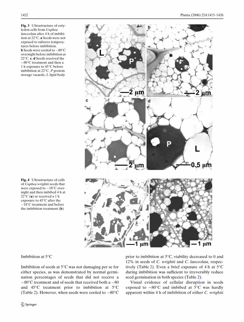
The data in Table 2 demonstrate that temperaturetreatments to control triacylglycerol phase prior toseed imbibition resulted in predictable changes to seedviability. Light microscopy and TEM were used toexamine the nature of cellular perturbations andwhether their incidence corresponded to patterns oflost viability. The ultrastructure of cells in cotyledonand axes of Cuphea seeds that were not cooled to¡80°C resembled that of other orthodox seeds; themassive accumulation of storage reserves obscureddetection of other organelles in the cytoplasm (Rossand Murphy 1992; Farrant et al. 1997; Walters et al.2005; Figs. 2a, b, 3a). Lipid bodies could be identiWedby the intact monolayer of polar lipid and protein com-pletely encompassing a spherical, non-staining oildroplet. Triacylglycerols in micrographs are electronlucent, partly because osmium bonds poorly to satu-rated alkanes and partly because ethanol and acetoneused during embedding extracted lipids (Mollenhauer1993). There were also several large electron-densespherical masses identiWed as protein storage vacuoles.
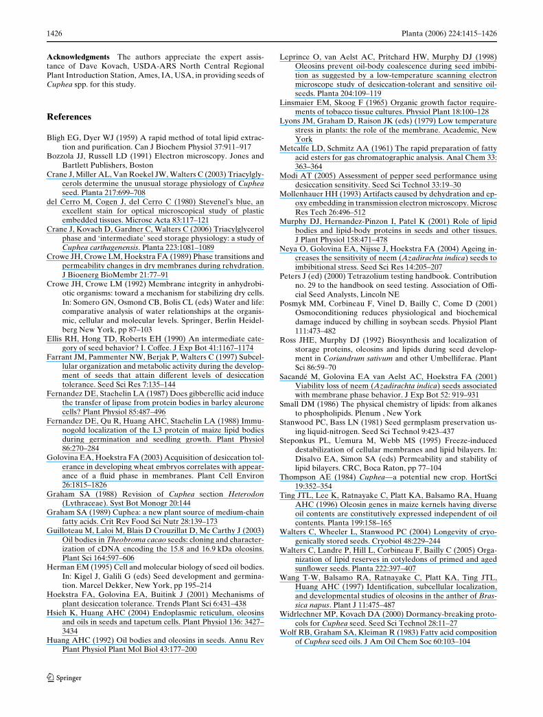
Consistent with viability assessments, remarkablecellular perturbations were observed in C. wrightiiseeds after they were cooled to ¡80°C and thenwarmed to 22°C and imbibed for 4–27 h (Figs. 2c, d,4a). Similar damage was also observed in C. wrightii
seeds initially cooled to –18°C (Fig. 4, Table 3), but notin C. lanceolata seeds (data not shown), presumablybecause the lipids in C. lanceolata crystallize at temper-atures < ¡18°C (Fig. 1). In damaged cells, evidence ofliposome membranes and spherical protein storagevacuoles was lost, and the cytoplasm was mostly elec-tron lucent, indicating loss of triacylglycerol compart-mentation. Electron lucent protrusions appear to cutinto irregularly shaped, dark staining masses that werelikely remnants of protein storage vacuoles. In C.wrightii, 100% of the cells in seeds cooled to ¡80°Cand then imbibed at 22°C exhibited visual signs ofdamage in light and electron micrographs (Table 2).The same type of cellular damage occurred in similarproportions of cells within both axes and cotyledonswhen seeds of this species were cooled to ¡18°C andimbibed at 22°C for 17 h (Table 3). Despite the intra-cellular damage apparent in electron micrographs ofcold-treated C. wrightii seeds, cell shape remainedintact. (Fig. 4a) and electrolyte leakage was the sameas seeds that did not receive a low temperature treat-ment (Table 2). These observations suggest that theplasma membrane was intact in C. wrightii seedscooled to ¡80°C and then warmed to 22°C beforeimbibition, since high levels of electrolyte leakage areexpected when the plasma membrane ruptures(Posmyk et al. 2001; Modi 2005). Cellular perturba-tions were not observed in C. lanceolata seeds sub-jected to the same series of temperature treatments(Fig. 3b, Table 2). Conductivity measurements revealed
Table 1 Fatty acid composition of total and polar lipid fractions within seeds of Cuphea wrightii and C. lanceolata and its eVect on melt-ing temperature
a Value obtained online by searching for data available for Cuphea wrightii and Cuphea lanceolata accessions on the Genetic ResourcesInformation Network (http://www.ars-grin.gov/npgs/)b Predicted temperature of lipid melt based on weighted average of transition temperatures for simple triacylglycerols and hydrated dia-cylcerol-phosphotidylcholines with given fatty acid side chains. The data for simple triacylglycerols and diacylcerol-phosphotidylcho-lines in excess water are taken from Small (1986)
Fatty acid Chain length Temperature of melt (°C)b Lipid composition (%)
C. wrightii C. lanceolata
Triacyl (B’) Polar (gel) GRINa Total Polar GRINa Total Polar
Caprylic C8 ¡21 1 0 0 1 0 0Capric C10 18 -15 28 14 14 75 75 0Lauric C12 35 0 56 68 53 2 3 3Myristic C14 47 23 5 11 3 3 3 3Palmitic C16 57 42 2 3 14 4 5 30Stearic C18 64 54 0 0 0 1 2 4Oleic C18:1 ¡12 ¡20 0 1 5 0 5 18Linoleic C18:2 ¡23 ¡30 6 3 11 7 7 42Linolenic C18:3 ¡34 ¡35 0 0 0 0 0 0Sum 98 100 100 93 100 100Predicted melt temperature (oC)b
32.3 0 17.7 ¡0.8
123

1420 Planta (2006) 224:1415–1426
123
Tab
le2
Ass
essm
ents
of v
iabi
lity
and
cellu
lar
inte
grit
y of
Cup
hea
seed
s im
bibe
d at
22
or 5
°C a
fter
rec
eivi
ng a
ser
ies
of te
mpe
ratu
re t
reat
men
ts t
o co
ntro
l lip
id p
hase
Aft
er th
e im
bibi
tion
per
iod,
see
ds w
ere
tran
sfer
red
to r
oom
tem
pera
ture
for
viab
ility
mea
sure
men
tsa
Per
cent
age
of c
ells
exh
ibit
ing
dam
age
obse
rved
usi
ng li
ght m
icro
scop
yb
Not
ava
ilabl
ec D
amag
e ob
serv
ed u
sing
tra
nsm
issi
on e
lect
ron
mic
rosc
opy
rese
mbl
ed t
hat o
f a
dyin
g ce
lld
Seed
s w
ere
imbi
bed
for
4h
at 5
°C s
eeds
and
the
n tr
ansf
erre
d to
22°
C f
or le
akag
e m
easu
rem
ents
(im
med
iate
ly)
and
embe
ddin
g (a
fter
24
h)
Spec
ies
Exp
osur
e te
mpe
ratu
res
for
dry
seed
s (°
C)
Imbi
biti
on c
ondi
tion
Lip
id p
hase
du
ring
imbi
biti
on
Via
bilit
y as
sess
men
t
Coo
led
toW
arm
ed t
oT
emp.
(o C
)D
urat
ion
(h)
Ger
min
atio
n (%
)A
xis
cult
ure
(%)
Tet
razo
lium
stai
ning
(%
)E
lect
roly
tele
akag
e (�
A m
g¡1 h¡
1 )
Vis
ual
dam
age
a (%)
Cup
hea
wri
ghtii
Non
eN
one
224
Flu
id74
§1
86
100
21§
510
§10
¡80
2222
4C
ryst
al0
9§
225
19§
710
0§
0 ¡
8045
224
Flu
id73
§4
90§
310
023
§4
0 N
one
Non
e5
4F
luid
73§
3N
abN
aN
a0
¡80
55
4C
ryst
al0
Na
Na
Na
25§
13¡
805
524
Cry
stal
0N
aN
aN
a10
0§
0 ¡
805
5/22
4/24
dC
ryst
al0
Na
Na
29§
685
§10
¡80
455
4F
luid
56§
6N
aN
aN
aN
aC
uphe
a la
nceo
lata
Non
eN
one
224
Flu
id81
§2
7010
06§
10
¡80
2222
4F
luid
97§
289
100
6§
10
¡80
4522
4F
luid
96§
372
967§
30
Non
eN
one
54
Flu
id80
§3
Na
Na
Na
0¡
805
54
Cry
stal
12§
12N
aN
aN
a0
¡80
55
24C
ryst
al0
Na
Na
Na
0¡
805
5/22
4/24
dC
ryst
al0
Na
Na
8§
210
0§
0c
¡80
455
4F
luid
95§
1N
aN
aN
ana

Planta (2006) 224:1415–1426 1421
three-fold lower electrolyte levels in leachate of C.lanceolata seeds compared to C. wrightii seeds, regard-less of seed treatment (Table 2).
Warming seeds to 45°C before imbibition at 22°Crestored high germination percentages in C. wrightiiseeds that were cooled to ¡18 or ¡80°C (Table 2). Pre-warmed seeds had organelles resembling protein andlipid bodies (Figs. 2e, f, 4b); however, the appearance of
the storage reserves was substantially changed. Theaverage cross-sectional area of the lipid bodies nearlydoubled while the cross-sectional area of the proteinstorage vacuoles was halved in cells of C. wrightii seedsthat were restored by the 45°C pulse (Fig. 5a, b). Incontrast, the lipid body size in cells of C. lanceolataremained constant throughout temperature Xuctuations,but the size of the protein storage vacuoles was reduced.
Fig. 2 Ultrastructure of coty-ledon cells from Cuphea wrightii seeds after 4 h of imbibition at 22°C. a, b Seeds were not exposed to subzero temperatures before imbibi-tion. c, d Seeds were cooled to ¡80°C overnight before imbi-bition at 22°C. e, f Seeds re-ceived the ¡80°C treatment and then a 1 h exposure to 45°C before imbibition at 22°C. P protein storage vacu-ole; L lipid body
123
1422 Planta (2006) 224:1415–1426
Imbibition at 5°C
Imbibition of seeds at 5°C was not damaging per se foreither species, as was demonstrated by normal germi-nation percentages of seeds that did not receive a¡80°C treatment and of seeds that received both a ¡80and 45°C treatment prior to imbibition at 5°C(Table 2). However, when seeds were cooled to ¡80°C
prior to imbibition at 5°C, viability decreased to 0 and12% in seeds of C. wrightii and C. lanceolata, respec-tively (Table 2). Even a brief exposure of 4 h at 5°Cduring imbibition was suYcient to irreversibly reduceseed germination in both species (Table 2).
Visual evidence of cellular disruption in seedsexposed to ¡80°C and imbibed at 5°C was hardlyapparent within 4 h of imbibition of either C. wrightii
Fig. 3 Ultrastructure of coty-ledon cells from Cuphea lanceolata after 4 h of imbibi-tion at 22°C. a Seeds were not exposed to subzero tempera-tures before imbibition. b Seeds were cooled to ¡80°C overnight before imbibition at 22°C. c, d Seeds received the ¡80°C treatment and then a 1 h exposure to 45°C before imbibition at 22°C. P protein storage vacuole; L lipid body
Fig. 4 Ultrastructure of cells of Cuphea wrightii seeds that were exposed to ¡18°C over-night and then imbibed 4 h at 22°C (a) or received a 1 h exposure to 45°C after the ¡18°C treatment and before the imbibition treatment (b)
123

Planta (2006) 224:1415–1426 1423
or C. lanceolata (Figs. 6 a, b, 5 a, b, Table 2), but wasobvious within 24 h of imbibition at 5°C (Fig. 6 c, d,Table 2). Cells of C. wrightii imbibed for 24 h at a
constant 5°C lacked lipid and protein storage vacu-oles with regular, spherical structure (Fig. 6c). Mas-sive cell damage with similar characteristics to thosedepicted in Figs. 2c, 2d, and 4a was observed in seedsof C. wrightii that were imbibed for 4 h at 5°C andthen transferred to 22°C for an additional 24 h ofimbibition (Fig. 6e, cf 5). Evidence of lipid bodyfusion and disruption of cellular structures were alsoobserved in cells of C. lanceolata seeds that wereimbibed for 24 h (Fig. 6d) or 7 days (data not shown)at a constant 5°C. The loss of organelles and the pres-ence of granulated masses were evidence that thesecells were dying; however, cells of C. lanceolata seedsdid not exhibit the striking cellular perturbationsobserved within C. wrightii seeds. Despite low viabil-ity (Table 2), cell structure seemed to be maintainedin C. lanceolata seeds that were brieXy imbibed at 5°Cand then transferred to 22°C for an additional 24 h(Figs. 5, 6f, Table 2). The slow development of visualdamage in seeds imbibed at 5°C suggests that theobserved cellular disruptions are both time and tem-perature dependent.
Discussion
We have shown that germination success of seeds fromtwo species of Cuphea can be manipulated predictablyby temperature treatments that control triacylglycerolphase in dry seeds. Lipids within the seeds of the twospecies contain diVering proportions of capric and lau-ric acids which confer a »10°C diVerence in the meltingtemperature of the triacylglycerols. Seeds with crystal-lized triacylglycerols do not survive imbibition. TheseWndings are important for genebank operators whohave been unable to store germplasm from tropicalplant species and for oilseed producers in temperateareas who use cultivars with high amounts of saturatedor monounsaturated fatty acids.
Our research on triacylglycerol-mediated damageduring cold exposure shares tenets with past researchlinking phase changes in polar lipids with chilling or
Table 3 Cellular integrity within Cuphea wrightii seeds imbibed at 22°C after being cooled to ¡18°C to crystallize triacylglycerols andthen warmed to 22 or 45°C to retain or melt, respectively, the lipid crystals
Cells were embedded after 4 h imbibitiona Percentage of cells exhibiting damage observed using light microscopy
Exposure temperatures for dry seeds (°C) Imbibition condition Lipid phase during imbibition
Visual damage (%)a
Cooled to Warmed to Temp. (oC) Duration (h) Axes Cotyledons
none none 22 17 crystal 0 0¡18 22 22 17 Xuid 70 § 30 90 § 10¡18 45 22 17 crystal 0 3 § 1
Fig. 5 Average cross sectional areas (§ SE) of lipid bodies (a)and protein storage vacuoles (b) measured in Cuphea wrightii andC. lanceolata cells from electron micrographs similar to those pre-sented in Figs. 2, 3, 4, 6. Column 1 seeds were not cooled prior to4 h imbibition at 22°C; column 2 seeds were cooled to ¡80°Cprior to 4 h imbibition at 22°C; column 3 seeds were cooled to¡80°C and then warmed to 45°C prior to 4 h imbibition at 22°C;column 4 seeds were cooled to ¡80oC and then imbibed for 4 h at5°C; column 5 seeds were cooled to ¡80°C and then imbibed for4 h at 5°C and 24 h at 22°C. Distinct lipid bodies and protein stor-age vacuoles could not be observed (*). Mean separation testsdetected signiWcant diVerences in cross sectional areas forC. wrightii (bars labeled a, b) and C. lanceolata (bars labeled y, z)at the � = 0.05 level
0
10
5
30
25
20
15
Cuphea lanceolataCuphea wrightii
y
a
zb
z zb
z
Pro
tein
bod
y cr
oss
sect
iona
l are
a (µ
m2 ) b
0
1
2
3
4
5
6
Cuphea lanceolataCuphea wrightii
b
a
b
y
yzyzyz
z
Lipi
d bo
dy c
ross
sec
tiona
l are
a (µ
m2 ) a
**
**1 2 3 4 5
1 2 3 4 5
123

1424 Planta (2006) 224:1415–1426
desiccation damage (e.g., Lyons et al. 1979; Croweet al. 1989; Crowe and Crowe 1992; Steponkus et al.1995; Hoekstra et al. 2001). Unlike systems studiedearlier, the plasma membrane does not appear to bethe site of damage in imbibing Cuphea seeds. Cupheaseeds are tolerant of desiccation and so would likelypossess suYcient protective mechanisms to avoid manyof the hypothesized changes in membranes duringexposure to low temperature or moisture. Further, the
seeds did not exhibit the classic physiological symptomof membrane damage, leakage of cellular constituentsincluding electrolytes, when they were exposed tolethal temperature treatments (Table 2), suggestingthat the plasma membrane retained function evenwhen intracellular constituents were disrupted. Thesimilar transition temperatures for the polar lipidfraction of the two species, predicted from fatty acidcompositions (Table 1), do not explain the diVerence in
Fig. 6 Ultrastructure of cells of Cuphea wrightii (a, c, e) and C. lanceolata (b, d, f) seeds cooled to ¡80°C and imbibed at 5°C for 4 h (a, b), 24 h (c, d), or 5°C for 4 h and then 22°C for 24 h (e, f) prior to embed-ding. P protein storage vacu-oles; L lipid body; N nucleus
123

Planta (2006) 224:1415–1426 1425
temperature response between the two species. Pre-sumably the polar lipids would be in the liquid crystal-line phase at the imbibition temperatures studied here(Table 1), and so phase transitions within membranesare a less likely mechanism of damage in Cuphea seeds.Nonetheless, the idea that water interactions with crys-tallized lipid are damaging has precedence in the semi-nal work on hydration of gel phase polar lipids,published in the imbibitional chilling literature (Croweet al. 1989; Hoekstra et al. 2001).
All intracellular structure was lost in C. wrightiiseeds that were cooled to < ¡18°C, rewarmed to· 22°C and imbibed. Triacylglycerols remained crys-tallized in seeds receiving this type of treatment. Cellu-lar disintegration was evident during early imbibition(Figs. 2c, d, 4a, 6c), and suggests that damage occurredduring the transition from an anhydrous to fullyhydrated state. The same type of damage was rarelyobserved in cells of C. lanceolata seeds (Table 2,Fig. 6d), probably because their triacylglycerolsrequired extreme cooling to ¡80°C to crystallize andreadily melted when seeds were warmed to room tem-perature (Fig. 1). Visual evidence of cell damage in C.lanceolata seeds imbibed at 5°C was not observed ini-tially (Fig. 6a, b), but took time to develop (Fig. 6c, d).Often the rapid cellular disintegration observed inimbibing C. wrightii seeds containing crystallized tria-cylglycerols was not observed in C. lanceolata seedsimbibed at 5°C, even though this treatment was lethalto the seeds (Table 2). In many cases, lethally treatedcells of C. lanceolata seeds exhibited a disorganizedultrastructure, which progressed with imbibition time.The timing and temperature dependence of cell disrup-tion in Cuphea seeds during early imbibition suggeststhat it arises from biophysical rather than metabolicmechanisms. The delayed appearance of massive cellu-lar disruption in seeds imbibed at 5°C suggests that theprimary lesion has not yet been detected.
The reversibility of conditions that impose damageprovides important insight into the mechanism causingcell perturbations. Heating seeds could reverse poten-tially lethal eVects of cooling dry seeds to temperaturesthat melted triacylglycerols prior to imbibition (heatpulses to 45°C and 22°C were required for C. wrightiiand C. lanceolata, respectively). Even though germina-tion was normal in seeds receiving this brief heat pulse,cell ultrastructure within C. wrightii seeds was diVerentfrom that of the controls. After the heat pulse, proteinstorage vacuoles were smaller and lipid bodies werelarger (Fig. 5) and electron dense material was mixedinto the triacylglycerol matrix (Fig. 2e, f). Thesecellular rearrangements are consistent with the closeassociation of protein and lipid bodies previously
observed (Fernandez and Staehelin 1987) and perhapssuggest that protein reserves were subdivided andredistributed into the lipid bodies. One may speculatethat the new oil bodies arise from a change in physicalinteractions among cellular components during crystal-lization and melting, and that these forces are diVerentthan those present during liposome and protein storagevacuole biogenesis that lead to controlled deposition ofreserves during embryogenesis. Interestingly, heatpulses delivered after imbibition begins are ineVectiveat restoring viability. This supports the conclusion thatthe primary lesion for the type of damage we arereporting occurs during the initial stages of imbibitionand is not apparent from the electron micrographs pre-sented here.
We speculate that the lethal eVect of water on crys-tallized triacylglycerols arises from an altered balanceof hydrophilic and hydrophobic interactions that arenecessary when anhydrous cells transition to an aque-ous environment. The increased hydrophobicity of thecrystallized triacylglycerols when water enters the cellmay promote preferential interactions of liposomemembrane components with other membrane systemsand cause massive decompartmentalization. Steponkusand colleagues proposed interlamellar interactionsamong polar lipids as a mechanism of damage whenunprotected cells were severely desiccated duringexposure to subzero temperatures (Steponkus et al.1995) and these interactions have also been proposedto explain lateral movement of enzymes among organ-elles (Fernandez and Staehelin 1987). Alternatively,oleosin orientation in lipid bodies may be aVectedwhen triacylglycerols crystallize, thereby aVecting thestability of the organelle and the mode in which ithydrates during imbibition.
Conclusions
Seeds of some species are lethally damaged duringimbibition following cold storage, and this sensitivityhas hindered their preservation in genebanks. We havelinked the temperature combinations that impart dam-age to the phase behavior of triacylglycerols withinseeds of Cuphea species. Severe cellular perturbationsoccur early during imbibition of sensitive seeds and areirreversible once imbibition commences. Imbibition ofseeds with crystallized lipids potentially alters intermo-lecular interactions that are fundamental to cell organi-zation and stability in the hydrated state. Continuedresearch in this area is needed to ensure optimum stor-age conditions for germplasm and to promote standestablishment of seeds with altered lipid compositions.
123
1426 Planta (2006) 224:1415–1426
Acknowledgments The authors appreciate the expert assis-tance of Dave Kovach, USDA-ARS North Central RegionalPlant Introduction Station, Ames, IA, USA, in providing seeds ofCuphea spp. for this study.
References
Bligh EG, Dyer WJ (1959) A rapid method of total lipid extrac-tion and puriWcation. Can J Biochem Physiol 37:911–917
Bozzola JJ, Russell LD (1991) Electron microscopy. Jones andBartlett Publishers, Boston
Crane J, Miller AL, Van Roekel JW, Walters C (2003) Triacylgly-cerols determine the unusual storage physiology of Cupheaseed. Planta 217:699–708
del Cerro M, Cogen J, del Cerro C (1980) Stevenel’s blue, anexcellent stain for optical microscopical study of plasticembedded tissues. Microsc Acta 83:117–121
Crane J, Kovach D, Gardner C, Walters C (2006) Triacylglycerolphase and ‘intermediate’ seed storage physiology: a study ofCuphea carthagenensis. Planta 223:1081–1089
Crowe JH, Crowe LM, Hoekstra FA (1989) Phase transitions andpermeability changes in dry membranes during rehydration.J Bioenerg BioMembr 21:77–91
Crowe JH, Crowe LM (1992) Membrane integrity in anhydrobi-otic organisms: toward a mechanism for stabilizing dry cells.In: Somero GN, Osmond CB, Bolis CL (eds) Water and life:comparative analysis of water relationships at the organis-mic, cellular and molecular levels. Springer, Berlin Heidel-berg New York, pp 87–103
Ellis RH, Hong TD, Roberts EH (1990) An intermediate cate-gory of seed behavior? I. CoVee. J Exp Bot 41:1167–1174
Farrant JM, Pammenter NW, Berjak P, Walters C (1997) Subcel-lular organization and metabolic activity during the develop-ment of seeds that attain diVerent levels of desiccationtolerance. Seed Sci Res 7:135–144
Fernandez DE, Staehelin LA (1987) Does gibberellic acid inducethe transfer of lipase from protein bodies in barley aleuronecells? Plant Physiol 85:487–496
Fernandez DE, Qu R, Huang AHC, Staehelin LA (1988) Immu-nogold localization of the L3 protein of maize lipid bodiesduring germination and seedling growth. Plant Physiol86:270–284
Golovina EA, Hoekstra FA (2003) Acquisition of desiccation tol-erance in developing wheat embryos correlates with appear-ance of a Xuid phase in membranes. Plant Cell Environ26:1815–1826
Graham SA (1988) Revision of Cuphea section Heterodon(Lythraceae). Syst Bot Monogr 20:144
Graham SA (1989) Cuphea: a new plant source of medium-chainfatty acids. Crit Rev Food Sci Nutr 28:139–173
Guilloteau M, Laloi M, Blais D Crouzillat D, Mc Carthy J (2003)Oil bodies in Theobroma cacao seeds: cloning and character-ization of cDNA encoding the 15.8 and 16.9 kDa oleosins.Plant Sci 164:597–606
Herman EM (1995) Cell and molecular biology of seed oil bodies.In: Kigel J, Galili G (eds) Seed development and germina-tion. Marcel Dekker, New York, pp 195–214
Hoekstra FA, Golovina EA, Buitink J (2001) Mechanisms ofplant desiccation tolerance. Trends Plant Sci 6:431–438
Hsieh K, Huang AHC (2004) Endoplasmic reticulum, oleosinsand oils in seeds and tapetum cells. Plant Physiol 136: 3427–3434
Huang AHC (1992) Oil bodies and oleosins in seeds. Annu RevPlant Physiol Plant Mol Biol 43:177–200
Leprince O, van Aelst AC, Pritchard HW, Murphy DJ (1998)Oleosins prevent oil-body coalescence during seed imbibi-tion as suggested by a low-temperature scanning electronmicroscope study of desiccation-tolerant and sensitive oil-seeds. Planta 204:109–119
Linsmaier EM, Skoog F (1965) Organic growth factor require-ments of tobacco tissue cultures. Physiol Plant 18:100–128
Lyons JM, Graham D, Raison JK (eds) (1979) Low temperaturestress in plants: the role of the membrane. Academic, NewYork
Metcalfe LD, Schmitz AA (1961) The rapid preparation of fattyacid esters for gas chromatographic analysis. Anal Chem 33:363–364
Modi AT (2005) Assessment of pepper seed performance usingdesiccation sensitivity. Seed Sci Technol 33:19–30
Mollenhauer HH (1993) Artifacts caused by dehydration and ep-oxy embedding in transmission electron microscopy. MicroscRes Tech 26:496–512
Murphy DJ, Hernandez-Pinzon I, Patel K (2001) Role of lipidbodies and lipid-body proteins in seeds and other tissues.J Plant Physiol 158:471–478
Neya O, Golovina EA, Nijsse J, Hoekstra FA (2004) Ageing in-creases the sensitivity of neem (Azadirachta indica) seeds toimbibitional stress. Seed Sci Res 14:205–207
Peters J (ed) (2000) Tetrazolium testing handbook. Contributionno. 29 to the handbook on seed testing. Association of OY-cial Seed Analysts, Lincoln NE
Posmyk MM, Corbineau F, Vinel D, Bailly C, Come D (2001)Osmoconditioning reduces physiological and biochemicaldamage induced by chilling in soybean seeds. Physiol Plant111:473–482
Ross JHE, Murphy DJ (1992) Biosynthesis and localization ofstorage proteins, oleosins and lipids during seed develop-ment in Coriandrum sativum and other Umbelliferae. PlantSci 86:59–70
Sacandé M, Golovina EA van Aelst AC, Hoekstra FA (2001)Viability loss of neem (Azadirachta indica) seeds associatedwith membrane phase behavior. J Exp Bot 52: 919–931
Small DM (1986) The physical chemistry of lipids: from alkanesto phospholipids. Plenum , New York
Stanwood PC, Bass LN (1981) Seed germplasm preservation us-ing liquid-nitrogen. Seed Sci Technol 9:423–437
Steponkus PL, Uemura M, Webb MS (1995) Freeze-induceddestabilization of cellular membranes and lipid bilayers. In:Disalvo EA, Simon SA (eds) Permeability and stability oflipid bilayers. CRC, Boca Raton, pp 77–104
Thompson AE (1984) Cuphea—a potential new crop. HortSci19:352–354
Ting JTL, Lee K, Ratnayake C, Platt KA, Balsamo RA, HuangAHC (1996) Oleosin genes in maize kernels having diverseoil contents are constitutively expressed independent of oilcontents. Planta 199:158–165
Walters C, Wheeler L, Stanwood PC (2004) Longevity of cryo-genically stored seeds. Cryobiol 48:229–244
Walters C, Landre P, Hill L, Corbineau F, Bailly C (2005) Orga-nization of lipid reserves in cotyledons of primed and agedsunXower seeds. Planta 222:397–407
Wang T-W, Balsamo RA, Ratnayake C, Platt KA, Ting JTL,Huang AHC (1997) IdentiWcation, subcellular localization,and developmental studies of oleosins in the anther of Bras-sica napus. Plant J 11:475–487
Widrlechner MP, Kovach DA (2000) Dormancy-breaking proto-cols for Cuphea seed. Seed Sci Technol 28:11–27
Wolf RB, Graham SA, Kleiman R (1983) Fatty acid compositionof Cuphea seed oils. J Am Oil Chem Soc 60:103–104
123