Role of CCL3/MIP1α and CCL5/RANTES during acute Trypanosoma cruzi infection in rats
15
Role of CCL3/MIP-1α and CCL5/RANTES during acute Trypanosoma cruzi infection in rats Ester Roffê a,* , Fabiano Oliveira b , Adriano L.S. Souza a , Vanessa Pinho a , Danielle G. Souza a , Patrícia R.S. Souza a , Remo C. Russo a , Helton C. Santiago a , Álvaro J. Romanha c , Herbert B. Tanowitz d , Jesus G. Valenzuela b , and Mauro M. Teixeira a a Departamento de Bioquímica e Imunologia, Instituto de Ciências Biológicas, Universidade Federal de Minas Gerais, Belo Horizonte, Minas Gerais, Brazil b Vector Molecular Biology Unit, Laboratory of Malaria and Vector Research, National Institute of Allergy and Infectious Diseases, NIH, Maryland, USA c Centro de Pesquisas René Rachou/Fiocruz, Belo Horizonte, Minas Gerais, Brazil d Department of Pathology, Albert Einstein College of Medicine, Bronx, NY, USA Abstract Chagas’ disease is caused by Trypanosoma cruzi infection and is characterized by chronic fibrogenic inflammation and heart dysfunction. Chemokines are produced during infection and drive tissue inflammation. In rats, acute infection is characterized by intense myocarditis and regression of inflammation after control of parasitism. We investigated the role of CCL3 and CCL5 during infection by using DNA vaccination encoding for each chemokine separately or simultaneously. MetRANTES treatment was used to evaluate the role of CCR1 and CCR5, the receptors for CCL3 and CCL5. Vaccination with CCL3 or CCL5 increased heart parasitism and decreased local IFN-γ production, but did not influence intensity of inflammation. Simultaneous treatment with both plasmids or treatment with MetRANTES enhanced cardiac inflammation, fibrosis and parasitism. In conclusion, chemokines CCL3 and CCL5 are relevant, but not essential, for control of T. cruzi infection in rats. On the other hand, combined blockade of these chemokines or their receptors enhanced tissue inflammation and fibrosis, clearly contrasting with available data in murine models of T. cruzi infection. These data reinforce the important role of chemokines during T. cruzi infection but suggest that caution must be taken when expanding the therapeutic modulation of the chemokine system in mice to the human infection. Keywords Chemokines; CCR5; Trypanosoma cruzi; Myocarditis 1. Introduction Chagas’ heart disease is caused by infection with Trypanosoma cruzi and is characterized by chronic fibrogenic inflammation and loss of function of the heart [1]. It is estimated that 200,000 new cases and 21,000 disease-associated deaths occur annually in Latin America © 2010 Elsevier Masson SAS. All rights reserved. * Corresponding author. 9000 Rockville Pike, MSC-1886, Bldg. 10, Room 11N07, NIH, Bethesda MD 20892, USA. Tel.: +1 301 402 6491; fax: +1 301 402 4369. [email protected] (E. Roffê). NIH Public Access Author Manuscript Microbes Infect. Author manuscript; available in PMC 2011 September 5. Published in final edited form as: Microbes Infect. 2010 August ; 12(8-9): 669–676. doi:10.1016/j.micinf.2010.04.011. NIH-PA Author Manuscript NIH-PA Author Manuscript NIH-PA Author Manuscript
-
Upload
independent -
Category
Documents
-
view
2 -
download
0
Transcript of Role of CCL3/MIP1α and CCL5/RANTES during acute Trypanosoma cruzi infection in rats
Role of CCL3/MIP-1α and CCL5/RANTES during acuteTrypanosoma cruzi infection in rats
Ester Roffêa,*, Fabiano Oliveirab, Adriano L.S. Souzaa, Vanessa Pinhoa, Danielle G. Souzaa,Patrícia R.S. Souzaa, Remo C. Russoa, Helton C. Santiagoa, Álvaro J. Romanhac, Herbert B.Tanowitzd, Jesus G. Valenzuelab, and Mauro M. Teixeiraa
aDepartamento de Bioquímica e Imunologia, Instituto de Ciências Biológicas, UniversidadeFederal de Minas Gerais, Belo Horizonte, Minas Gerais, BrazilbVector Molecular Biology Unit, Laboratory of Malaria and Vector Research, National Institute ofAllergy and Infectious Diseases, NIH, Maryland, USAcCentro de Pesquisas René Rachou/Fiocruz, Belo Horizonte, Minas Gerais, BrazildDepartment of Pathology, Albert Einstein College of Medicine, Bronx, NY, USA
AbstractChagas’ disease is caused by Trypanosoma cruzi infection and is characterized by chronicfibrogenic inflammation and heart dysfunction. Chemokines are produced during infection anddrive tissue inflammation. In rats, acute infection is characterized by intense myocarditis andregression of inflammation after control of parasitism. We investigated the role of CCL3 andCCL5 during infection by using DNA vaccination encoding for each chemokine separately orsimultaneously. MetRANTES treatment was used to evaluate the role of CCR1 and CCR5, thereceptors for CCL3 and CCL5. Vaccination with CCL3 or CCL5 increased heart parasitism anddecreased local IFN-γ production, but did not influence intensity of inflammation. Simultaneoustreatment with both plasmids or treatment with MetRANTES enhanced cardiac inflammation,fibrosis and parasitism. In conclusion, chemokines CCL3 and CCL5 are relevant, but not essential,for control of T. cruzi infection in rats. On the other hand, combined blockade of these chemokinesor their receptors enhanced tissue inflammation and fibrosis, clearly contrasting with availabledata in murine models of T. cruzi infection. These data reinforce the important role of chemokinesduring T. cruzi infection but suggest that caution must be taken when expanding the therapeuticmodulation of the chemokine system in mice to the human infection.
KeywordsChemokines; CCR5; Trypanosoma cruzi; Myocarditis
1. IntroductionChagas’ heart disease is caused by infection with Trypanosoma cruzi and is characterized bychronic fibrogenic inflammation and loss of function of the heart [1]. It is estimated that200,000 new cases and 21,000 disease-associated deaths occur annually in Latin America
© 2010 Elsevier Masson SAS. All rights reserved.*Corresponding author. 9000 Rockville Pike, MSC-1886, Bldg. 10, Room 11N07, NIH, Bethesda MD 20892, USA. Tel.: +1 301 4026491; fax: +1 301 402 4369. [email protected] (E. Roffê).
NIH Public AccessAuthor ManuscriptMicrobes Infect. Author manuscript; available in PMC 2011 September 5.
Published in final edited form as:Microbes Infect. 2010 August ; 12(8-9): 669–676. doi:10.1016/j.micinf.2010.04.011.
NIH
-PA Author Manuscript
NIH
-PA Author Manuscript
NIH
-PA Author Manuscript
[2]. Specific treatment of the infection with benznidazole is effective in the acute phase butnot shown to halt progression of the chronic forms of Chagas’ disease [2].
Chemokines are a group of mediators of the inflammatory process thought to play anessential role in the recruitment and activation of leukocytes in various models ofinflammatory diseases [3]. Specific chemokines are produced in tissue in response to T.cruzi infection and are crucial to define the leukocyte subtypes that compose theinflammatory infiltrate in the heart of infected animals [4–6]. CC-chemokines preferentiallyattract mononuclear cells to sites of chronic inflammation and mononuclear cellspredominate in lesions of patients with Chagas’ disease and in experimental T. cruziinfection [7–9]. Although some studies have evaluated the role of chemokines inexperimental T. cruzi infection [4,5,10], few studies have addressed the role of specific CC-chemokines, especially in the rat model. In rats, acute infection is characterized by intensemyocarditis, with many mononuclear cells surrounding amastigote nests. Interestingly, highlevels of IL-10 are found in the myocardium and myocarditis regresses after control ofparasitism [6].
It has been shown that chemokine-encoding DNA vaccines are able to induce high titers ofneutralizing antibodies against the targeted chemokine in rats [11,12,6]. For example, wehave previously shown that Holtzman rats immunized with a CCL4/MIP-1β-encoding DNAvaccine had enhanced heart inflammation but unchanged heart parasite load when infectedwith T. cruzi. The latter study suggested that CCL4 could be recruiting regulatory cells tothe infected tissue and, hence, regulating the intensity of the inflammatory response to theinfection [6]. The aim of the present work was to study the role of the chemokines CCL3and CCL5 in rats infected with T. cruzi. We also investigated the effect of MetRANTEStreatment as this drug is a functional antagonist of the receptors at which CCL3 and CCL5act; i.e. CCR1 and CCR5.
2. Materials and methods2.1. Animals and infection
Holtzman rats, 90 days old and 300–400 g of weight, were obtained from CEBIO/UFMG(Minas Gerais, Brasil) and maintained in the animal facilities of the Laboratório deImunofarmacologia, with filtered water and food ad libitum. Animals were infectedintraperitoneally with 104 blood forms of the CL-Brener clone of T. cruzi per 50 g of bodyweight. All procedures had prior approval from the local animal ethics committee (CETEA/UFMG) and are in accordance with international guidelines for animal procedures. Themyocardium was obtained in days corresponding to the acute (15, 20 and 30) and chronic(65) phases of infection, transversally sectioned and divided in 3 defined parts to detectionof cytokines by ELISA, to histopathology and to collagen quantification of hydroxyproline,an indirect measurement of tissue fibrosis.
2.2. Rat CCL3/MIP-1α and CCL5/RANTES cloning and construction of the vaccinationplasmid
The genes coding for CCL3 and CCL5 were amplified using primers to CCL3 and CCL5 byRT-PCR reactions in myocardial samples from acutely T. cruzi-infected rats using high-fidelity platinum Taq polymerase (GIBCO BRL). The signal peptide-encoding sequence tosecretion present in the chemokine genes was identified by using the program “SignalPeptide” and specific primers to amplify the chemokine genes lacking signal-sequence tosecretion were constructed, as the VR2001-TOPO DNA plasmid vector (Vical) has thissequence [13]. The PCR products were immediately cloned into TOPO TA cloning vectorPCRII (Invitrogen) following the manufacturer’s specifications. The construction of
Roffê et al. Page 2
Microbes Infect. Author manuscript; available in PMC 2011 September 5.
NIH
-PA Author Manuscript
NIH
-PA Author Manuscript
NIH
-PA Author Manuscript
vaccination plasmids was done as previously described [13]. After visualization of the PCRproducts on a 2% agarosis gel, we sequenced the positive PCR products using CEQ2000DNA (Beckman Coulter). For the vaccine construct, we chose a sample that contained theentire sequence of CCL3 or CCL5 in the right orientation and in the correct open-readingframe after the tissue plasminogen activator signal peptide. Cells containing the CCL3 geneon VR2001 and CCL5 gene on VR2001 were grown overnight at 37 °C on 1L of Luria brothwith kanamycin (100 µg/ml) and plasmid isolation was performed using Wizard Maxiprepkit. After plasmid isolation, the constructs containing VR2001-CCL3, VR2001-CCL5 andVR2001 alone (control) were washed three times with ultrapure water using an Amicon-100(Millipore). The concentration of the samples was measured by UV absorbance.
2.3. ImmunizationImmunizations with CCL3 or CCL5 constructs or control plasmid were performed byintradermal injection of 30 µg of DNA, suspended in 30 µl of sterile and apyrogenicultrapure water, per immunization. In addition, some groups of rats were immunized withboth CCL3 and CCL5 constructs (total of 60 µg of DNA). Intradermal immunization wasmade into the foot of rats after anesthesia with ketamine–xylazine. Boosters were giventwice a week for 2 weeks – a total of 4 boosters. Two weeks after the last immunization, ratswere infected as described above and euthanized at 20 (acute phase) or 65 days (chronicphase) after infection.
2.4. In vivo readout assay for screening the neutralizing activity of rat anti-CCL5 seraSera obtained from CCL5- or control plasmid-vaccinated animals (20 days after infection, n= 5) were pooled, diluted at 1:20 in PBS and given s.c. to rats. After 1 h, 300 ng ofrecombinant rat CCL5 (Peprotech) or sterile PBS was injected i. p. and animals killed 18 hlater. Cells were harvested with 15 ml PBS, total cell counts performed in a Neubauerchamber using Turk’s stain and differential cell counts on cytospin preparations (ShandonIII) stained with May–Grumwald–Giemsa using standard morphologic criteria to identifycell types. There was no effect of the sera on baseline (PBS-induced) recruitment of cells(data not shown).
2.5. Treatment with MetRANTESMetRANTES (donated by Dr. Amanda Proudfoot, Merck-Serono Pharmaceuticals,Switzerland) is a modified form of human CCL5/RANTES that works as a functionalantagonist of CCR1 and CCR5 receptors, the receptors at which CCL3 and CCL5 bind. Ratswere treated subcutaneously with 150 µg of MetRANTES or saline daily, from days 10–20after infection. Rats were euthanized at day 20.
2.6. Cytokine and chemokine detection by ELISAA portion of the heart was processed in a solution containing protease inhibitors andcentrifuged, as previously described [14]. The supernantant was used to investigate theconcentration of the cytokines IFN-γ and IL-4 and the chemokines CCL3 and CCL5 byELISA (Peprotech, USA), according to the instructions of the manufacturer.
2.7. Histopathological analysisA portion of the heart was fixed in 4% buffered paraformaldehyde and 7 µm sections stainedwith hematoxylin and eosin to quantify inflammation and infection. Cardiac parasitism andinflammation were analyzed with a Zeiss integrating eyepiece with 100 hits (Öberkohen,Germany) at a final magnification of 320×. A total of 4000 hits were evaluated in eachsection of cardiac tissue. The infection and inflammation indices represent the number ofhits covered by amastigote nests and inflammatory cells, respectively.
Roffê et al. Page 3
Microbes Infect. Author manuscript; available in PMC 2011 September 5.
NIH
-PA Author Manuscript
NIH
-PA Author Manuscript
NIH
-PA Author Manuscript
2.8. Hydroxyproline quantificationFragments of 100 mg of myocardium were removed for hydroxyproline determination, as anindirect measure of collagen content [15]. Briefly, tissues were homogenized in saline 0.9%,frozen at −70 °C and lyophilized. The assay was performed with 20 mg of dry tissue, whichwas subjected to alkaline hydrolysis in 300 µl H2O plus 75 µl NaOH 10 M at 120 °C for 20min. An aliquot of 50 µl of the hydrolyzed tissue was added to 450 µl of Chloramine Toxidizing reagent (Chloramine T 0.056 M, n-propanol 10% in acetate/citrate buffer pH 6.5)and allowed to react for 20 min. A hydroxyproline standard curve was prepared likewise.Color was developed by addition of 500 µl of 1 M p-dimethyl-aminebenzaldehyde diluted inn-propanol/perchloric acid 2:1 v/v. Absorbance was read at 550 nm.
2.9. Statistical analysesResults are shown as means ± S.E.M.. Differences between groups were compared usingStudent’s t test (two sets of data) or one-way ANOVA (three or more sets of data), followedby Student–Newman–Keuls post hoc test. Differences were considered significant at p <0.05.
3. Results3.1. CCL3 and CCL5 are produced in the myocardium of T. cruzi-infected rats
Initial experiments were conducted to characterize the kinetics of chemokine expression ininfected rats. A previous study from our group has shown that the chemokine CCL5 wasdetectable in the myocardium of infected rats from day 15 till day 30 after infection. Levelsdropped to background at 65 days after infection [6]. Likewise, CCL3 was alreadydetectable in high levels in the myocardium of infected rats at day 15. Levels of CCL3dropped by day 30 after infection and reached basal levels at day 65 (Fig. 1), a time at whichinfection and inflammation had subsided [6]. Based on the data above and on evaluation oftissue sections of infected animals, days 20 and 65 were used to study the acute and chronicphases of infection, respectively.
3.2. Effects of CCL3- and CCL5-encoding DNA vaccines in T. cruzi-infected ratsInitial experiments evaluated the efficacy of the CCL3- and the CCL5-encoding vaccines.The administration of commercially available rat CCL3 failed to induce the recruitment ofleukocytes in the peritoneal cavity of rats (data not shown). In order to define the efficacy ofthe vaccine at inhibiting CCL3 production in vivo, we measured CCL3 levels in the hearts ofinfected animals given CCL3 or control plasmids. Levels of CCL3 were significantlyreduced in rats given CCL3 plasmid as compared to animals given control plasmid (Levelsof CCL3 in the heart of infected rats: control plasmid-treated, 4517 ± 472 pg/100 mg hearttissue; CCL3 plasmid-treated, 2696 ± 228 pg/100 mg heart tissue, n = 6, P < 0.001).
We also analyzed the effect of a DNA vaccine encoding CCL5 in T. cruzi-infected rats. Theefficacy of the CCL5-encoding DNA vaccination was evaluated by assessing the ability ofsera from vaccinated rats to neutralize the chemo-attractant activity of CCL5. Sera fromanimals immunized with CCL5 DNA decreased the migration of leukocytes to the peritonealcavity after injection of 300 ng of rat CCL5 when compared to treatment with sera fromanimals immunized with control plasmid (rat CCL5 + control plasmid serum, 3780 ± 877 ×104 cells/cavity; rat CCL5 + CCL5 plasmid serum, 1140 ± 311 × 104 cells/cavity; n = 5, P <0.01).
In T. cruzi-infected animals, myocarditis was clearly observed at day 20 after infection.Inflammatory lesions were predominantly characterized by mononuclear cells that infiltratedcardiac fibers (Fig. 2A). The inflammatory infiltrates were diffuse (Fig. 2A) and amastigote
Roffê et al. Page 4
Microbes Infect. Author manuscript; available in PMC 2011 September 5.
NIH
-PA Author Manuscript
NIH
-PA Author Manuscript
NIH
-PA Author Manuscript
pseudocysts were present, accompanied or not by inflammatory cells (Fig. 2A).Inflammatory and infection indices were not different in T. cruzi-infected animals thatreceived or not the control plasmid, indicating that vaccination with empty plasmid had noeffect in the course of infection (data not shown).
In both CCL3- and CCL5-vaccinated infected animals, there was no significant increase ofinflammation in the heart at day 20 after infection (Fig. 2B and C, respectively). Theinflammatory lesions were also quantitatively similar to the ones found in control-plasmidvaccinated group (Fig. 2E). In contrast, number of amastigote nests was greater in bothCCL3- (Fig. 2B and F) and CCL5-vaccinated animals (Fig. 2C and F) in comparison tocontrol plasmid-vaccinated animals (Fig. 2A and F).
Next, we evaluated levels of the cytokines IFN-γ and IL-4 in the heart of infected rats. Theadministration of CCL3 and CCL5 plasmids was accompanied by a significant inhibition inthe local production of IFN-γ (810 ± 200 pg/100 mg of tissue, plasmid-vaccinated group;427 ± 191 pg/100 mg of tissue, CCL3-vaccinated group; 401 ± 54 pg/100 mg of tissue,CCL5-vaccinated group), while levels of IL-4 were similar to those of control plasmid-treated rats (4039 ± 1468 pg/100 mg of tissue, plasmid-vaccinated group; 3182 ± 912 pg/100mg of tissue, CCL3-vaccinated group; 4448 ± 2327 pg/100 mg of tissue, CCL5-vaccinatedgroup).
3.3. Effects of a cocktail of CCL3/CCL5-encoding DNA vaccines in T. cruzi-infected ratsTo evaluate the effect of simultaneous blockade of CCL3 and CCL5 in T. cruzi-infectedanimals, we used a cocktail of plasmids containing CCL3 and CCL5 DNA in a separategroup of rats. CCL3/CCL5-vaccinated rats had increased cardiac inflammation compared tocontrol plasmid-vaccinated animals or animals vaccinated with either CCL3 or CCL5plasmid separately (Fig. 2D and E). Multifocal inflammation, with high predominance ofmononuclear cells was found in animals vaccinate with the combined vaccines (Fig. 2D).Moreover, enhanced heart parasitism was observed (Fig. 2F). However, administration ofthe combined CCL3/CCL5 vaccine did not enhance cardiac parasitism above that observedwhen either CCL3 or CCL5 vaccines were given alone (Fig. 2F). So, there was nosynergistic or additive effect of CCL3 and CCL5 in terms of parasitism.
Collagen deposition in tissues and fibrosis are important characteristics of chronic T. cruziinfection [16]. Infection of rats with T. cruzi resulted in no increase of cardiac content ofhydroxyproline, a marker of tissue collagen deposition, at days 20 or 65 after infection (Fig.3). This is consistent with the idea that infection in rats usually develops without muchchronic damage [17]. Vaccination with plasmids encoding for CCL3 or CCL5 resulted in noincrease of hydroxyproline content at days 20 or 65 after infection. However, the combinedvaccination with both CCL3 and CCL5 plasmids resulted in a significant increase ofhydroxyproline levels at day 65 after infection (Fig. 3).
3.4. Effects of the treatment with MetRANTES in T. cruzi-infected ratsMetRANTES is a modified human CCL5/RANTES that acts in vivo as an inhibitor of CCR1and CCR5, the receptors for CCL3 and CCL5. Overall, the inflammatory infiltrates inMetRANTES-treated rats were predominantly mononuclear and qualitatively similar to theinflammation found in vehicle-given animals (Fig. 4A and B). However, daily treatmentwith MetRANTES between days 10 and 20 after infection significantly increasedinflammatory (Fig. 4A and D) and infection indices (Fig. 4A, E). The enhancedinflammatory response observed in treated animals was accompanied by significant highercollagen deposition in the heart of MetRANTES- than vehicle-treated rats, as assessed byhydroxyproline assay (Fig. 3F).
Roffê et al. Page 5
Microbes Infect. Author manuscript; available in PMC 2011 September 5.
NIH
-PA Author Manuscript
NIH
-PA Author Manuscript
NIH
-PA Author Manuscript
MetRANTES treatment reduced levels of IFN-γ in the heart of infected animals (3418 ±1035 pg/100 mg of tissue, saline-treated group; 1911 ± 678 pg/100 mg of tissue,MetRANTES-treated group). IL-4 levels in the heart were also significantly reduced inMetRANTES-treated rats (1035 ± 238 pg/100 mg of tissue, saline-treated group; 697 ± 283pg/100 mg of tissue, MetRANTES-treated group).
4. DiscussionThe data reported in the present study show that CCL3 and CCL5 play a non redundant rolein controlling the initial phases of T. cruzi replication in rats. This was evidenced by anincrease in number of amastigote nests and reduction in levels of IFN-γ in the heart ofanimals vaccinated with DNA vaccines encoding for CCL3 or CCL5. Vaccination with acocktail containing both CCL3 and CCL5 or treatment with MetRANTES resulted in greaterparasitism that was of similar intensity to that observed in animals vaccinated with CCL3 orCCL5 alone. However, combined vaccination or treatment with MetRANTES resulted in anincrease in the inflammatory response and collagen deposition in the heart of infectedanimals.
Previous data have shown the relevance of chemokines and chemokine receptors incontrolling T. cruzi burden in vivo, including studies evaluating the role of CXCL9/CXCL10[10], CCR2 [18] and CCR5 [5,19]. Treatment of T. cruzi-infected mice with anti-CCL5 and/or anti-CXCL10 antibodies did not alter T. cruzi burden in mice [10]. In contrast, CCL5 (orCCL3)-encoding DNA vaccination increased parasite burden in the heart of rats. Thedifferences between these studies could reflect the use of different animal models/species(mice vs rats). Alternatively, the antibody schedule may have not been able to depletecompletely the relevant ligand. It is clear, however, that single blockade of CCL3 or CCL5is sufficient to impair temporarily the control of parasite replication in rats. The ability ofchemokines to activate macrophages [20] and cardiomyocytes [21] to produce NO and killT. cruzi may be an explanation for our in vivo observations.
An alternative explanation derives from the finding that there was lower production of IFN-γin myocardium of CCL3- and CCL5-vaccinated rats. The role of CCL5 in inducing IFN-γproduction and recruiting polarized type 1, activated T cells, has been previously described[22–24]. Administration of CCL3 may exacerbate inflammatory bowel disease in mice andthis was associated with increased IFN-γ production by lymphocytes in lamina propria [25].CCL3 and CCL5 may attract and activate type 1 polarized T cells, increasing IFN-γproduction, which is important to control T. cruzi replication. CCR5-binding chemokines arepostulated to have a role in recruiting polarized type 1 T cells [26]. Whatever the mechanismresponsible for the decreased IFN-γ production, the diminished levels of this cytokine canprovide an alternative explanation for the enhanced tissue parasitism observed afterchemokine DNA vaccination.
It is of note that treatment with CCL3 or CCL5-encoding DNA did not modify totalleukocyte influx in the heart of infected animals. However, when both CCL3 and CCL5were blocked by using CCL3/CCL5-encoding DNA vaccines, inflammation wassignificantly enhanced. In rats vaccinated with a plasmid encoding CCL4, which bindssolely to CCR5, exacerbated myocarditis was also found [27]. Similarly, increasedparasitism, exacerbated myocarditis and increased collagen content were observed when ratswere treated with MetRANTES. In contrast, MetRANTES treatment reduced T lymphocyteinfiltration in the heart of T. cruzi-infected mice without affecting parasite load [4] andCCR5 deletion was also shown to reduce dramatically cardiac inflammation in mice [5]. Thelatter studies highlight the importance of CCR5-mediated events for leukocyte recruitmentin murine, but not rat, models of T. cruzi infection. Therefore, whereas in the mouse, CCR5
Roffê et al. Page 6
Microbes Infect. Author manuscript; available in PMC 2011 September 5.
NIH
-PA Author Manuscript
NIH
-PA Author Manuscript
NIH
-PA Author Manuscript
appears to be essential for the recruitment of leukocytes to the heart, in rats, CCL3 andCCL5 and their receptors CCR1 and CCR5 appear not to be relevant for total leukocyteinfiltration.
An important finding was that despite the initial increase of parasitism, control of infectionwas eventually attained in all rats. These results suggest that CCL3 and CCL5 are importantbut not essential in the system to control parasite replication. Alternatively, the degree ofblockade attained with the DNA vaccination or MetRANTES treatment was not sufficient toblunt an anti-parasite response. For example, in mice, treatment with MetRANTES [4] oranti-CCL5 [10] had little effect on parasite burden. In contrast, experiments in CCR5-deleted mice showed that parasite levels were greatly enhanced [5]. Therefore, it is possiblethat partial CCR5 blockade is sufficient to control parasitism both in mice [4,10] and in rats(here). Despite the eventual control of parasitemia and tissue parasitism, there was anincrease in cardiac fibrosis in rats that received the CCL3/CCL5 vaccine or were treatedwith MetRANTES. The results with MetRANTES once again contrast with the situation inmice where administration of MetRANTES significantly decreased fibrosis in chronically-infected animals [27]. In humans, CCR5 expression on peripheral blood mononuclear cellswas decreased in patients with more severe disease and correlated with the degree of heartdysfunction [28]. The latter study suggests that loss of CCR5 correlates and may beimportant for heart dysfunction in patients. This is similar to our findings in rats in whichadministration of MetRANTES or treatment with CCL3/CCL5 vaccine led to worsening ofdisease. In that regard, results from mice suggesting that blockade/antagonism of chemokinereceptors may be useful in chronically-infected patients should be taken with great care, asthe disease in these animals may not mimic the human situation. This is not to say that ratsare more useful than mice to study T. cruzi infection, but results do highlight the need toexpand the immunopathology of experimental T. cruzi infection to species other than mice.
In summary, our data show that the chemokines CCL3 and CCL5 are relevant, but notessential, for the control of T. cruzi infection in rats. On the other hand, combined blockadeof these chemokines or their receptors enhanced tissue inflammation and fibrosis, clearlycontrasting with available data in murine models of T. cruzi infection [4,10]. Altogetherthese data reinforce the important role of chemokines in the context of T. cruzi infection butsuggest that caution must be taken when expanding the therapeutic modulation of thechemokine system in mice to the human infection.
AcknowledgmentsWe are grateful to Rosana Oliveira Alves (CPqRR/Fiocruz, Minas Gerais, Brasil) for infecting the animals, to Dr.Geovanni D. Cassali (UFMG, Minas Gerais, Brasil) for helping with the histopathological description), and toValdinéria Borges, Luíza Silva and Carlos Henrique Silva, by the technical support.
This work was supported by Conselho Nacional de Pesquisa (CNPq, Brazil), Fundação de Amparo à Pesquisa doEstado de Minas Gerais (FAPEMIG, Brazil), Coordenação de Aperfeiçoamento de Pessoal de Nível Superior(CAPES, Brazil), Fogarty International Research Collaboration Award (NIH/FIRCA, 1 R03-TW006857-01A1,USA), and NIH (AI-076248, USA).
References1. Rocha MO, Ribeiro AL, Teixeira MM. Clinical management of chronic Chagas cardiomyopathy.
Front Biosci. 2003; 8:44–54.2. Urbina JA, Docampo R. Specific chemotherapy of Chagas disease: controversies and advances.
Trends Parasitol. 2003; 19:495–501. [PubMed: 14580960]3. Charo IF, Ransohoff RM. The many roles of chemokines and chemokine receptors in inflammation.
N. Engl. J. Med. 2006; 354:610–621. [PubMed: 16467548]
Roffê et al. Page 7
Microbes Infect. Author manuscript; available in PMC 2011 September 5.
NIH
-PA Author Manuscript
NIH
-PA Author Manuscript
NIH
-PA Author Manuscript
4. Marino APMP, Silva AA, Santos PVA, Pinto LMO, Gazzinelli RT, Teixeira MM, Lannes-Vieira J.Regulated on activation, normal T cell expressed and secreted (RANTES) antagonist (Met-RANTES) controls the early phase of Trypanosoma cruzi-elicited myocarditis. Circulation. 2004;110:1443–1449. [PubMed: 15337689]
5. Machado FS, Koyama NS, Carregaro V, Ferreira BR, Milanezi CM, Teixeira MM, Rossi MA, SilvaJS. CCR5 plays a critical role in the development of myocarditis and host protection in miceinfected with Trypanosoma cruzi. J. Infect. Dis. 2005; 191:627–636. [PubMed: 15655788]
6. Roffê E, Souza ALS, Caetano BC, Machado PP, Barcelos LS, Russo RC, Santiago HC, Souza DG,Pinho V, Tanowitz HB, Camargos ER, Bruña-Romero O, Teixeira MM. A DNA vaccine encodingCCL4/MIP-1beta enhances myocarditis in experimental Trypanosoma cruzi infection in rats.Microbes Infect. 2006; 8:2745–2755. [PubMed: 16979363]
7. Reis DD, Jones EM, Tostes S Jr, Lopes ER, Gazzinelli G, Colley DG, McCurley TL.Characterization of inflammatory infiltrates in chronic chagasic myocardial lesions: presence oftumor necrosis factor-alpha+ cells and dominance of granzyme A+, CD8+ lymphocytes. Am. J.Trop. Med. Hyg. 1993; 48:637–644. [PubMed: 8517482]
8. Higuchi MD, Reis MM, Aiello VD, Benvenuti LA, Gutierrez PS, Bellotti G, Pileggi F. Associationof an increase in CD8+ T cells with the presence of Trypanosoma cruzi antigens in chronic, human,chagasic myocarditis. Am. J. Trop. Med. Hyg. 1997; 56:485–489. [PubMed: 9180594]
9. Molina HA, Kierszenbaum F. Kinetics of development of inflammatory lesions in myocardial andskeletal muscle in experimental Trypanosoma cruzi infection. J. Parasitol. 1988; 74:370–374.[PubMed: 3132546]
10. Hardison JL, Wrightsman RA, Carpenter PM, Lane TE, Manning JE. The chemokines CXCL9 andCXCL10 promote a protective immune response but do not contribute to cardiac inflammationfollowing infection with Trypanosoma cruzi. Infect. Immun. 2006; 74:125–134. [PubMed:16368965]
11. Youssef S, Wildbaum G, Maor G, Lanir N, Gour-Lavie A, Grabie N, Karin N. Long-lastingprotective immunity to experimental autoimmune encephalomyelitis following vaccination withnaked DNA encoding C–C chemokines. J. Immunol. 1998; 161:3870–3879. [PubMed: 9780152]
12. Youssef S, Maor G, Wildbaum G, Grabie N, Gour-Lavie A, Karin N. C–C chemokine-encodingDNA vaccines enhance breakdown of tolerance to their gene products and treat ongoing adjuvantarthritis. J. Clin. Invest. 2000; 106:361–371. [PubMed: 10930439]
13. Oliveira F, Kamhawi S, Seitz AE, Pham VM, Guigal PM, Fischer L, Ward J, Valenzuela JG. Fromtranscriptome to immunome: identification of DTH inducing proteins from a Phlebotomus ariasisalivary gland cDNA library. Vaccine. 2006; 24:374–390. [PubMed: 16154670]
14. Souza DG, Cara DC, Cassali GD, Coutinho SF, Silveira MR, Andrade SP, Poole SP, Teixeira MM.Effects of the PAF receptor antagonist UK74505 on local and remote reperfusion injuriesfollowing ischaemia of the superior mesenteric artery in the rat. Br. J. Pharmacol. 2000; 131:1800–1808. [PubMed: 11139461]
15. Reddy GK, Enwemeka CS. A simplified method for the analysis of hidroxyproline in biologicaltissues. Clin. Biochem. 1996; 29:225–229. [PubMed: 8740508]
16. Pereira Barretto AC, Mady C, Arteaga-Fernandez E, Stolf N, Lopes EA, Higuchi ML, Bellotti G,Pileggi F. Right ventricular endomyocardial biopsy in chronic Chagas’ disease. Am. Heart J. 1986;111:307–312. [PubMed: 3946173]
17. Ramirez, LE.; Silva, VD.; Lages-Silva, E.; Chapadeiro, E. Doença de Chagas – Manual ParaExperimentação Animal, Fiocruz/Instituto Oswaldo Cruz, Rio de Janeiro. Araújo-Jorge, TC.;Castro, SL., editors. 2000. p. 140-142.
18. Hardison JL, Kuziel WA, Manning JE, Lane TE. Chemokine CC receptor 2 is important for acutecontrol of cardiac parasitism but does not contribute to cardiac inflammation after infection withTrypanosoma cruzi. J. Infect. Dis. 2006; 193:1584–1588. [PubMed: 16652288]
19. Hardison JL, Wrightsman RA, Carpenter PM, Kuziel WA, Lane TE, Manning JE. The CCchemokine receptor 5 is important in control of parasite replication and acute cardiac inflammationfollowing infection with Trypanosoma cruzi. Infect. Immun. 2006; 74:135–143. [PubMed:16368966]
Roffê et al. Page 8
Microbes Infect. Author manuscript; available in PMC 2011 September 5.
NIH
-PA Author Manuscript
NIH
-PA Author Manuscript
NIH
-PA Author Manuscript
20. Aliberti JC, Machado FS, Souto JT, Campanelli AP, Teixeira MM, Gazzinelli RT, Silva JS. Beta-Chemokines enhance parasite uptake and promote nitric oxide-dependent microbiostatic activity inmurine inflammatory macrophages infected with Trypanosoma cruzi. Infect. Immun. 1999;67:4819–4826. [PubMed: 10456936]
21. Machado FS, Martins GA, Aliberti JC, Mestriner FL, Cunha FQ, Silva JS. Trypanosoma cruzi-infected cardiomyocytes produce chemokines and cytokines that trigger potent nitric oxide-dependent trypanocidal activity. Circulation. 2000; 102:3003–3008. [PubMed: 11113053]
22. Makino Y, Cook DN, Smithies O, Hwang OY, Neilson EG, Turka LA, Sato H, Wells AD, DanoffTM. Impaired T cell function in RANTES-deficient mice. Clin. Immunol. 2002; 102:302–309.[PubMed: 11890717]
23. Weber C, Weber KS, Klier C, Gu S, Wank R, Horuk R, Nelson PJ. Specialized roles of thechemokine receptors CCR1 and CCR5 in the recruitment of monocytes and T(H)1-like/CD45RO(+) T cells. Blood. 2001; 97:1144–1146. [PubMed: 11159551]
24. Santiago HC, Oliveira CF, Santiago L, Ferraz FO, de Souza DG, de-Freitas LA, Afonso LC,Teixeira MM, Gazzinelli RT, Vieira LQ. Involvement of the chemokine RANTES (CCL5) inresistance to experimental infection with Leishmania major. Infect. Immun. 2004; 72:4918–4923.[PubMed: 15271961]
25. Pender SL, Chance V, Whiting CV, Buckley M, Edwards M, Pettipher R, MacDonald TT.Systemic administration of the chemokine macrophage inflammatory protein 1alpha exacerbatesinflammatory bowel disease in a mouse model. Gut. 2005; 54:1114–1120. [PubMed: 16009684]
26. Sallusto F, Lanzavecchia A, Mackay CR. Chemokines and chemokine receptors in T-cell primingand Th1/Th2-mediated responses. Immunol. Today. 1998; 19:568–574. [PubMed: 9864948]
27. Medeiros GA, Silvério JC, Marino AP, Roffê E, Vieira V, Kroll-Palhares K, Carvalho CE, SilvaAA, Teixeira MM, Lannes-Vieira J. Treatment of chronically Trypanosoma cruzi-infected micewith a CCR1/CCR5 antagonist (Met-RANTES) results in amelioration of cardiac tissue damage.Microbes Infect. 2009; 11:264–273. [PubMed: 19100857]
28. Talvani A, Rocha MO, Ribeiro AL, Correa-Oliveira R, Teixeira MM. Chemokine receptorexpression on the surface of peripheral blood mononuclear cells in Chagas disease. J. Infect. Dis.2004; 189:214–220. [PubMed: 14722885]
Roffê et al. Page 9
Microbes Infect. Author manuscript; available in PMC 2011 September 5.
NIH
-PA Author Manuscript
NIH
-PA Author Manuscript
NIH
-PA Author Manuscript
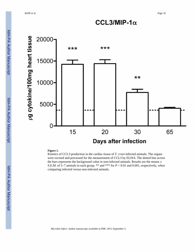
Figure 1.Kinetics of CCL3 production in the cardiac tissue of T. cruzi-infected animals. The organswere excised and processed for the measurement of CCL3 by ELISA. The dotted line acrossthe bars represents the background value in non-infected animals. Results are the means ±S.E.M. of 5–7 animals in each group. ** and *** for P < 0.01 and 0.001, respectively, whencomparing infected versus non-infected animals.
Roffê et al. Page 10
Microbes Infect. Author manuscript; available in PMC 2011 September 5.
NIH
-PA Author Manuscript
NIH
-PA Author Manuscript
NIH
-PA Author Manuscript
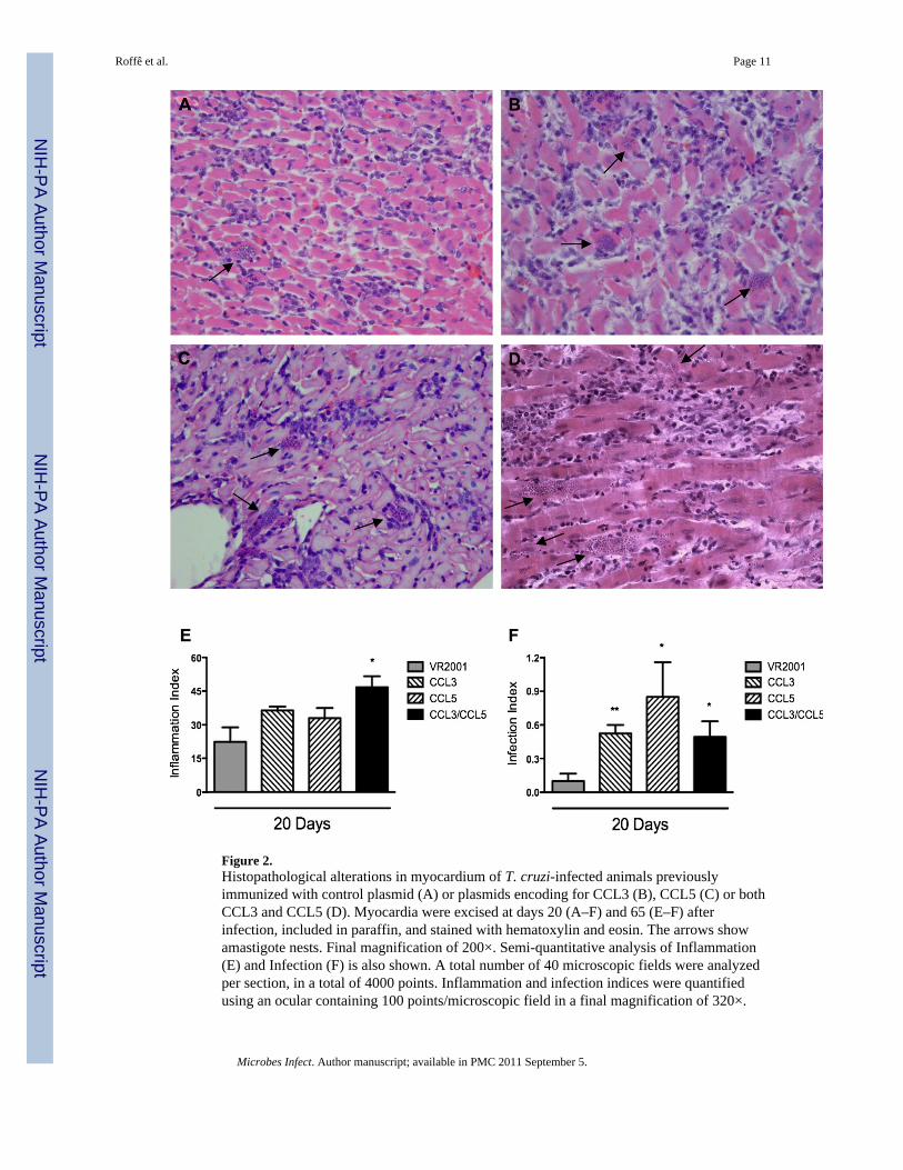
Figure 2.Histopathological alterations in myocardium of T. cruzi-infected animals previouslyimmunized with control plasmid (A) or plasmids encoding for CCL3 (B), CCL5 (C) or bothCCL3 and CCL5 (D). Myocardia were excised at days 20 (A–F) and 65 (E–F) afterinfection, included in paraffin, and stained with hematoxylin and eosin. The arrows showamastigote nests. Final magnification of 200×. Semi-quantitative analysis of Inflammation(E) and Infection (F) is also shown. A total number of 40 microscopic fields were analyzedper section, in a total of 4000 points. Inflammation and infection indices were quantifiedusing an ocular containing 100 points/microscopic field in a final magnification of 320×.
Roffê et al. Page 11
Microbes Infect. Author manuscript; available in PMC 2011 September 5.
NIH
-PA Author Manuscript
NIH
-PA Author Manuscript
NIH
-PA Author Manuscript
Results are the means ± S.E.M. of 5–7 animals in each group. * and ** for P < 0.05 and 0.01respectively, when comparing chemokine- versus control plasmid-immunized animals.
Roffê et al. Page 12
Microbes Infect. Author manuscript; available in PMC 2011 September 5.
NIH
-PA Author Manuscript
NIH
-PA Author Manuscript
NIH
-PA Author Manuscript
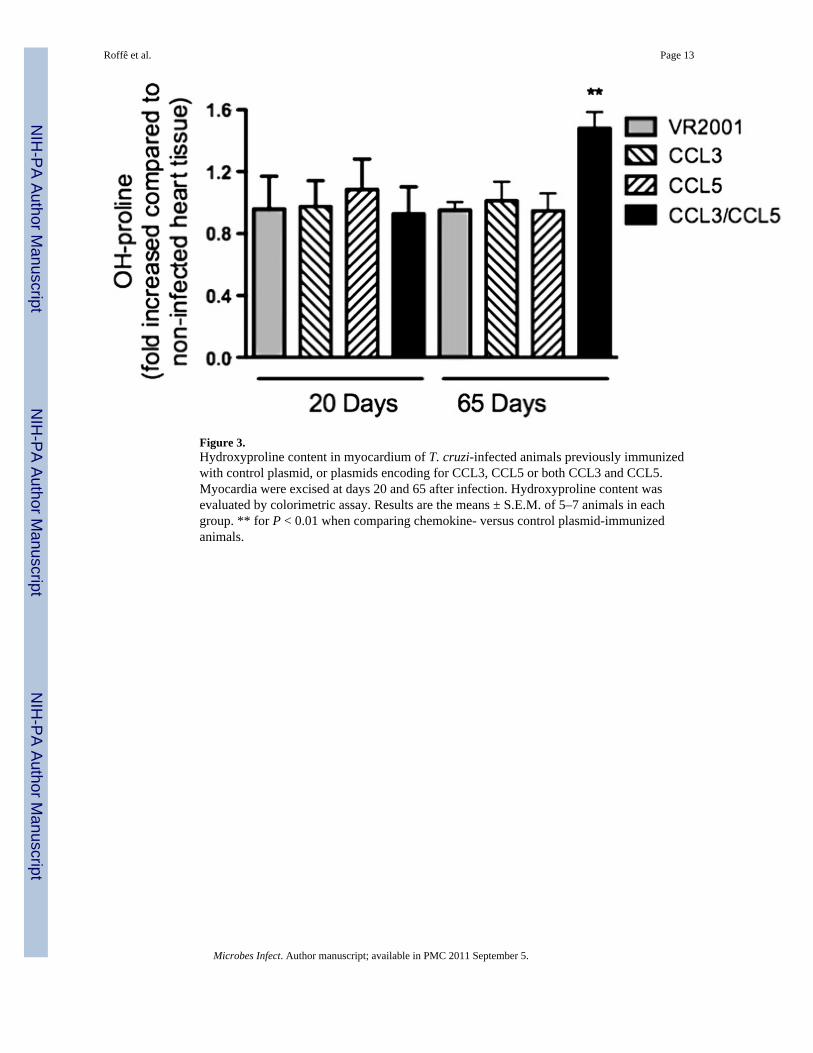
Figure 3.Hydroxyproline content in myocardium of T. cruzi-infected animals previously immunizedwith control plasmid, or plasmids encoding for CCL3, CCL5 or both CCL3 and CCL5.Myocardia were excised at days 20 and 65 after infection. Hydroxyproline content wasevaluated by colorimetric assay. Results are the means ± S.E.M. of 5–7 animals in eachgroup. ** for P < 0.01 when comparing chemokine- versus control plasmid-immunizedanimals.
Roffê et al. Page 13
Microbes Infect. Author manuscript; available in PMC 2011 September 5.
NIH
-PA Author Manuscript
NIH
-PA Author Manuscript
NIH
-PA Author Manuscript
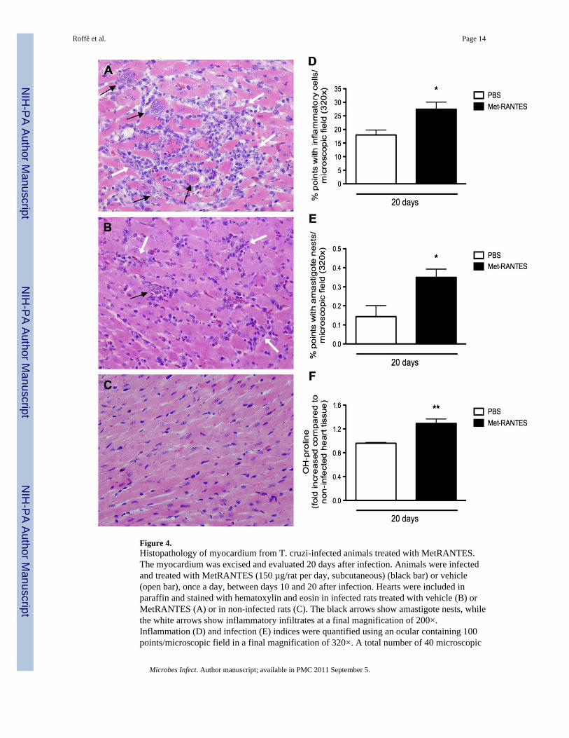
Figure 4.Histopathology of myocardium from T. cruzi-infected animals treated with MetRANTES.The myocardium was excised and evaluated 20 days after infection. Animals were infectedand treated with MetRANTES (150 µg/rat per day, subcutaneous) (black bar) or vehicle(open bar), once a day, between days 10 and 20 after infection. Hearts were included inparaffin and stained with hematoxylin and eosin in infected rats treated with vehicle (B) orMetRANTES (A) or in non-infected rats (C). The black arrows show amastigote nests, whilethe white arrows show inflammatory infiltrates at a final magnification of 200×.Inflammation (D) and infection (E) indices were quantified using an ocular containing 100points/microscopic field in a final magnification of 320×. A total number of 40 microscopic
Roffê et al. Page 14
Microbes Infect. Author manuscript; available in PMC 2011 September 5.
NIH
-PA Author Manuscript
NIH
-PA Author Manuscript
NIH
-PA Author Manuscript
fields were analyzed per section, in a total of 4000 points. Hydroxyproline content (F) wasmeasured using a colorimetric assay. Results are the means ± S.E.M. of 5–7 animals in eachgroup. * and ** for P < 0.05 and 0.01 respectively, when comparing MetRANTES versusvehicle-treated animals.
Roffê et al. Page 15
Microbes Infect. Author manuscript; available in PMC 2011 September 5.
NIH
-PA Author Manuscript
NIH
-PA Author Manuscript
NIH
-PA Author Manuscript