Cell Culture-Based Assessment of Toxicity and Therapeutics ...
13
Citation: Asuzu, P.C.; Trompeter, N.S.; Cooper, C.R.; Besong, S.A.; Aryee, A.N.A. Cell Culture-Based Assessment of Toxicity and Therapeutics of Phytochemical Antioxidants. Molecules 2022, 27, 1087. https://doi.org/10.3390/ molecules27031087 Academic Editors: José Pinela, Lillian Barros and Maria Ines Dias Received: 4 January 2022 Accepted: 3 February 2022 Published: 6 February 2022 Publisher’s Note: MDPI stays neutral with regard to jurisdictional claims in published maps and institutional affil- iations. Copyright: © 2022 by the authors. Licensee MDPI, Basel, Switzerland. This article is an open access article distributed under the terms and conditions of the Creative Commons Attribution (CC BY) license (https:// creativecommons.org/licenses/by/ 4.0/). molecules Review Cell Culture-Based Assessment of Toxicity and Therapeutics of Phytochemical Antioxidants Peace C. Asuzu 1 , Nicholas S. Trompeter 2 , Carlton R. Cooper 3 , Samuel A. Besong 1 and Alberta N. A. Aryee 1, * 1 Department of Human Ecology (Food Science and Biotechnology Program), College of Agriculture, Science and Technology, Delaware State University, Dover, DE 19901, USA; [email protected] (P.C.A.); [email protected] (S.A.B.) 2 Department of Biomedical Engineering, University of Delaware, Newark, DE 19716, USA; [email protected] 3 Center for Translational Cancer Research, Department of Biological Sciences, University of Delaware, Newark, DE 19716, USA; [email protected] * Correspondence: [email protected]; Tel.: +1-302-857-6422 Abstract: Plant-derived natural products are significant resources for drug discovery and develop- ment including appreciable potentials in preventing and managing oxidative stress, making them promising candidates in cancer and other disease therapeutics. Their effects have been linked to phytochemicals such as phenolic compounds and their antioxidant activities. The abundance and complexity of these bio-constituents highlight the need for well-defined in vitro characterization and quantification of the plant extracts/preparations that can translate to in vivo effects and hopefully to clinical use. This review article seeks to provide relevant information about the applicability of cell-based assays in assessing anti-cytotoxicity of phytochemicals considering several traditional and current methods. Keywords: medicinal plants; antioxidant activity; cell lines; cytotoxicity; apoptosis; bioprinting 1. Introduction Cancer is one of the leading causes of death worldwide. It is the first or second leading cause of death prior to age 70 in 112 of 183 countries and third or fourth leading cause in a further 23 countries, according to World Health Organization (WHO) estimates in 2019 [1]. The rising prominence of cancer as a leading cause of death in combination with limited clinical interventions clearly compromises the effects of treatment on population trends in cancer mortality, even in developed countries [2]. Although a combination of screening and treatment is progressively effective in reducing mortality from some cancers, an expected global cancer burden of 28.4 million cases in 2040, a rise of 47% from 2020 values, necessitates the development of new tools to address the unmet needs in cancer management [1]. Although newer, more specific treatments are showing promising results, they can be expensive, and further research is required to determine how to best use these drugs, as well as the toxicities associated with their use [3]. The most common types of cancer treatments available today are chemotherapy, surgery, and radiotherapy. Chemotherapy is curative in subsets of patients presenting with advanced disease, including Hodgkin’s and non-Hodgkin’s lymphoma, acute lymphoblas- tic and acute myelogenous leukemia, germ cell cancer, small cell lung cancer, ovarian cancer, and choriocarcinoma [4,5]. Chemotherapy has also been used as a neoadjuvant therapy to reduce the size of solid tumors before surgical removal, and adjuvant therapy has been used after surgery or radiotherapy, with promising results [4]. However, for some other advanced cancers, including prostate cancer, a curative treatment regimen is yet to be discovered. Scientists are returning to the drawing board to find new therapies or new combinations of therapies to further improve cancer treatment outcomes. Molecules 2022, 27, 1087. https://doi.org/10.3390/molecules27031087 https://www.mdpi.com/journal/molecules
-
Upload
khangminh22 -
Category
Documents
-
view
7 -
download
0
Transcript of Cell Culture-Based Assessment of Toxicity and Therapeutics ...
�����������������
Citation: Asuzu, P.C.; Trompeter,
N.S.; Cooper, C.R.; Besong, S.A.;
Aryee, A.N.A. Cell Culture-Based
Assessment of Toxicity and
Therapeutics of Phytochemical
Antioxidants. Molecules 2022, 27, 1087.
https://doi.org/10.3390/
molecules27031087
Academic Editors: José Pinela,
Lillian Barros and Maria Ines Dias
Received: 4 January 2022
Accepted: 3 February 2022
Published: 6 February 2022
Publisher’s Note: MDPI stays neutral
with regard to jurisdictional claims in
published maps and institutional affil-
iations.
Copyright: © 2022 by the authors.
Licensee MDPI, Basel, Switzerland.
This article is an open access article
distributed under the terms and
conditions of the Creative Commons
Attribution (CC BY) license (https://
creativecommons.org/licenses/by/
4.0/).
molecules
Review
Cell Culture-Based Assessment of Toxicity and Therapeutics ofPhytochemical AntioxidantsPeace C. Asuzu 1, Nicholas S. Trompeter 2, Carlton R. Cooper 3, Samuel A. Besong 1 and Alberta N. A. Aryee 1,*
1 Department of Human Ecology (Food Science and Biotechnology Program), College of Agriculture, Scienceand Technology, Delaware State University, Dover, DE 19901, USA; [email protected] (P.C.A.);[email protected] (S.A.B.)
2 Department of Biomedical Engineering, University of Delaware, Newark, DE 19716, USA; [email protected] Center for Translational Cancer Research, Department of Biological Sciences, University of Delaware,
Newark, DE 19716, USA; [email protected]* Correspondence: [email protected]; Tel.: +1-302-857-6422
Abstract: Plant-derived natural products are significant resources for drug discovery and develop-ment including appreciable potentials in preventing and managing oxidative stress, making thempromising candidates in cancer and other disease therapeutics. Their effects have been linked tophytochemicals such as phenolic compounds and their antioxidant activities. The abundance andcomplexity of these bio-constituents highlight the need for well-defined in vitro characterization andquantification of the plant extracts/preparations that can translate to in vivo effects and hopefullyto clinical use. This review article seeks to provide relevant information about the applicability ofcell-based assays in assessing anti-cytotoxicity of phytochemicals considering several traditional andcurrent methods.
Keywords: medicinal plants; antioxidant activity; cell lines; cytotoxicity; apoptosis; bioprinting
1. Introduction
Cancer is one of the leading causes of death worldwide. It is the first or second leadingcause of death prior to age 70 in 112 of 183 countries and third or fourth leading causein a further 23 countries, according to World Health Organization (WHO) estimates in2019 [1]. The rising prominence of cancer as a leading cause of death in combination withlimited clinical interventions clearly compromises the effects of treatment on populationtrends in cancer mortality, even in developed countries [2]. Although a combination ofscreening and treatment is progressively effective in reducing mortality from some cancers,an expected global cancer burden of 28.4 million cases in 2040, a rise of 47% from 2020values, necessitates the development of new tools to address the unmet needs in cancermanagement [1]. Although newer, more specific treatments are showing promising results,they can be expensive, and further research is required to determine how to best use thesedrugs, as well as the toxicities associated with their use [3].
The most common types of cancer treatments available today are chemotherapy,surgery, and radiotherapy. Chemotherapy is curative in subsets of patients presenting withadvanced disease, including Hodgkin’s and non-Hodgkin’s lymphoma, acute lymphoblas-tic and acute myelogenous leukemia, germ cell cancer, small cell lung cancer, ovariancancer, and choriocarcinoma [4,5]. Chemotherapy has also been used as a neoadjuvanttherapy to reduce the size of solid tumors before surgical removal, and adjuvant therapyhas been used after surgery or radiotherapy, with promising results [4]. However, for someother advanced cancers, including prostate cancer, a curative treatment regimen is yet tobe discovered. Scientists are returning to the drawing board to find new therapies or newcombinations of therapies to further improve cancer treatment outcomes.
Molecules 2022, 27, 1087. https://doi.org/10.3390/molecules27031087 https://www.mdpi.com/journal/molecules

Molecules 2022, 27, 1087 2 of 13
Increased reactive oxygen species (ROS) levels have been found in almost all cancersand are thought to play an important role in the initiation and progression of cancers [6].These highly reactive ions and molecules are produced during normal metabolism of cellsbut are present in higher levels in cancer cells due to increased metabolic activity, mito-chondrial dysfunction, peroxisome activity, increased cellular receptor signaling, oncogeneactivity, increased activity of oxidases, cyclooxygenases, lipoxygenases and thymidinephosphorylase, or through crosstalk with infiltrating immune cells [6]. ROS are managedunder normal physiological conditions, through detoxification by non-enzymatic moleculessuch as glutathione, or through antioxidant enzymes, which specifically scavenge differentkinds of ROS [6]. With increasing interest in natural products, scientists continue to con-sider plants, which are natural sources of exogenous antioxidants, as possible sources ofeffective treatments for different cancers.
2. Medicinal Plants in Cancer Treatment and Management
Phytochemicals are classified as primary or secondary metabolites based on their rolein plant metabolism [7]. Secondary metabolites are chemically active compounds includingalkaloids, anthocyanins, flavonoids, terpenoids, tannins, steroids, saponins, coumarins,phenolics and antioxidants. These are often produced in response to stress, are morecomplex in structure, and are less widely distributed than the primary metabolites [7,8].They are pharmacologically active as anti-oxidative, anti-allergic, anti-bacterial, anti-fungal,anti-diabetic, anti-inflammatory and anti-carcinogenic compounds [8–10]. It is common fora single plant to produce many secondary metabolites with a wide range of chemical andbiological properties, providing a range for bioactive substances [10].
In the last decades, several plants have been confirmed to contain chemo-preventiveand therapeutic agents for various cancers [11–20]. These studies show the effectivenessand synergistic effects of phytochemicals in plant extracts in various diseases [15,21,22].Researchers have discovered that polyphenols are good antioxidants, capable of neutraliz-ing the destructive reactivity of reactive oxygen/nitrogen species produced as byproductsof metabolism [23]. In addition, epidemiological studies have revealed that polyphenolsprovide significant protection against development of several chronic conditions suchas cardiovascular diseases (CVDs), cancer, diabetes, infections, aging and asthma [23].Phenolic phytochemicals are the largest category of phytochemicals and the most widelydistributed in the plant kingdom [24].
There have been studies to examine the effect of crude plant extracts or fractions con-taining phenolic compounds on cancer cells to test the hypothesis that potent antioxidantspossess anticancer potential. Some of these studies revealed that the efficacy of phenoliccompounds in inhibiting cancer activity differs based on the structure of the phenoliccompound and its molecular target [25]. Phenolic compounds can directly scavenge freeradicals after entering cells and activate several cellular signaling pathways (CSP), includ-ing nuclear factor erythroid-2 (NFE2)-related factor 2 (Nrf2)-Kelch-like ECH associatedprotein 1 (Keap1) complex [26]. When activated, the Nrf2-Keap1 complex induces cellu-lar defense mechanisms, including phase II detoxifying enzymes, phase III transporters,anti-oxidative stress proteins, and other stress-defense molecules that protect normal cellsfrom ROS and reactive metabolites of carcinogenic species [26]. Another CSP that can beactivated by phenolics is the mitogen-activated protein kinases (MAPKs) cascade, whichhelps regulate proliferation, differentiation, stress reduction, and apoptosis in cells [26].
Anticancer activity of phenolic compounds has been studied with the use of crudeextracts containing mixtures of phenolic compounds and with isolated phenolic com-pounds. Some examples of crude extracts with reported anticancer activity include:Pandanus amaryllifolius extracts containing gallic acid, cinnamic acid and ferulic acid, withreported in vitro inhibition of breast cancer cell lines [25]. Several Teucrium species extractscontaining hydroxycinnamic acid derivatives, phenylethanoid glycosides, flavonoid glyco-sides, and flavonoid aglycones, with reported antiproliferative and proapoptotic activitiesin HCT-116 colon cancer cell lines [27,28]; Baccharis trimera extracts containing gallic acid,
Molecules 2022, 27, 1087 3 of 13
pyrogallol, syringic acid and caffeic acid, with reported suppression of tumor cell colonyformation and proliferation of SiHa cell line (isolated from a primary uterine squamouscell carcinoma); and Prunus africanus extracts, containing artraric and ferulic acids andN-butylbenzene-sulfonamide (NBBS) with reported antiproliferative effect on prostatecancer cells [15,29].
Over 60% of currently used anti-cancer agents are estimated to be derived fromnatural sources, such as plants, marine organisms, and microorganisms [15,17,30,31]. Goodexamples of plant sources include Prunus africana [15,17], African cherry (Prunus africana(Hook.f.) Kalkman) or Pygeum africanum (Hook. f.), bitter almond, African prune, and red.Besides its use for timber, it is employed as a medicinal plant, whose leaves, roots and barkare used in traditional medicine in Africa [15,32–35]. This is not surprising since variousbioactive substances with anti-inflammatory, anti-cancer, and anti-viral properties havebeen identified in different members of the genus Prunus [33,35–37]. Many phytochemicalsfrom medicinal plants have been discovered to have significant anticancer properties, andmany more are yet to be discovered.
3. Determining Anti-Cancer Potential of Phytochemicals
Phytochemicals and their derivate metabolites present in plants have been shown topossess several beneficial effects in humans. Some of the more widely known phytochemi-cals with anticancer properties include vincristine, vinblastine, camptothecin, bleomycin,paclitaxel, and Taxol among others [30,38]. Different mechanisms have been proposedfor the anticancer effect of various phytochemicals, with some exerting additive and/orsynergistic effects with other phytochemicals. Some of the mechanisms that have been iden-tified include selective killing of rapidly dividing cells, targeting of atypically expressedmolecular factors, anti-oxidation, modification of cell growth factors, and inhibition ofinappropriate angiogenesis and induction of apoptosis [38]. An example is ellagic acid,found in pomegranates, which induces apoptosis in prostate and breast cancer cells andsuppresses metastatic processes of many cancer types [38]. Curcumin, found in turmeric, isattributed to cause apoptosis in cancer cells without cytotoxic effect on healthy cells viaseveral mechanisms, including the regulation of cell proliferation, cell survival, and caspaseactivation pathways [26,39]. While many phytochemicals that can serve as anticancer drugsby themselves are yet to be discovered, those already discovered can serve as models forthe preparation of more effective formulations by applying methods such as total or combi-natorial synthesis, or biosynthetic pathway manipulation [16,30]. This concept was appliedto overcome the severe toxicity of earlier formulations of paclitaxel by utilizing an albumin-bound nanoparticle technology, which concentrates the drug in tumors [16]. With new andmore effective technologies and better understanding of cancer biology, phytochemicalsand their derivatives are bound to play a pivotal role in cancer chemotherapy.
3.1. Cancer Cell Lines
Cancer cell lines are useful tools because they provide a multifaceted model of thebiological mechanisms involved in cancer development and progression. The use of cancercell lines improved the knowledge of deregulated genes and signaling pathways involvedin cancer progression; cell lines have also been used to define potential molecular markersfor cancer screening and prognosis [40]. Numerous cell lines with their unique propertiesand characteristics are currently available for in vitro study of different types of cancer [41].
Cell lines are easy to handle and manipulate genetically/epigenetically by usingdemethylation agents, small interfering ribonucleic acid (siRNA), expression vectors, andthey can be pharmacologically manipulated through cytostatics (cell growth inhibitors).Cell lines are homogenous, providing identical tumor cells for easier analysis unlike inheterogeneous solid tumors. However, to imitate in vivo tumor characteristics as closelyas possible, a cancer cell line panel representative of the heterogeneity observed in theprimary tumors can be used. Cancer cell lines are pure populations of tumor cells and havea high degree of similarity with the initial tumor. Because of the homogeneity of cell lines,
Molecules 2022, 27, 1087 4 of 13
results of experiments using correct conditions are easily reproducible [40]. In addition,there is a substantial number and variety of cancer cell lines available (Table 1). Despitethese advantages, some drawbacks of using cancer cell lines include cross-contaminationwith HeLa cells, genomic instability leading to differences between the original tumor andthe specific cell line, changes in the morphology, gene expression, and cellular pathwaysof cell lines from culture conditions required to maintain them (i.e., culture adaption),and infections with mycoplasma [40]. Furthermore, it is difficult to establish long-termcancer cell lines of certain types of tumors, including prostate cancer tumors [40,42]. Thelimited number of cell line models for prostate cancer research stems from the difficultyin propagating prostate cancer cells in vitro for extended periods. Investigators have beenable to generate only seven cell lines that were previously available through public cellline repositories, but these do not represent the spectrum of clinical disease. New cell lines,which demonstrate the commonly observed clinical phenotypes, are clearly needed [42].
Since the isolation of the first cell line in the 1950s (i.e., HeLa cells), a variety of cancercell lines have been developed for preliminary drug testing [43]. The different cell linesrequire different media, growth factors, and supplements to remain viable over time, as theconstituents of culture media affect the cell lines. In a study by Kim et al., human breastcancer cells (MDA-MB-231) cultured in minimum essential medium (MEM), Dulbecco’smodified Eagle’s medium (DMEM), or Roswell Park Memorial Institute (RPMI)-1640medium and containing different concentrations of fetal bovine serum (FBS) or differentsera (equine or bovine) showed significant changes in gene expression [44]. They reportedthat about 25% of genes were expressed at significantly different levels by cells grown inMEM, DMEM, or RPMI-1640 media based on genome-wide expression analysis [44]. Inanother study, lung cancer cells (A549) and hepatocellular cancer cells (HepG2) cultured inHam’s F-12 nutrient mix (F12), RPMI, DMEM, and MEM revealed a significantly increasedproliferation rate for A549 cells in DMEM compared to the other media tested, and thelowest rate for both A549 and HepG2 cells in MEM, confirmed by assaying conditionedmedia for basal level ATP at 72 h [45]. This underscores the significant effect of growthconditions and/or environment on cells in drug discovery experiments, and the need forspecificity to ensure results are reproducible.
There are a handful of prostate cancer cell lines in use today, most of which have beenestablished from metastatic deposits [46]. The LNCaP cell line, isolated from a subclavianlymph node metastasis of prostate cancer, maintains several key markers including prostate-specific antigen (PSA), prostate specific membrane antigen (PSMA) and the androgenreceptor (AR) [47]. The LNCaP cell line is androgen sensitive (AS) and expresses ARand PSA mRNA/protein [41,48]. It has a doubling time of 60–72 h, is responsive to TGF-α, EGF and IGF-1, which are known to promote cancer development and progression,and has a 50% success rate after xenografting, with a tumor doubling time of 86 h whencombined with a Matrigel™ formulation [41]. In another study, LNCaP cells, amongothers, were injected subcutaneously between the scapulae of pfp−/−/rag2−/− doubleknock-out mice, resulting in primary tumor growth and pulmonary metastases in 100%of LNCaP-injected mice, and detection of DNA of 266 circulating tumor cells (CTC) permL of blood and 35 disseminated tumor cells (DTC) per mL bone marrow after Alu-PCRanalysis [49]. Through passage and hormonal manipulation in vivo, the lineage-relatedLNCaP sublines have resulted in a series of cells that mimic the progression of prostatecancer from the original AS LNCaP cell line to the androgen-independent (AI) C4-2 andC4-2B cell lines [47,50].
An AI cell line, C4-2, reproducibly and consistently follows the metastatic patterns ofhormone-refractory prostate cancer by producing lymph node and bone metastases wheninjected either subcutaneously or orthotopically in either hormonally intact or castratedhosts. This model enables the study of factors that determine the predilection of prostatecancer cells for the skeletal microenvironment [50]. These C4-2 cells have a doubling time ofabout 48 h, are androgen independent, express an androgen receptor, metastasize to lymphnodes, and produce PSA [41,46]. The AI C4-2 cell line differs from its parent AS LNCaP,
Molecules 2022, 27, 1087 5 of 13
with differential expression of 38 genes between the two cell lines (≥2-fold change, 95% CI),14 of which expressed at higher levels in LNCaP than in C4-2 cells, while the remaining24 were expressed at lower levels in LNCaP than in C4-2 cells. In addition, the AI C4-2cell line is highly tumorigenic and metastatic, including spontaneous metastasis to bone,whereas the AS LNCaP cell line is only weakly tumorigenic and is non-metastatic [47].
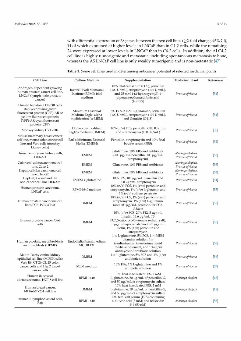
Table 1. Some cell lines used in determining anticancer potential of selected medicinal plants.
Cell Line Culture Medium Supplementation Medicinal Plant Reference
Androgen-dependent growinghuman prostate cancer cell line,LNCaP (lymph node prostate
cancer)
Roswell Park MemorialInstitute (RPMI) 1640
medium
10% fetal calf serum (FCS), penicillin(100 U/mL), streptomycin (100 U/mL),
and 25 mM 4-(2-hydroxyethyl)-1-piperazineethanesulfonic acid
(HEPES)
Prunus africana [51]
Human hepatoma Hep3B cellsstablyexpressing green
fluorescent protein (GFP)-AR oryellow fluorescent protein(YFP)-AR-cyan fluorescent
protein (CFP)
Minimum EssentialMedium Eagle, alpha
modification (α-MEM)
5% FCS, 2 mM L-glutamine, penicillin(100 U/mL), streptomycin (100 U/mL),
and Genticin (G418)Prunus africana [51]
Monkey kidney CV1 cells Dulbecco’s modifiedEagle’s medium (DMEM)
10% (v/v) FCS, penicillin (100 IU/mL)and streptomycin (100 IU/mL) Prunus africana [37]
Mouse mammary breast cancercell line, mouse colon cancer cell
line and Vero cells (monkeykidney cells)
Earl’s Minimum EssentialMedia (EMEM)
Penicillin, streptomycin and 10% fetalbovine serum (FBS) Prunus africana [52]
Human embryonic kidney cells,HEK293 EMEM
Glutamine, 10% FBS and antibiotics(100 µg/mL penicillin, 100 µg/mL
streptomycin)
Moringa oleifera,Prunus africana [53]
Colorectal adenocarcinoma cellline, Caco-2 EMEM Glutamine, 10% FBS and antibiotics Moringa oleifera,
Prunus africana [53]
Hepatocellular carcinoma cellline, HepG2 EMEM Glutamine, 10% FBS and antibiotics Moringa oleifera,
Prunus africana [53]
HepG-2, Caco-2 and thenon-cancer cell line HEK293 EMEM + glutamine 10% FBS, 100 µg/mL penicillin and
100 µg/mL streptomycin Prunus africana [54]
Human prostate carcinomaLNCaP cells RPMI-1640 medium
10% (v/v) FCS, 1% (v/v) penicillin andstreptomycin, 1% (v/v) L-glutamin and
1% (v/v) sodium pyruvatePrunus africana [55]
Human prostate carcinoma celllines PC3, PC3-ARwt DMEM
10% (v/v) FCS, 1% (v/v) penicillin andstreptomycin, 1% (v/v) L-glutamin(and 600 µg/mL geneticin for PC3-
ARwt)
Prunus africana [55]
Human prostate cancer C4-2cells DMEM
10% (v/v) FCS, 20% F12, 5 µg/mLInsulin, 13.6 pg/mL T3
(3,3′,5-triiodo-L-thyronine sodium salt),5 µg/mL apotransferrin, 0.25 µg/mL
Biotin, 1% (v/v) penicillin andstreptomycin
Prunus africana [55]
Human prostatic myofibroblastsand fibroblasts (HPMF)
Endothelial basal mediumMCDB 131
1 × L-glutamine, 5% FCS, 1 ×MEMvitamins solution, 1×
insulin-tranferrin-selenium liquidmedia supplement, and 1% (v/v)antimycotic/ antibiotic solution
Prunus africana [56]
Madin-Darby canine kidneyepithelial cell line (MDCK cells) DMEM 1 × L-glutamine, 5% FCS and 1% (v/v)
antibiotic solution Prunus africana [56]
Vero E6, CT 26-CL 25 coloncancer cells and Hep2 throat
cancer cellsMEM medium 10% FBS, 1% L-glutamine and 1%
antibiotic solution Prunus africana [57]
Human ileoceacaladenocarcinoma, HCT-8 cell line RPMI-1640
10% heat inactivated FBS, 2 mML-glutamine, 50 µg/mL of penicillin-G,and 50 µg/mL of streptomycin sulfate
Moringa oleifera [18]
Human breast cancer,MDA-MB-231 cell line DMEM
10% heat inactivated FBS, 2 mML-glutamine, 50 µg/mL of penicillin-G,and 50 µg/mL of streptomycin sulfate
Moringa oleifera [18]
Human B-lymphoblastoid cells,Raji RPMI-1640
10% fetal calf serum (FCS) containingn-butyric acid (3 mM) and teleocidin
B-4 (50 nM)Moringa oleifera [58]
Molecules 2022, 27, 1087 6 of 13
3.2. Recent Advances in Cell Culture Models for Testing Anticancer Drugs
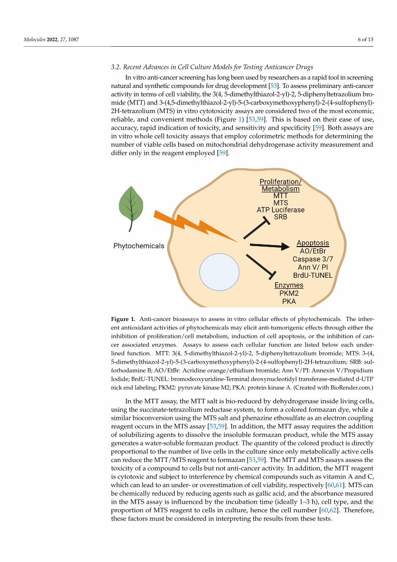
In vitro anti-cancer screening has long been used by researchers as a rapid tool in screeningnatural and synthetic compounds for drug development [53]. To assess preliminary anti-canceractivity in terms of cell viability, the 3(4, 5-dimethylthiazol-2-yl)-2, 5-diphenyltetrazolium bro-mide (MTT) and 3-(4,5-dimethylthiazol-2-yl)-5-(3-carboxymethoxyphenyl)-2-(4-sulfophenyl)-2H-tetrazolium (MTS) in vitro cytotoxicity assays are considered two of the most economic,reliable, and convenient methods (Figure 1) [53,59]. This is based on their ease of use,accuracy, rapid indication of toxicity, and sensitivity and specificity [59]. Both assays arein vitro whole cell toxicity assays that employ colorimetric methods for determining thenumber of viable cells based on mitochondrial dehydrogenase activity measurement anddiffer only in the reagent employed [59].
Figure 1. Anti-cancer bioassays to assess in vitro cellular effects of phytochemicals. The inher-ent antioxidant activities of phytochemicals may elicit anti-tumorigenic effects through either theinhibition of proliferation/cell metabolism, induction of cell apoptosis, or the inhibition of can-cer associated enzymes. Assays to assess each cellular function are listed below each under-lined function. MTT: 3(4, 5-dimethylthiazol-2-yl)-2, 5-diphenyltetrazolium bromide; MTS: 3-(4,5-dimethylthiazol-2-yl)-5-(3-carboxymethoxyphenyl)-2-(4-sulfophenyl)-2H-tetrazolium; SRB: sul-forhodamine B; AO/EtBr: Acridine orange/ethidium bromide; Ann V/PI: Annexin V/PropidiumIodide; BrdU-TUNEL: bromodeoxyuridine-Terminal deoxynucleotidyl transferase-mediated d-UTPnick end labeling; PKM2: pyruvate kinase M2; PKA: protein kinase A. (Created with BioRender.com.)
In the MTT assay, the MTT salt is bio-reduced by dehydrogenase inside living cells,using the succinate-tetrazolium reductase system, to form a colored formazan dye, while asimilar bioconversion using the MTS salt and phenazine ethosulfate as an electron couplingreagent occurs in the MTS assay [53,59]. In addition, the MTT assay requires the additionof solubilizing agents to dissolve the insoluble formazan product, while the MTS assaygenerates a water-soluble formazan product. The quantity of the colored product is directlyproportional to the number of live cells in the culture since only metabolically active cellscan reduce the MTT/MTS reagent to formazan [53,59]. The MTT and MTS assays assess thetoxicity of a compound to cells but not anti-cancer activity. In addition, the MTT reagentis cytotoxic and subject to interference by chemical compounds such as vitamin A and C,which can lead to an under- or overestimation of cell viability, respectively [60,61]. MTS canbe chemically reduced by reducing agents such as gallic acid, and the absorbance measuredin the MTS assay is influenced by the incubation time (ideally 1–3 h), cell type, and theproportion of MTS reagent to cells in culture, hence the cell number [60,62]. Therefore,these factors must be considered in interpreting the results from these tests.
Molecules 2022, 27, 1087 7 of 13
The sulforhodamine B (SRB) assay is a rapid, sensitive, and inexpensive method fordetermining cell growth, utilizing a bright pink anionic dye that binds electrostatically tobasic amino acids of trichloroacetic acid fixed cells. The protein-bound dye is extracted withTris (tris (hydroxymethyl) aminomethane) base to quantify the protein content indirectlywith spectrophotometry [63]. The endpoint of the SRB assay is non-destructive, stable,does not require time-sensitive measurement, and it is comparable with other fluorescenceassays [60,64]. However, it is labor intensive, requiring several washing steps [63,65].
A known characteristic of cancer cell growth and metastasis is the ability of the cells toescape apoptosis because of a mutation in tumor suppressor genes. Induction of apoptosisis thus used as an important indicator of the ability of chemotherapeutic agents to inhibittumor growth and progression. The acridine orange/ethidium bromide (AO/EB) apoptosisassay is used to study changes in cellular and nuclear morphology and characteristics ofapoptosis under a fluorescent microscope [53]. Both AO and EB bind to DNA and RNA byintercalation between adjacent base pairs, but AO stains both live and dead cells while EBstains dead cells only. Live cells appear green under the microscope, and early apoptoticcells have a bright green nucleus due to chromatin condensation and nuclear fragmentation,while late apoptotic cells appear orange because they take up EB, and necrotic cells willstain orange but have a normal nuclear morphology [53,66]. After cells are counted underthe microscope, an apoptotic index is calculated.
The living status of a cell can be determined by measuring the amount of ATP inthe cell, since ATP is necessary for life and function of all cells, and levels of cytoplasmicATP decrease in cases of injury and hypoxia. After a cell is lysed, ATP is free to reactwith luciferin and luciferase, producing high quantum chemiluminescence that is linearlyproportional to the ATP concentration under optimum conditions. Compared to the MTTassay, the luciferase assay had higher sensitivity and reproducibility over several days andwas able to detect the viability of cells with cell counts as low as 2000 cells/well comparedto a minimum of 25,000 cells/well required for the MTT assay mentioned above. TheATP assay has been reported as sensitive compared to MTT and calcein assays, used todetermine the potency of cytotoxic agents [67]. This high sensitivity of the ATP assayallowed for detection of cytotoxic agent-induced ATP breakdown after incubation periodsas short as 1 h, which provides an additional advantage over the MTT assay that requiresapproximately 72 h of incubation. A further advantage of the ATP assay was the shortmeasurement time of 15 s per well, compared to the MTT assay, which required a 1–2 hsolubilization step of the formazan before an absorption measurement [67]. However, theATP assay cannot differentiate between cytostatic and cytotoxic cellular effects [64].
Many anticancer drugs in use today inactivate target cells by inducing apoptosis [68].As one of the later steps in apoptosis, DNA fragmentation, a process resulting from theactivation of nucleases that break down DNA into small fragments, can be used as a mea-sure of anticancer agent bioactivity [68]. When anticancer agents break down DNA, theyexpose many 3′-hydroxyl ends to which fluorescein deoxythymidine analog, 5-bromo-2′-deoxyuridine 5′-tri-phosphate (BrdUTP) molecules attach, with the help of terminaldeoxynucleotidyl transferase (TdT). One of the TdT dUTP Nick-End Labeling (TUNEL)assay methods involves the attachment of a fluorescein deoxythymidine analog, 5-bromo-2′-deoxyuridine 5′-triphosphate (BrdUTP) molecule [68]. After it is assimilated into theDNA, BrdU can be detected by an anti-BrdU antibody using standard immunohistochem-ical techniques, fluorescence microscopy or flow cytometry [68]. TUNEL assays havebeen used in the evaluation of many anticancer compounds, including derivatives of be-tulinic acid and botulin and 5-fluorouracil [68]. The harsh denaturing conditions necessaryfor the binding of anti-BrdU cause cell disruption and protein degradation, which is alimitation, particularly if concurrent protein content measurement or molecular analysisis required [64,69].
Although two-dimensional (2D) cell culture systems involving the growth of a mono-layer of cells on a plastic surface or in vivo animals were the standard for drug testing, datafrom 2D models are often misleading, resulting in difficulties with translational efficacy
Molecules 2022, 27, 1087 8 of 13
in vivo [70,71]. This is mostly because while convenient, 2D systems are over simplisticrepresentations of the in vivo complex tissue architecture, which fail to incorporate thebiochemical and biomechanical crosstalk between tumors and the surrounding tumormicroenvironment [70,72,73]. The absence of drug transport barriers, extracellular matrixand blood vessels, immune cells, gradients of oxygen tension, extracellular pH, nutrients,catabolites present normally in tumor conditions in vivo, coupled with short-term cultureconditions in 2D systems, may select for cytotoxic drugs that prove insufficient in pre-clinical and clinical settings [70,72,74]. Therefore, in a bid to overcome these limitations andavoid the ethical concerns involving animal testing, new test systems such as the Boyden’schamber, three-dimensional (3D) cultures, microfluidic device systems, and models createdusing 3D bioprinting were developed [72].
One of the new systems used to model the complex in vivo intercellular interactionsin vitro is the Boyden chamber, consisting of two chambers containing media and par-titioned by a semi-permeable membrane [72,75]. To study cell migration in a Boydenchamber, cells are seeded in the upper chamber and allowed to migrate under the influenceof a concentration gradient of chemotactic substances added to the media in the lowerchamber media [75,76]. Cell migration is assessed by measuring the optical density oflabeled cell extracts and corresponds to the effectiveness of the biologically active sub-stance [75]. The Boyden chamber was used to assess and compare the invasive activity ofspheroids containing only tumor cells and spheroids containing a mixture of tumor andstem cells, showing an increased invasion of the heterogeneous spheroids when comparedto spheroid containing only tumor cells [72,77]. Although easy to use, the Boyden chamberdoes not allow for direct cell–cell interactions, limiting its ability to fully reproduce in vivoconditions and creating a preference for other evolving methods such as 3D culture andmicrofluidic systems [75].
An ideal 3D system would mimic a specific solid tumor microenvironment, wherecells are able to replicate and interact with other cells, while promoting differentiation [78].Most 3D systems, however, do not exactly simulate in vivo conditions, although they aremore representative than 2D models. Three-dimensional systems may be classified as free-floating anchorage-independent systems, scaffold-based systems, and organoids, whichare hybrid 3D culture models composed of spheroids [78]. Regardless of the class of 3Dsystem used, research has shown that cancer cells grown in 3D culture may respond todrug treatment similarly to cancer cells in the native environment [70,72,78]. In addition,differences in apoptotic sensitivity to chemotherapeutic agents have been noted in non-malignant and malignant mammary cell lines between 2D and 3D cultured cells [79,80].In another study, BT-549, BT-474, and T-47D breast cancer cells in a 2D culture were lessresistant to paclitaxel and doxorubicin compared to a 3D culture of the same cells [72,81].Three-dimensional cell culture systems also provide an alternative to suspension cultures,which are necessary for growing poorly adherent cancer cells and non-solid tumor cellssuch as leukemia [70]. In addition, the development of organoid cultures has created moreways to carry out high-throughput drug screening using 3D culture, which may facilitatepersonalized cancer treatments, biomarker discovery, and mechanistic studies on drugresistance [73]. Organoid cultures have been successfully used to model pancreatic ductaladenocarcinoma from patient derived xenograft tumors and from patient prostate cancerbone metastasis [72].
Bioprinting allows for the creation of various models that mimic the processes thatoccur in the tumor microenvironment (TME) and is a method for constructing complex 3Dbiological structures. This is achieved by printing a bio ink composed of an extracellularmatrix (ECM) or other synthetic substrate and cells layer-by-layer in a computer-designedpattern [72,82]. In this way, a system that mimics the TME of cervical cancer, triple-negativebreast cancer with fibroblasts, and patient-derived cancer cells, fibroblasts, and endothelialcells have been created [82]. Three-dimensional bioprinting has been used to highlight thetrophic role of stromal or immune cells in breast cancer cells cultured with fibroblasts inspheroids which remained viable for over 30 d and were resistant to paclitaxel, unlike the

Molecules 2022, 27, 1087 9 of 13
homogenous breast cancer spheroids [72]. Three-dimensional bioprinting also makes itpossible to study immune cell behavior in the TME. For example, glioblastoma cells in a 3Dbioprinting model were shown to polarize actively recruited macrophages in glioblastoma-associated macrophages, enhancing the proliferation and invasiveness of the glioblastomacells [72].
Three-dimensional bioprinting can also be used to design systems that simulateaberrant tumor vascularization to better understand tumor biology and for in vitro drugtesting [72,82]. Microfluidic systems involve the use of small devices designed for cellcultures to mimic perfusion, thus allowing for steady supply of oxygen and nutrients tocells and the removal of wastes [72,78]. They make it possible to control fluid flow, tem-perature, hydrodynamic and hydraulic pressures, shear, and chemical gradients in vitroto simulate physiological conditions in the TME [72]. The device may be designed with abarrier between compartments or a non-physical barrier such as a biomimetic extracellularmatrix to divide the compartments in the device [78]. Microfluidic systems can be usedto simulate a metastatic model of tumors, allowing researchers to study the effects ofanti-metastatic drugs on tumor cell migration. For example, a microfluidic system usingcollagen–matrigel hydrogel matrices was used to reproduce the microenvironment andexperimental conditions to study the migration and invasion of H1299 lung adenocarci-noma cells [72]. Different forms of microfluidic systems such as well plate, droplet, andcontinuous flow microfluidics are amenable to high-throughput drug screening, makingthem desirable for anticancer drug screening [83].
3.3. Real-Time Assessments of Cell Culture Assays
With the establishment of 3D cultures, there is a need for monitoring and recordingthe cultures in 3D in real time and over the time needed for progression to occur, namely,in 4D [84]. Real-time image-based analysis of cellular response to drug activity in vitro andin vivo may expedite drug development timelines, decrease costs, provide better under-standing of adaptive response and increase clinical predictivity when used with relevantmodel systems [85]. While traditional time-lapse epifluorescent and confocal microscopesprovide detailed temporal and spatial analysis of cellular function, they are restricted to oneor a few samples per experiment, limiting their application for drug discovery. However,new generation live-cell imaging microscopes allow for examination of dynamic cellularprocesses in response to multiple molecular or pharmacological interventions [85]. Theseinclude the IncuCyte™; Cell-IQ™ and Biostation CT™, which are equipped with softwareto remotely control image acquisition, filter optic configurations and image analysis, andare optimized for long-term kinetic studies across multi-well plates [85].
The standard IncuCyte-FLR™ system can accommodate up to six 384-well platesand can automatically monitor cell growth, cell migration into a wounded monolayer,angiogenesis and apoptosis [85]. In a recent study, the Sartorius IncuCyte® system wasused to investigate the killing potential of immune cells on cancer cell lines, tracking livingcells labeled by a red fluorescent protein, and cell death through the green fluorescentsignal generated when apoptotic pathways are activated [86]. The Cell-IQ® system is afully integrated incubator, with continuous live cell imaging and an automated analysisplatform that combines phase-contrast microscopy and fluorescent image acquisition withan analyzer software package for the quantification of migration image data [85,87]. Pro-cesses such as cell attachment, migration velocity, migration direction, neurite outgrowth,vesicle formation, angiogenesis and stem cell differentiation have been documented usingthe Cell-IQ™ system, which allows for a robust kinetic study of phenotypic responseto drug treatment [85]. The Nikon Biostation CT™ platform is reported to be the firstmulti-objective fluorescent and phase contrast microscope combined with automated platehandling robotics within a cell-culture incubator [85]. The Biostation CT is used to demon-strate reduced spheroid migration velocity and suppressed spheroid fusion of human breastcancer cell lines BT474 and T47D when exposed to trastuzumab and paclitaxel, correlatedby ATP quantification cell viability testing [79].
Molecules 2022, 27, 1087 10 of 13
The ability of these systems to document real-time drug response in cells have theadded advantage of making it easier to quantify transient phenotypic responses, optimizetime points for endpoint studies, determine accurate dosing and scheduling regimens,identify cancer cell adaptive responses, and facilitate more robust quantitative analysisfrom less specimens [85]. The combinations of functions in one place, the ability to maintainsteady environmental conditions, and remotely controlled multiple phases of experimentsare attractive features of these automated live-cell analysis systems.
4. Conclusions
The increasing prevalence of cancers worldwide has made the development of quickermethods to create, develop, and test new anticancer drugs a necessity. Anticancer bioassayshave proven to be powerful tools in the drug discovery process and preclinical authen-tication. However, caution should be exercised in comparing results from the differentassays, as they usually target different mechanisms. The choice of an anti-cancer bioassayto use depends, to a large extent, on the researcher’s objective, the target cancer cells andphytochemical composition of the medicinal plant, the availability of reagents and cost ofreagents, and the experience of the research team. It is also important that a few knownanticancer compounds of known potency are included for comparison with potentialmedicinal plant extracts, regardless of what method is used for cytotoxicity screening, forobjectivity and relevance. Cell-based anti-cancer bioassays have much to offer prior toanimal testing. Because of lower cost, the investigator has more control of confrontingvariables, and models can be developed to predict or approximate the phytochemical (i.e.,natural drug) effect in animal and, with additional research, human subjects.
Author Contributions: P.C.A. wrote the primary manuscript; A.N.A.A., S.A.B., N.S.T. and C.R.C.edited the manuscript. All authors have read and agreed to the published version of the manuscript.
Funding: This study was supported by a grant from Evans-Allen (project # DELXHMEC2017).
Institutional Review Board Statement: Not applicable.
Informed Consent Statement: Not applicable.
Data Availability Statement: Not applicable.
Conflicts of Interest: The authors declare no conflict of interest.
References1. Sung, H.; Ferlay, J.; Siegel, R.L.; Laversanne, M.; Soerjomataram, I.; Jemal, A.; Bray, F. Global Cancer Statistics 2020: GLOBOCAN
Estimates of Incidence and Mortality Worldwide for 36 Cancers in 185 Countries. CA Cancer J. Clin. 2021, 71, 209–249. [CrossRef][PubMed]
2. Danaei, G.; Vander Hoorn, S.; Lopez, A.D.; Murray, C.J.; Ezzati, M. Causes of cancer in the world: Comparative risk assessment ofnine behavioural and environmental risk factors. Lancet 2005, 366, 1784–1793. [CrossRef]
3. Herbst, R.S.; Bajorin, D.F.; Bleiberg, H.; Blum, D.; Hao, D.; Johnson, B.E.; Ozols, R.F.; Demetri, G.D.; Ganz, P.A.; Kris, M.G.; et al.Clinical Cancer Advances 2005: Major research advances in cancer treatment, prevention, and screening—A report from theAmerican Society of Clinical Oncology. J. Clin. Oncol. 2006, 24, 190–205. [CrossRef] [PubMed]
4. Arruebo, M.; Vilaboa, N.; Sáez-Gutierrez, B.; Lambea, J.; Tres, A.; Valladares, M.; González-Fernández, Á. Assessment of theEvolution of Cancer Treatment Therapies. Cancers 2011, 3, 3279–3330. [CrossRef] [PubMed]
5. DeVita, V.T., Jr.; Chu, E. A history of cancer chemotherapy. Cancer Res. 2008, 68, 8643–8653. [CrossRef]6. Liou, G.-Y.; Storz, P. Reactive oxygen species in cancer. Free Radic. Res. 2010, 44, 479–496. [CrossRef]7. Krishnaiah, D.; Sarbatly, R.; Bono, A. Phytochemical antioxidants for health and medicine—A move towards nature. Biotechnol.
Mol. Biol. Rev. 2007, 1, 97–104.8. Kumari, P.; Kumari, C.; Singh, P.S. Phytochemical screening of selected medicinal plants for secondary metabolites. Int. J. Life-Sci.
Sci. Res. 2017, 3, 1151–1157. [CrossRef]9. Gupta, A.; Naraniwal, M.; Kothari, V. Modern extraction methods for preparation of bioactive plant extracts. Int. J. Appl. Nat. Sci.
2012, 1, 8–26.10. Ncube, A.; Afolayan, A.; Okoh, A. Assessment techniques of antimicrobial properties of natural compounds of plant origin:
Current methods and future trends. Afr. J. Biotechnol. 2008, 7, 1797–1806. [CrossRef]
Molecules 2022, 27, 1087 11 of 13
11. Albuquerque Costa, R.; de Sousa, O.V.; Hofer, E.; Mafezoli, J.; Barbosa, F.G.; dos Fernandes Vieira, R.H.S. Thiocarbamates fromMoringa oleifera seeds bioactive against virulent and multidrug-resistant Vibrio species. BioMed Res. Int. 2017, 2017, 1–6. [CrossRef][PubMed]
12. Barbieri, L.; Zamboni, M.; Lorenzoni, E.; Montanaro, L.; Sperti, S.; Stirpe, F. Inhibition of protein synthesis in vitro by proteinsfrom the seeds of Momordica charantia (bitter pear melon). Biochem. J. 2015, 186, 443–452. [CrossRef] [PubMed]
13. Bijina, B.; Chellappan, S.; Basheer, S.M.; Elyas, K.K.; Bahkali, A.H.; Chandrasekaran, M. Protease inhibitor from Moringa oleiferaleaves: Isolation, purification, and characterization. Process. Biochem. 2011, 46, 2291–2300. [CrossRef]
14. Igwe, K.; Madubuike, A.; Ikenga, C.; Otuokere, I.; Amaku, F. Studies of the medicinal plant Pausinystalia yohimbe ethanol leafextract phytocomponents by GCMS analysis. Int. J. Sci. Res. Manag. 2016, 4, 4116–4122. [CrossRef]
15. Komakech, R.; Kang, Y.; Lee, J.; Omujal, F. A review of the potential of phytochemicals from Prunus africana (Hook f.) Kalkmanstem bark for chemoprevention and chemotherapy of prostate cancer. Evid.-Based Complement. Altern. Med. 2017, 2017, 1–10.[CrossRef]
16. Huang, M.; Lu, J.-J.; Ding, J. Natural Products in Cancer Therapy: Past, Present and Future. Nat. Prod. Bioprospect. 2021, 11, 5–13.[CrossRef]
17. Shenouda, N.; Sakla, M.; Newton, L.; Besch-Williford, C.; Greenberg, N.; MacDonald, R.; Lubahn, D. Phytosterol Pygeum africanumregulates prostate cancer in vitro and in vivo. Endocrine 2007, 31, 72–81. [CrossRef]
18. Al-Asmari, A.K.; Albalawi, S.M.; Athar, M.T.; Khan, A.Q.; Al-Shahrani, H.; Islam, M. Moringa oleifera as an Anti-Cancer Agentagainst Breast and Colorectal Cancer Cell Lines. PLoS ONE 2015, 10, e0135814. [CrossRef] [PubMed]
19. Eweka, A.O.; Om’Iniabohs, F.A.E.; Momodu, O. The histological effects of mixed diet containing Pausinystalia yohimbe groundstem bark on the kidney of adult Wistar rats (Rattus norvegicus). Biol. Med. 2010, 2, 30–36.
20. Lo, H.-Y.; Ho, T.-Y.; Lin, C.; Li, C.-C.; Hsiang, C.-Y. Momordica charantia and Its Novel Polypeptide Regulate Glucose Homeostasisin Mice via Binding to Insulin Receptor. J. Agric. Food Chem. 2013, 61, 2461–2468. [CrossRef]
21. Karan, M.; Kumar Jena, A.; Sharma, N.; Vasisht, K.; Efferth, T. A systematic analysis of Prunus species with a focus on managementplan of Prunus africana (Hook.f.) Kalkman: An autochthon plant of Africa. Eur. J. Med. Plants 2017, 20, 1–24. [CrossRef]
22. Wavinya Nyamai, D.; Musyoki, M.A.; Wambua, F.; Matheri, F. Phytochemical Profile of Prunus africana Stem Bark from Kenya.J. Pharmacogn. Nat. Prod. 2015, 1, 8. [CrossRef]
23. Pandey, K.B.; Rizvi, S.I. Plant polyphenols as dietary antioxidants in human health and disease. Oxid. Med. Cell. Longev. 2009, 2,270–278. [CrossRef] [PubMed]
24. Saxena, M.; Saxena, J.; Nema, R.; Singh, D.; Gupta, A. Phytochemistry of medicinal plants. J. Pharmacogn. Phytochem. 2013, 1,168–182. [CrossRef]
25. Anantharaju, P.G.; Gowda, P.C.; Vimalambike, M.G.; Madhunapantula, S.V. An overview on the role of dietary phenolics for thetreatment of cancers. Nutr. J. 2016, 15, 99. [CrossRef]
26. Lee, J.H.; Khor, T.O.; Shu, L.; Su, Z.-Y.; Fuentes, F.; Kong, A.-N.T. Dietary Phytochemicals and Cancer Prevention: Nrf2 signaling,Epigenetics, and Cell DeathMechanisms in Blocking Cancer Initiation and Progression. Pharmacol. Ther. 2013, 137, 153–171.[CrossRef]
27. Stankovic, M.S.; Niciforovic, N.; Topuzovic, M.; Solujic, S. Total phenolic content, flavonoid concentrations and antioxidantactivity, of the whole plant and plant parts extracts from Teucrium montanum L. var. montanum, f. supinum (L.) reichenb.Biotechnol. Biotechnol. Equip. 2011, 25, 2222–2227. [CrossRef]
28. Mitreski, I.; Petreska Stanoeva, J.; Stefova, M.; Stefkov, G.; Kulevanova, S. Polyphenols in Representative Teucrium Species in theFlora of R. Macedonia: LC/DAD/ESI-MS n Profile and Content. Nat. Prod. Comm. 2014, 9. [CrossRef]
29. Asuzu, P.; Aryee, A.; Trompeter, N.; Mann, Y.; Besong, S.; Duncan, R.; Cooper, C. In Vitro Assessment of Efficacy and Cytotoxicityof Prunus africana Extracts on Prostate Cancer C4-2 Cells. bioRxiv 2021. [CrossRef]
30. Cragg, G.M.; Pezzuto, J.M. Natural Products as a Vital Source for the Discovery of Cancer Chemotherapeutic and Chemopreven-tive Agents. Med. Princ. Pract. 2016, 25 (Suppl. S2), 41–59. [CrossRef]
31. Singh, M.; Patra, S.; Singh, R.K. Common techniques and methods for screening of natural products for developing of anticancerdrugs. In Evolutionary Diversity as a Source for Anticancer Molecules; Academic Press: Cambridge, MA, USA, 2021.
32. Das, S. Antibacterial activity of Prunus africana stem bark extract against Shigella spp. World J. Pharm. Pharm. Sci. 2017, 6,1155–1160. [CrossRef]
33. Jeruto, P.; Mutai, C.; Catherine, L.; Ouma, G. Phytochemical constituents of some medicinal plants. J. Anim. Plant Sci. 2011, 9,1201–1210.
34. Madivoli, E.S.; Maina, E.G.; Kairigo, P.K.; Murigi, M.K.; Ogilo, J.K.; Nyangau, J.O.; Kimani, P.K.; Kipyegon, C.; Madivoli, E.In vitro antioxidant and antimicrobial activity of Prunus africana (Hook. f.) Kalkman (bark extracts) and Harrisonia abyssinica Oliv.extracts (bark extracts): A comparative study. J. Med. Plants Econ. Dev. 2018, 2, 1–9. [CrossRef]
35. Ngule, M.C.; Ndiku, M.H.; Ramesh, F. Chemical constituents screening and in vitro antibacterial assessment of Prunus africanabark hydromethanolic extract. J. Nat. Sci. Res. 2014, 4, 85–90.
36. Kadu, C.; Parich, A.; Schueler, S.; Konrad, H.; Muluvi, G.M.; Eyog-Matig, O.; Muchugi, A.; Williams, V.L.; Ramamonjisoa, L.;Kapinga, C.; et al. Bioactive constituents in Prunus africana: Geographical variation throughout Africa and associations withenvironmental and genetic parameters. Phytochemistry 2012, 83, 70–78. [CrossRef] [PubMed]
Molecules 2022, 27, 1087 12 of 13
37. Schleich, S.; Papaioannou, M.; Baniahmad, A.; Matusch, R. Activity-guided isolation of an antiandrogenic compound of Pygeumafricanum. Planta Med. 2006, 72, 547–551. [CrossRef] [PubMed]
38. Singh, S.; Sharma, B.; Kanwar, S.S.; Kumar, A. Lead Phytochemicals for Anticancer Drug Development. Front. Plant Sci. 2016,7, 1667. [CrossRef]
39. Wang, H.; Khor, T.O.; Shu, L.; Su, Z.-Y.; Fuentes, F.; Lee, J.-H.; Kong, A.-N.T. Plants vs. Cancer: A Review on NaturalPhytochemicals in Preventing and Treating Cancers and Their Druggability. Anticancer. Agents Med. Chem. 2012, 12, 1281–1305.[CrossRef]
40. Ferreira, D.; Adega, F.; Chaves, R. The Importance of Cancer Cell Lines as in vitro Models in Cancer Methylome Analysis andAnticancer Drugs Testing. In Oncogenomics and Cancer Proteomics—Novel Approaches in Biomarkers Discovery and Therapeutic Targetsin Cancer; IntechOpen: London, UK, 2013; pp. 139–166.
41. Cunningham, D.; You, Z. In vitro and in vivo model systems used in prostate cancer research. J. Biol. Methods 2015, 2, e17.[CrossRef]
42. Vela, I.; Chen, Y. Prostate cancer organoids: A potential new tool for testing drug sensitivity. Expert Rev. Anticancer Ther. 2015, 15,261–263. [CrossRef]
43. Goodspeed, A.; Heiser, L.M.; Gray, J.W.; Costello, J.C. Tumor-derived cell lines as molecular models of cancer pharmacogenomics.Mol. Cancer Res. 2016, 14, 3–13. [CrossRef] [PubMed]
44. Kim, S.W.; Kim, S.-J.; Langley, R.R.; Fidler, I.J. Modulation of the cancer cell transcriptome by culture media formulations and celldensity. Int. J. Oncol. 2015, 46, 2067–2075. [CrossRef] [PubMed]
45. Arodin Selenius, L.; Wallenberg Lundgren, M.; Jawad, R.; Danielsson, O.; Björnstedt, M. The Cell Culture Medium AffectsGrowth, Phenotype Expression and the Response to Selenium Cytotoxicity in A549 and HepG2 Cells. Antioxidants 2019, 8, 130.[CrossRef] [PubMed]
46. Russell, P.J.; Kingsley, E.A. Human prostate cancer cell lines. In Prostate Cancer Methods and Protocols; Russell, P.J., Jackson, P.,Kingsley, E., Eds.; Springer: Totowa, NJ, USA, 2003; Volume 81, pp. 21–40. [CrossRef]
47. Chen, Q.; Watson, J.T.; Marengo, S.R.; Decker, K.S.; Coleman, I.; Nelson, P.S.; Sikes, R.A. Gene expression in the LNCaP humanprostate cancer progression model: Progression associated expression in vitro corresponds to expression changes associated withprostate cancer progression in vivo. Cancer Lett. 2006, 244, 274–288. [CrossRef]
48. Liu, A.Y.; Brubaker, K.D.; Goo, Y.A.; Quinn, J.E.; Kral, S.; Sorensen, C.M.; Vessella, R.L.; Belldegrun, A.S.; Hood, L.E. Lineagerelationship between LNCaP and LNCaP-derived prostate cancer cell lines. Prostate 2004, 60, 98–108. [CrossRef]
49. Lange, T.; Ullrich, S.; Müller, I.; Nentwich, M.F.; Stübke, K.; Feldhaus, S.; Knies, C.; Hellwinkel, O.J.C.; Vessella, R.L.;Abramjuk, C.; et al. Human Prostate Cancer in a Clinically Relevant Xenograft Mouse Model: Identification of β(1,6)-BranchedOligosaccharides as a Marker of Tumor Progression. Clin. Cancer Res. 2012, 18, 1364–1373. [CrossRef]
50. Thalmann, G.N.; Sikes, R.A.; Wu, T.T.; Degeorges, A.; Chang, S.M.; Ozen, M.; Pathak, S.; Chung, L.W.K. LNCaP progressionmodel of human prostate cancer: Androgen-independence and osseous metastasis. Prostate 2000, 44, 91–103. [CrossRef]
51. Hessenkemper, W.; Roediger, J.; Bartsch, S.; Houtsmuller, A.B.; van Royen, M.E.; Petersen, I.; Grimm, M.-O.; Baniahmad, A. Anatural androgen receptor antagonist induces cellular senescence in prostate cancer cells. Mol. Endocrinol. 2014, 28, 1831–1840.[CrossRef]
52. Nabende, P.; Karanja, S.; Mwatha, J.; Wachira, S. Anti-proliferative activity of Prunus africana, Warburgia stuhlmannii andMaytenus senegalensis extracts in breast and colon cancer cell lines. Eur. J. Med. Plants 2015, 5, 366–376. [CrossRef]
53. Chepkoech, M.F. Phytochemistry and anti-cancer potential of compounds isolated from Kenyan medicinal plants, Moringa oleiferaand Prunus africana. Phytochemistry 2015, 1–199.
54. Maiyo, F.; Moodley, R.; Singh, M. Phytochemistry, cytotoxicity and apoptosis studies of B-sitosterol-3-oglucoside and B -amyrinfrom prunus Africana. Afr. J. Tradit. Complement. Altern. Med. 2016, 13, 105–112. [CrossRef] [PubMed]
55. Papaioannou, M.; Schleich, S.; Prade, I.; Degen, S.; Roell, D.; Schubert, U.; Tanner, T.; Claessens, F.; Matusch, R.; Baniahmad, A.The natural compound atraric acid is an antagonist of the human androgen receptor inhibiting cellular invasiveness and prostatecancer cell growth. J. Cell. Mol. Med. 2019, 13, 2210–2223. [CrossRef] [PubMed]
56. Boulbès, D.; Soustelle, L.; Costa, P.; Haddoum, M.; Bali, J.P.; Hollande, F.; Magous, R. Pygeum africanum extract inhibits proliferationof human cultured prostatic fibroblasts and myofibroblasts. BJU Int. 2006, 98, 1106–1113. [CrossRef] [PubMed]
57. Yiaile, A.L. In Vitro Antiploriferative Activity, Phytochemical Composition and Toxicity Studies of Fagaropsis Angolensis andPrunus Africana Crude Extracts. Master’s Thesis, University of Nairobi, Nairobi, Kenya, 2017.
58. Murakami, A.; Kitazono, Y.; Jiwajinda, S.; Koshimizu, K.; Ohigashi, H. Niaziminin, a thiocarbamate from the leaves of Moringaoleifera, holds a strict structural requirement for inhibition of tumor-promoter-induced epstein- barr virus activation. Planta Med.1998, 64, 319–323. [CrossRef]
59. McCauley, J.; Zivanovic, A.; Skropeta, D. Bioassays for anticancer activities. Methods Mol. Biol. 2013, 1055, 191–205. [CrossRef]60. Aslantürk, Ö.S. In Vitro Cytotoxicity and Cell Viability Assays: Principles, Advantages, and Disadvantages. In Genotoxicity—A
Predictable Risk to Our Actual World; IntechOpen: London, UK, 2018.61. Chakrabarti, R.; Kundu, S.; Kumar, S.; Chakrabarti, R. Vitamin A as an enzyme that catalyzes the reduction of MTT to formazan
by vitamin C. J. Cell. Biochem. 2000, 80, 133–138. [CrossRef]62. Lee, J.W.; Shin, K.D.; Kim, K.-H.; Kim, E.-J.; Han, S.-S.; Jeong, T.C.; Koh, W.S. A Three-step Method of Immunotoxicity Assessment.
Toxicol. Res. 2000, 16, 317–323.

Molecules 2022, 27, 1087 13 of 13
63. Kumar, S.; Bajaj, S.; Bodla, R. Preclinical screening methods in cancer. Indian J. Pharmacol. 2016, 48, 481–486. [CrossRef]64. Adan, A.; Kiraz, Y.; Baran, Y. Cell Proliferation and Cytotoxicity Assays. Curr. Pharm. Biotechnol. 2016, 17, 1213–1221. [CrossRef]65. Vichai, V.; Kirtikara, K. Sulforhodamine B colorimetric assay for cytotoxicity screening. Nat. Protoc. 2006, 1, 1112–1116. [CrossRef]66. Liu, K.; Liu, P.-c.; Liu, R.; Wu, X. Dual AO/EB staining to detect apoptosis in osteosarcoma cells compared with flow cytometry.
Med. Sci. Monit. Basic Res. 2015, 21, 15–20. [CrossRef] [PubMed]67. Mueller, H.; Kassack, M.U.; Wiese, M. Comparison of the usefulness of the MTT, ATP, and calcein assays to predict the potency of
cytotoxic agents in various human cancer cell lines. J. Biomol. Screen. 2004, 9, 506–515. [CrossRef] [PubMed]68. Moorthi, C.; Kathiresan, K.; Krishnan, K.; Manavalan, R. In-vitro Cell Based Assay: A Preferred Anticancer Drug Screening
Techniques for The Academic Researchers. J. Pharm. Res. 2011, 4, 671–675.69. Salic, A.; Mitchison, T.J. A chemical method for fast and sensitive detection of DNA synthesis in vivo. Proc. Natl. Acad. Sci. USA
2008, 105, 2415–2420. [CrossRef]70. Gurski, L.A.; Petrelli, N.J.; Jia, X.; Farach-Carson, M.C. 3D Matrices for Anti-Cancer Drug Testing and Development. Oncol. Issues
2017, 25, 20–25. [CrossRef]71. Lovitt, C.; Shelper, T.; Avery, V. Advanced cell culture techniques for cancer drug discovery. Biology 2014, 3, 345–367. [CrossRef]72. Kitaeva, K.V.; Rutland, C.S.; Rizvanov, A.A.; Solovyeva, V.V. Cell Culture Based in vitro Test Systems for Anticancer Drug
Screening. Front. Bioeng. Biotechnol. 2020, 8, 322. [CrossRef]73. Meijer, T.G.; Naipal, K.A.; Jager, A.; Van Gent, D.C. Ex vivo tumor culture systems for functional drug testing and therapy
response prediction. Future Sci. OA 2017, 3, FSO190. [CrossRef]74. Rathod, C.P.; Dhawale, S.C.; Kshirsagar, R.V. Recent Trends in Screening and Evaluation Methods of Anticancer Drugs. Indo Am.
J. Pharm. Res. 2011, 111, 506–515.75. Blatt, N.L.; Mingaleeva, R.N.; Khaiboullina, S.F.; Lombardi, V.C.; Rizvanov, A.A. Application of cell and tissue culture systems for
anticancer drug screening. World Appl. Sci. J. 2013, 23, 315–325. [CrossRef]76. Falasca, M.; Raimondi, C.; Maffucci, T. Boyden chamber. Methods Mol. Biol. 2011, 769, 87–95. [CrossRef]77. Wessely, A.; Waltera, A.; Reichert, T.E.; Stöckl, S.; Grässel, S.; Bauer, R.J. Induction of ALP and MMP9 activity facilitates invasive
behavior in heterogeneous human BMSC and HNSCC 3D spheroids. FASEB J. 2019, 33, 11884–11893. [CrossRef]78. Langhans, S.A. Three-dimensional in vitro cell culture models in drug discovery and drug repositioning. Front. Pharmacol. 2018,
9, 6. [CrossRef]79. Sakamoto, R.; Rahman, M.M.; Shimomura, M.; Itoh, M.; Nakatsura, T. Time-lapse imaging assay using the BioStation CT: A
sensitive drug-screening method for three-dimensional cell culture. Cancer Sci. 2015, 106, 757–765. [CrossRef]80. Weaver, V.M.; Lelièvre, S.A.; Lakins, J.N.; Chrenek, M.A.; Jones, J.C.R.; Giancotti, F.G.; Werb, Z.; Bissell, M.J. beta4 integrin-
dependent formation of polarized three-dimensional architecture confers resistance to apoptosis in normal and malignantmammary epithelium. Cancer Cell 2002, 2, 205–216. [CrossRef]
81. Imamura, Y.; Mukohara, T.; Shimono, Y.; Funakoshi, Y.; Chayahara, N.; Toyoda, M.; Kiyota, N.; Takao, S.; Kono, S.;Nakatsura, T.; et al. Comparison of 2D- and 3D-culture models as drug-testing platforms in breast cancer. Oncol. Rep. 2015, 33,1837–1843. [CrossRef] [PubMed]
82. Han, S.; Kim, S.; Chen, Z.; Shin, H.K.; Lee, S.Y.; Moon, H.E.; Paek, S.H.; Park, S. 3D bioprinted vascularized tumour for drugtesting. Int. J. Mol. Sci. 2020, 21, 2993. [CrossRef] [PubMed]
83. Regnault, C.; Dheeman, D.S.; Hochstetter, A. Microfluidic devices for drug assays. High-Throughput 2018, 7, 18. [CrossRef][PubMed]
84. Ji, K.; Sameni, M.; Osuala, K.O.; Moin, K.; Mattingly, R.R.; Sloane, B.F. Spatio-temporal modeling and live-cell imaging ofproteolysis in the 4D microenvironment of breast cancer. Cancer Metastasis Rev. 2019, 38, 445–454. [CrossRef] [PubMed]
85. Isherwood, B.J.; Timpson, P.; McGhee, E.J.; Anderson, K.I.; Canel, M.; Serrels, A.; Brunton, V.G.; Carragher, N.O. Live Cell in Vitroand in Vivo Imaging Applications: Accelerating Drug Discovery. Pharmaceutics 2011, 3, 141–170. [CrossRef]
86. Lanigan, T.M.; Rasmussen, S.M.; Weber, D.P.; Athukorala, K.S.; Campbell, P.L.; Fox, D.A.; Ruth, J.H. Real time visualization ofcancer cell death, survival and proliferation using fluorochrome-transfected cells in an IncuCyte® imaging system. J. Biol. Methods2020, 7. [CrossRef] [PubMed]
87. Parr, C.; Ali, A.Y. Boswellia frereana suppresses HGF-mediated breast cancer cell invasion and migration through inhibition ofc-Met signalling. J. Transl. Med. 2018, 16, 281. [CrossRef] [PubMed]