New Approaches to Cryopreservation of Cells, Tissues, and ...
19
Review Article Transfus Med Hemother 2019;46:197–215 New Approaches to Cryopreservation of Cells, Tissues, and Organs Michael J. Taylor a–c Bradley P. Weegman a Simona C. Baicu a Sebastian E. Giwa a a Sylvatica Biotech, Inc., North Charleston, SC, USA; b Department of Mechanical Engineering, Carnegie Mellon University, Pittsburgh, PA, USA; c Department of Medicine, University of Arizona, Tucson, AZ, USA Received: March 5, 2019 Accepted: March 7, 2019 Published online: June 4, 2019 Dr. Michael J. Taylor Sylvatica Biotech, Inc. 2231 Technical Parkway, Suite D North Charleston, SC 29406 (USA) E-Mail Mike @sylvaticabio.com © 2019 S. Karger AG, Basel E-Mail [email protected] www.karger.com/tmh DOI: 10.1159/000499453 Keywords Cryopreservation · Vitrification · Nanowarming · Isochoric cryopreservation · Non-Newtonian cryoprotection · Liquidus tracking Abstract In this concept article, we outline a variety of new approach- es that have been conceived to address some of the remain- ing challenges for developing improved methods of bio- preservation. This recognizes a true renaissance and variety of complimentary, high-potential approaches leveraging in- spiration by nature, nanotechnology, the thermodynamics of pressure, and several other key fields. Development of an organ and tissue supply chain that can meet the healthcare demands of the 21st century means overcoming twin chal- lenges of (1) having enough of these lifesaving resources and (2) having the means to store and transport them for a variety of applications. Each has distinct but overlapping lo- gistical limitations affecting transplantation, regenerative medicine, and drug discovery, with challenges shared among major areas of biomedicine including tissue engi- neering, trauma care, transfusion medicine, and biomedical research. There are several approaches to biopreservation, the optimum choice of which is dictated by the nature and complexity of the tissue and the required length of storage. Short-term hypothermic storage at temperatures a few de- grees above the freezing point has provided the basis for nearly all methods of preserving tissues and solid organs that, to date, have proved refractory to cryopreservation techniques successfully developed for single-cell systems. In essence, these short-term techniques have been based on designing solutions for cellular protection against the effects of warm and cold ischemia and basically rely upon the pro- tective effects of reduced temperatures brought about by Arrhenius kinetics of chemical reactions. However, further optimization of such preservation strategies is now seen to be restricted. Long-term preservation calls for much lower temperatures and requires the tissue to withstand the rigors of heat and mass transfer during protocols designed to op- timize cooling and warming in the presence of cryoprotec- tive agents. It is now accepted that with current methods of cryopreservation, uncontrolled ice formation in structured tissues and organs at subzero temperatures is the single most critical factor that severely restricts the extent to which tissues can survive procedures involving freezing and thaw- ing. In recent years, this major problem has been effectively circumvented in some tissues by using ice-free cryopreser- vation techniques based upon vitrification. Nevertheless, despite these promising advances there remain several rec- ognized hurdles to be overcome before deep-subzero cryo- preservation, either by classic freezing and thawing or by vit- rification, can provide the much-needed means for biobank- ing complex tissues and organs for extended periods of weeks, months, or even years. In many cases, the approaches outlined here, including new underexplored paradigms of high-subzero preservation, are novel and inspired by mech- anisms of freeze tolerance, or freeze avoidance, in nature. Others apply new bioengineering techniques such as nano- technology, isochoric pressure preservation, and non-New- tonian fluids to circumvent currently intractable problems in cryopreservation. © 2019 S. Karger AG, Basel
-
Upload
khangminh22 -
Category
Documents
-
view
0 -
download
0
Transcript of New Approaches to Cryopreservation of Cells, Tissues, and ...

Review Article
Transfus Med Hemother 2019;46:197–215
New Approaches to Cryopreservation of Cells, Tissues, and Organs
Michael J. Taylor
a–c Bradley P. Weegman
a Simona C. Baicu
a
Sebastian E. Giwa
a a
Sylvatica Biotech, Inc., North Charleston, SC, USA; b Department of Mechanical Engineering, Carnegie Mellon University, Pittsburgh, PA, USA; c Department of Medicine, University of Arizona, Tucson, AZ, USA
Received: March 5, 2019Accepted: March 7, 2019Published online: June 4, 2019
Dr. Michael J. TaylorSylvatica Biotech, Inc.2231 Technical Parkway, Suite DNorth Charleston, SC 29406 (USA)E-Mail Mike @ sylvaticabio.com
© 2019 S. Karger AG, Basel
E-Mail [email protected]/tmh
DOI: 10.1159/000499453
KeywordsCryopreservation · Vitrification · Nanowarming · Isochoric cryopreservation · Non-Newtonian cryoprotection · Liquidus tracking
AbstractIn this concept article, we outline a variety of new approach-es that have been conceived to address some of the remain-ing challenges for developing improved methods of bio-preservation. This recognizes a true renaissance and variety of complimentary, high-potential approaches leveraging in-spiration by nature, nanotechnology, the thermodynamics of pressure, and several other key fields. Development of an organ and tissue supply chain that can meet the healthcare demands of the 21st century means overcoming twin chal-lenges of (1) having enough of these lifesaving resources and (2) having the means to store and transport them for a variety of applications. Each has distinct but overlapping lo-gistical limitations affecting transplantation, regenerative medicine, and drug discovery, with challenges shared among major areas of biomedicine including tissue engi-neering, trauma care, transfusion medicine, and biomedical research. There are several approaches to biopreservation, the optimum choice of which is dictated by the nature and complexity of the tissue and the required length of storage. Short-term hypothermic storage at temperatures a few de-grees above the freezing point has provided the basis for nearly all methods of preserving tissues and solid organs that, to date, have proved refractory to cryopreservation techniques successfully developed for single-cell systems. In
essence, these short-term techniques have been based on designing solutions for cellular protection against the effects of warm and cold ischemia and basically rely upon the pro-tective effects of reduced temperatures brought about by Arrhenius kinetics of chemical reactions. However, further optimization of such preservation strategies is now seen to be restricted. Long-term preservation calls for much lower temperatures and requires the tissue to withstand the rigors of heat and mass transfer during protocols designed to op-timize cooling and warming in the presence of cryoprotec-tive agents. It is now accepted that with current methods of cryopreservation, uncontrolled ice formation in structured tissues and organs at subzero temperatures is the single most critical factor that severely restricts the extent to which tissues can survive procedures involving freezing and thaw-ing. In recent years, this major problem has been effectively circumvented in some tissues by using ice-free cryopreser-vation techniques based upon vitrification. Nevertheless, despite these promising advances there remain several rec-ognized hurdles to be overcome before deep-subzero cryo-preservation, either by classic freezing and thawing or by vit-rification, can provide the much-needed means for biobank-ing complex tissues and organs for extended periods of weeks, months, or even years. In many cases, the approaches outlined here, including new underexplored paradigms of high-subzero preservation, are novel and inspired by mech-anisms of freeze tolerance, or freeze avoidance, in nature. Others apply new bioengineering techniques such as nano-technology, isochoric pressure preservation, and non-New-tonian fluids to circumvent currently intractable problems in cryopreservation. © 2019 S. Karger AG, Basel
Taylor/Weegman/Baicu/GiwaTransfus Med Hemother 2019;46:197–215198DOI: 10.1159/000499453
Introduction
The ability to replace organs and tissues on demand could save or improve millions of lives each year globally and create public health benefits on par with curing can-cer [1, 2]. Unmet needs for organ and tissue preservation place enormous logistical limitations on transplantation, regenerative medicine, drug discovery, and a variety of rapidly advancing areas spanning biomedicine. In a re-cent commentary in Nature Biotechnology [1], we, to-gether with a cohort of experts in these fields, have out-lined the unrealized potential of organ transplantation and the key role preservation plays in harnessing this potential.
Here, we outline some new approaches to cryopreser-vation with the potential to reach these goals for the first time. In some cases, these novel approaches are inspired by nature with a focus on high-subzero preservation, in-cluding attempts to harness thermodynamic equilibrium pressure-enabled (isochoric) techniques. In addition, we highlight some new bioengineering applications that en-hance biopreservation technology using nanotechnology, as well as non-Newtonian and rheomagnetic fluids.
Developing an organ and tissue supply chain that can meet the healthcare demands of the 21st century means overcoming twin challenges: (1) having enough of these lifesaving resources and (2) having the means to store and transport them for a variety of applications, each with dis-tinct but overlapping logistical needs. The first and con-ceptually more straightforward challenge has been the subject of heroic efforts in science, medicine, and public policy, ranging from increasing organ donation and uti-lization to engineering laboratory-grown tissues, bioarti-ficial organs, and “humanized” porcine organs for trans-plantation. These efforts are intertwined with, and heav-ily dependent on, meeting the other challenge: preserving organs and tissues during procurement (or manufactur-ing), storage, transport, and other steps of the supply chain. The latter challenge is the subject of this article, in which we summarize some new and varied approaches to improving the cryobanking of cells, tissues, and organs. Almost all of these novel approaches are conceived and designed to circumvent the barriers that have hindered the long-anticipated goal of biobanking to alleviate the global shortage of organs for transplantation.
Short organ preservation times impose numerous constraints on transplantation, contributing to the organ shortage, exacerbating graft rejection, and limiting the length and quality of life of transplant recipients [1, 3–13]. In addition to their role in helping to address organ short-age, advances in organ banking stand to greatly expand options for donor-recipient matching [1, 3, 6, 7] and eq-uitable allocation; enhance screening for transmissible diseases and malignancies [1, 8–10]; decrease costs and
enable flexible scheduling of surgeries [1, 14]; and allow assessment of organ quality before transplantation [3, 11–13]. Advanced organ preservation modalities could even provide the opportunity for cutting-edge pretrans-plant interventions with the potential to improve trans-plant outcomes, including immunomodulation [15, 16] or gene therapy [17] and other approaches for functional augmentation [3, 18] in specialized laboratories [1, 4, 5]. Importantly, extending organ preservation times to just 5–7 days could allow current clinical immune tolerance induction protocols [19–22], which are showing promise in live kidney donation at several centers [23, 24] to be used in the context of deceased organ donation – reduc-ing or eliminating the need for lifelong immunosuppres-sion, thereby improving the lives of transplant recipients and saving the healthcare system in the USA roughly USD 100 million each year in costs for immunosuppres-sion [25]. Recently, the goal of developing new approach-es to organ preservation has become an increasing na-tional priority in the USA [1, 2, 26–34] due to the recog-nition that preservation challenges are shared among major areas of biomedicine, including tissue engineering, trauma care, transfusion medicine, and biomedical re-search.
Despite much progress over the last decades, many cell types (stem cells [35, 36], hepatocytes [37], and granulo-cytes [38]), important tissues (e.g., skin, blood vessels, and cartilage), and reproductive organs (ovaries and tes-tes in oncofertility contexts) remain difficult to effective-ly preserve with good viability and function. The difficul-ties are even more severe for solid organs and vascular-ized composite allograft tissues (e.g., limbs), which currently are limited to hours of viability.
Organ preservation has recently been identified as a key priority by the NIH and the Multi-Agency Tissue En-gineering Science working group [39], the Obama White House [34], the American Society of Transplantation [27], and other major transplant societies [1], which have noted its critical importance to efforts to expand the or-gan donor pool [1–5, 13, 40–42] and biomanufactured tissues and organs [32, 34, 39, 43] and to develop an in-frastructure for widespread clinical xenotransplantation (increasingly considered to be years, not decades, away) [1, 44, 45].
Current Status of Cryopreservation and Constraints on the Biobanking of Tissues and Organs
There are several approaches to biopreservation, the optimum choice of which is dictated by the nature and complexity of the tissue and the required length of stor-age. With the exception of normothermic organ culture, and more recently normothermic organ perfusion [3, 41,
Novel Approaches to Cryopreservation 199Transfus Med Hemother 2019;46:197–215DOI: 10.1159/000499453
46], all approaches to biopreservation aim to stabilize bi-ological tissues by inhibiting metabolism and significant-ly retarding the chemical and biochemical processes re-sponsible for degradation during ex vivo storage [47–49].
Short-term preservation of tissues and organs that cannot yet be successfully cryopreserved because they sustain too much injury at deep-subzero temperatures can be achieved using hypothermic storage at tempera-tures a few degrees above the freezing point. This has pro-vided the basis for all methods of preserving tissues and solid organs that, to date, have proved refractory to cryo-preservation techniques successfully developed for sin-gle-cell systems. Hypothermic preservation has devel-oped over decades into a dedicated science, and some general principles have emerged and been reviewed ex-tensively [47–54]. In essence, hypothermic preservation has relied principally on the protective effects of reduced temperatures brought about by Arrhenius kinetics of chemical reactions [48, 52]. The focus over several de-cades has been to design solutions for cellular protection against the effects of warm and cold ischemia [48–50, 55, 56]. However, it is generally accepted that the era of fur-ther optimization of preservation strategies based upon these principles is at an end and further advances in the quality and length of storage call for new ideas and ap-proaches to tackle the remaining problems that hinder the biobanking of tissues and organs.
Long-term preservation calls for much lower tempera-tures than does short-term hypothermic storage, and it requires the tissue to withstand the rigors of heat and mass transfer during protocols designed to optimize cool-ing and warming in the presence of cryoprotective agents (CPAs). Classic cryopreservation involving freezing, while successful for many isolated cells in suspension, has failed to provide the means of banking more complex tis-sues and organs. While composed of individual cells, which in themselves may (or may not) be readily cryopre-served, the unique physical properties of tissues and or-gans have been shown to significantly affect the biological response to freezing and thawing [reviewed in 57–59]. The diversity of cell types and cell densities, as well as the morphological differences between constituent cells, sig-nificantly affects the osmotic and thermal state of tissues and organs. This has dramatic implications for the cool-ing and thawing rates that can be attained and, hence, for the response of tissues and organs to freezing and thaw-ing. In addition, the requisite cell-cell and cell-matrix in-teractions in a tissue have been implicated in the poor survival of tissues following freezing [60–62]. In tissue and organ systems, ice crystallization becomes much more complicated and difficult to balance through the use of conventional cryoprotectants and cooling/thawing rates, due to thermal and mass transfer limitations creat-ing damaged cellular zones within the tissue or organ. It
is now accepted that with current methods, uncontrolled ice formation in structured tissues during cryopreserva-tion is the single most critical factor that severely restricts the extent to which tissues can survive cryopreservation procedures involving freezing and thawing [57, 58, 63, 64, 221].
In recent years, this major problem has been effective-ly circumvented in some tissues by using ice-free cryo-preservation techniques based upon vitrification [65–69]. Nevertheless, despite these promising advances there re-main a number of recognized hurdles to be overcome be-fore deep-subzero cryopreservation, either by classic freezing and thawing or by vitrification, can provide the much-needed means for biobanking complex tissues and organs for extended periods of weeks, months, or even years. These remaining challenges were the subject of a symposium (global summit) which served to examine the state of the art of biopreservation and to identify a road-map for progress towards this ultimate goal [2, 28, 70]. Leaders in the field gathered to consider the remaining challenges, which they collectively identified as:1. Control excessive ice formation2. Hold cryoprotectant toxicity at acceptable levels3. Limit disproportionate mechanical/thermodynamic
stress4. Control excessive injury from chilling5. Avoid unacceptable levels of ischemic injury6. Ensure acceptable repair and revival protocols
What has emerged from the 70 years of research into the cryobiological response of tissues and organs is that there is an interconnectedness between the biophysical, thermal, mechanical, and biological behaviors of the sys-tem which must be considered when developing strate-gies for low-temperature preservation [2, 28, 70]. The multidisciplinary summits and the National Science Foundation roadmap summarized how new approaches and their convergence can be brought in from at least 20 different fields to address these remaining challenges. In this concept article we outline a variety of new approach-es that have been conceived to address some of the re-maining challenges for developing improved methods of biopreservation. In many cases, these approaches are novel and inspired by mechanisms of freeze tolerance, or freeze avoidance, in nature, and others apply new bioen-gineering techniques such as nanotechnology, isochoric pressure preservation, and non-Newtonian fluids to cir-cumvent currently intractable problems in cryopreserva-tion. Although it will not be a focus in this review, it should be mentioned that machine perfusion technology has found a central role in the application of many of the new approaches to cryopreservation. Vascularized tissues and organs require perfusion for the various stages of preservation, such as preconditioning (including pre-equilibration with CPAs), preservation, and resuscita-

Taylor/Weegman/Baicu/GiwaTransfus Med Hemother 2019;46:197–215200DOI: 10.1159/000499453
tion/recovery. In addition, ex vivo machine perfusion provides a platform for post-preservation viability assess-ment and quality control prior to transplantation [49, 50, 53, 71–73].
With the recognition of the limits of conventional hy-pothermic storage and the variety of challenges that still remain for the cryopreservation of organs, attention is now being given to preservation in the intermediate tem-perature zone (+20 to –20 ° C) at normal atmospheric pressure, which we define as the “viginti zone” (Latin for twenty). At increased pressures and constant volume (isochoric), extended preservation is anticipated below –20 ° C in an equilibrium state, as discussed below. Figure 1 summarizes the variety of approaches that fall within this zone, all of which have been inspired to a greater or lesser degree by species in nature.
High-Subzero Preservation
Historically, organ preservation strategies have fo-cused on using the passive effects of cold – either hypo-thermic preservation (around +4 ° C) for short-term pres-ervation or vitrification and cryopreservation strategies at cryogenic temperatures (–120 to –195 ° C) for indefinite storage durations. In contrast to these extremes of pres-ervation, either by hypothermic storage above the freez-ing point or by cryopreservation at deep subzero tem-peratures, there is an opportunity to consider preserva-
tion at intermediate temperatures, which potentially offer extended durations of storage compared with current practices of hypothermic preservation. This could elimi-nate some of the logistical constraints on organ trans-plantation outlined above, and also allow time for im-mune tolerance induction, which would be transforma-tional for organ transplantation [1].
Cryopreservation and vitrification hold promise, but the major challenges we have summarized with respect to ice formation, scalability to large systems, mechanical and thermal stresses, cryoprotectant toxicity, etc., have so far proven difficult to overcome. Yet, in nature, at least 45 vertebrate species (including mammals) can survive days, weeks, or even months at high-subzero body tempera-tures in a state of “suspended animation,” without tissue damage [74–76]. Numerous mechanisms that adapt tis-sues to this temperature range have been uncovered, in-cluding upregulation of stress response pathways [77], programmed suppression of metabolism [78], and syn-thesis of antifreeze proteins that prevent ice nucleation [79, 80]. However, extending hypothermic storage to high-subzero temperatures by implementing strategies employed in nature is an essentially unexplored domain with high potential to markedly impact clinical organ preservation. For convenience, we will define these new approaches to preservation at intermediate temperatures as “high-subzero preservation,” and we will discuss three specific aspects, namely: (a) supercooling, (b) partial freezing, and (c) equilibrium nonfrozen subzero preser-
Fig. 1. New nature-inspired and bioengineering approaches to cryopreservation compared with classic methods.
Novel Approaches to Cryopreservation 201Transfus Med Hemother 2019;46:197–215DOI: 10.1159/000499453
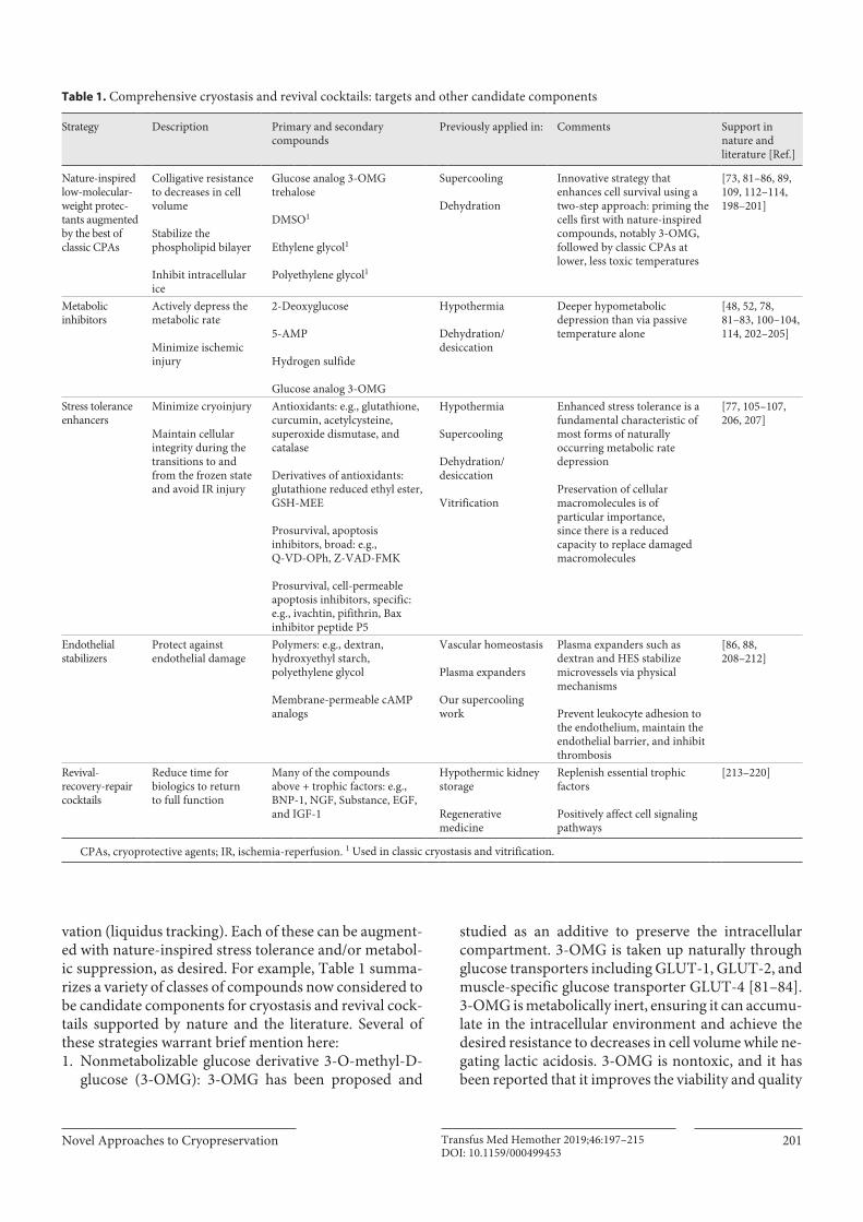
vation (liquidus tracking). Each of these can be augment-ed with nature-inspired stress tolerance and/or metabol-ic suppression, as desired. For example, Table 1 summa-rizes a variety of classes of compounds now considered to be candidate components for cryostasis and revival cock-tails supported by nature and the literature. Several of these strategies warrant brief mention here:1. Nonmetabolizable glucose derivative 3-O-methyl-D-
glucose (3-OMG): 3-OMG has been proposed and
studied as an additive to preserve the intracellular compartment. 3-OMG is taken up naturally through glucose transporters including GLUT-1, GLUT-2, and muscle-specific glucose transporter GLUT-4 [81–84]. 3-OMG is metabolically inert, ensuring it can accumu-late in the intracellular environment and achieve the desired resistance to decreases in cell volume while ne-gating lactic acidosis. 3-OMG is nontoxic, and it has been reported that it improves the viability and quality
Table 1. Comprehensive cryostasis and revival cocktails: targets and other candidate components
Strategy Description Primary and secondary compounds
Previously applied in: Comments Support in nature and literature [Ref.]
Nature-inspired low-molecular-weight protec-tants augmented by the best of classic CPAs
Colligative resistance to decreases in cell volume
Stabilize the phospholipid bilayer
Inhibit intracellular ice
Glucose analog 3-OMG trehalose
DMSO1
Ethylene glycol1
Polyethylene glycol1
Supercooling
Dehydration
Innovative strategy that enhances cell survival using a two-step approach: priming the cells first with nature-inspired compounds, notably 3-OMG, followed by classic CPAs at lower, less toxic temperatures
[73, 81–86, 89, 109, 112–114, 198–201]
Metabolic inhibitors
Actively depress the metabolic rate
Minimize ischemic injury
2-Deoxyglucose
5-AMP
Hydrogen sulfide
Glucose analog 3-OMG
Hypothermia
Dehydration/desiccation
Deeper hypometabolic depression than via passive temperature alone
[48, 52, 78, 81–83, 100–104, 114, 202–205]
Stress tolerance enhancers
Minimize cryoinjury
Maintain cellular integrity during the transitions to and from the frozen state and avoid IR injury
Antioxidants: e.g., glutathione, curcumin, acetylcysteine, superoxide dismutase, and catalase
Derivatives of antioxidants: glutathione reduced ethyl ester, GSH-MEE
Prosurvival, apoptosis inhibitors, broad: e.g., Q-VD-OPh, Z-VAD-FMK
Prosurvival, cell-permeable apoptosis inhibitors, specific: e.g., ivachtin, pifithrin, Bax inhibitor peptide P5
Hypothermia
Supercooling
Dehydration/desiccation
Vitrification
Enhanced stress tolerance is a fundamental characteristic of most forms of naturally occurring metabolic rate depression
Preservation of cellular macromolecules is of particular importance, since there is a reduced capacity to replace damaged macromolecules
[77, 105–107, 206, 207]
Endothelial stabilizers
Protect against endothelial damage
Polymers: e.g., dextran, hydroxyethyl starch, polyethylene glycol
Membrane-permeable cAMP analogs
Vascular homeostasis
Plasma expanders
Our supercooling work
Plasma expanders such as dextran and HES stabilize microvessels via physical mechanisms
Prevent leukocyte adhesion to the endothelium, maintain the endothelial barrier, and inhibit thrombosis
[86, 88, 208–212]
Revival-recovery-repair cocktails
Reduce time for biologics to return to full function
Many of the compounds above + trophic factors: e.g., BNP-1, NGF, Substance, EGF, and IGF-1
Hypothermic kidney storage
Regenerative medicine
Replenish essential trophic factors
Positively affect cell signaling pathways
[213–220]
CPAs, cryoprotective agents; IR, ischemia-reperfusion. 1 Used in classic cryostasis and vitrification.
Taylor/Weegman/Baicu/GiwaTransfus Med Hemother 2019;46:197–215202DOI: 10.1159/000499453
of hepatocytes preserved in vitro [85] and, as outlined below, enables supercooling of whole livers [86]
2. Polyethylene glycol (PEG): this polymer has histori-cally been used as a cryoprotectant, as it disrupts hy-drogen bonding in aqueous solutions and depresses freezing temperatures [87]. Studies have shown that it provides a range of other protective benefits during preservation, such as membrane and tight junction stabilization, preventing edema and acting as an anti-oxidant, thereby enhancing cellular stress tolerance by preventing lipid peroxidation [88, 89]. Low-molecu-lar-weight PEG is nontoxic at high-subzero tempera-tures, and its benefits for hepatocyte storage have re-cently been reported [89], as well as for whole liver banking in a supercooled state [86]
3. Synthetic ice modulators: these are compounds that influence the formation and growth of ice nuclei and crystals by various purported mechanisms [90, 91]. This general classification embraces several categories of molecules that have been shown to modulate ice for-mation and growth. The synthetic polymers polyvinyl alcohol (X-1000) and polyglycerol (Z-1000) have been shown to effectively suppress ice nucleation events in aqueous systems even at concentrations as low as 1 ppm, much lower than most other ice control agents, by selectively binding surfaces of molecules that would otherwise promote the formation of ice nuclei [79, 80]. These synthetic ice blockers are inspired by natural antifreeze proteins found in polar fish and insects that can remain in a supercooled state for extended periods without damage [80]. Antifreeze proteins have shown the ability to protect rat hearts during supercooling [92–94]. X- and Z-1000 are nontoxic, readily permeate cell membranes, and remain active at temperatures ranging from 0 ° C all the way to glass transition tem-peratures (below –120 ° C) [79, 80]. They have been employed to preserve a variety of cell types with no reported toxicity [67, 79, 80, 95, 96]. New nature-in-spired approaches in this area of research are focused on synthesizing novel chemically defined molecules with greater potency, less toxicity, and high stability [97–99]
4. Metabolic rate depression: naturally occurring hypo-metabolic states involve molecular and biochemical strategies that actively suppress metabolism [78]. While cooling roughly halves the rate of biological re-actions for every change of 10°C [52], not all cellular metabolic reactions are completely impaired [48, 52]. In nature, decreases in metabolism precede changes in body temperature and synergize with passive cooling effects [100]. Conditioning cells to achieve suppressed metabolism promotes greater tolerance to ischemia and stress during high-subzero preservation. 3-OMG has an inhibitory effect on glycolysis; studies in animal
models have shown that blunting mitochondrial res-piration can prevent the pathological consequences of ischemia-reperfusion injury [101–103] and inhibit cell death [81, 104]
5. Enhancement of stress tolerance: activation of cyto-protective pathways in nature is core to most forms of metabolic depression [105]. This includes the active regulation of antioxidant defenses (e.g., the increased expression of reactive oxygen species-scavenging en-zymes [77]), heat shock proteins involved in the main-tenance of protein stability/folding, and the preven-tion of protein aggregation [106] and prosurvival sig-nals (e.g., Akt signaling) [107], each with functions that serve to maintain cellular integrity during the stressful temperature transitions. This heightened stress tolerance extends the range of stressors that pre-served cells/tissues can cope with
SupercoolingMammals like the arctic ground squirrel hibernate/
supercool with body temperatures of –3 ° C (–8 ° C exper-imentally) for up to 3 weeks, with every organ “banked” without injury. Even Caiman crocodilus, which can grow to over 2.5 m (over 58 kg), can supercool to below –5 ° C [74]. Many species in nature, including mammals, can sustain subfreezing body temperatures for weeks or lon-ger, supercooling to avoid ice formation [74–76, 108]. Two of the most prominent strategies involve (1) synthe-sis of high amounts of low-molecular-weight carbohy-drates, which provide colligative resistance to detrimental decreases in cell volume, stabilize phospholipid bilayers, and restrict the formation of intracellular ice, and (2) syn-thesis of ice-blocking molecules that bind molecular sur-faces around which ice would otherwise form. It is an-ticipated that an effective translation of these strategies to human organs may be achieved through the application of low-molecular-weight cryoprotectants and synthetic ice blockers.
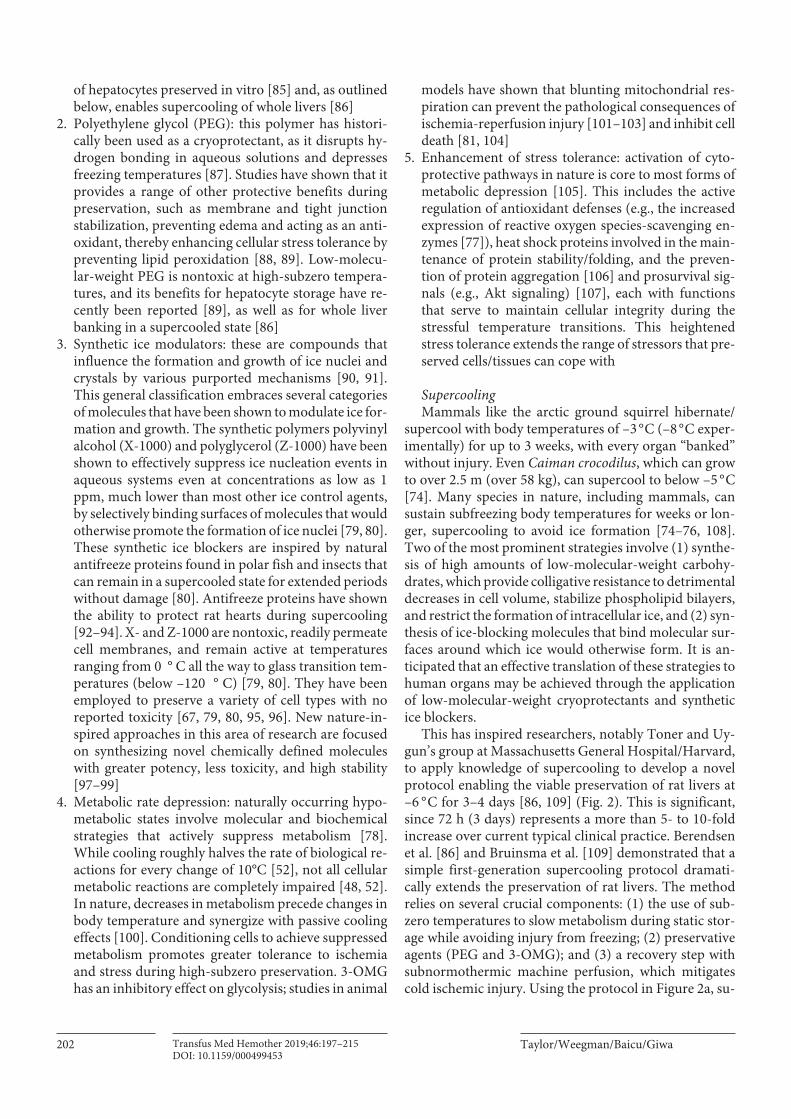
This has inspired researchers, notably Toner and Uy-gun’s group at Massachusetts General Hospital/Harvard, to apply knowledge of supercooling to develop a novel protocol enabling the viable preservation of rat livers at –6 ° C for 3–4 days [86, 109] (Fig. 2). This is significant, since 72 h (3 days) represents a more than 5- to 10-fold increase over current typical clinical practice. Berendsen et al. [86] and Bruinsma et al. [109] demonstrated that a simple first-generation supercooling protocol dramati-cally extends the preservation of rat livers. The method relies on several crucial components: (1) the use of sub-zero temperatures to slow metabolism during static stor-age while avoiding injury from freezing; (2) preservative agents (PEG and 3-OMG); and (3) a recovery step with subnormothermic machine perfusion, which mitigates cold ischemic injury. Using the protocol in Figure 2a, su-
Novel Approaches to Cryopreservation 203Transfus Med Hemother 2019;46:197–215DOI: 10.1159/000499453
percooled whole rat livers were stored for 3 days retaining full viability (Fig. 2b, c) [86]. Recipients appeared com-pletely healthy and did not display any signs of jaundice. Liver enzymes (ALT and AST) had fallen to normal levels by week 3, and all animals eventually regained their nor-mal weight. Without optimization, storage was extended by supercooling to 4 days, but at a reduced survival of 58%. By comparison, posttransplantation survival with classic hypothermic preservation was 0% at 3 or 4 days. These studies included control transplants to identify which steps of the developed protocol were critical. No survival was achieved after 4 days if (1) supercooling was replaced with ice-cold storage at +4 ° C, (2) final machine perfusion was skipped, (3) preloading 3-OMG was skipped, or (4) preservative PEG was not added (i.e., stor-age in standard University of Wisconsin solution), indi-cating that all four components were critical for 3- to 4-day preservation.
These recent achievements represent significant ad-vances in establishing the feasibility of supercooling pres-ervation of mammalian organs and pave the way for translation to organs with even shorter ischemic toler-ance times, such as hearts and lungs. The feasibility of optimizing and translating supercooling preservation to hearts is supported by the fact that Amir et al. [92–94] have reported rat heart preservation 7 times longer than with conventional methods using a solution containing only antifreeze proteins and no other cryoprotectants, as well as by our own results with nonfrozen heart preserva-tion under pressure discussed further below. These early achievements in supercooling preservation also highlight some of the nature-inspired strategies that contributed to the demonstrated successes outlined here.
Notwithstanding the recent developments in super-cooling preservation of organs, the approach is nonethe-less a non-equilibrium process with inherent risks of de-stabilization in the form of random ice nucleation. More-
Fig. 2. Supercooled rat livers successfully transplanted after 3–4 days of storage. The 30-day survival rate was 100% for livers stored 3 days, and 58% for livers stored for 4 days. By comparison, re-cipients of livers stored in UW medium for 3 or 4 days perished within 48 h. 3-OMG, 3-O-methyl-D-glucose; HP, hypothermic preservation in UW medium at 4 ° C; SNMP, subnormothermic machine perfusion (21 ° C); UW, University of Wisconsin. a Sche-matic of the supercooling temperature profile. Loading of 3-OMG, as an additive to Williams’ E-based medium, is performed by SNMP for 1 h through the portal vein. Cooling to 4 ° C (1 ° C/min) is carried out during perfusion. The liver is then flushed with 4 ° C-
cold UW solution containing 5% w/v 35 kDa PEG, and slowly cooled to –6 ° C (1 ° C/10 min). Storage is maintained for up to 96 h, after which rewarming to 4 ° C takes place (1 ° C/10 min). The liver is then flushed with 21 ° C-warm, oxygenated SNMP medium, recovered with 3 h of SNMP, and transplanted orthotopically. b Kaplan-Meier curve of transplantation recipients (n > 6 in all groups shown). c Posttransplantation trends in transaminase out-put that normalize in a month; 3 months after transplantation, the differences had completely vanished (data not shown). Adapted from Berendsen et al. [86].
Taylor/Weegman/Baicu/GiwaTransfus Med Hemother 2019;46:197–215204DOI: 10.1159/000499453
over, since ice nucleation is a stochastic event, the probability of ice formation increases with both the size of the compartment/system and the degree of undercool-ing. Most recently, advances have been reported in the use of surface sealing of water with an oil phase to sig-nificantly diminish the primary heterogeneous nucle-ation at the water/air interface. Huang et al. [110] achieved deep supercooling (down to –20 ° C) of large volumes of water (up to 100 mL) for long periods (up to 100 days) simultaneously via this approach. Furthermore, in these preliminary studies they demonstrated the utility of deep supercooling in extended (100-day) preservation of hu-man red blood cells [110]. Apart from the inherent risk of uncontrolled nucleation, a further hazard in non-equilib-rium undercooling is related to the fact that the morphol-ogy of the ice crystals forming in highly supercooled solu-tions presents a sharper dendritic – and potentially more damaging – shape when compared with equilibrium freezing processes [111]. These constraints on high-sub-zero preservation by supercooling have encouraged the pursuit of alternative nature-inspired strategies involving thermodynamic equilibrium in the presence or absence of ice.
Controlled, Partial-Ice FreezingOne good example from the at least 45 animals that
can survive long periods of time at high-subzero temper-atures in a state of “suspended animation” is the wood frog (Rana sylvatica), surviving with 65–70% of the total body water as extracellular ice [112]. One of the most crit-
ical strategies for freeze tolerance involves the synthesis of high amounts of low-molecular-weight carbohydrates (glucose in wood frogs), which provide colligative resis-tance to detrimental decreases in cell volume while also serving to stabilize the phospholipid bilayer of mem-branes and to restrict the formation of intracellular ice [112–114]. Importantly, research has shown that simple augmentation strategies such as increasing endogenous levels of cryoprotectants (via injection) prior to freezing have the capacity to improve survival and extend the time in a frozen state in a certain context from 2 weeks to 49 days [113]. A translation of nature’s cryostasis strategies to human systems may be achieved through low-molec-ular-weight CPAs, in particular the aforementioned non-metabolizable glucose derivative 3-OMG and oligosac-charides, of which trehalose has received notable atten-tion in recent years [54, 115, 116].
Studies on diverse freeze-tolerant species have shown that ice-nucleating agents play a critical role in survival of freezing. Ice-nucleating agents in the blood and in the gut/skin induce controlled freezing of extracellular water at multiple nucleation sites. In this way, secure and pro-tective extracellular freezing occurs before any nucleating components present in cells trigger injurious freezing. It is generally accepted that minimization of cryoinjury may be achieved when ice nucleation occurs as near as possible to the equilibrium freezing point – i.e., the highest tem-perature which promotes ice crystallization and propaga-tion [117, 118]. It therefore has been hypothesized that through the application of biocompatible ice nucleators,
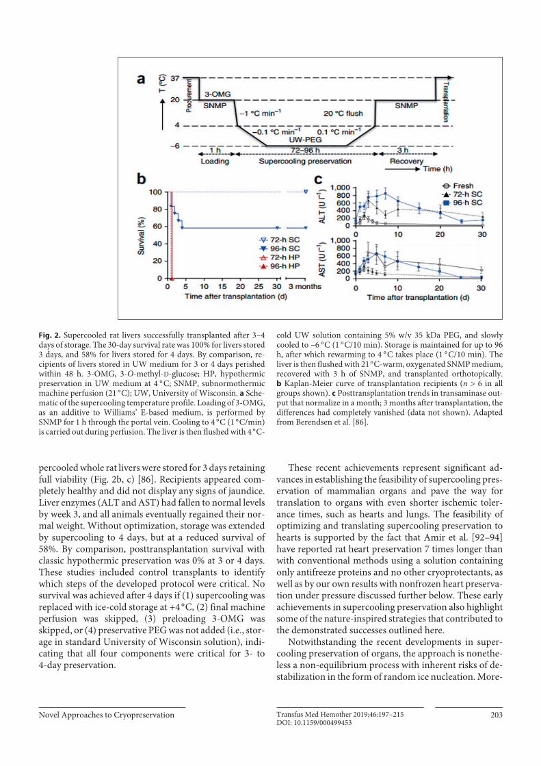
Fig. 3. A Section of the phase diagram for DMSO-H2O showing the equilibrium melting curve in blue (Tm liquidus). The stepped line above the curve represents a scheme for incremental equilibration of a tissue with sufficient cryoprotectant such that the system does not freeze during cooling. B Function of smooth muscle tissue after cooling to –21 ° C in either the frozen (F) or unfrozen (NF) state.
Histograms of post-warming contractility (mean ± SEM) normal-ized to the control responses derived prior to cooling. Avoidance of ice is critical for good survival with high function, and liquidus tracking ensures that the system remains in equilibrium and ice-free during subzero cooling [63].

Novel Approaches to Cryopreservation 205Transfus Med Hemother 2019;46:197–215DOI: 10.1159/000499453
controlled, slow propagation of freezing at relatively high subzero temperatures can be achieved and thereby cryo-damage be minimized. Preliminary support for this ap-proach in a simple blood vessel model has recently been provided [116].
Equilibrium Nonfrozen Subzero Preservation (Liquidus Tracking)Notwithstanding the strategy adopted by the arctic
wood frog for high-subzero survival in a partially frozen state, there is extensive evidence that mammalian organ banking might be better served by freeze avoidance rath-er than freeze tolerance [108, 112], which is also employed in nature. A third option is to leverage the strengths of these two approaches for nonfrozen subzero storage in thermodynamic equilibrium.
To this end, an alternative cryopreservation strategy has been conceived that relies upon incorporation of clas-sic cryoprotectants into the cryostasis cocktail to main-tain the system above the equilibrium freezing point at all stages during cooling. This approach is based upon the same principles of equilibrium cryopreservation as origi-nally proposed by Farrant [119] (reviewed by Taylor et al. [69]) and more recently studied and referred to in the lit-erature as “liquidus tracking” [120–123] – a process used to sequentially increase the concentration of CPAs in a stepwise manner during cooling to minimize solute tox-icity and osmotic shock at each temperature and to en-sure an ultrastable equilibrium state where it is impossi-ble for ice to form.
Equilibrium phase diagrams, such as that illustrated in Figure 3, provide a useful tool for designing cryopreserva-tion protocols based upon the principle of “liquidus tracking.” Figure 3 also illustrates how these equilibrium phase diagrams have been used to design experiments to specifically investigate the role of ice formation in cryo-injury to multicellular tissues [63]. Such studies have pro-vided compelling evidence for the profound effect of avoiding (uncontrolled and nonlimited) ice during cryo-preservation. Moreover, the amount of ice and its loca-tion within the tissue has been shown to impact the struc-tural and functional integrity of tissues [63, 64, 69].
Bioengineering Applications
Vitrification, the transformation of liquid water into a glass instead of crystalline ice during cooling, is one of the more promising technologies for avoiding ice damage to biological systems during cryopreservation [68, 69]. In fact, in the years since Rall and Fahy’s seminal publication on the successful vitreous preservation of murine embry-os [65], there has been a phenomenal increase in the number of publications related to vitrification as the most
promising approach to avoiding ice damage during the cryopreservation of multicellular tissues and organs [67–69, 124–128]. Early studies of vitrification were conduct-ed by Luyet in the 1930s. He reported successful vitrifica-tion with moss [129], frog sperm [130], chick embryo heart [131], vinegar eels [132], and other materials. Luyet also studied the effect of pressure on cells [133–136], pre-sumably as a means to improve the probability for vitri-fication, and he reported a detrimental effect of elevated pressures on the systems he was studying. Nevertheless, vitrification has emerged as a primary approach to cryo-preservation, underpinned by the extensive foundational work by Fahy and his colleagues. Of particular note is the fundamental work by Fahy and McFarlane [126], who es-tablished that several factors affect the probability for vit-rification. Luyet’s original approach used high cooling rates during cooling to cryogenic temperatures in order to reduce the probability for ice nucleation. However, while rapid cooling is effective for achieving vitrification of small volumes, it is not feasible for large organs or tis-sues due to limitations to heat transfer. Fahy’s group took another approach and focused on developing vitrification solutions, which replace part of the water in the organ with glass-promoting solutes and thereby facilitate vitri-fication at lower cooling rates [66]. This has been estab-lished to be a more effective approach for use in tissues and organs.
Until recently [137], these advances in vitrification had not been matched by advances in rewarming of vitri-fied tissues without ice growth and fracturing or in ad-dressing the inherent toxicity of the high concentrations of CPAs required. The CPA concentration necessary for vitrification, and the associated toxicity, can be mini-mized by cooling and warming at the fastest rate possible. Rapid warming is especially important, as the critical warming rates (CWRs) needed to avoid devitrification (the process of crystallization during warming) are typi-cally an order of magnitude higher than the correspond-ing critical cooling rates needed to achieve a vitrified state [138]. Successful rewarming depends on two factors: (1) the warming rate and (2) uniform warming. Heating must be uniform to avoid stress on the tissue that often results in fractures or cracks [138]. Hence, it is well estab-lished in the field of vitreous cryopreservation that the critical cooling rate of large biosamples, tissues, and or-gans is not a constraint using state-of-the-art vitrification cocktails, but achieving a CWR to avoid devitrification and fracturing during rewarming remains one of the principal remaining challenges [33].
NanowarmingThe conflicting requirements to accomplish both rap-
id and uniform warming in large systems can be satisfied by applying heat transfer methods capable of warming

Taylor/Weegman/Baicu/GiwaTransfus Med Hemother 2019;46:197–215206DOI: 10.1159/000499453
tissue from within, rather than relying exclusively on warming through surface conduction. Electromagnetic warming (or “microwave” [139, 140] or “dielectric” [141, 142] warming) and, more recently, magnetic nanoparti-cle warming [137, 143, 144] (nanowarming) and warm-ing with metal forms [145] have been proposed and stud-ied for faster and more uniform heating of tissue during recovery from the vitrified state.
There has been limited study of electromagnetic warm-ing for cryopreserved organs, tissues, and blood products since the 1970s and 1980s. The reader is referred to the work of Pegg’s and Gao’s groups for a review of the status and challenges to this approach, to which uniform heat-ing remains a major obstacle [141, 142, 146, 147].
Generally, the CWR and uniformity requirements have only been fulfilled in small-scale systems of 1–3 mL (Fig. 4) [148] – until recently having used nanowarming, which takes advantage of the ability of metallic nanopar-ticles to transform a radio frequency or “light energy” into heat [149], as evidenced by a report in Science Transla-tional Medicine from the Bischof group [137]. In that ar-ticle, the authors demonstrated their scalable and bio-compatible nanowarming technology using radio fre-
quency-excited iron oxide nanoparticles (IONPs) to uniformly warm large vitrified volumes (up to 80 mL) at over 100 ° C/min and avoid (a) ice crystallization and (b) fracturing while (c) decreasing the total concentration of toxic CPAs needed [137]. This technique has also been applied in the past to cancer therapies with IONPs [150, 151]. For tissue and organ heating, Bischof’s group also explore the dispersion of the IONPs within the CPA used to preserve tissues. One of the challenges presented by this approach is IONP stability within the strongly ionic environment of CPAs. However, coating the IONPs with mesoporous silica has been shown to improve stability [152, 153]. Moreover, this coating has demonstrated a high stability within biological environments and has maintained that stability within CPAs [154, 155].
By limiting the risk of ice formation during rewarm-ing, rapid nanowarming can also help decrease the con-centration of CPAs required and thus toxicity to tissues. With these and other advancements, vitreous cryopreser-vation now, for the first time, holds promise to be scalable to whole-organ cryobanking and to help transform trans-plantation in key ways [1–7, 13, 14, 19–22, 40–42, 156].
Fig. 4. Schematic illustrating a tissue nanowarming system [137]. Nanowarm-ing scale: up from 1 to 15 kW inductive heating system. The 15-kW systems enable heating up to 80 mL. The illustration shows the limitations of convective cooling and rewarming (C, D) compared to nanowarm-ing (E, F) of vitrification (success and fail-ure) of 0.5- to 2.5-cm-radius cylinders. Failure and success (red and green shad-ing) is defined by the critical minimum cooling and warming rates for VS55 vitrifi-cation solution and a thermal stress < 3.2 MPa [137].
Novel Approaches to Cryopreservation 207Transfus Med Hemother 2019;46:197–215DOI: 10.1159/000499453
Isochoric CryopreservationHigh-Subzero Isochoric PreservationHuman life occurs in a relatively constant atmospher-
ic pressure environment (near sea level), and hence most research into cell and tissue preservation has been per-formed under constant-standard pressure (isobaric) con-ditions (near 1 atm of pressure). At the same time, alter-native thermodynamic systems exist (and, based on na-ture, are consistent with healthy life). As such, the basic principles of biological thermodynamic preservation un-der constant-volume (isochoric) conditions with chang-ing pressures have recently been explored [157–160]. This has enabled a novel paradigm for biopreservation that utilizes more thermodynamic degrees of freedom and hence can avoid ice formation while keeping CPA toxicity low – across biologics of any size. Based on this concept, Rubinsky’s group have introduced an innova-tive, but very simple and cost-effective, technology for re-ducing the temperature of “hypothermic storage” to as low as –20 ° C in a thermodynamic nonfrozen equilibrium state. This isochoric process can also be combined with other powerful strategies employed by supercooling and hibernating animals in nature, such as those described above.
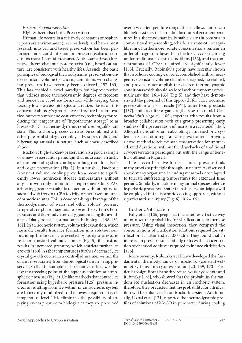
Isochoric high-subzero preservation is a good example of a new preservation paradigm that addresses virtually all the remaining shortcomings in long-duration tissue and organ preservation (Fig. 1). In a nutshell, isochoric (constant-volume) cooling provides a means to signifi-cantly lower nonfrozen storage temperatures without any – or with only minimum – requirements for CPAs, achieving greater metabolic reduction without injury as-sociated with freezing, CPA toxicity, or increased amounts of osmotic solutes. This is done by taking advantage of the thermodynamics of water and other solutes’ pressure temperature phase diagrams to lower the system’s tem-perature and thermodynamically guaranteeing the avoid-ance of dangerous ice formation in the biologic [158, 159, 161]. In an isochoric system, volumetric expansion, which normally results from ice formation in a solution sur-rounding the tissue, is prevented by using a pressure- resistant constant-volume chamber (Fig. 5); this instead results in increased pressure, which restricts further ice growth [159]. As the temperature is further decreased, ice crystal growth occurs in a controlled manner within the chamber separately from the biological sample being pre-served, so that the sample itself remains ice-free, well be-low the freezing point of the aqueous solution at atmo-spheric pressure (Fig. 5). Unlike methods that control ice formation using hyperbaric pressure [126], pressure in-creases resulting from ice within in an isochoric system are inherently minimized to the lowest required at each temperature level. This eliminates the possibility of ap-plying excess pressure to biologics as they are preserved
over a wide temperature range. It also allows nonfrozen biologic systems to be maintained at subzero tempera-tures in a thermodynamically stable state (in contrast to conventional supercooling, which is a state of nonequi-librium). Furthermore, solute concentrations remain an order of magnitude lower than the toxic levels occurring under traditional isobaric conditions [162], and the con-centrations of CPAs required are significantly lower [163]. Crucially, Rubinsky’s group have recently shown that isochoric cooling can be accomplished with an inex-pensive constant-volume chamber designed, assembled, and proven to accomplish the desired thermodynamic conditions which should scale to isochoric systems of vir-tually any size [161–163] (Fig. 5), and they have demon-strated the potential of this approach for basic isochoric preservation of fish muscle [164], other food products [157], and an entire organism (the research model Cae-norhabditis elegans) [165], together with results from a broader collaboration with our group presenting early studies of the preservation of hearts in a rat model [166]. Altogether, equilibrium subcooling in an isochoric sys-tem – i.e., isochoric high-subzero preservation – provides a novel method to achieve stable preservation for unprec-edented durations, without the drawbacks of traditional cryopreservation paradigms but with the range of bene-fits outlined in Figure 1.
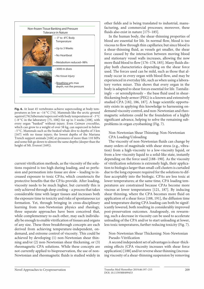
Life – even in active forms – under pressure finds many proofs of principle throughout nature. As discussed above, many organisms, including mammals, are adapted to tolerate subfreezing temperatures for extended time periods. Similarly, in nature many animal species tolerate hyperbaric pressures greater than those we anticipate will be employed in the isochoric cooling approach, without significant tissue injury (Fig. 6) [167–169].
Isochoric VitrificationFahy et al. [126] proposed that another effective way
to improve the probability for vitrification is to increase pressure. Using visual inspection, they compared the concentrations of vitrification solutions required for vit-rification at 1 atm and at 1,000 atm. They found that an increase in pressure substantially reduces the concentra-tion of chemical additives required to induce vitrification [126].
More recently, Rubinsky et al. have developed the fun-damental thermodynamics of isochoric (constant-vol-ume) systems for cryopreservation [20, 159, 170]. Par-ticularly significant is the theoretical work by Szobota and Rubinsky [158], who showed that the probability for ran-dom ice nucleation decreases in an isochoric system; therefore, they predicted that the probability for vitrifica-tion will be enhanced in an isochoric system. Addition-ally, Ukpai et al. [171] reported the thermodynamic pro-files of solutions of Me2SO in pure water during cooling

Taylor/Weegman/Baicu/GiwaTransfus Med Hemother 2019;46:197–215208DOI: 10.1159/000499453
to and warming from cryogenic temperatures in an iso-choric system. Subsequently, together with Rubinsky’s group, we showed that pressure measurement is impor-tant for designing and control of cryopreservation proto-cols in constant-volume (isochoric) systems and con-firmed that any ice formation is associated with an in-crease in pressure, and that therefore pressure can be used as a measure for the occurrence of vitrification. However, when ice is not formed, the pressure in the isochoric sys-tem does not increase during cooling, and in systems that vitrify, the absence of a pressure increase can confirm vit-rification, observed pressure increases during a vitrifica-tion protocol can indicate devitrification, and temporal or static pressure measurements can be used as an objec-tive substitute for or adjunct to more subjective visual in-spection methods [28, 171, 172]. A comparison with re-sults from the literature shows that the concentration of CPAs needed for vitrification in an isochoric chamber is
substantially lower than that needed for vitrification in isobaric systems at 1 atm and in hyperbaric systems at 1,000 atm. In addition, isochoric chambers are much more effective in promoting vitrification than hyperbaric pressure chambers; in addition, they are less expensive and easier to design and implement.
Non-Newtonian and Rheomagnetic FluidsSince this is a concept paper, we choose to outline a
novel concept recently introduced by Kilbride and Morris [173, 174] – after iteration of a concept that won the Breakthrough Ideas Hackathon at the first global Organ Banking Summit at Stanford – and highly relevant to the topic under discussion but not yet investigated widely in the context of cryopreservation.
In vitrification of larger tissue systems, viscosity is a catch-22: it is required to be high during cooling/warm-ing and low during CPA loading/unloading. This limits
Fig. 5. Top panel: schematic of an isochor-ic (constant-volume) system, in which the biologic in solution is cooled; the tempera-ture is progressively reduced from left to right to control ice nucleation separated from the biological sample in a thermody-namically equilibrated way. Isochoric pres-ervation apparatus. Lower left panel: iso-choric preservation vessel, pressure and temperature measurement, and DAC-computer connection assembly. Lower right panel: example of a system for cooling the isochoric chambers. The cooling bath (NESLAB RT-140) with hoses running out of the cooling bath and into the foam bath. One hose outputs the cooling fluid from the cooling bath to the foam bath. Another hose carries fluid from the foam bath to the cooling bath. The fluid used is a 50% by vol-ume ethylene glycol/water mixture. (Draw-ings courtesy of P.A. Perez.)
Novel Approaches to Cryopreservation 209Transfus Med Hemother 2019;46:197–215DOI: 10.1159/000499453
current vitrification methods, as the viscosity of the solu-tions required is too high during loading, and so perfu-sion and permeation into tissue are slow – leading to in-creased exposure to toxic CPAs, which counteracts the protective benefits that the CPAs provide. After loading, viscosity needs to be much higher, but currently this is only achieved through deep cooling – a process that takes considerable time with larger tissues and increases both the exposure time to toxicity and risks of spontaneous ice formation. Yet, through bringing in cross-disciplinary learning from non-Newtonian physics and rheology, three separate approaches have been conceived that, while complementary to each other, may each individu-ally be enough to enable vitrification of tissues and organs of any size. These three breakthrough concepts are each derived from achieving temperature-independent, on-demand, and extreme control of viscosity. This could be achieved by developing (1) non-Newtonian shear thin-ning and/or (2) non-Newtonian shear thickening, or (3) rheomagnetic CPA solutions. While these concepts are not currently applied to biopreservation, the use of non-Newtonian and rheomagnetic fluids is studied widely in
Non-frozen Tissue Banking and Pressure Tolerance in Nature
• -3° to -8°C Body Temperature
• Up to 3 Weeks
• No Heartbeat
• Metabolism reduced~98%
• 3000 m dives
• No tissue injury
• Breathing sets max depth, not the pressure
Fig. 6. At least 45 vertebrates achieve supercooling at body tem-peratures as low as –14 ° C [74]. Mammals like the arctic ground squirrel [76] hibernate/supercool with body temperatures of –3 ° C (–8 ° C in the laboratory [75, 108]) for up to 3 weeks [108], with every organ “banked” without injury. Even Caiman crocodilus, which can grow to a weight of over 58 kg, can supercool to below –5 ° C. Mammals such as the beaked whale dive to depths of 3 km [167] with no tissue injury; the lowest depths of the Mariana Trench support animals [168] at pressures of more than 110 MPa and some fish go down to almost the same depths (deeper than the height of Mt. Everest [169]).
other fields and is being translated to industrial, manu-facturing, and commercial processes; moreover, these fluids also exist in nature [175–185].
In the human body, the shear-thinning properties of blood are essential for life. In normal flow, blood is too viscous to flow through thin capillaries; but since blood is a shear-thinning fluid, as vessels get smaller, the shear force caused by the interaction between moving blood and stationary vessel walls increases, allowing the now more fluid blood to flow [176–178, 185]. Many fluids dis-play both characteristics depending on the shear force used. The forces used can be mild, such as those that al-ready occur in every organ with blood flow, and may be experienced in everyday life, such as when using a labora-tory vortex mixer. This shows that every organ in the body is adapted to shear forces essential for life. Tantaliz-ingly – or serendipitously – the base fluid used in shear-thickening body armor (PEG) is a known and extensively studied CPA [182, 186, 187]. A huge scientific opportu-nity exists in applying this knowledge to harnessing on-demand viscosity control, and non-Newtonian and rheo-magnetic solutions could be the foundation of a highly significant advance, helping to solve the remaining sub-problems in organ cryobanking (Fig. 7).
Non-Newtonian Shear Thinning: Non-Newtonian CPA Loading/UnloadingThe viscosity of non-Newtonian fluids can change by
many orders of magnitude with shear stress (e.g., vibra-tion) from a high-viscosity to a low-viscosity liquid, or from a low-viscosity liquid to a solid-like state, instantly depending on the force used [188–190]. As the viscosity of vitrification solutions is extremely high, their applica-tion to biologics larger than small-cell clusters is curtailed due to the long exposure required for the solutions to dif-fuse acceptably into the biologic. CPAs are less toxic at lower temperatures; at the same time, CPA loading tem-peratures are constrained because CPAs become more viscous at lower temperatures [121, 187]. By inducing shear thinning, where the CPA becomes more fluid on application of a shear force [188, 191], the diffusion time and temperature during CPA loading can both be signif-icantly lowered, both resulting in considerably improved post-preservation outcomes. Analogously, on rewarm-ing, such a decrease in viscosity can be used to accelerate unloading of the CPA and/or to start unloading at lower, less toxic temperatures, further reducing toxicity (Fig. 7).
Non-Newtonian Shear Thickening: Non-Newtonian Pseudo-VitrificationA second independent set of advantages is shear-thick-
ening effects (CPA viscosity increases with shear force application) [188] and/or reverse shear thinning (increas-ing viscosity of a shear-thinning suspension by removing

Taylor/Weegman/Baicu/GiwaTransfus Med Hemother 2019;46:197–215210DOI: 10.1159/000499453
shear stress). With either approach, after the loading steps have been completed, the new shear force can be varied in such a way as to substantially increase the viscosity of the solution-infused tissue so that it enters a shear force-induced pseudo-“vitrified” state during cool down. This increase in viscosity (1) reduces toxicity due to lower mo-lecular diffusion, (2) inhibits ice formation and damage, and (3) allows a decrease in the CPA concentration re-quired. All these benefits can be leveraged again upon re-warming. Crucially, these effects only need to be applied in temperature zones down to –60/–70 ° C [69], since at lower temperatures, the risk of ice growth during cooling or of devitrification during rewarming is no longer a problem even with current CPAs and cooling/warming protocols. This means that no shear forces at all would be applied in temperature regions approaching the glass transition temperature (Tg), at which tissues become brittle [69].
Rheomagnetic Pseudo-VitrificationA third independent approach to extreme viscosity
control is to utilize rheomagnetic fluids. These are fluids containing superparamagnetic (magnetic in the presence of a magnetic field) or ferromagnetic (permanently mag-netic) particles whose viscosity can be controlled on de-mand with the use of a magnetic field. The mechanism is based on magnetic fields that can be used to induce re-
versible particle aggregation [181], and particle aggrega-tion drastically increases fluid viscosity [192–194]. This can induce changes in viscosity of many orders of magni-tude; hence, fluids can be loaded as low-viscosity suspen-sions initially, and upon application of the magnetic field, viscosity can be drastically increased after loading. Fur-thermore, shear stress can be applied to samples in addi-tion to magnetic fields in order to fine-tune viscosity. The fact that many rheomagnetic fluids are also shear thin-ning [191, 195, 196] provides further freedom in match-ing the best characteristics for biological systems.
Upon relaxation of force (shear stress or magnetic), the material reverts back to its natural state, rendering each of the three processes completely reversible [188–190, 197]. While the three approaches can be combined, it is important to stress that just one of the approaches could be enough to make viable, reversible vitrification of large tissues and organ systems work.
Non-Newtonian fluids have been studied in great de-tail and have found a huge diversity of applications, of which those in body armor are especially impressive [175, 176, 178–183]. At the same time, while not widely known, many fluids have non-Newtonian characteristics, and the range of different shear forces that can be applied pro-vides a large set of options. The likelihood of success is especially enhanced by the fact that by adding nanopar-ticles to solutions, almost all fluids adopt non-Newtonian
Fig. 7. Scheme illustrating three new technological approaches which could be used during both CPA loading/cooling and warm-ing/CPA unloading. (1) On-demand non-Newtonian decrease in viscosity allows for accelerated CPA loading of tissues at lower temperatures, in turn leading to less toxicity and/or permitting the use of increased CPA concentrations. (2) On-demand non-New-tonian increase in viscosity enables ice avoidance and reduced tox-icity due to lower molecular diffusion. The first effect allows de-creased CPA concentration needs and the second effect allows in-creased CPA concentrations. All of these effects can be leveraged during both cooling and warming. Each of the on-demand increas-
es in viscosity could also be achieved via a rheomagnetic approach. (3) Once the system is at temperatures below –60/–70 ° C, the risk of ice growth during cooling or of devitrification during rewarm-ing is no longer a problem even with current protocols, and pres-ervation can proceed without any active viscosity control. At the same time, because any or all of these steps potentially allow the use of higher CPA concentrations, new cooling and warming pro-tocols that proceed at slower speeds could now be used. The abil-ity (1) to go slower and (2) thereby also to gain more degrees of freedom for annealing protocols should also permit decreased risks of fracturing or cracking. CPA, cryoprotective agent.

Novel Approaches to Cryopreservation 211Transfus Med Hemother 2019;46:197–215DOI: 10.1159/000499453
features [195] and can be calibrated based on the form and size of the nanoparticles – natural phenomena that could be exploited for improved methods of cryopreser-vation [196].
Acknowledgements
We gratefully acknowledge the input from and resources of our active collaborators in the various areas of research outlined in this article. These collaborators include: Prof. Mehmet Toner, Prof. Korkut Uygun, and Dr. Shannon Tessier (Harvard and Massachu-setts General Hospital); Prof. John Bischof and Dr. Erik Finger (University of Minnesota); Prof. Boris Rubinsky (University of California – Berkeley); Dr. John Morris and Dr. Peter Kilbride (As-ymptote-GE Healthcare, Cambridge, UK); Dr. Chris Hogan (Uni-versity of Minnesota); Dr. Charles Lee (University of North Caro-lina-Charlotte); and Dr. Kelvin Brockbank (Tissue Testing Tech-
nologies). We also gratefully acknowledge the input from Jedd Lewis and Alyssa Ward at the Organ Preservation Alliance regard-ing the road mapping and Harvard Organ Banking Summit. The research reported was supported, in part, by grants from the Na-tional Institutes of Health (1R43HD089832, 2R44AI124835, and 1R43DK113537) and Department of Defense (contract: W81X-WH-15-C-0190).
Statement of Ethics
The authors have no ethical conflicts to disclose.
Disclosure Statement
The authors are all employed by Sylvatica Biotech, Inc.
References
1 Giwa S, Lewis JK, Alvarez L, Langer R, Roth AE, Church GM, et al. The promise of organ and tissue preservation to transform medi-cine. Nat Biotechnol. 2017 Jun; 35(6): 530–42.
2 Lewis JK, Bischof JC, Braslavsky I, Brockbank KG, Fahy GM, Fuller BJ, et al. The Grand Challenges of Organ Banking: proceedings from the first global summit on complex tis-sue cryopreservation. Cryobiology. 2016 Apr;
72(2): 169–82. 3 Ravikumar R, Leuvenink H, Friend PJ. Nor-
mothermic liver preservation: a new para-digm? Transpl Int. 2015 Jun; 28(6): 690–9.
4 Hosgood SA, van Heurn E, Nicholson ML. Normothermic machine perfusion of the kid-ney: better conditioning and repair? Transpl Int. 2015 Jun; 28(6): 657–64.
5 Whitson BA, Black SM. Organ assessment and repair centers: the future of transplanta-tion is near. World J Transplant. 2014 Jun;
4(2): 40–2. 6 Sack K. In discarding of kidneys, system re-
veals its flaws. The New York Times. 2012 Sep 19.
7 Wigfield CH, Cypel M, Yeung J, Waddell T, Alex C, Johnson C, et al. Successful emergent lung transplantation after remote ex vivo per-fusion optimization and transportation of do-nor lungs. Am J Transplant. 2012 Oct; 12(10):
2838–44. 8 Srinivasan A, Burton EC, Kuehnert MJ, Rupp-
recht C, Sutker WL, Ksiazek TG, et al.; Rabies in Transplant Recipients Investigation Team. Transmission of rabies virus from an organ donor to four transplant recipients. N Engl J Med. 2005 Mar; 352(11): 1103–11.
9 US Center for Disease Control. CDC con-firms rabies death in organ transplant recipi-ent. Media Statement. Atlanta: CDC; 2013.
10 Centers for Disease Control and Prevention (CDC). HIV transmitted from a living organ donor—New York City, 2009. MMWR Morb Mortal Wkly Rep. 2011 Mar; 60(10): 297–301.
11 Machuca TN, Cypel M. Ex vivo lung perfu-sion. J Thorac Dis. 2014 Aug; 6(8): 1054–62.
12 Hosgood SA, Barlow AD, Hunter JP, Nichol-son ML. Ex vivo normothermic perfusion for quality assessment of marginal donor kidney transplants. Br J Surg. 2015 Oct; 102(11):
1433–40.13 Bruinsma BG, Sridharan GV, Weeder PD,
Avruch JH, Saeidi N, Özer S, et al. Metabolic profiling during ex vivo machine perfusion of the human liver. Sci Rep. 2016 Mar; 6(1):
22415.14 Hanson SG, Bentley TS. 2014 U.S. organ and
tissue transplant cost estimates and discus-sion. Seattle, WA: Milliman; 2014.
15 Jamieson RW, Friend PJ. Organ reperfusion and preservation. Front Biosci. 2008 Jan;
13(13): 221–35.16 Mohamed MS. Translational insights on lung
transplantation: learning from immunology. Iran J Immunol. 2015 Sep; 12(3): 156–65.
17 Cypel M, Liu M, Rubacha M, Yeung JC, Hi-rayama S, Anraku M, et al. Functional repair of human donor lungs by IL-10 gene therapy. Sci Transl Med. 2009 Oct; 1(4): 4ra9.
18 Lv X, Tan J, Liu D, Wu P, Cui X. Intratrache-al administration of p38α short-hairpin RNA plasmid ameliorates lung ischemia-reperfu-sion injury in rats. J Heart Lung Transplant. 2012 Jun; 31(6): 655–62.
19 Scandling JD, Busque S, Dejbakhsh-Jones S, Benike C, Millan MT, Shizuru JA, et al. Toler-ance and chimerism after renal and hemato-poietic-cell transplantation. N Engl J Med. 2008 Jan; 358(4): 362–8.
20 Scandling JD, Busque S, Shizuru JA, Engle-man EG, Strober S. Induced immune toler-ance for kidney transplantation. N Engl J Med. 2011 Oct; 365(14): 1359–60.
21 Leventhal JR, Elliott MJ, Yolcu ES, Bozulic LD, Tollerud DJ, Mathew JM, et al. Immune reconstitution/immunocompetence in recip-ients of kidney plus hematopoietic stem/fa-cilitating cell transplants. Transplantation. 2015 Feb; 99(2): 288–98.
22 Szabolcs P, Burlingham WJ, Thomson AW. Tolerance after solid organ and hematopoi-etic cell transplantation. Biol Blood Marrow Transplant. 2012 Jan; 18(1 Suppl):S193–200.
23 Prof. David Sachs [personal communication].24 Prof. Gerald Brandacher [personal communi-
cation].25 Page TF, Woodward RS. Cost of lifetime im-
munosuppression coverage for kidney trans-plant recipients. Health Care Financ Rev. 2008 Winter; 30(2): 95–104.
26 Organ Banking Summit program 2017 [Inter-net]. Available from: https://obs2017.dryfta.com/en/.
27 McDannell L. White House highlights AST’s new initiative with Organ Preservation Alli-ance [Internet]. 2016 Dec 22. Available from: https://www.myast.org/about-ast/white-house-highlights-asts-new-initiative-organ-preservation-alliance.
28 Organ Bioengineering and Banking Beta Roadmap Workshop. Program guide and preliminary roadmap concepts [Internet]. Organ Preservation Alliance; 2015. Available from: https://issuu.com/pylyp/docs/new_or-gan_conf-03.
29 Klassen DK, Shepard BM. Looking at the fu-ture of solid organ transplants [Internet]. Ne-phrology News & Issues. 2016 Sep 15. Avail-able from: https://static1.squarespace.com/static/57d7d2ad15d5db47a2e81a13/t/5974c65fd482e9419b148181/1500825186688/Klassen%2C+2016+-+Future+of+solid+organ +transplants.pdf.
30 Marshall A. Buying time for transplants. Nat Biotechnol. 2018; 36: 776.
31 Silberman S. Libraries of flesh: the sorry state of human tissue storage. WIRED. 2010 May 24.
32 Khademhosseini A, Langer R. A decade of progress in tissue engineering. Nat Protoc. 2016 Oct; 11(10): 1775–81.

Taylor/Weegman/Baicu/GiwaTransfus Med Hemother 2019;46:197–215212DOI: 10.1159/000499453
33 Alvarez LC. Compendium of Organ & Tissue Banking Concepts 2015 [Internet]. Available from: http://science.dodlive.mil/files/2015/ 01/Organ-and-Tissue-Banking-Compendi-um-2015-Jan.pdf.
34 Fact Sheet. Obama Administration announc-es key actions to reduce the organ waiting list [Internet]. Available from: https://www.whitehouse.gov/sites/whitehouse.gov/files/documents/White_House_Organ_Summit_Fact_Sheet_FINALv2.pdf
35 Katkov II, Kan NG, Cimadamore F, Nelson B, Snyder EY, Terskikh AV. DMSO-Free Pro-grammed Cryopreservation of Fully Dissoci-ated and Adherent Human Induced Pluripo-tent Stem Cells. Stem Cells Int. 2011; 2011:
981606.36 Heng BC, Kuleshova LL, Bested SM, Liu H,
Cao T. The cryopreservation of human em-bryonic stem cells. Biotechnol Appl Biochem. 2005 Apr; 41(Pt 2): 97–104.
37 Stéphenne X, Najimi M, Sokal EM. Hepato-cyte cryopreservation: is it time to change the strategy? World J Gastroenterol. 2010 Jan;
16(1): 1–14.38 Vian AM, Higgins AZ. Membrane permeabil-
ity of the human granulocyte to water, di-methyl sulfoxide, glycerol, propylene glycol and ethylene glycol. Cryobiology. 2014 Feb;
68(1): 35–42.39 Multi-Agency Tissue Engineering Sciences
(MATES) Interagency Working Group. MATES: Advancing Tissue Science and Stra-tegic Report. 2007.
40 Cypel M, Yeung JC, Keshavjee S. Novel ap-proaches to expanding the lung donor pool: donation after cardiac death and ex vivo con-ditioning. Clin Chest Med. 2011 Jun; 32(2):
233–44.41 Vogel T, Brockmann JG, Friend PJ. Ex-vivo
normothermic liver perfusion: an update. Curr Opin Organ Transplant. 2010 Apr;
15(2): 167–72.42 Messer S, Ardehali A, Tsui S. Normothermic
donor heart perfusion: current clinical experi-ence and the future. Transpl Int. 2015 Jun;
28(6): 634–42.43 Department of Defense; Additive Manufac-
turing, Biopharmaceutical, Economy, Educa-tion, Health, Innovation, Manufacturing, Public Private Partnerships, STEM, Work-force. DoD Announces Award of New Ad-vanced Tissue Biofabrication Manufacturing Innovation Hub in Manchester. New Hamp-shire; 2016.
44 Andrea B, Annegret W, Seebach D, Rieben R. Symposium on Xenotransplantation 2013;
189.45 Reardon S. New life for pig-to-human trans-
plants. Nature. 2015 Nov; 527(7577): 152–4.46 Reddy SP, Brockmann J, Friend PJ. Normo-
thermic perfusion: a mini-review. Transplan-tation. 2009 Mar; 87(5): 631–2.
47 Taylor MJ. Tissue preservation. In: Baust JG, Baust JM, editors. Advances in biopreserva-tion. Boca Raton: Taylor & Francis; 2007. p. 157–96.
48 Taylor MJ. Biology of cell survival in the cold: the basis for biopreservation of tissues and or-gans. In: Baust JG, Baust JM, editors. Advanc-es in biopreservation. Boca Raton: Taylor & Francis; 2007. p. 15–62.
49 Guibert EE, Petrenko AY, Balaban CL, Somov AY, Rodriguez JV, Fuller BJ. Organ Preserva-tion: Current Concepts and New Strategies for the Next Decade. Transfus Med Hemo-ther. 2011; 38(2): 125–42.
50 Fuller BJ, Lee CY. Hypothermic perfusion preservation: the future of organ preservation revisited? Cryobiology. 2007 Apr; 54(2): 129–45.
51 Taylor MJ, Elrifai AM, Bailes JE. Hypother-mia in Relation to the Acceptable Limits of Ischemia for Bloodless Surgery. London, UK, Greenwich, CT: JAI Press Inc.; 1996.
52 Taylor MJ. Hypothermia. In: Fink G, editor. Encyclopedia of stress. 2nd ed. Oxford: Aca-demic Press; 2007. p. 428–38.
53 Uygun K, Lee CY. Organ preservation and re-engineering. Methods in Bioengineering. Artech House; 2011.
54 Burlage LC, Tessier SN, Etra JW, Uygun K, Brandacher G. Advances in machine perfu-sion, organ preservation, and cryobiology: potential impact on vascularized composite allotransplantation. Curr Opin Organ Trans-plant. 2018 Oct; 23(5): 561–7.
55 Brockbank KG, Taylor MJ. Tissue preserva-tion. In: Baust JG, Baust JM, editors. Advanc-es in biopreservation. Boca Raton: Taylor & Francis; 2006. p. 157–96.
56 Belzer FO, Southard JH. Principles of solid-organ preservation by cold storage. Trans-plantation. 1988 Apr; 45(4): 673–6.
57 Pegg DE, Jacobsen IA, Armitage WJ, Taylor MJ. Mechanisms of injury in organs. In: Pegg DE, Jacobsen IA, editors. Organ preservation II. Edinburgh: Churchill Livingston; 1979. p. 132–46.
58 Karlsson JO, Toner M. Long-term storage of tissues by cryopreservation: critical issues. Biomaterials. 1996 Feb; 17(3): 243–56.
59 Acker JP. Biopreservation of cells and engi-neered tissues. In: Lee K, Kaplan D, editors. Tissue engineering II: basics of tissue engi-neering and tissue applications. New York: Springer; 2006. p. 157–87.
60 Armitage WJ, Juss BK. Freezing monolayers of cells without gap junctions. Cryobiology. 2003 Apr; 46(2): 194–6.
61 Acker JP, Larese A, Yang H, Petrenko A, Mc-Gann LE. Intracellular ice formation is affect-ed by cell interactions. Cryobiology. 1999 Jun;
38(4): 363–71.62 Higgins AZ, Karlsson JO. Effects of intercel-
lular junction protein expression on intracel-lular ice formation in mouse insulinoma cells. Biophys J. 2013 Nov; 105(9): 2006–15.
63 Taylor MJ, Pegg DE. The effect of ice forma-tion on the function of smooth muscle tissue stored at –21 or –60 degrees C. Cryobiology. 1983 Feb; 20(1): 36–40.
64 Hunt CJ, Taylor MJ, Pegg DE. Freeze-substi-tution and isothermal freeze-fixation studies to elucidate the pattern of ice formation in smooth muscle at 252 K (–21 degrees C). J Mi-crosc. 1982 Feb; 125(Pt 2): 177–86.
65 Rall WF, Fahy GM. Ice-free cryopreservation of mouse embryos at –196 degrees C by vitri-fication. Nature. 1985 Feb; 313(6003): 573–5.
66 Fahy GM, Wowk B, Pagotan R, Chang A, Phan J, Thomson B, et al. Physical and bio-logical aspects of renal vitrification. Organo-genesis. 2009 Jul; 5(3): 167–75.
67 Fahy GM, Wowk B, Wu J, Phan J, Rasch C, Chang A, et al. Cryopreservation of organs by vitrification: perspectives and recent advanc-es. Cryobiology. 2004 Apr; 48(2): 157–78.
68 Fahy GM, Wowk B. Principles of cryopreser-vation by vitrification. Methods Mol Biol. 2015; 1257: 21–82.
69 Taylor MJ, Song YC, Brockbank KG. Vitrifi-cation in tissue preservation: new develop-ments. In: Fuller BJ, Lane N, Benson EE, edi-tors. Life in the frozen state. CRC Press; 2004. p. 603–41.
70 Scudellari M. Core Concept: cryopreserva-tion aims to engineer novel ways to freeze, store, and thaw organs. Proc Natl Acad Sci USA. 2017 Dec; 114(50): 13060–2.
71 Taylor MJ, Baicu SC. Current state of hypo-thermic machine perfusion preservation of organs: the clinical perspective. Cryobiology. 2010 Jul; 60(3 Suppl):S20–35.
72 Bruinsma BG, Berendsen TA, Izamis ML, Yarmush ML, Uygun K. Determination and extension of the limits to static cold storage using subnormothermic machine perfusion. Int J Artif Organs. 2013 Nov; 36(11): 775–80.
73 Berendsen TA, Bruinsma BG, Lee J, D’Andrea V, Liu Q, Izamis ML, et al. A simplified sub-normothermic machine perfusion system re-stores ischemically damaged liver grafts in a rat model of orthotopic liver transplantation. Transplant Res. 2012 May; 1(1): 6.
74 Lowe CH, Lardner PJ, Halpern EA. Super-cooling in reptiles and other vertebrates. Comp Biochem Physiol A Comp Physiol. 1971 May; 39(1): 125–35.
75 Popovic P, Popovic V. Survival of newborn ground squirrels after supercooling or freez-ing. Am J Physiol. 1963 May; 204(5): 949–52.
76 Richter MM, Williams CT, Lee TN, Tøien Ø, Florant GL, Barnes BM, et al. Thermogenic capacity at subzero temperatures: how low can a hibernator go? Physiol Biochem Zool. 2015 Jan-Feb; 88(1): 81–9.
77 Joanisse DR, Storey KB. Oxidative damage and antioxidants in Rana sylvatica, the freeze-tolerant wood frog. Am J Physiol. 1996 Sep;
271(3 Pt 2):R545–53.78 Storey KB, Storey JM. Metabolic rate depres-
sion in animals: transcriptional and transla-tional controls. Biol Rev Camb Philos Soc. 2004 Feb; 79(1): 207–33.
79 Wowk B, Fahy GM. Inhibition of bacterial ice nucleation by polyglycerol polymers. Cryobi-ology. 2002 Feb; 44(1): 14–23.
80 Wowk B, Leitl E, Rasch CM, Mesbah-Karimi N, Harris SB, Fahy GM. Vitrification en-hancement by synthetic ice blocking agents. Cryobiology. 2000 May; 40(3): 228–36.
81 Ratan RR, Siddiq A, Aminova L, Lange PS, Langley B, Ayoub I, et al. Translation of is-chemic preconditioning to the patient: prolyl hydroxylase inhibition and hypoxia inducible factor-1 as novel targets for stroke therapy. Stroke. 2004 Nov; 35(11 Suppl 1): 2687–9.
82 Lesnefsky EJ, Chen Q, Moghaddas S, Hassan MO, Tandler B, Hoppel CL. Blockade of elec-tron transport during ischemia protects car-diac mitochondria. J Biol Chem. 2004 Nov;
279(46): 47961–7.83 Blackstone E, Morrison M, Roth MB. H2S
induces a suspended animation-like state in mice. Science. 2005 Apr; 308(5721): 518.
Novel Approaches to Cryopreservation 213Transfus Med Hemother 2019;46:197–215DOI: 10.1159/000499453
84 Klepper J, Garcia-Alvarez M, O’Driscoll KR, Parides MK, Wang D, Ho YY, et al. Erythro-cyte 3-O-methyl-D-glucose uptake assay for diagnosis of glucose-transporter-protein syn-drome. J Clin Lab Anal. 1999; 13(3): 116–21.
85 Sugimachi K, Roach KL, Rhoads DB, Tomp-kins RG, Toner M. Nonmetabolizable glucose compounds impart cryotolerance to primary rat hepatocytes. Tissue Eng. 2006 Mar; 12(3):
579–88.86 Berendsen TA, Bruinsma BG, Puts CF, Saeidi
N, Usta OB, Uygun BE, et al. Supercooling en-ables long-term transplantation survival fol-lowing 4 days of liver preservation. Nat Med. 2014 Jul; 20(7): 790–3.
87 Elliott GD, Wang S, Fuller BJ. Cryoprotec-tants: a review of the actions and applications of cryoprotective solutes that modulate cell recovery from ultra-low temperatures. Cryo-biology. 2017 Jun; 76: 74–91.
88 Dutheil D, Underhaug Gjerde A, Petit-Paris I, Mauco G, Holmsen H. Polyethylene glycols interact with membrane glycerophospholip-ids: is this part of their mechanism for hypo-thermic graft protection? J Chem Biol. 2009 Mar; 2(1): 39–49.
89 Puts CF, Berendsen TA, Bruinsma BG, Ozer S, Luitje M, Usta OB, et al. Polyethylene glycol protects primary hepatocytes during super-cooling preservation. Cryobiology. 2015 Aug;
71(1): 125–9.90 Eisenberg DP, Taylor MJ, Jimenez-Rios JL,
Rabin Y. Thermal expansion of vitrified blood vessels permeated with DP6 and synthetic ice modulators. Cryobiology. 2014 Jun; 68(3):
318–26.91 Eisenberg DP, Taylor MJ, Rabin Y. Thermal
expansion of the cryoprotectant cocktail DP6 combined with synthetic ice modulators in presence and absence of biological tissues. Cryobiology. 2012 Oct; 65(2): 117–25.
92 Amir G, Rubinsky B, Horowitz L, Miller L, Leor J, Kassif Y, et al. Prolonged 24-hour sub-zero preservation of heterotopically trans-planted rat hearts using antifreeze proteins derived from arctic fish. Ann Thorac Surg. 2004 May; 77(5): 1648–55.
93 Amir G, Horowitz L, Rubinsky B, Yousif BS, Lavee J, Smolinsky AK. Subzero nonfreezing cryopresevation of rat hearts using antifreeze protein I and antifreeze protein III. Cryobiol-ogy. 2004 Jun; 48(3): 273–82.
94 Amir G, Rubinsky B, Basheer SY, Horowitz L, Jonathan L, Feinberg MS, et al. Improved vi-ability and reduced apoptosis in sub-zero 21-hour preservation of transplanted rat hearts using anti-freeze proteins. J Heart Lung Transplant. 2005 Nov; 24(11): 1915–29.
95 Ting AY, Yeoman RR, Lawson MS, Zelinski MB. Synthetic polymers improve vitrification outcomes of macaque ovarian tissue as as-sessed by histological integrity and the in vitro development of secondary follicles. Cryobiol-ogy. 2012 Aug; 65(1): 1–11.
96 Marco-Jimenez F, Berlinguer F, Leoni GG, Succu S, Naitana S. Effect of “ice blockers” in solutions for vitrification of in vitro matured ovine oocytes. Cryo Letters. 2012 Jan-Feb;
33(1): 41–4.
97 Briard JG, Poisson JS, Turner TR, Capic-ciotti CJ, Acker JP, Ben RN. Small molecule ice recrystallization inhibitors mitigate red blood cell lysis during freezing, transient warming and thawing. Sci Rep. 2016 Mar;
6(1): 23619. 98 Capicciotti CJ, Kurach JD, Turner TR, Man-
cini RS, Acker JP, Ben RN. Small molecule ice recrystallization inhibitors enable freez-ing of human red blood cells with reduced glycerol concentrations. Sci Rep. 2015 Apr;
5(1): 9692. 99 Briard JG, Jahan S, Chandran P, Allan D,
Pineault N, Ben RN. Small-Molecule Ice Re-crystallization Inhibitors Improve the Post-Thaw Function of Hematopoietic Stem and Progenitor Cells. ACS Omega. 2016 Nov;
1(5): 1010–8.100 Watts P, Øritsland N, Jonkel C, Ronald K.
Mammalian hibernation and the oxygen consumption of a denning black bear (Ur-sus americanas). Comp Biochem Physiol Part A Physiol. 1981; 69(1): 121–3.
101 Huber R, Spiegel T, Büchner M, Riepe MW. Graded reoxygenation with chemical inhi-bition of oxidative phosphorylation im-proves posthypoxic recovery in murine hip-pocampal slices. J Neurosci Res. 2004 Feb;
75(3): 441–9.102 Burwell LS, Nadtochiy SM, Brookes PS.
Cardioprotection by metabolic shut-down and gradual wake-up. J Mol Cell Cardiol. 2009 Jun; 46(6): 804–10.
103 Chen Q, Camara AK, Stowe DF, Hoppel CL, Lesnefsky EJ. Modulation of electron trans-port protects cardiac mitochondria and de-creases myocardial injury during ischemia and reperfusion. Am J Physiol Cell Physiol. 2007 Jan; 292(1):C137–47.
104 Hand SC, Menze MA, Borcar A, Patil Y, Covi JA, Reynolds JA, et al. Metabolic re-structuring during energy-limited states: in-sights from Artemia franciscana embryos and other animals. J Insect Physiol. 2011 May; 57(5): 584–94.
105 Storey KB, Storey JM. Tribute to P. L. Lutz: putting life on ‘pause’—molecular regula-tion of hypometabolism. J Exp Biol. 2007 May; 210(Pt 10): 1700–14.
106 Kiss AJ, Muir TJ, Lee RE Jr, Costanzo JP. Seasonal variation in the hepatoproteome of the dehydration and freeze-tolerant wood frog, Rana sylvatica. Int J Mol Sci. 2011;
12(12): 8406–14.107 Zhang J, Storey KB. Akt signaling and freez-
ing survival in the wood frog, Rana sylvatica. Biochim Biophys Acta. 2013 Oct; 1830(10):
4828–37.108 Barnes BM. Freeze avoidance in a mammal:
body temperatures below 0 degree C in an Arctic hibernator. Science. 1989 Jun;
244(4912): 1593–5.109 Bruinsma BG, Berendsen TA, Izamis ML,
Yeh H, Yarmush ML, Uygun K. Supercool-ing preservation and transplantation of the rat liver. Nat Protoc. 2015 Mar; 10(3): 484–94.
110 Huang H, Yarmush ML, Usta OB. Long-term deep-supercooling of large-volume water and red cell suspensions via surface sealing with immiscible liquids. Nat Com-mun. 2018 Aug; 9(1): 3201.
111 Costanzo JP, Grenot C, Lee RE Jr. Super-cooling, ice inoculation and freeze tolerance in the European common lizard, Lacerta vi-vipara. J Comp Physiol B. 1995; 165(3): 238–44.
112 do Amaral MC, Lee RE Jr, Costanzo JP. He-patocyte responses to in vitro freezing and β-adrenergic stimulation: insights into the extreme freeze tolerance of subarctic Rana sylvatica. J Exp Zool A Ecol Genet Physiol. 2015 Feb; 323(2): 89–96.
113 Layne JR Jr, Costanzo JP, Lee RE Jr. Freeze duration influences postfreeze survival in the frog Rana sylvatica. J Exp Zool. 1998 Feb; 280(2): 197–201.
114 Storey KB. Living in the cold: freeze-in-duced gene responses in freeze-tolerant ver-tebrates. Clin Exp Pharmacol Physiol. 1999 Jan; 26(1): 57–63.
115 Tessier SN, Cronin SE, Pendexter CA, Özer S, de Vries RJ, Nagpal S, et al. Partial freez-ing: a nature-inspired strategy for organ banking. Cryobiology. 2018; 81: 220.
116 Tessier SN, Weng L, Moyo WD, Au SH, Wong KH, Angpraseuth C, et al. Effect of Ice Nucleation and Cryoprotectants during High Subzero-Preservation in Endothelial-ized Microchannels. ACS Biomater Sci Eng. 2018; 4(8): 3006–15.
117 Costanzo JP, Lee RE Jr, Wright MF. Glucose loading prevents freezing injury in rapidly cooled wood frogs. Am J Physiol. 1991 Dec;
261(6 Pt 2):R1549–53.118 Morris GJ, Acton E. Controlled ice nucle-
ation in cryopreservation—a review. Cryo-biology. 2013 Apr; 66(2): 85–92.
119 Farrant J. Mechanism of cell damage during freezing and thawing and its prevention. Nature. 1965; 205(4978): 1284–7.
120 Kay AG, Hoyland JA, Rooney P, Kearney JN, Pegg DE. A liquidus tracking approach to the cryopreservation of human cartilage allografts. Cryobiology. 2015 Aug; 71(1): 77–84.
121 Puschmann E, Selden C, Butler S, Fuller B. Liquidus tracking: controlled rate vitrifica-tion for the cryopreservation of larger vol-umes and tissues. Cryo Letters. 2014 Jul-Aug; 35(4): 345–55.
122 Yu XY, Chen GM, Zhang SZ. A refinement to the liquidus-tracking method for vitreous preservation of articular cartilage. Cryo Let-ters. 2013 May-Jun; 34(3): 267–76.
123 Pegg DE, Wang L, Vaughan D. Cryopreser-vation of articular cartilage. Part 3: the liq-uidus-tracking method. Cryobiology. 2006 Jun; 52(3): 360–8.
124 Fahy GM, Wowk B, Wu J. Cryopreservation of complex systems: the missing link in the regenerative medicine supply chain. Reju-venation Res. 2006; 9(2): 279–91.
125 Wowk B, Fahy GM, Ahmedyar S, Taylor MJ, Rabin Y. Vitrification tendency and sta-bility of DP6-based vitrification solutions for complex tissue cryopreservation. Cryo-biology. 2018 Jun; 82: 70–7.
126 Fahy GM, MacFarlane DR, Angell CA, Meryman HT. Vitrification as an approach to cryopreservation. Cryobiology. 1984 Aug; 21(4): 407–26.

Taylor/Weegman/Baicu/GiwaTransfus Med Hemother 2019;46:197–215214DOI: 10.1159/000499453
127 Taylor MJ, Song YC, Kheirabadi BS, Light-foot FG, Brockbank KG. Vitrification fulfills its promise as an approach to reducing freeze-induced injury in a multicellular tis-sue. Adv Heat Mass Transf Biotechnol. HTD; 1999. vol. 3, p. 93–102.
128 Song YC, Khirabadi BS, Lightfoot F, Brock-bank KG, Taylor MJ. Vitreous cryopreserva-tion maintains the function of vascular grafts. Nat Biotechnol. 2000 Mar; 18(3): 296–9.
129 Luyet BJ, Gehenio PM. The survival of moss vitrified in liquid air and its relation to water content. Biodynamica. 1938; 42: 1–7.
130 Luyet BJ, Hodapp EL. Revival of Frog’s Spermatozoa Vitrified in Liquid Air. Proc Soc Exp Biol Med. 1938; 39(3): 433–4.
131 Gonzales F, Luyet B. Resumption of heart-beat in chick embryo frozen in liquid nitro-gen. Biodynamica. 1950 May; 7(126-128):
1–5.132 Luyet BJ, Gehenio PM. Survival of vinegar
eels after congelation in liquid nitrogen. Anat Rec. 1950; 108: 544.
133 Luyet BJ. On the mechanism of cellular death by high pressure cytological modifica-tions accompanying death in yeast. C R Hebd Seances Acad Sci. 1937; 204: 1506–8.
134 Luyet BJ, Hodapp EL. Mechanism of cellu-lar death by high pressure, compression of yeast in sodium chloride solutions. Proc Soc Exp Biol Med. 1937; 36(5): 615–7.
135 Luyet R, Guilliermond A. The mechanism of the death of cells by high pressures the intensity and the duration of pressures le-thal for yeast. C R Hebd Seances Acad Sci. 1937; 204: 1214–5.
136 Hodapp EL, Luyet BJ. On the mechanism of death by hydrostatic pressure in paramecia. Biodynamica. 1947 May; 6(109-112): 101–9.
137 Manuchehrabadi N, Gao Z, Zhang J, Ring HL, Shao Q, Liu F, et al. Improved tissue cryopreservation using inductive heating of magnetic nanoparticles. Sci Transl Med. 2017 Mar; 9(379):eaah4586.
138 Steif PS, Palastro MC, Rabin Y. The Effect of Temperature Gradients on Stress Develop-ment During Cryopreservation via Vitrifi-cation. Cell Preserv Technol. 2007; 5(2):
104–15.139 Hirsch J, Bach R, Menzebach A, Welters ID,
Dietrich GV, Hempelmann G. Temperature course and distribution during plasma heat-ing with a microwave device. Anaesthesia. 2003 May; 58(5): 444–7.
140 Zhang Q, Jackson TH, Ungan A. A continu-ous hybrid microwave heating process for producing rapid and uniform rewarming of cryopreserved tissues. Ann N Y Acad Sci. 1998 Sep; 858: 253–61.
141 Wusteman M, Robinson M, Pegg D. Vitrifi-cation of large tissues with dielectric warm-ing: biological problems and some ap-proaches to their solution. Cryobiology. 2004 Apr; 48(2): 179–89.
142 Pan J, Shu Z, Ren S, Gao D. Determination of Dielectric Properties of Cryoprotective Agent Solutions with a Resonant Cavity for the Electromagnetic Rewarming in Cryo-preservation. Biopreserv Biobank. 2017 Oct; 15(5): 404–9.
143 Bischof J. 11. Nanowarming: a new concept in tissue and organ preservation. Cryobiol-ogy. 2015; 71(1): 167.
144 Bischof JC, Diller KR. From Nanowarming to Thermoregulation: New Multiscale Ap-plications of Bioheat Transfer. Annu Rev Biomed Eng. 2018 Jun; 20(1): 301–27.
145 Manuchehrabadi N, Shi M, Roy P, Han Z, Qiu J, Xu F, et al. Ultrarapid Inductive Re-warming of Vitrified Biomaterials with Thin Metal Forms. Ann Biomed Eng. 2018 Nov; 46(11): 1857–69.
146 Robinson MP, Pegg DE. Rapid electromag-netic warming of cells and tissues. IEEE Trans Biomed Eng. 1999 Dec; 46(12): 1413–25.
147 Robinson MP, Wusteman MC, Wang L, Pegg DE. Electromagnetic re-warming of cryopreserved tissues: effect of choice of cryoprotectant and sample shape on unifor-mity of heating. Phys Med Biol. 2002 Jul;
47(13): 2311–25.148 Finger EB. Organ preservation [Internet].
Medscape 2015 [cited 2017 Jul 3]. Availa- ble from: http://emedicine.medscape.com/article/431140-overview.
149 Etheridge ML, Xu Y, Rott L, Choi J, Glasmacher B, Bischof JC. RF heating of magnetic nanoparticles improves the thaw-ing of cryopreserved biomaterials. Technol-ogy (Singap). 2014; 2(3): 229–42.
150 Hirsch LR, Stafford RJ, Bankson JA, Sershen SR, Rivera B, Price RE, et al. Nanoshell-me-diated near-infrared thermal therapy of tu-mors under magnetic resonance guidance. Proc Natl Acad Sci USA. 2003 Nov; 100(23):
13549–54.151 Thiesen B, Jordan A. Clinical applications of
magnetic nanoparticles for hyperthermia. Int J Hyperthermia. 2008 Sep; 24(6): 467–74.
152 Hurley KR, Lin YS, Zhang J, Egger SM, Haynes CL. Effects of Mesoporous Silica Coating and Post-Synthetic Treatment on the Transverse Relaxivity of Iron Oxide Nanoparticles. Chem Mater. 2013 May;
25(9): 1968–78.153 Lin YS, Haynes CL. Synthesis and Charac-
terization of Biocompatible and Size-Tun-able Multifunctional Porous Silica Nanopar-ticles. Chem Mater. 2009; 21(17): 3979–86.
154 Hurley KR, Ring HL, Etheridge M, Zhang J, Gao Z, Shao Q, et al. Predictable Heating and Positive MRI Contrast from a Mesopo-rous Silica-Coated Iron Oxide Nanoparti-cle. Mol Pharm. 2016 Jul; 13(7): 2172–83.
155 Zhang J, Ring HL, Hurley KR, Shao Q, Carl-son CS, Idiyatullin D, et al. Quantification and biodistribution of iron oxide nanopar-ticles in the primary clearance organs of mice using T1 contrast for heating. Magn Reson Med. 2017 Aug; 78(2): 702–12.
156 Ward AM, Klassen DK, Franz KM, Giwa S, Lewis JK. Social, economic, and policy im-plications of organ preservation advances. Curr Opin Organ Transplant. 2018 Jun;
23(3): 336-346.157 Lyu C, Nastase G, Ukpai G, Serban A, Ru-
binsky B. A comparison of freezing-damage during isochoric and isobaric freezing of the potato. PeerJ. 2017 May; 5:e3322.
158 Szobota SA, Rubinsky B. Analysis of iso-choric subcooling. Cryobiology. 2006 Aug;
53(1): 139–42.159 Rubinsky B, Perez PA, Carlson ME. The
thermodynamic principles of isochoric cryopreservation. Cryobiology. 2005 Apr;
50(2): 121–38.160 Rubinsky B. Biological matter in isochoric
systems. Cryobiology. 2015; 71(1): 172.161 Preciado JA, Rubinsky B. Isochoric preser-
vation: a novel characterization method. Cryobiology. 2010 Feb; 60(1): 23–9.
162 Pérez PA. Thermodynamics and heat trans-fer analysis for isochoric cryopreservation [PhD thesis in Engineering-Mechanical En-gineering]. Berkeley (CA): University of California; 2006.
163 Preciado J, Rubinsky B. The fundamentals of isochoric freezing and its role in the cryo-preservation of biological materials [PhD thesis]. Berkeley (CA): University of Cali-fornia; 2007. Available from: http://grad-works.umi.com/33/06/3306298.html.
164 Năstase G, Lyu C, Ukpai G, Şerban A, Ru-binsky B. Isochoric and isobaric freezing of fish muscle. Biochem Biophys Res Com-mun. 2017 Apr; 485(2): 279–83.
165 Mikus H, Miller A, Nastase G, Serban A, Shapira M, Rubinsky B. The nematode Cae-norhabditis elegans survives subfreezing temperatures in an isochoric system. Bio-chem Biophys Res Commun. 2016 Aug;
477(3): 401–5.166 Wan L, Powell-Palm MJ, Lee C, Gupta A,
Weegman BP, Clemens MG, et al. Preserva-tion of rat hearts in subfreezing temperature isochoric conditions to –8 ° C and 78 MPa. Biochem Biophys Res Commun. 2018 Feb;
496(3): 852–7.167 Schorr GS, Falcone EA, Moretti DJ, An-
drews RD. First long-term behavioral re-cords from Cuvier’s beaked whales (Ziphius cavirostris) reveal record-breaking dives. PLoS One. 2014 Mar; 9(3):e92633.
168 Kobayashi H, Hatada Y, Tsubouchi T, Nagahama T, Takami H. The Hadal Amphi-pod Hirondellea gigas possessing a unique cellulase for digesting wooden debris buried in the deepest seafloor. PLoS One. 2012;
7(8):e42727.169 Yancey PH, Gerringer ME, Drazen JC, Row-
den AA, Jamieson A. Marine fish may be biochemically constrained from inhabiting the deepest ocean depths. Proc Natl Acad Sci USA. 2014 Mar; 111(12): 4461–5.
170 Rubinsky B. From ice in the veins, through unfrozen fish and frozen frogs to isochoric preservation, ad astra. Cryobiology. 2015;
71(1): 167–8.171 Ukpai G, Năstase G, Șerban A, Rubinsky B.
Pressure in isochoric systems containing aqueous solutions at subzero centigrade temperatures. PLoS One. 2017 Aug; 12(8): e0183353.
172 Zhang Y, Ukpai G, Grigoropoulos A, Pow-ell-Palm MJ, Weegman BP, Taylor MJ, et al. Isochoric vitrification: an experimental study to establish proof of concept. Cryo-biology. 2018 Aug; 83: 48–55.
Novel Approaches to Cryopreservation 215Transfus Med Hemother 2019;46:197–215DOI: 10.1159/000499453
173 Kilbride P, Rull MV, Townsend A, Wilson H, Morris J. Shear-thickening fluids in bio-logically relevant agents. Biorheology. 2019. [Epub ahead of print]
174 Kilbride P, Morris GJ. Methods for cryo-preservation. 2017. Available from: https://p a t e n t s . g o o g l e . c o m / p a t e n t /WO2017072499A1/en.
175 National Cancer Institute. Biorepositories and Biospecimen Research Branch. Avail-able from: http://biospecimens.cancer.gov/about/overview.asp.
176 Pries AR, Secomb TW. Rheology of the mi-crocirculation. Clin Hemorheol Microcirc. 2003; 29(3-4): 143–8.
177 Gijsen FJ, van de Vosse FN, Janssen JD. The influence of the non-Newtonian properties of blood on the flow in large arteries: steady flow in a carotid bifurcation model. J Bio-mech. 1999 Jun; 32(6): 601–8.
178 Merrill EW, Pelletier GA. Viscosity of hu-man blood: transition from Newtonian to non-Newtonian. J Appl Physiol. 1967 Aug;
23(2): 178–82.179 Gopalakrishnan R, McMurry PH, Hogan CJ
Jr. The Bipolar Diffusion Charging of Nanoparticles: A Review and Development of Approaches for Non-Spherical Particles. Aerosol Sci Technol. 2015; 49(12): 1181–94.
180 Adam KT, Helen JW. The fluid dynamics of the chocolate fountain. Eur J Phys. 2016;
37(1): 15803.181 Hong R, Cima MJ, Weissleder R, Josephson
L. Magnetic microparticle aggregation for viscosity determination by MR. Magn Re-son Med. 2008 Mar; 59(3): 515–20.
182 Wagner NJ, Wetzel ED. Advanced body ar-mor utilizing shear thickening fluids. Patent US7498276 B2. 2007. Available from: h t t p s : / / w w w . g o o g l e . c o m / p a t e n t s /US7498276.
183 Lee YS, Wetzel ED, Wagner NJ. The ballistic impact characteristics of Kevlar® woven fabrics impregnated with a colloidal shear thickening fluid. J Mater Sci. 2003; 38(13):
2825–33.184 Wagner NJ, Brady JF. Shear thickening in
colloidal dispersions. Phys Today. 2009;
62(10): 27–32.185 Chien S, Usami S, Dellenback RJ, Gregersen
MI, Nanninga LB, Guest MM. Blood viscos-ity: influence of erythrocyte aggregation. Science. 1967 Aug; 157(3790): 829–31.
186 Fuller BJ. Cryoprotectants: the essential an-tifreezes to protect life in the frozen state. Cryo Letters. 2004 Nov-Dec; 25(6): 375–88.
187 Fuller BJ, Lane N, Benson EE. Life in the fro-zen state. 2004.
188 Hartnett JP, Cho YI. Nonnewtonian fluids. In: Rohsenow WM, Hartnett JP, Cho YI, editors. Handbook of heat transfer. 3rd ed. New York: McGraw-Hill; 1998. p. 10.1–10.53.
189 Schowalter WR. Mechanics of non-Newto-nian fluids. Oxford: Pergamon Press; 1978.
190 Skelland AH. Non-Newtonian flow and heat transfer. Wiley; 1967.
191 Cheng X, McCoy JH, Israelachvili JN, Co-hen I. Imaging the microscopic structure of shear thinning and thickening colloidal sus-pensions. Science. 2011 Sep; 333(6047):
1276–9.
192 Duan F, Kwek D, Crivoi A. Viscosity affect-ed by nanoparticle aggregation in Al2O3-water nanofluids. Nanoscale Res Lett. 2011 Mar; 6(1): 248.
193 Mansfield ML, Douglas JF, Garboczi EJ. In-trinsic viscosity and the electrical polariz-ability of arbitrarily shaped objects. Phys Rev E Stat Nonlin Soft Matter Phys. 2001 Dec; 64(6 Pt 1): 061401.
194 Rubio-Hernández FJ, Ayúcar-Rubio MF, Velázquez-Navarro JF, Galindo-Rosales FJ. Intrinsic viscosity of SiO2, Al2O3 and TiO2 aqueous suspensions. J Colloid Interface Sci. 2006 Jun; 298(2): 967–72.
195 Hojjat M, Etemad SG, Bagheri R, Thibault J. Rheological characteristics of non-Newto-nian nanofluids: experimental investiga-tion. Int Commun Heat Mass Transf. 2011;
38(2): 144–8.196 Barnes HA. Shear-thickening (“dilatancy”)
in suspensions of nonaggregating solid par-ticles dispersed in Newtonian liquids. J Rhe-ol (NYNY). 1989; 33(2): 329–66.
197 Bender J, Wagner NJ. Reversible shear thickening in monodisperse and bidisperse colloidal dispersions. J Rheol (NYNY). 1996; 40(5): 899–916.
198 Crowe JH, Crowe LM, Chapman D. Preser-vation of membranes in anhydrobiotic or-ganisms: the role of trehalose. Science. 1984 Feb; 223(4637): 701–3. https://doi.org/ 10.1126/science.223.4637.701.
199 Eroglu A, Russo MJ, Bieganski R, Fowler A, Cheley S, Bayley H, et al. Intracellular treha-lose improves the survival of cryopreserved mammalian cells. Nat Biotechnol. 2000 Feb;
18(2): 163–7.200 Crowe JH, Crowe LM. Preservation of
mammalian cells – learning nature’s tricks. Nat Biotechnol. 2000 Feb; 18(2): 145–6.
201 Crowe JH, Crowe LM, Wolkers WF, Oliver AE, Ma X, Auh JH, et al. Stabilization of dry Mammalian cells: lessons from nature. In-tegr Comp Biol. 2005 Nov; 45(5): 810–20.
202 Dark J, Miller DR, Zucker I. Reduced glu-cose availability induces torpor in Siberian hamsters. Am J Physiol. 1994 Aug; 267(2 Pt 2):R496–501.
203 Menze MA, Chakraborty N, Clavenna M, Banerjee M, Liu XH, Toner M, et al. Meta-bolic preconditioning of cells with AICAR-riboside: improved cryopreservation and cell-type specific impacts on energetics and proliferation. Cryobiology. 2010 Aug; 61(1):
79–88.204 Borcar A, Menze MA, Toner M, Hand SC.
Metabolic preconditioning of mammalian cells: mimetic agents for hypoxia lack fidel-ity in promoting phosphorylation of pyru-vate dehydrogenase. Cell Tissue Res. 2013 Jan; 351(1): 99–106.
205 Corton JM, Gillespie JG, Hawley SA, Hardie DG. 5-Aminoimidazole-4-carboxamide ri-bonucleoside. A specific method for activat-ing AMP-activated protein kinase in intact cells? Eur J Biochem. 1995 Apr; 229(2): 558–65.
206 Moubasher AE, El Din AM, Ali ME, El-sherif WT, Gaber HD. Catalase improves motility, vitality and DNA integrity of cryo-preserved human spermatozoa. Androlo-gia. 2013 Apr; 45(2): 135–9.
207 Baust JM, Vogel MJ, Van Buskirk R, Baust JG. A molecular basis of cryopreservation failure and its modulation to improve cell survival. Cell Transplant. 2001; 10(7): 561–71.
208 Leung AD, Wong KH, Tien J. Plasma ex-panders stabilize human microvessels in microfluidic scaffolds. J Biomed Mater Res A. 2012 Jul; 100(7): 1815–22.
209 Killinger WA Jr, Dorofi DB, Keagy BA, Johnson G Jr. Endothelial cell preservation using organ storage solutions. Transplanta-tion. 1992 May; 53(5): 979–82.
210 Oltean M, Joshi M, Björkman E, Oltean S, Casselbrant A, Herlenius G, et al. Intralumi-nal polyethylene glycol stabilizes tight junc-tions and improves intestinal preservation in the rat. Am J Transplant. 2012 Aug; 12(8):
2044–51.211 Marsh DC, Lindell SL, Fox LE, Belzer FO,
Southard JH. Hypothermic preservation of hepatocytes. I. Role of cell swelling. Cryobi-ology. 1989 Dec; 26(6): 524–34.
212 Kayano K, Toda K, Naka Y, Okada K, Oz MC, Pinsky DJ. Superior protection in or-thotopic rat lung transplantation with cyclic adenosine monophosphate and nitroglycer-in-containing preservation solution. J Tho-rac Cardiovasc Surg. 1999 Jul; 118(1): 135–44.
213 Williams GT, Smith CA, Spooncer E, Dex-ter TM, Taylor DR. Haemopoietic colony stimulating factors promote cell survival by suppressing apoptosis. Nature. 1990 Jan;
343(6253): 76–9.214 Wyllie AH. Apoptosis and the regulation of
cell numbers in normal and neoplastic tis-sues: an overview. Cancer Metastasis Rev. 1992 Sep; 11(2): 95–103.
215 Zhou XM, Liu Y, Payne G, Lutz RJ, Chitten-den T. Growth factors inactivate the cell death promoter BAD by phosphorylation of its BH3 domain on Ser155. J Biol Chem. 2000 Aug; 275(32): 25046–51.
216 McAnulty JF, Reid TW, Waller KR, Murphy CJ. Successful six-day kidney preservation using trophic factor supplemented media and simple cold storage. Am J Transplant. 2002 Sep; 2(8): 712–8.
217 Schwab IR, Dries D, Cullor J, Smith W, Mannis M, Reid T, et al. Corneal storage medium preservation with defensins. Cor-nea. 1992 Sep; 11(5): 370–5.
218 Chan EY, Stang SL, Bottorff DA, Stone JC. Hypothermic stress leads to activation of Ras-Erk signaling. J Clin Invest. 1999 May;
103(9): 1337–44.219 Arai S, Mochida S, Ohno A, Ishikawa K,
Matsui A, Arai M, et al. Decreased expres-sion of receptors for vascular endothelial growth factor and sinusoidal endothelial cell damage in cold-preserved rat livers. Transplant Proc. 1999 Sep; 31(6): 2668–72.
220 Wilhelm SM, Simonson MS, Robinson AV, Stowe NT, Schulak JA. Cold ischemia in-duces endothelin gene upregulation in the preserved kidney. J Surg Res. 1999 Jul; 85(1):
101–8.221 Pegg DE. The relevance of ice crystal for-
mation for the cryopreservation of tissues and organs. Cryobiology. 2010 Jul; 60(3 Suppl):S36–44.