Pulmonary Hypertension in Sickle-Cell Disease: Comorbidities and Echocardiographic Findings
12
PULMONARY HYPERTENSION IN SICKLE CELL DISEASE: Relevance to Children Gregory J. Kato, Vascular Medicine Branch, National Heart, Lung and Blood Institute, and Critical Care Medicine Department, Clinical Center, National Institutes of Health, Bethesda, Maryland, USA Onyinye C. Onyekwere, and Center for Sickle Cell Disease, Howard University College of Medicine, Washington, DC, USA Mark T. Gladwin Vascular Medicine Branch, National Heart, Lung and Blood Institute, and Critical Care Medicine Department, Clinical Center, National Institutes of Health, Bethesda, Maryland, USA Abstract Pulmonary arterial hypertension (PAH), once considered a rare complication of sickle cell disease (SCD) and thalassemia, appears to be more common in adults with hemoglobinopathy than previously appreciated. On prospective screening of adults with SCD, approximately one-third of adults are found on echocardiography to have a tricuspid regurgitant jet velocity (TRV) of 2.5 m/s or higher, many of whom are asymptomatic. Dyspnea on exertion is the most common presenting symptom. This TRV abnormality is a marker for approximately 40% 3-year mortality in adults, and it is associated with laboratory values suggestive of more severe intravascular hemolysis. Release of hemoglobin and arginase from lysed red cells causes scavenging of nitric oxide (NO) and catabolism of L-arginine, the obligate substrate for NO synthase. The resulting impairment in NO bioavailability is associated with pulmonary vasoconstriction, endothelial dysfunction, thrombosis, and eventual development of plexogenic arterial lesions, the histological hallmark of all forms of PAH. Undoubtedly, additional pathophysiological mechanisms will also play a role in its multifactorial pathogenesis. Early data from children with SCD indicate a similar prevalence of elevated TRV, but the prognostic implications of this remain to be established. Individual patient diagnosis of PAH requires confirmation by right heart catheterization studies and individualized management. Hemolysis-associated PAH with impairments in NO bioavailability is being identified in thalassemia and other hemolytic disorders, and may be a general consequence of long-standing, severe intravascular hemolytic anemia. Keywords sickle cell; thalassemia; pulmonary hypertension; nitric oxide; arginase Pulmonary hypertension is a complication of sickle cell disease (SCD) and thalassemia gaining considerable attention in the past 4 years. Cor pulmonale, the clinical syndrome of right ventricular failure, has been considered for decades to be a rare, but well-known, terminal complication in adults with both hemoglobinopathies [1,2]. As the survival of patients with hemoglobinopathies has improved dramatically in the last forty years, the prevalence of such chronic complications has increased. Well-documented studies of pulmonary arterial Address correspondence to Gregory J. Kato, MD, Director, Sickle Cell Vascular Disease Unit, Vascular Medicine Branch, National Heart, Lung and Blood Institute, National Institutes of Health, 10 Center Drive, MSC 1476, Building 10-CRC, Room 5-5140, Bethesda, MD 20892-1476, USA. E-mail: [email protected] NIH Public Access Author Manuscript Pediatr Hematol Oncol. Author manuscript; available in PMC 2007 November 7. Published in final edited form as: Pediatr Hematol Oncol. 2007 ; 24(3): 159–170. NIH-PA Author Manuscript NIH-PA Author Manuscript NIH-PA Author Manuscript
-
Upload
johnshopkins -
Category
Documents
-
view
0 -
download
0
Transcript of Pulmonary Hypertension in Sickle-Cell Disease: Comorbidities and Echocardiographic Findings

PULMONARY HYPERTENSION IN SICKLE CELL DISEASE:Relevance to Children
Gregory J. Kato,Vascular Medicine Branch, National Heart, Lung and Blood Institute, and Critical Care MedicineDepartment, Clinical Center, National Institutes of Health, Bethesda, Maryland, USA
Onyinye C. Onyekwere, andCenter for Sickle Cell Disease, Howard University College of Medicine, Washington, DC, USA
Mark T. GladwinVascular Medicine Branch, National Heart, Lung and Blood Institute, and Critical Care MedicineDepartment, Clinical Center, National Institutes of Health, Bethesda, Maryland, USA
AbstractPulmonary arterial hypertension (PAH), once considered a rare complication of sickle cell disease(SCD) and thalassemia, appears to be more common in adults with hemoglobinopathy thanpreviously appreciated. On prospective screening of adults with SCD, approximately one-third ofadults are found on echocardiography to have a tricuspid regurgitant jet velocity (TRV) of 2.5 m/sor higher, many of whom are asymptomatic. Dyspnea on exertion is the most common presentingsymptom. This TRV abnormality is a marker for approximately 40% 3-year mortality in adults, andit is associated with laboratory values suggestive of more severe intravascular hemolysis. Release ofhemoglobin and arginase from lysed red cells causes scavenging of nitric oxide (NO) and catabolismof L-arginine, the obligate substrate for NO synthase. The resulting impairment in NO bioavailabilityis associated with pulmonary vasoconstriction, endothelial dysfunction, thrombosis, and eventualdevelopment of plexogenic arterial lesions, the histological hallmark of all forms of PAH.Undoubtedly, additional pathophysiological mechanisms will also play a role in its multifactorialpathogenesis. Early data from children with SCD indicate a similar prevalence of elevated TRV, butthe prognostic implications of this remain to be established. Individual patient diagnosis of PAHrequires confirmation by right heart catheterization studies and individualized management.Hemolysis-associated PAH with impairments in NO bioavailability is being identified in thalassemiaand other hemolytic disorders, and may be a general consequence of long-standing, severeintravascular hemolytic anemia.
Keywordssickle cell; thalassemia; pulmonary hypertension; nitric oxide; arginase
Pulmonary hypertension is a complication of sickle cell disease (SCD) and thalassemia gainingconsiderable attention in the past 4 years. Cor pulmonale, the clinical syndrome of rightventricular failure, has been considered for decades to be a rare, but well-known, terminalcomplication in adults with both hemoglobinopathies [1,2]. As the survival of patients withhemoglobinopathies has improved dramatically in the last forty years, the prevalence of suchchronic complications has increased. Well-documented studies of pulmonary arterial
Address correspondence to Gregory J. Kato, MD, Director, Sickle Cell Vascular Disease Unit, Vascular Medicine Branch, NationalHeart, Lung and Blood Institute, National Institutes of Health, 10 Center Drive, MSC 1476, Building 10-CRC, Room 5-5140, Bethesda,MD 20892-1476, USA. E-mail: [email protected]
NIH Public AccessAuthor ManuscriptPediatr Hematol Oncol. Author manuscript; available in PMC 2007 November 7.
Published in final edited form as:Pediatr Hematol Oncol. 2007 ; 24(3): 159–170.
NIH
-PA Author Manuscript
NIH
-PA Author Manuscript
NIH
-PA Author Manuscript

hypertension (PAH) have drawn the attention of the hematology community to its frequentoccurrence and poor prognosis [3–7]. This article will review the current understanding ofhemoglobinopathy-associated pulmonary hypertension, with special attention to the mostrecently developing data in children.
PREVALENCE AND PROGNOSISIn the last few years, echocardiographic screening studies have suggested that the prevalenceof hemoglobinopathy-associated PAH is much higher than previously known. In SCD,approximately one-third of adult patients have an elevated tricuspid regurgitant jet velocity(TRV) of 2.5 m/s or higher, a threshold that correlates in right heart catheterization studies toa pulmonary artery systolic pressure of at least 30 mm Hg [8–10]. Even though this thresholdrepresents quite mild pulmonary hypertension, SCD patients with TRV above this thresholdhave a 9- to 10-fold higher risk for early mortality than those with a lower TRV [8,9]. Our ownupdate to these mortality figures indicate that adults with SCD and TRV ≥2.5 m/s have 40%mortality at 40 months of follow-up. It is as yet unproven whether PAH is the actual cause ofdeath in these cases, although PAH is a frequent cause of death in other studies [11–13]. If itis, then patients with SCD are dying of PAH before the pressures rise to the levels commonlyseen in the terminal phase of primary pulmonary hypertension. It appears that the baselinecompromised oxygen delivery and co-morbid organ dysfunction of SCD diminishes thephysiological reserve to tolerate even modest pulmonary arterial pressures. Large-scalescreening in thalassemia intermedia and major also has indicated a high prevalence and adverseprognosis for PAH as estimated by echocardiography [14–17].
PATHOLOGYAutopsy studies in SCD have revealed histopathological findings common to all forms of PAH,including plexiform and concentric medial hyperplastic pulmonary vascular lesions, and in situpulmonary arterial thrombosis, although interpretations of these findings has varied (Figure 1)[12,13,18,19]. Similar autopsy findings also have been observed in thalassemia, particularlypulmonary thrombi in patients who had previously undergone splenectomy [20–24]. It remainsundetermined in what proportion of these cases pulmonary thrombosis is a cause or effect ofPAH. In summary, the histopathology of PAH in SCD and thalassemia is associated withvasoconstriction, proliferative vascular smooth muscle and irregular endothelium inpulmonary arteries with associated thrombosis, all combining to produce luminal narrowing,and eventual right ventricular failure (Figure 1).
The pathophysiology of PAH in hemoglobinopathies is undoubtedly multifactorial, butepidemiological and biochemical data support a prominent role for intravascular hemolysisinducing a state of vascular dysfunction. Clinical variables independently associated with PAHin adults with SCD include the following: low hemoglobin levels, suggestive of more severehemolytic anemia; high steady-state serum lactate dehydrogenase (LDH) levels, largelyreflecting intravascular hemolysis [25]; high serum creatinine levels, indicative of renalinsufficiency; high serum direct bilirubin and alkaline phosphatase levels, suggestingcholestatic hepatic dysfunction; low serum transferrin or high serum ferritin levels, indicativeof iron overload (Table 1) [8]. In addition, the prevalence of an elevated TRV appears to risewith age during adulthood, exceeding 65% after 50 years of age [8,9] One study has conflictingfindings regarding the relationship of high TRV to systolic blood pressure [9]. Another hasfound an additional correlation of high TRV to proteinuria [10]. Overall, there is a relationshipof elevated TRV to the degree of intravascular hemolysis, and the degrees of renal and hepaticdysfunction, and to iron overload. Except for the contribution of hemolysis, there are no studiesto indicate whether the other factors contribute to the development of PAH, or whether theyreflect other organs concurrently affected by vasculopathy.
Kato et al. Page 2
Pediatr Hematol Oncol. Author manuscript; available in PMC 2007 November 7.
NIH
-PA Author Manuscript
NIH
-PA Author Manuscript
NIH
-PA Author Manuscript

Our group has accumulated additional biochemical and physiological evidence implicatingintravascular hemolysis in the development of impaired nitric oxide bioavailability and chronicvascular dysfunction [26,27]. Nitric oxide (NO) is a master regulator of vascular health,producing vasodilation and increased blood flow, in part by promoting vasodilation, and byinhibiting endothelial adhesion molecule expression, vascular smooth muscle proliferation,platelet aggregation and blood coagulation [28]. Pathological processes in SCD accelerate thedestruction of NO, and limit the compensatory increase in NO production. Hemoglobindecompartmentalized from the red cell into blood plasma by intravascular hemolysis reachessteady-state levels of 5–10 μM, and at times exceeding 50 μM [29–31]. This hemoglobin reactswith NO in a rapid, nearly diffusion-limited reaction to produce methemoglobin and inertnitrate [29]. This plasma hemoglobin is associated with a relative resistance of vasodilation toNO donors in patients with SCD [32]. It is also clear that from canine hemolysis studies thatplasma hemoglobin scavenges NO and produces pulmonary vasoconstriction, hypertension,and renal dysfunction, all partly reversible by inhaled NO [27]. In addition, NO is alsoconsumed by reaction with reactive oxygen species produced as a by-product of the highlyexpressed enzymatic activities of xanthine oxidase and NADPH oxidase (Table 2) [33,34].Lastly, more recent studies suggest that hemolysis leads to uncoupling of endothelial NOsynthase activity, likely secondary to heme-mediated oxidative damage to the enzyme, alsoproducing reactive oxygen species [35]. These mechanisms may combine additively tomarkedly accelerate NO destruction.
Intravascular hemolysis also limits the expected compensatory increase in NO synthaseactivity. Arginase, concurrently released from red cells into blood plasma during intravascularhemolysis, converts plasma L-arginine to ornithine, depleting plasma levels of L-arginine, theobligate substrate for NO synthase [36]. The arginine:ornithine ratio, a convenient marker ofplasma arginase activity, correlates with pulmonary hypertension and risk of death in patientswith sickle cell disease and thalassemia [36,37]. In this manner, intravascular hemolysiscontributes to both decreased production and increased destruction of NO, compounded byreactive oxygen species produced as a side product of xanthine oxidase and NADPH oxidaseactivity, which are both expressed abundantly in SCD. It is also conceivable that oxidant stressdue to chemistry generated by iron and heme released from intravascular hemolysis may alsodeplete NO [38,39]. Multiple mechanisms directly and indirectly attributable to hemolysisreduce NO bioavailability in SCD and thalassemia. A similar mechanism also may pertain inmalaria and paroxysmal nocturnal hemoglobinuria [40,41].
CLINICAL PRESENTATION AND COURSEThe most common presenting symptom for PAH in SCD is worsening dyspnea on exertion.This is often attributed by the clinician to anemia, and cardiopulmonary evaluation may bedelayed. Symptomatic patients may also have systolic systemic hypertension and peripheraledema. Clubbing of the digits is commonly observed [42]. Laboratory tests in adults with SCDfrequently display markers of hemolysis, renal insufficiency, iron overload, and a subtlecholestatic abnormality, out of proportion to other patients with SCD. Such laboratory markersinclude lower hemoglobin and serum transferrin, higher reticulocyte count, and higher levelsof serum lactate dehydrogenase, aspartate aminotransferase, alkaline phosphatase, creatinineand ferritin. The plasma arginine:ornithine ratio is often <0.7. Proteinuria may also be found.Symptomatic patients often have a TRV ≥ 3 m/s. Chest x-ray and computed tomography ofthe chest may show prominent vascularity, and enlargement of the pulmonary artery. In moreadvanced cases, computed tomography scans may demonstrate bibasilar interstitial fibrosisand a mosaic perfusion pattern. The diagnosis of PAH must be confirmed with right heartcatheterization studies, with expected findings summarized elsewhere [3,42]. Besides thefrequent contribution of hemolysis-associated pulmonary vasoconstriction, in a minority of
Kato et al. Page 3
Pediatr Hematol Oncol. Author manuscript; available in PMC 2007 November 7.
NIH
-PA Author Manuscript
NIH
-PA Author Manuscript
NIH
-PA Author Manuscript

patients, the pulmonary hypertension may be compounded by mitral valvular insufficiency orleft ventricular diastolic dysfunction [43].
As might be expected, in mild PAH, the patients may report no symptoms, but screeningechocardiography reveals a TRV of 2.5–2.9 m/s. The same laboratory abnormalities as abovemay be seen, but typically to a milder degree than in patients with more severe PAH. This milddegree of elevation in the TRV frequently goes unreported on echocardiography reports, withthe inappropriate interpretation that it is too small to have clinical consequences. However,adults with SCD have severe anemia with very high cardiac output, less cardiopulmonaryreserve, and frequent co-morbid organ dysfunction, and even this small degree of TRVelevation epidemiologically is associated with early mortality.
Adults with SCD during vaso-occlusive pain crisis (VOC) may sometimes develop peripheralor periorbital edema. Although this may be related to overly vigorous hydration, many of thesepatients have acutely elevated TRV during VOC, with the edema and TRV both returning tonormal afterward [44]. These findings suggest that NO bioavailability falls further duringhyperhemolysis associated with VOC [30,31,45], leading to acute worsening of pulmonaryvasoconstriction and consequent right ventricular failure [44]. This may contribute to deathsobserved during VOC [12,44,46].
Hemoglobin SC disease has a lower prevalence of PAH than homozygous SCD or S-β0-thalassemia, as might be expected based on the lower hemolytic rate [8;10] Fetal hemoglobinexpression, while protective for other complications of SCD, has been variably associated withlower rates of PAH [9]. The effect of coinheritance of α-thalassemia and SCD on PAHprevalence has not been yet reported.
TREATMENTThere are currently several FDA-approved drugs for the treatment of pulmonary hypertension,but there are few studies of the efficacy or toxicities of these drugs in patients withhemoglobinopathies. In published pilot experience by our group and others, thephosphodiesterase-5 inhibitor sildenafil reduces pulmonary pressures and improvescardiopulmonary performance in patients with SCD and thalassemia with pulmonaryhypertension [47,48]. The oral endothelin receptor blocker bosentan is being tested in patientswith SCD and PAH, but no results are available as yet. There are minimal published experiencesas yet in SCD with FDA-approved intravenous, subcutaneous, or inhaled prostacyclins [49],although our group has found anecdotal success using intravenous epoprostenol to stabilizetwo SCD patients with life-threatening right ventricular failure due to PAH. Additional clinicaltrials are needed to help establish the safety and efficacy of these agents in hemoglobinopathypatients.
HIGH TRV IN PEDIATRIC PATIENTS WITH SCDMuch less is known regarding the prevalence and natural history of PAH in pediatric patientswith hemoglobinopathies. However, the results of 11 small screening studies have beenpublished or presented in abstract form, and all the authors have consented to have their datasummarized here (Table 3). Remarkably, aggregated screening results from over 600 childrenwith SCD indicate that 30% have with TRV ≥ 2.5 m/s, nearly identical to 32% in thesummarized screening experience in adults with SCD. Six studies have reported a combinedprevalence of 8% for TRV ≥ 3 m/s, suggestive of moderate to severe PAH, slightly less thanthe combined prevalence of 14% in adults. Many of these pediatric studies indicate severalcorrelations of high TRV to the same variables seen in adults: low hemoglobin levels, highreticulocyte counts, bilirubin, and LDH levels (personal communication, Stephen C. Nelson,Farzana D. Pashankar, and Margaret T. Lee) [50–54]. As in adults, these markers implicate
Kato et al. Page 4
Pediatr Hematol Oncol. Author manuscript; available in PMC 2007 November 7.
NIH
-PA Author Manuscript
NIH
-PA Author Manuscript
NIH
-PA Author Manuscript

hemolysis-associated mechanisms in pediatric PAH. A correlation of high TRV to systolicblood pressure has also been observed in children, confirming a relationship seen in one studyin adults (personal communication, Farzana D. Pashkar) [55].
Overall, preliminary studies suggest a provocatively high prevalence of elevated TRV inchildren with SCD. There are several caveats in these data. These pediatric studies are a mixtureof retrospective and prospective designs with variable entry criteria and most have yet toundergo peer review. Many of them likely reflect a bias toward increased screening in moresymptomatic patients. Prospective screening studies are needed to indicate the true prevalenceand prognosis of elevate TRV in the pediatric SCD population.
DOES A HIGH TRV MEAN PAH?The TRV has been a highly useful tool in adults with SCD, in whom there is a good statisticalcorrelation with right heart catheterization, the gold standard for the diagnosis of PAH. TheTRV is very useful in epidemiological studies and for screening purposes. However, itsspecificity is not sufficient to diagnose PAH in individual patients. In our own anecdotalobservations, young adults with particularly high cardiac output occasionally have a mildlyelevated TRV without evidence on right heart catheterization of increased pulmonary pressuresor vascular resistance. In adults, a high TRV is a marker that predicts early mortality in SCD,and might be a possible indication to consider general measures such as institution ofhydroxyurea or chronic transfusion therapy. Pulmonary vasodilator therapy should beconsidered only in those patients with PAH confirmed by right heart catheterization. It is notyet proven whether such interventions prolong survival in SCD patients with elevated TRV.In children, the sensitivity and specificity of the TRV for identifying PAH requires more study.
IS IT BETTER TO DIAGNOSE PAH AT A YOUNG AGE?Lessons learned in PAH due to other causes make a case for screening for PAH in childrenwith SCD. In an example familiar to pediatricians, diagnosis of an atrial septal defect inchildhood generally leads to surgical repair and reversal of PAH with no significant sequelae.However, if diagnosis of the defect is delayed to early adulthood, PAH may become more fixedwith potentially irreversible histological and functional changes, leading to early death or needfor lung transplantation.
We propose that a similar sequence of events may occur in patients with SCD (Figure 1). Inthis model, hemolysis-associated impairment of NO bioavailability over decades causeschronic vasoconstriction and mild pulmonary hypertension. At this point, decreasing hemolysisby chronic transfusion might ameliorate the NO consumption and deficiency, and therebyreverse the pulmonary vasoconstriction and pulmonary hypertension. There are some dataavailable to support this model. Lezcano et al. have reported that chronic transfusion lowersplasma hemoglobin in children with sickle cell disease [56]. Furthermore, one group hasindicated that children with SCD on chronic transfusion have lower average TRV and estimatedRV pressure than those not chronically transfused [57]. In an informative case, a 5-year oldchild with a TRV of 3.1 m/s was treated with chronic transfusion therapy, on which the TRVdeclined to 0.7 m/s after 19 months, supporting a potential for reversal of high TRV in youngchildren (personal communication, Margaret T. Lee).
By age 20–50 years, we suggest that more extensive vascular smooth muscle hyperplasia willhave occurred, with luminal narrowing that may not respond to therapeutic maneuvers to reducehemolysis (Figure 1, bottom two stages). In even more advanced cases, irregular, chronicallyactivated endothelium may have accrued in situ thrombosis and plexogenic changes thatdramatically increase pulmonary vascular resistance. In such advanced stages, reduction ofhemolysis by transfusion may be ineffective, and only pharmacological treatment with
Kato et al. Page 5
Pediatr Hematol Oncol. Author manuscript; available in PMC 2007 November 7.
NIH
-PA Author Manuscript
NIH
-PA Author Manuscript
NIH
-PA Author Manuscript

pulmonary vasodilator drugs may produce improvement. These drugs are expected to improvepulmonary pressures and cardiopulmonary performance, but gradual progression of the diseaseis still likely, as in other forms of PAH.
This is a model that merits additional testing, and several sickle cell centers are accumulatingmore TRV data in their pediatric patients with SCD. Such prospectively gathered data,supplemented by long-term follow-up data, will provide valuable information regarding theprognostic significance of an elevated TRV in childhood. This will guide future clinicaldecision making regarding the appropriateness of hydroxyurea, transfusions, or pulmonaryvasodilator therapy.
Acknowledgements
G.J.K. and M.T.G. are supported by intramural funding from the National Heart, Lung and Blood Institute and theClinical Center of the National Institutes of Health. M.T.G. also receives research support through a cooperativeresearch and development agreement with INO Therapeutics. O.C.O. receives support from Novartis Pharmaceuticalsand INO Therapeutics. We thank Stephen C. Nelson, Farzana D. Pashankar, and Margaret T. Lee for sharingunpublished data.
References1. Moser KM, Shea JG. The relationship between pulmonary infarction, cor pulmonale and the sickle
states. Am J Med 1957;22:561–579. [PubMed: 13410950]2. Koren A, Garty I, Antonelli D, et al. Right ventricular cardiac dysfunction in beta-thalassemia major.
Am J Dis Child 1987;141:93–96. [PubMed: 3788890]3. Castro O, Hoque M, Brown BD. Pulmonary hypertension in sickle cell disease: cardiac catheterization
results and survival. Blood 2003;101:1257–1261. [PubMed: 12393669]4. Collins FS, Orringer EP. Pulmonary hypertension and cor pulmonale in the sickle hemoglobinopathies.
Am J Med 1982;73:814–821. [PubMed: 7148875]5. Sutton LL, Castro O, Cross DJ, et al. Pulmonary hypertension in sickle cell disease. Am J Cardiol
1994;74:626–628. [PubMed: 8074054]6. Aessopos A, Stamatelos G, Skoumas V, et al. Pulmonary hypertension and right heart failure in patients
with beta-thalassemia intermedia. Chest 1995;107:50–53. [PubMed: 7813310]7. Atichartakarn V, Likittanasombat K, Chuncharunee S, et al. Pulmonary arterial hypertension in
previously splenectomized patients with beta-thalassemic disorders. Int J Hematol 2003;78:139–145.[PubMed: 12953808]
8. Gladwin MT, Sachdev V, Jison ML, et al. Pulmonary hypertension as a risk factor for death in patientswith sickle cell disease. N Engl J Med 2004;350:886–895. [PubMed: 14985486]
9. Ataga KI, Sood N, De GG, et al. Pulmonary hypertension in sickle cell disease. Am J Med2004;117:665–669. [PubMed: 15501204]
10. De Castro LM, Jonassaint JC, Graham FL, et al. Pulmonary hypertension in SS, SC and Sβthalassemia: prevalence, associated clinical syndromes, and mortality. Blood 2004;104:462a.[PubMed: 15044250]
11. Powars DR, Chan LS, Hiti A, et al. Outcome of sickle cell anemia: a 4-decade observational studyof 1056 patients. Medicine (Baltimore) 2005;84:363–376. [PubMed: 16267411]
12. Manci EA, Culberson DE, Yang YM, et al. Causes of death in sickle cell disease: an autopsy study.Br J Haematol 2003;123:359–365. [PubMed: 14531921]
13. Adedeji MO, Cespedes J, Allen K, et al. Pulmonary thrombotic arteriopathy in patients with sicklecell disease. Arch Pathol Lab Med 2001;125:1436–1441. [PubMed: 11697998]
14. Aessopos A, Farmakis D, Karagiorga M, et al. Cardiac involvement in thalassemia intermedia: amulticenter study. Blood 2001;97:3411–3416. [PubMed: 11369631]
15. Aessopos A, Farmakis D, Deftereos S, et al. Thalassemia heart disease: a comparative evaluation ofthalassemia major and thalassemia intermedia. Chest 2005;127:1523–1530. [PubMed: 15888823]
16. Du ZD, Roguin N, Milgram E, et al. Pulmonary hypertension in patients with thalassemia major. AmHeart J 1997;134:532–537. [PubMed: 9327712]
Kato et al. Page 6
Pediatr Hematol Oncol. Author manuscript; available in PMC 2007 November 7.
NIH
-PA Author Manuscript
NIH
-PA Author Manuscript
NIH
-PA Author Manuscript

17. Wu KH, Chang JS, Su BH, et al. Tricuspid regurgitation in patients with beta-thalassemia major. AnnHematol 2004;83:779–783. [PubMed: 15449031]
18. Gerry JL, Bulkley BH, Hutchins GM. Clinicopathologic analysis of cardiac dysfunction in 52 patientswith sickle cell anemia. Am J Cardiol 1978;42:211–216. [PubMed: 150786]
19. Haque AK, Gokhale S, Rampy BA, et al. Pulmonary hypertension in sickle cell hemoglobinopathy:a clinicopathologic study of 20 cases. Hum Pathol 2002;33:1037–1043. [PubMed: 12395378]
20. Grisaru D, Rachmilewitz EA, Mosseri M, et al. Cardiopulmonary assessment in beta-thalassemiamajor. Chest 1990;98:1138–1142. [PubMed: 2225958]
21. Sonakul D, Pacharee P, Laohapand T, et al. Pulmonary artery obstruction in thalassaemia. SoutheastAsian J Trop Med Public Health 1980;11:516–523. [PubMed: 7221695]
22. Sonakul D, Thakerngpol K, Pacharee P. Cardiac pathology in 76 thalassemic patients. Birth DefectsOrig Artic Ser 1988;23:177–191. [PubMed: 3390539]
23. Sonakul D, Fucharoen S. Pulmonary thromboembolism in thalassemic patients. Southeast Asian JTrop Med Public Health 1992;23(Suppl 2):25–28. [PubMed: 1298988]
24. Hahalis G, Manolis AS, Apostolopoulos D, et al. Right ventricular cardiomyopathy in beta-thalassaemia major. Eur Heart J 2002;23:147–156. [PubMed: 11785997]
25. Kato GJ, McGowan VR, Machado RF, et al. Lactate dehydrogenase as a biomarker of hemolysis-associated nitric oxide resistance, priapism, leg ulceration, pulmonary hypertension and death inpatients with sickle cell disease. Blood 2006;107:2279–2285. [PubMed: 16291595]
26. Rother RP, Bell L, Hillmen P, et al. The clinical sequelae of intravascular hemolysis and extracellularplasma hemoglobin: a novel mechanism of human disease. JAMA 2005;293:1653–1662. [PubMed:15811985]
27. Minneci PC, Deans KJ, Zhi H, et al. Hemolysis-associated endothelial dysfunction mediated byaccelerated NO inactivation by decompartmentalized oxyhemoglobin. J Clin Invest 2005;115:3409–3417. [PubMed: 16294219]
28. Walford G, Loscalzo J. Nitric oxide in vascular biology. J Thromb Haemost 2003;1:2112–2118.[PubMed: 14521592]
29. Reiter CD, Wang X, Tanus-Santos JE, et al. Cell-free hemoglobin limits nitric oxide bioavailabilityin sickle-cell disease. Nat Med 2002;8:1383–1389. [PubMed: 12426562]
30. Neely CL, Wajima T, Kraus AP, et al. Lactic acid dehydrogenase activity and plasma hemoglobinelevations in sickle cell disease. Am J Clin Pathol 1969;52:167–169. [PubMed: 5807978]
31. Naumann HN, Diggs LW, Barreras L, et al. Plasma hemoglobin and hemoglobin fractions in sicklecell crisis. Am J Clin Pathol 1971;56:137–147. [PubMed: 5567718]
32. Gladwin MT, Schechter AN, Ognibene FP, et al. Divergent nitric oxide bioavailability in men andwomen with sickle cell disease. Circulation 2003;107:271–278. [PubMed: 12538427]
33. Aslan M, Freeman BA. Oxidant-mediated impairment of nitric oxide signaling in sickle cell disease—mechanisms and consequences. Cell Mol Biol (Noisy-le-grand) 2004;50:95–105. [PubMed:15040433]
34. Wood KC, Hebbel RP, Granger DN. Endothelial cell NADPH oxidase mediates the cerebralmicrovascular dysfunction in sickle cell transgenic mice. FASEB J 2005;19:989–991. [PubMed:15923406]
35. Hsu LL, Champion HC, Campbell-Lee SA, et al. Hemolysis in sickle cell mice causes pulmonaryhypertension due to global impairment in nitric oxide bioavailability. Blood. in press
36. Morris CR, Kato GJ, Poljakovic M, et al. Dysregulated arginine metabolism, hemolysis-associatedpulmonary hypertension and mortality in sickle cell disease. JAMA 2005;294:81–90. [PubMed:15998894]
37. Morris CR, Kuypers FA, Kato GJ, et al. Hemolysis-associated pulmonary hypertension in thalassemia.Ann N Y Acad Sci 2005;1054:481–5. 481–485. [PubMed: 16339702]
38. Hebbel RP. Auto-oxidation and a membrane-associated ‘Fenton reagent’: a possible explanation fordevelopment of membrane lesions in sickle erythrocytes. Clin Haematol 1985;14:129–140.[PubMed: 2985310]
39. Sadrzadeh SM, Graf E, Panter SS, et al. Hemoglobin: a biologic Fenton reagent. J Biol Chem1984;259:14354–14356. [PubMed: 6094553]
Kato et al. Page 7
Pediatr Hematol Oncol. Author manuscript; available in PMC 2007 November 7.
NIH
-PA Author Manuscript
NIH
-PA Author Manuscript
NIH
-PA Author Manuscript

40. Gramaglia I, Sobolewski P, Meays D, et al. Low nitric oxide bioavailability contributes to the genesisof experimental cerebral malaria. Nat Med 2006;12:1417–1422. [PubMed: 17099710]
41. Heller PG, Grinberg AR, Lencioni M, et al. Pulmonary hypertension in paroxysmal nocturnalhemoglobinuria. Chest 1992;102:642–643. [PubMed: 1643968]
42. Machado RF, Gladwin MT. Chronic sickle cell lung disease: new insights into the diagnosis,pathogenesis and treatment of pulmonary hypertension. Br J Haematol 2005;129:449–464. [PubMed:15877728]
43. Sachdev V, Machado RF, Shizukuda Y, et al. Diastolic dysfunction is an independent risk factor fordeath in patients with sickle cell disease. J Am Coll Cardiol 2007;49:472–479. [PubMed: 17258093]
44. Machado RF, Mack AK, Martyr S, et al. Severity of pulmonary hypertension during vaso-occlusivepain crisis and exercise in patients with sickle cell disease. Br J Haematol 2007;136:319–325.[PubMed: 17156401]
45. Ballas SK, Marcolina MJ. Hyperhemolysis during the evolution of uncomplicated acute painfulepisodes in patients with sickle cell anemia. Transfusion (Paris) 2006;46:105–110.
46. Parfrey NA, Moore W, Hutchins GM. Is pain crisis a cause of death in sickle cell disease? Am J ClinPathol 1985;84:209–212. [PubMed: 4025226]
47. Machado RF, Martyr S, Kato GJ, et al. Sildenafil therapy in patients with sickle cell disease andpulmonary hypertension. Br J Haematol 2005;130:445–453. [PubMed: 16042696]
48. Derchi G, Forni GL, Formisano F, et al. Efficacy and safety of sildenafil in the treatment of severepulmonary hypertension in patients with hemoglobinopathies. Haematologica 2005;90:452–458.[PubMed: 15820939]
49. Kaur K, Brown B, Lombardo F. Prostacyclin for secondary pulmonary hypertension. Ann Intern Med2000;132:165. [PubMed: 10644284]
50. Young EM, Zilberman MV, Du W, et al. Pulmonary hypertension in pediatric patients with sicklecell disease: a retrospective study. Blood 2004;104:23b.
51. Onyekwere OC, Campbell AD, Teshome M, et al. Pulmonary hypertension in sickle cell diseasechildren and adolescents. Pediatr Cardiol. in press
52. Ambrusko SJ, Gunawardena S, Sakara A, et al. Elevation of tricuspid regurgitant jet velocity, a markerfor pulmonary hypertension in children with sickle cell disease. Pediatr Blood Cancer 2006;47:907–913. [PubMed: 16496290]
53. Liem RI, Willingham NM, Young LT, et al. Tricuspid regurgitant jet velocity is signficantly associatedwith hemolysis in the evaluation of pulmonary hypertension in children and young adults with sicklecell disease. Blood 2006;108:356a.
54. Sedrak A, Rao SP, Miller ST, et al. Pulmonary hypertension in children and adolescents with sicklecell disease. Blood 2006;108:21b–22b.
55. Gladwin MT, Sachdev V, Jison ML, et al. Pulmonary hypertension as a risk factor for death in patientswith sickle cell disease. N Engl J Med 2004;350:886–895. [PubMed: 14985486]
56. Lezcano NE, Odo N, Kutlar A, et al. Regular transfusion lowers plasma free hemoglobin in childrenwith sickle-cell disease at risk for stroke. Stroke 2006;37:1424–1426. [PubMed: 16627796]
57. Joyce K, Sable C, Martin B, et al. Pulmonary artery hypertension in children with sickle cell disease:is chronic transfusion protective? Blood 2006;108:356a.
58. Morris CR, Gardner J, Hagar W, et al. Pulmonary hypertension in sickle cell disease: a commoncomplication for both adults and children. Blood 2004;104:463a.
59. Suell MN, Bezold LI, Okcu MF, et al. Increased pulmonary artery pressures among adolescents withsickle cell disease. J Pediatr Hematol Oncol 2005;27:654–658. [PubMed: 16344670]
60. Qureshi N, Joyce JJ, Qi N, et al. Right ventricular abnormalities in sickle cell anemia: evidence of aprogressive increase in pulmonary vascular resistance. J Pediatr 2006;149:23–27. [PubMed:16860121]
Kato et al. Page 8
Pediatr Hematol Oncol. Author manuscript; available in PMC 2007 November 7.
NIH
-PA Author Manuscript
NIH
-PA Author Manuscript
NIH
-PA Author Manuscript
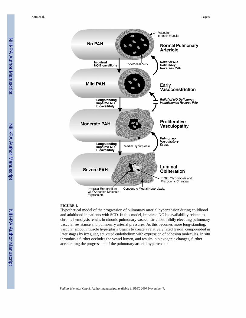
FIGURE 1.Hypothetical model of the progression of pulmonary arterial hypertension during childhoodand adulthood in patients with SCD. In this model, impaired NO bioavailability related tochronic hemolysis results in chronic pulmonary vasoconstriction, mildly elevating pulmonaryvascular resistance and pulmonary arterial pressures. As this becomes more long-standing,vascular smooth muscle hyperplasia begins to create a relatively fixed lesion, compounded inlater stages by irregular, activated endothelium with expression of adhesion molecules. In situthrombosis further occludes the vessel lumen, and results in plexogenic changes, furtheraccelerating the progression of the pulmonary arterial hypertension.
Kato et al. Page 9
Pediatr Hematol Oncol. Author manuscript; available in PMC 2007 November 7.
NIH
-PA Author Manuscript
NIH
-PA Author Manuscript
NIH
-PA Author Manuscript

NIH
-PA Author Manuscript
NIH
-PA Author Manuscript
NIH
-PA Author Manuscript
Kato et al. Page 10
TABLE 1Factors Linked Epidemiologically to PAH in Adults with SCD
Low hemoglobinHigh LDHHigh creatinine levelsHigh direct bilirubinHigh alkaline phosphataseLow serum transferrinHigh ferritin levelsLow arginine:ornithine ratioHigh systolic blood pressureIncreasing ageProteinuria
Pediatr Hematol Oncol. Author manuscript; available in PMC 2007 November 7.

NIH
-PA Author Manuscript
NIH
-PA Author Manuscript
NIH
-PA Author Manuscript
Kato et al. Page 11
TABLE 2Mechanisms Implicated in the Vascular Dysfunction of SCD
NO scavenging by cell-free plasma hemoglobinPlasma L-arginine depletion by cell-free plasma arginaseOxidation of NO by reactive oxygen species produced by xanthine oxidase and NADPH oxidaseEndothelial adhesion to red cells, white cells and platelets
Pediatr Hematol Oncol. Author manuscript; available in PMC 2007 November 7.

NIH
-PA Author Manuscript
NIH
-PA Author Manuscript
NIH
-PA Author Manuscript
Kato et al. Page 12TA
BLE
3Su
mm
ary
of R
epor
ts o
f Ech
ocar
diog
raph
ic S
cree
ning
in C
hild
ren
and
Adu
lts w
ith S
ickl
e C
ell D
isea
se
TR
V ≥
2.5
TR
V ≥
3R
epor
ted
sign
ifica
nt c
orre
latio
ns
Aut
hor
Loc
atio
nA
ges (
year
s)Sc
reen
ed n
nPe
rcen
tage
nPe
rcen
tage
Mor
ris e
t al.
[58]
CA
7–17
6015
25n.
r.Y
oung
et a
l. [5
0]M
I0–
1836
24a
66n.
r.Lo
w H
b, P
lt; h
igh
retic
, AC
SN
elso
n et
al.b
MN
4–18
5315
283
5H
igh
abso
lute
retic
Ony
ekw
ere
et a
l. [5
1]D
C, M
I8–
2452
2446
612
Low
Hb;
hig
h LD
H, b
iliru
bin
Pash
anka
r et a
l.cC
T0–
1825
624
14
Low
Hb;
hig
h SB
PLe
e at
al.d
NY
7–19
5512
22n.
r.H
igh
retic
, LD
H, A
STSu
ell e
t al.
[59]
TX12
–22
8021
264
5N
one
Am
brus
ko e
t al.
[52]
PA0–
2144
1330
614
Low
Hb;
hig
h re
tic, C
VD
, chr
onic
Tx
Qur
eshi
et a
l. [6
0]C
A0–
2131
5a16
n.r.
n.r.
Liem
et a
l. [5
3]IL
10–2
043
1330
n.r.
Low
Hb;
hig
h LD
H, r
etic
, WB
CSe
drak
et a
l. [5
4]N
Y5–
2148
48
n.r.
Hig
h bi
lirub
inJo
yce
et a
l. [5
7]D
C0–
2176
2938
68
n.r.
Gla
dwin
et a
l. [8
]M
D, D
C≥
1819
563
3217
9Lo
w H
b, T
f; hi
gh L
DH
, alk
pho
s, SB
PA
taga
et a
l. [9
]N
C≥1
860
1830
712
Low
Hb,
SB
PD
e C
astro
et a
l. [1
0]N
C≥1
812
540
3228
22Lo
w H
bPe
diat
ric to
tals
603
181
3026
8A
dult
tota
ls38
012
132
5214
Not
e. T
RV
, tric
uspi
d re
gurg
itant
jet v
eloc
ity; n
.r., n
ot re
porte
d; H
b, h
emog
lobi
n; P
lt, p
late
let c
ount
; ret
ic, r
etic
uloc
yte
coun
t; A
CS,
acu
te c
hest
synd
rom
e in
cide
nce;
LD
H, s
erum
lact
ate
dehy
drog
enas
e;SB
P, sy
stol
ic b
lood
pre
ssur
e; A
ST, s
erum
asp
arta
te a
min
otra
nsfe
rase
; CV
D, h
isto
ry o
f cer
ebro
vasc
ular
dis
ease
; Tx,
tran
sfus
ion
ther
apy;
Tf,
seru
m tr
ansf
errin
; alk
pho
s, se
rum
alk
alin
e ph
osph
atas
e;W
BC
, whi
te b
lood
cel
l cou
nt.
a Rep
orte
d nu
mbe
r of p
atie
nts w
ith e
stim
ated
righ
t ven
tricu
lar s
ysto
lic p
ress
ure
>30
mm
Hg
inst
ead
of T
RV
≥ 2
.5, w
hich
is a
ppro
xim
atel
y eq
uiva
lent
.
b Pers
onal
com
mun
icat
ion,
Ste
phen
Nel
son.
c Pers
onal
com
mun
icat
ion,
Far
zana
Pas
hank
ar.
d Pers
onal
com
mun
icat
ion,
Mar
gare
t T. L
ee.
Pediatr Hematol Oncol. Author manuscript; available in PMC 2007 November 7.