Prevalence of Intracardiac Shunting in Children with Sickle Cell Disease and Stroke
6
Prevalence of Intracardiac Shunting in Children with Sickle Cell Disease and Stroke Michael M. Dowling, MD, PhD, Nancy Lee, BS, Charles T. Quinn, MD, MS, Zora R. Rogers, MD, Deborah Boger, RN, MSN, CPNP, Naveed Ahmad, MD, MPH, Claudio Ramaciotti, MD, and George R. Buchanan, MD Objective To determine the prevalence of potential intracardiac shunts, including patent foramen ovale (PFO), in children with sickle cell disease (SCD) and stroke. Study design We performed a transthoracic echocardiogram (TTE) on 40 children with SCD (39 with hemoglobin SS and 1 with sickle-beta 0 thalassemia) and earlier stroke (overt stroke in 30, silent infarction in 10). We compared 3 TTE techniques: conventional 2-dimensional imaging, color Doppler ultrasound, and intravenous agitated saline contrast injection for the detection of intracardiac shunts. We also evaluated the clinical, laboratory, and radio- graphic findings of the children with and without shunts. Results We identified PFO or other potential intracardiac shunts in 10 of 40 children with SCD and earlier stroke (25%; 95% CI, 11.6-38.4). With contrasted TTE, we failed to detect potential shunts in 2 children. In a comparison group of 60 children with stroke but without SCD, retrospective review of clinical echocardiograms identified PFO in 7 of 60 (11.7%; 95% CI, 3.6-19.8). Clinical features significantly associated with the presence of intracardiac shunts were stroke in the setting of vaso-occlusive crisis (P = .026) and headache at stroke onset (P = .014). Conclusion One-quarter of children with SCD and stroke have potential intracardiac shunts. A combination of echocardiographic techniques is required for optimal shunt detection. Intracardiac shunting could be a risk factor for stroke in children with SCD because they are predisposed to thrombosis and elevations of right heart pressure, which could promote paradoxical embolization across an intracardiac shunt. (J Pediatr 2010;-:---). C hildren with sickle cell disease (SCD) sustain strokes at a rate 220 times higher than other children. 1 Clinically overt stroke occurs in 8% to 11% of patients, and as many as 35% of patients have clinically silent stroke detectable only with magnetic resonance imaging (MRI). 2 The identified risk factors for stroke in SCD include previous stroke, sickle vasculopathy, severe anemia, and acute chest syndrome. 3 Transcranial Doppler ultrasound (TCD) can identify children with SCD at highest risk of stroke, but the only treatment shown to prevent stroke is chronic transfusion, with risks of infection and iron overload. 4 Stroke may occur before preventive transfusion therapy can be initiated, and some children with normal TCD study results still sustain stroke. Identification of additional risk factors could lead to improved risk stratification and new strategies for stroke prevention. In children and young adults without SCD, intracardiac shunting, primarily via a patent foramen ovale (PFO), has been identified as a risk factor for stroke. The mechanism is presumed to be ‘‘paradoxical embolization,’’ in which venous emboli avoid filtration in the pulmonary vasculature by right-to-left shunting through a hole in the heart. 5,6 Observations of focal neurologic deficits and MRI lesions consistent with embolic phenomenon in patients with SCD and fat embolization syndrome after vaso-occlusive crisis (VOC) support a role of paradoxical embolization in the neurologic complications of SCD. 7,8 The only route for venous fat emboli from necrotic bone marrow to reach the brain is via right-to-left shunting either at the cardiac or pulmonary level. Few earlier reports have described the prevalence of PFO or other intracardiac shunts in children with SCD and stroke. We hypothesized that intracardiac shunts, potentially treatable with percutaneous transcatheter closure, 9 may play a causal role in stroke in children with SCD. We conducted a pilot study to determine the prev- alence of PFO or other intracardiac shunts in children with SCD and earlier stroke and to evaluate different echocardiographic shunt detection methods in this population. We compared the prevalence of detectible PFO and other intra- cardiac shunts with the prevalence in published reports of children and young adults and with that in a comparison group of children without SCD who From the Departments of Pediatrics (C.Q., Z.R., C.R., G. B., M.D.) and Neurology (M.D.), University of Texas Southwestern Medical Center at Dallas, Dallas, TX; Southwestern Comprehensive Sickle Cell Center and The Center for Cancer and Blood Disorders (N.L., D.B.), and Research Departments (N.A.), Children’s Medical Center Dallas, Dallas, TX Supported by the National Heart, Lung, and Blood Insti- tute Comprehensive Sickle Cell Center Program (U54 HL 70588) and the Children’s Clinical Research Advisory Committee at Children’s Medical Center Dallas. M.D. and C.Q. are supported by the North and Central Texas Clinical and Translational Science Initiative from the NIH KL2 RR024983. M.D. is supported by the First American Real Estate Information Services, Inc. The authors de- clare no conflicts of interest. 0022-3476/$ - see front matter. Copyright Ó 2010 Mosby Inc. All rights reserved. 10.1016/j.jpeds.2009.10.012 MRI Magnetic resonance imaging PFO Patent foramen ovale SCD Sickle cell disease TCD Transcranial Doppler ultrasound TEE Transesophageal echocardiogram TTE Transthoracic echocardiogram VOC Vaso-occlusive crisis 1
-
Upload
independent -
Category
Documents
-
view
0 -
download
0
Transcript of Prevalence of Intracardiac Shunting in Children with Sickle Cell Disease and Stroke

Prevalence of Intracardiac Shunting in Children with Sickle Cell Diseaseand Stroke
Michael M. Dowling, MD, PhD, Nancy Lee, BS, Charles T. Quinn, MD, MS, Zora R. Rogers, MD, Deborah Boger, RN, MSN, CPNP,
Naveed Ahmad, MD, MPH, Claudio Ramaciotti, MD, and George R. Buchanan, MD
Objective To determine the prevalence of potential intracardiac shunts, including patent foramen ovale (PFO), inchildren with sickle cell disease (SCD) and stroke.Study design We performed a transthoracic echocardiogram (TTE) on 40 children with SCD (39 with hemoglobinSS and 1 with sickle-beta0 thalassemia) and earlier stroke (overt stroke in 30, silent infarction in 10). We compared 3TTE techniques: conventional 2-dimensional imaging, color Doppler ultrasound, and intravenous agitated salinecontrast injection for the detection of intracardiac shunts. We also evaluated the clinical, laboratory, and radio-graphic findings of the children with and without shunts.Results We identified PFO or other potential intracardiac shunts in 10 of 40 children with SCD and earlier stroke(25%; 95% CI, 11.6-38.4). With contrasted TTE, we failed to detect potential shunts in 2 children. In a comparisongroup of 60 children with stroke but without SCD, retrospective review of clinical echocardiograms identified PFO in7 of 60 (11.7%; 95% CI, 3.6-19.8). Clinical features significantly associated with the presence of intracardiac shuntswere stroke in the setting of vaso-occlusive crisis (P = .026) and headache at stroke onset (P = .014).Conclusion One-quarter of children with SCD and stroke have potential intracardiac shunts. A combination ofechocardiographic techniques is required for optimal shunt detection. Intracardiac shunting could be a risk factorfor stroke in children with SCD because they are predisposed to thrombosis and elevations of right heart pressure,which could promote paradoxical embolization across an intracardiac shunt. (J Pediatr 2010;-:---).
Children with sickle cell disease (SCD) sustain strokes at a rate 220 times higher than other children.1 Clinically overtstroke occurs in 8% to 11% of patients, and as many as 35% of patients have clinically silent stroke detectable onlywith magnetic resonance imaging (MRI).2 The identified risk factors for stroke in SCD include previous stroke, sickle
vasculopathy, severe anemia, and acute chest syndrome.3 Transcranial Doppler ultrasound (TCD) can identify children withSCD at highest risk of stroke, but the only treatment shown to prevent stroke is chronic transfusion, with risks of infectionand iron overload.4 Stroke may occur before preventive transfusion therapy can be initiated, and some children with normalTCD study results still sustain stroke. Identification of additional risk factors could lead to improved risk stratification andnew strategies for stroke prevention.
In children and young adults without SCD, intracardiac shunting, primarily via a patent foramen ovale (PFO), has been identifiedas a risk factor for stroke. The mechanism is presumed to be ‘‘paradoxical embolization,’’ in which venous emboli avoid filtration inthe pulmonary vasculature by right-to-left shunting through a hole in the heart.5,6 Observations of focal neurologic deficits and MRIlesions consistent with embolic phenomenon in patients with SCD and fat embolization syndrome after vaso-occlusive crisis (VOC)support a role of paradoxical embolization in the neurologic complications of SCD.7,8 The only route for venous fat emboli fromnecrotic bone marrow to reach the brain is via right-to-left shunting either at the cardiac or pulmonary level.
Few earlier reports have described the prevalence of PFO or other intracardiac shunts in children with SCD and stroke. Wehypothesized that intracardiac shunts, potentially treatable with percutaneous transcatheter closure,9 may play a causal role in
From the Departments of Pediatrics (C.Q., Z.R., C.R., G.B., M.D.) and Neurology (M.D.), University of TexasSouthwestern Medical Center at Dallas, Dallas, TX;Southwestern Comprehensive Sickle Cell Center andThe Center for Cancer and Blood Disorders (N.L., D.B.),and Research Departments (N.A.), Children’s Medical
stroke in children with SCD. We conducted a pilot study to determine the prev-alence of PFO or other intracardiac shunts in children with SCD and earlierstroke and to evaluate different echocardiographic shunt detection methods inthis population. We compared the prevalence of detectible PFO and other intra-cardiac shunts with the prevalence in published reports of children and youngadults and with that in a comparison group of children without SCD who
Center Dallas, Dallas, TX
Supported by the National Heart, Lung, and Blood Insti-tute Comprehensive Sickle Cell Center Program (U54 HL70588) and the Children’s Clinical Research AdvisoryCommittee at Children’s Medical Center Dallas. M.D. andC.Q. are supported by the North and Central TexasClinical and Translational Science Initiative from the NIHKL2 RR024983. M.D. is supported by the First AmericanReal Estate Information Services, Inc. The authors de-clare no conflicts of interest.
0022-3476/$ - see front matter. Copyright � 2010 Mosby Inc.
All rights reserved. 10.1016/j.jpeds.2009.10.012
MRI Magnetic resonance imaging
PFO Patent foramen ovale
SCD Sickle cell disease
TCD Transcranial Doppler ultrasound
TEE Transesophageal echocardiogram
TTE Transthoracic echocardiogram
VOC Vaso-occlusive crisis
1
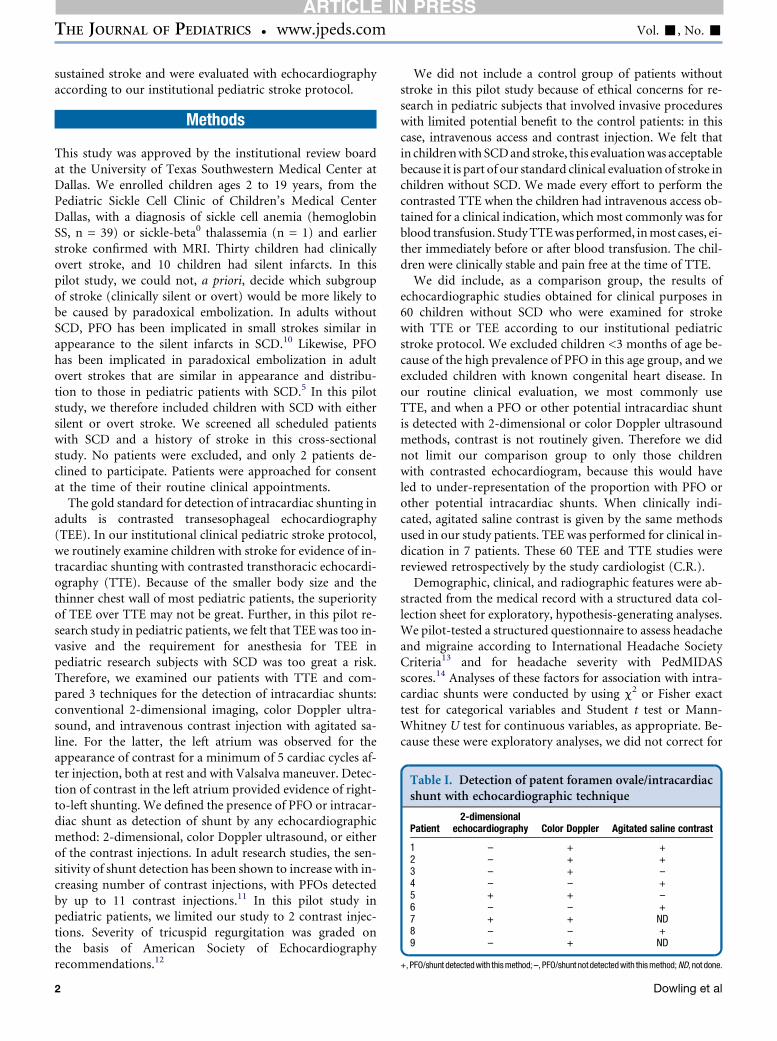
Table I. Detection of patent foramen ovale/intracardiacshunt with echocardiographic technique
Patient2-dimensional
echocardiography Color Doppler Agitated saline contrast
1 – + +2 – + +3 – + –4 – – +5 + + –6 – – +7 + + ND8 – – +9 – + ND
+, PFO/shunt detected with this method; –, PFO/shunt not detected with this method; ND, not done.
THE JOURNAL OF PEDIATRICS � www.jpeds.com Vol. -, No. -
ARTICLE IN PRESS
sustained stroke and were evaluated with echocardiographyaccording to our institutional pediatric stroke protocol.
Methods
This study was approved by the institutional review boardat the University of Texas Southwestern Medical Center atDallas. We enrolled children ages 2 to 19 years, from thePediatric Sickle Cell Clinic of Children’s Medical CenterDallas, with a diagnosis of sickle cell anemia (hemoglobinSS, n = 39) or sickle-beta0 thalassemia (n = 1) and earlierstroke confirmed with MRI. Thirty children had clinicallyovert stroke, and 10 children had silent infarcts. In thispilot study, we could not, a priori, decide which subgroupof stroke (clinically silent or overt) would be more likely tobe caused by paradoxical embolization. In adults withoutSCD, PFO has been implicated in small strokes similar inappearance to the silent infarcts in SCD.10 Likewise, PFOhas been implicated in paradoxical embolization in adultovert strokes that are similar in appearance and distribu-tion to those in pediatric patients with SCD.5 In this pilotstudy, we therefore included children with SCD with eithersilent or overt stroke. We screened all scheduled patientswith SCD and a history of stroke in this cross-sectionalstudy. No patients were excluded, and only 2 patients de-clined to participate. Patients were approached for consentat the time of their routine clinical appointments.
The gold standard for detection of intracardiac shunting inadults is contrasted transesophageal echocardiography(TEE). In our institutional clinical pediatric stroke protocol,we routinely examine children with stroke for evidence of in-tracardiac shunting with contrasted transthoracic echocardi-ography (TTE). Because of the smaller body size and thethinner chest wall of most pediatric patients, the superiorityof TEE over TTE may not be great. Further, in this pilot re-search study in pediatric patients, we felt that TEE was too in-vasive and the requirement for anesthesia for TEE inpediatric research subjects with SCD was too great a risk.Therefore, we examined our patients with TTE and com-pared 3 techniques for the detection of intracardiac shunts:conventional 2-dimensional imaging, color Doppler ultra-sound, and intravenous contrast injection with agitated sa-line. For the latter, the left atrium was observed for theappearance of contrast for a minimum of 5 cardiac cycles af-ter injection, both at rest and with Valsalva maneuver. Detec-tion of contrast in the left atrium provided evidence of right-to-left shunting. We defined the presence of PFO or intracar-diac shunt as detection of shunt by any echocardiographicmethod: 2-dimensional, color Doppler ultrasound, or eitherof the contrast injections. In adult research studies, the sen-sitivity of shunt detection has been shown to increase with in-creasing number of contrast injections, with PFOs detectedby up to 11 contrast injections.11 In this pilot study inpediatric patients, we limited our study to 2 contrast injec-tions. Severity of tricuspid regurgitation was graded onthe basis of American Society of Echocardiographyrecommendations.12
2
We did not include a control group of patients withoutstroke in this pilot study because of ethical concerns for re-search in pediatric subjects that involved invasive procedureswith limited potential benefit to the control patients: in thiscase, intravenous access and contrast injection. We felt thatin children with SCD and stroke, this evaluation was acceptablebecause it is part of our standard clinical evaluation of stroke inchildren without SCD. We made every effort to perform thecontrasted TTE when the children had intravenous access ob-tained for a clinical indication, which most commonly was forblood transfusion. Study TTE was performed, in most cases, ei-ther immediately before or after blood transfusion. The chil-dren were clinically stable and pain free at the time of TTE.
We did include, as a comparison group, the results ofechocardiographic studies obtained for clinical purposes in60 children without SCD who were examined for strokewith TTE or TEE according to our institutional pediatricstroke protocol. We excluded children <3 months of age be-cause of the high prevalence of PFO in this age group, and weexcluded children with known congenital heart disease. Inour routine clinical evaluation, we most commonly useTTE, and when a PFO or other potential intracardiac shuntis detected with 2-dimensional or color Doppler ultrasoundmethods, contrast is not routinely given. Therefore we didnot limit our comparison group to only those childrenwith contrasted echocardiogram, because this would haveled to under-representation of the proportion with PFO orother potential intracardiac shunts. When clinically indi-cated, agitated saline contrast is given by the same methodsused in our study patients. TEE was performed for clinical in-dication in 7 patients. These 60 TEE and TTE studies werereviewed retrospectively by the study cardiologist (C.R.).
Demographic, clinical, and radiographic features were ab-stracted from the medical record with a structured data col-lection sheet for exploratory, hypothesis-generating analyses.We pilot-tested a structured questionnaire to assess headacheand migraine according to International Headache SocietyCriteria13 and for headache severity with PedMIDASscores.14 Analyses of these factors for association with intra-cardiac shunts were conducted by using c2 or Fisher exacttest for categorical variables and Student t test or Mann-Whitney U test for continuous variables, as appropriate. Be-cause these were exploratory analyses, we did not correct for
Dowling et al
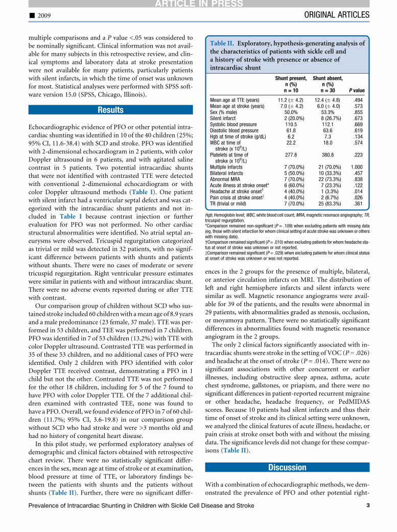
Table II. Exploratory, hypothesis-generating analysis ofthe characteristics of patients with sickle cell anda history of stroke with presence or absence ofintracardiac shunt
Shunt present,n (%)n = 10
Shunt absent,n (%)
n = 30 P value
Mean age at TTE (years) 11.2 (� 4.2) 12.4 (� 4.8) .494Mean age at stroke (years) 7.0 (� 4.2) 6.0 (� 4.0) .573Sex (% male) 50.0% 53.3% .855Silent infarct 2 (20.0%) 8 (26.7%) .673Systolic blood pressure 110.5 112.1 .669Diastolic blood pressure 61.8 63.6 .619Hgb at time of stroke (g/dL) 6.2 7.3 .134WBC at time of
stroke (x 109/L)22.2 18.0 .574
Platelets at time ofstroke (x 109/L)
277.8 380.8 .223
Multiple infarcts 7 (70.0%) 21 (70.0%) 1.000Bilateral infarcts 5 (50.0%) 10 (33.3%) .457Abnormal MRA 7 (70.0%) 22 (73.3%) .838Acute illness at stroke onset* 6 (60.0%) 7 (23.3%) .122Headache at stroke onset† 4 (40.0%) 1 (3.3%) .014Pain crisis at stroke onsetz 4 (40.0%) 2 (6.7%) .026TR (trivial or mild) 7 (70.0%) 25 (83.3%) .361
Hgb, Hemoglobin level; WBC, white blood cell count; MRA, magnetic resonace angiography; TR,tricuspid regurgitation.*Comparison remained non-significant (P = .109) when excluding patients with missing data(eg, those with silent infarction for whom clinical setting of acute stroke was unknown or otherswith missing data).†Comparison remained significant (P = .010) when excluding patients for whom headache sta-tus at onset of stroke was unknown or not reported.zComparison remained significant (P = .029) when excluding patients for whom clinical statusat onset of stroke was unknown or was not reported.
- 2009 ORIGINAL ARTICLES
ARTICLE IN PRESS
multiple comparisons and a P value <.05 was considered tobe nominally significant. Clinical information was not avail-able for many subjects in this retrospective review, and clin-ical symptoms and laboratory data at stroke presentationwere not available for many patients, particularly patientswith silent infarcts, in which the time of onset was unknownfor most. Statistical analyses were performed with SPSS soft-ware version 15.0 (SPSS, Chicago, Illinois).
Results
Echocardiographic evidence of PFO or other potential intra-cardiac shunting was identified in 10 of the 40 children (25%;95% CI, 11.6-38.4) with SCD and stroke. PFO was identifiedwith 2-dimensional echocardiogram in 2 patients, with colorDoppler ultrasound in 6 patients, and with agitated salinecontrast in 5 patients. Two potential intracardiac shuntsthat were not identified with contrasted TTE were detectedwith conventional 2-dimensional echocardiogram or withcolor Doppler ultrasound methods (Table I). One patientwith silent infarct had a ventricular septal defect and was cat-egorized with the intracardiac shunt patients and not in-cluded in Table I because contrast injection or furtherevaluation for PFO was not performed. No other cardiacstructural abnormalities were identified. No atrial septal an-eurysms were observed. Tricuspid regurgitation categorizedas trivial or mild was detected in 32 patients, with no signif-icant difference between patients with shunts and patientswithout shunts. There were no cases of moderate or severetricuspid regurgitation. Right ventricular pressure estimateswere similar in patients with and without intracardiac shunt.There were no adverse events reported during or after TTEwith contrast.
Our comparison group of children without SCD who sus-tained stroke included 60 children with a mean age of 8.9 yearsand a male predominance (23 female, 37 male). TTE was per-formed in 53 children, and TEE was performed in 7 children.PFO was identified in 7 of 53 children (13.2%) with TTE withcolor Doppler ultrasound. Contrasted TTE was performed in35 of these 53 children, and no additional cases of PFO wereidentified. Only 2 children with PFO identified with colorDoppler TTE received contrast, demonstrating a PFO in 1child but not the other. Contrasted TTE was not performedfor the other 18 children, including for 5 of the 7 found tohave PFO with color Doppler TTE. Of the 7 additional chil-dren examined with contrasted TEE, none was found tohave a PFO. Overall, we found evidence of PFO in 7 of 60 chil-dren (11.7%; 95% CI, 3.6-19.8) in our comparison groupwithout SCD who had stroke and were >3 months old andhad no history of congenital heart disease.
In this pilot study, we performed exploratory analyses ofdemographic and clinical factors obtained with retrospectivechart review. There were no statistically significant differ-ences in the sex, mean age at time of stroke or at examination,blood pressure at time of TTE, or laboratory findings be-tween the patients with shunts and the patients withoutshunts (Table II). Further, there were no significant differ-
Prevalence of Intracardiac Shunting in Children with Sickle Cell D
ences in the 2 groups for the presence of multiple, bilateral,or anterior circulation infarcts on MRI. The distribution ofleft and right hemisphere infarcts and silent infarcts weresimilar as well. Magnetic resonance angiograms were avail-able for 39 of the patients, and the results were abnormal in29 patients, with abnormalities graded as stenosis, occlusion,or moyamoya pattern. There were no statistically significantdifferences in abnormalities found with magnetic resonanceangiogram in the 2 groups.
The only 2 clinical factors significantly associated with in-tracardiac shunts were stroke in the setting of VOC (P = .026)and headache at the onset of stroke (P = .014). There were nosignificant associations with other concurrent or earlierillnesses, including obstructive sleep apnea, asthma, acutechest syndrome, gallstones, or priapism, and there were nosignificant differences in patient-reported recurrent migraineor other headache, headache frequency, or PedMIDASscores. Because 10 patients had silent infarcts and thus theirtime of onset of stroke and its clinical setting were unknown,we analyzed the clinical features of acute illness, headache, orpain crisis at stroke onset both with and without the missingdata. The significance levels did not change for these compar-isons (Table II).
Discussion
With a combination of echocardiographic methods, we dem-onstrated the prevalence of PFO and other potential right-
isease and Stroke 3
THE JOURNAL OF PEDIATRICS � www.jpeds.com Vol. -, No. -
ARTICLE IN PRESS
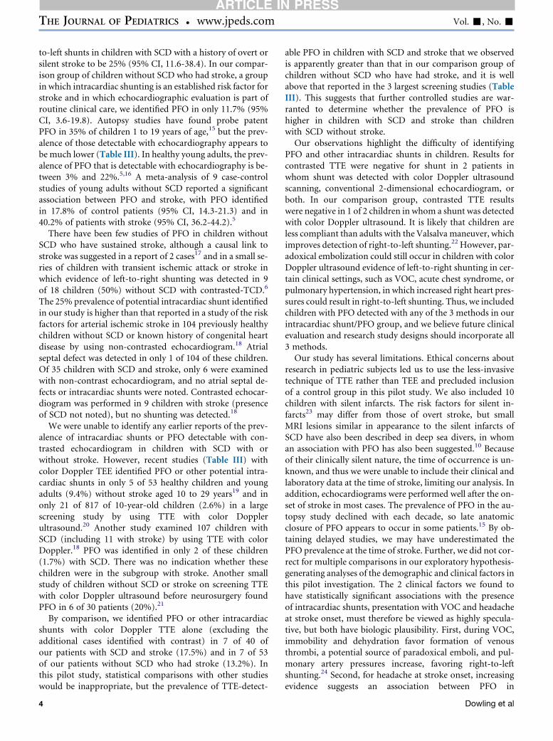
to-left shunts in children with SCD with a history of overt orsilent stroke to be 25% (95% CI, 11.6-38.4). In our compar-ison group of children without SCD who had stroke, a groupin which intracardiac shunting is an established risk factor forstroke and in which echocardiographic evaluation is part ofroutine clinical care, we identified PFO in only 11.7% (95%CI, 3.6-19.8). Autopsy studies have found probe patentPFO in 35% of children 1 to 19 years of age,15 but the prev-alence of those detectable with echocardiography appears tobe much lower (Table III). In healthy young adults, the prev-alence of PFO that is detectable with echocardiography is be-tween 3% and 22%.5,16 A meta-analysis of 9 case-controlstudies of young adults without SCD reported a significantassociation between PFO and stroke, with PFO identifiedin 17.8% of control patients (95% CI, 14.3-21.3) and in40.2% of patients with stroke (95% CI, 36.2-44.2).5
There have been few studies of PFO in children withoutSCD who have sustained stroke, although a causal link tostroke was suggested in a report of 2 cases17 and in a small se-ries of children with transient ischemic attack or stroke inwhich evidence of left-to-right shunting was detected in 9of 18 children (50%) without SCD with contrasted-TCD.6
The 25% prevalence of potential intracardiac shunt identifiedin our study is higher than that reported in a study of the riskfactors for arterial ischemic stroke in 104 previously healthychildren without SCD or known history of congenital heartdisease by using non-contrasted echocardiogram.18 Atrialseptal defect was detected in only 1 of 104 of these children.Of 35 children with SCD and stroke, only 6 were examinedwith non-contrast echocardiogram, and no atrial septal de-fects or intracardiac shunts were noted. Contrasted echocar-diogram was performed in 9 children with stroke (presenceof SCD not noted), but no shunting was detected.18
We were unable to identify any earlier reports of the prev-alence of intracardiac shunts or PFO detectable with con-trasted echocardiogram in children with SCD with orwithout stroke. However, recent studies (Table III) withcolor Doppler TEE identified PFO or other potential intra-cardiac shunts in only 5 of 53 healthy children and youngadults (9.4%) without stroke aged 10 to 29 years19 and inonly 21 of 817 of 10-year-old children (2.6%) in a largescreening study by using TTE with color Dopplerultrasound.20 Another study examined 107 children withSCD (including 11 with stroke) by using TTE with colorDoppler.18 PFO was identified in only 2 of these children(1.7%) with SCD. There was no indication whether thesechildren were in the subgroup with stroke. Another smallstudy of children without SCD or stroke on screening TTEwith color Doppler ultrasound before neurosurgery foundPFO in 6 of 30 patients (20%).21
By comparison, we identified PFO or other intracardiacshunts with color Doppler TTE alone (excluding theadditional cases identified with contrast) in 7 of 40 ofour patients with SCD and stroke (17.5%) and in 7 of 53of our patients without SCD who had stroke (13.2%). Inthis pilot study, statistical comparisons with other studieswould be inappropriate, but the prevalence of TTE-detect-
4
able PFO in children with SCD and stroke that we observedis apparently greater than that in our comparison group ofchildren without SCD who have had stroke, and it is wellabove that reported in the 3 largest screening studies (TableIII). This suggests that further controlled studies are war-ranted to determine whether the prevalence of PFO ishigher in children with SCD and stroke than childrenwith SCD without stroke.
Our observations highlight the difficulty of identifyingPFO and other intracardiac shunts in children. Results forcontrasted TTE were negative for shunt in 2 patients inwhom shunt was detected with color Doppler ultrasoundscanning, conventional 2-dimensional echocardiogram, orboth. In our comparison group, contrasted TTE resultswere negative in 1 of 2 children in whom a shunt was detectedwith color Doppler ultrasound. It is likely that children areless compliant than adults with the Valsalva maneuver, whichimproves detection of right-to-left shunting.22 However, par-adoxical embolization could still occur in children with colorDoppler ultrasound evidence of left-to-right shunting in cer-tain clinical settings, such as VOC, acute chest syndrome, orpulmonary hypertension, in which increased right heart pres-sures could result in right-to-left shunting. Thus, we includedchildren with PFO detected with any of the 3 methods in ourintracardiac shunt/PFO group, and we believe future clinicalevaluation and research study designs should incorporate all3 methods.
Our study has several limitations. Ethical concerns aboutresearch in pediatric subjects led us to use the less-invasivetechnique of TTE rather than TEE and precluded inclusionof a control group in this pilot study. We also included 10children with silent infarcts. The risk factors for silent in-farcts23 may differ from those of overt stroke, but smallMRI lesions similar in appearance to the silent infarcts ofSCD have also been described in deep sea divers, in whoman association with PFO has also been suggested.10 Becauseof their clinically silent nature, the time of occurrence is un-known, and thus we were unable to include their clinical andlaboratory data at the time of stroke, limiting our analysis. Inaddition, echocardiograms were performed well after the on-set of stroke in most cases. The prevalence of PFO in the au-topsy study declined with each decade, so late anatomicclosure of PFO appears to occur in some patients.15 By ob-taining delayed studies, we may have underestimated thePFO prevalence at the time of stroke. Further, we did not cor-rect for multiple comparisons in our exploratory hypothesis-generating analyses of the demographic and clinical factors inthis pilot investigation. The 2 clinical factors we found tohave statistically significant associations with the presenceof intracardiac shunts, presentation with VOC and headacheat stroke onset, must therefore be viewed as highly specula-tive, but both have biologic plausibility. First, during VOC,immobility and dehydration favor formation of venousthrombi, a potential source of paradoxical emboli, and pul-monary artery pressures increase, favoring right-to-leftshunting.24 Second, for headache at stroke onset, increasingevidence suggests an association between PFO in
Dowling et al

Table III. Echocardiographic detection of patent foramen ovale or intracardiac shunting in children
Study, year Patient age Number of patientsShunt with TTEDoppler, n (%)
Shunt with TTE Doppleror contrast, n (%)
Shunt withTEE, n (%)
Shunt with anymethod, n (%) Comments
Hagen et al, 198415 1-9 years 100 – – – 34% Autopsy dataProbe patent PFONon-SCD
10-19 years 100 36%1-99 years 965 27.3%
Overell et al, 20005 <55 years Meta-analysis – – – Stroke 225/566 (39.8%) Meta-analysis of 9 casecontrol studies of young adultsNon-SCD
1022 Control 81/456 (17.8%)
Fuchs et al, 199821 1-14 years 30 6/30 (20%) – – 6/30 (20%) Children withoutstroke screened for PFO beforeneurosurgeryNon-SCD
Fischer et al, 199519 10-29 years 53 – – 5/53 (9.4%) 5/53 (9.4%) TEE performedfor clinical indication:stroke/TIA in 391/1000Proportion withstroke not reportedfor 10- to 29-year-old subgroupNon-SCD
10-99 years 1000 92/1000 (9.2%) 92/1000 (9.2%)
Ganesan et al, 200318 21days-19.7 years 104 1 ASD/104 (0.96%) – 0/36 1/104 (0.96%) Previously healthychildren with strokeNon-SCD
Basso et al, 200420 10 years 817 20 PFO and 1 VSD,21/817 (2.6%)
– – 21/817 (2.6%) Healthy childrenwithout strokeNon-SCD
Caldas et al, 200828 3-18 years 107 2/107 (1.9%) – – 2/107 (1.9%) Children with SCD(11/107 with stroke)
Present study, patientswith SCD and stroke
2-19 years 40 6 PFO and 1 VSD,7/40 (17.5%)
9 PFO and 1 VSD,10/40 (25%)
– 10/40 (25%) Children with SCD withovert (30) or silent (10) stroke
Present study, patientswithout SCD withstroke
0.25-19 years 60 7/53 (13.2%) 7/53 (13.2%) 0/7 (0%) 7/60 (11.7%) Children withstroke Excluding thosewith known congenitalheart diseaseNon-SCD
ASD, atrial septal defect; VSD, ventricular septal defect.
-2009
OR
IGIN
AL
AR
TICLES
Pre
vale
nce
of
Intra
card
iac
Shuntin
gin
Child
ren
with
Sic
kle
Cell
Dis
ease
and
Stro
ke
5
AR
TIC
LE
INP
RE
SS

THE JOURNAL OF PEDIATRICS � www.jpeds.com Vol. -, No. -
ARTICLE IN PRESS
migraine.25-27 These potential associations will need to beexplored more fully in larger studies.
Intracardiac shunts such as PFO could be an independentmodifiable risk factor for stroke or recurrent stroke in childrenwith SCD. A large, controlled study of the prevalence of intra-cardiac shunts or PFO in children with SCD could demon-strate that intracardiac shunts are a risk factor for stroke inSCD. This could lead to improved risk stratification in screen-ing and therapeutic trials and offers the possibility of a novelpreventative treatment for stroke in children with SCD. n
Submitted for publication Sep 20, 2008; last revision received Aug 5, 2009;
accepted Oct 13, 2009.
Reprint requests: Michael M. Dowling, MD, PhD, Department of Pediatrics,
Division of Pediatric Neurology, University of Texas Southwestern Medical
Center, 5323 Harry Hines Blvd, Dallas, TX 75390-9063. E-mail: Michael.
References
1. Earley CJ, Kittner SJ, Feeser BR, Gardner J, Epstein A, Wozniak MA, et al.
Stroke in children and sickle-cell disease: Baltimore-Washington Coop-
erative Young Stroke Study. Neurology 1998;51:169-76.
2. Steen RG, Emudianughe T, Hankins GM, Wynn LW, Wang WC,
Xiong X, et al. Brain imaging findings in pediatric patients with sickle
cell disease. Radiology 2003;228:216-25.
3. Quinn CT, Miller ST. Risk factors and prediction of outcomes in chil-
dren and adolescents who have sickle cell anemia. Hematol Oncol Clin
N Am 2004;18:1339-54.
4. Adams RJ, McKie VC, Hsu L, Files B, Vichinsky E, Pegelow C, et al. Pre-
vention of a first stroke by transfusions in children with sickle cell anemia
and abnormal results on transcranial Doppler ultrasonography. N Engl J
Med 1998;339:5-11.
5. Overell JR, Bone I, Lees KR. Interatrial septal abnormalities and stroke:
a metaanalysis of case-control studies. Neurology 2000;55:1172-9.
6. Benedik MP, Zaletel M, Megli NP, Podnar T. Patent foramen ovale and
unexplained ischemic cerebrovascular events in children. Catheter Car-
diovasc Interv 2007;70:999-1007.
7. Vichinsky E, Williams R, Das M, Earles AN, Lewis N, Adler A, et al. Pul-
monary fat embolism: a distinct cause of severe acute chest syndrome in
sickle cell anemia. Blood 1994;83:3107-12.
8. Horton DP, Ferriero DM, Mentzer WC. Nontraumatic fat embolism
syndrome in sickle cell anemia. Pediatr Neurol 1995;12:77-80.
9. Bartz PJ, Cetta F, Cabalka AK, Reeder GS, Squarcia U, Agnetti A, et al. Par-
adoxical emboli in children and young adults: role of atrial septal defect
and patent foramen ovale device closure. Mayo Clin Proc 2006;81:615-8.
10. Schwerzmann M, Seiler C, Lipp E, Guzman R, Lovblad KO, Kraus M,
et al. Relation between directly detected patent foramen ovale and ische-
mic brain lesions in sport divers. Ann Internal Med 2001;134:21-4.
11. Johansson MC, Helgason H, Dellborg M, Eriksson P. Sensitivity for
detection of patent foramen ovale increased with increasing num-
ber of contrast injections: a descriptive study with contrast trans-
6
esophageal echocardiography. J Am Soc Echocardiogr 2008;21:
419-24.
12. Zoghbi WA, Enriquez-Sarano M, Foster E, Grayburn PA, Kraft CD,
Levine RA, et al. Recommendations for evaluation of the severity of na-
tive valvular regurgitation with two-dimensional and Doppler echocar-
diography. J Am Soc Echocardiogr 2003;16:777-802.
13. Hershey AD, Winner P, Kabbouche MA, Gladstein J, Yonker M,
Lewis D, et al. Use of ICHD-II criteria in the diagnosis of pediatric mi-
graine. Headache 2005;45:1288-97.
14. Hershey AD, Powers SW, Vockell A-LB, LeCates SL, Kabbouche MA,
Maynard MK. PedMIDAS: development of a questionnaire to assess dis-
ability of migraines in children. Neurology 2001;75:2034-9.
15. Hagen PT, Scholz DG, Edwards WD. Incidence and size of patent fora-
men ovale during first 10 decades of life: an autopsy study of 965 normal
hearts. Mayo Clin Proc 1984;59:17-20.
16. Cramer SC. Patent foramen ovale and its relationship to stroke. Cardiol
Clin 2005;23:7-11.
17. Agnetti A, Carano N, Sani E, Tchana B, Allergri V, Bernasconi S, et al.
Cryptogenic stroke in children: possible role of patent foramen ovale.
Neuropediatrics 2006;37:53-6.
18. Ganesan V, Prengler M, McShane MA, Wade AM, Kirkham FJ. Investi-
gation of risk factors in children with arterial ischemic stroke. Ann Neu-
rol 2003;53:167-73.
19. Fisher DC, Fisher EA, Budd JH, Rosen SE, Goldman ME. The incidence
of patent foramen ovale in 1000 consecutive patients: a contrast transe-
sophageal echocardiography study. Chest 1995;107:1504-9.
20. Basso C, Boschello M, Perrone C, Mecenero A, Cera A, Bicego D, et al.
An echocardiographic survey of primary school children for bicuspid
aortic valve. Am J Cardiol 2004;93:661-3.
21. Fuchs G, Schwarz G, Stein J, Kaltenbock F, Baumgartner A,
Oberbauer RW. Doppler color-flow imaging: screeing of a patent fora-
men ovale in children scheduled for neurosurgery in the sitting position.
J Neurosurg Anesthesiol 1998;10:5-9.
22. Kenny D, Turner M, Martin R. When to close a patent foramen ovale.
Arch Dis Child 2008;93:255-9.
23. Kinney TR, Sleeper LA, Wang WC, Zimmerman RA, Pegelow CH,
Ohene-Frempong K, for the Cooperative Study of Sickle Cell Disease. Si-
lent cerebral infarcts in sickle cell anemia: a risk factor analysis. Pediatrics
1999;103:640-5.
24. Machado RF, Mack AK, Martyr S, Barnett C, MacArthur P, Sachdev V,
et al. Severity of pulmonary hypertension during vaso-occlusive pain cri-
sis and exercise in patients with sickle cell disease. Br J Haematol 2007;
136:319-25.
25. Morandi E, Anzola GP, Angeli S, Melzi G, Onorato E. Transcatheter clo-
sure of patent foramen ovale: a new migraine treatment? J Intervent Car-
diol 2003;16:39-42.
26. Post MC, Thijs V, Herroelen Budts WIHL. Closure of a patent foramen
ovale is associated with a decrease in prevalence of migraine. Neurology
2004;62:1439-40.
27. Schwerzmann M, Wiher S, Nedeltchev K, Mattle HP, Wahl A, Seiler C,
et al. Percutaneous closure of patent foramen ovale reduces the fre-
quency of migraine attacks. Neurology 2004;62:1399-401.
28. Caldas MC, Meira ZA, Barbosa MM. Evaluation of 107 patients with
sickle cell anemia through tissue Doppler and myocardial performance
index. J Am Soc Echocardiogr 2008;21:1163-7.
Dowling et al